Article

Higher Expressions of SHH and AR Are Associated with a Positive Receptor Status and Have Impact on Survival in a Cohort of Croatian Breast Cancer Patients

1
Department of Plastic, Reconstructive and Aesthetic Surgery, Dubrava University Hospital, School of Medicine, University of Zagreb, 10000 Zagreb, Croatia
2
Faculty of Medicine Osijek, Josip Juraj Strossmayer University of Osijek, 31000 Osijek, Croatia
3
Clinical Department of Pathology and Cytology, Dubrava University Hospital, School of Medicine, University of Zagreb, 10000 Zagreb, Croatia
4
Department of Medical Statistics and Medical Informatics, Faculty of Medicine Osijek, Josip Juraj Strossmayer University of Osijek, 31000 Osijek, Croatia
5
Division of Molecular Medicine, Ruđer Bošković Institute, 10000 Zagreb, Croatia
6
Faculty of Health Sciences, Libertas International University, 10000 Zagreb, Croatia
7
Department of Abdominal Surgery, Dubrava University Hospital, School of Medicine, University of Zagreb, 10000 Zagreb, Croatia
*
Authors to whom correspondence should be addressed.
Life 2022, 12(10), 1559; https://doi.org/10.3390/life12101559
Submission received: 10 September 2022 / Revised: 4 October 2022 / Accepted: 5 October 2022 / Published: 7 October 2022
(This article belongs to the Section Medical Research)

Abstract

:
Breast cancers (BC) are usually classified into four molecular subtypes according to the expression of estrogen (ER), progesterone (PR), and human epidermal growth factor 2 (HER2) receptors and proliferation marker Ki-67. Despite available anti-hormonal therapies and due to the inherent propensity of some subtypes to develop metastasis, there is a permanent need to discover new prognostic and predictive biomarkers, as well as therapeutic targets for BC. In this study, we used immunohistochemical staining to determine the expression of androgen receptor (AR) and sonic hedgehog protein (SHH), the main ligand of the Hedgehog-GLI (HH-GLI) signaling pathway, in 185 archival primary BC tissue samples and correlated it with clinicopathological characteristics, molecular subtypes, receptors statuses, and survival in a cohort of Croatian BC patients. Results showed that higher SHH and AR expressions were associated with positive receptor status, but increased SHH expression had a negative impact on survival in receptor-negative BCs. On the contrary, higher AR expression was mostly protective. However, multivariate analysis showed that only higher AR expression could be considered as an independent prognostic biomarker for poorer overall survival in triple-negative breast cancer patients (TNBC) (HR 10.9, 95% CI 1.43–83.67; p = 0.021), what could be Croatian population-related. SHH could be a potential target for treating TNBCs and HER2-enriched BCs, in cases where HH-GLI signaling is canonical (SHH-dependent).

1. Introduction

Breast cancer (BC) is the most commonly diagnosed cancer in all countries, even those with low and intermediate levels of income [1]. The incidence rates are the lowest in Asia and sub-Saharan Africa and the highest in North America, Australia/New Zealand, as well as western and northern Europe [2]. According to the Croatian National Cancer Registry, in 2020 there were 2894 newly diagnosed BC cases (incidence rate 120.3/100,000) and 832 deaths (mortality rate 32.8/100,000) by BC [3]. The societal changes brought by industrialization, such as increased fat intake, body weight, age at menarche and/or lactation, and reproductive patterns such as fewer pregnancies and later ages at first birth, are probably responsible for these global variances [2].
For every newly diagnosed BC, expression of estrogen receptor (ER), progesterone receptor (PR), human epidermal growth factor receptor 2 (HER2) and proliferation marker protein Ki-67 is routinely determined, and according to these characteristics molecular or intrinsic subtypes of BC are defined. A molecular subtype of BC is still among the most important factors in determining the treatment protocol for a patient in most countries. Luminal A (LumA) subtype of BCs expresses ER and PR, while Ki-67 is present in less than 20% of cancer cells. The expression of hormone receptors makes these tumors prone to anti-hormone therapy and considering the low presence of proliferation index, these tumors have better treatment outcome [4]. In the luminal B (LumB) subtype ER and PR are expressed, with PR in usually a lower percentage, but Ki-67 is present in 20% or more cancer cells. This subtype can also overexpress HER2. Even though they are similar to LumA, they are related to worse prognosis due to a higher proliferation index and this subtype presents up to 80% of all BC cases [4]. If tumor cells do not express ER or PR, but do overexpress HER2, the tumor is classified into HER2-enriched (HER2-E) molecular subtype. HER2-E is frequently occurring among younger women, related to lower differentiation and worse treatment outcome [4]. Triple-negative breast cancers (TNBCs) do not express any hormone receptor. They are low differentiated and highly aggressive, with fewer treatment options and a worse prognosis. Women with BRCA1 mutation most frequently develop this BC subtype [4].
Androgen receptor (AR) is not routinely checked in BC, but several studies have shown that 60–80% of BC patients overexpress AR, and they suggest that AR may be a predictive or prognostic factor as well as a therapeutic target in BC [5,6]. The function of AR appears to vary among the many BC subtypes, and its prognostic and predictive utility in BC patients is still debatable despite numerous publications covering the physiology of AR, AR-related processes, and AR-targeting therapeutics [7,8].
The Hedgehog-GLI (HH-GLI) pathway is a highly conserved signaling pathway from the cell membrane to the cell nucleus. Its role is crucial in stem cell renewal, tissue regeneration, and embryonic development. Core pathway components in mammals are three Hedgehog ligands: desert, Indian, and sonic hedgehog (DHH, IHH, and SHH, respectively); two transmembrane receptors, patched 1 (PTC1) and patched 2 (PTC2); transmembrane protein smoothened (SMO), and three transcriptional factors: GLI1, GLI2, GLI3, which are regulated by suppressor of fused (SUFU) protein [9]. HH ligand is modified in its secretory cell by the addition of a cholesterol and palmitate from palmitic acid into its active state [10]. Binding of HH ligand to PTC stops repression of SMO and that de-repression enables the release of GLIs from SUFU. GLIs translocate to the nucleus and trigger target genes transcription [11]. GLI1 is exclusively a transcriptional promotor while GLI2 and GLI3 can act as both promotors and suppressors. Described activation is considered as a canonical signalization, while non-canonical is an activation of GLI transcription factors independently of SMO [12].
The role of the HH-GLI pathway in breast cancer depends on the histological and molecular characteristics of the cancer, and this topic has been recently extensively reviewed by Riobo-Del Galdo et al. [13]. For instance, significantly higher levels of SHH and DHH were perceived in cancer tissue compared to healthy breast tissue [14]. Higher SHH and DHH expression also positively correlated with cancer invasiveness, lymph node metastases and recurrences [15,16]. In ER-positive (ER+) BCs it was shown that this signaling pathway increases in the proportion of cancer stem cells (CSC) and promotes invasiveness [14,17,18,19]. HER2-positive (HER2+) tumors had higher levels of SHH and GLI1 compared to healthy controls, but these higher levels had no significant correlation with clinical or pathological characteristics [20,21,22]. A study on BC cell lines demonstrated that inhibition of hedgehog acyltransferase HHAT, one of the main enzymes in SHH synthesis, led to a decrease in the growth of HER2+ tumor cells [16]. Several studies confirmed that higher expression of SHH and SMO in TNBC tumors positively correlates with higher nuclear grade, while higher disease stages correlated with SMO and GLI expression [14,15]. A high proportion of CSC was also observed, followed by a higher incidence of recurrences and metastases [23,24]. High SHH expression in in vitro conditions positively correlated with cancer cell proliferation, also canonical signaling promoted metastases development through neo-angiogenesis promotion [25,26]. Research on animals showed a predisposition for the development of bone metastases in TNBC tumors with high SHH levels [27,28].
Recently it was shown by Trnski et al. that in androgen-independent prostate cancer (PC) cells SHH binds to AR through its cholesterol modification and activates AR signaling, which sustained androgen independence [29]. Sabol et al. in their study gave an indication of potential direct interaction between SHH and ERα in the MCF-7 BC cell line [30]. We hypothesize that SHH can bind to sex hormone receptors with its cholesterol modification but before any mechanistical studies, first we wanted to determine the expression of AR and SHH in breast cancer tissue samples from the Croatian population and examine potential correlations between this expression and clinical and pathological characteristics, especially BC molecular subtypes and receptors statuses. We also wanted to assess the prognostic significance of SHH and AR expressions, which could bring those two proteins closer to clinical practice. To the best of our knowledge, this is the first study focusing on both SHH and AR in BC.

2. Materials and Methods

2.1. Archival Tumors Tissue Samples and Patients Data Collection

This study was conducted on 185 archival primary BC tissue samples collected from the Clinical Department of Pathology and Cytology, Dubrava University Hospital, Zagreb. Samples were obtained from women non-consecutively operated between 2010 and 2015 at the Department of Plastic, Reconstructive and Aesthetic Surgery at the same hospital. The following clinical and pathological data were collected from present medical documentation: date of diagnosis, patient’s age, tumor histotype, T, N, M status, the status of ER, PR and HER2 receptors, level of proliferation marker Ki-67, and presence of lymphovascular invasion, local and distant disease recurrence. Data on survival were collected from the Hospital Information System (BIS) and by phone calls to patients. The study was conducted according to the guidelines of the Declaration of Helsinki and approved by the Ethics Committee of the Dubrava University Hospital (protocol code 2020/2409-07; approved 24 September 2020). Description of patients and tumor samples included in this study is presented in Table 1.

2.2. Determination of Androgen Receptor Protein Expression

To determine AR protein expression, 2–3 µm thick sections were cut from archival paraffin blocks, followed by deparaffinization in thermostat. Deparaffinization was followed by pre-digestion in a thermal bath PT Link (DAKO, Glostrup, Denmark) with EnVision FLEX, High pH (K800021, DAKO) target retrieval solution. Immunohistochemical (IHC) staining was performed using automated DAKO Autostainer Link 48 (DAKO) system, with monoclonal mouse anti-human AR primary antibody, clone AR441 (M356201, DAKO), 1:75 dilution. Immunostains were detected by an indirect avidin-biotin complex technique using REAL EnVision Detection System (K5007, DAKO), followed by contrasting with hematoxylin (1 min), rinsing in an ascending series of alcohol (70–100%) and xylene, and applying a coverslip. Human breast tissue embedded in paraffin was used as a positive control. All prepared slides were microscopically analyzed with an Olympus BX53 microscope (Olympus, Tokyo, Japan) by an experienced pathologist. Androgen receptor positivity for each tissue sample was expressed as the percentage of stained tumor nuclei.

2.3. Determination of Sonic Hedgehog Protein Expression

To determine SHH protein expression, 2–3 µm thick sections were cut from archival paraffin blocks, followed by deparaffinization in thermostat, and pre-digestion in Ventana BenchMark Ultra (Roche Diagnostics, Basel, Switzerland) instrument with thermal stands and ULTRA Cell Conditioning Solution (950–224, Roche Diagnostics, Basel, Switzerland). IHC staining was performed using Ventana BenchMark automated immunohistochemical system (Roche Diagnostics), with polyclonal rabbit anti-SHH (H-160) primary antibody (sc-9024, Santa Cruz Biotechnology, Dallas, TX, USA), 1:50 dilution. The ultraView Universal DAB Detection Kit (760–500, Roche Diagnostics) was used for immunostaining visualization, followed by contrasting with hematoxylin (1 min), rinsing in an ascending series of alcohol (70–100%) and xylene, and applying a coverslip. As a positive control, ureteral tissue embedded in paraffin was used. All prepared slides were analyzed with an Olympus BX53 microscope (Olympus) by an experienced pathologist.
For SHH expression, both staining intensity and percentage of stained tumor cells were evaluated. Staining intensity was expressed numerically as 0 (negative), 1 (weak), 2 (medium), and 3 (strong intensity). If any sample showed different staining intensities, first was determined which part of the tumor (percentage of tumor cells) was stained with a stronger intensity, while the remaining part of the tumor tissue was stained with a weaker intensity. The total expression of SHH was expressed as an immunoreactive score (IRS), which is the sum of the product of the stronger intensity with the percentage of cells with the stronger intensity and the product of the weaker intensity with the percentage of cells with the weaker intensity.

2.4. Statistical Analysis

The normality of distribution of continuous variables was tested with the Shapiro–Wilk test. Cut-off values used to dichotomize protein expression into ‘low’ and ‘high’ categories were determined using the area under the receiver operator characteristic curve (AUC-ROC) analysis. The chi-square test and Fisher’s exact test were used to assess the association and distribution of categorical variables. Categorical variables were presented with number of samples and percentage. Survival curves were calculated with the Kaplan-Meier method and compared by the log-rank test. The Cox proportional hazards regression model with a forward stepwise variable selection was used for multivariate analysis. Statistical analysis was performed using MedCalc Statistical Software v20.114 (MedCalc Software bv, Ostend, Belgium). A two-tailed p-value less than 0.05 was considered statistically significant.

3. Results

3.1. Association of Clinicopathological Characteristics with Molecular Subtype and Receptor Status in a Cohort of Breast Cancer Patients from Croatia

In this study, a cohort of 185 breast cancer patients operated in a single Croatian institution was included to infer the association of clinicopathological characteristics with molecular subtype and receptor status. Results of the statistical analysis are presented in Supplemental Table S1. It could be seen that there was a trend for TNBC patients to be younger at diagnosis, but age was not related to the status of any of the studied receptors. The coexistence of ductal carcinoma in situ (DCIS) in invasive ductal breast cancer (IDC) was significantly more frequent in sex hormone receptor-positive patients, or more precisely, exclusively in luminal A and luminal B subtypes (p = 0.0002). TNBC patients had the highest proportion of tumors larger than 2 cm (p = 0.0003), which was also observed in ER- (p = 0.0004) and PR- (p = 0.019) patients, and conversely, in HER2+ (p = 0.003) patients. HER2+ patients also had a larger number of positive lymph nodes compared to HER2- (p = 0.002). Significantly higher Ki-67 positivity was observed in patients with ER- (p < 0.0001), PR- (p = 0.0005) or HER2+ (p = 0.0003) receptor status, the same as for larger tumors. Lymphovascular invasion (LVI) was most often present in patients with luminal B BC (p = 0.029). Death (by any cause) was not significantly more frequent in any of the subtypes but was marginally more frequent in ER- patients. In the same subcohort of patients, there were significantly more cases of disease recurrence (p = 0.014). Development of distant metastasis was most frequent in TNBC patients (p = 0.002), and it was associated with ER- (p = 0.001) and PR- (p = 0.004) status.

3.2. Expression of Sonic Hedgehog Protein and Androgen Receptor and Its Association with a Molecular Subtype and Receptor Status of Breast Cancer Patients from Croatia

AR and SHH protein expression was determined immunohistochemically on 185 BC formalin-fixed, paraffin-embedded tissue samples. AR was expressed in 91.4% (169/285) of samples. Median AR positivity was 80%, interquartile range 50–90%. Figure 1 and Supplemental Figure S1 show different percentages of AR+ tumor nuclei.
SHH was expressed in 98.4% (182/18) of tissue samples. Median SHH IRS was 150, interquartile range 100–190. Figure 2 and Supplemental Figure S2 show different levels of SHH protein expression.
Next, we tried to associate AR and SHH protein expression with clinicopathological characteristics and receptor status of BC patients. Results are presented in Table 2. In general, AR expression was significantly associated with more characteristics than SHH expression. AR expression was higher in older patients (p = 0.017) and those with the coexistence of DCIS in IDC (p = 0.017), and that was also the trend for higher SHH expression. Both higher AR and SHH expression were observed in smaller tumors (p = 0.002 and p = 0.026, respectively), while higher AR was also observed in patients with lower Ki-67 (p = 0.001). There was also a trend for higher both AR and SHH expression in patients with a higher number of positive lymph nodes. AR expression was highest in LumA, and lowest in TNBC patients. Later was also observed for SHH expression, while SHH was highest in HER2-E patients. This was also observed in relation to the status of each studied receptor: AR expression was significantly higher in ER+ (p < 0.0001), PR+ (p < 0.0001), and HER2+ (p = 0.008) patients, while SHH was significantly higher in ER+ (p = 0.041) and HER2+ (p = 0.028), and a similar trend was observed in PR+ patients. Both disease recurrence and distant metastases were significantly more frequent in patients with lower AR expression (p = 0.003 and p = 0.020, respectively), while disease recurrence was marginally associated with higher SHH expression.
We also wanted to explore if there was any relation between SHH and AR expression, however, correlation analysis showed there was no statistically significant correlation between SHH and AR expression in any subgroup of BC patients (Table 3).

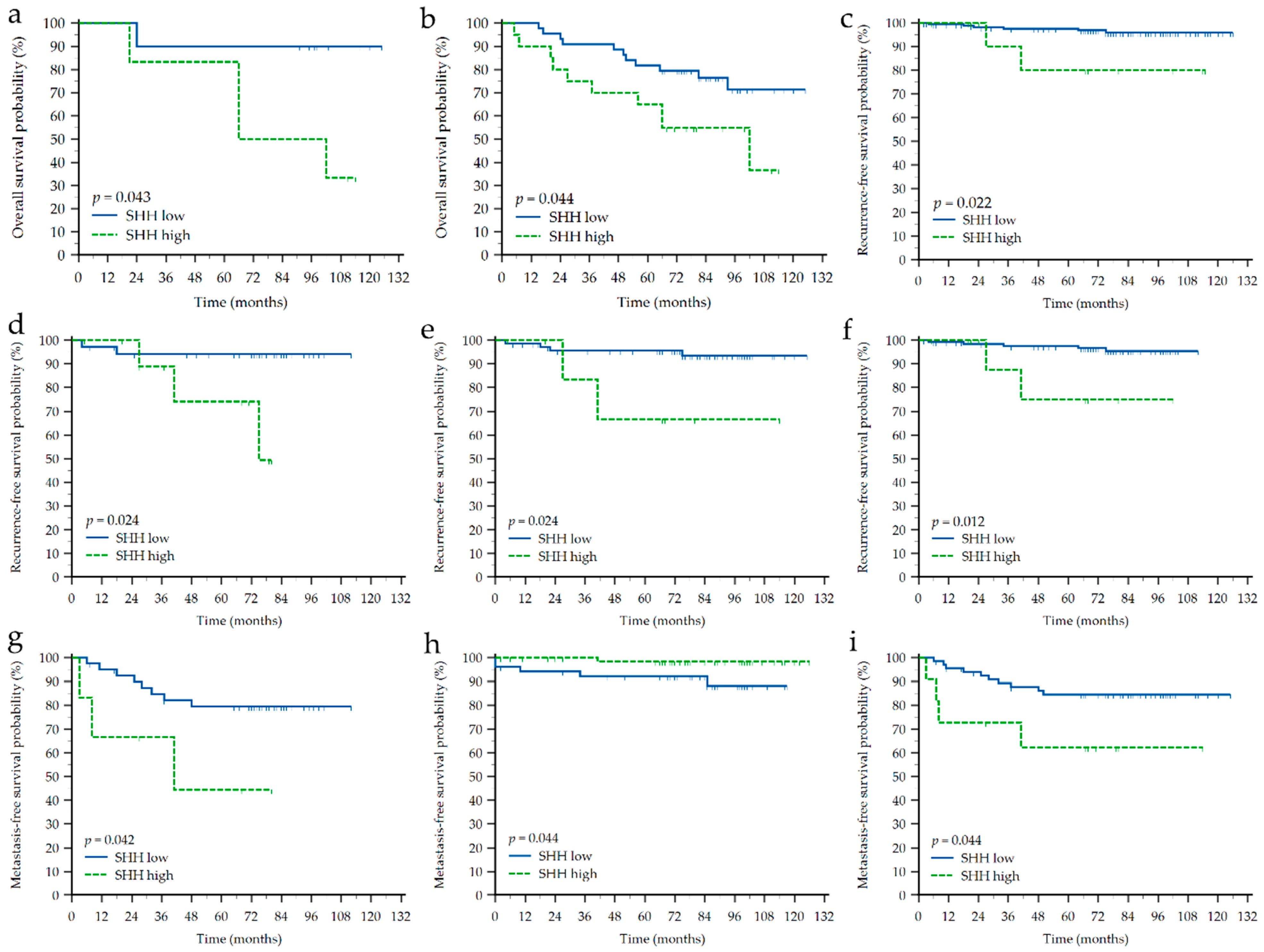
3.3. Impact of Clinicopathological Characteristics and Sonic Hedgehog Protein and Androgen Receptor Expressions on Survival of Croatian Patients with Breast Cancer

Furthermore, we wanted to assess the prognostic significance of SHH and AR expression in BC patients. Survival data were collected for all 185 patients. The median follow-up time was 82 months, range 2 to 126 months. Overall survival (OS) was defined as the time from diagnosis to the death from any cause or last check-up, recurrence-free survival (RFS) or time to recurrence was defined as the time from diagnosis to the local or regional disease recurrence or last check-up, while metastasis-free survival (MFS) or time to metastasis was defined the time from diagnosis to the appearance of distant metastasis or last check-up.
Kaplan-Meier survival analysis was performed on the whole cohort of patients as well as separately in each of the molecular subtypes and according to the receptors’ status. Results are presented in Supplemental Table S2 and Table 4. Generally, higher T and M stage showed a negative impact on OS and MFS in the complete cohort as well as in TNBC, ER- and PR- patients, while histotype did not show any significant impact. Higher SHH expression showed negative impact on OS in HER2-E (p = 0.043) and ER- (p = 0.044) patients, and negative impact on MFS in TNBC (p = 0.042) and PR- (p = 0.044) patients (Figure 3 and Figure 4). However, higher SHH expression showed to be associated with a longer time before developing metastasis in ER+ (p = 0.044) patients. Higher AR expression showed negative impact on OS in TNBC (p = 0.006), ER- (p = 0.022) and PR- (p = 0.040) patients, while positive impact on OS in HER2+ (p = 0.012) patients. Higher AR expression also showed positive impact on MFS in the complete cohort (p = 0.018), TNBC (p = 0.008), ER- (p = 0.024), PR- (p = 0.044) and HER2+ (p = 0.034) patients. Interestingly, RFS was mostly associated with AR or SHH expression, and not clinicopathological characteristics. For instance, higher SHH expression was related to shorter RFS in the complete cohort (p = 0.022), TNBC (p = 0.024), PR- (p = 0.024) and HER2- (p = 0.012) patients, while to the contrary, higher AR expression was positive prognostic biomarker for longer RFS in the complete cohort (p = 0.001), ER+ (p = 0.045), PR- (p = 0.029), PR+ (p = 0.042) and HER2- (p = 0.015) patients (Figure 3 and Figure 4).
Finally, to infer if either SHH or AR could be considered as an independent prognostic biomarker, the Cox regression analysis was performed. We analyzed only those combinations of endpoints and subgroups of BC patients in which, besides SHH or AR expression, at least one clinicopathological characteristic showed a statistically significant impact on survival rate (Table 4). Results are presented in Supplemental Tables S3 and S4. Only higher AR expression showed to be an independent prognostic biomarker for shorter OS in TNBC patients (hazard ratio 10.9, 95% confidence interval 1.43–83.67; p = 0.021).

4. Discussion

Newly diagnosed breast cancer is most commonly classified into one of several molecular or intrinsic subtypes according to the status or level of ER and PR sex hormone receptor expression, growth factor receptor HER2, and proliferation marker Ki-67. These subtypes present distinct biological features resulting in different treatment approaches and clinical outcomes [31]. BC-related deaths are caused by the development of metastases, which is inherent to TNBC and HER2-E subtypes, but also induced by a development of therapy resistance [32,33]. Therefore, there is a permanent need to discover new and better prognostic and predictive biomarkers, as well as new targeted or combination therapies [34]. One potential source of such biomarkers and therapeutical targets is the HH-GLI pathway, a developmental signaling pathway whose role in BC etiopathology and significant overexpression of pathway’s members in BC tissues compared to healthy controls have been confirmed in a multitude of studies (reviewed in [13]). However, an association of the expression of the HH-GLI pathway members with BC subtypes, clinicopathological characteristics and prognosis are still inconsistent and often contradictory. This is usually explained by differences in study populations and sample sizes, methodologies used for expression determination, approaches used for quantifying the level of expression and cut-off values used to dichotomize expression into ‘low’ and ‘high’ categories. Instigated by the recent discovery that SHH, the main ligand and activator of signal transduction through the HH-GLI pathway, can physically bind to AR in androgen-independent PC cell lines [29] and that there are indications for potential physical interaction between SHH and ERα [30], in this study we wanted to determine the expression of AR and SHH in breast cancer tissue samples and correlate it with clinicopathological characteristics, receptors statuses and survival in a cohort of BC patients from Croatia.
In our study, SHH was expressed in the vast majority of BC tissue samples. We found a negative correlation between SHH expression and tumor size, which is opposite to previous studies [21,35,36]. This could be explained by a disproportionately smaller number of T3 tumors in our cohort or the fact that studies that have shown a positive correlation between SHH expression and tumor size primarily studied SHH mRNA expression. However, when we correlated SHH expression, dichotomized according to the median value, with three tumor size groups, the highest expression was observed for T3, then for T1, and the lowest for the T2 group (data not presented). But since we had only nine T3 samples, we decided that merging T2 and T3 groups would give more statistically reliable results. SHH was the most highly expressed in HER2-E tumors, less in luminal, and least in TNBC. When we further subdivided our cohort according to the status of each receptor, we observed that SHH expression was significantly higher in ER+ and HER2+ subgroups, and this is consistent with previous findings [15,36,37,38]. So far, the role of this pathway has been mostly studied in hormone receptor-positive and TNBCs, while there is a very small number of published studies describing crosstalk between HER2 and HH-GLI pathways. However, it is known that HER2 activates that pathway through the PI3K/AKT/mTOR signaling in human esophageal adenocarcinoma [39]. An aspirant regulator of SHH expression in BC is the transcription factor nuclear factor NF-kappa-B [40,41,42]. NF-kappa-B binds to the SHH promoter when it is demethylated, while it is known that levels of methylation regulate SHH expression [43]. Also, hypomethylation of SHH promoter was observed in BCs with increased levels of SHH and nuclear staining of NF-kappa-B [20]. In ER-positive BCs increased SHH expression is caused by estrogen, while it is known that estrogen also increases levels of GLI transcription factors [14,18]. In TNBC, activation of the HH-GLI pathway can be both canonical and non-canonical, while paracrine secretion of SHH by TNBC cells affects tumor stroma which contributes to tumor growth, invasion, and metastasis [21,44]. In our study, we observed a marginal positive association of SHH expression with nodal involvement which was observed in several previous studies [36,45]. We did not confirm the previously observed association of increased SHH expression with local [19,45,46] and distant [15,21,36] disease relapses, but in our cohort, there were only eight cases of local and 19 of distant relapses. Despite that, and although there was a relatively small number of patients who died during the follow-up period (24%), we observed a significant negative impact of increased SHH expression on different types of survival in our cohort of BC patients, which has been consistently reported previously [19,21,36,45,47,48]. However, in our study SHH expression has not proven either its significance or ‘independence’ from established BC prognostic biomarkers in the Cox regression analyses, which could be easily addressed to the small number of patients with a certain characteristic who reached studied endpoints. Nevertheless, one of the most interesting findings of our study is the observation that although an increased SHH expression was associated with a receptor-positive status, an increased expression mostly had a negative impact on the survival of sex hormone receptor-negative and TNBC patients, while the opposite was observed for HER2-E patients in whom a higher SHH expression was associated with both HER2+ status and worse prognosis. This was additionally proven by the observation that higher SHH expression showed a positive impact on longer MFS only in ER+ patients. These findings indicate a differential impact or significance of commonly observed increased SHH expression (or activated HH-GLI signaling pathway in general) in BCs, further distinguishing its protective or harmful impact regarding the receptor’s status. The present body of evidence could point to a hypothesis that the activated HH-GLI pathway is protective for sex hormone receptor-positive BCs, but harmful for TNBC and HER2-E BC patients. If additionally proven, this could lead to the application of SHH-inhibitors for the treatment of triple-negative, or hormone receptor-negative in general, and HER2-E BCs [49]. There are several chemical approaches on diminishing SHH activity. For instance, the SE1 monoclonal antibody prevents the binding of SHH to PTC1 [50], but the effectiveness of this and other anti-SHH antibodies against BC has so far been proven only preclinically [21,51,52]. Other inhibitors of SHH/PTC1 interaction are a small molecule robotnikinin [53], pyrimidine derivative 7_3d3 [54], and macrocyclic peptide inhibitor HL2-m5 [55]. On the other side, the final step in posttranslational processing of SHH is catalyzed by hedgehog acyltransferase HHAT so its inhibitors such as RU-SKI 43 can affect SHH processing and thus its activity as a ligand [56]. It was observed that RU-SKI 43 reduces proliferation and anchorage-independent growth of BC cells [22]. Here we must emphasize that all those approaches would not be effective if the HH-GLI pathway is activated in a non-canonical, ligand-independent manner, e.g., by crosstalk with other signaling pathways [57], and also that in the same subtype of BC both types of pathway activation could be present. In those cases, either inhibitors of downstream signaling, such as GLI-inhibitor GANT61 [58], or combination therapy would be more useful [59]. However, although there have been several ongoing clinical trials (reviewed in [13]), no inhibitors of the HH-GLI pathway, either as monotherapy or in combination, are being used in common clinical practice to treat BC.
The importance of AR in BC is being increasingly emphasized in scientific literature and although it is still not determined routinely in clinics, according to some studies, it is overexpressed in 60–80% of BCs, which is why it could have therapeutic and prognostic importance [60]. Most studies attributed a protective effect of AR on BC, although there is still no consensus on this [61,62,63]. On the contrary, quadruple negative breast cancers (QNBCs), so those that lack in ER, PR, HER2, and AR expressions, are considered more aggressive with the worst prognosis [64]. In our study, we find positive AR nuclear staining in more than 90% of samples. Higher AR expression was associated with higher age, the coexistence of DCIS in IDC, smaller tumors, lower Ki-67, luminal and hormone receptor-positive BCs, and fewer local and distant disease relapses, which is all consistent with previous studies and also confirms the protective effect of AR in BC [8,60,62,63]. AR expression was not present in 9% of our samples, all belonging to the TNBC subtype, but this subgroup, which can be considered as QNBC, did not show any differences compared to TNBC cases with AR expression (data not shown). AR was expressed in 66% of our TNBC cases and this subtype generally showed the lowest AR expression, which is consistent with previous studies [60,61,65,66]. Interestingly, higher AR expression showed to be a marker for shorter OS in receptor-negative and TNBCs, of which multivariate Cox regression analyses showed that AR expression could be considered as an independent biomarker for poorer OS only for TNBCs, which would contradict generally considered protective role of AR in BC. However, one multi-institutional meta-analysis showed that the protective or harmful effect of AR on the prognosis of TNBC patients could be population-specific, namely, Bhattarai et al. observed that AR-positive status was a marker of good prognosis in US and Nigerian cohorts, a marker of poor prognosis in Norwegian, Icelandic and Indian cohorts, and neutral in UK cohort [66]. This is a very interesting observation that needs confirmation in a larger population of TNBC patients from Croatia. However, our findings that higher AR expression is protective for RFS and MFS could indicate that this population-specific observation could be not BC-related.
The main reason why we were also interested in AR expression in BC was the idea that physical interaction between SHH and AR observed in PC cell lines could be also present in BCs that express AR. However, our correlation analyses show that there was no correlation between the expressions of those two proteins in any of the subgroups of BC patients related to clinicopathological characteristic, molecular subtype, or receptor status, although the correlation between SHH and AR expression was previously observed in PC tissue samples [67]. We must be aware that physical interaction between those two proteins has been proven in androgen-independent PC cells, which is not a native condition for the PC cell line used in that study [29]. Since we analyzed only primary, treatment-naïve BCs, maybe different results would be observed if we analyze local or distant metastases, or treatment-resistant tissue samples.
We are aware of the limits of our study. Our cohort of Croatian BC patients is relatively small and may be non-representative since all patients are coming from a single center. A small number of patients in some of the subgroups or those who have reached studied endpoints may have influenced observed results. Most observed p-values were relatively high and would not remain significant after an adjustment for multiple comparisons. Nevertheless, observed results have encouraged us to continue with research on the role and prognostic significance of the HH-GLI pathway in different molecular subtypes of BCs, and that the possible physical interaction of SHH with sex hormone receptors could be a potential therapeutic target for BC.

5. Conclusions

Expression of SHH protein in the Croatian cohort of breast cancer patients was associated with a molecular subtype or more precisely, higher SHH expression was observed in receptor-positive BCs. Conversely, the negative impact of increased SHH expression on survival was observed in receptor-negative BCs, indicating a distinct impact regarding the receptors’ statuses. Increased expression of androgen receptor was associated with clinicopathological characteristics which are associated with less malignant BCs, thus proving its protective role in BC. However, the observation that higher AR expression is an independent prognostic biomarker for worse overall survival of TNBC patients could be Croatian-specific. Additional studies are needed to dis/prove the prognostic and predictive significance of interactions between the Hedgehog-GLI signaling pathway and hormone receptors in BC.

Supplementary Materials

The following supporting information can be downloaded at: https://www.mdpi.com/article/10.3390/life12101559/s1, Figure S1: Original photomicrograph of Figure 1; Figure S2: Original photomicrograph of Figure 2; Table S1: Association of clinicopathological characteristics with molecular subtype and receptor status; Table S2: Impact of clinicopathological characteristics and protein expressions on different types of survival; Table S3: Cox regression model analysis of overall survival; Table S4: Cox regression model analysis of metastasis-free survival.

Author Contributions

Conceptualization, I.B., Z.V., K.M. and P.O.; methodology, Č.T.-L., K.K. and J.Č.; validation, Č.T.-L. and J.Č.; formal analysis, I.B., K.K. and J.Č.; investigation, I.B., D.E., R.Ž., B.G., H.T., D.C., J.J. and V.L.; resources, Č.T.-L. and D.E.; data curation, I.B., D.E., R.Ž., B.G., H.T., D.C., J.J. and V.L.; writing—original draft preparation, I.B. and Z.V.; writing—review and editing, K.M. and P.O.; visualization, Č.T.-L. and K.K.; funding acquisition, Z.V., K.M. and P.O. All authors have read and agreed to the published version of the manuscript.

Funding

This research was funded by the Terry Fox Foundation with a donation collected during the Terry Fox Run in Zagreb in 2019. The work of doctoral student J.Č. has been supported by the “Young Researchers’ Career Development Project-Training of Doctoral Students” no. DOK-2021-02-5821 of the Croatian Science Foundation.

Institutional Review Board Statement

The study was conducted according to the guidelines of the Declaration of Helsinki and approved by the Ethics Committee of the Dubrava University Hospital (protocol code 2020/2409-07; approved 24 September 2020).

Informed Consent Statement

Patient consent was waived due to the usage of archival tissue samples.

Data Availability Statement

Individual per patient data are available from the corresponding authors upon reasonable request.

Acknowledgments

We would like to acknowledge the help and effort received from the staff of the Clinical Department of Pathology and Cytology from the Dubrava University Hospital. Their contribution was invaluable to make this research happen. We would also like to thank the Croatian League Against Cancer and all organizers and participants of the Terry Fox Run Croatia in 2019 for helping us to acquire the funding for this research.

Conflicts of Interest

The authors declare no conflict of interest. The funders had no role in the design of the study; in the collection, analyses, or interpretation of data; in the writing of the manuscript; or in the decision to publish the results.

References

  1. Anderson, B.O.; Yip, C.H.; Smith, R.A.; Shyyan, R.; Sener, S.F.; Eniu, A.; Carlson, R.W.; Azavedo, E.; Harford, J. Guideline implementation for breast healthcare in low-income and middle-income countries: Overview of the Breast Health Global Initiative Global Summit 2007. Cancer 2008, 113, 2221–2243. [Google Scholar] [CrossRef]
  2. W.H.O. The Global Cancer Observatory. 2018 Statistics. Available online: http://gco.iarc.fr/today/data/factsheets/populations/900-world-fact-sheets.pdf (accessed on 10 September 2022).
  3. Incidencija i Mortalitet od Raka u EU-27 Zemljama za 2020. Godinu. Available online: https://www.hzjz.hr/sluzba-epidemiologija-prevencija-nezaraznih-bolesti/incidencija-i-mortalitet-od-raka-u-eu-27-zemljama-za-2020-godinu/ (accessed on 10 September 2022).
  4. Prat, A.; Pineda, E.; Adamo, B.; Galvan, P.; Fernandez, A.; Gaba, L.; Diez, M.; Viladot, M.; Arance, A.; Munoz, M. Clinical implications of the intrinsic molecular subtypes of breast cancer. Breast 2015, 24, S26–S35. [Google Scholar] [CrossRef] [Green Version]
  5. Huang, R.; Han, J.; Liang, X.; Sun, S.; Jiang, Y.; Xia, B.; Niu, M.; Li, D.; Zhang, J.; Wang, S.; et al. Androgen Receptor Expression and Bicalutamide Antagonize Androgen Receptor Inhibit beta-Catenin Transcription Complex in Estrogen Receptor-Negative Breast Cancer. Cell Physiol. Biochem. 2017, 43, 2212–2225. [Google Scholar] [CrossRef]
  6. Murria, R.; Palanca, S.; de Juan, I.; Alenda, C.; Egoavil, C.; Segui, F.J.; Garcia-Casado, Z.; Juan, M.J.; Sanchez, A.B.; Segura, A.; et al. Immunohistochemical, genetic and epigenetic profiles of hereditary and triple negative breast cancers. Relevance in personalized medicine. Am. J. Cancer Res. 2015, 5, 2330–2343. [Google Scholar]
  7. Hickey, T.E.; Robinson, J.L.; Carroll, J.S.; Tilley, W.D. Minireview: The androgen receptor in breast tissues: Growth inhibitor, tumor suppressor, oncogene? Mol. Endocrinol. 2012, 26, 1252–1267. [Google Scholar] [CrossRef]
  8. Salvi, S.; Bonafe, M.; Bravaccini, S. Androgen receptor in breast cancer: A wolf in sheep’s clothing? A lesson from prostate cancer. Semin. Cancer Biol. 2020, 60, 132–137. [Google Scholar] [CrossRef]
  9. Sabol, M.; Trnski, D.; Musani, V.; Ozretic, P.; Levanat, S. Role of GLI Transcription Factors in Pathogenesis and Their Potential as New Therapeutic Targets. Int. J. Mol. Sci. 2018, 19, 2562. [Google Scholar] [CrossRef] [Green Version]
  10. Porter, J.A.; Young, K.E.; Beachy, P.A. Cholesterol modification of hedgehog signaling proteins in animal development. Science 1996, 274, 255–259. [Google Scholar] [CrossRef]
  11. Katoh, Y.; Katoh, M. Hedgehog target genes: Mechanisms of carcinogenesis induced by aberrant hedgehog signaling activation. Curr. Mol. Med. 2009, 9, 873–886. [Google Scholar] [CrossRef] [PubMed]
  12. Pietrobono, S.; Gagliardi, S.; Stecca, B. Non-canonical Hedgehog Signaling Pathway in Cancer: Activation of GLI Transcription Factors Beyond Smoothened. Front. Genet. 2019, 10, 556. [Google Scholar] [CrossRef] [PubMed] [Green Version]
  13. Riobo-Del Galdo, N.A.; Lara Montero, A.; Wertheimer, E.V. Role of Hedgehog Signaling in Breast Cancer: Pathogenesis and Therapeutics. Cells 2019, 8, 375. [Google Scholar] [CrossRef] [PubMed]
  14. Souzaki, M.; Kubo, M.; Kai, M.; Kameda, C.; Tanaka, H.; Taguchi, T.; Tanaka, M.; Onishi, H.; Katano, M. Hedgehog signaling pathway mediates the progression of non-invasive breast cancer to invasive breast cancer. Cancer Sci. 2011, 102, 373–381. [Google Scholar] [CrossRef] [PubMed]
  15. Noman, A.S.; Uddin, M.; Rahman, M.Z.; Nayeem, M.J.; Alam, S.S.; Khatun, Z.; Wahiduzzaman, M.; Sultana, A.; Rahman, M.L.; Ali, M.Y.; et al. Overexpression of sonic hedgehog in the triple negative breast cancer: Clinicopathological characteristics of high burden breast cancer patients from Bangladesh. Sci. Rep. 2016, 6, 18830. [Google Scholar] [CrossRef] [Green Version]
  16. Benvenuto, M.; Masuelli, L.; De Smaele, E.; Fantini, M.; Mattera, R.; Cucchi, D.; Bonanno, E.; Di Stefano, E.; Frajese, G.V.; Orlandi, A.; et al. In vitro and in vivo inhibition of breast cancer cell growth by targeting the Hedgehog/GLI pathway with SMO (GDC-0449) or GLI (GANT-61) inhibitors. Oncotarget 2016, 7, 9250–9270. [Google Scholar] [CrossRef] [Green Version]
  17. Kurebayashi, J.; Kanomata, N.; Yamashita, T.; Shimo, T.; Moriya, T. Antitumor and anticancer stem cell activities of eribulin mesylate and antiestrogens in breast cancer cells. Breast Cancer 2016, 23, 425–436. [Google Scholar] [CrossRef]
  18. O’Brien, C.S.; Farnie, G.; Howell, S.J.; Clarke, R.B. Breast cancer stem cells and their role in resistance to endocrine therapy. Horm. Cancer 2011, 2, 91–103. [Google Scholar] [CrossRef] [PubMed] [Green Version]
  19. Jeng, K.S.; Sheen, I.S.; Jeng, W.J.; Yu, M.C.; Hsiau, H.I.; Chang, F.Y. High expression of Sonic Hedgehog signaling pathway genes indicates a risk of recurrence of breast carcinoma. OncoTargets Ther. 2013, 7, 79–86. [Google Scholar] [CrossRef] [PubMed] [Green Version]
  20. Cui, W.; Wang, L.H.; Wen, Y.Y.; Song, M.; Li, B.L.; Chen, X.L.; Xu, M.; An, S.X.; Zhao, J.; Lu, Y.Y.; et al. Expression and regulation mechanisms of Sonic Hedgehog in breast cancer. Cancer Sci. 2010, 101, 927–933. [Google Scholar] [CrossRef] [PubMed]
  21. O’Toole, S.A.; Machalek, D.A.; Shearer, R.F.; Millar, E.K.; Nair, R.; Schofield, P.; McLeod, D.; Cooper, C.L.; McNeil, C.M.; McFarland, A.; et al. Hedgehog overexpression is associated with stromal interactions and predicts for poor outcome in breast cancer. Cancer Res. 2011, 71, 4002–4014. [Google Scholar] [CrossRef] [Green Version]
  22. Matevossian, A.; Resh, M.D. Hedgehog Acyltransferase as a target in estrogen receptor positive, HER2 amplified, and tamoxifen resistant breast cancer cells. Mol. Cancer 2015, 14, 72. [Google Scholar] [CrossRef] [Green Version]
  23. Ginestier, C.; Hur, M.H.; Charafe-Jauffret, E.; Monville, F.; Dutcher, J.; Brown, M.; Jacquemier, J.; Viens, P.; Kleer, C.G.; Liu, S.; et al. ALDH1 is a marker of normal and malignant human mammary stem cells and a predictor of poor clinical outcome. Cell Stem Cell 2007, 1, 555–567. [Google Scholar] [CrossRef] [PubMed]
  24. Charafe-Jauffret, E.; Ginestier, C.; Iovino, F.; Tarpin, C.; Diebel, M.; Esterni, B.; Houvenaeghel, G.; Extra, J.M.; Bertucci, F.; Jacquemier, J.; et al. Aldehyde dehydrogenase 1-positive cancer stem cells mediate metastasis and poor clinical outcome in inflammatory breast cancer. Clin. Cancer Res. 2010, 16, 45–55. [Google Scholar] [CrossRef] [PubMed] [Green Version]
  25. Cao, X.; Geradts, J.; Dewhirst, M.W.; Lo, H.W. Upregulation of VEGF-A and CD24 gene expression by the tGLI1 transcription factor contributes to the aggressive behavior of breast cancer cells. Oncogene 2012, 31, 104–115. [Google Scholar] [CrossRef] [PubMed] [Green Version]
  26. Harris, L.G.; Pannell, L.K.; Singh, S.; Samant, R.S.; Shevde, L.A. Increased vascularity and spontaneous metastasis of breast cancer by hedgehog signaling mediated upregulation of cyr61. Oncogene 2012, 31, 3370–3380. [Google Scholar] [CrossRef] [Green Version]
  27. Johnson, R.W.; Nguyen, M.P.; Padalecki, S.S.; Grubbs, B.G.; Merkel, A.R.; Oyajobi, B.O.; Matrisian, L.M.; Mundy, G.R.; Sterling, J.A. TGF-beta promotion of Gli2-induced expression of parathyroid hormone-related protein, an important osteolytic factor in bone metastasis, is independent of canonical Hedgehog signaling. Cancer Res. 2011, 71, 822–831. [Google Scholar] [CrossRef] [Green Version]
  28. Sterling, J.A.; Oyajobi, B.O.; Grubbs, B.; Padalecki, S.S.; Munoz, S.A.; Gupta, A.; Story, B.; Zhao, M.; Mundy, G.R. The hedgehog signaling molecule Gli2 induces parathyroid hormone-related peptide expression and osteolysis in metastatic human breast cancer cells. Cancer Res. 2006, 66, 7548–7553. [Google Scholar] [CrossRef] [PubMed] [Green Version]
  29. Trnski, D.; Sabol, M.; Tomic, S.; Stefanac, I.; Mrcela, M.; Musani, V.; Rincic, N.; Kurtovic, M.; Petric, T.; Levanat, S.; et al. SHH-N non-canonically sustains androgen receptor activity in androgen-independent prostate cancer cells. Sci. Rep. 2021, 11, 14880. [Google Scholar] [CrossRef]
  30. Sabol, M.; Trnski, D.; Uzarevic, Z.; Ozretic, P.; Musani, V.; Rafaj, M.; Cindric, M.; Levanat, S. Combination of cyclopamine and tamoxifen promotes survival and migration of mcf-7 breast cancer cells--interaction of hedgehog-gli and estrogen receptor signaling pathways. PLoS ONE 2014, 9, e114510. [Google Scholar] [CrossRef] [PubMed] [Green Version]
  31. Yersal, O.; Barutca, S. Biological subtypes of breast cancer: Prognostic and therapeutic implications. World J. Clin. Oncol. 2014, 5, 412–424. [Google Scholar] [CrossRef] [PubMed]
  32. Kennecke, H.; Yerushalmi, R.; Woods, R.; Cheang, M.C.; Voduc, D.; Speers, C.H.; Nielsen, T.O.; Gelmon, K. Metastatic behavior of breast cancer subtypes. J. Clin. Oncol. 2010, 28, 3271–3277. [Google Scholar] [CrossRef]
  33. Luque-Bolivar, A.; Perez-Mora, E.; Villegas, V.E.; Rondon-Lagos, M. Resistance and Overcoming Resistance in Breast Cancer. Breast Cancer 2020, 12, 211–229. [Google Scholar] [CrossRef] [PubMed]
  34. Maric, P.; Ozretic, P.; Levanat, S.; Oreskovic, S.; Antunac, K.; Beketic-Oreskovic, L. Tumor markers in breast cancer—Evaluation of their clinical usefulness. Coll. Antropol. 2011, 35, 241–247. [Google Scholar]
  35. Cochrane, C.R.; Szczepny, A.; Watkins, D.N.; Cain, J.E. Hedgehog Signaling in the Maintenance of Cancer Stem Cells. Cancers 2015, 7, 1554–1585. [Google Scholar] [CrossRef] [PubMed]
  36. Riaz, S.K.; Khan, J.S.; Shah, S.T.A.; Wang, F.; Ye, L.; Jiang, W.G.; Malik, M.F.A. Involvement of hedgehog pathway in early onset, aggressive molecular subtypes and metastatic potential of breast cancer. Cell Commun. Signal. 2018, 16, 3. [Google Scholar] [CrossRef] [PubMed] [Green Version]
  37. Koga, K.; Nakamura, M.; Nakashima, H.; Akiyoshi, T.; Kubo, M.; Sato, N.; Kuroki, S.; Nomura, M.; Tanaka, M.; Katano, M. Novel link between estrogen receptor alpha and hedgehog pathway in breast cancer. Anticancer Res. 2008, 28, 731–740. [Google Scholar] [PubMed]
  38. Im, S.; Choi, H.J.; Yoo, C.; Jung, J.H.; Jeon, Y.W.; Suh, Y.J.; Kang, C.S. Hedgehog related protein expression in breast cancer: Gli-2 is associated with poor overall survival. Korean J. Pathol. 2013, 47, 116–123. [Google Scholar] [CrossRef]
  39. Kebenko, M.; Drenckhan, A.; Gros, S.J.; Jucker, M.; Grabinski, N.; Ewald, F.; Grottke, A.; Schultze, A.; Izbicki, J.R.; Bokemeyer, C.; et al. ErbB2 signaling activates the Hedgehog pathway via PI3K-Akt in human esophageal adenocarcinoma: Identification of novel targets for concerted therapy concepts. Cell. Signal. 2015, 27, 373–381. [Google Scholar] [CrossRef]
  40. Kasperczyk, H.; Baumann, B.; Debatin, K.M.; FuMa, S. Characterization of sonic hedgehog as a novel NF-kappaB target gene that promotes NF-kappaB-mediated apoptosis resistance and tumor growth in vivo. FASEB J. 2009, 23, 21–33. [Google Scholar] [CrossRef] [PubMed]
  41. Nakashima, H.; Nakamura, M.; Yamaguchi, H.; Yamanaka, N.; Akiyoshi, T.; Koga, K.; Yamaguchi, K.; Tsuneyoshi, M.; Tanaka, M.; Katano, M. Nuclear factor-kappaB contributes to hedgehog signaling pathway activation through sonic hedgehog induction in pancreatic cancer. Cancer Res. 2006, 66, 7041–7049. [Google Scholar] [CrossRef] [Green Version]
  42. Shostak, K.; Chariot, A. NF-kappaB, stem cells and breast cancer: The links get stronger. Breast Cancer Res. 2011, 13, 214. [Google Scholar] [CrossRef]
  43. Wang, L.H.; Choi, Y.L.; Hua, X.Y.; Shin, Y.K.; Song, Y.J.; Youn, S.J.; Yun, H.Y.; Park, S.M.; Kim, W.J.; Kim, H.J.; et al. Increased expression of sonic hedgehog and altered methylation of its promoter region in gastric cancer and its related lesions. Mod. Pathol. 2006, 19, 675–683. [Google Scholar] [CrossRef] [PubMed] [Green Version]
  44. Habib, J.G.; O’Shaughnessy, J.A. The hedgehog pathway in triple-negative breast cancer. Cancer Med. 2016, 5, 2989–3006. [Google Scholar] [CrossRef] [PubMed]
  45. Noman, A.S.; Uddin, M.; Chowdhury, A.A.; Nayeem, M.J.; Raihan, Z.; Rashid, M.I.; Azad, A.K.; Rahman, M.L.; Barua, D.; Sultana, A.; et al. Serum sonic hedgehog (SHH) and interleukin-(IL-6) as dual prognostic biomarkers in progressive metastatic breast cancer. Sci. Rep. 2017, 7, 1796. [Google Scholar] [CrossRef] [PubMed]
  46. Bieche, I.; Lerebours, F.; Tozlu, S.; Espie, M.; Marty, M.; Lidereau, R. Molecular profiling of inflammatory breast cancer: Identification of a poor-prognosis gene expression signature. Clin. Cancer Res. 2004, 10, 6789–6795. [Google Scholar] [CrossRef] [PubMed] [Green Version]
  47. Duan, Z.H.; Wang, H.C.; Zhao, D.M.; Ji, X.X.; Song, M.; Yang, X.J.; Cui, W. Cooperatively transcriptional and epigenetic regulation of sonic hedgehog overexpression drives malignant potential of breast cancer. Cancer Sci. 2015, 106, 1084–1091. [Google Scholar] [CrossRef] [PubMed] [Green Version]
  48. Ten Haaf, A.; Bektas, N.; von Serenyi, S.; Losen, I.; Arweiler, E.C.; Hartmann, A.; Knuchel, R.; Dahl, E. Expression of the glioma-associated oncogene homolog (GLI) 1 in human breast cancer is associated with unfavourable overall survival. BMC Cancer 2009, 9, 298. [Google Scholar] [CrossRef] [PubMed] [Green Version]
  49. Quaglio, D.; Infante, P.; Di Marcotullio, L.; Botta, B.; Mori, M. Hedgehog signaling pathway inhibitors: An updated patent review (2015-present). Expert Opin. Ther. Pat. 2020, 30, 235–250. [Google Scholar] [CrossRef]
  50. Maun, H.R.; Wen, X.; Lingel, A.; de Sauvage, F.J.; Lazarus, R.A.; Scales, S.J.; Hymowitz, S.G. Hedgehog pathway antagonist 5E1 binds hedgehog at the pseudo-active site. J. Biol. Chem. 2010, 285, 26570–26580. [Google Scholar] [CrossRef] [Green Version]
  51. Katano, M. Hedgehog signaling pathway as a therapeutic target in breast cancer. Cancer Lett. 2005, 227, 99–104. [Google Scholar] [CrossRef]
  52. Das, S.; Tucker, J.A.; Khullar, S.; Samant, R.S.; Shevde, L.A. Hedgehog signaling in tumor cells facilitates osteoblast-enhanced osteolytic metastases. PLoS ONE 2012, 7, e34374. [Google Scholar] [CrossRef]
  53. Stanton, B.Z.; Peng, L.F.; Maloof, N.; Nakai, K.; Wang, X.; Duffner, J.L.; Taveras, K.M.; Hyman, J.M.; Lee, S.W.; Koehler, A.N.; et al. A small molecule that binds Hedgehog and blocks its signaling in human cells. Nat. Chem. Biol. 2009, 5, 154–156. [Google Scholar] [CrossRef] [Green Version]
  54. Yun, T.; Wang, J.; Yang, J.; Huang, W.; Lai, L.; Tan, W.; Liu, Y. Discovery of Small Molecule Inhibitors Targeting the Sonic Hedgehog. Front. Chem. 2020, 8, 498. [Google Scholar] [CrossRef]
  55. Owens, A.E.; de Paola, I.; Hansen, W.A.; Liu, Y.W.; Khare, S.D.; Fasan, R. Design and Evolution of a Macrocyclic Peptide Inhibitor of the Sonic Hedgehog/Patched Interaction. J. Am. Chem. Soc. 2017, 139, 12559–12568. [Google Scholar] [CrossRef]
  56. Petrova, E.; Rios-Esteves, J.; Ouerfelli, O.; Glickman, J.F.; Resh, M.D. Inhibitors of Hedgehog acyltransferase block Sonic Hedgehog signaling. Nat. Chem. Biol. 2013, 9, 247–249. [Google Scholar] [CrossRef] [Green Version]
  57. Levanat, S.; Sabol, M.; Musani, V.; Ozretic, P.; Trnski, D. Hedgehog Signaling Pathway as Genetic and Epigenetic Target in Ovarian Tumors. Curr. Pharm. Des. 2017, 23, 73–94. [Google Scholar] [CrossRef] [PubMed]
  58. Riaz, S.K.; Ke, Y.; Wang, F.; Kayani, M.A.; Malik, M.F.A. Influence of SHH/GLI1 axis on EMT mediated migration and invasion of breast cancer cells. Sci. Rep. 2019, 9, 6620. [Google Scholar] [CrossRef] [Green Version]
  59. Bhateja, P.; Cherian, M.; Majumder, S.; Ramaswamy, B. The Hedgehog Signaling Pathway: A Viable Target in Breast Cancer? Cancers 2019, 11, 1126. [Google Scholar] [CrossRef] [Green Version]
  60. Collins, L.C.; Cole, K.S.; Marotti, J.D.; Hu, R.; Schnitt, S.J.; Tamimi, R.M. Androgen receptor expression in breast cancer in relation to molecular phenotype: Results from the Nurses’ Health Study. Mod. Pathol. 2011, 24, 924–931. [Google Scholar] [CrossRef] [Green Version]
  61. Chen, M.; Yang, Y.; Xu, K.; Li, L.; Huang, J.; Qiu, F. Androgen Receptor in Breast Cancer: From Bench to Bedside. Front. Endocrinol. 2020, 11, 573. [Google Scholar] [CrossRef]
  62. Kensler, K.H.; Poole, E.M.; Heng, Y.J.; Collins, L.C.; Glass, B.; Beck, A.H.; Hazra, A.; Rosner, B.A.; Eliassen, A.H.; Hankinson, S.E.; et al. Androgen Receptor Expression and Breast Cancer Survival: Results From the Nurses’ Health Studies. J. Natl. Cancer Inst. 2019, 111, 700–708. [Google Scholar] [CrossRef]
  63. Kensler, K.H.; Regan, M.M.; Heng, Y.J.; Baker, G.M.; Pyle, M.E.; Schnitt, S.J.; Hazra, A.; Kammler, R.; Thurlimann, B.; Colleoni, M.; et al. Prognostic and predictive value of androgen receptor expression in postmenopausal women with estrogen receptor-positive breast cancer: Results from the Breast International Group Trial 1-98. Breast Cancer Res. 2019, 21, 30. [Google Scholar] [CrossRef] [PubMed] [Green Version]
  64. Huang, M.; Wu, J.; Ling, R.; Li, N. Quadruple negative breast cancer. Breast Cancer 2020, 27, 527–533. [Google Scholar] [CrossRef]
  65. Bozovic-Spasojevic, I.; Zardavas, D.; Brohee, S.; Ameye, L.; Fumagalli, D.; Ades, F.; de Azambuja, E.; Bareche, Y.; Piccart, M.; Paesmans, M.; et al. The Prognostic Role of Androgen Receptor in Patients with Early-Stage Breast Cancer: A Meta-analysis of Clinical and Gene Expression Data. Clin. Cancer Res. 2017, 23, 2702–2712. [Google Scholar] [CrossRef] [PubMed] [Green Version]
  66. Bhattarai, S.; Klimov, S.; Mittal, K.; Krishnamurti, U.; Li, X.B.; Oprea-Ilies, G.; Wetherilt, C.S.; Riaz, A.; Aleskandarany, M.A.; Green, A.R.; et al. Prognostic Role of Androgen Receptor in Triple Negative Breast Cancer: A Multi-Institutional Study. Cancers 2019, 11, 995. [Google Scholar] [CrossRef] [PubMed] [Green Version]
  67. Zhang, X.; Zhang, Y.; Lin, F.; Shi, X.; Xiang, L.; Li, L. Shh Overexpression Is Correlated with GRP78 and AR Expression in Primary Prostate Cancer: Clinicopathological Features and Outcomes in a Chinese Cohort. Cancer Manag. Res. 2020, 12, 1569–1578. [Google Scholar] [CrossRef]
Figure 1. Representative images of tissue samples with different percentages of tumor nuclei with androgen receptor protein expression. (a) 0% of AR+ tumor nuclei (200× magnification); (b) 30% of AR+ tumor nuclei (200×); (c) 60% of AR+ tumor nuclei (200×); (d) 100% of AR+ tumor nuclei (200×).
Life 12 01559 g001
Figure 2. Representative images of tissue samples with different intensities of sonic hedgehog protein expression, expressed as immunoreactive score (IRS). (a) IRS 0 (100× magnification); (b) IRS 100 (200×); (c) IRS 200 (200×); (d) IRS 300 (200×).
Life 12 01559 g002
Figure 3. Kaplan-Meier survival curves depicting significant impact of sonic hedgehog (SHH) protein expression on different types of survival in different groups of breast cancer patients. (a) Impact on overall survival (OS) in HER2-enriched (n = 16); (b) OS in ER-negative (n = 64); (c) Impact on recurrence-free survival (RFS) in the whole cohort (N = 185); (d) RFS in TNBC (n = 47); (e) RFS in PR-negative (n = 79); (f) RFS in HER2-negative (n = 142); (g) Impact on metastasis-free survival (MFS) in TNBC (n = 47); (h) MFS in ER-positive (n = 121); (i) MFS in PR-negative (n = 79). Shown are p-values for the long-rank test. Tick marks indicate censored cases.
Life 12 01559 g003
Figure 4. Kaplan-Meier survival curves depicting a significant impact of androgen receptor (AR) protein expression on different types of survival in different groups of breast cancer patients. (a) Impact on overall survival (OS) in TNBC patients (n = 47); (b) OS in ER-negative (n = 64); (c) OS in PR-negative (n = 79); (d) OS in HER2-positive (n = 64); (e) Impact on recurrence-free survival (RFS) in the whole cohort (N = 185); (f) RFS in ER-positive (n = 121); (g) RFS in PR-negative (n = 79); (h) RFS in PR-positive (n = 106); (i) RFS in HER2-negative (n = 142); (j) Impact on metastasis-free survival (MFS) in the whole cohort (N = 185); (k) MFS in TNBC (n = 47); (l) MFS in ER-negative (n = 64); (m) MFS in PR-negative (n = 79); (n) MFS in HER2-positive (n = 43). Shown are p-values for the long-rank test. Tick marks indicate censored cases.
Life 12 01559 g004
Table 1. Baseline clinicopathological characteristics of Croatian cohort of breast cancer patients whose tumor tissue samples were used in this study.
Characteristicn Out of 185 (%)
Age * (years)60 (28–87)
Histotype
Invasive ductal carcinoma160 (86.5)
Invasive ductal carcinoma and ductal carcinoma in situ 25 (13.5)
Tumor size (T)
T1 (≤2 cm)105 (56.8)
T2 (2.1–5 cm)71 (38.4)
T3 (>5.1 cm)9 (4.9)
Nodal involvement (N)
N0 (0)113 (61.4)
N1 (1–3)46 (25.0)
N2 (4–9)14 (7.6)
N3 (≥10)11 (6.0)
Distant metastasis (M)
M0183 (98.9)
M12 (1.1)
Ki-67
<20%53 (28.6)
≥20%132 (71.4)
Lymphovascular invasion
Absent116 (62.7)
Present69 (37.3)
Molecular subtype
Luminal A48 (25.9)
Luminal B74 (40.0)
HER2-enriched16 (8.6)
Triple-negative47 (25.4)
Survival
Alive140 (75.7)
Deceased45 (24.3)
Recurrence
Absent177 (95.7)
Present8 (4.3)
Metastasis
Absent166 (89.7)
Present19 (10.3)
* Data are presented as median (range).
Table 2. Association of SHH and AR protein expression with clinicopathological characteristics and receptor status of Croatian breast cancer patients. Statistically significant p-values are in bold.
CharacteristicSHH AR
‘Low’‘High’p-Value‘Low’‘High’p-Value
Age
≤50 years20 (43.5)26 (56.5)0.07027 (58.7)19 (41.3)0.017
>50 years40 (29.0)98 (71.0) 53 (38.4)85 (61.6)
Histotype
IDC81 (50.6)79 (49.4)0.08458 (36.2)102 (63.7)0.017
IDC + DCIS8 (32.0)17 (68.0) 3 (12.0)22 (88.0)
Tumor size
T143 (41.0)62 (59.0)0.02635 (33.3)70 (66.7)0.002
T2 + T346 (57.5)34 (42.5) 45 (56.2)35 (43.7)
Nodal involvement
0–3124 (78.0)35 (22.0)0.05217 (10.7)142 (89.3)0.087
≥415 (60.0)10 (40.0) 0 (0.0)25 (100.0)
Ki-67
<20%23 (43.4)30 (56.6)0.4188 (15.1)45 (84.9)0.001
≥20%66 (50.0)66 (50.0) 53 (40.2)79 (59.8)
LVI
Absent91 (78.4)25 (21.6)0.25642 (36.2)74 (63.8)0.226
Present49 (71.0)20 (29.0) 19 (27.5)50 (72.5)
Molecular subtype *
Luminal A22 (45.8)26 (54.2)0.03716 (33.3)32 (66.7)<0.0001
Luminal B37 (50.0)37 (50.0) 33 (44.6)41 (55.4)
HER2-E5 (31.2)11 (68.8) 12 (75.0)4 (25.0)
Triple-negative32 (68.1)15 (31.9) 40 (85.1)7 (14.9)
Receptor status
ER-negative31 (48.4)33 (51.6)0.04143 (67.2)21 (32.8)<0.0001
ER-positive40 (33.1)81 (66.9) 10 (8.3)111 (91.7)
PR-negative31 (39.2)48 (60.8)0.08944 (55.7)35 (44.3)<0.0001
PR-positive29 (27.4)77 (72.6) 9 (8.5)97 (91.5)
HER2-negative80 (56.3)62 (43.7)0.02832 (22.5)110 (77.5)0.008
HER2-positive16 (37.2)27 (62.8) 2 (4.7)41 (95.3)
Survival
Alive101 (72.1)39 (27.9)0.20973 (52.1)67 (47.9)0.239
Deceased28 (62.2)17 (37.8) 28 (62.2)17 (37.8)
Recurrence
Absent165 (93.2)12 (6.8)0.05733 (18.6)144 (81.4)0.003
Present6 (75.0)2 (25.0) 5 (62.5)3 (37.5)
Metastasis
Absent77 (46.4)89 (53.6)0.16767 (40.4)99 (59.6)0.020
Present12 (63.2)7 (36.8) 13 (68.4)6 (31.6)
* Protein expression was dichotomized using the median value of expression. AR—androgen receptor; DCIS—ductal carcinoma in situ; ER—estrogen receptor; HER2-E—HER2-enriched; IDS—invasive ductal carcinoma; LVI—lymphovascular invasion; PR—progesterone receptor; SHH—sonic hedgehog protein; T—tumor size; TNBC—triple-negative breast cancer.
Table 3. Correlation between SHH and AR protein expression in breast cancer patients classified by baseline clinicopathological characteristics and receptor status.
CharacteristicCategoryρp-Value
All patients 0.070.336
Age≤50 years0.140.356
>50 years0.020.787
HistotypeIDC0.100.218
IDC + DCIS−0.130.533
Tumor size<2 cm−0.030.749
≥2 cm0.170.134
Nodal involvement0–30.060.474
≥40.130.534
Ki-67<20%0.180.205
≥20%0.040.615
Lymphovascular invasionAbsent0.030.765
Present0.140.243
Molecular subtypeLuminal A0.140.337
Luminal B−0.060.597
HER2-E0.110.680
TNBC−0.030.846
Receptor statusER-negative0.100.437
ER-positive0.020.847
PR-negative0.100.398
PR-positive0.040.717
HER2-negative0.100.247
HER2-positive−0.060.721
SurvivalAlive0.080.341
Deceased0.050.765
RecurrenceAbsent0.090.210
Present−0.550.157
MetastasisAbsent0.090.246
Present0.030.900
ρ—Spearman’s rank correlation coefficient.
Table 4. Hazard ratios with 95% confidence intervals only for clinicopathological characteristics and sonic hedgehog protein and androgen receptor expressions with statistically significant (p < 0.05) impact on different types of survival in a cohort of Croatian breast cancer patients (N = 185). The p-values for the log-rank test are presented in Supplemental Table S2.
Characteristic Molecular SubtypeReceptor Status
AllLumALumBHER2-ETNBCER-ER+PR-PR+HER2-HER2+
Age ≤50 vs. >50 yearsOS2.5 (1.29–4.84) 3.7 (1.32–10.23)3.2 (1.25–8.42) 2.6 (1.07–6.49) N.A.
RFS N.A.
T <2 vs. ≥2 cmOS2.7 (1.49–4.91) 3.1 (1.12–8.38)2.6 (1.10–6.27) 2.9 (1.29–6.44) 3.6 (1.78–7.15)
MFS5.0 (1.98–12.41) 3.6 (1.08–12.11)4.3 (1.42–12.77) 4.3 (1.52–12.37) 5.7 (1.95–16.52)
N 0–3 vs. ≥4OS4.5 (1.78–11.16) 7.0 (1.48–32.96) 7.0 (1.26–39.06)4.4 (1.20–16.02)4.2 (1.16–14.97)5.1 (1.61–16.41) 14.7 (3.83–56.50)
MFS19.9 (4.88–81.40) 11.2 (1.29–96.24) 19.0 (2.64–137.16)9.6 (1.89–48.20)47.2 (3.77–591.45)10.20 (2.28–45.47)24.9 (1.39–445.77)133.9 (18.92–947.40)
Ki-67 <20% vs. ≥20%MFS3.3 (1.25–8.81) 3.9 (1.35–10.99)
LVI no vs. yesOS 4.0 (1.07–15.11)
SHH ‘low’ vs. ‘high’OS 6.7 (1.06–42.82) 2.7 (1.03–6.96)
RFS30.8 (1.63–580.74) 12.8 (1.40–116.76) 29.23 (1.56–547.50) 56.3 (2.46–1288.75)
MFS 8.2 (1.08–62.79) 0.19 (0.038–0.958)5.4 (1.05–28.23)
AR ‘low’ vs. ‘high’OS 4.1 (1.48–11.23)3.1 (1.17–8.20) 2.8 (1.05–7.47) N.A.
RFS0.05 (0.009–0.323) N.A.0.16 (0.032–0.834)N.A.0.10 (0.016–0.639)
MFS0.33 (0.133–0.824) N.A.N.A. N.A. N.A.
AR—androgen receptor; HER2-E—HER2-enriched; LumA—luminal A; LumB—luminal B; LVI—lymphovascular invasion; MFS—metastasis-free survival; N—number of positive lymph nodes; N.A.—no patients with certain characteristics have reach endpoint; OR—overall survival; RFS—recurrence-free survival; SHH—sonic hedgehog protein; T—tumor size; vs.—versus.
Publisher’s Note: MDPI stays neutral with regard to jurisdictional claims in published maps and institutional affiliations.

