Article

Photorhabdus sp. ETL Antimicrobial Properties and Characterization of Its Secondary Metabolites by Gas Chromatography–Mass Spectrometry

by
Tshikala Eddie Lulamba
,
Ezekiel Green
and
Mahloro Hope Serepa-Dlamini
*
Department of Biotechnology and Food Technology, University of Johannesburg, Doornfontein Campus, P.O. Box 17011, Johannesburg 2028, South Africa
*
Author to whom correspondence should be addressed.
Life 2021, 11(8), 787; https://doi.org/10.3390/life11080787
Submission received: 22 June 2021 / Revised: 29 July 2021 / Accepted: 3 August 2021 / Published: 4 August 2021

Abstract

:
Entomopathogenic nematodes (EPNs) are known to be highly pathogenic to insect pests, due to their associated symbiotic bacteria, which produce virulence factors, exo-enzymes and other harmful secondary metabolites to conquer, kill, and degrade their insect hosts. However, these properties are not fully characterized. This study reports on the antimicrobial activities of Photorhabdus sp. strain ETL, symbiotically associated to an insect pathogenic nematode, Heterorhabditis zealandica, against human pathogenic bacteria and toxigenic fungi, as well as the non-targeted profiling of its secondary metabolites (SMs) using gas chromatography coupled to high-resolution time-of-flight mass spectrometry. Fatty acids including 3-eicosene, (E)-; 5-eicosene, (E)-; eicosene; 9-octadecenamide; undecanoic acid with shown antimicrobial activities were detected. This provided more insight on the composition and bioactivities of SMs produced by the Photorhabdus sp.

1. Introduction

The entomopathogenic nematode bacteria (EPNB), Xenorhabdus and Photorhabdus (Enterobacteriales: Morganellaceae) [1], are known to produce inside their insects host an arsenal of virulence factors, antibiotic and exo-enzymatic compounds with pesticidal, pharmaceutical, and other beneficial properties [2,3,4,5,6]. These bioactive compounds have been grouped as bacteriocins, xenorhabdins, xenorxides, xenocoumacins, indole (including nematophin, genistein), stilbene and anthraquinone derivatives, and chitinases [2,3,7]. Therefore, EPNB have been considered among the major natural sources of novel bioactive molecules [3,8] that are important in the development of new drugs, agrochemicals, as well as food and biofuel industrial compounds [9,10,11].
Members of the Photorhabdus genus have an extensive secondary metabolism producing various secondary metabolites (SMs) that are thought to be virulent although their specific functions are not fully understood [4,12,13]. Secondary metabolism, although some SMs are produced during the exponential phase or bacterial growth, is generally activated during the post-exponential or stationary phase of the bacterial growth, which is during the mutual association with their nematode vectors [4]. Therefore, secondary metabolism, apart from producing anti-hosts effectors, is also important to support both nematode growth and development [4,6]. This was observed through the functions of some SMs such as multipotent stilbene (ST) antibiotics, antraquinone (AQ) and the ability to fluoresce, and through its ability to provide important nutrients to the nematode as large intracellular inclusions composed of crystal proteins with essential amino acids [4]. The ST antibiotic is required to initiate the recovery program of the nematode free-living life stage Infective Juvenile (IJ) during the mutual association [4], and during the nematode normal cycle of reproduction, the ST might act as a food signal stimulating the recovery of IJs, therefore functioning as a link between food availability with both nematode reproduction and development [4]. However, EPNB occurs in two phenotypic variants viz. primary and secondary variants, which differ according to morphology and SMs production [4,6]. The phenotypic primary variant has a normal secondary metabolism, and is the original bacterium variant, isolated from the EPN. In contrast, the secondary variant which is the resulting switching off of some secondary metabolism activities including the production of ST antibiotic, AQ pigment and the ability to fluoresce, and inefficiently produce intracellular inclusions, can arise probably to adapt to a life without the nematode in the environment [4,6]. Secondary variants can also arise after repeated in vivo and in vitro sub-culturing [14] during the bacterial stationary phase and when nematodes emerge from the cadaver [14,15], and this is influenced by nutrient limitation and redox stress [4,16,17]. This phenomenon of phenotypic variation is reversible with the Xenorhabdus and has not been reported for Photorhabdus spp. [14]. This phenomenon is critical bacterial SMs production because it cannot be manipulated during in vitro growth and a poor understanding of the biology of these bacteria can result in lower quality (variety and number) of produced SMs [14], in addition to other influencing factors, such us suitable medium, monoxenic conditions and adequate oxygen [15]. Secondary metabolites include numerous inert and mobile volatile low molecular weight natural products of diverse structures [18,19]. They are mainly divided into terpene, phenol and nitrogen containing compounds [20]. Volatile compounds serve to prevent or protect the host against pathogens [21].
A range of biochemical and genetic approaches can be utilized to identify and characterize these SMs. This is because some genetic loci that are involved in the production of these molecules are cryptic, therefore are not expressed under normal laboratory conditions [4]. Although the first natural products from Photorhabdus and other EPNB have been known for almost 30 years, a huge variety of new compounds have been identified in the last 5–10 years, mainly due to the application of modern mass spectrometry. Metabolomics has been recently implemented to advance the knowledge of the biology of both primary and secondary metabolites, and to identify new metabolites, as well as both their chemotaxonomy and alterations caused by them [22]. Mass spectrometry (MS) coupled with gas/liquid chromatography (GC-MS/LC-MS), or Nuclear Magnetic Resonance (NMR) analytical techniques, are generally used in metabolomics [23]. Liquid chromatography-MS can be used for unknown high molecular weight compounds, such as lipids, and for the profiling of low molecular weight compounds, such as saponins and phenylpropanoids [24], whereas gas chromatography–mass spectrometry (GC-MS) is used for low molecular weight compounds [25]. Non-volatile metabolites, not responding to the GC-MS approach, can be separated and characterized through reverse-phase column of the liquid chromatography–mass spectrometry (LC-MS) approach [20].
This study reports on the SMs produced in vitro by the South Africa-native EPNB, P. heterorhabditis strain ETL, using GC-MS, and analysis of its antimicrobial properties against selected human microbial pathogens and toxigenic fungi.

2. Materials and Methods

Photorhabdus heterorhabditis strain ETL conserved in glycerol stocks at −80 °C was reconstituted in nutrient agar (NA) (Merck KGaA, Darmstadt, Germany), supplemented with 0.025% bromothymol blue (MerckMillipore, Billerica, MA, USA) and 0.004% triphenyltetrazolium chloride (PanReac AppliChem, Barcelone, Spain), and MacConkey agar (Merck KGaA, Darmstadt, Germany).

2.1. Selection of the Optimal Nutrient Medium for Metabolite Production

Five loopfuls of the log phase of P. heterorhabditis strain ETL obtained from NBTA agar plates were inoculated into 100 mL separate flasks containing 20 mL of Luria–Bertani broth (LB), nutrient broth (NB) (Neogen® Culture Mediam, Lansing, MI, USA), NB supplemented with canola oil, and NB supplemented with canola oil and glucose [26]. The media were adjusted to final pH of 7.0 [27]. The flasks were simultaneously incubated at 28 °C for 48 h in the dark on an orbital shaker at 160 rpm. At one-hour intervals, the cell cultures were measured at 600 nm with an S-20 Boeco Spectrophotometer for 24 h. The media with highest bacterial yield was deduced to be the one suitable for the bacterial growth. This experiment was carried out in triplicate.

2.2. Extraction of Secondary Metabolites

Luria–Bertani broth (8 L) was prepared in Erlenmeyer flasks and autoclaved at 121 °C for 15 min. Flasks were inoculated with the P. heterorhabditis strain ETL and incubated in a shaking incubator at 125 rpm, at 30 °C for 7 days [28]. Thereafter, sterilized XAD-7-HP resin (SIGMA, South Africa, BCBR6696V) (20 g/L of the culture) was added and further shaken for 2 h. The resin was filtered through cheesecloth and eluted with acetone three times. Acetone was removed using a Rotary evaporator. The remaining water was extracted with ethyl acetate three times, using the separating funnel, and concentrated using a rotary evaporator [29]. The crude extract was re-suspended in 1 mL of methanol (chromatographic grade) in a dark amber vial for analysis after filtration.

2.3. Determination of Anti-Microbial Activity

2.3.1. Minimum Inhibitory Concentrations (MIC)

The minimum inhibitory concentrations (MIC) and agar well diffusion methods were used to determine the antibacterial and antifungal activity of the Photorhabdus heterorhabditis strain ETL secondary metabolites. The following human pathogenic bacterial strains Bacillus cereus (ATCC 10876), Enterococcus faecium (ATTC 13048), Escherichia coli (ATCC 10536), Klebsiella pneumonia (ATCC 10031), K. oxytoca (ATCC 13182), Pseudomonas aeruginosa (NCTC 10662), Staphylococcus aureus (ATCC 25923), S. saprophyticus (ATCC 15305), S. epidermidis (ATCC 14990), Veillonella parvula (ATCC 10790), and Mycobacterium smegmatis (ATCC 21293), and fungal strains Aspergillus flavus, A. niger, and A. parasiticus obtained from the Food, Environment and Health Research Group (FEHRG) laboratories, Faculty of Health Sciences, University of Johannesburg, South Africa, were used. This was carried out following the methods outlined by Andrews [30], Rodriguez-Tudela [31], and Balouiri et al. [11]. Accordingly, the bacterial strains were inoculated in Mueller–Hinton broth and allowed to grow overnight in an incubator at 37 °C for 24 to 36 h, and compared to a 0.5 McFarland’s standard. Streptomycin standard solution (~1 mg/mL in 1 mM EDTA (Ethylenediamine tetraacetic acid) analytical grade) at 0.032 mg/mL (in sterile distilled water), was used as the positive control, while 0.1% DMSO (Dimethyl sulfoxide) was used as a negative control.
The crude secondary metabolite extract was weighed (0.173 g) into an empty sterile McCartney bottle and dissolved in 0.1% (w/v) DMSO to make a stock solution of 32 mg/mL. This solution was two-fold serially diluted using the Mueller–Hinton broth down to 0.03125 mg/mL. The 96 well microtiter plates were used. The outer wells were filled with sterile distilled water, while 100 µL of standardized bacterial cultures, grown overnight, were horizontally added into each inner well, with five repeats vertically. In vertical order, a decreasing concentration (from 16 to 0.03125 mg/mL) of 100 µL of the extract was added into each well. The plates were covered and incubated overnight at 37 °C. Thereafter, 10 µL of 0.02% (w/v) of Resazurin sodium salt solution was added, and further incubated for 2 h. Changes of colour from blue to pink to clear, upon reduction in the amount of oxygen within the medium, was observed as an indication of the microbial metabolism [32]. After a visual inspection for colour changes, the known metabolite concentration, in a well with slight colour change, in each horizontal row, was used as the MIC. The Agar disk-diffusion and Agar well diffusion methods were followed for fungal species.

2.3.2. Agar Disk-Diffusion Method

The basic method, agar disk-diffusion, followed by the agar well diffusion method as described by Balouiri et al. [11] were used for the MIC testing, with minor modification. The toxigenic fungal strains A. flavus, A. niger, and A. parasiticus were used. Briefly, non-supplemented potato dextrose agar (PDA) plates were inoculated with the fungal strains adjusted to (0.4–5) × 106 CFU/mL in potato dextrose broth (PDB). Five discs of filter paper (Whatman No. 1, Sigma Aldrich (Pty) Ltd., Jet Park 1459, South Africa) of about 6 mm in diameter, flooded with different concentrations of the crude secondary metabolite extract (two-fold serially diluted from 4 to 0.5 mg/mL), were placed on the agar surface then incubated at 36 °C for 5 days. Clotrimazole was used as the positive control. Thereafter, plates in triplicates were visually examined for the inoculum growth inhibition by the extract diffused within the agar. Diameters of zones of inhibition were compare with the CLSI (Clinical and Laboratory Standards Institute) standards.

2.3.3. Agar Well Diffusion Method

The agar well diffusion method by Balouiri et al. [11] was used for antifungal testing to better determine the bacterial zone of inhibition. Its procedure is similar to the disk-diffusion method, but for the inoculation, a volume of the microbial inoculum was spread over the entire agar surface. Then, 5 holes of a diameter ranging from 6 to 8 mm were punched aseptically using a sterile cork borer, and 20 to 100 µL of the crude secondary metabolite extract at 1 mg/mL was added into each well. A hole for Clotrimazole, used as positive control, was also punched. Then, plates were incubated at 36 °C for 5 days in triplicate. Secondary metabolites diffused in the medium were observed for growth inhibition of the tested fungal strains.

2.3.4. Determination of Anti-Microbial Activity Methods’ Validation

The determination of anti-microbial activity methods was validated by a one-way analysis of variance (ANOVA) with Tukey and Duncan post hoc tests using IBM SPSS Statistics 26 (SPSS/IBM, Chicago, IL, USA); mean values of the repeated experiments (3×) were compared. Significant differences at the p ≤ 0.05 level of probability were then reported.

2.4. Profiling of Volatile Compounds

The Pegasus gas chromatography (GC) coupled to a high-resolution time-of-flight mass spectrometry (MS) (GC-HRTOF-MS) system (LECO Corporation, St Joseph, MI, USA) equipped with an Agilent 7890A gas chromatograph (Agilent Technologies, Inc., Wilmington, DE, USA), a Gerstel MPS multipurpose autosampler (Gerstel Inc., Mülheim an der Ruhr, Germany) and a Rxi®-5 ms column (30 m × 0.25 mm ID × 0.25 μm) (Restek, Bellefonte, PA, USA) was used, operating in high-resolution. The instrument was successfully mass calibrated, prior to use, to assure accurate mass data collection. Mass calibration and pre-analysis calibration were performed using the compound perfluorotributylamine (PFTBA), and 11 masses including: CF3 (m/z 68.9952), C2F4 (m/z 99.9936), C2F4N (113.9967), C2F5 (m/z 130.9920), C3F6 (m/z 149.9904), C4F9 (m/z 218.9856), C5F10N (m/z 263.9871), C8F16N (m/z 413.9775), C9F18N (m/z 463.9743) and C9F20N (m/z 501.9711), respectively. The intensity and resolution values, 41392, 40200, respectively, as well as a mass accuracy RMS (root mean square) value <1 ppm, were observed.
One microliter of each sample was injected into the instrument in spitless mode and was carried by helium gas constantly pumped at 1 mL/min with inlet and transfer line temperatures of 250 and 225 °C, respectively. The oven was heated at successive ‘rests’ at increasing temperatures, designed to bake out the column. This consisted of an initial temperature of 70 °C, and 0.5 min rest; ramp up to 150 °C at 10 °C/min, and 2 min rest; and ramp up to 330 °C at 10 °C/min, and 3 min rest. The recommended rate and system extraction frequency was used for the MS data acquisition. This consisted of the rates of 13 spectra/s, 30 to 1000 m/z, and electron ionization and ion source temperature at 70 eV and 250 °C, respectively, and extraction frequency of 1.25 kHz. The ChromaTOF® software was used for data analysis. The biological sample extraction was conducted in triplicate and each replicate analyzed twice (six analytical injections).

Data Processing and Statistical Analysis

The collected GC-HRTOF-MS dataset was converted to mzML format using the LECO ChromaTOF-HRT software and then processed (peak picking and alignment) on the XCMS open-source tool (https://xcmsonline.scripps.edu/, accessed on 19 October 2019). The resulting peak list variables, retention times (min), mass-to-charge ratios (m/z) and integrated peak areas were corrected. Statistically significant metabolites were identified based on their mass spectra and retention time using the National Institute of Standards and Technology (NIST), Mainlib and Feihn metabolomics libraries, and both online open chemistry databases and platforms, such as PubChem at the National Institutes of Health (NIH), Chemical Entities of Biological Interest (ChEBI) of the European Molecular Biology Laboratory-European Bioinformatics Institute (EMBL-EBI), and existing literature.

3. Results and Discussion

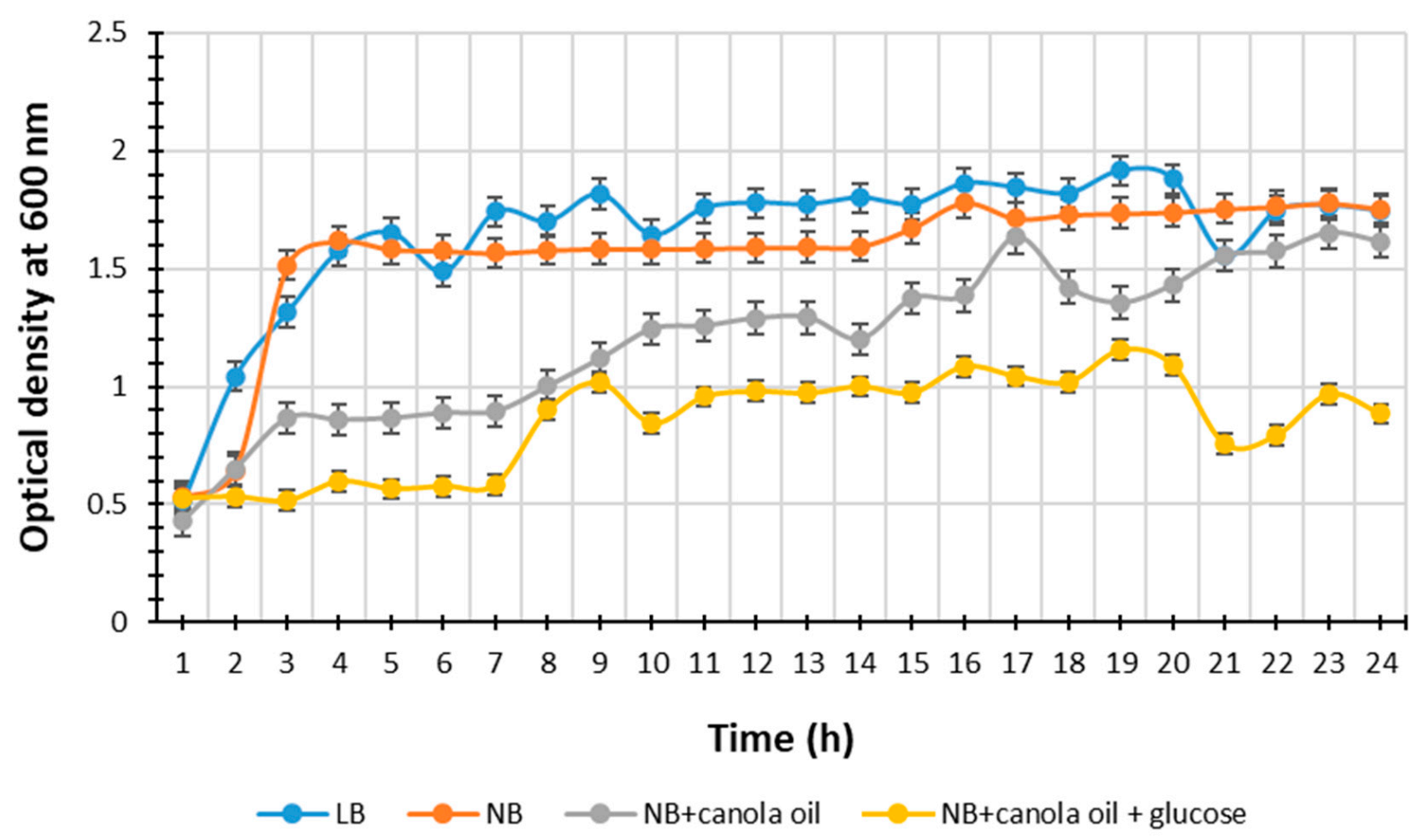
3.1. Selection of the Optimal Nutrient Medium for Metabolite Production

Distinctive color changes were observed (results not shown) in all media types, NB, NB with canola oil, NB with canola oil and glucose, and LB incubated with P. heterorhabditis strain ETL which indicated the secretion of secondary metabolites (SMs) by the bacterium as it proliferated and served as means to visually monitor the purity of the bacterial cultures, as some of these SMs have antibiotic activity [5] against opportunistic microorganisms that might contaminate the media. The results of P. heterorhabditis strain ETL growth are presented in Figure 1. The highest bacterial yields (Optical density, OD) value over the time (h) taken for the bacterial growth in LB broth, NB, NB supplemented with canola oil, and NB supplemented with canola oil and glucose was 1.9 at the 20th h, 1.8 at the 16th h, 1.6 at the 17th h, and 1.2 at the 19th h, respectively.
The media type significantly affected the bacterial growth, and thus SMs secretion. The broth LB showed a highest bacterial yield, suggesting an increase in both bacterial growth and metabolism, due to nutrients availability, leading to an even increase in antibiotic production during the bacterial exponential growth phase, and to most SMs secretion during the bacterial stationery growth phase [4,33]. The NB medium also resulted in high microbial yield (Figure 1). These results are in accordance with the findings by Wang et al. [27], suggesting that these two media LB and NB are suitable for EPNB production because of the sodium chloride, component of the LB broth, which increases the metabolite production due to its osmolarity in the presence of good nitrogen sources [27]. The tryptone present in LB broth and both peptone and yeast extracts present in NB medium are good nitrogen sources [27].

3.2. Determination of Anti-Microbial Activity

Minimum Inhibitory Concentration (MIC)
The inhibition values of the crude extract of P. heterorhabditis strain ETL are presented in Table 1. The MIC values ranged from 4.0 to 0.0625 mg/mL. The crude extract MIC values at ≤1 mg/mL are considered active [34]. Thus, the observed inhibition values of up to 0.0625 mg/mL showed the potential of the SM to be used for new anti-microbial compounds development.
The extract from strain ETL had the most significant MIC values between 0.0625 and 1.000 mg/mL against B. cereus, E. coli, K. oxytoca, K. pneumoniae, S. aureus, P. aeruginosa, S. epidermidis, and S. saprophyticus species. The most significant MIC value of 0.0625 mg/mL was seen on pathogenic E. coli. Other notable inhibitory activities were seen on S. saprophyticus (0.125 mg/mL) and M. smegmatis and S. aureus (0.250 mg/mL). The crude extract from the strain ETL had higher fungal MIC properties (≤1 mg/mL of MIC) against A. flavus and A. niger than a crude extract from another reported EPNB (4 mg/mL ant 5 mg/mL for A. flavus and A. niger, respectively) [35], although a pur- antifungal compound (cyclo(l-Pro-d-Leu)) isolated from it showed greater MIC values of 0.008 mg/mL and 0.032 mg/mL against A. flavus and A. niger, respectively [35]. Members of the Photorhabdus produce numerous SMs with various antimicrobial activities [5,12] to suit the entomopathogenic lifestyle with their nematode vectors and to contribute to the symbiotic association with the nematode host by providing a monoxenic growth environment while killing off competitive microbes and insect larvae, and providing nutrients for both the nematodes and bacteria [4,36,37,38,39]. This could justify the notable inhibitory activity found in this study. Furthermore, the outer membrane in the Gram-negative organisms that is generally responsible for the more resistance to antibiotics than the Gram-positive bacteria, due to the exclusion of certain drugs and antibiotics from penetrating the cell [40], could also justify the fact that Gram-positive bacteria were more susceptible to the strain ETL crude extract’ SMs. These results suggest that strain ETL SMs can be further explored for the development of new pharmaceutical products against pathogens.

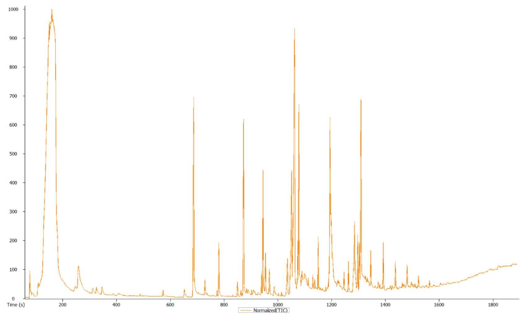
3.3. Overview and Exploration of the Acquired GC-MS Data

Considering the inherent chemo-diversity and multidimensionality of the extracted metabolome, the high-resolution GC-TOF-MS platform allowed the simultaneous detection of multiple analytes with high sensitivity, providing more detailed characterization of the metabolic inventory of the sample. Thus, visual inspection of the generated mass chromatogram pointed to 160 metabolic profiles of the analyzed sample, as shown in Figure 2. Table 2 presents a summary of the obtained precursors, fragments and retention time for the identified compounds, after the chromatogram peaks of the identified compounds being confirmed using online databases and the existing literature.
About 22 volatile compounds, in various chemical groups, were identified from P. heterorhabditis strain ETL, as shown in Table 2. This number is below the number of natural products produced by members of the Photorhabdus genus [4]. This could be due to SMs’ cryptic genes not expressed under normal laboratory conditions [4], the unique extraction method [73], the media used for in vitro culturing, and both the unique approach and instrument used in this study. More SMs are still yet to be identified and characterized by using alternative methods. The strain ETL volatile compounds were found to be similar (or partly similar) to those produced by other pathogenic bacteria and fungi, including Breibacillus brevis (Bacillales: Paenibacillaceae), Paracoccus pantotrophus (Rhodobacterales: Rhodobacteraceae), Streptomyces sp. (Actinomycetales: Streptomycetaceae), Aspergillus nidulans (Eurotiales: Trichocomaceae) and Lentinula edodes (Agaricales: Omphalotaceae); and (medicinal) plants, including Ammodaucus leucotricus (Umbelliferae: Apiaceae), Bidens pilosa var. radiate (Malvales: Malvaceae), Monochaetia kansensis (Xylariales: Amphisphaeriaceae), Rhaponticum acaule (Asterales: Asterales), Tamarix aphylla L. (Caryophyllales: Tamaricaceae) and Woodfordia Fruticosa (L.) kurz (Myrtales:Lythraceae). However, no literature support was found regarding the tetradonium bromide and nonanamide compounds found in strain ETL.
The functions and applications of the strain ETL-identified natural products are not fully understood; however, when reviewing the literature, valuable activities and putative roles in various industries were identified. Fatty acids including 3-eicosene, (E)-; 5-eicosene, (E)-; eicosene; 9-octadecenamide; undecanoic acid, methyl ester; benzenepropanoic acid, 3,5-bis(1,1-dimethylethyl)-4-hydroxy, methyl ester (FAME); and diethyl phthalate (FAEE) were detected among the strain ETL-produced natural products (Table 2). Most fatty acids, including FAMEs are recognized to have antifungal and antioxidant activities [74]. Some methyl esters (FAME), such as hexadecanoic acid, methyl ester, can be used as as biodiesel fuel [75] and FAME undecanoic acid, methyl ester as a component in lipases from A. nidulans, was found to have antimicrobial activities [66]. Lipases (and proteinases) have important functions in EPNB parasitism, as some are essential for insect colonization; they are involved in immunosuppression and tissue degradation [4,5,76,77]. Lipases in EPNB contribute efficiently in the degradation (and neutralization) of insect larvae adipose tissue which is a dynamic tissue implicated in many metabolic functions and is the main producer of key insect host proteins, such as both Pattern Recognition Proteins/Receptors (PRPs) and antimicrobial peptides (AMPs) [78,79,80]. The PRPs and AMPs are involved in either the humoral or the cellular immune response [5]. Thus, these lipases are an important source of energy and nutrients for both the bacterium and its nematode vector [5] and have been shown to contribute to host specificity through bioactivities against selected hosts [76].
The alcoholic compounds, 2-Pentanone, 4-hydroxy-4-methyl- (Table 2), have been found among the major components of essential oils of A. leucotricus (Umbelliferae: Apiaceae) to have a strong antibacterial activity against S. aureus, E. coli and K. pneumonia [41]. The bromomethane component of the tris (trifluoromethyl) bromomethane (Table 2) was produced both industrially and biologically and was used extensively as a pesticide until being phased out by most countries in the early 2000s [58,59]. The ketone compound, ergotaman-3′,6′,18-trione, 9,10-dihydro-12′-hydroxy-2′-methyl-5′(phenylmethyl), (5′à,10à)- was found to have a biocontrol activity against Lasiodiplodia theobromae (Botryosphaeriales: Botryosphaeriaceae), a plant pathogen causing black spot disease on wax apple and is reportedly a potential biocontrol agent against pathogens on fruits [71]. Diethyl phthalate derivatives showed antimicrobial activity against A. niger, A. flavus, Trichoderma harizanum (Hypocreales: Hypocreaceae), T. viridae and Rhizoctonia solani (Cantharellales: Ceratobasidiaceae) [81]. Pyrrolo[1,2-a]pyrazine-1,4-dione, hexahydro-3-(2-methylpropyl)-, having antioxidant properties and antimicrobial activities (Table 2), might reportedly have a potential future in agriculture [62]. The benzenepropanoic acid (carboxylic acid) component of the benzenepropanoic acid, 3,5-bis(1,1-dimethylethyl)-4-hydroxy-, methyl ester (Table 2), as with other members of the phenylpropanoids class, are used in food as antioxidants, sweeteners, emulsifiers, and flavorings for ice cream, bakery, and confectionary. In cosmetics, they are used as a flagrant agent giving a floral scent to bath gels, detergents (both liquid and powders), fabric softeners, perfumes and soaps [63]. The diethyl phthalate (Table 2) can be useful in the development of useful agricultural, pharmaceutical and industrial products. Finally, tridecane is used for the development of solvents and fuels [42,43], indicating the diverse usefulness of the SMs from Photorhabdus, not only in pharmaceutical but other industries as well. These volatile organic compounds showed the diverse useful applications of the SMs from P. heterorhabditis strain ETL and can possibly play a critical role in the food, cosmetic and drug discovery industries. Despite these findings, some of these produced natural products including their physiological and/or biological role(s) are still not fully clear. This is because the inference of roles for some natural products is not as easy as it is for antibiotically active natural products in the bacterial ecosystem, and antibiotics in sub-inhibitory concentrations induce changes in both the mRNA and the levels of proteins produced in other bacteria [82,83], thus serving as signal molecules in natural environments. Therefore, future studies to determine the genetic characteristics and mechanisms involved in the P. heterorhabditis strain ETL SMs production, as well as the genetic evolutionary differences and similarities with other EPNB are needed.

4. Conclusions

The EPNB produce numerous SMs with various bioactivities, and their functions are not fully understood. Nevertheless, evidence shows that P. heterorhabditis strain ETL and other EPNB may provide substantial benefits to agriculture, pharmaceutical industries and the environment. In brief, this study determined the antimicrobial activity of the crude extracts of the EPNB, P. heterorhabditis, which showed notable inhibitory activities. The antibacterial results show the potential use of this bacterium for the isolation of pure bioactive compounds and possible drug discovery. Additionally, a useful fingerprint on major volatile SMs produced by P. heterorhabditis is provided. Bioactive compounds with pesticidal, pharmaceutical, medicinal, agricultural, food and cosmetic industrial potentials were identified. This thus provided more insight on the composition and bioactivities of SMs produced by the P. heterorhabditis strain ETL.

Author Contributions

Conceptualization, M.H.S.-D. and T.E.L.; methodology, M.H.S.-D. and T.E.L.; software, T.E.L.; validation, E.G., M.H.S.-D. and T.E.L.; formal analysis, T.E.L.; investigation, M.H.S.-D. and T.E.L.; resources, E.G. and M.H.S.-D.; data curation, T.E.L.; writing—original draft preparation, T.E.L.; writing—review and editing, E.G., M.H.S.-D. and T.E.L.; visualization, M.H.S.-D. and T.E.L.; supervision, M.H.S.-D.; project administration, M.H.S.-D.; funding acquisition, M.H.S.-D. and E.G. All authors have read and agreed to the published version of the manuscript.

Funding

This research was funded by NRF-ZAR, Thuthuka grant number TTK170405225920 and the APC was funded by the University of Johannesburg.

Institutional Review Board Statement

Not applicable.

Informed Consent Statement

Not applicable.

Data Availability Statement

None.

Acknowledgments

Special thanks to the department Biotechnology and Food Technology technical staff for their assistance.

Conflicts of Interest

The authors declare no conflict of interest. The funders had no role in the design of the study; in the collection, analyses, or interpretation of data; in the writing of the manuscript, or in the decision to publish the results.

References

  1. Morales-López, S.; Yepes, J.A.; Prada-Herrera, J.C.; Torres-Jiménez, A. Enterobacteria in the 21st century: A review focused on taxonomic changes. J. Infect. Dev. Ctries. 2019, 13, 265–273. [Google Scholar] [CrossRef]
  2. Webster, J.M.; Genhui, C.; Kaiji, H.; Li, J. Bacterial metabolites. In Entomopathogenic Nematology; Gaugler, R., Ed.; CABI: New York, NY, USA, 2002; pp. 99–114. [Google Scholar]
  3. Ji, D.J.; Yi, Y.K.; Kang, G.H. Identification of an antibacterial compound, benzylideneacetone, from Xenorhabdus nematophila against major plant-pathogenic bacteria. FEMS Microbiol. Lett. 2004, 239, 241–248. [Google Scholar] [CrossRef] [Green Version]
  4. Clarke, D.J. The Regulation of secondary metabolism in Photorhabdus. In The Molecular Biology of Photorhabdus Bacteria. Current Topics in Microbiology and Immunology; Richard, H., Ed.; Springer: Cham, Switzerland, 2017; Volume 402, pp. 81–102. [Google Scholar]
  5. Salazar-Gutiérrez, J.D.; Castelblanco, A.; Rodríguez-Bocanegra, M.X.; Teran, W.; Sáenz-Aponte, A. Photorhabdus luminescens subsp. akhurstii SL0708 pathogenicity in Spodoptera frugiperda (Lepidoptera: Noctuidae) and Galleria mellonella (Lepidoptera: Pyralidae). J. Asia-Pac. Entomol. 2017, 20, 1112–1121. [Google Scholar] [CrossRef]
  6. Eckstein, S.; Heermann, R. Regulation of phenotypic switching and heterogeneity in Photorhabdus luminescens cell populations. J. Mol. Biol. 2019, 431, 4559–4568. [Google Scholar] [CrossRef]
  7. Dreyer, J.; Malan, A.P.; Dicks, L.M.T. Bacteria of the genus Xenorhabdus, a novel source of bioactive compounds. Front. Microbiol. 2018, 9, 3177. [Google Scholar] [CrossRef] [PubMed] [Green Version]
  8. Mukendi, J.P.K.; Kimbita, E.; Mbanzulu, K.M.; Maindo, P.P.M.; Misinzo, G. Morphological and molecular detection of canine dirofilarial species of veterinary and medical importance in Morogoro municipality, Tanzania. Vet. Parasitol. 2016, 220, 1–3. [Google Scholar] [CrossRef]
  9. Guschin, A.; Ryzhikh, P.; Rumyantseva, T.; Gomberg, M.; Unemo, M. Treatment efficacy, treatment failures and selection of macrolide resistance in patients with high load of Mycoplasma genitalium during treatment of maleurethritis with Josamycin. BMC Infect. Dis. 2015, 15, 1–7. [Google Scholar] [CrossRef] [PubMed] [Green Version]
  10. Martin, I.; Sawatzky, P.; Liu, G.; Mulvey, M.R. Antimicrobial resistance to Neisseria gonorrhoeae in Canada: 2009–2013. Can. Commun. Dis. Rep. 2015, 41, 40–41. [Google Scholar] [CrossRef]
  11. Balouiri, M.; Sadiki, M.; Ibnsouda, S.K. Methods for in vitro evaluating antimicrobial activity: A review. J. Pharm. Anal. 2016, 6, 71–79. [Google Scholar] [CrossRef] [Green Version]
  12. Bode, H.B. Entomopathogenic bacteria as a source of secondary metabolites. Curr. Opin. Chem. Biol. 2009, 13, 224–230. [Google Scholar] [CrossRef]
  13. Waterfield, N.; Ciche, T.; Clarke, D. Photorhabdus and a host of hosts. Annu. Rev. Microbiol. 2009, 63, 557–574. [Google Scholar] [CrossRef]
  14. Ehlers, R.-U.; Shapiro-Ilan, D.I. Mass production. In Nematodes as Biological Control Agents; Grewal, P.S., Ehlers, R., Shapiro-Ilan, D.I., Eds.; CABI: Cambridge, UK, 2005; pp. 65–78. [Google Scholar]
  15. Hazir, S.; Kaya, H.K.; Stock, S.P.; Keskün, N. Entomopathogenic nematodes (Steinernematidae and Heterorhabditidae) for biological control of soil pests. Turk. J. Biol. 2003, 27, 181–202. [Google Scholar]
  16. Derzelle, S.; Turlin, E.; Duchaud, S.; Pages, S.; Kunst, F.; Givaudan, A.; Danchin, A. The PhoPPhoQ two component regulatory system of Photorhabdus luminescens is essential for virulence in insects. J. Bacteriol. 2004, 186, 1270–1279. [Google Scholar] [CrossRef] [Green Version]
  17. Blackburn, D.; Wood, P.L., Jr.; Burk, T.J.; Crawford, B.; Wright, S.M.; Adams, B.J. Evolution of virulence in Photorhabdus spp., entomopathogenic nematode symbionts. Syst. Appl. Microbiol. 2016, 39, 173–179. [Google Scholar] [CrossRef] [PubMed]
  18. Mabona, U.; Viljoen, A.; Shikanga, E.; Marston, A.; Van vuuren, S. Antimicrobial activity of Southern African medicinal plants with dermatological relevance: From an ethno-pharmacological screening approach, to combination studies and the isolation of a bioactive compound. J. Ethnopharmacol. 2013, 148, 45–55. [Google Scholar] [CrossRef]
  19. Nazzaro, F.; Fratianni, F.; De Martino, L.; Coppola, R.; De Feo, V. Effect of essential oils on pathogenic bacteria. Pharmaceuticals 2013, 6, 1451–1474. [Google Scholar] [CrossRef] [PubMed]
  20. Zhong, J.J. Plant Secondary Metabolites. In Comprehensive Biotechnology; Moo-Young, M., Ed.; Elsevier: Pergamon, Turkey, 2011; pp. 299–308. [Google Scholar]
  21. Ezra, D.; Hess, W.M.; Strobel, G. New endophytic isolates of Muscodor albus, a volatile-antibiotic-producing fungus. Microbiology 2004, 150, 4023–4031. [Google Scholar] [CrossRef] [Green Version]
  22. Tugizimana, F.; Piater, L.; Dubery, I. Plant metabolomics: A new frontier in phytochemical analysis. S. Afr. J. Sci. 2013, 109, 1–11. [Google Scholar] [CrossRef]
  23. Nakabayashi, R.; Yamazaki, M.; Saito, K. A polyhedral approach for understanding flavonoid biosynthesis in Arabidopsis. New Biotechnol. 2010, 27, 829–836. [Google Scholar] [CrossRef] [PubMed] [Green Version]
  24. Matsuda, F.; Hirai, M.Y.; Sasaki, E.; Akiyama, K.; Yonekura-Sakakibara, K.; Provart, N.J.; Sakurai, T.; Shimada, Y.; Saito, K. AtMetExpress development: A phytochemical atlas of Arabidopsis development. Plant Physiol. 2010, 152, 566–578. [Google Scholar] [CrossRef] [Green Version]
  25. Rukmini, K.; Devi, P.S. GC-MS Analysis and phytochemical screening of a rare Pteridophyte Nephrolepis Cardifolia (L.) Presl. From Tirumala Hills. Int. J. Pharm. Sci. Rev. Res. 2014, 3, 13–19. [Google Scholar]
  26. Torre, M. Challenges for mass production of nematodes in submerged culture. Biotechnol. Adv. 2003, 21, 407–416. [Google Scholar] [CrossRef]
  27. Wang, Y.; Fang, X.; An, F.; Wang, G.; Zhang, X. Improvement of antibiotic activity of Xenorhabdus bovienii by medium optimization using response surface methodology. Microb. Cell Fact. 2011, 10, 98. [Google Scholar] [CrossRef] [PubMed] [Green Version]
  28. Sandhu, S.S.; Kumar, S.; Aharwal, R.P.; Chaturvedi, S. Anti-bacterial potential of endophytic fungi isolated from Saraca indica. J. Biol. Chem. Sci. 2014, 1, 24–34. [Google Scholar]
  29. Maloney, K.N.; Macmillan, J.B.; Kauffman, C.A.; Jensen, P.R.; Dipasquale, A.G.; Rheingold, A.L.; Fenical, W. Lodopyridone, a structurally unprecedented alkaloid from a marine actinomycete. Org. Lett. 2009, 11, 5422. [Google Scholar] [CrossRef] [PubMed] [Green Version]
  30. Andrews, J.M. Determination of minimum inhibitory concentrations. J. Antimicrob. Chemother. 2001, 48 (Suppl. 1), 5–16. [Google Scholar] [CrossRef] [PubMed] [Green Version]
  31. Rodríguez-Tudela, J.; Barchiesi, F.; Bille, J.; Chryssanthou, E.; Cuenca-Estrella, M.; Denning, D.; Donnelly, J.P.; Dupont, B.; Fegeler, W.; Moore, C.; et al. Method for the determination of minimum inhibitory concentration (MIC) by broth dilution of fermentative yeasts. Eur. Soc. Clin. Microbiol. Infect. Dis. 2002, 9, 1–8. [Google Scholar] [CrossRef] [Green Version]
  32. Rezende, N.; Jayme, C.C.; Brassesco, M.S.; Tedesco, A.C.; Oliveira, H.F. Standardization of a resazurin-based assay for the evaluation of metabolic activity in oral squamous carcinoma and glioblastoma cells. Photodiagn. Photodyn. Ther. 2019, 26, 371–374. [Google Scholar] [CrossRef]
  33. Thomashow, L.S. Biocontrol of plant root pathogens. Curr. Opin. Biotechnol. 1996, 7, 343–347. [Google Scholar] [CrossRef]
  34. Niño, J.; Correa, Y.M.; Mosquera, O.M. Antibacterial, antifungal, and cytotoxic activities of 11 Solanaceae plants from Colombian biodiversity. Pharm. Biol. 2006, 44, 14–18. [Google Scholar] [CrossRef]
  35. Kumar, S.N.; Mohandas, S.; Nambisan, B. Purification of an antifungal compound, cyclo(l-Pro-d-Leu) for cereals produced by Bacillus cereus subsp. thuringiensis associated with entomopathogenic nematode. Microbiol. Res. 2013, 168, 278–288. [Google Scholar] [CrossRef]
  36. Thomas, G.M.; Poinar, G.O. Xenorhabdus gen. nov., a genus of entomopathogenic, nematophilic bacteria of the family Enterobacteriaceae. Int. J. Syst. Evol. Microbiol. 1979, 29, 352–360. [Google Scholar] [CrossRef] [Green Version]
  37. Akhurst, R.J. Morphological and functional dimorphism in Xenorhabdus spp., bacteria symbiotically associated with the insect pathogenic nematodes Neoaplectana and Heterorhabditis. J. Gen. Microbiol. 1980, 121, 303–309. [Google Scholar] [CrossRef] [Green Version]
  38. Akhurst, R.J. Antibiotic activity of Xenorhabdus spp., bacteria symbiotically associated with insect pathogenic nematodes of the families Heterorhabditidae and Steinernematidae. J. Gen. Microbiol. 1982, 128, 3061–3065. [Google Scholar] [CrossRef] [PubMed]
  39. Thaler, J.; Duvic, B.; Givaudan, A.; Boemare, N. Isolation and entomotoxic properties of the Xenorhabdus nematophilus F1 lecithinase. Appl. Environ. Microbiol. 1998, 64, 2367–2373. [Google Scholar] [CrossRef] [Green Version]
  40. Diblasi, L.; Arrighi, F.; Silva, J.; Bardon, A.; Cartagena, E. Penicillium commune metabolic profile as a promising source of antipathogenic natural products. Nat. Prod. Res. 2015, 29, 2181–2187. [Google Scholar] [CrossRef] [PubMed]
  41. Gherraf, N.; Zellagui, A.; Kabouche, A.; Lahouel, M.; Salhi, R.; Rhouati, S. Chemical constituents and antimicrobial activity of essential oils of Ammodaucus Leucotricus. Arab. J. Chem. 2013. [Google Scholar] [CrossRef] [Green Version]
  42. Chen, J.; Wei, J.H.; Cai, S.F.; Zhang, H.J.; Zhang, X.H.; Liang, W.J.; Wu, S.G. Chemical constituents in whole herb of Bidens pilosa var. radiata. J. Chin. Med. Mater. 2013, 36, 410–413. [Google Scholar]
  43. Molfetta, I.; Ceccarini, L.; Macchia, M.; Flamini, G.; Cioni, P.L. Abelmoschus esculentus (L.) Moench. and Abelmoschus moschatus Medik: Seeds production and analysis of the volatile compounds. Food Chem. 2013, 141, 34–40. [Google Scholar] [CrossRef]
  44. Begum, I.F.; Mohankumar, R.; Jeevan, M.; Ramani, K. GC–MS analysis of bio-active molecules derived from Paracoccus pantotrophus FMR19 and the antimicrobial activity against bacterial pathogens and MDROs. Indian J. Microbiol. 2016, 56, 426–432. [Google Scholar] [CrossRef] [Green Version]
  45. Yogeswari, S.; Ramalakshmi, S.; Neelavathy, R.; Muthumary, J. Identification and comparative studies of different volatile fractions from Monochaetia kansensis by GCMS. Glob. J. Pharmacol. 2012, 6, 65–71. [Google Scholar]
  46. Jonsson, S.; Vavilin, V.A.; Svensson, B.H. Phthalate hydrolysis under landfill conditions. Water Sci. Technol. A J. Int. Assoc. Water Pollut. Res. 2006, 53, 119–127. [Google Scholar] [CrossRef] [PubMed]
  47. Matsuda, T.; Kurohane, K.; Imai, Y. Di-(2-ethylhexyl) phthalate enhances skin sensitization to isocyanate haptens in mice. Toxicol. Lett. 2010, 192, 97–100. [Google Scholar] [CrossRef]
  48. Ishido, M.; Suzuki, J. Classification of phthalates based on an in vitro neurosphere assay using rat mesencephalic neural stem cells. J. Toxicol. Sci. 2014, 39, 25–32. [Google Scholar] [CrossRef] [Green Version]
  49. Guo, Y.; Wang, L.; Kannan, K. Phthalates and parabens in personal care products from China: Concentrations and human exposure. Arch. Environ. Contam. Toxicol. 2014, 66, 113–119. [Google Scholar] [CrossRef]
  50. Alrumman, S.A. Phytochemical and antimicrobial properties of Tamarix aphylla L. leaves growing naturally in the Abha Region, Saudi Arabia. Arab. J. Sci. Eng. 2016, 41, 2123–2129. [Google Scholar] [CrossRef]
  51. Chen, N.; Fang, G.; Zhou, D.; Gao, J. Effects of clay minerals on diethyl phthalate degradation in Fenton reactions. Chemosphere 2016, 165, 52–58. [Google Scholar] [CrossRef]
  52. Gardner, S.T.; Wood, A.T.; Lester, R.; Onkst, P.E.; Burnham, N.; Perygin, D.H.; Rayburn, J. Assessing differences in toxicity and teratogenicity of three phthalates, Diethyl phthalate, Di-n-propyl phthalate, and Di-n-butyl phthalate, using Xenopus laevis embryos. J. Toxicol. Environ. Health Part A 2016, 79, 71–82. [Google Scholar] [CrossRef] [PubMed]
  53. Zhang, X.; Sarmah, A.K.; Bolan, N.S.; He, L.; Lin, X.; Che, L.; Tang, C.; Wang, H. Effect of aging process on adsorption of diethyl phthalate in soils amended with bamboo biochar. Chemosphere 2016, 142, 28–34. [Google Scholar] [CrossRef] [PubMed]
  54. Yousefzadeh, S.; Ahmadi, E.; Gholami, M.; Ghaffari, H.R.; Azari, A.; Ansari, M.; Miri, M.; Sharafi, K.; Rezaei, S. A comparative study of anaerobic fixed film baffled reactor and up-flow anaerobic fixed film fixed bed reactor for biological removal of diethyl phthalate from wastewater: A performance, kinetic, biogas, and metabolic pathway study. Biotechnol. Biofuels 2017, 10, 139. [Google Scholar] [CrossRef] [PubMed]
  55. Zhu, F.; Mao, C.; Du, D. Time-resolved immunoassay based on magnetic particles for the detection of diethyl phthalate in environmental water samples. Sci. Total Environ. 2017, 601–602, 723–731. [Google Scholar] [CrossRef]
  56. Phillips, S.; Rao, M.R.K.; Prabhu, K.; Priya, M.; Kalaivani, S.; Ravi, A.; Dinakar, S. Preliminary GC-MS analysis of an Ayurvedic medicine “Kulathadi Kashayam”. J. Chem. Pharm. Res. 2015, 7, 393–400. [Google Scholar]
  57. Kwak, A.-M.; Lee, I.-K.; Lee, S.-Y.; Yun, B.-S.; Kang, H.-W. Oxalic acid from Lentinula edodes culture filtrate: Antimicrobial activity on phytopathogenic bacteria and qualitative and quantitative analyses. Mycobiology 2016, 44, 338–342. [Google Scholar] [CrossRef] [Green Version]
  58. Bulathsinghala, A.T.; Shaw, I.C. The toxic chemistry of methyl bromide. Hum. Exp. Toxicol. 2014, 33, 81–91. [Google Scholar] [CrossRef] [PubMed]
  59. Juergensmeyer, M.A.; Gingras, B.A.; Scheffrahn, R.H.; Weinberg, M.J. Methyl bromide fumigant lethal to Bacillus anthracis spores. J. Environ. Health 2007, 69, 24–26. [Google Scholar]
  60. Balachandara, R.; Karmegam, N.; Saravanan, M.; Subbaiya, R.; Gurumoorthy, P. Synthesis of bioactive compounds from vermicast isolated actinomycetes species and its antimicrobial activity against human pathogenic bacteria. Microb. Pathog. 2018, 121, 155–165. [Google Scholar] [CrossRef]
  61. Gopi, M.; Dhayanithi, N.B.; Devi, K.N.; Kumar, T.T.A. Marine natural product, Pyrrolo[1,2-a]pyrazine-1,4-dione, hexahydro- (C7H10N202) of antioxidant properties from Bacillus species at Lakshadweep archipelago. J. Coast. Life Med. 2014, 2, 632–637. [Google Scholar]
  62. Awla, H.K.; Kadir, J.; Othman, R.; Rashid, T.S.; Wong, M.-Y. Bioactive compounds produced by Streptomyces sp. isolate UPMRS4 and antifungal activity against Pyricularia oryzae. Am. J. Plant Sci. 2016, 7, 1077–1085. [Google Scholar] [CrossRef] [Green Version]
  63. Gover, N.; Patni, V. Phytochemical characterization using various solvent extracts and GC-MS analysis of methanolic extract of Woodfordia Fruticosa (L.) kurz. Leaves. Int. J. Pharm. Pharm. Sci. 2013, 5, 4. [Google Scholar]
  64. Beulah, G.G.; Soris, P.T.; Mohan, V.R. GC-MS determination of bioactive compounds of Dendrophthoe falcata (L.F) Ettingsh: An epiphytic plant. Int. J. Health Sci. Res. 2018, 8, 261–269. [Google Scholar]
  65. Boussaada, O.; Ammar, S.; Saidana, D.; Chriaa, J.; Chraif, I.; Daami, M.; Helal, A.N.; Mighri, Z. Chemical composition and antimicrobial activity of volatile components from capitula and aerial parts of Rhaponticum acaule DC growing wild in Tunisia. Microbiol. Res. 2008, 163, 87–95. [Google Scholar] [CrossRef]
  66. Brito-Madurro, A.G.; Prade, R.A.; Madurro, J.M.; Santos, M.A.; Peres, N.T.; Cursino-Santos, J.R.; Martinez-Rossi, N.M.; Rossi, A. A single amino acid substitution in one of the lipases of Aspergillus nidulans confers resistance to the antimycotic drug undecanoic acid. Biochem. Genet. 2008, 46, 557–565. [Google Scholar] [CrossRef] [PubMed]
  67. Narra, N.; Kaki, S.S.; Prasad, R.B.N.; Misra, S.; Dhevendar, K.; Kontham, V.; Korlipara, P.V. Synthesis and evaluation of anti-oxidant and cytotoxic activities of novel 10-undecenoic acid methyl ester based lipoconjugates of phenolic acids. Beilstein J. Org. Chem. 2017, 13, 26–32. [Google Scholar] [CrossRef] [PubMed] [Green Version]
  68. Hadi, M.Y.; Mohammed, G.J.; Hameed, I.H. Analysis of bioactive chemical compounds of Nigella sativa using gas chromatography-mass spectrometry. J. Pharmacogn. Phytother. 2016, 8, 8–24. [Google Scholar]
  69. Hussein, H.M.; Hameed, I.H.; Ibraheem, O.H. Antimicrobial activity and spectral chemical analysis of methanolic leaves extract of adiantum capillus-veneris using GC-MS and FT-IR spectroscopy. Int. J. Pharmacogn. Phytochem. Res. 2016, 8, 369–385. [Google Scholar]
  70. Poonsri, W.; Pluempanupat, W.; Chitchirachan, P.; Bullangpoti, V.; Koul, O. Insecticidal alkanes from Bauhinia scandens var. horsfieldii against Plutella xylostella L. (Lepidoptera: Plutellidae). Ind. Crops Prod. 2015, 65, 170–174. [Google Scholar] [CrossRef]
  71. Che, L.; Liu, B.; Ruan, C.; Tang, J.; Huang, D. Biocontrol of Lasiodiplodia theobromae, which causes black spot disease of harvested was apple fruit, using a strain of Breibacillus brevis FJAT-0809-GLX. Crop Prot. 2015, 67, 178–183. [Google Scholar] [CrossRef]
  72. Rajkumar, S.; Jebanesan, A. Mosquitocidal activities of octacosane from Moschosma polystachyum Linn. (lamiaceae). J. Ethnopharmacol. 2004, 90, 87–89. [Google Scholar] [CrossRef] [PubMed]
  73. Gbashi, S.; Adebo, O.A.; Piater, L.; Madala, N.E.; Njobeh, P.B. Subcritical water extraction of biological materials. Sep. Purif. Rev. 2017, 46, 21–34. [Google Scholar] [CrossRef]
  74. Pinto, M.E.A.; Araujo, S.G.; Morais, M.I.; Sa, N.O.; Lima, C.M.; Rosa, C.A.; Siqueira, E.P.; Johann, S.; Lima, L.A.R.S. Antifungal and antioxidant activity of fatty acid methyl esters from vegetable oils. An. Acad. Bras. Ciências 2017, 89, 1671–1681. [Google Scholar] [CrossRef] [Green Version]
  75. Knothe, G. Avocado and olive oil methyl esters. Biomass Bioenergy 2013, 58, 143–148. [Google Scholar] [CrossRef]
  76. Dowling, A.J. Identifying anti-host effectors in Photorhabdus. In The Molecular Biology of Photorhabdus Bacteria. Current Topics in Microbiology and Immunology; Richard, H., Ed.; Springer: Cham, Switzerland, 2017; Volume 402, pp. 25–38. [Google Scholar]
  77. Eleftherianos, I.; Shokal, U.; Yadav, S.; Kenney, E.; Maldonado, T. Insect immunity to entomopathogenic nematodes and their mutualistic bacteria. In The Molecular Biology of Photorhabdus Bacteria. Current Topics in Microbiology and Immunology; Richard, H., Ed.; Springer: Cham, Switzerland, 2017; Volume 402, pp. 123–156. [Google Scholar]
  78. Eleftherianos, I.; Ffrench-Constant, R.; Clarke, D.J.; Dowling, A.J. Dissecting the immune response to the entomopathogen Photorhabdus. Trends Microbiol. 2010, 18, 552–560. [Google Scholar] [CrossRef] [Green Version]
  79. Eleftherianos, I.; Reynolds, S.; Castillo, J. Insect immune responses to nematode parasites. Trends Parasitol. 2011, 27, 537–547. [Google Scholar]
  80. Hoshizaki, D. Fat body. In The Insects: Structure and Function; Chapman, R., Ed.; Cambridge University Press: New York, NY, USA, 2013; p. 903. [Google Scholar]
  81. Raman, N.; Parameswari, S. Designing and synthesis of antifungal active macrocyclic ligand and its complexes derived from diethylphthalate and benzidine. Mycobiology 2007, 35, 65–68. [Google Scholar] [CrossRef] [PubMed] [Green Version]
  82. Davies, J.; Spiegelman, G.B.; Yim, G. The world of subinhibitory antibiotic concentrations. Curr. Opin. Microbiol. 2006, 9, 445–453. [Google Scholar] [CrossRef] [PubMed]
  83. Davies, J.; Ryan, K.S. Introducing the parvome: Bioactive compounds in the microbial world. ACS Chem. Biol. 2012, 7, 252–259. [Google Scholar] [CrossRef]
Figure 1. Absorbance over time at 600 nm of P. heterorhabditis strain ETL in Luria–Bertani broth, nutrient broth, nutrient broth supplemented with canola oil and nutrient broth supplemented with canola oil and glucose at approximately 25 °C on a shaker at 160 rpm for 24 h. Bars indicate the standard deviation of the repeated triplicates.
Life 11 00787 g001
Figure 2. GC-MS chromatogram of Photorhabdus heterorhabditis strain ETL’s crude extract.
Life 11 00787 g002
Table 1. Minimum inhibitory concentration (MIC) and both Agar disk-diffusion and Agar well diffusion methods values of the antimicrobial activity tests carried out on the secondary metabolites from Photorhabdus heterorhabditis strain ETL.
Test OrganismGram Reaction of Test
Microorganisms
MIC
(mg/mL)
Positive Control
(Streptomycin) (mg/mL)
Bacteria
Pseudomonas aeruginosaNegative0.83 ± 0.28 cd0.025
Klebsiella oxytocaNegative0.42 ± 0.14 ab1
Escherichia coliNegative0.062 ± 0 a1
Staphylococcus aureusPositive0.25 ± 0 ab<0.031
Staphylococcus epidermidisPositive1.00 ± 0 d1.000
Klebsiella pneumoniaeNegative0.42 ± 0.14 ab2.000
Veillonella parvulaNegative4.00 ± 0 e0.062
Enterococcus faeciumPositive4.00 ± 0 e0.025
Bacillus cereusPositive0.500 ± 0 bc0.5
Staphylococcus saprophyticusPositive0125 ± 0 a0.062
Mycobacterium smegmatisPositive0.25 ± 0 ab0.5
FungiFungal MIC
(mg/mL)
ZI
(diameter in mm)
Clotrimazole
Aspergillus flavus1.00 ± 0 d14 ± 1.21
Aspergillus niger0.83 ± 0.28 cd12 ± 11
Aspergillus parasiticus1.00 ± 0 d12 ± 1.31
a,b,c,d,e Mean values in the same column not sharing the same superscript are significantly different (p < 0.05). Values within columns are means (left) and standard deviation (right), and ZI: zone of inhibition.
Table 2. Summary of the precursors, fragments and retention time for significant compounds identified in Photorhabdus heterorhabditis strain ETL’s crude extract.
Rt (s)m/zActual MassesMFNameMC/Compound NatureActivity/Function (References)
1.159.547103.065243.018008C6H12O22-Pentanone, 4-hydroxy-4-methyl-Alcohol Strong antibacterial activity [41].
2.324.79131.12443.054469C13H28TridecaneLong-chain alkaneVolatile oil component of various fuels and solvents; a distillation chaser in research laboratories [42,43].
3.702.79206.1668191.143147C14H22OPhenol, 2,5-bis(1,1-dimethylethyl)-Phenol/Aromatic hydro carbonAntibacterial activity [44].
4.780.266141.162757.070034C16H34HexadecaneAlkane long chain hydrocarbonAntimicrobial and antioxidant activity [45].
5.782.647202.1097149.023421C12H14O4Diethyl PhthalateDiester of phthalic acid (FAEE), ethyl ester, Phthalate esterAntimicrobial activity, a teratogenic agent, neurotoxin, plasticiser, and an endocrine disruptor [46,47,48,49,50,51,52,53,54,55].
6.849.532147.092843.054409C19H39ClNonadecane, 1-chloro-Alkane (long-chain)Antioxidant [56].
7.871.555241.275358.065283C17H38BrNTetradonium BromideNitrogen compound (germicidal detergent) -
8.883.831140.155957.070068C22H42O4Oxalic acid, isobutyl hexadecyl esterEster, Organic acidOxalic acid has antimicrobial activity [57].
9.910.782131.113869.057689C4BrF9Tris(trifluoromethyl) bromomethaneBromomethane (or methyl bromide)/organobromine compound.Pesticide [58,59].
10939.259131.086643.054488C18H363-Octadecene, (E)-AlkeneAntimicrobial activity [60].
11966.787163.086870.065210C11H18N2O2Pyrrolo[1,2-a]pyrazine-1,4-dione, hexahydro-3-(2-methylpropyl)-Antibiotic compoundAntioxidant properties, antimicrobial activity [40,61,62].
12.1046.92292.202157.070074C18H28O3Benzenepropanoic acid, 3,5-bis(1,1-dimethylethyl)-4-hydroxy-, methyl esterAromatic acid ester (FAME)/Benzenepropanoic acid (carboxylic acid) Fixative, or preservative agents used in foods, cosmetics, and medicines [63].
13.1060.08233.1522149.023469C25H40O4Phthalic acid, 6-ethyl-3-octyl heptyl esterEster/Plasticizer
Compound
Antimicrobial, antifouling [64].
14.1066.93124.112359.036837C9H19NONonanamideAmide-
15.1073.98139.147243.054469C20H403-Eicosene, (E)-Alkene/Long chain fatty acidAntibacterial activity [44,65].
16.1074.22192.98255.054478C20H405-Eicosene, (E)-Alkene/Long chain fatty acid[44,65].
17.1077.64183.210257.070030C20H42EicosaneAlkane/Long chain fatty acidAntibacterial activity [44,65].
18.1153.56199.168774.036306C12H24O2Undecanoic acid, methyl esterFAME/Medium-chain fatty acidsAntimicrobial, antioxidant [66,67].
19.1284.1262.252759.036757C18H35NO9-OctadecenamideAmide/Fatty acid amideAnti-inflammatory
and antibacterial.
activities [68,69].
20.1296.83219.044857.070020C27H56HeptacosaneAlkane Insecticidal activity [70].
21.1302.48244.120470.065251C33H37N5O5Ergotaman-3—,6′,18-trione, 9,10-dihydro-12′-hydroxy-2′-methyl-5′-(phenylmethyl)-, (5′a,10a)-Ketone Antimicrobial (antifungal) [71].
22.1437225.2612BPI(57.070047)C28H58OctacosaneAlkaneInsecticidal activity [72].
FAEE—fatty acid ethyl ester, FAME—Fatty acid methyl esters, m/z—mass-to-charge ratio, MC—metabolite class, MF—molecular formula, Rt—retention time (s).
Publisher’s Note: MDPI stays neutral with regard to jurisdictional claims in published maps and institutional affiliations.

