
In Situ 3D-Imaging of the Inner Ear Synapses with a Cochlear Implant

 , , and
Abstract
1. Introduction
2. Materials and Methods
2.1. Animals
2.2. Tissue Preparation
2.3. Immunostaining
2.4. Confocal Laser Scanning Microscopy (CLSM)
3. Results
4. Discussion
5. Conclusions
Author Contributions
Funding
Institutional Review Board Statement
Informed Consent Statement
Data Availability Statement
Acknowledgments
Conflicts of Interest
References
- Spoendlin, H. Degeneration behaviour of the cochlear nerve. Arch. Klin. Exp. Ohren. Nasen. Kehlkopfheilkd. 1971, 200, 275–291. [Google Scholar] [CrossRef] [PubMed]
- Dodson, H.C.; Mohuiddin, A. Response of spiral ganglion neurones to cochlear hair cell destruction in the guinea pig. J. Neurocytol. 2000, 29, 525–537. [Google Scholar] [CrossRef] [PubMed]
- Ylikoski, J. Correlative studies on the cochlear pathology and hearing loss in guinea-pigs after intoxication with ototoxic antibiotics. Acta Otolaryngol. 1974, 326, 1–62. [Google Scholar]
- Wang, Q.; Green, S.H. Functional role of neurotrophin-3 in synapse regeneration by spiral ganglion neurons on inner hair cells after excitotoxic trauma in vitro. J. Neurosci. 2011, 31, 7938–7949. [Google Scholar] [CrossRef] [PubMed]
- Kujawa, S.G.; Liberman, M.C. Synaptopathy in the noise-exposed and aging cochlea: Primary neural degeneration in acquired sensorineural hearing loss. Hear. Res. 2015, 330, 191–199. [Google Scholar] [CrossRef]
- Liberman, M.C.; Kujawa, S.G. Cochlear synaptopathy in acquired sensorineural hearing loss: Manifestations and mechanisms. Hear. Res. 2017, 349, 138–147. [Google Scholar] [CrossRef]
- Moser, T.; Starr, A. Auditory neuropathy-neural and synaptic mechanisms. Nat. Rev. Neurol. 2016, 12, 135–149. [Google Scholar] [CrossRef] [PubMed]
- Hu, N.; Rutherford, M.A.; Green, S.H. Protection of cochlear synapses from noise-induced excitotoxic trauma by blockade of Ca2+-permeable AMPA receptors. Proc. Natl. Acad. Sci. USA 2020, 201914247. [Google Scholar] [CrossRef]
- Shi, L.; Liu, K.; Wang, H.; Zhang, Y.; Hong, Z.; Wang, M.; Wang, X.; Jiang, X.; Yang, S. Noise induced reversible changes of cochlear ribbon synapses contribute to temporary hearing loss in mice. Acta Otolaryngol. 2015, 135, 1093–1102. [Google Scholar] [CrossRef]
- Wang, H.; Zhao, N.; Yan, K.; Liu, X.; Zhang, Y.; Hong, Z.; Wang, M.; Yin, Q.; Wu, F.; Lei, Y.; et al. Inner hair cell ribbon synapse plasticity might be molecular basis of temporary hearing threshold shifts in mice. Int. J. Clin. Exp. Pathol. 2015, 8, 8680–8691. [Google Scholar]
- Sergeyenko, Y.; Lall, K.; Charles Liberman, M.; Kujawa, S.G. Age-related cochlear synaptopathy: An early-onset contributor to auditory functional decline. J. Neurosci. 2013, 33, 13686–13694. [Google Scholar] [CrossRef] [PubMed]
- Shearer, A.E.; Hansen, M.R. Auditory synaptopathy, auditory neuropathy, and cochlear implantation. Laryngoscope Investig. Otolaryngol. 2019, 4, 429–440. [Google Scholar] [CrossRef] [PubMed]
- Lenarz, T.; Buechner, A.; Lesinski-Schiedat, A.; Timm, M.; Salcher, R. Hearing Preservation with a New Atraumatic Lateral Wall Electrode. Otol. Neurotol. 2020. [Google Scholar] [CrossRef]
- Stankovic, K.M.; Adachi, O.; Tsuji, K.; Kristiansen, A.G.; Adams, J.C.; Rosen, V.; McKenna, M.J. Differences in gene expression between the otic capsule and other bones. Hear. Res. 2010, 265, 83–89. [Google Scholar] [CrossRef] [PubMed]
- Montgomery, S.C.; Cox, B.C. Whole mount dissection and immunofluorescence of the adult mouse cochlea. J. Vis. Exp. 2016, 2016, e53561. [Google Scholar] [CrossRef]
- Wright, G.D.; Horn, H.F. Three-dimensional image analysis of the mouse cochlea. Differentiation 2016, 91, 104–108. [Google Scholar] [CrossRef] [PubMed]
- Scheper, V.; Paasche, G.; Miller, J.M.; Warnecke, A.; Berkingali, N.; Lenarz, T.; Stöver, T. Effects of delayed treatment with combined GDNF and continuous electrical stimulation on spiral ganglioncell survival in deafened guinea pigs. J. Neurosci. Res. 2009, 87, 1389–1399. [Google Scholar] [CrossRef]
- Astolfi, L.; Simoni, E.; Giarbini, N.; Giordano, P.; Pannella, M.; Hatzopoulos, S.; Martini, A. Cochlear implant and inflammation reaction: Safety study of a new steroid-eluting electrode. Hear. Res. 2016, 336, 44–52. [Google Scholar] [CrossRef]
- Neal, C.; Kennon-McGill, S.; Freemyer, A.; Shum, A.; Staecker, H.; Durham, D. Hair cell counts in a rat model of sound damage: Effects of tissue preparation & identification of regions of hair cell loss. Hear. Res. 2015, 328, 120–132. [Google Scholar] [CrossRef]
- Konerding, W.S.; Janssen, H.; Hubka, P.; Tornøe, J.; Mistrik, P.; Wahlberg, L.; Lenarz, T.; Kral, A.; Scheper, V. Encapsulated cell device approach for combined electrical stimulation and neurotrophic treatment of the deaf cochlea. Hear. Res. 2017, 350, 110–121. [Google Scholar] [CrossRef]
- Hardie, N.A.; MacDonald, G.; Rubel, E.W. A new method for imaging and 3D reconstruction of mammalian cochlea by fluorescent confocal microscopy. Brain Res. 2004, 1000, 200–210. [Google Scholar] [CrossRef] [PubMed]
- Rau, T.S.; Harbach, L.; Pawsey, N.; Kluge, M.; Erfurt, P.; Lenarz, T.; Majdani, O. Insertion trauma of a cochlear implant electrode array with Nitinol inlay. Eur. Arch. Oto-Rhino-Laryngol. 2016, 273, 3573–3585. [Google Scholar] [CrossRef] [PubMed]
- Hurley, P.A.; Clarke, M.; Crook, J.M.; Wise, A.K.; Shepherd, R.K. Cochlear immunochemistry-A new technique based on gelatin embedding. J. Neurosci. Methods 2003, 129, 81–86. [Google Scholar] [CrossRef]
- Kopecky, B.J.; Duncan, J.S.; Elliott, K.L.; Fritzsch, B. Three-dimensional reconstructions from optical sections of thick mouse inner ears using confocal microscopy. J. Microsc. 2012, 248, 292–298. [Google Scholar] [CrossRef]
- MacDonald, G.H.; Rubel, E.W. Three-dimensional imaging of the intact mouse cochlea by fluorescent laser scanning confocal microscopy. Hear. Res. 2008, 243, 1–10. [Google Scholar] [CrossRef]
- Risoud, M.; Tardivel, M.; Lemesre, P.E.; Bonne, N.X.; Vincent, C. Optimised immunofluorescence method on cleared intact Mongolian gerbil cochlea. Eur. Ann. Otorhinolaryngol. Head Neck Dis. 2020, 137, 145–150. [Google Scholar] [CrossRef]
- Risoud, M.; Sircoglou, J.; Dedieu, G.; Tardivel, M.; Vincent, C.; Bonne, N.X. Imaging and cell count in cleared intact cochlea in the Mongolian gerbil using laser scanning confocal microscopy. Eur. Ann. Otorhinolaryngol. Head Neck Dis. 2017, 134, 221–224. [Google Scholar] [CrossRef]
- Wrzeszcz, A.; Steffens, M.; Balster, S.; Warnecke, A.; Dittrich, B.; Lenarz, T.; Reuter, G. Hydrogel coated and dexamethasone releasing cochlear implants: Quantification of fibrosis in guinea pigs and evaluation of insertion forces in a human cochlea model. J. Biomed. Mater. Res. Part B Appl. Biomater. 2015, 103, 169–178. [Google Scholar] [CrossRef]
- Wrzeszcz, A.; Reuter, G.; Nolte, I.; Lenarz, T.; Scheper, V. Spiral ganglion neuron quantification in the guinea pig cochlea using Confocal Laser Scanning Microscopy compared to embedding methods. Hear. Res. 2013, 306, 145–155. [Google Scholar] [CrossRef]
- Scheper, V.; Hoffmann, A.; Gepp, M.M.; Schulz, A.; Hamm, A.; Pannier, C.; Hubka, P.; Lenarz, T.; Schwieger, J. Stem cell based drug delivery for protection of auditory neurons in a guinea pig model of cochlear implantation. Front. Cell. Neurosci. 2019, 13, 177. [Google Scholar] [CrossRef]
- Nolte, L.; Tinne, N.; Schulze, J.; Heinemann, D.; Antonopoulos, G.C.; Meyer, H.; Nothwang, H.G.; Lenarz, T.; Heisterkamp, A.; Warnecke, A.; et al. Scanning laser optical tomography for in toto imaging of the murine cochlea. PLoS ONE 2017, 12, e0175431. [Google Scholar] [CrossRef] [PubMed]
- Perin, P.; Voigt, F.F.; Bethge, P.; Helmchen, F.; Pizzala, R. iDISCO+ for the study of neuroimmune architecture of the rat auditory brainstem. Front. Neuroanat. 2019, 13, 15. [Google Scholar] [CrossRef] [PubMed]
- Wood, M.B.; Nowak, N.; Mull, K.; Goldring, A.; Lehar, M.; Fuchs, P.A. Acoustic Trauma Increases Ribbon Number and Size in Outer Hair Cells of the Mouse Cochlea. J. Assoc. Res. Otolaryngol. 2021, 22, 19–31. [Google Scholar] [CrossRef]
- Shi, L.; Liu, L.; He, T.; Guo, X.; Yu, Z.; Yin, S.; Wang, J. Ribbon synapse plasticity in the cochleae of guinea pigs after noise-induced silent damage. PLoS ONE 2013, 8, e81566. [Google Scholar] [CrossRef]
- Schmitz, F.; Königstorfer, A.; Südhof, T.C. RIBEYE, a component of synaptic ribbons: A protein’s journey through evolution provides insight into synaptic ribbon function. Neuron 2000, 28, 857–872. [Google Scholar] [CrossRef]
- Furman, A.C.; Kujawa, S.G.; Charles Liberman, M. Noise-induced cochlear neuropathy is selective for fibers with low spontaneous rates. J. Neurophysiol. 2013, 110, 577–586. [Google Scholar] [CrossRef]
- Klotz, L.; Wendler, O.; Frischknecht, R.; Shigemoto, R.; Schulze, H.; Enz, R. Localization of group II and III metabotropic glutamate receptors at pre- and postsynaptic sites of inner hair cell ribbon synapses. FASEB J. 2019. [Google Scholar] [CrossRef]
- Brody, K.M.; Hampson, A.J.; Cho, H.; Johnson, P.; Leary, O. A new method for three-dimensional immunofluorescence study of the cochlea. Hear. Res. 2020, 107956. [Google Scholar] [CrossRef]
- Sly, D.J.; Campbell, L.; Uschakov, A.; Saief, S.T.; Lam, M.; O’Leary, S.J. Applying neurotrophins to the round window rescues auditory function and reduces inner hair cell synaptopathy after noise-induced hearing loss. Otol. Neurotol. 2016, 37, 1223–1230. [Google Scholar] [CrossRef]
- Martinez-Monedero, R.; Liu, C.; Weisz, C.; Vyas, P.; Fuchs, P.A.; Glowatzki, E. GluA2-containing AMPA receptors distinguish ribbon-associated from ribbonless afferent contacts on rat cochlear hair cells. eNeuro 2016, 3, 11080–11085. [Google Scholar] [CrossRef] [PubMed]
- Santi, P.A. Light sheet fluorescence microscopy: A review. J. Histochem. Cytochem. 2011, 59, 129–138. [Google Scholar] [CrossRef] [PubMed]
- Hutson, K.A.; Pulver, S.H.; Ariel, P.; Naso, C.; Fitzpatrick, D.C. Light sheet microscopy of the gerbil cochlea. J. Comp. Neurol. 2021, 757–785. [Google Scholar] [CrossRef]
- Elisa, Z.; Toon, B.; De Smedt, S.C.; Katrien, R.; Kristiaan, N.; Kevin, B. Technical implementations of light sheet microscopy. Microsc. Res. Tech. 2018, 81, 941–958. [Google Scholar] [CrossRef]
- Hutson, K.A.; Pulver, S.H.; Fitzpatrick, D.C. Further Application of Light Sheet Microscopy of the Gerbil Cochlea. In Proceedings of the ARO Midwinter Meeting 2020, San Jose, CA, USA, 25–29 January 2020; p. 616. [Google Scholar]
- Chakraborty, T.; Driscoll, M.K.; Jeffery, E.; Murphy, M.M.; Roudot, P.; Chang, B.-J.; Vora, S.; Wong, W.M.; Nielson, C.D.; Zhang, H.; et al. Light-sheet microscopy of cleared tissues with isotropic, subcellular resolution. Nat. Methods 2019, 16, 1109–1113. [Google Scholar] [CrossRef]
- Lu, C.H.; Tang, W.C.; Liu, Y.T.; Chang, S.W.; Wu, F.C.M.; Chen, C.Y.; Tsai, Y.C.; Yang, S.M.; Kuo, C.W.; Okada, Y.; et al. Lightsheet localization microscopy enables fast, large-scale, and three-dimensional super-resolution imaging. Commun. Biol. 2019, 2, 1–10. [Google Scholar] [CrossRef] [PubMed]
- Dean, K.M.; Roudot, P.; Welf, E.S.; Danuser, G.; Fiolka, R. Deconvolution-free Subcellular Imaging with Axially Swept Light Sheet Microscopy. Biophys. J. 2015, 108, 2807–2815. [Google Scholar] [CrossRef] [PubMed]
- Manley, H.R.; Potter, D.L.; Heddleston, J.M.; Chew, T.L.; Keightley, M.C.; Lieschke, G.J. Frontline Science: Dynamic cellular and subcellular features of migrating leukocytes revealed by in vivo lattice lightsheet microscopy. J. Leukoc. Biol. 2020, 108, 455–468. [Google Scholar] [CrossRef]


| Target Structure | Primary Antibody | Secondary Antibody | Excitation Wavelength (nm) | Emission Color |
|---|---|---|---|---|
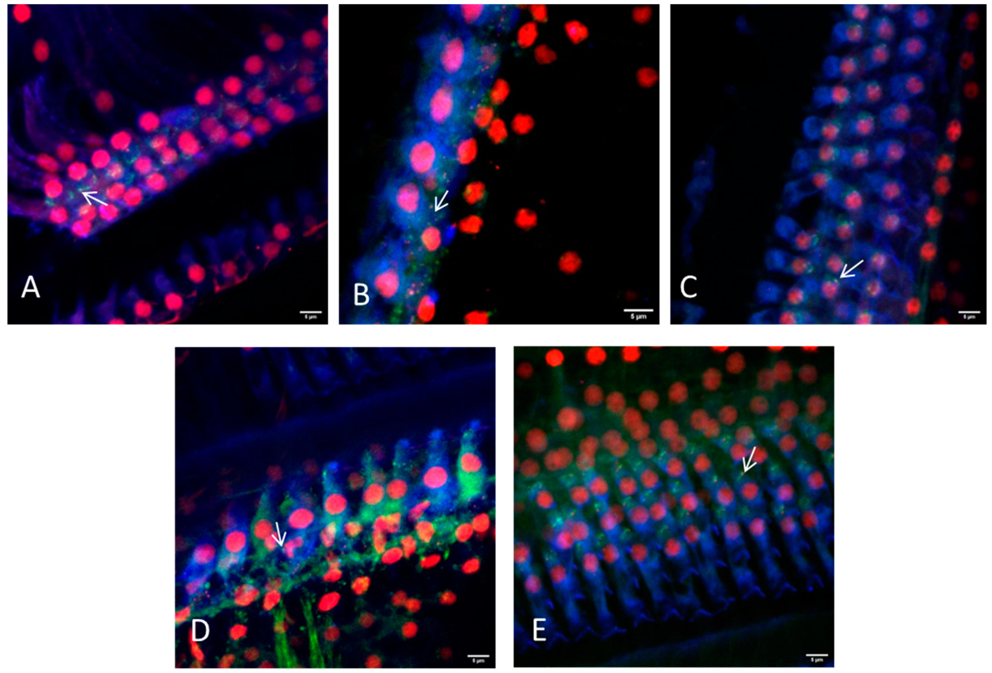
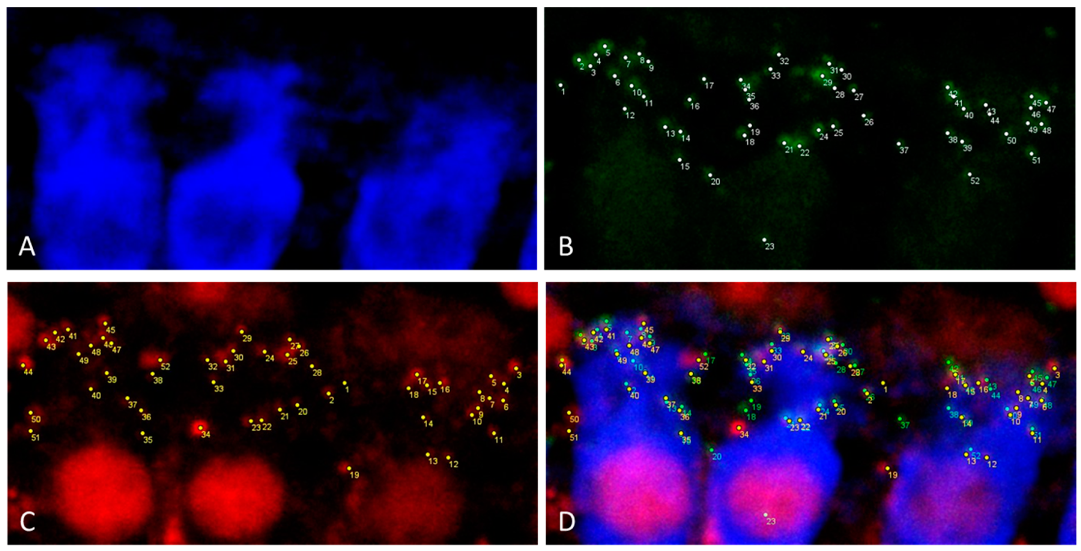
| Hair cell | Rabbit anti myosin VIIa, Novus Biol. NB 120-3481 | Goat anti rabbit IgG-Cy5, Jackson #111-175-144 | 650 | Blue |
| Postsynaptic density | Mouse anti PSD95, Merck MAB1596 | Goat anti mouse IgG2a-Alexa488, Thermo Fisher #21131 | 488 | Green |
| Ribbon synapse | Mouse anti CtBP2, BD #612044 | Goat anti mouse IgG1-Alexa568, Thermo Fisher #21124 | 568 | Red |
| I—Intact Cochlea | II—Bony Capsule Dissected | III—Dissected in Three Sections | |
|---|---|---|---|
| Preparation time | +++ | + | ++ |
| Time for microscopy | + | +++ | ++ |
| 20× objective | + | ++ | +++ |
| 63× objective | − | + | + |
| Stable sample positioning | ++ | ++ | +/− |
| Basilar membrane damage due to preparation | +++ | +/− | + |
| Overview image | +++ | + | ++ |
| Synapse imaging | − | + | ++ |
| Lateral wall tissue | intact | Often disrupted | Mostly intact |
Publisher’s Note: MDPI stays neutral with regard to jurisdictional claims in published maps and institutional affiliations. |
© 2021 by the authors. Licensee MDPI, Basel, Switzerland. This article is an open access article distributed under the terms and conditions of the Creative Commons Attribution (CC BY) license (https://creativecommons.org/licenses/by/4.0/).
Share and Cite
Malfeld, K.; Armbrecht, N.; Volk, H.A.; Lenarz, T.; Scheper, V. In Situ 3D-Imaging of the Inner Ear Synapses with a Cochlear Implant. Life 2021, 11, 301. https://doi.org/10.3390/life11040301
Malfeld K, Armbrecht N, Volk HA, Lenarz T, Scheper V. In Situ 3D-Imaging of the Inner Ear Synapses with a Cochlear Implant. Life. 2021; 11(4):301. https://doi.org/10.3390/life11040301
Chicago/Turabian StyleMalfeld, Kathrin, Nina Armbrecht, Holger A. Volk, Thomas Lenarz, and Verena Scheper. 2021. "In Situ 3D-Imaging of the Inner Ear Synapses with a Cochlear Implant" Life 11, no. 4: 301. https://doi.org/10.3390/life11040301
APA StyleMalfeld, K., Armbrecht, N., Volk, H. A., Lenarz, T., & Scheper, V. (2021). In Situ 3D-Imaging of the Inner Ear Synapses with a Cochlear Implant. Life, 11(4), 301. https://doi.org/10.3390/life11040301