Article

Yuxiensis granularis gen. et sp. nov., a Novel Quellkörper-Bearing Fungal Taxon Added to Scortechiniaceae and Inclusion of Parasympodiellaceae in Coronophorales Based on Phylogenetic Evidence

by
Digvijayini Bundhun
1,2,3,
Dhanushka N. Wanasinghe
1,4,*,
Sajeewa S. N. Maharachchikumbura
5,
Darbhe J. Bhat
6,
Shi-Ke Huang
3,
Saisamorn Lumyong
7,8,9,
Peter E. Mortimer
1,4 and
Kevin D. Hyde
2,3,7,10,*
1
Centre for Mountain Futures (CMF), Kunming Institute of Botany, Honghe County 654400, China
2
Innovative Institute of Plant Health, Zhongkai University of Agriculture and Engineering, Haizhu District, Guangzhou 510225, China
3
Center of Excellence in Fungal Research, Mae Fah Luang University, Chiang Rai 57100, Thailand
4
CIFOR-ICRAF China Program, World Agroforestry Centre, Kunming 650201, China
5
School of Life Science and Technology, University of Electronic Science and Technology of China, Chengdu 611731, China
6
No. 128/1-J, Azad Housing Society, Curca, P.O., Goa Velha 403108, India
7
Research Center of Microbial Diversity and Sustainable Utilization, Chiang Mai University, Chiang Mai 50200, Thailand
8
Department of Biology, Faculty of Science, Chiang Mai University, Chiang Mai 50200, Thailand
9
Academy of Science, The Royal Society of Thailand, Bangkok 10300, Thailand
10
CAS Key Laboratory for Plant Biodiversity and Biogeography of East Asia (KLPB), Kunming Institute of Botany, Chinese Academy of Sciences, Kunming 650201, China
*
Authors to whom correspondence should be addressed.
Life 2021, 11(10), 1011; https://doi.org/10.3390/life11101011
Submission received: 30 August 2021 / Revised: 17 September 2021 / Accepted: 19 September 2021 / Published: 25 September 2021

Abstract

:
An undetermined saprobic fungal taxon from Yunnan (China) is revealed as a new genus in Scortechiniaceae (Coronophorales). The novel taxon, Yuxiensis, is characterized by immersed to erumpent, semi-globose ascomata, which are not surrounded by any tomentum or conspicuous subiculum, a subcylindrical quellkörper in the centrum, clavate asci with long pedicels and allantoid hyaline ascospores with granular contents. Maximum likelihood and Bayesian posterior probability analyses based on LSU, ITS, tef1 and rpb2 sequence data depict a close phylogenetic relationship of the new genus to Pseudocatenomycopsis, hence, confirming its placement in Scortechiniaceae. Parasympodiellaceae, thus far belonging to Parasympodiellales, is transferred to Coronophorales based on multi-gene phylogenetic evidence. Additionally, the incertae sedis monotypic genus Arthrocristula is treated as a synonym of Parasympodiella, with Arthrocristula hyphenata recombined as Parasympodiella hyphenata comb. nov., as the type strain of Arthrocristula hyphenata clusters inside the Parasympodiellaceae clade along with other Parasympodiella taxa.

1. Introduction

Members of Coronophorales are generally found in terrestrial habitats. These fungi occur as saprobes on woody substrates, with no specific host per se, and their diversity spans tropical and temperate regions [1,2,3,4]. The order is accommodated in Hypocreomycetidae and it includes six families, viz. Bertiaceae, Ceratostomataceae, Chaetosphaerellaceae, Coronophoraceae, Nitschkiaceae and Scortechiniaceae, classified based on molecular data and morphology [2,5,6,7,8].
Scortechiniaceae was introduced by Huhndorf et al. [9] to accommodate three saprobic genera, Euacanthe, Neofracchiaea and Scortechinia using morphological characteristics and LSU sequence data. Mugambi and Huhndorf [2] later added six additional genera, Biciliospora, Coronophorella, Cryptosphaerella, Scortechiniella, Scortechiniellopsis and Tympanopsis to the family. All genera formed a well-supported clade in a multi-gene phylogeny based on LSU, tef1 and rpb2 sequence data [9]. Interestingly, all these taxa contain a distinctive phenotypic character in their centrum, namely, the quellkörper, which demarcates them from taxa belonging to other families in the order [2,9]. The quellkörper has been described as a subcylindrical to inverted-conical structure located in the centrum, made up of a mass of thick-walled, hyaline cells tightly arranged in a circular manner [2,9,10]. It is usually attached to the roof of the ascoma, orienting downward, and can extend until the base of the sporocarp [2,9,10]. The structure is mainly thought to be involved in ascospore discharge [2,9,10]. The quellkörper has, over the years, been considered and justified by taxonomists as the primary family-level character [2,3,9].
The monotypic taxon, Pseudocatenomycopsis, was then recently added to Scortechiniaceae based on LSU and ITS sequence data. The genus is only known in its asexual morph [11]. Latest taxonomic revisions of the family conducted by Huang et al. [12] revealed that Cryptosphaerella had to be transferred to Sordariomycetes genera incertae sedis based on the morphology of its type. The taxa Neocryptosphaerella and Pseudocryptosphaerella have instead been introduced to accommodate the Cryptosphaerella taxa having sequence data in GenBank at present [12].
Another order, Parasympodiellales, belonging to the same subclass, Hypocreomycetidae, was established by Hernández-Restrepo et al. [13], along with its type family, Parasympodiellaceae, to accommodate Parasympodiella, whose taxa formed a monophyletic clade in their phylogenetic tree. Members of Parasympodiellales are only known in their asexual morphs and are mainly characterized by sympodial conidiogenous cells and thallic-arthric conidia [13,14,15,16]. Furthermore, they usually have a stylaspergillus-like synasexual morph [13,16]. In the multi-gene phylogenetic analyses conducted by Hyde et al. [8], it was observed that Parasympodiellales clustered as sister to Scortechiniaceae (Coronophorales), following which the authors mentioned the status of this order requiring further revision.
Similarly, many species of Sordariomycetes still require taxonomic revision, especially those which have so far been identified based on morphology alone. Such is the case for Arthrocristula hyphenata, belonging to the monotypic genus Arthrocristula [17], currently placed in Ascomycota genera incertae sedis [18]. DNA sequence data for A. hyphenata were recently retrieved by Vu et al. [19] and deposited in GenBank. With both the morphology and molecular data for the taxon now available, it becomes largely feasible to determine the correct placement of A. hyphenata.
The present study aims to introduce a new fungal genus, Yuxiensis, in Scortechiniaceae, collected on a woody host in Yuxi (Yunnan, China) based on morphology and phylogenetic analyses of combined LSU–ITS–tef1rpb2 sequence data. Furthermore, Parasympodiellaceae is included in Coronophorales based on phylogenetic evidence. Arthrocristula is synonymized under Parasympodiella, with Arthrocristula hyphenata combined as Parasympodiella hyphenata Bundhun & K.D. Hyde, comb. nov., and is accommodated in Parasympodiellaceae based on DNA sequence data analyzed in the present study.

2. Materials and Methods

2.1. Specimen Collection and Morphological Studies

Dead twigs of an undetermined deciduous host were collected from Yuxi, Yunnan Province, China, during the dry season in May 2019. The samples were taken to the mycology laboratory at the Kunming Institute of Botany, in a plastic Ziploc bag and stored inside a paper envelope. External examinations were made using a Motic SMZ 168 Series stereo-microscope. Morphological characters were examined by hand sectioning of sporocarps and placed on water-mounted glass slides. Microscopic photography was conducted using a Nikon ECLIPSE 80i compound microscope (Nikon, Tokyo, Japan) fitted with a Canon EOS 600D camera. The structures which were observed and measured include diameter, height, shape, and color of ascomata; ascomatal wall’s width and cell structure; quellkörper shape; shape, length, and width of asci and shape, size, and ornamentation of ascospores. The Tarosoft (R) Image Frame Work version 0.9.7. program was used for the measurements of photomicrograph structures. Images used for figures were processed with the Adobe Photoshop CS6 Extended version 13.0.1 software (Adobe Systems, San Jose, California). The holotype was deposited in the herbarium of Cryptogams Kunming Institute of Botany Academia Sinica (HKAS). Both Facesoffungi [20] and Index Fungorum [21] numbers were obtained.

2.2. DNA Extraction, PCR Amplification and Sequencing

No culture could be obtained for the collected sample despite several trials on various media, including malt extract agar, potato dextrose agar, corn meal agar, or water agar under different incubation conditions. Therefore, DNA was extracted directly from the fruiting bodies of the fungus as outlined by Wanasinghe et al. [22].
According to the manufacturer’s instructions, DNA was extracted from fresh sporocarps using the Biospin Fungus Genomic DNA Extraction Kit-BSC14S1 (BioFlux, P.R. China). Primers LR0R/LR5 [23], ITS5/ITS4 [24], EF1-983F/EF1-2218R [25], and fRPB2-5f/fRPB2-7cR [26,27] were used to amplify the DNA sequences of the partial 28S large subunit rDNA (LSU), internal transcribed spacers (ITS), translation elongation factor 1-α (tef1), and RNA polymerase II second largest subunit (rpb2). The total volume of 25 μL contained 12.5 μL of 2× PCR Master Mix with dye (0.1 U Taq Polymerase/μL, 500 μm dNTP each), 20 mM Tris-HCl (pH 8.3, 100 mM KCl, 3 mM MgCl2), 1 μL of each primer, 9.5 μL of double-distilled water, and 1 μL (100–500 ng) of DNA template.
The PCR protocols were programmed as described in Wanasinghe et al. [28]. The PCR products were verified by staining with ethidium bromide on 1% agarose electrophoresis gels. They were then purified according to the company protocols and DNA sequencing was performed at Shanghai Sangon Biological Engineering Technology & Services Co. (Shanghai, P.R. China). Forward and reverse DNA sequence data were obtained and analyzed. Consensus sequences were generated using the SeqMan software (DNAStar, Inc., Madison, WI, USA). The newly acquired sequence data from the present study were deposited in GenBank for subsequent studies [29] (Table 1).

2.3. Phylogenetic Analyses

Verified sequences were initially used for BLASTn analyses, following which closely related sequences were downloaded from GenBank based on BLAST similarities and relevant publications [2,8] (Table 1). Alignment of each locus was performed using MUSCLE in MEGA X (Molecular Evolutionary Genetics Analysis), using default conditions for gap openings and gap extension penalties. It was then improved whenever necessary in the BioEdit v.7.0.5.2 software [30].
Maximum likelihood (ML) and Bayesian posterior probability (BYPP) analyses were conducted using both individual and combined datasets. Prior to ML analysis, the sequence alignments were converted from FASTA into PHYLIP format using the ALTER (alignment transformation environment, http://www.singgroup.org/ALTER/, accessed on 30 August 2021) bioinformatics web tool [31]. They were then used to generate ML trees using RAxML-HPC2 on XSEDE (v.8.2.10) [32] with the GTRGAMMA substitution model and bootstrapping with 1000 replicates.
The BYPP analysis was generated using Markov Chain Monte Carlo sampling in MrBayes v3.1.2 [33,34]. MrModeltest v.2.3 [35] was used to estimate the best evolutionary model for each gene region under the Akaike Information Criterion (AIC) implemented in PAUP v.4.0b10 [36]. The best-fit model was determined as GTR+I+G for LSU, tef1, and rpb2 while GTR+G for ITS. Six simultaneous Markov chains were run for 4.5M generations with trees sampled every 100th generation. The first 20% of generated trees were the burn-in phase and discarded. The remaining 80% of trees were used to calculate posterior probabilities in the majority rule consensus tree. Phylograms were configured in FigTree v.1.4.0 [37] and modified in Microsoft PowerPoint (2013). The final alignments and phylogenetic tree were deposited in TreeBASE, submission ID: 28713 (http://www.treebase.org/ , accessed on 30 August 2021).

3. Results

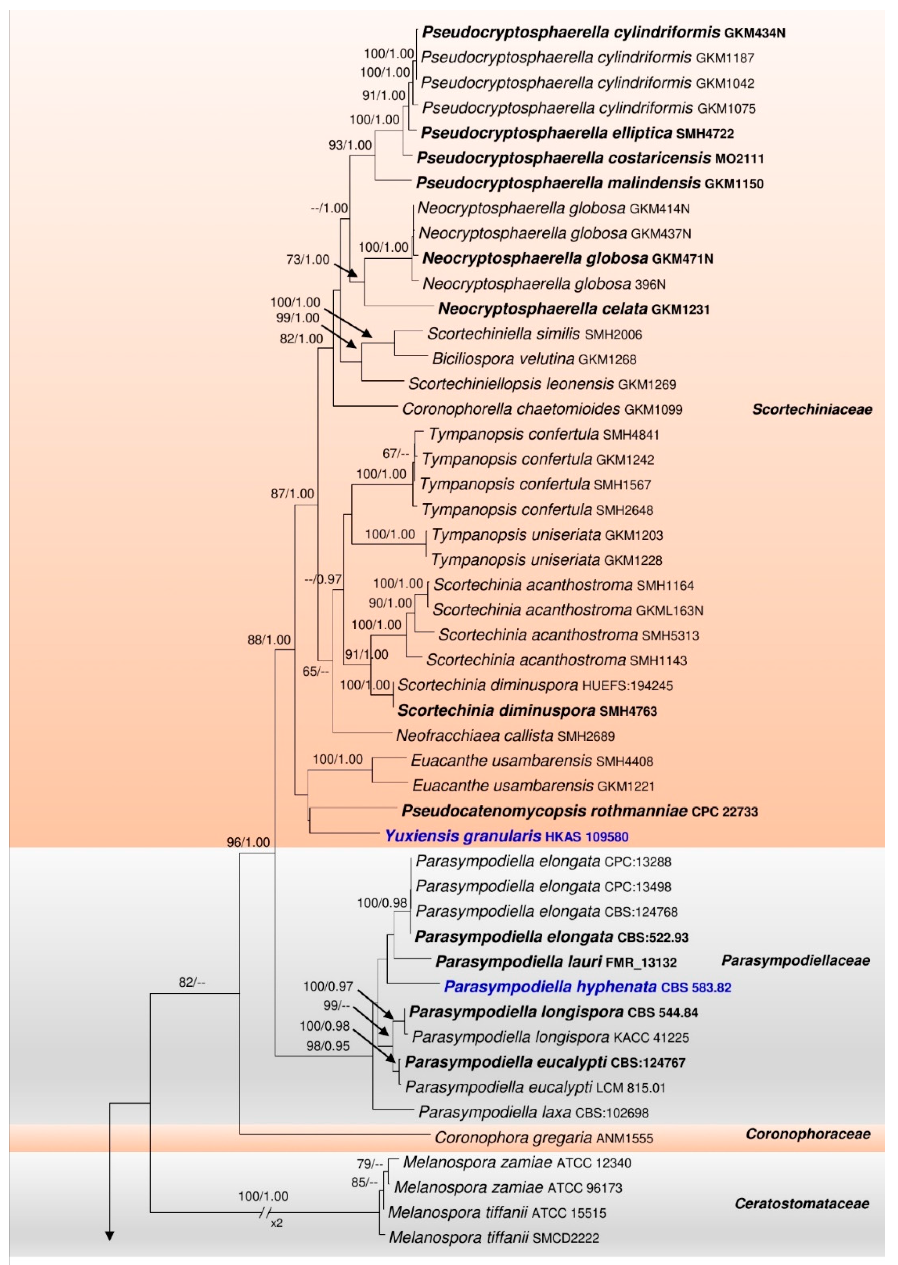
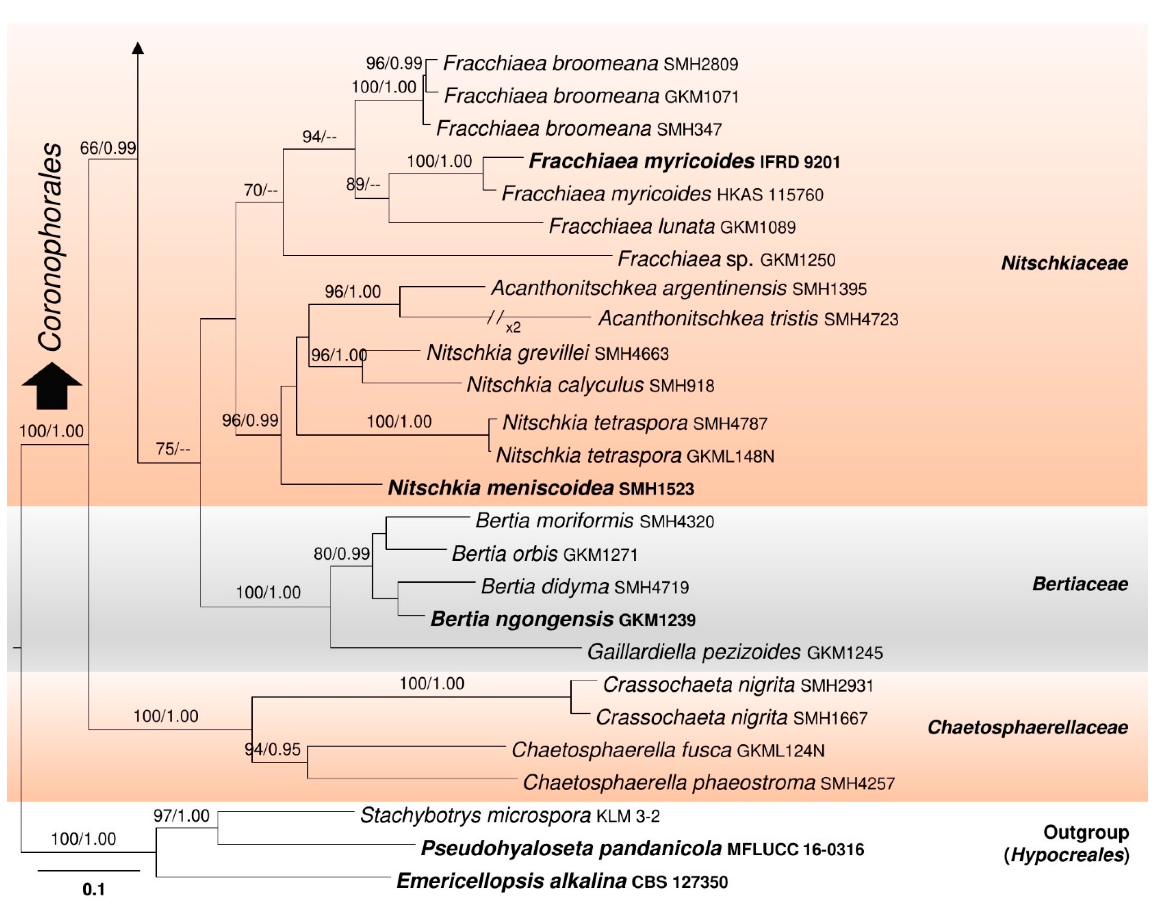
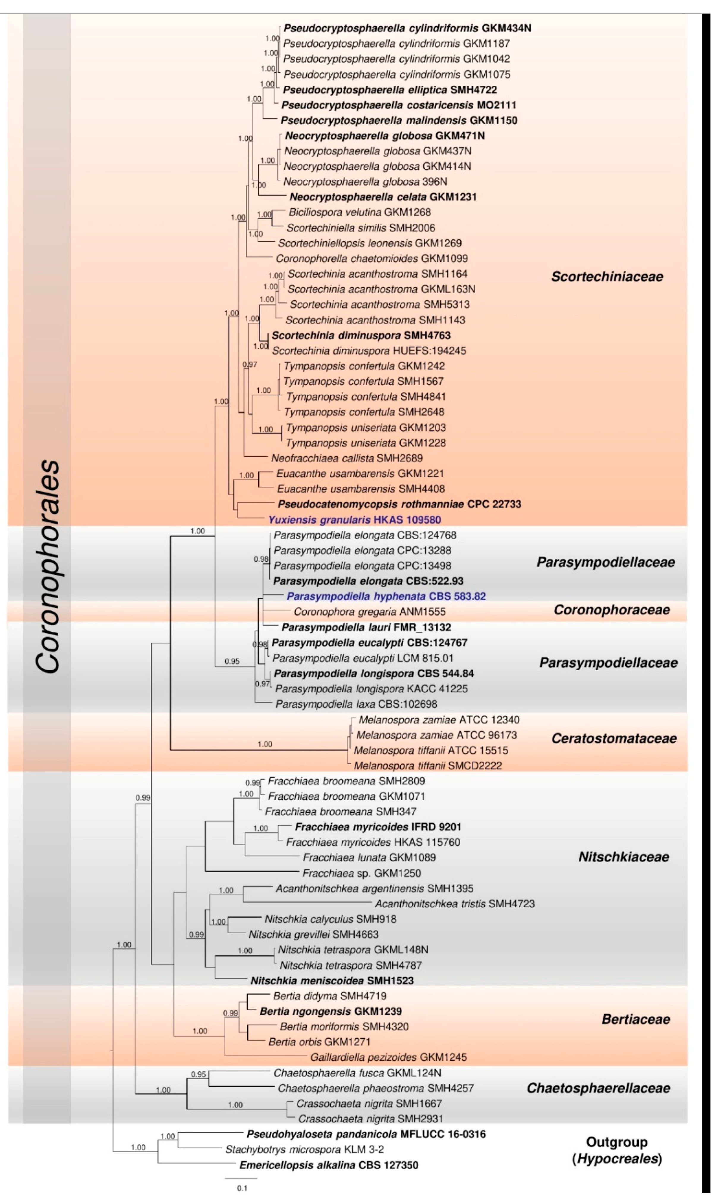
3.1. Phylogenetic Analyses

The final concatenated LSU–ITS–tef1rpb2 alignment (Figure 1) comprised 75 strains including the outgroup taxa Emericellopsis alkalina (CBS 127350), Pseudohyaloseta pandanicola (MFLUCC 16-0316), and Stachybotrys microspora (KLM 3-2). The manually adjusted dataset consisted of 3845 characters including gaps (LSU: 1086, ITS: 768, tef1: 813, rpb2: 1178). The best scoring RAxML tree with final optimization had a likelihood value of −41,633.775219. The matrix had 2266 distinct alignment patterns, with 49.47% of gaps and completely undetermined characters. Estimated base frequencies were as follows: A = 0.236728, C = 0.275120, G = 0.286267, T = 0.201885, with substitution rates AC = 1.265283, AG = 3.588580, AT = 1.636499, CG = 1.073052, CT = 7.584875, GT = 1.000000. The gamma distribution shape parameter α = 0.342349 and Tree-length = 7.680873.
The families Bertiaceae, Ceratostomataceae, Chaetosphaerellaceae, Coronophoraceae, Nitschkiaceae, Parasympodiellaceae and Scortechiniaceae grouped together, each forming a monophyletic clade in the ML tree (Figure 1). The tree topology resulting from the BYPP analysis mainly differed from the ML one with regard to the placement of Coronophora gregaria (ANM1555) (Coronophoraceae), which clustered within Parasympodiellaceae (Figure A1).
Parasympodiellaceae formed a sister clade with Scortechiniaceae with statistical support of 96% ML, 1.00 BYPP. It comprised ten strains of Parasympodiella and a strain of ‘Arthrocristula hyphenata’ (CBS 583.82), which clustered together with 98% ML and 0.95 BYPP statistical support (Figure 1).
Our strain HKAS 109580 formed a distinct lineage within Scortechiniaceae, placed sister to Pseudocatenomycopsis rothmanniae (CPC 22733) with low statistical support (Figure 1).

3.2. Taxonomy

In this section, the amended descriptions and notes for Coronophorales, Parasympodiellaceae, and Parasympodiella are given. Furthermore, descriptions, notes, and illustrations are given for the following taxa: Parasympodiella hyphenata comb. nov. and Yuxiensis granularis gen. et sp. nov.

3.2.1. Coronophorales Nannf., Nova Acta R. Soc. Scient. Upsal., Ser. 4 8(no. 2): 54 (1932) Amend

Index Fungorum number: IF 501516; Facesoffungi number: FoF 06517
= Parasympodiellales Hern.-Restr., Gené, R.F. Castañeda & Crous 2017
Saprobic on leaves, wood, or associated with sclerotia. Sexual morph: see Mugambi and Huhndorf [2], Hyde et al. [8], Nannfeldt [10]. Asexual morph: Hyphomycetous. Conidiophores simple or branched, septate, straight to flexuous, brown, smooth or comprising rough swellings along the whole length in some genera or geniculations in others. Conidiogenous cells apical, lateral or intercalary, usually hyaline, often sympodial, blastic or thallic. Conidia hyaline or brown, aseptate to septate, globose, oval, elliptical, ellipsoidal, pyriform, cylindrical, oblong or spindle-shaped, smooth or verrucose, solitary or produced in branched or unbranched chains. Synasexual morph: stylaspergillus-like (see Hernández-Restrepo et al. [13], Cheewangkoon et al. [16]), or arthrocristula-like: Conidiophores branched, brown, smooth. Conidiogenous cells apical, lateral, sympodial, thallic. Conidia pale brown or brown, aseptate to septate, cylindrical to oblong, produced in unbranched chains.
Type family: Coronophoraceae Höhn.
Notes: The phylogenetic analyses based on the combined LSU–ITS–tef1rpb2 sequence data, conducted in the present study, supports the inclusion of Parasympodiellales in Coronophorales, as Parasympodiellaceae is sister to Scortechiniaceae with 96% ML and 1.00 BYPP statistical support within the order (Figure 1). The description for the asexual morph of taxa in Coronophorales is therefore emended to include the morphological characteristics of Parasympodiellaceae.

3.2.2. Parasympodiellaceae Hern.-Restr., Gené, Guarro & Crous, in Hernández-Restrepo, Gené, Castañeda-Ruiz, Mena-Portales, Crous & Guarro, Stud. Mycol. 86: 87 (2017) Amend

Index Fungorum number: IF 820298; Facesoffungi number: FoF 06518
Saprobic on leaves and twigs or associated with sclerotia. Sexual morph: Undetermined. Asexual morph: Hyphomycetous. Conidiophores micro- to macronematous, brown, usually unbranched, septate. Conidiogenous cells thallic, terminal or intercalary, sympodial. Conidia thallic-arthric, aseptate or septate, cylindrical, hyaline, often in chains, with schizolytic secession. Synasexual morph: stylaspergillus-like (see Hernández-Restrepo et al. [13], Cheewangkoon et al. [16]) or arthrocristula-like: Conidiophores micro- to macronematous, brown, branched. Conidiogenous cells thallic, terminal, lateral, sympodial. Conidia thallic-arthric, aseptate or septate, cylindrical to oblong, pale brown or brown, generally in chains, with rhexolytic secession.
Type genus: Parasympodiella Ponnappa
Notes: Parasympodiellaceae is now included in Coronophorales, which at present comprises seven families (this study). This family, in turn, accommodates the single genus Parasympodiella.

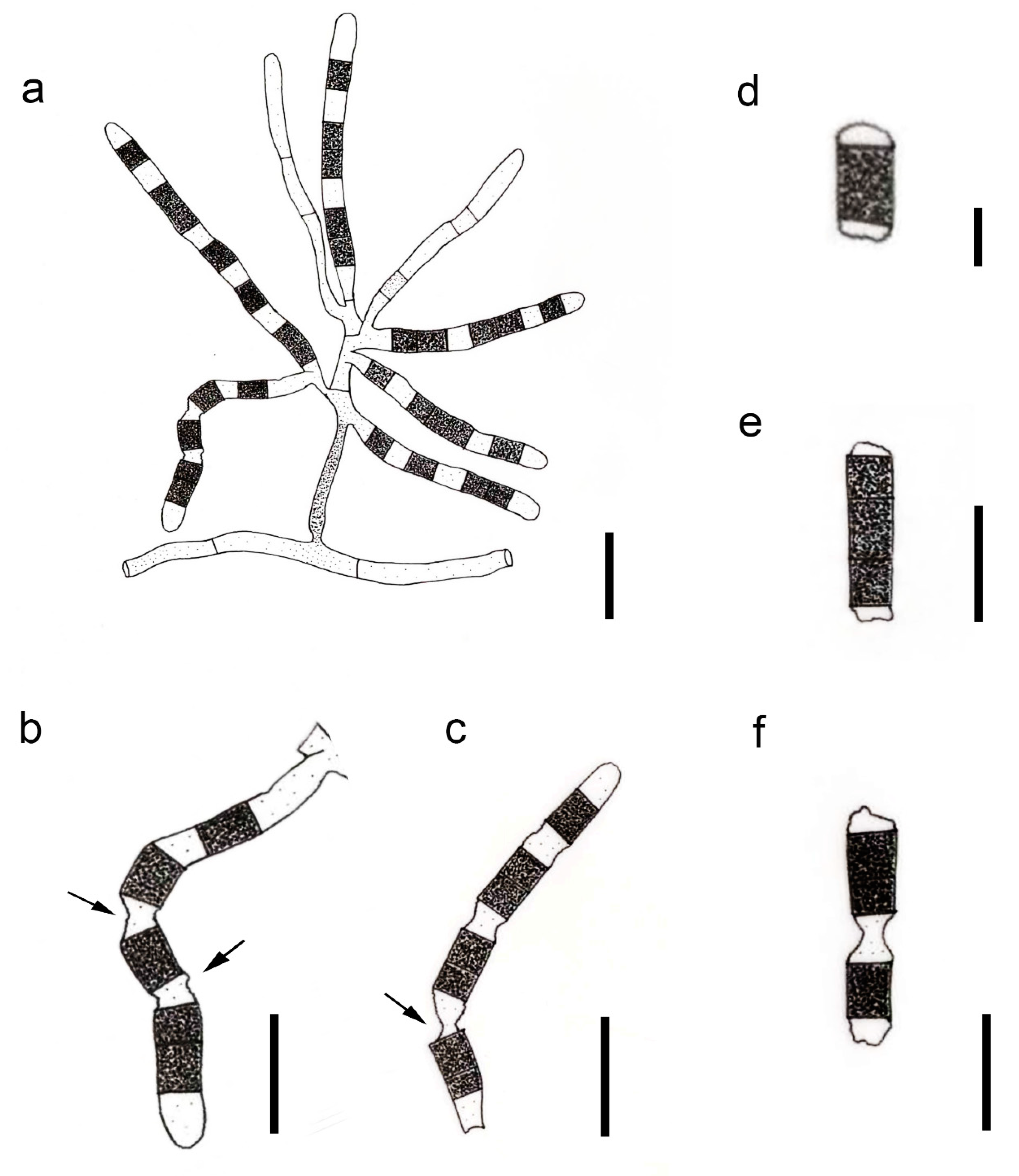
3.2.3. Parasympodiella Ponnappa Trans. Br. Mycol. Soc. 64(2): 344 (1975) Amend

Index Fungorum number: IF 9226; Facesoffungi number: FoF 05188
= Arthrocristula Sigler, M.T. Dunn & J.W. Carmich., Mycotaxon 15: 409 (1982)
Saprobic on leaves and twigs or associated with sclerotia. Sexual morph: Undetermined. Asexual morph: Hyphomycetous. Conidiophores micro- to macronematous, brown, generally unbranched, septate, straight to geniculate. Conidiogenous cells thallic, terminal or intercalary, indeterminate, sympodial, unbranched. Conidia thallic-arthric, produced in unbranched chains, cylindrical, aseptate or septate, hyaline, seceding schizolytically, often with a septal plug. Synasexual morph: stylaspergillus-like (see Hernández-Restrepo et al. [13], Cheewangkoon et al. [16]) or arthrocristula-like: Conidiophores micro- to macronematous, brown, branched. Conidiogenous cells thallic, terminal, lateral, indeterminate, sympodial, unbranched. Conidia thallic-arthric, produced in unbranched chains, aseptate or septate, pale brown or brown, cylindrical to oblong, with rhexolytic secession, with frills of remnant cells at each end.
Type species: Parasympodiella laxa (Subram. & Vittal) Ponnappa
Notes: With the exact taxonomic placement being uncertain, the hyphomycetous taxon Arthrocristula has so far been maintained in Ascomycota genera incertae sedis [18,38]. In the present study, the type strain of A. hyphenata (CBS 583.82) was found to cluster within Parasympodiellaceae, indicating a close phylogenetic affinity to Parasympodiella (Figure 1). Morphologically, the conidiophores of Parasympodiella are unbranched or sparingly branched, thick-walled, and brown, becoming paler towards the conidiogenous regions. They are often geniculate, with terminal or intercalary sympodial conidiogenous cells, which are mostly thallic. The secession of conidia occurs schizolytically [13,14,16]. The conidiophores of Arthrocristula are, however, well-branched with no conspicuous geniculation and are initially hyaline, becoming thick-walled and brown upon maturity. The conidiogenous cells secede rhexolytically into arthroconidia, leaving the mature conidia with remnants of the separating cells at each end with small frills. The terminal cells of the conidiogenous hyphae remain as empty cells [17]. Considering these morphological differences, the two genera can be considered as distinct. However, given that Parasympodiella has a stylaspergillus-like synasexual morph, the fact that Arthrocristula also is another synasexual morph of Parasympodiella cannot be ruled out. Fungi have been reported to have two or more morphologically distinct asexual morphs [39,40,41]. Moreover, phylogeny supports the inclusion of Arthrocristula in Parasympodiella (Figure 1). Therefore, the former is synonymized under Parasympodiella in the present study.

3.2.4. Parasympodiella hyphenata (Sigler, M.T. Dunn & J.W. Carmich.) Bundhun & K.D. Hyde, comb. nov.

Index Fungorum number: IF 558677; Facesoffungi number: FoF 10183, Figure 2
Arthrocristula hyphenata Sigler, M.T. Dunn & J.W. Carmich., Mycotaxon 15: 409 (1982)
Associated with sclerotia of Sclerotinia minor in soil. Sexual morph: Undetermined. Asexual morph: Hyphomycetous. Mycelium in vitro comprises narrow, septate vegetative hyphae (2–3 µm diam.), hyaline when immature and brown at maturity. Conidiophores originating from vegetative hyphae, undifferentiated, initially hyaline, turning brown and thick-walled when mature, branched, narrow at first, widening and extending apically and laterally after that to give rise to fertile conidiogenous hyphae. Conidiogenous cells (80–100 × 4–5.5 µm), delimited by a basal septum, developing successively, generally unbranched, indeterminate, initially non-septate, hyaline, becoming randomly septate and pigmented in alternate cells on maturity, undergoing rhexolytic secession to give rise to arthroconidia. Conidia (5–12 × 6–7 µm), thallic-arthric, brown, aseptate to septate, cylindrical to oblong, formed in a chain with hyaline separating cells, with fragments of intervening cell walls remaining attached to both ends of conidium after secession, terminal cells of the chains remain empty. Chlamydospores in vitro (12–16 × 8–13 µm), thick-walled, brown, terminal and intercalary (adapted from Sigler et al. [17]).
Notes: The type and single taxon of Arthrocristula, A. hyphenata is combined under Parasympodiella following the synonymy of Arthrocristula under Parasympodiella in the present study. Parasympodiella hyphenata comb. nov. has been reported from the sclerotium of Sclerotinia minor buried in the soil [17]. It is mainly characterized by branched conidiophores and alternatively pigmented conidiogenous cells, which produce conidia by seceding rhexolytically [17].

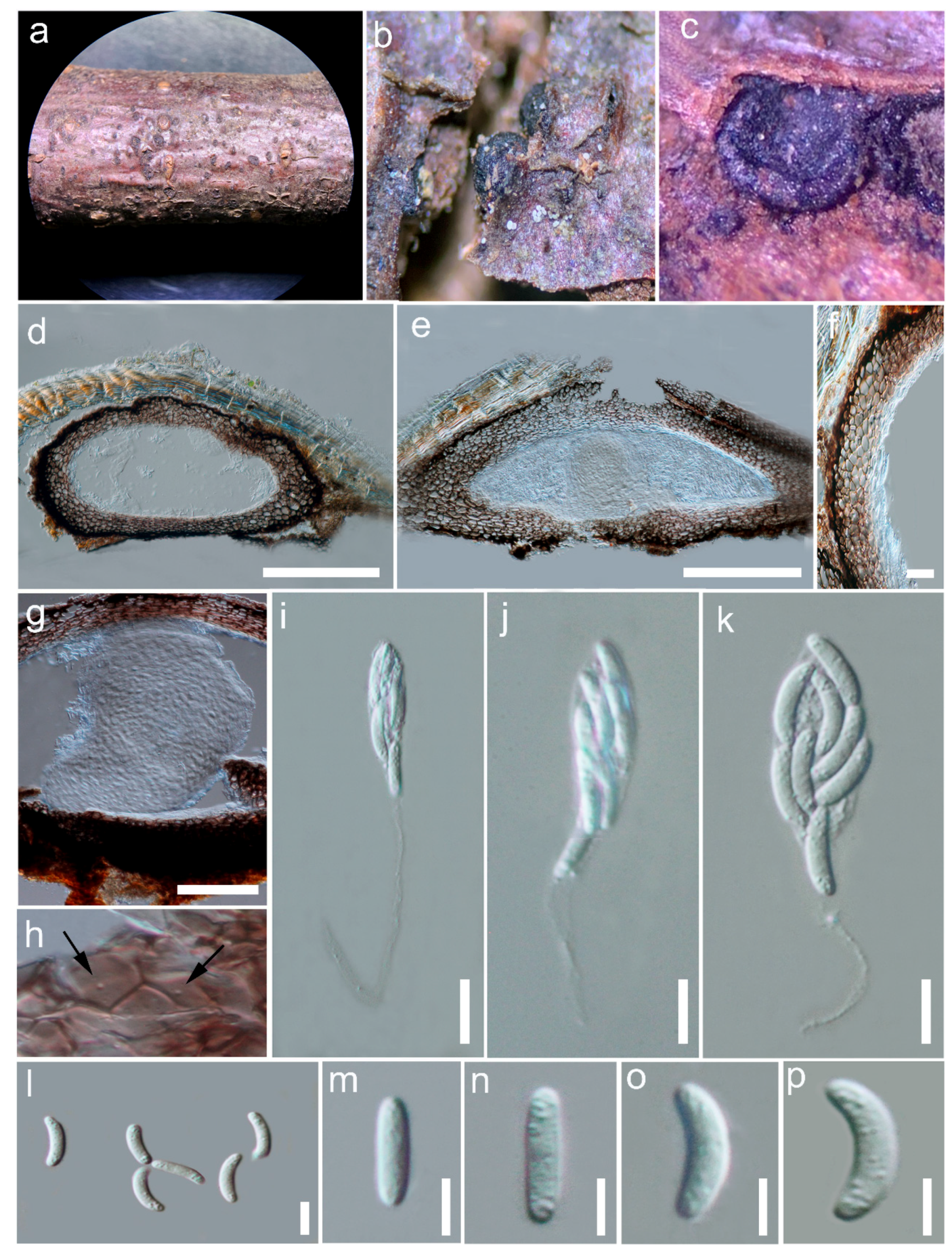
3.2.5. Yuxiensis Bundhun & K.D. Hyde, gen. nov.

Index Fungorum number: IF 558675; Facesoffungi number: FoF 10184
Etymology–Referring to the city Yuxi in China.
Saprobic on dead wood. Sexual morph: Ascomata immersed to erumpent, appearing superficial on worn off substrate, astromatic to stromatic, aggregated, black, non-ostiolate, semi-globose when fresh, collapsing when dry, without hair or bristles, subiculum inconspicuous, coriaceous. Ascomatal wall comprising 2–3 types of layers; outermost layer heavily pigmented, composed of thick-walled, very dark brown cells, inner layer comprising thick-walled cells of textura globulosa to textura angularis, innermost layer composed of flattened, thin-walled cells of textura prismatica toward the locule; Munk pores visible, few per cell. Hamathecium composed of large, subcylindrical quellkörper, attached to the roof of the centrum and extending until base of the ascoma; paraphyses indistinct. Asci 8-spored, unitunicate, clavate, long-pedicellate, rounded at apex, lacking an apical ring, thin-walled, evanescent. Ascospores irregularly arranged, cylindrical to allantoid, hyaline, aseptate, with granular contents, lacking mucilaginous sheath or appendage. Asexual morph: Undetermined.
Type: Yuxiensis granularis Bundhun, Wanas. & K.D. Hyde
Notes: Yuxiensis is introduced in Scortechiniaceae as a new quellkörper-bearing taxon distinct from all other genera in the family, based on LSU, ITS, tef1 and rpb2 sequence data. The new genus has a close phylogenetic affinity to Pseudocatenomycopsis even though this relationship is not statistically significant (Figure 1). This low support may possibly be accounted for by insufficient taxon sampling. Nevertheless, the new taxon being introduced in the present study constantly clusters with Pseudocatenomycopsis in all phylogenies (single, not shown; and concatenated, Figure 1). The asexual morph for Yuxiensis could not be obtained in the present study. It thus cannot be morphologically compared with Pseudocatenomycopsis, which has been described in its asexual morph only. Pseudocatenomycopsis has been introduced from Zambia as a saprobe on the stem of Rothmannia engleriana (Rubiaceae) [11]. The new genus is also phylogenetically close to Euacanthe (Figure 1). It morphologically differs from Euacanthe in terms of ascomatal position and surface as well as ascospore ornamentation [12,43].

3.2.6. Yuxiensis granularis Bundhun, Wanas. & K.D. Hyde, sp. nov.

Index Fungorum number: IF 558676; Facesoffungi number: FoF 10185, Figure 3
Etymology—The specific epithet refers to the granular contents of the ascospores.
Holotype–HKAS 109580
Saprobic on dead twigs of deciduous hosts in terrestrial habitats. Sexual morph: Ascomata 250–400 μm high, 550–700 μm diam. ( x ¯ = 309 × 642 μm, n = 5), astromatic to stromatic, immersed to erumpent, appearing superficial after substrate has worn away, aggregated, black, non-ostiolate, semi-globose when fresh, collapsing upon drying, without hair or bristles, with inconspicuous subiculum, coriaceous. Ascomatal wall made up of 2–3 layers, almost equally thickened, 40–70 μm wide at the apex and base, 50–70 μm wide at the sides; heavily pigmented at outermost layer, composed of thick-walled, very dark brown cells, with inner layer comprising thick-walled cells (25–45 μm) of textura globulosa to textura angularis, innermost layer composed of flattened, thin-walled cells of textura prismatica toward the locule; Munk pores visible, few per cell. Hamathecium made up of subcylindrical quellkörper, 290 μm long and 220 μm wide. Asci 40–80 × 5–10 μm ( x ¯ = 55.8 × 8.4 μm, n = 10), spore bearing part 15–30 μm, pedicel 15–50 μm, 8-spored, unitunicate, clavate, long-pedicellate, rounded at apex, lacking an apical ring, thin-walled, evanescent. Ascospores 8–15 × 2–3 μm ( x ¯ = 11.7 × 2.3 μm, n = 35), irregularly arranged, cylindrical to allantoid, hyaline, unicellular, aseptate, with granular contents, lacking mucilaginous sheath or appendage. Asexual morph: Undetermined.
Material examined: CHINA, Yunnan, Yuxi, Yi and Dai Autonomous County, Yuanjiang Hani, 23.74074 N, 102.17735 E, on woody litter of an undetermined deciduous host, 24 May 2019, 1345 msl, D.N. Wanasinghe, DW0636-19 (HKAS 109580, holotype).
Notes: In the multi-gene phylogeny, Yuxiensis granularis is more closely related to Pseudocatenomycopsis rothmanniae, followed by Euacanthe usambarensis (=Euacanthe foveolata [12]) (Figure 1). The LSU sequence of Yuxiensis granularis is 95% similar to Pseudocatenomycopsis rothmanniae (GenBank KF777237; similarity = 869/910(95%), Gaps = 3/910(0%)). The ITS sequence of Yuxiensis granularis is 85% similar to Pseudocatenomycopsis rothmanniae (GenBank KF777185; similarity = 470/552(85%), Gaps = 10/552(1%)). Pseudocatenomycopsis rothmanniae has only LSU and ITS sequence data deposited in GenBank, and hence the protein-coding genes, tef1 and rpb2 could not be compared. Morphological comparison between the two taxa is currently unfeasible since the single species of Pseudocatenomycopsis, P. rothmanniae, has been introduced in its asexual morph [11], while the asexual morph for Yuxiensis granularis could not be obtained.
Yuxiensis granularis resembles Euacanthe usambarensis in having ascomata which become collabent on drying and 8-spored, clavate asci [2,12,43]. However, the new species has immersed to erumpent, glabrous ascomata that are not surrounded by any conspicuous subiculum or tomentum, whereas Euacanthe usambarensis comprises superficial ascomata with a setose surface, sitting on a dense subiculum [12,43]. The asci of Yuxiensis granularis are long-pedicellate whereas those of Euacanthe usambarensis have short pedicels [12,43]. Furthermore, while the ascospore contents of Yuxiensis granularis are granular, those of Euacanthe usambarensis generally have two guttules [12,43]. There are also more than 2.5% nucleotide differences in LSU out of 925 base pairs among the strains of Yuxiensis granularis (HKAS 109580) and Euacanthe usambarensis (GKM1221 and SMH4408), while more than 2.5% nucleotide differences in tef1 and rpb2 out of 731 and 1178 base pairs respectively between the strains of Yuxiensis granularis (HKAS 109580) and Euacanthe usambarensis (GKM1221).

4. Discussion

Sordariomycetes is a frequently-studied class, with several taxa having been recently introduced or revised [4,5,8,12,44]. The present study corroborates this fact, as supported, firstly, by establishing a new saprobic genus, Yuxiensis, in Scortechiniaceae based on a dual taxonomic approach. In addition to phylogeny, the familial placement of the new genus within Scortechiniaceae is morphologically confirmed by the presence of the quellkörper in its centrum. Within Scortechiniaceae, Yuxiensis shares many overlapping characters with the other genera. For instance, like almost all the other genera in the family with a known sexual morph, Yuxiensis comprises ascomata which collapse upon drying, presence of munk pores in the ascomatal wall and inconspicuous paraphyses [4,8]. It also has 8-spored asci, similar to Biciliospora, Coronophorella, Euacanthe, Scortechinia, and Tympanopsis and long-pedicellate asci like Biciliospora, Neofracchiaea, Scortechinia, Scortechiniella, and Scortechiniellopsis [2,4,8]. It is equally similar to most taxa of Neocryptosphaerella and Pseudocryptosphaerella in that its ascomata are immersed to erumpent, appearing superficial when the substrate has worn away [2,12]. The new genus however, demarcates itself from the other genera in the family by several ways. Its ascomata are not seated on or surrounded by a well-developed, conspicuous subiculum unlike many taxa of Biciliospora, Coronophorella, Euacanthe, Neofracchiaea, Scortechinia, Scortechiniella, Scortechiniellopsis, or Tympanopsis [2,4,8]. The ascomata of Yuxiensis are also devoid of a tomentose or setose surface as compared to Euacanthe, Neofracchiaea, or some taxa of Neocryptosphaerella and Pseudocryptosphaerella [2,8]. Moreover, the ascospores of Yuxiensis do not have conspicuous guttules unlike those of Euacanthe, Neocryptosphaerella, and Pseudocryptosphaerella taxa and they lack appendage-like wall extensions on both ends, contrary to Biciliospora and Scortechiniella [2,45]. Since Yuxiensis is phylogenetically closely related to Pseudocatenomycopsis and Euacanthe (Figure 1), more details about their morpho-molecular comparisons have been given in the result parts 3.2.5 and 3.2.6 above. Based on all these morphological as well as phylogenetic differences, Yuxiensis is herein introduced as a new genus.
The inclusion of Parasympodiellales in Coronophorales in the present study also points toward the continuous amendment in the classification of Sordariomycetes. Herein, while phylogeny supports the addition of Parasympodiellaceae to Coronophorales, this inclusion is equally supported by the fact that taxa of Parasympodiellaceae have similar morphological characters with several taxa in other families of Coronophorales (Ceratostomataceae, Chaetosphaerellaceae, Scortechiniaceae ) in terms of unbranched or branched, generally brown and often septate conidiophores or conidia produced in chains [4,8,11]. Parasympodiellaceae distinguishes itself from the other families mainly by its sympodial and unbranched conidiogenous cells which undergo schizolytic or rhexolytic secession to form arthroconidia. Furthermore, the incertae sedis taxon Arthrocristula is synonymized under Parasympodiella, with Arthrocristula hyphenata recombined to Parasympodiella hyphenata and representing another synasexual morph of Parasympodiella. This arthrocristula-like synasexual morph of Parasympodiella is typically characterized by branched conidiophores and conidiogenous cells which secede rhexolytically to give rise to arthroconidia. It is also different from the stylaspergillus-like synasexual morph of Parasympodiella which is generally characterized by pale brown, phialidic conidiogenous cells originating from terminal or intercalary vesicle-like cells and filiform conidia which are produced in slimy masses [13].
An additional collection of Fracchiaea myricoides (HKAS 115760) was also made in the present study, and sequence data for the same have been used in the phylogenetic analyses and deposited in GenBank (Table 1). The latter species was initially introduced as Coronophora myricoides based on LSU and ITS sequence data and the differences mentioned between this taxon and the type species of Coronophora, C. gregaria was mainly based on the shapes of the ascomata and ascospores [46]. Huang et al. [12] recently synonymized this species to Fracchiaea myricoides; our collection supports this synonymy and the inclusion of ‘Coronophora myricoides’ in Fracchiaea (Nitschkiaceae) (Figure 1).
Despite the advancement towards a natural classification of Sordariomycetes, uncertainties and confusions still prevail, as we note in the case of Parasympodiella longispora (CBS 544.84 and KACC 41225) (in Parasympodiellaceae clade, Figure 1). The latter is currently known as ‘Bahusakala longispora’ in Index Fungorum and MycoBank, with Parasympodiella longispora as an (obligate) synonym. The type strain of ‘Bahusakala longispora’, CBS 544.84, sequenced by Vu et al. [19], clusters in the Parasympodiellaceae clade (Figure 1) with good statistical support. Bahusakala longispora was introduced by Tokumasu and Tubaki [47] as a taxon with conidiophores that are sympodial, rarely branched, erect in the lower part and become geniculate (zig-zag, as mentioned in the original description) in the upper part. Furthermore, the conidiogenous cells are hyaline, originating at regular intervals, while the arthroconidia, subhyaline to pale yellow. Chlamydospores are produced in vegetative hyphae. However, the species was later synonymized to Parasympodiella longispora since its morphology (based on its type) matched the description of Parasympodiella more accurately [48]. Bahusakala taxa have been reported to have conidiophores that branch at irregular intervals to produce brown conidiogenous hyphae at the terminal and intercalary positions. The conidia are usually brown to dark brown and originate from random disarticulation of the main conidiophore axes and conidiogenous hyphae [42,48,49,50]. Based on the description of its type (and placement in the present phylogenetic tree), the species is better accommodated in Parasympodiella than Bahusakala. We may as well adopt a broader taxonomic perspective and decide that both Parasympodiella and Bahusakala are congeneric since, despite their morphological differences, the two genera are also characterized by similar features. Both are hyphomycetes with erect and septate conidiophores and produce arthroconidia which secede schizolytically [42,50]. However, no molecular data for the type species, B. olivaceonigra is yet available to enable any definite phylogenetic placement and eventually to confirm a taxonomic conclusion for Bahusakala.
We also take note that the GenBank accession numbers of the sequence data for Neocryptosphaerella globosa (GKM471N) that we use in our phylogeny have been assigned under different strain numbers, namely, LSU (GenBank FJ968977: strain GKM469N), tef1 (GenBank FJ969036: strain GKM471N), and rpb2 (GenBank FJ968935: strain GKM469N). In the original manuscript [2], these accession numbers are under the strain number Neocryptosphaerella globosa GKM471N. Therefore, we followed the original paper.
This remarkable finding of a new genus in a rarely collected order indicates how little we know of the fungal diversity of Yunnan and the broader region, including areas such as Thailand and Laos [51], with recent studies showing large numbers of novel taxa being discovered [52,53]. Further studies in other countries and habitats across this region will surely result in the discovery of numerous other taxa in Parasympodiella, Yuxiensis, and other poorly known taxa of Coronophorales [54].

Author Contributions

Conceptualization, D.B., D.N.W. and K.D.H.; data curation, D.N.W.; formal analysis, D.B., D.N.W., S.S.N.M. and S.-K.H.; funding acquisition, D.N.W. and K.D.H.; investigation, D.B. and S.S.N.M.; methodology, D.B., D.N.W. and S.S.N.M.; resources, D.N.W., P.E.M. and K.D.H.; supervision, K.D.H.; writing—original draft, D.B., D.N.W., S.S.N.M., D.J.B. and S.-K.H.; writing—review & editing, S.L., P.E.M. and K.D.H. All authors have read and agreed to the published version of the manuscript.

Funding

Dhanushka N. Wanasinghe would like to thank CAS President’s International Fellowship Initiative (PIFI) for funding his postdoctoral research (number 2021FYB0005) and the Postdoctoral Fund from Human Resources and Social Security Bureau of Yunnan Province. Kevin D. Hyde thanks the Thailand Research Fund, grant RDG6130001 entitled “Impact of climate change on fungal diversity and biogeography in the Greater Mekong Sub-region”.

Institutional Review Board Statement

Not applicable.

Informed Consent Statement

Not applicable.

Data Availability Statement

The nucleotide sequences generated in the present study are deposited in GenBank (Table 1). The final alignment and phylogenetic tree have been submitted to TreeBASE (submission ID: 28713, http://www.treebase.org/, accessed on 30 August 2021). Specimen has been deposited in the herbarium of Cryptogams Kunming Institute of Botany Academia Sinica (HKAS).

Acknowledgments

Digvijayini Bundhun gratefully acknowledges Mae Fah Luang University, the Mushroom Research Foundation and the Center of Excellence in Fungal Research, Thailand, for research support. Ruvishika S. Jayawardena, Eleni Gentekaki, Rajesh Jeewon, Chitrabhanu S. Bhunjun, and Vedprakash G. Hurdeal are thanked for their advice and support throughout this study. Kevin D. Hyde thanks Chiang Mai University for the award of a Visiting Professorship. All authors thank Shaun Pennycook and Paul Kirk for their valuable suggestions.

Conflicts of Interest

The authors declare no conflict of interest.

Appendix A

Figure A1. Phylogram inferred from the Bayesian analysis of LSU–ITS–tef1rpb2 matrix for Coronophorales. The tree is rooted with Emericellopsis alkalina (CBS 127350), Pseudohyaloseta pandanicola (MFLUCC 16-0316), and Stachybotrys microspora (KLM 3-2). BYPP (≥0.95 PP) supports are given above or below the branches. Type strains are in bold while novelty and the recombined taxon are in blue.
Life 11 01011 g0a1

References

  1. Nannfeldt, J.A. Studien über die Morphologie und Systematik der nichtlichenisierten inoperculaten Discomyceten. Nova Acta Regiae Soc. Sci. Upsal. 1932, 8, 1–368. [Google Scholar]
  2. Mugambi, G.K.; Huhndorf, S.M. Multigene phylogeny of the Coronophorales: morphology and new species in the order. Mycologia 2010, 102, 185–210. [Google Scholar] [CrossRef] [Green Version]
  3. Carneiro de Almeida, D.A.; Gusmão, L.F.P.; Miller, A.N. Brazilian Semi-Arid Ascomycetes II: New and interesting records of Bertiaceae, Nitschkiaceae and Scortechiniaceae (Coronophorales, Sordariomycetes). Nova Hedwigia 2016, 102, 513–522. [Google Scholar] [CrossRef]
  4. Maharachchikumbura, S.S.N.; Hyde, K.D.; Jones, E.B.G.; McKenzie, E.H.C.; Bhat, J.D.; Dayarathne, M.D.; Huang, S.K.; Norphanphoun, C.; Senanayake, I.C.; Perera, R.H.; et al. Families of Sordariomycetes. Fungal Divers. 2016, 79, 1–317. [Google Scholar] [CrossRef]
  5. Maharachchikumbura, S.S.N.; Hyde, K.D.; Jones, E.B.G.; McKenzie, E.H.C.; Huang, S.K.; Abdel-Wahab, M.A.; Daranagama, D.A.; Dayarathne, M.D.; D’souza, M.J.; Goonasekara, I.D.; et al. Towards a natural classification and backbone tree for Sordariomycetes. Fungal Divers. 2015, 72, 199–301. [Google Scholar] [CrossRef]
  6. Hongsanan, S.; Maharachchikumbura, S.S.N.; Hyde, K.D.; Samarakoon, M.C.; Jeewon, R.; Zhao, Q.; Al-Sadi, A.M.; Bahkali, A.H. An updated phylogeny of Sordariomycetes based on phylogenetic and molecular clock evidence. Fungal Divers. 2017, 84, 25–41. [Google Scholar] [CrossRef]
  7. Hyde, K.D.; Maharachchikumbura, S.S.N.; Hongsanan, S.; Samarakoon, M.C.; Lücking., R.; Pem, D.; Harishchandra, D.; Jeewon, R.; Zhao, R.L.; Xu, J.C.; et al. The ranking of fungi: A tribute to David L. Hawksworth on his 70th birthday. Fungal Divers. 2017, 84, 1–23. [Google Scholar] [CrossRef]
  8. Hyde, K.D.; Norphanphoun, C.; Maharachchikumbura, S.S.N.; Bhat, D.J.; Jones, E.B.G.; Bundhun, D.; Chen, Y.J.; Bao, D.F.; Boonmee, S.; Calabon, M.S.; et al. Refined families of Sordariomycetes. Mycosphere 2020, 11, 305–1059. [Google Scholar] [CrossRef]
  9. Huhndorf, S.M.; Miller, A.N.; Fernandez, F.A. Molecular systematics of the Coronophorales and new species of Bertia, Lasiobertia and Nitschkia. Mycol. Res. 2004, 108, 1384–1398. [Google Scholar] [CrossRef]
  10. Nannfeldt, J.A. Stray studies in the Coronophorales (Pyrenomycetes) 1–3. Svensk Botanisk Tidskrift 1975, 69, 49–66. [Google Scholar]
  11. Crous, P.W.; Wingfield, M.J.; Guarro, J.; Cheewangkoon, R.; van der Bank, M.; Swart, W.J.; Stchigel, A.M.; Cano-Lira, J.F.; Roux, J.; Madrid, H.; et al. Fungal Planet description sheets: 154–213. Persoonia 2013, 31, 188–296. [Google Scholar] [CrossRef]
  12. Huang, S.K.; Hyde, K.D.; Maharachchikumbura, S.S.N.; McKenzie, E.H.C.; Wen, T.C. Taxonomic studies of Coronophorales and Niessliaceae (Hypocreomycetidae). Mycosphere 2021, 12, 875–992. [Google Scholar] [CrossRef]
  13. Hernández-Restrepo, M.; Gené, J.; Castañeda-Ruiz, R.F.; Mena-Portales, J. Phylogeny of saprobic microfungi from Southern Europe. Stud. Mycol. 2017, 86, 53–97. [Google Scholar] [CrossRef] [PubMed]
  14. Subramanian, C.V.; Vittal, B.P. Three new Hyphomycetes from litter. Can J Bot 1973, 51, 1127–1132. [Google Scholar] [CrossRef]
  15. Crous, P.W.; Wingfield, M.J.; Kendrick, W.B. Foliicolous dematiaceous hyphomycetes from Syzygium cordatum. Can J Bot 1995, 73, 224–234. [Google Scholar] [CrossRef]
  16. Cheewangkoon, R.; Groenewald, J.Z.; Summerell, B.A.; Hyde, K.D.; To-anun, C.; Crous, P.W. Myrtaceae, a cache of fungal biodiversity. Persoonia 2009, 23, 55–85. [Google Scholar] [CrossRef] [Green Version]
  17. Sigler, L.; Dunn, M.T.; Carmichael, J.W. Arthrocristula and Arthropsis, two new Hyphomycetes with dematiaceous arthroconidia. Mycotaxon 1982, 15, 409–419. [Google Scholar]
  18. Wijayawardene, N.N.; Hyde, K.D.; Al-Ani, L.K.T.; Tedersoo, L.; Haelewaters, D.; Rajeshkumar, K.C.; Zhao, R.L.; Aptroot, A.; Leontyev, D.V.; Saxena, R.K.; et al. Outline of Fungi and fungus-like taxa. Mycosphere 2020, 11, 1060–1456. [Google Scholar] [CrossRef]
  19. Vu, D.; Groenewald, M.; De Vries, M.; Gehrmann, T.; Stielow, B.; Eberhardt, U.; Al-Hatmi, A.; Groenewald, J.Z.; Cardinali, G.; Houbraken, J.; et al. Large-scale generation and analysis of filamentous fungal DNA barcodes boosts coverage for kingdom fungi and reveals thresholds for fungal species and higher taxon delimitation. Stud. Mycol. 2019, 92, 135–154. [Google Scholar] [CrossRef]
  20. Jayasiri, S.C.; Hyde, K.D.; Ariyawansa, H.A.; Bhat, J.; Buyck, B.; Cai, L.; Dai, Y.C.; Abd-Elsalam, K.A.; Ertz, D.; Hidayat, I.; et al. The Faces of Fungi database: Fungal names linked with morphology, phylogeny and human impacts. Fungal Divers. 2015, 74, 3–18. [Google Scholar] [CrossRef]
  21. Index Fungorum. 2021. Available online: http://www.indexfungorum.org/names/Names.asp (accessed on 30 May 2021).
  22. Wanasinghe, D.N.; Phukhamsakda, C.; Hyde, K.D.; Jeewon, R.; Lee, H.B.; Jones, E.B.G.; Tibpromma, S.; Tennakoon, D.S.; Dissanayake, A.J.; Jayasiri, S.C.; et al. Fungal diversity notes 709–839: Taxonomic and phylogenetic contributions to fungal taxa with an emphasis on fungi on Rosaceae. Fungal Divers. 2018, 89, 1–236. [Google Scholar] [CrossRef]
  23. Vilgalys, R.; Hester, M. Rapid genetic identification and mapping of enzymatically amplified ribosomal DNA from several Cryptococcus species. J. Bacteriol. Res. 1990, 172, 4238–4246. [Google Scholar] [CrossRef] [Green Version]
  24. White, T.J.; Bruns, T.; Lee, S.J.W.T.; Taylor, J.W. Amplification and direct sequencing of fungal ribosomal RNA genes for phylogenetics. PCR—Protoc. Appl. 1990, 18, 315–322. [Google Scholar]
  25. Carbone, I.; Kohn, L.M. A method for designing primer sets for speciation studies in filamentous ascomycetes. Mycologia 1999, 91, 553–556. [Google Scholar] [CrossRef]
  26. Liu, Y.J.; Whelen, S.; Hall, B.D. Phylogenetic relationships among ascomycetes: Evidence from an RNA polymerse II subunit. Mol. Biol. Evol. 1999, 16, 1799–1808. [Google Scholar] [CrossRef]
  27. Sung, G.-H.; Sung, J.-M.; Hywel-Jones, N.L.; Spatafora, J.W. A multi-gene phylogeny of Clavicipitaceae (Ascomycota, Fungi): Identification of localized incongruence using a combinational bootstrap approach. Mol. Phylogenet. Evol. 2007, 44, 1204–1223. [Google Scholar] [CrossRef] [PubMed]
  28. Wanasinghe, D.N.; Wijayawardene, N.N.; Xu, J.; Cheewangkoon, R.; Mortimer, P.E. Taxonomic novelties in Magnolia-associated pleosporalean fungi in the Kunming Botanical Gardens (Yunnan, China). PLoS One 2020, 15, e0235855. [Google Scholar] [CrossRef]
  29. Dissanayake, A.J.; Bhunjun, C.S.; Maharachchikumbura, S.S.N.; Liu, J.K. Applied aspects of methods to infer phylogenetic relationships amongst fungi. Mycosphere 2020, 11, 2652–2676. [Google Scholar] [CrossRef]
  30. Hall, T.A. BioEdit: A user-friendly biological sequence alignment editor and analysis program for Windows 95/98/NT. Nucleic Acids Symp. Ser. 1999, 41, 95–98. [Google Scholar] [CrossRef]
  31. Glez-Pea, D.; Gmez-Blanco, D.; Reboiro-Jato, M.; Fdez-Riverola, F.; Posada, D. ALTER: Program-oriented conversion of DNA and protein alignments. Nucleic Acids Res 2010, 38, 14–18. [Google Scholar] [CrossRef]
  32. Stamatakis, A. RAxML version 8: A tool for phylogenetic analysis and post-analysis of large phylogenies. Bioinformatics 2014, 30, 1312–1313. [Google Scholar] [CrossRef]
  33. Huelsenbeck, J.P.; Ronquist, F. MRBAYES: Bayesian inference of phylogenetic trees. Bioinformatics 2001, 17, 754–755. [Google Scholar] [CrossRef] [Green Version]
  34. Zhaxybayeva, O.; Gogarten, J.P. Bootstrap, Bayesian probability and maximum likelihood mapping: Exploring new tools for comparative genome analyses. BMC Genomics 2002, 3, 4. [Google Scholar] [CrossRef] [PubMed] [Green Version]
  35. Nylander, J.A.A. MrModeltest, Version 2; Program Distributed by the Author; Evolutionary Biology Centre, Uppsala University: Uppsala, Sweden, 2004; Available online: https://github.com/Nylander (accessed on 30 May 2021).
  36. Swofford, D.L. PAUP* Version 4.0 b10. Phylogenetic Analysis Using Parsimony (*and Other Methods); Sinauer Associates: Sunderland, Massachusetts, 2002. [Google Scholar]
  37. Rambaut, A. FigTree. v. 1.4.0. 2012. Available online: http://tree.bio.ed.ac.uk/software/figtree/ (accessed on 2 June 2021).
  38. Wijayawardene, N.N.; Hyde, K.D.; Lumbsch, H.T.; Liu, J.K.; Maharachchikumbura, S.S.N.; Ekanayaka, A.H.; Tian, Q.; Phookamsak, R. Outline of Ascomycota: 2017. Fungal Divers. 2018, 88, 167–263. [Google Scholar] [CrossRef]
  39. Holubová-Jechová, V. Studies on Hyphomycetes from Cuba VII. Seven new taxa of dematiaceous Hyphomycetes. Česká Mykologie 1988, 42, 23–30. [Google Scholar]
  40. Liu, M.; Rombach, M.C.; Humber, R.A.; Hodge, K.T. What’s in a Name? Aschersonia insperata: A new pleoanamorphic fungus with characteristics of Archersonia and Hirsutella. Mycologia 2005, 97, 246–253. [Google Scholar] [CrossRef] [Green Version]
  41. Ulloa, M.; Hanlin, R.T. Nuevo Diccionario Ilustrado de Micología; American Phytopathological Society: Saint Paul, MN, USA, 2006; p. 674. [Google Scholar]
  42. Seifert, K.A.; Morgan-Jones, G.; Gams, W.; Kendrick, B. The Genera of Hyphomycetes; CBS Biodiversity Series; CBS-KNAW Fungal Biodiversity Centre: Utrecht, The Netherlands, 2011; Volume 9, pp. 1–997. [Google Scholar]
  43. Subramanian, C.V.; Sekar, G. Coronophorales from India – a monograph. Kavaka 1990, 18, 19–91. [Google Scholar]
  44. Huang, S.K.; Hyde, K.D.; Mapook, A.; Maharachchikumbura, S.S.N.; Bhat, D.J.; McKenzie, E.H.C.; Jeewon, R.; Wen, T.C. Taxonomic studies of some often over-looked Diaporthomycetidae and Sordariomycetidae. Fungal Divers 2021, in press. [Google Scholar]
  45. Sivanesan, A. Two new genera of Coronophorales with descriptions and key. Trans Brit Mycol Soc. 1974, 62, 35–43. [Google Scholar] [CrossRef]
  46. Hyde, K.D.; Hongsanan, S.; Jeewon, R.; Bhat, D.J.; McKenzie, E.H.C.; Jones, E.B.G.; Phookamsak, R.; Ariyawansa, H.A.; Boonmee, S.; Zhao, Q.; et al. Fungal diversity notes 367–490: Taxonomic and phylogenetic contributions to fungal taxa. Fungal Divers. 2016, 80, 1–270. [Google Scholar] [CrossRef]
  47. Tokumasu, S.; Tubaki, K. Bahusakala longispora sp. nov., and its geographical distribution in the pine forests of Japan. Trans. Mycol. Soc. Japan 1983, 24, 425–431. [Google Scholar]
  48. Tokumasu, S. Parasympodiella longispora, comb. nov., and its distribution in pine forests. Trans. Mycol. Soc. Japan 1987, 28, 19–26. [Google Scholar]
  49. Guarro, J.; Calvo, M.A.; Vicente, E. Contribución al estudio de los Hyphomycetes de España. IV. Acta Bot. Malac. 1980, 6, 43–52. [Google Scholar] [CrossRef]
  50. Pem, D.; Jeewon, R.; Bhat, D.J.; Doilom, M.; Boonmee, S.; Hongsanan, S.; Promputtha, I.; Xu, J.C.; Hyde, K.D. Mycosphere notes 275-324: A morpho-taxonomic revision and typification of obscure Dothideomycetes genera (incertae sedis). Mycosphere 2019, 10, 1115–1246. [Google Scholar] [CrossRef]
  51. Hyde, K.D.; Norphanphoun, C.; Chen, J.; Dissanayake, A.J.; Doilom, M.; Hongsanan, S.; Jayawardena, R.S.; Jeewon, R.; Perera, R.H.; Thongbai, B.; et al. Thailand’s amazing diversity – up to 96% of fungi in northern Thailand are novel. Fungal Divers. 2018, 93, 215–239. [Google Scholar] [CrossRef]
  52. Hyde, K.D.; Tennakoon, D.S.; Jeewon, R.; Bhat, D.J.; Maharachchikumbura, S.S.N.; Rossi, W.; Leonardi, M.; Lee, H.B.; Mun, H.Y.; Houbraken, J.; et al. Fungal diversity notes 1036–1150: taxonomic and phylogenetic contributions on genera and species of fungal taxa. Fungal Divers. 2019, 96, 1–242. [Google Scholar] [CrossRef]
  53. Hyde, K.D.; Dong, Y.; Phookamsak, R.; Jeewon, R.; Bhat, D.J.; Jones, E.B.G.; Liu, N.-G.; Abeywickrama, P.D.; Mapook, A.; Wei, D.; et al. Fungal diversity notes 1151–1276: taxonomic and phylogenetic contributions on genera and species of fungal taxa. Fungal Divers. 2020, 100, 5–277. [Google Scholar] [CrossRef] [Green Version]
  54. Hyde, K.D.; Jeewon, R.; Chen, Y.J.; Bhunjun, C.S.; Calabon, M.S.; Jiang, H.B.; Lin, C.G.; Norphanphoun, C.; Sysouphanthong, P.; Pem, D.; et al. The numbers of fungi: Is the descriptive curve flattening? Fungal Divers. 2020, 103, 219–271. [Google Scholar] [CrossRef]
Figure 1. Phylogram generated from maximum likelihood (RAxML) based on LSU–ITS–tef1rpb2 matrix for Coronophorales. The tree is rooted with Emericellopsis alkalina (CBS 127350), Pseudohyaloseta pandanicola (MFLUCC 16-0316) and Stachybotrys microspora (KLM 3-2). Maximum likelihood bootstrap (≥65) and BYPP (≥0.95) supports are shown, respectively, above or below the branches. Type strains are in bold while novelty and the recombined taxon are in blue.
Life 11 01011 g001aLife 11 01011 g001b
Figure 2. Parasympodiella hyphenata redrawn from Sigler et al. [17] and Seifert et al. [42]. (a) Conidiogenous hyphae originating from a conidiophore. (b,c) Conidiogenous hyphae, with rhexolytically dehiscing intervening cells (shown by arrows) and darker pigmented area denoting conidia. (df) Arthroconidia with remnants of cell walls attached at both ends. Scale bars: (ac) = 30 µm, (d) = 5 µm, (e,f) = 10 µm (scale bars adapted based on original description in Sigler et al. [17]).
Life 11 01011 g002
Figure 3. Yuxiensis granularis (HKAS 109580, holotype). (a) Appearance of ascomata on twig. (b) Close-up of ascomata. (c) Collabent ascoma. (d,e) Longitudinal sections of ascoma. (f) Peridium. (g) Quellkörper. (h) Munk pores (arrows). (ik) Asci. (lp) Ascospores. Scale bars: (d,e) = 200 µm, (f) = 30 µm, (g) = 100 µm, (ik) = 10 µm, (lp) = 5 µm.
Life 11 01011 g003
Table 1. Taxa used in this study and their corresponding GenBank accession numbers. Generated sequence data for the new taxon are in bold.
TaxaStrainsGenBank Accession Numbers
LSUITStef1rpb2
Acanthonitschkea argentinensisSMH1395AY695259-FJ969042FJ968943
Acanthonitschkea tristisSMH4723FJ968949-FJ969043-
Bertia didymaSMH4719FJ968958---
Bertia ngongensisGKM1239 *FJ968954---
Bertia moriformisSMH4320AY695260--AY780151
Bertia orbisGKM1271FJ968955-FJ969009-
Biciliospora velutinaGKM1268FJ968964-FJ969018FJ968932
Chaetosphaerella fuscaGKML124NFJ968967-FJ969002-
Chaetosphaerella phaeostromaSMH4257AY695264-FJ969004FJ968940
Coronophora gregariaANM1555--FJ969007FJ968938
Coronophorella chaetomioidesGKM1099FJ968969-FJ969034FJ968922
Crassochaeta nigritaSMH1667AY695265---
SMH2931AY695266---
Emericellopsis alkalinaCBS 127350 *MH875970MH864534KC998993KC999029
Euacanthe usambarensisGKM1221FJ968978-FJ969026FJ968927
SMH4408AY695267---
Fracchiaea broomeanaSMH347FJ968979-FJ969041FJ968947
SMH2809AY695268-FJ969039FJ968942
GKM1071--FJ969040FJ968919
Fracchiaea myricoidesIFRD 9201 *KX856174KX856173--
HKAS 115760MZ713199MZ713184MZ712579MZ712580
Fracchiaea lunataGKM1089---FJ968921
Fracchiaea sp.GKM1250--FJ969005-
Gaillardiella pezizoidesGKM1245FJ968981-FJ969006-
Melanospora tiffaniiATCC 15515AY015630--AY015637
SMCD2222FJ748915FJ748921--
Melanospora zamiaeATCC 12340AY046579--AY046580
ATCC 96173AY057906---
Neocryptosphaerella celataGKM1231 *FJ968975-FJ969035FJ968929
Neocryptosphaerella globosaGKM471N *FJ968977-FJ969036FJ968935
GKM437N--FJ969038-
GKM414N--FJ969037-
396NFJ968976---
Neofracchiaea callistaSMH2689AY695269-FJ969020FJ968941
Nitschkia calyculusSMH918FJ968983---
Nitschkia grevilleiSMH4663AY346294---
Nitschkia meniscoideaSMH1523 *AY695270---
Nitschkia tetrasporaGKML148NFJ968987-FJ969011FJ968936
SMH4787FJ968984-FJ969010-
Parasympodiella elongataCBS:522.93 *GQ303314GQ303283--
CBS:124768GQ303311GQ303280--
CPC:13288GQ303312GQ303281--
CPC:13498GQ303313GQ303282--
Parasympodiella eucalyptiCBS:124767 *GQ303315GQ303284--
LCM 815.01-MF495381--
Parasympodiella hyphenataCBS 583.82 *MH873274MH861530--
Parasympodiella lauriFMR_13132 *KY853518KY853457--
Parasympodiella laxaCBS 102698GQ303316GQ303285--
Parasympodiella longisporaCBS 544.84 *MH873476MH861778--
KACC 41225-GQ272636--
Pseudocatenomycopsis rothmanniaeCPC 22733 *KF777237KF777185--
Pseudocryptosphaerella costaricensisMO2111 *FJ968971-FJ969028-
Pseudocryptosphaerella cylindriformisGKM434N *FJ968972-FJ969031FJ968934
GKM1187GQ217531-FJ969033FJ968925
GKM1042FJ968973-FJ969032FJ968918
GKM1075--FJ969030FJ968920
Pseudocryptosphaerella ellipticaSMH4722 *FJ968974-FJ969029FJ968944
Pseudocryptosphaerella malindensisGKM1150 *FJ968970-FJ969027FJ968923
Pseudohyaloseta pandanicolaMFLUCC 16-0316 *MH376737MH388363MH388398MH412733
Scortechinia acanthostromaSMH1164FJ968989-FJ969014FJ968924
SMH1143FJ968988-FJ969012FJ968948
GKML163NFJ968991-FJ969015-
SMH5313FJ968990-FJ969013-
Scortechinia diminusporaSMH4763 *FJ968992---
HUEFS:194245KT003703---
Scortechiniella similisSMH2006FJ968994-FJ969019FJ968945
Scortechiniellopsis leonensisGKM1269FJ968993-FJ969021FJ968933
Stachybotrys microsporaKLM 3-2KU760387KU760377KU760392KU760397
Tympanopsis confertulaGKM1242FJ968997-FJ969023FJ968930
SMH1567FJ969001--FJ968939
SMH4841FJ968998-FJ969024FJ968946
SMH2648AY695272---
Tympanopsis uniseriataGKM1203FJ968999-FJ969016FJ968926
GKM1228FJ969000-FJ969017-
Yuxiensis granularisHKAS 109580 *MZ713198MZ713183MZ712577MZ712578
Type strains are indicated in ‘*’.
Publisher’s Note: MDPI stays neutral with regard to jurisdictional claims in published maps and institutional affiliations.

