Article

Chemical Characteristics of Freshwater and Saltwater Natural and Cultured Pearls from Different Bivalves

Bahrain Institute for Pearls & Gemstones (DANAT), WTC East Tower, P.O. Box 17236 Manama, Bahrain
*
Author to whom correspondence should be addressed.
Minerals 2019, 9(6), 357; https://doi.org/10.3390/min9060357
Submission received: 19 April 2019 / Revised: 26 May 2019 / Accepted: 5 June 2019 / Published: 12 June 2019
(This article belongs to the Special Issue Mineralogy and Geochemistry of Gems)

Abstract

:
The present study applied Laser Ablation-Inductively Coupled Plasma-Mass Spectrometry (LA-ICP-MS) on a large number of natural and cultured pearls from saltwater and freshwater environments, which revealed that freshwater (natural and cultured) pearls contain relatively higher quantities of manganese (Mn) and barium (Ba) and lower sodium (Na), magnesium (Mg) and strontium (Sr) than saltwater (natural and cultured) pearls. A few correlations between the host animal’s species and chemical elements were found; some samples from Pinctada maxima (P. maxima) are the only studied saltwater samples with 55Mn >20 ppmw, while some P. radiata are the only studied saltwater samples with 24Mg <65 ppmw and some of the P. imbricata are the only studied saltwater samples with 137Ba >4.5 ppmw. X-ray luminescence reactions of the studied samples has confirmed a correlation between its yellow-green intensity and manganese content in aragonite, where the higher Mn2+ content, the more intense the yellow-green luminescence becomes. Luminescence intensity in some cases is lower even if manganese increases, either because of pigments or because of manganese self-quenching. X-ray luminescence can be applied in most cases to separate saltwater from freshwater samples; only samples with low manganese content (55Mn <50 ppmw) might be challenging to identify. One of the studied natural freshwater pearls contained vaterite sections which react by turning orange under X-ray due to a different coordination of Mn2+ in vaterite than that in aragonite.

1. Introduction

Pearls are probably the most appreciated organogenic gems and they are either natural or cultured. Natural pearls (NPs) are secreted accidentally, without human intervention within naturally formed sacs (cysts made of epithelium cells), by molluscs such as bivalves or gastropods and very rarely also by cephalopods. Cultured pearls (CPs) are formed by molluscs within a pearl sac (cyst) produced with human intervention; e.g., after transplantation of epithelial cells cut from the mantle—a.k.a. tissue—(with or without the implantation of a bead) by human. Pearls are also classified, following their external appearance, into nacreous and non-nacreous forms. Under an optical microscope, nacreous (natural and cultured) pearls show terrace like structures (sometimes looking like fingerprints) composed of aragonite and organic matter (mixture of beta-chitin and acidic glycoproteins) stacked in a “brick-wall” pattern (i.e., sheet nacre). All pearls without nacreous appearance are considered non-nacreous. The vast majority of natural and cultured pearls used in jewellery are found in bivalves and they have a nacreous surface which is entirely made out of aragonite [1].
Natural and cultured pearls may be separated following the growth environment of their host mollusc; the environment is either freshwater (i.e., living in rivers or lakes; FW) or saltwater (i.e., living in sea; SW). Usually, the implanted bead in cultured pearls (both SW and FW) is cut from a freshwater bivalve shell [2].
Freshwater cultured and natural pearls contain more manganese (Mn) than their saltwater counterparts. As a consequence, eye visible (nowadays captured with a digital camera in most cases) reactions under X-rays (a.k.a. X-ray luminescence) are different for samples found in different water environments. Freshwater (natural and cultured) pearls luminate a green-yellow colour and saltwater pearls (natural and cultured without bead) remain inert [3,4,5]. However, some cultured saltwater pearls with a bead (made out of freshwater shell) can also give a green–yellow form of luminescence under X-rays. This reaction is due to the manganese content of freshwater bead and the relatively thin nacre (i.e., thickness of nacre material covering the bead) [5]. Cathodoluminescence (CL) microscopy and spectroscopy were also used for pearl characterization, and it was suggested that Mn2+ differs from cultured freshwater to natural freshwater and to natural saltwater pearls [6].
Energy dispersive X–ray fluorescence (EDXRF), a non-destructive method commonly used on gems, is applied to separate freshwater from saltwater samples [7,8,9]. Saltwater samples contain more Sr and less Mn than freshwater samples. The SrO/MnO ratio was suggested to be used for freshwater and saltwater pearls separation, as it is >12 for saltwater pearls and <12 for freshwater pearls [8]. EDXRF is also used to detect treatments used on natural and cultured pearls to improve their colour—with inorganic substances such as silver, iodine, bromine etc. [10,11].
Few examples of Laser Ablation-Inductively Coupled Plasma-Mass Spectrometry (LA-ICP-MS) analysis on cultured and natural freshwater and saltwater pearls from different regions can be found in literature [12,13,14,15,16]. However, this method is widely applied on other biogenic calcium carbonates (of freshwater and saltwater) such as molluscs shells and corals, as they are considered to be valuable environmental monitors and archives of paleoclimates [17,18,19,20,21,22]. This is because of their immobility and their trace elements content, which could be linked with the water conditions they grew in, making them a useful indicator of climate pollution and ecosystem changes. Biological factors such as growth rate, age etc. can also influence biogenic carbonate chemistry, making the interpretation of chemical elements incorporation in calcium carbonate challenging [23].
For the present work, 1113 natural and cultured nacreous pearls were studied using X-ray luminescence and LA-ICP-MS. The studied samples were collected from various bivalves and different geographic areas. This study was carried out in order to better study natural and cultured pearls’ chemical characteristics and look for potential differences linked to their origin (environment, host animal and/or geographic). This is the first study that combines these methods on such a large group of (natural and cultured) pearls.

2. Materials and Methods

All studied 1113 samples are listed in Table 1; 999 samples were natural saltwater pearls found in three different bivalves and areas. Two hundred forty-eight were natural saltwater pearls from P. radiata, fished in the early 1980s in the Arabian Gulf, around 15 km NNE off Bahrain (“main heirats”) for a project conducted by the Bahrain Centre for Studies and Research, headed by Dr. Hashim Al-Sayed (ex-dean of College of Science, University of Bahrain), principally for environmental studies. The samples currently belong to the Bahrain National Museum. The pearls have various shapes, sized from 2.3 to 8.8 mm and weigh from 0.2 to 4.3 carats (1 carat = 0.2 grams). Their colours vary from light cream to yellow.
Eighty natural saltwater pearls were from P. margaritifera, fished early 1990s from the Red Sea, off Hurghada (Egypt) were also studied. All samples were reportedly found in the same animal. The samples have various shapes, are sized from 1.5 to 5.4 mm, weigh from 0.1 to 1.1 carats and their colours vary from white to light grey.
The rest of the studied natural saltwater pearls were six hundred seventy-one samples from Venezuela, part of a private collection. These samples were reportedly collected in the Pre to Early-Colombian Era from P. imbricata and kept in a jar for several years [9]. The pearls also have various shapes, sized from 2.9 to 19.5 mm, weigh from 0.2 to 47.5 carats and their colours vary from white to cream and sometimes light grey and light yellow.
Fifty-nine cultured saltwater pearls from two different animals and three different areas were also studied. These are of P. maxima; where 21 samples with bead and 32 samples without bead are from different farms off Indonesia and 3 samples with bead from a farm off Burma as well as 3 samples (with bead) of P. fucata cultivated off Vietnam (Halong Bay). The samples have various shapes, from 3.5 to 13.3 mm in size, weigh from 0.3 to 28.2 carats and their colours vary from light cream to light yellow. Thus, a total number of 1058 of saltwater pearls (natural and cultured) were studied for the present work.
Twelve natural freshwater pearls found in Margaritifera margaritifera in the Spey river (Scotland) during two different expeditions at the same season in the late 1980s were also studied. The samples have various shapes, are 4.6–9.1 mm in size, weigh from 0.4 to 3.8 carats and their colours vary from white to light grey to light yellow.
Twenty-six natural freshwater pearls reportedly found in USA freshwaters and from various animals belonging to Unionidae (Unionidae indet.) were studied. The samples have various shapes, sized from 2.8 to 8.9 mm, weigh from 0.2 to 4.9 carats and their colours vary from white to light yellow to light grey to light purple to brown.
Seventeen cultured freshwater pearls, without bead, were all reportedly cultivated in Chinese freshwaters into animals from Hyriopsis sp.; which today dominate the gem market. The samples have various shapes, sized from 4.4 to 10.6 mm, weigh from 0.4 to 8.3 carats and their colours vary from white to cream to purple and to grey. A total number of 55 freshwater pearls (natural and cultured) were studied for the present work.
X-ray luminescence was studied with a PXI GenX-100 (Pacific X-ray Imaging, San Diego, CA, USA) under 100 kV and 5 mA (500 W), the samples were placed around 20 cm from the X–ray tube. A Nikon D850 camera (Nikon, Tokyo, Japan) was used with an AF–S Micro-Nikkor 105 mm lens (Nikon, Tokyo, Japan), utilizing an exposure time of 8 seconds, F6.3 aperture and ISO Hi 0.7. Natural and cultured pearls may change their colour after exposure to X-ray irradiation, but no alteration of samples surface was noticed after the performed measurement. The samples used for more than 100 times as references for X-ray luminescence may turn darker in colour.
Laser Ablation-Induced Coupled Plasma-Mass Spectrometer (LA-ICP-MS) chemical analysis were performed using a iCAP Q (Thermo Fisher Scientific; Waltham, MA, USA) Induced Coupled Plasma-Mass Spectrometer (ICP-MS) coupled with a Q-switched Nd:YAG Laser Ablation (LA) device operating at a wavelength of 213 nm (Electro Scientific Industries/New Wave Research, Fremont, CA, USA/San Diego, CA, USA). A laser spot of 40 μm in diameter was used, along with a fluence of around 5 J/cm2 and a 10 Hz repetition rate. Laser warm up/background time was 20 s, its dwell time was 30 s, and its wash out time was 50 s. For the ICP-MS operations, the forward power was set at ~1550 W and the typical nebulizer gas (argon) flow was ~1.0 L/min and the carrier gas (helium) set at ~0.80 L/min. The criteria for the alignment and tuning sequence were to maximize Cobalt (Co), Lanthanum (La), Thorium (Th), and Uranium (U) counts and keep the ThO/Th ratio below 2%. A MACS-3 standard synthetic calcium carbonate (CaCO3) pellet was used to minimize matrix effects [24]. The time-resolved signal was processed in Qtegra ISDS 2.10 software using calcium (43Ca) as the internal standard applying 40.04 wt % theoretical value—calculated from pure aragonite (CaCO3). Several isotopes were measured, but in this study only sodium (23Na), strontium (88Sr), barium (137Ba) and lead (208Pb) as well as manganese (55Mn) and magnesium (24Mg) are presented. These isotopes were selected as they present low to no matrix and gas blank related interferences (Mn and Mg are relatively high) [16,24]. 1022 samples were analysed with one spot only (all natural saltwater pearls from Bahrain and Venezuela, 59 out of 80 natural saltwater pearls from Egypt and 44 out of 53 cultured saltwater pearls from Indonesia) and in 91 samples three spots were analysed (Table 1). Chemical elements can be incorporated differently in various calcium carbonate polymorphs. All chemical analyses were acquired on spots made of aragonite (checked with Raman spectroscopy) with nacreous microstructure (checked with optical microscope). Limits of detection (LOD) and limits of quantification (LOQ) for each of the abovementioned elements are shown in Table 2. These limits differ from day to day (for every set of measurements) so they are presented as ranges from the lowest to the highest.
Raman spectra were acquired in a Renishaw inVia spectrometer from 100 to 2000 cm−1, coupled with an optical microscope, 514 nm excitation wavelength (diode-pumped solid-state laser), 1800 grooves/mm grating, notch filter, 40 microns slit, a spectral resolution of around 2 cm−1 and calibrated using a diamond at 1331.8 cm−1. A laser power of 5 mW on the sample (to avoid any destruction of fragile organic matter) was used to acquire all Raman spectra, 50× long distance objective lens, an acquisition time was 30 seconds and 7 accumulations.

3. Results and Discussion

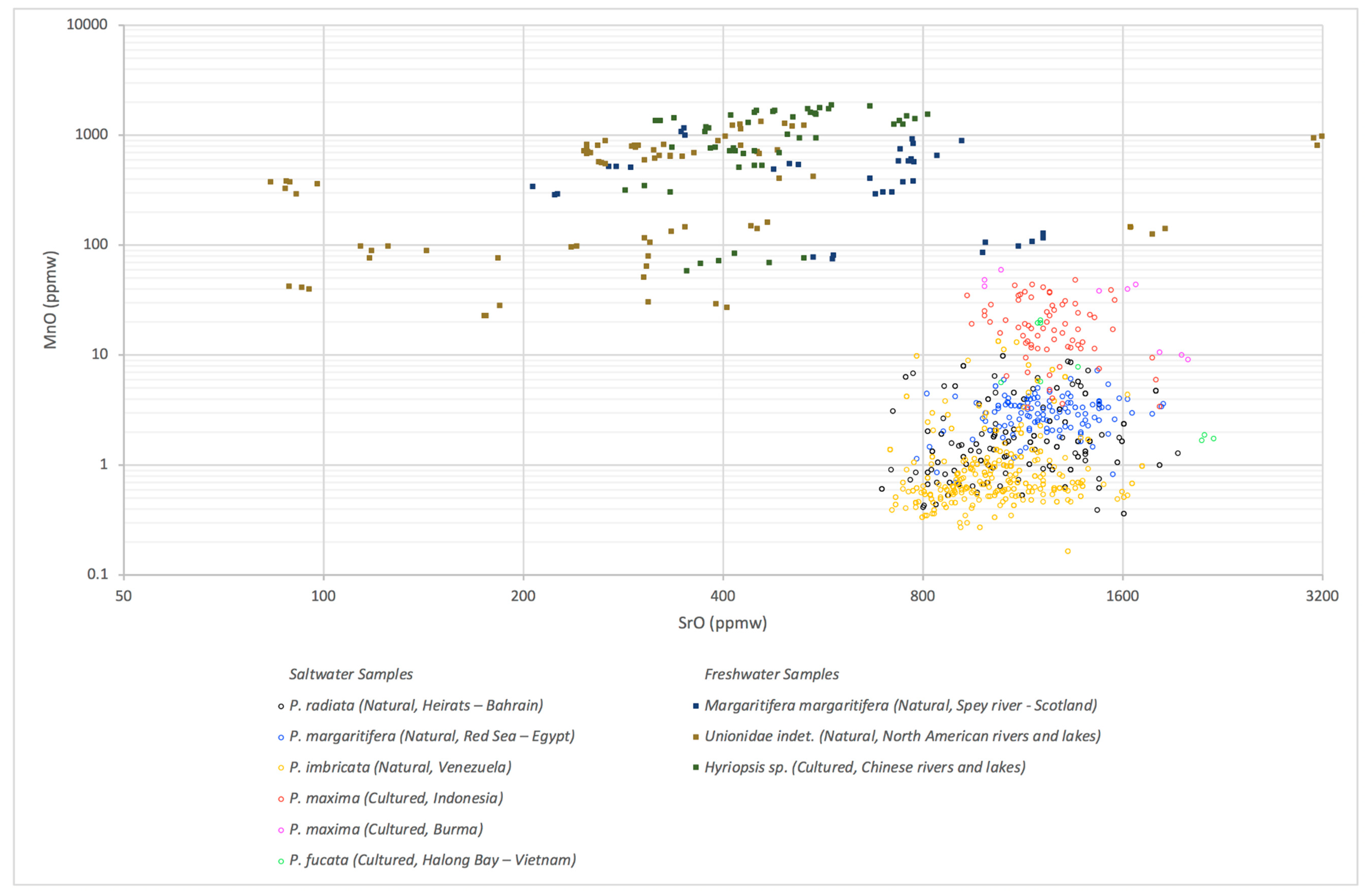
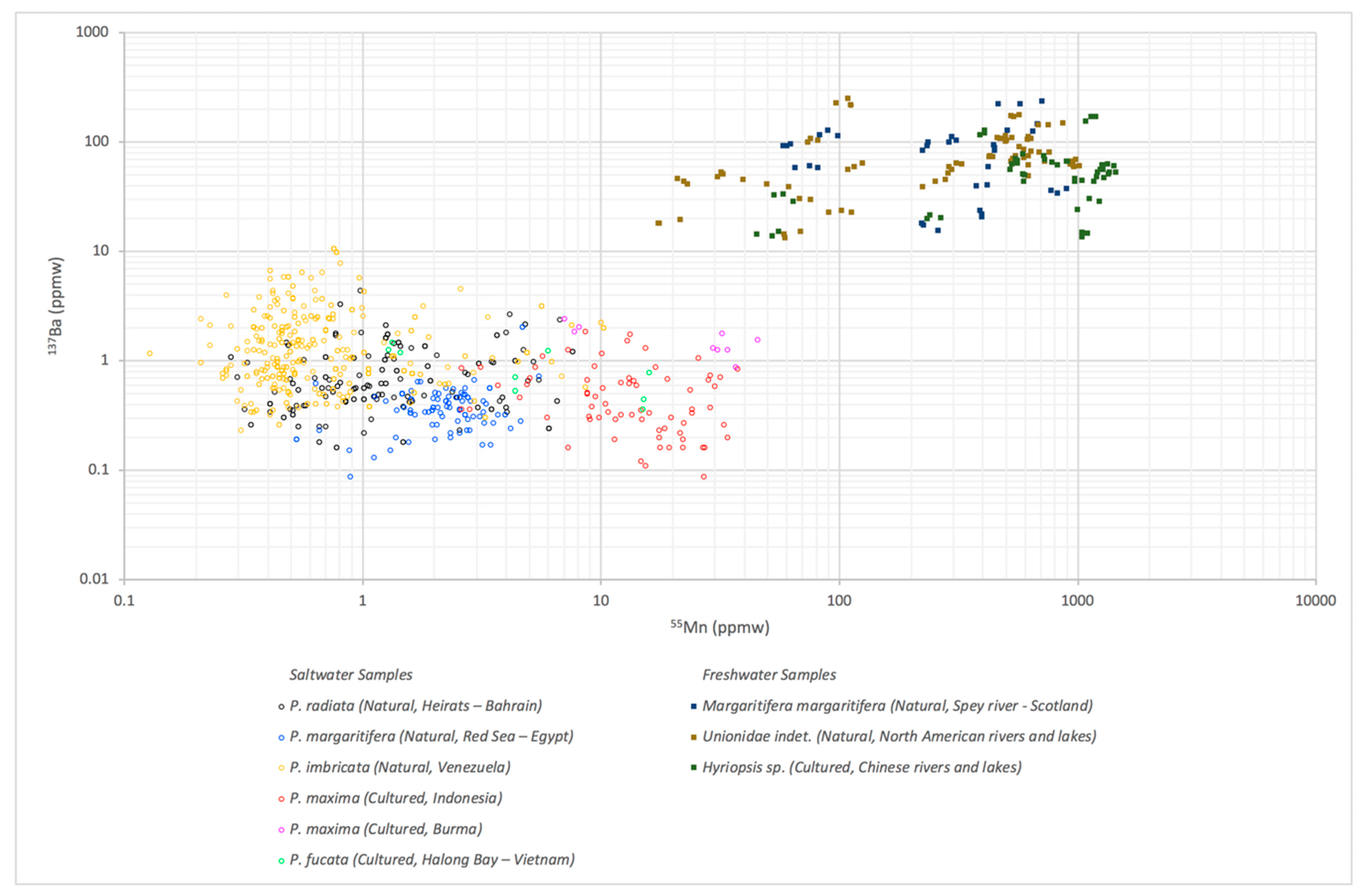
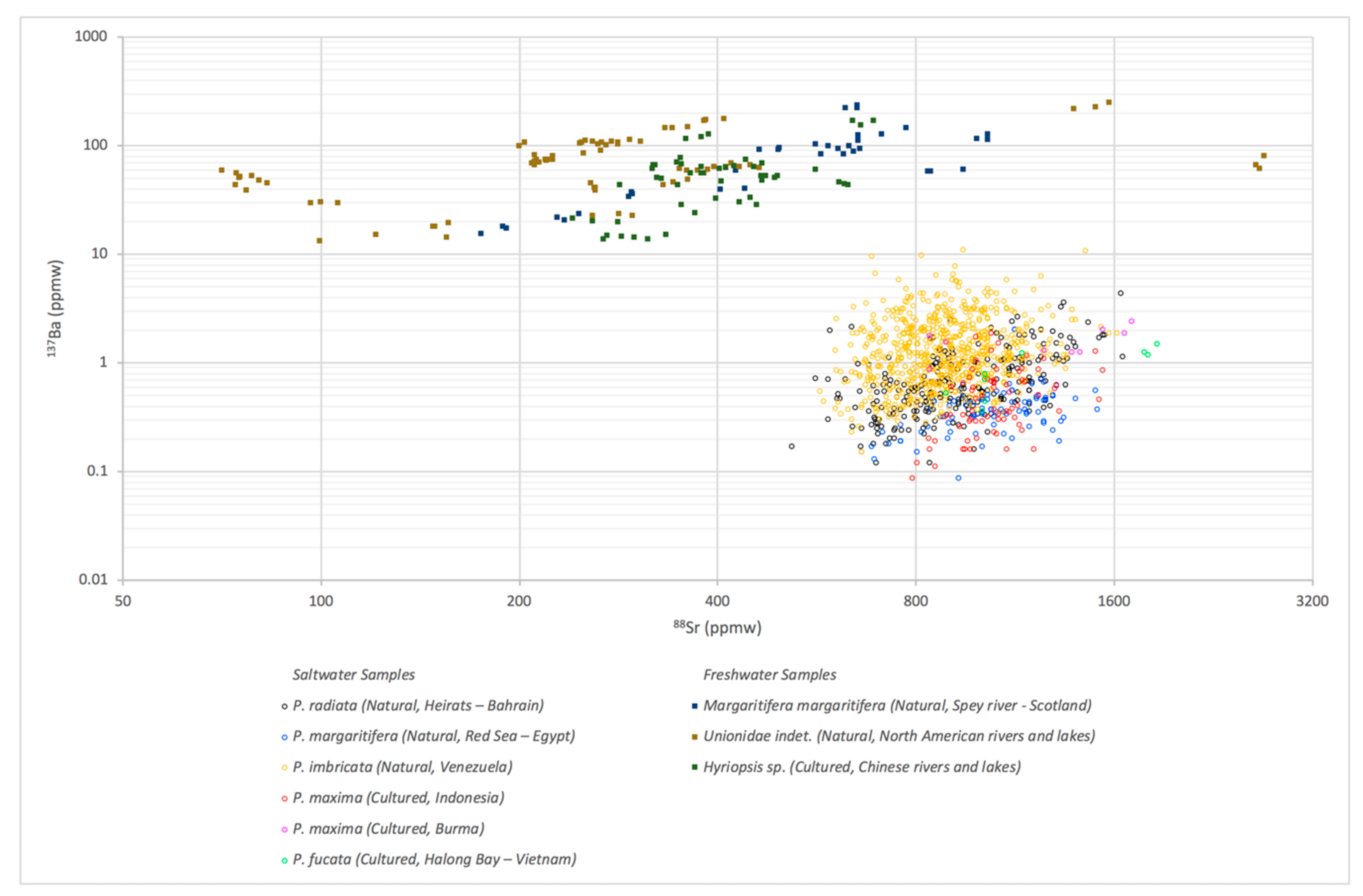
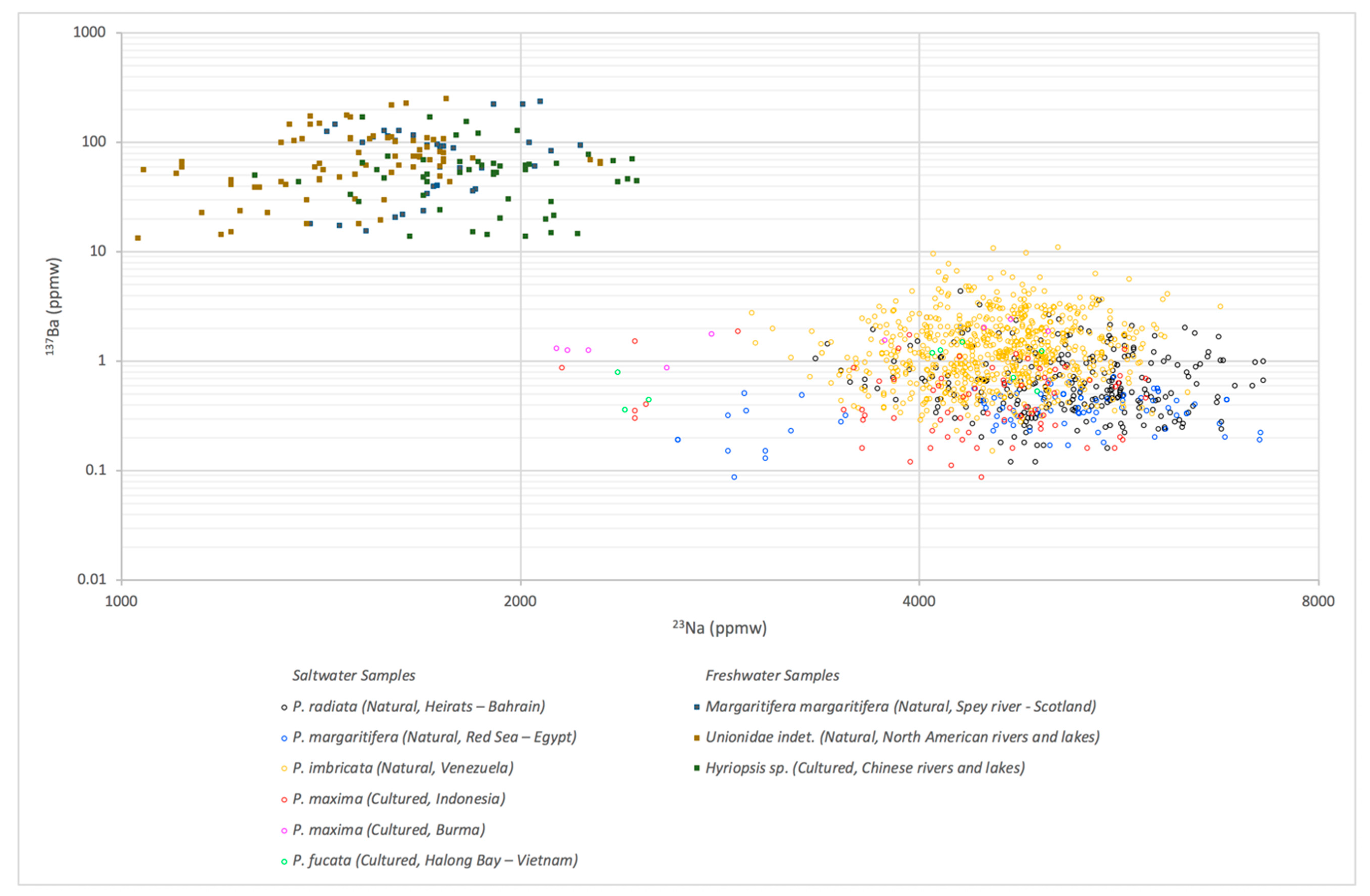
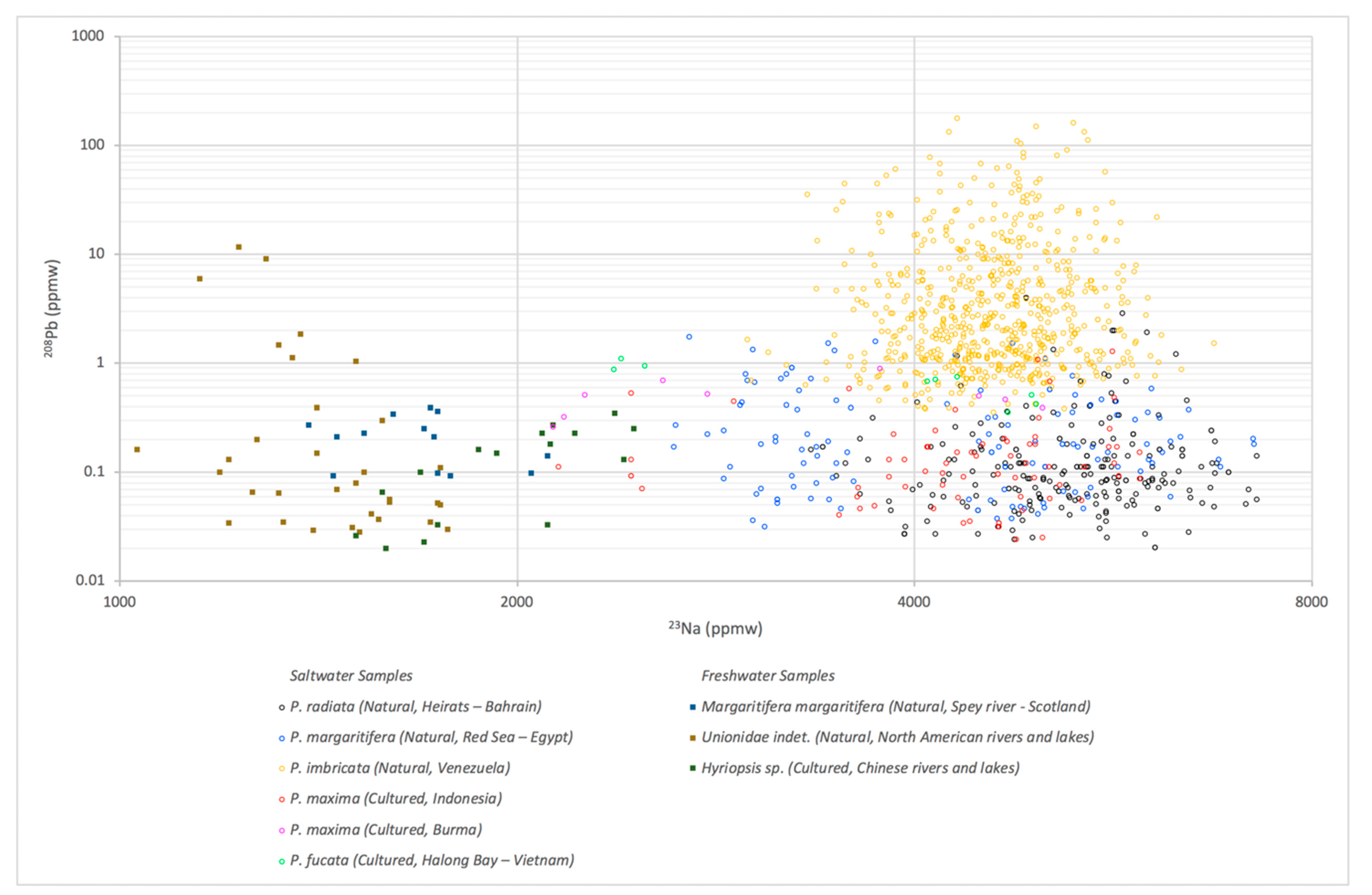
3.1. LA-ICP-MS Results

LA-ICP-MS data are presented in Table 3 where the results of saltwater and freshwater samples are listed together and in Table 4 the results by bivalve are listed. All acquired individual data-points are presented in Tables S1–S9. Measured freshwater samples contain higher 55Mn than measured saltwater samples (see again Table 3). Manganese content of water is considered one of the important factors affecting molluscs and shells 55Mn content. Rivers and lakes contain a higher content of 55Mn than sea water [6,7,25].
Most (40 out of 55) of the studied freshwater samples present 55Mn >200 ppmw. In some samples 55Mn is higher than 1000 ppmw and up to 1440 ppmw. Large amount of Mn2+ incorporation into biogenic aragonite has been attributed to local crystallographic alterations [26]. On the other hand, some freshwater samples (15 out of 55) present relatively low 55Mn concentrations (<150 ppmw). It has been suggested that 55Mn found in nacreous aragonitic freshwater mollusc shells is linked with the availability of Mn2+ in the sediment-water interface [27]. Manganese variations of shells were also linked to local geology, anthropogenic factors, animal’s age and growth rates as well as phytoplankton blooms, water temperature and pH as well as others [28,29]. However, measured natural pearls from Margaritifera margaritifera bivalve fished off the same season (two consecutive years) at the same location are presenting great variability of 55Mn (58–896 ppmw; Table 4).
All studied saltwater cultured pearls, as well as around half of the natural saltwater pearls present detectable 55Mn using LA-ICP-MS reaching up to 45.7 ppmw (see again Table 3 and Table 4). Noteworthy, 1/3 of the cultured saltwater samples present 55Mn >15 ppmw and all studied natural saltwater pearls contained 55Mn <15 ppmw. Half of the studied natural saltwater pearls did not present any detectable amounts of 55Mn. Moreover, three natural freshwater pearls from US presented amounts of 55Mn similar to those presented to some cultured saltwater pearls from P. maxima bivalve from Indonesia and Burma (Table 4).
88Sr in studied saltwater samples is relatively higher than in freshwater samples (Table 3). Most studied freshwater samples present concentrations from 70.6 ppmw to 800 ppmw, with only four natural freshwater pearls presenting 88Sr >800 ppmw. There are two natural pearls from Margaritifera margaritifera presenting 88Sr values of 874 and 1150 ppmw and two natural samples from Unionidae indet. with about 1487 ppmw and 2663 ppmw. The other 24 studied natural samples from Unionidae indet. presented 88Sr <470 ppmw. It was suggested that the variation of 88Sr in aragonitic (nacreous and non-nacreous) bivalves is influenced by the animal’s growth rates (which is linked with water temperature as well as other parameters) and physiological processes; 88Sr variations are not under environmental control [30,31,32]. Strontium content measured in nacreous aragonitic freshwater bivalve inner shells has been recently suggested as a good proxy of water’s 88Sr [27].
Figure 1 is a binary plot of manganese oxide (MnO) and strontium oxide (SrO). This plot is similar to those previously presented using EDXRF chemical analysis [7,8,9], taking into account that the limit of detection for MnO is higher for EDXRF than for LA-ICP-MS. It was formerly published that the SrO/MnO ratio, measured with EDXRF, is separating freshwater (here presented as filled squares in the figure) from saltwater pearls, as it is >12 for saltwater pearls and <12 for freshwater pearls [8]. Most of saltwater samples can be separated using this ratio as they present higher strontium and lower manganese with a SrO/MnO ratio of >100. However, for the freshwater samples, most present lower strontium and higher manganese samples with a ratio of <1. Chemical analysis of some freshwater samples might lead to wrong conclusions if the previously published plots and ratio are used [8,9], these are the freshwater (natural and cultured) pearls with 55Mn <150 ppmw (i.e., MnO <ca. 194 ppmw) or 88Sr >950ppmw (i.e., SrO >ca. 1124 ppmw). There are two natural freshwater pearls presenting SrO/MnO ratio >12. The SrO/MnO ratio of the studied samples is still not overlapping; all the studied saltwater samples present SrO/MnO ratio >17 and freshwater samples <16.
Interestingly, all studied freshwater samples presented higher 137Ba concentration than the saltwater samples; no overlapping values between the studied freshwater and saltwater samples were observed (Table 3). Barium is present in various concentrations in freshwater and saltwater [33] and in some studies barium uptake of a nacreous and non-nacreous shell was principally related to water’s barium content [34,35,36]. A strong inverse correlation was also found between salinity and barium in an aragonitic non-nacreous shell [30]. Other factors are also playing a role as the content is highly variable from a freshwater sample to another; even for those collected at the same region. For instance, measured 137Ba of natural pearls from Margaritifera margaritifera bivalves collected from the Spey river (Scotland) varied from 15.3 to 233 ppmw (Table 4). Food supply and shell growth have also been suggested as influencing factors [37,38] along with another yet undetermined environmental factor [35]. Animal age might also play a role. The vast majority of saltwater samples present a detectable 137Ba, while only 30 samples out of 1058 sample present undetectable samples. Also, only 120 samples out of the studied 1058 samples present 137Ba above 2.5 ppmw. All these samples are natural pearls; 116 sample found in P. imbricata. Few of these are samples with 137Ba >4.5 ppmw.
Figure 2 is the binary plot of 55Mn and 137Ba of the studied samples. All freshwater samples present higher 55Mn and 137Ba (shown as coloured filled squares in the plot) than the studied saltwater samples (shown as coloured empty circles in the figure). The population fields of these two group of samples are well apart, and the plot can be used to efficiently separate saltwater and freshwater (natural and cultured) pearls. In Figure 3, freshwater and saltwater samples are well separated. Moreover, freshwater samples present a somehow linear trend for 137Ba vs. 88Sr plots. This is not observed for saltwater samples and it further supports previous studies mentioning that 88Sr incorporation differs in freshwater and saltwater bivalves [27]. This should be further studied on samples from known molluscs and areas like the studied natural pearls founded in Margaritifera margaritifera bivalve from the river Spey (Scotland).
23Na is the most abundant element present in the studied samples in both saltwater and freshwater (Table 3) with higher concentrations for the first than for the latter. Sodium concentration of biogenic carbonate is linked with water salinity and pH [25,36]. All studied samples present diverse concentrations, not linked to specific bivalves or regions. Even the natural saltwater pearls found into the same P. margaritifera bivalve present a great variability of 23Na from 2630 to 7240 ppmw (Table 4) and the natural freshwater pearls from Spey river with 23Na from 1030 to 2450 ppmw. This may indicate that other factors can also influence cultured and natural pearls’ sodium concentration, such as the age of the pearl sac’s epithelial cells where it was found into. In the 23Na and 137Ba diagram presented in Figure 4, saltwater samples are more highly separated than the freshwater samples (filled squares).
Measured 24Mg content was also lower on the studied freshwater samples than on the saltwater (Table 3). However, some saltwater samples present relatively low manganese content, similar to the content of freshwater samples. The lowest content on saltwater samples was measured for P. radiata, where 45 samples out of 284 sample has 24Mg <65 ppmw (Table 3). Samples found in the same animal (see again Table 4 for P. margaritifera) have a great variation of 24Mg (from 67.6 to 675 ppmw). It has been suggested that Mg content is not (or is little) related to water conditions (e.g., temperature) as previously believed, but instead is related to physiological processes [30,34,39].
208Pb content was relatively low on all the studied samples, sometimes including BQL. All studied freshwater samples present below 0.4 ppmw of 208Pb. Solely two freshwater samples, both natural from Unionidae indet., presented concentrations of >0.4 pppmw, one presented around 1.5 ppmw and the other around 9 ppmw. Most of the studied saltwater samples presented below 1.5 ppmw 208Pb. Only five samples from P. radiata containing above 1.5 ppmw 208Pb and up to 3.91 ppmw (Table 4). However, around 2/3 of the studied P. imbricata samples contained above 1.5 ppmw 208Pb, with around 1/3 above 3.91 ppmw and up to 177 ppmw (see also Figure 5). It has been suggested that 208Pb measured in aragonite bivalve shells can be linked with anthropogenic lead pollution [40]. Anthropogenic small-scale lead pollution might be the reason of 208Pb >1 ppmw concentration in eight natural freshwater samples from P. radiata. Relatively high concentrations of 208Pb observed in P. imbricata natural pearls are probably superficial due to samples storage, as some natural pearls fished near Central America around 16th century were stored in lead boxes (e.g., natural pearls of Santa Margarita shipwreck treasures were found [41]). However, a possible link with polluted water caused by human and lead content cannot be excluded.

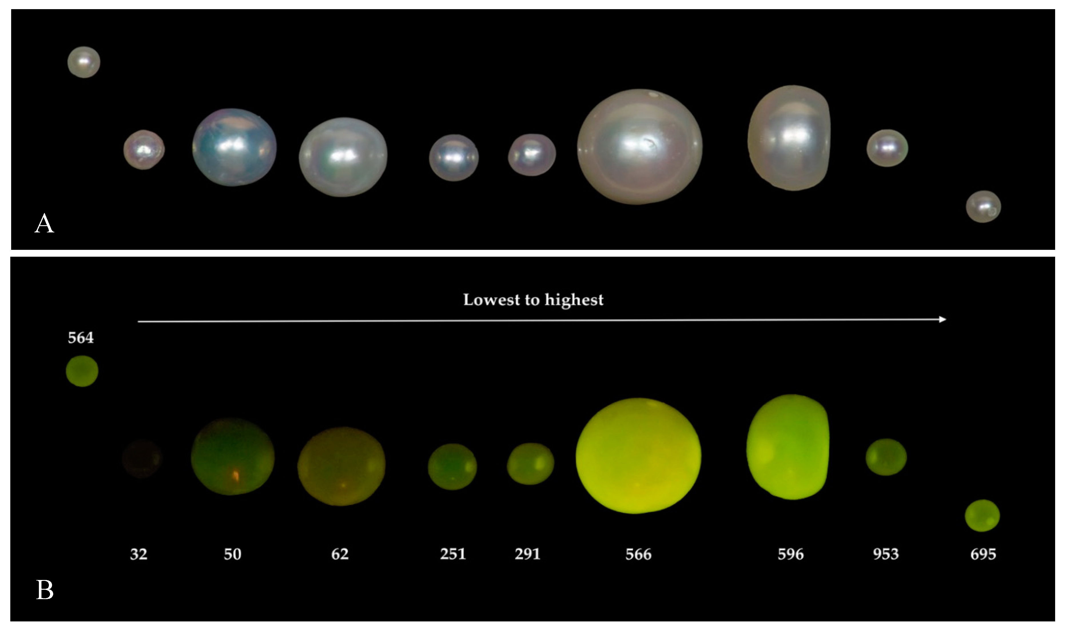
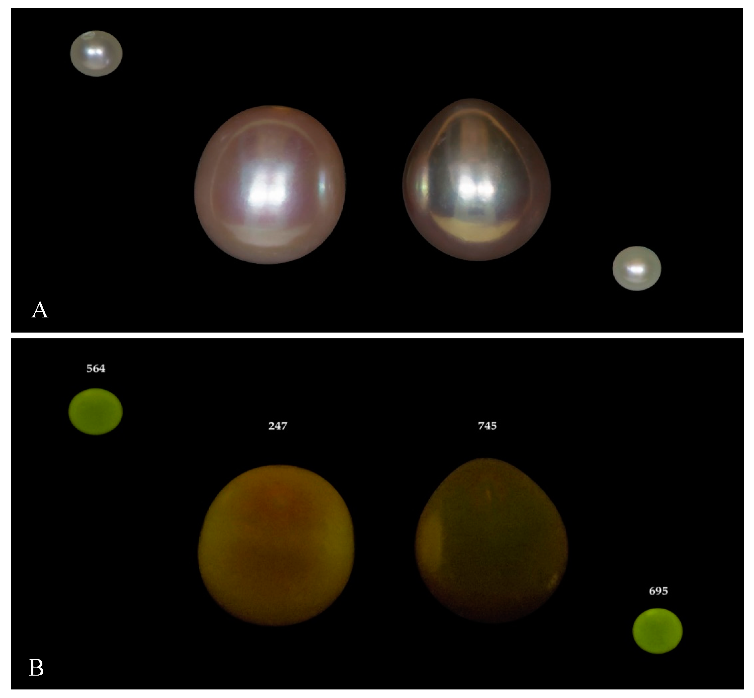
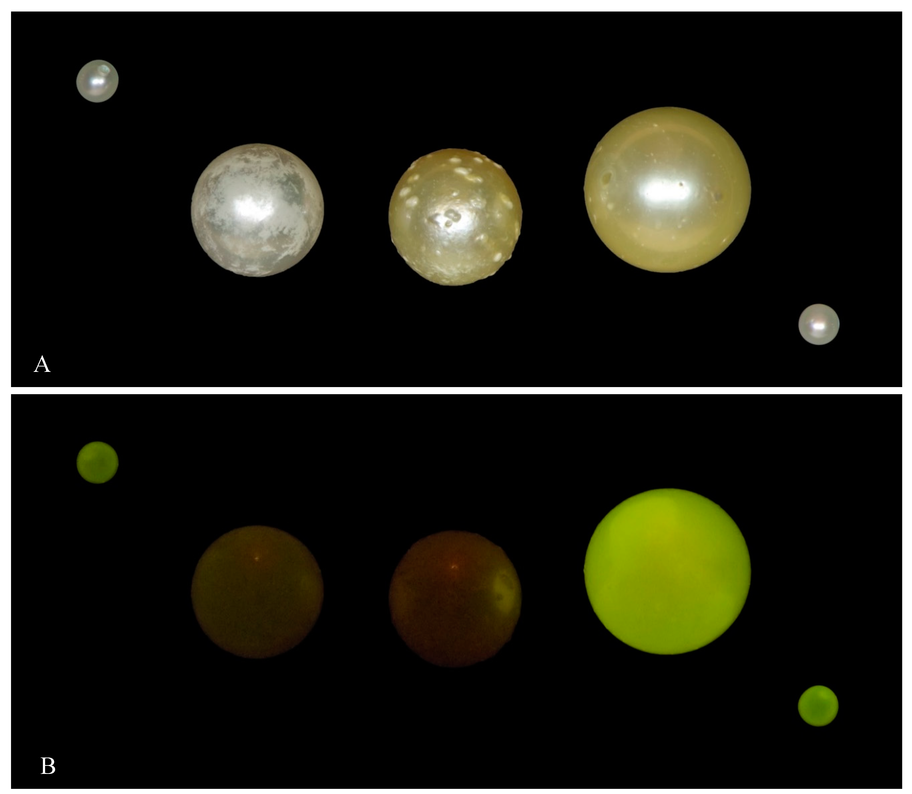
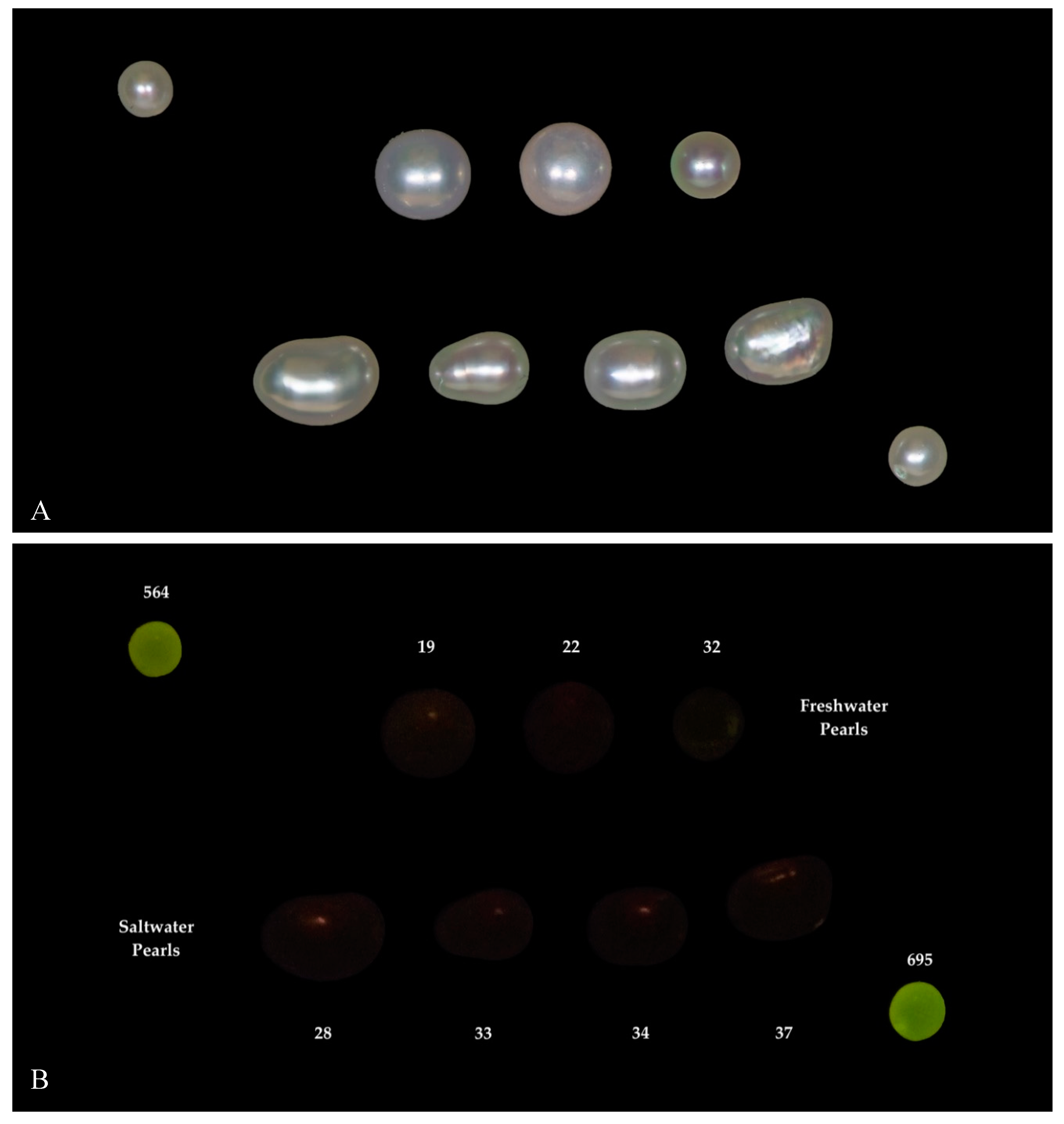
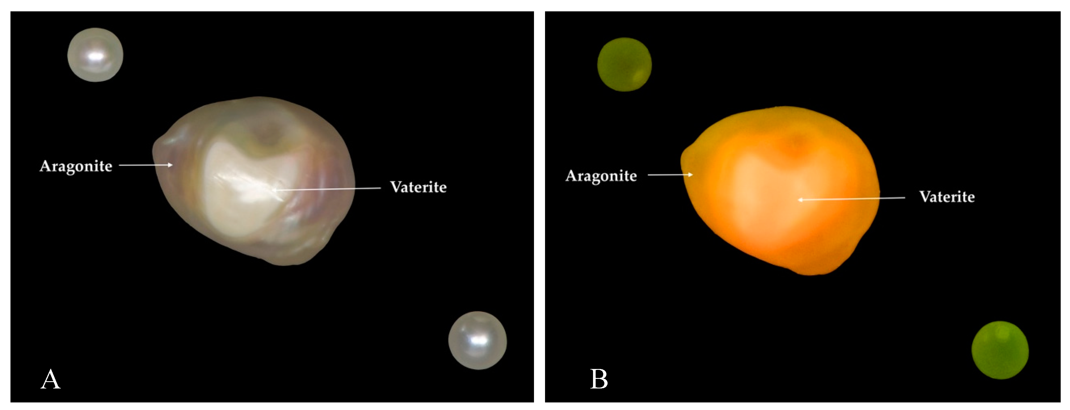
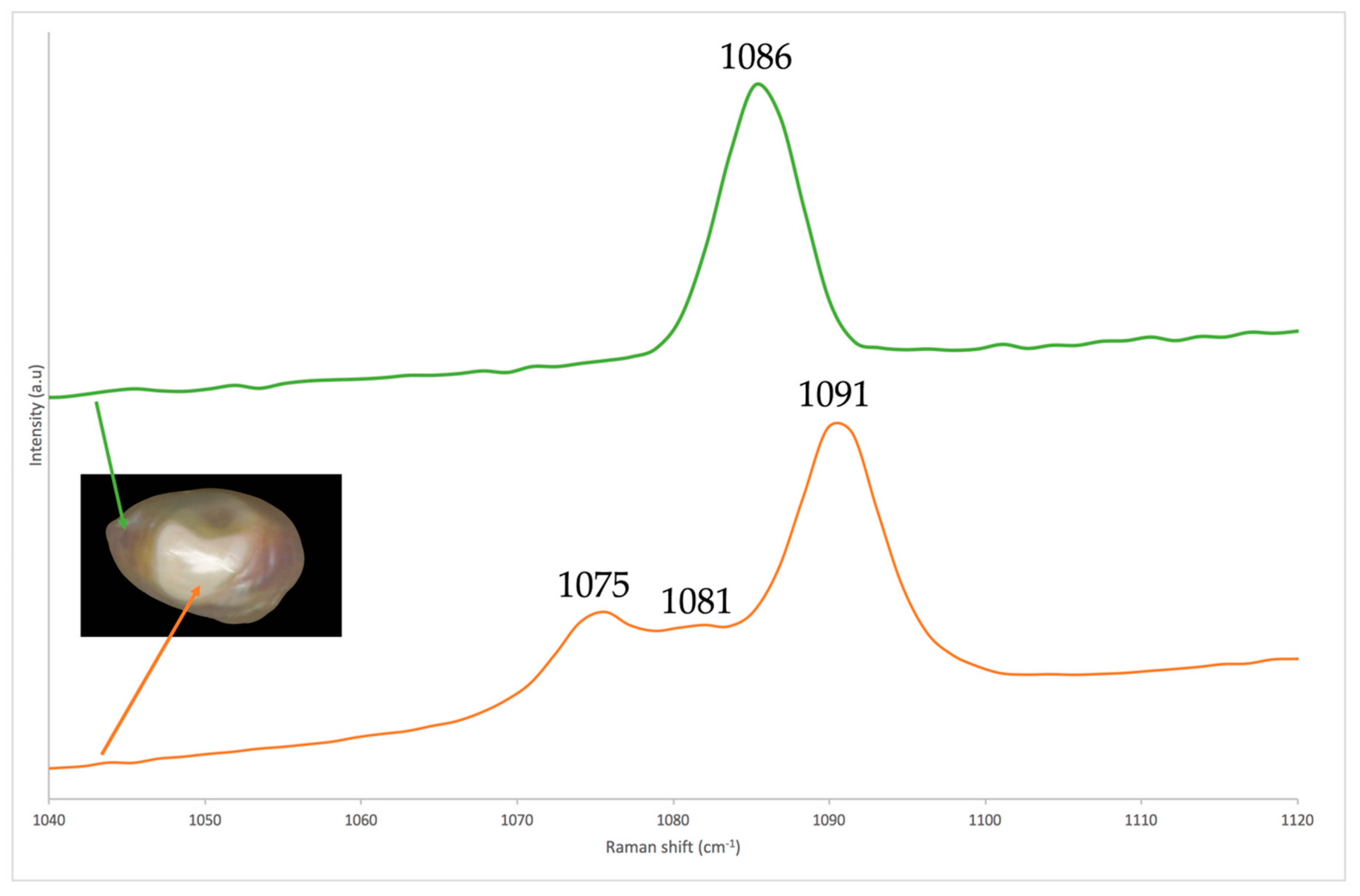
3.2. X-ray Luminescence and Raman Spectroscopy

Under X-rays, most natural and cultured (without bead) saltwater pearls remain inert and most natural and cultured freshwater pearls presented yellow-green luminescence of medium to high intensity (Figure 6, Figure 7, Figure 8 and Figure 9). This luminescence is attributed to 9-fold coordination of oxygen around Mn2+ in aragonite [42]. Luminescence intensity is correlated with an Mn2+ content, as the higher Mn2+ content, the more intense the yellow-green luminescence appears to be [42,43] (Figure 6). Meanwhile, it has been noticed that a higher Mn2+ content could cause lower luminescence (see again Figure 6). Similar to what was previously observed with CL spectroscopy (and X-ray induced luminescence) of biogenic aragonites, this is probably due to self-quenching of manganese. The presence of Fe2+ (as well as Cu2+, Co2+ and Ni2+) could quench Mn2+ luminescence as well [42,43].
Some coloured natural and cultured pearls present photoluminescence phenomena [44]. None of the studied samples presented any reaction under X-rays related to pigments. It was previously mentioned that natural (and artificial) pigments of freshwater samples suppress luminescence under X-rays [5]. The studied freshwater samples confirm that coloured samples with higher 55Mn (measured with LA-ICP-MS) present lower luminescence than less coloured samples with lower manganese (measured with LA-ICP-MS); see Figure 7.
Some saltwater cultured pearls, with freshwater bead, also present intense yellow-green luminescence (Figure 8). The amount of 55Mn measured on these samples is from 8 to 38 ppmw and it is not directly linked to the luminescence. The reaction under X-rays is due to the content of Mn2+ of the bead of these saltwater cultured samples and the relatively thin nacre thickness (i.e., thickness of the layer of saltwater nacre deposited around the freshwater bead) of the studied samples [5]. In Figure 8, the samples with the highest fluorescence has the thinnest nacre thickness (0.65 mm; measured using X-ray microradiography) and the other two samples have similar relatively thick nacre thickness (1.8 mm).
As mentioned above, some of the studied freshwater samples present 55Mn <150 ppmw. This samples present luminescence of low to medium intensity under X-rays (see examples in Figure 6 and Figure 9). On the other hand, some of the studied saltwater samples present detectable manganese measured with LA-ICP-MS. On our samples, X-ray luminescence appeared with low intensity at the studied samples only when 55Mn was higher than 10 ppmw. However, this will be different, if different parameters (e.g., less X-ray power) are used to acquire luminescence images. All the studied freshwater samples presented luminescence as well as some of the studied saltwater samples of low intensity. The latter were sometimes difficult to separate them from the freshwater samples containing low manganese (Figure 9). The amount of manganese in freshwater samples is low and below the detection limit of some EDXRF instruments and might lead to wrong conclusions. This samples might be identified by EDXRF if barium is taken into consideration or with LA-ICP-MS using the plots presented from Figure 2, Figure 3 and Figure 4.
One of the studied natural freshwater pearls presented a section with intense orange luminescence under X-rays; and phosphorescence which lasted for about 3 seconds (Figure 10). Orange luminescence activated by CL was previously observed on some cultured freshwater pearls, without bead, from China and this was attributed to calcite emitting Mn2+ [6]. Raman spectra acquired on the section where orange luminescence presents (and which, macroscopically and under optical microscope, appears with different lustre than the rest of the sample; refer to Figure 10A) have shown Raman bands at around 1091, 1081 and 1075 cm−1 due to ν1 symmetric stretching of carbonate ions in vaterite (Figure 11) and not at 1086 cm−1 due to ν1 symmetric stretching of carbonate ions in aragonite as on the section where medium yellow-green luminescence was observed [45]. CL and X-ray induced luminescence spectra of Mn2+ in calcite and vaterite are very close, which suggest a 6-fold coordination of oxygen around both calcite and vaterite [42]. Sections of vaterite were already previously identified in freshwater cultured pearls without bead from Japan and China [45,46,47]; but not in natural freshwater pearls.

4. Conclusions

LA-ICP-MS measurements on freshwater samples reveal higher 55Mn and 137Ba and lower 23Na, 24Mg and 88Sr concentrations than the studied saltwater samples. Only 137Ba concentrations of the studied samples do not present any overlap; the other concentrations slightly overlap. Plots combining 137Ba with 55Mn, 23Na and 88Sr clearly separate freshwater samples from saltwater samples. Very little correlation between the studied chemical elements of natural and cultured pearls and host animal’s species was found. Saltwater samples from P. maxima are the only studied saltwater samples which contain 55Mn >20 ppmw. On the other hand, only some saltwater samples from P. radiata could present 24Mg <65 ppmw and saltwater samples from P. imbricata could present 137Ba >2.5 ppmw. It seems that all the chemical elements studied here are linked to a combination of environmental and physiological factors.
Most studied freshwater samples can be separated from the saltwater samples as X-ray luminescence intensity is correlated with Mn2+ content in aragonite. Some of the studied freshwater samples present similar manganese content with some saltwater samples as well as similar low intensity luminescence under X-rays. The growth environment of these samples could only inadequately be identified by LA-ICP-MS or a well calibrated EDXRF with a good limit of detection for manganese and barium.
Studies on a bigger number of samples from known animals and various regions (with known geographic coordinates) that take into account the local environmental factors, along with statistical analysis (e.g., principal components), should be performed in order to confirm the abovementioned differences and better understand the link between the chemical elements and the natural and cultured pearls. Studying natural pearls is not an easy task because they are very rarely found and in parallel rarely used for scientific purposes. Additional measurements of isotopes might shed more light on a possible link between environmental factors and natural (or cultured) pearls. In order to inspect the possible role of organic matter, TGA analysis and chemical analysis would also be useful. It is also important to further study the possible role of elements such us Fe2+, Cu2+, Co2+ and Ni2+ which could quench Mn2+ luminescence and their effect on the separation of saltwater from freshwater (natural and cultured) pearls. Orange coloured X-ray luminescence of freshwater samples should also be further investigated and its link with vaterite should be confirmed with spectroscopic means.

Supplementary Materials

The following are available online at https://www.mdpi.com/2075-163X/9/6/357/s1, Table S1: LA-ICP-MS analysis in ppmw of the studied natural pearls samples from P. radiata (Heirats, Bahrain), Table S2: LA-ICP-MS analysis in ppmw of the studied natural pearls samples from P. margaritifera (Red Sea, Egypt), Table S3: LA-ICP-MS analysis in ppmw of the studied natural pearls samples from P. imbricata (Venezuela), Table S4: LA-ICP-MS analysis in ppmw of the studied cultured pearls samples from P. maxima (Indonesia), Table S5: LA-ICP-MS analysis in ppmw of the studied cultured pearls samples from P. maxima (Burma), Table S6: LA-ICP-MS analysis in ppmw of the studied cultured pearls samples from P. fucata (Halong Bay, Vietnam), Table S7: LA-ICP-MS analysis in ppmw of the studied natural pearls samples from Margaritifera margaritifera (Spey river, Scotland), Table S8: LA-ICP-MS analysis in ppmw of the studied natural pearls samples from Unionidae indet. (North American rivers and lakes), Table S9: LA-ICP-MS analysis in ppmw of the studied cultured pearls samples from Hyriopsis sp. (Chinese rivers and lakes).

Author Contributions

S.K. formulated the paper designed the experiments, participated to the data interpretation and wrote the manuscript. F.M. prepared the experiments for X-ray luminescence analysis, did part of LA-ICP-MS analysis, data reduction, edited the manuscript and draw its figures and tables. H.A. prepared all the images of the manuscript. F.A. and L.F. performed most of LA-ICP-MS analysis on the samples and S.S. has designed, supervised and evaluated LA-ICP-MS analysis. A.A. selected the samples, designed the experiments and edited the manuscript.

Funding

This research received no external funding.

Acknowledgments

The authors wish to thank the National Museum of Bahrain which loaned the natural saltwater pearls from P. radiata fished off Bahrain, Peter Balogh for the natural saltwater pearls from Pinctada imbricata fished off Venezuela and Peter Piva for the natural saltwater pearls from Pinctada margaritifera fished off Egypt. The rest of the samples are from DANAT reference collection.

Conflicts of Interest

The authors declare no conflict of interest.

References

  1. Gauthier, J.P.; Karampelas, S. Pearls and corals: “Trendy biomineralizations”. Elements 2009, 5, 179–180. [Google Scholar] [CrossRef]
  2. Cartier, L.; Krzemnicki, M.S. New developments in cultured pearls production: Use of organic and baroque shell nuclei. Aust. Gemmol. 2013, 25, 6–13. [Google Scholar]
  3. Schiffmann, C.A. Pearl identification: Some laboratory experiments. J. Gemmol. 1971, 12, 284–296. [Google Scholar] [CrossRef]
  4. Lorenz, I.; Schmetzer, K. Possibilities and limitations in radiographic determination of pearls. J. Gemmol. 1986, 20, 114–123. [Google Scholar] [CrossRef]
  5. Hänni, H.A.; Kiefert, L.; Giese, P. X-ray luminescence, a valuable test in pearl identification. J. Gemmol. 2005, 29, 325–329. [Google Scholar] [CrossRef]
  6. Habermann, D.; Banerjee, A.; Meijer, J.; Stephan, A. Investigation of manganese in salt- and freshwater pearls. Nucl. Instrum. Methods 2001, 181, 739–743. [Google Scholar] [CrossRef]
  7. Gütmannsbauer, W.; Hänni, H.A. Structural and chemical investigations on shell and pearls of nacre forming salt- and freshwater bivalve mollusks. J. Gemmol. 1994, 24, 241–252. [Google Scholar] [CrossRef]
  8. Karampelas, S.; Kiefert, L. Gemstones and minerals. In Analytical Archaeometry: Selected Topics, 1st ed.; Edwards, H.G.M., Vandenabeele, P., Eds.; Royal Society of Chemistry: London, UK, 2012; pp. 291–317. ISBN 978-1-84973-162-1. [Google Scholar]
  9. Zhou, C.; Hodgins, G.; Lange, T.; Saruwatari, K.; Sturman, N.; Kiefert, L.; Schollenbruch, K. Saltwater pearls from the pre- to early Columbian era: A gemological and radiocarbon dating study. Gems Gemol. 2017, 53, 286–295. [Google Scholar] [CrossRef]
  10. Goebel, M.; Dirlam, D.M. Polynesian black pearls. Gems Gemol. 1989, 25, 130–148. [Google Scholar] [CrossRef]
  11. Elen, S. Spectral reflectance and fluorescence characteristics of natural-color and heat-treated “Golden” south sea cultured pearls. Gems Gemol. 2001, 37, 114–123. [Google Scholar] [CrossRef]
  12. Jacob, D.E.; Wehrmeister, U.; Hager, T.; Hofmeister, W. Identifying Japanese freshwater cultured pearls from Lake Kasumigaura. J. Gemmol. 2006, 22, 539–541. [Google Scholar]
  13. Scarratt, K.; Bracher, P.; Bracher, M.; Atawi, A.; Safar, A.; Saeseaw, S.; Homkrajae, A.; Sturman, N. Natural pearls from Australian Pinctada maxima. Gems Gemol. 2012, 48, 236–261. [Google Scholar] [CrossRef]
  14. Hänni, H.A.; Cartier, L.E. Tracing cultured pearls from farm to consumer: A review of potential methods and solutions. J. Gemmol. 2013, 33, 185–191. [Google Scholar] [CrossRef]
  15. Sturman, N.; Homkrajae, A.; Manustrong, A.; Somsaard, N. Observations on pearls reportedly from the Pinnidae family (pen pearls). Gems Gemol. 2014, 50, 202–215. [Google Scholar] [CrossRef]
  16. Homkrajae, A.; Sun, Z.; Blodgett, T.; Zhou, C. Provenance discrimination of freshwater pearls by laser ablation–inductively coupled plasma–mass spectrometry (LA-ICP-MS) and linear discriminant analysis (LDA). Gems Gemol. 2019, 55, 47–60. [Google Scholar]
  17. Perkins, W.T.; Fuge, R.; Pearce, N.J.G. Quantitative analysis of trace elements in carbonates using laser ablation inductively coupled plasma mass spectrometry. J. Anal. Atom. Spectr. 1991, 6, 445–449. [Google Scholar] [CrossRef]
  18. Sinclair, D.J.; Kinsley, L.P.J.; McCulloch, M.T. High resolution analysis of trace elements in corals by laser-ablation ICP-MS. Geochim. Cosmochim. Acta 1998, 62, 1889–1901. [Google Scholar] [CrossRef]
  19. Gillikin, D.P. Geochemistry of Marine Bivalve Shells: The Potential for Paleoenvironmental Reconstruction. Ph.D. Thesis, Vrije University, Brussel, Belgium, 2005; 265p. [Google Scholar]
  20. Jacob, D.E.; Soldati, A.L.; Wirth, R.; Huth, J.; Wehrmeister, U.; Hofmeister, W. Nanostructure, composition and mechanisms of bivalve shell growth. Geochim. Cosmochim. Acta 2009, 72, 5401–5415. [Google Scholar] [CrossRef]
  21. Mertz-Kraus, R.; Brachert, T.C.; Jochum, K.P.; Reuter, M.; Stoll, B. LA-ICP-MS analyses on coral growth increments reveal heavy winter rain in the eastern Mediterranean at 9 Ma. Palaeogeogr. Palaeoclimatol. Palaeoecol. 2009, 273, 25–40. [Google Scholar] [CrossRef]
  22. Phung, A.T.; Baeyens, W.; Leermakers, M.; Goderis, S.; Vanhaecke, F.; Gao, Y. Reproducibility of laser ablation–inductively coupled plasma–mass spectrometry (LA–ICP–MS) measurements in mussel shells and comparison with micro-drill sampling and solution ICP–MS. Talanta 2013, 396, 42–50. [Google Scholar] [CrossRef]
  23. Strasser, C.A.; Mullineaux, L.S.; Walther, B.D. Growth rate and age effects on Mya Arenaria shell chemistry: Implications for biogeochemical studies. J. Exp. Mar. Biol. Ecol. 2008, 355, 153–163. [Google Scholar] [CrossRef]
  24. Jochum, K.P.; Scholz, D.; Stoll, B.; Weis, U.; Wilson, S.A.; Yang, Q.; Schwalb, A.; Borner, N.; Jacob, D.E.; Andreae, M.O. Accurate trace element analysis of speleothems and biogenic calcium carbonates by LA-ICP-MS. Chem. Geol. 2012, 318–319, 31–44. [Google Scholar] [CrossRef]
  25. Gordon, C.M.; Carr, R.A.; Larson, R.E. The influence of environmental factors on the sodium and manganese content of barnacle shells. Limnol. Oceanogr. 1970, 15, 461–466. [Google Scholar] [CrossRef]
  26. Soldati, A.L.; Jacob, D.E.; Glatzel, P.; Swarbrick, J.C.; Geck, J. Element substitution by living organisms: The case of manganese in mollusc shell aragonite. Sci. Rep. 2016, 6, 22514. [Google Scholar] [CrossRef] [PubMed]
  27. Geeza, T.J.; Gillikin, D.P.; Goodwin, D.H.; Evans, S.D.; Watters, T.; Warner, N.R. Controls on magnesium, manganese, strontium, and barium concentrations recorded in freshwater mussel shells from Ohio. Chem. Geol. 2019. [Google Scholar] [CrossRef]
  28. Carroll, M.; Romanek, C.S. Shell layer variation in trace element concentration for the freshwater bivalve Elliptio complanta. Geo-Mar. Lett. 2008, 28, 369–381. [Google Scholar] [CrossRef]
  29. Kelemen, Z.; Gillikin, D.P.; Bouillon, S. Relationship between river water chemistry and shell chemistry of two tropical African freshwater bivalve species. Chem. Geol. 2019. [Google Scholar] [CrossRef]
  30. Poulain, C.; Gillikin, D.P.; Thebault, J.; Munaron, J.M.; Bohn, M.; Robert, R.; Paulet, Y.M.; Lorrain, A. An evaluation of Mg/Ca, Sr/Ca, and Ba/Ga ratios as environmental proxies bivalve shells. Chem. Geol. 2015, 396, 42–50. [Google Scholar] [CrossRef]
  31. Wanamaker, D.; Gillikin, D.P. Strontium, magnesium and barium incorporation in aragonitic shells of junevile Arctica islandica: Insights from temperature controlled experiments. Chem. Geol. 2019. [Google Scholar] [CrossRef]
  32. Stecher, H.A.; Krantz, D.E.; Lord, C.J.; Luther, G.W.; Bock, K.W. Profiles of strontium and barium in Mercenaria mercenaria and Spisula solidisima shells. Geochim. Cosmochim. Acta 1996, 60, 3445–3456. [Google Scholar] [CrossRef]
  33. Schroeder, H.A.; Tipton, I.H.; Nason, A.P. Trace metals in man: Strontium and barium. J. Chronic Dis. 1972, 25, 491–517. [Google Scholar] [CrossRef]
  34. Gillikin, D.P.; Dehairs, F.; Lorrain, A.; Steenmans, D.; Baeyens, W.; Andre, L. Barium uptake into the shells of the common mussel (Mytilus edulis) and the potentials for estuarine paleo-chemistry reconstruction. Geochim. Cosmochim. Acta 2006, 70, 395–407. [Google Scholar] [CrossRef]
  35. Gillikin, D.P.; Lorrain, A.; Paulet, Y.M. Synchronous barium peaks in high-resolution profiles of calcite and aragonite marine bivalve shells. Geo-Mar. Lett. 2008, 28, 351–358. [Google Scholar] [CrossRef] [Green Version]
  36. Zhao, L.; Schone, B.R.; Mertz-Kraus, R.; Yang, F. Controls on strontium and barium incorporation into freshwater bivalve shells (Corbicula fluminea). Palaeogeogr. Palaeoclimatol. Palaeoecol. 2017, 465, 386–394. [Google Scholar] [CrossRef]
  37. Zhao, L.; Schone, B.R.; Mertz-Kraus, R. Sodium provides unique insights into transgenerational effects of ocean acidification on bivalve shell formation. Sci. Total Environ. 2017, 577, 360–366. [Google Scholar] [CrossRef] [PubMed]
  38. Herath, D.; Jacob, D.E.; Jones, H.; Fallon, S.J. Potential of shells of three species of Eastern Australian freshwater mussels (Bivalvia: Hyriidae) as environmental proxy archives. Mar. Freshw. Res. 2018, 70, 255–269. [Google Scholar] [CrossRef]
  39. Foster, L.C.; Finch, A.A.; Allison, N.; Andersson, C.; Clarke, L.J. Mg in aragonitic bivalve shells: Seasonal variations and mode of incorporation in Arctica islandica. Chem. Geol. 2008, 254, 113–119. [Google Scholar] [CrossRef]
  40. Gillikin, D.P.; Dehairs, F.; Baeyens, W.; Navez, J.; Lorrain, A.; André, L. Inter- and intra-annual variations of Pb/Ca ratios in clam shells (Mercenaria mercenaria): A record of anthropogenic lead pollution. Mar. Pollut. Bull. 2005, 50, 1530–1540. [Google Scholar] [CrossRef]
  41. Collins, D. Thousands of Pearls Found in Shipwreck. Available online: https://www.cbsnews.com/news/thousands-of-pearls-found-in-shipwreck/ (accessed on 20 March 2019).
  42. Sommer, S.E. Cathodoluminescence of carbonates 1. Characterization of cathodoluminescence from carbonate solid solutions. Chem. Geol. 1972, 9, 257–273. [Google Scholar] [CrossRef]
  43. Götte, T.; Richter, D.K. Quantitative aspects of Mn activated cathodoluminescence of natural and synthetic aragonite. Sedimentology 2009, 56, 483–492. [Google Scholar] [CrossRef]
  44. Hainschwang, T.; Karampelas, S.; Fritsch, E.; Notari, F. Luminescence spectroscopy and microscopy applied to study gem materials: A case study of C centre containing diamonds. Mineral. Petrol. 2013, 107, 393–413. [Google Scholar] [CrossRef]
  45. Wehrmeister, U.; Jacob, D.E.; Soldati, A.L.; Häger, T.; Hofmeister, W. Vaterite in freshwater cultured pearls from China and Japan. J. Gemmol. 2007, 31, 269–276. [Google Scholar] [CrossRef]
  46. Qiao, L.; Feng, Q.L.; Li, Z. Special vaterite in freshwater lackluster pearls. Cryst. Growth Des. 2006, 7, 275–279. [Google Scholar] [CrossRef]
  47. Ma, H.; Su, A.; Zhang, B.; Li, R.K.; Zhou, L.; Wang, B. Vaterite or aragonite observed in the prismatic layer of freshwater-cultured pearls from South China. Prog. Nat. Sci. 2009, 19, 817–820. [Google Scholar] [CrossRef]
Figure 1. Binary plot of SrO vs. MnO.
Minerals 09 00357 g001
Figure 2. Binary plot of 55Mn vs. 137Ba of the studied samples.
Minerals 09 00357 g002
Figure 3. Binary plot of 88Sr vs. 137Ba of the studied samples.
Minerals 09 00357 g003
Figure 4. Binary plot of 137Ba vs. 23Na of the studied samples.
Minerals 09 00357 g004
Figure 5. Binary plot of 23Na vs. 208Pb of the studied samples.
Minerals 09 00357 g005
Figure 6. Freshwater samples under daylight (A) and under X-rays (B). The two freshwater samples upper left (diameter: 2.85 mm) and bottom right (diameter: 2.95 mm) used as reference. The concentrations of 55Mn in ppmw are mentioned below for the studied samples and above for the reference samples. For the samples 3 analyses with LA-ICP-MS were acquired, the average values are presented (rounded to the nearest one).
Minerals 09 00357 g006
Figure 7. Freshwater samples under daylight (A) and under X-rays (B). Two natural-coloured freshwater samples (left sample of pink colour and right samples of grey colour) with the amount 55Mn in ppmw. The two freshwater samples upper left (diameter: 2.85 mm) and bottom right (diameter: 2.95 mm) used as reference. For the samples 3 analyses with LA-ICP-MS were acquired, the average values are presented (rounded to the nearest one).
Minerals 09 00357 g007
Figure 8. Saltwater cultured pearls (with freshwater bead) under daylight (A) and under X-rays (B). The first and second pearls from the left has a similar nacre thickness that equals 1.8 mm, while the third one has a nacre thickness of 0.65 mm (measured using X-ray microradiography). The two freshwater samples upper left (diameter: 2.85 mm) and bottom right (diameter: 2.95 mm) used as reference.
Minerals 09 00357 g008
Figure 9. Three freshwater (upper row) and four saltwater (bottom row) samples under daylight (A) and under X-rays (B). The two freshwater samples upper left (diameter: 2.85 mm) and bottom right (diameter: 2.95 mm) used as reference. All samples present similar luminescence intensity and separation of freshwater samples from saltwater is challenging. The concentrations of 55Mn in ppmw are mentioned. For the samples 3 analyses with LA-ICP-MS that were acquired, the average values are presented (rounded to the nearest one).
Minerals 09 00357 g009
Figure 10. Natural freshwater pearl under daylight (A) and under X-rays (B); orange coloured part is made of vaterite (verified by Raman; see Figure 11). The two freshwater samples upper left (diameter: 2.85 mm) and bottom right (diameter: 2.95 mm) were used as references.
Minerals 09 00357 g010
Figure 11. Raman spectra on the white part with low lustre (bottom spectrum) and on the cream coloured part with medium lustre (top spectrum) showing respectively bands due to vaterite and calcite. Note that the spectra are stacked and shifted for clarity.
Minerals 09 00357 g011
Table 1. List of studied samples.
EnvironmentBivalveAreaNo. of Samples
SaltwaterP. radiata *Arabian Gulf (Heirats, Bahrain)248 ****1058
P. margaritifera *Red Sea (Hurghada, Egypt)80 (****)
P. imbricata *Venezuela671 ****
P. maxima **/***Indonesia53 (****)
P. maxima ***Burma3
P. fucata ***Vietnam (Halong Bay)3
FreshwaterMargaritifera margaritifera *Scotland (Spey river)1255
Unionidae indet. *North American rivers26
Hyriopsis sp. ***Chinese rivers and lakes17
* Natural pearls; ** Cultured pearls without bead; *** Cultured pearls with bead; **** 1 spot was analysed using LA-ICP-MS, (****) one spot was analysed using LA-ICP-MS in most pearls.
Table 2. Laser Ablation-Induced Coupled Plasma-Mass Spectrometer (LA-ICP-MS) detection limits and ranges in ppmw.
Limits23Na (ppmw)24Mg (ppmw)55Mn (ppmw)88Sr (ppmw)137Ba (ppmw)208Pb (ppmw)
LOD1.62–70.840.02–0.570.09–0.650.01–0.050.01–0.390.01–0.02
LOQ5.35–233.770.66–1.880.21–1.180.03-0.160.17–1.180.02–0.07
LOD: Limits of detection; LOQ: Limits of quantification.
Table 3. LA-ICP-MS analysis of saltwater and freshwater samples.
SamplesElementMin–Max (ppmw)Average (SD) (ppmw)Median (ppmw)
Saltwater23Na2130–72704711.27 (816.6)4720
24Mg29.5–950241.29 (111.13)240
55MnBQL–45.71.84 (5.01)BQL
88Sr518–1860941.2 (197.31)912
137BaBQL–111.18 (1.17)0.84
208PbBQL–1775.18 (14.88)0.95
Freshwater23Na1030–24501672.81 (303.89)1680
24Mg5.58–81.330.87 (16.45)28.3
55Mn17.4–1440504.95 (386.34)473
88Sr70.6–2700431.84 (389.41)350
137Ba13.2–24972.76 (48.25)61.6
208PbBQL–11.60.24 (1.24)BQL
BQL: Below quantification limit; SD: Standard deviation.
Table 4. LA-ICP-MS analysis of the studied samples by mollusc.
SamplesElementMin–Max (ppmw)Average (SD) (ppmw)Median (ppmw)
P. radiata
(Heirats, Bahrain)
Natural
23Na3340–72705249.52 (800.85)5250
24Mg29.5–477147.45 (88.18)131
55MnBQL–7.620.79 (1.38)BQL
88Sr518–1650943.9 (219.42)902
137BaBQL–4.320.78 (0.6)0.58
208PbBQL–3.910.17 (0.4)0.08
P. margaritifera
(Red Sea, Egypt)
Natural
23Na2630–72404442.05 (1204.07)4595
24Mg67.6–675362.72 (109.6)365
55MnBQL–5.52.24 (1)2.31
88Sr663–15601052.57 (180.98)1030
137BaBQL–2.020.26 (0.26)0.28
208PbBQL–1.740.3 (0.36)0.17
P. imbricata
(Venezuela)
Natural
23Na3200–62404621.64 (564.29)4600
24Mg108–531264.34 (72.01)258
55MnBQL–10.30.33 (0.95)BQL
88Sr572–1620898.07 (164.56)880
137BaBQL–111.56 (1.32)1.18
208Pb0.35–1778.58 (18.68)2.42
P. maxima
(Indonesia)
Cultured
23Na2150–59304430.28 (860.33)4460
24Mg71.1–279125.76 (55.38)110
55Mn2.6–37.415.37 (8.65)13.5
88Sr791–15401061.54 (151.92)1050
137Ba0.09–1.850.53 (0.38)0.38
208PbBQL–1.270.17 (0.22)0.11
P. maxima
(Burma)
Cultured
23Na2130–50003317.78 (1170.44)2790
24Mg107–172139.67 (23.58)139
55Mn7.04–45.725.82 (14.42)30.90
88Sr840–17001279.78 (364.37)1380
137Ba0.86–2.391.58 (0.47)1.55
208Pb0.2–1.160.43 (0.32)0.3
P. fucata
(Halong Bay, Vietnam)
Cultured
23Na2370–49503821.11 (1092.03)4150
24Mg237–950476.11 (298.01)306
55Mn1.29–15.97.2 (6.31)4.38
88Sr888–18601284.22 (403.18)1020
137Ba0.36–1.470.88 (0.41)0.78
208Pb0.35–1.080.7 (0.24)0.70
Margaritifera margaritifera
(Spey river, Scotland)
Natural
23Na1390–22201733.61 (202.26)1725
24Mg11.1–60.631.37 (11.79)29.15
55Mn58–896355.25 (234.92)343.5
88Sr175–1030559.94 (244.16)599
137Ba15.30–23385.96 (56.2)90.45
208PbBQL–0.390.08 (0.12)BQL
Unionidae indet.
(North American rivers and lakes)
Natural
23Na1030–23001519.48 (BQL)1510
24Mg9.21–81.334.06 (16.96)33.45
55Mn17.40–1020382.07 (298.5)427.5
88Sr70.6–2700389.12 (527.14)260.5
137Ba13.2–24977.04 (48.49)65.9
208PbBQL–11.60.44 (1.79)BQL
Hyriopsis sp.
(Chinese rivers and lakes)
Cultured
23Na1260–24501880.39 (BQL)1870
24Mg5.58–76.925.65 (17.51)19.20
55Mn45–1440798.54 (431.41)898
88Sr241–690406.75 (111.39)378
137Ba13.6–16956.88 (37.33)52
208PbBQL–0.350.04 (0.09)BQL
BQL: Below quantification limit; SD: Standard deviation.

