
First Record of Romanechite in the Apulian Karst (Southern Italy) Resulting from the Interaction of Limestones and Clay Minerals


Abstract
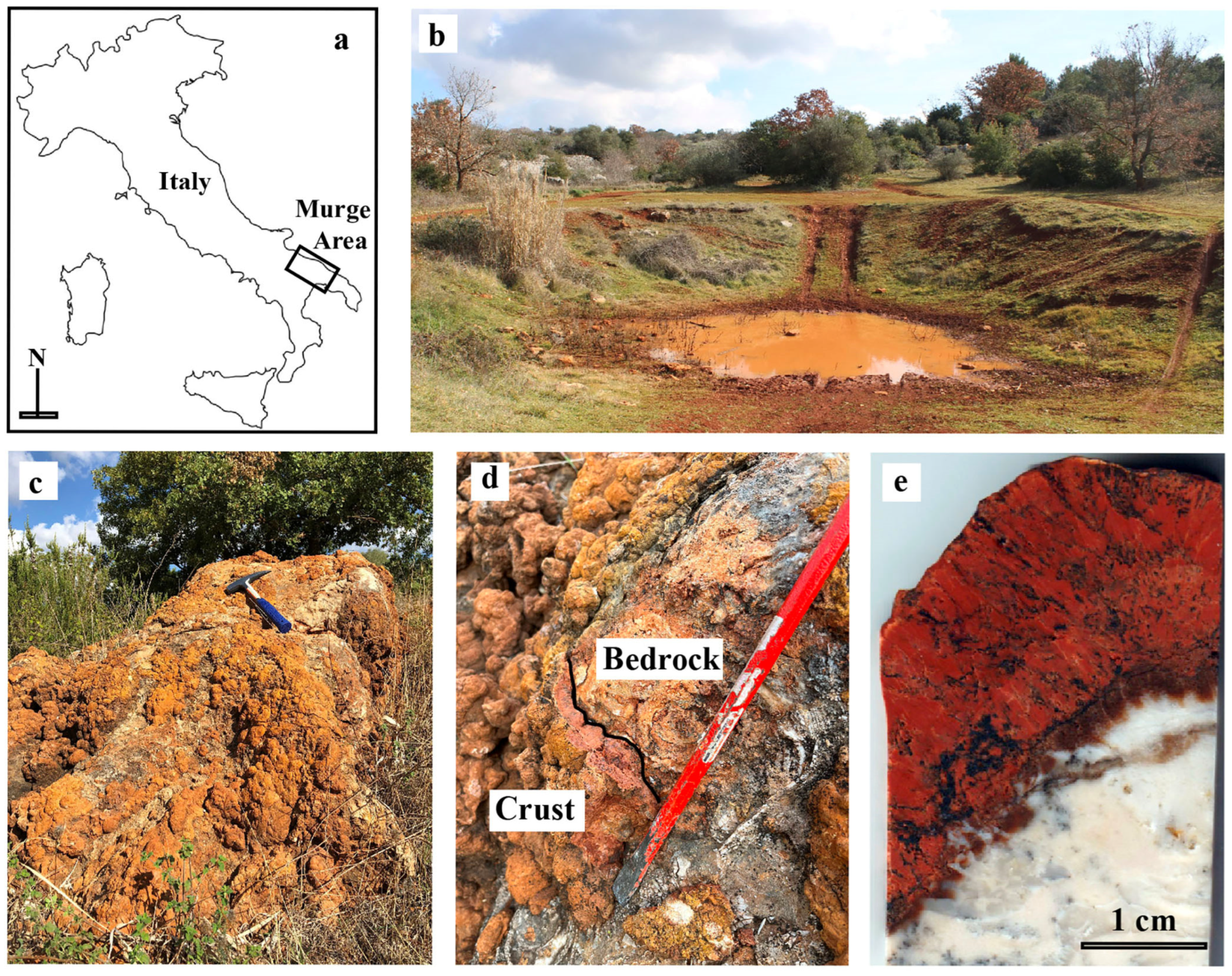
1. Introduction
2. Materials and Methods
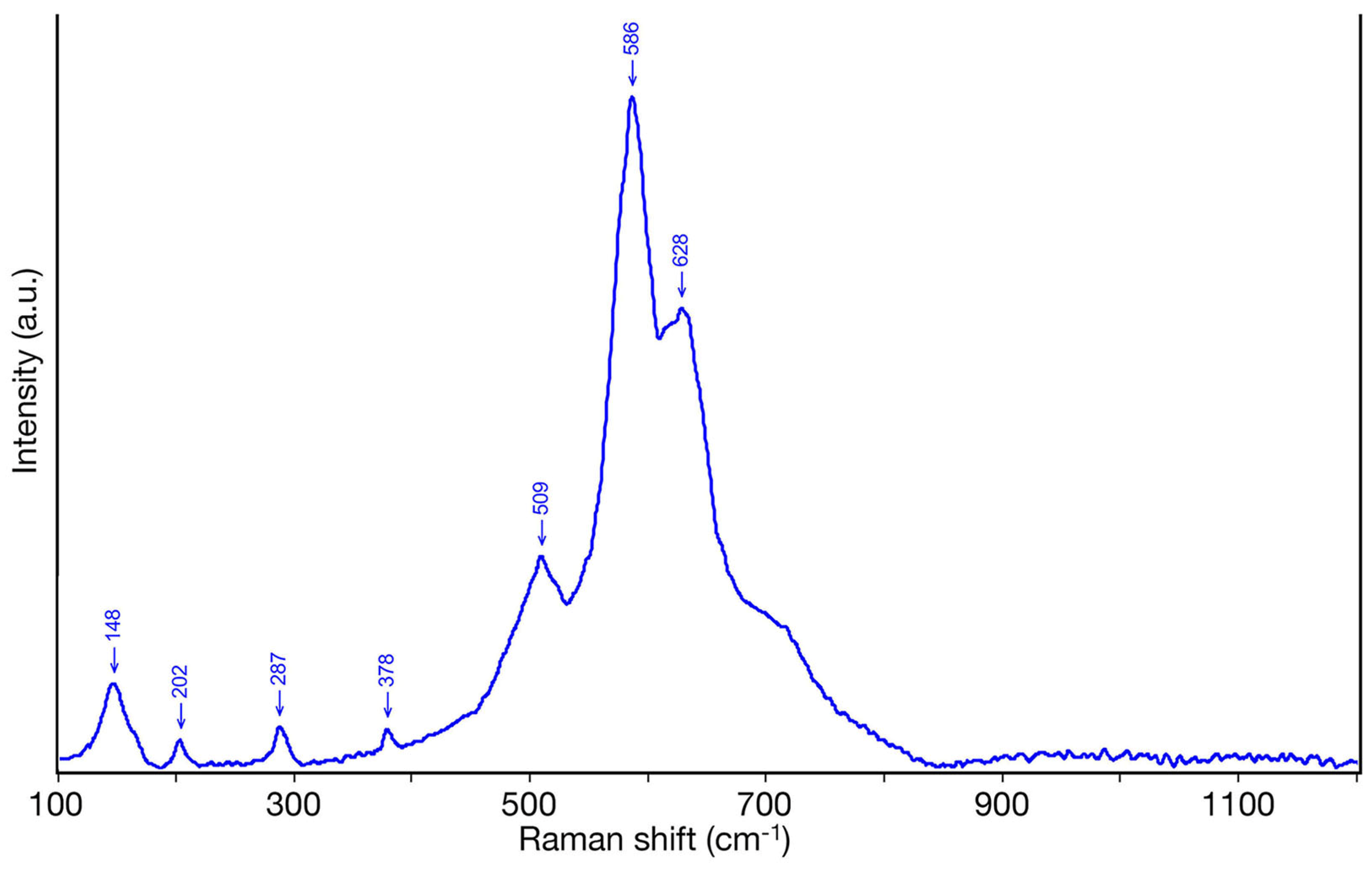
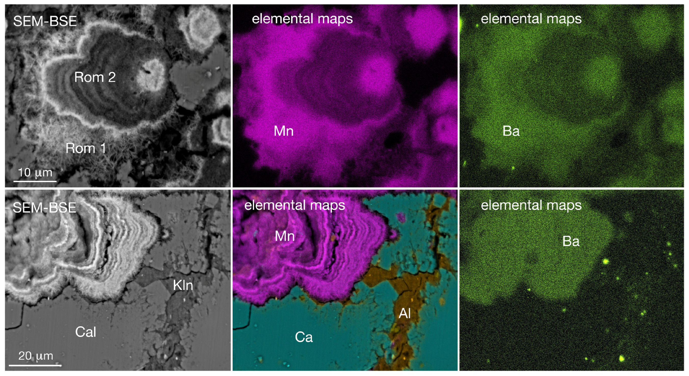
3. Results
4. Discussion and Conclusions
Author Contributions
Funding
Data Availability Statement
Acknowledgments
Conflicts of Interest
References
- Post, J.E.; McKeown, D.A.; Heaney, P.J. Raman spectroscopy study of manganese oxides: Tunnel structures. Am. Mineral. 2020, 105, 1175–1190. [Google Scholar] [CrossRef]
- Le Goff, P.; Baffier, N.; Bach, S.; Pereira-Ramos, J.P. Synthesis, ion exchange and electrochemical properties of lamellar phyllomanganates of the birnessite group. Mater. Res. Bull. 1996, 31, 63–75. [Google Scholar] [CrossRef]
- Post, J.E.; McKeown, D.A.; Heaney, P.J. Raman spectroscopy study of manganese oxides: Layer structures. Am. Mineral. 2021, 106, 351–366. [Google Scholar] [CrossRef]
- Manning, B.A.; Fendorf, S.E.; Bostick, B.; Suarez, D.L. Arsenic (III) oxidation and arsenic(V) adsorption reactions on synthetic birnessite. Environ. Sci. Technol. 2002, 36, 976–981. [Google Scholar] [CrossRef] [PubMed]
- Feng, X.H.; Zhai, L.M.; Tan, W.F.; Liu, F.; He, J.Z. Adsorption and redox reactions of heavy metals on synthesized Mn oxide minerals. Environ. Pollut. 2007, 147, 366–373. [Google Scholar] [CrossRef] [PubMed]
- Lopano, C.L.; Heaney, P.J.; Post, J.E. Cs-exchange in birnessite: Reaction mechanisms inferred from time-resolved X-ray diffraction and transmission electron microscopy. Am. Mineral. 2009, 94, 816–826. [Google Scholar] [CrossRef]
- Kwon, K.D.; Refson, K.; Sposito, G. Understanding the trends in transition metal sorption by vacancy sites in birnessite. Geochim. Cosmochim. Ac. 2013, 101, 222–232. [Google Scholar] [CrossRef]
- Bernardini, S.; Bellatreccia, F.; Casanova Municchia, A.; Della Ventura, G.; Sodo, A. Raman spectra of natural manganese oxides. J. Raman Spectrosc. 2019, 50, 873–888. [Google Scholar] [CrossRef]
- Micheletti, F.; Fornelli, A.; Spalluto, L.; Parise, M.; Gallicchio, S.; Tursi, F.; Festa, V. Petrographic and geochemical inferences for genesis of Terra Rossa: A case study from the Apulian Karst (southern Italy). Minerals 2023, 13, 499. [Google Scholar] [CrossRef]
- Rojkovič, I.; Aubrecht, R.; Mišík, M. Mineral and chemical composition of manganese hardgrounds in Jurassic limestones of the Western Carpathians. Geol. Carpath. 2003, 54, 317–328. [Google Scholar]
- Pharoe, B.K.; Evdokimov, A.N.; Gembitskaya, I.M.; Bushuyev, Y.Y. Mineralogy, geochemistry and genesis of the post-Gondwana supergene manganese deposit of the Carletonville-Ventersdorp area, North West Province, South Africa. Ore Geol. Rev. 2020, 120, 103372. [Google Scholar] [CrossRef]
- Beukes, N.J.; Smith, A. Paleoproterozoic banded Iron formation-hosted high grade hematite iron ore deposits of Transvaal supergroup, South Africa. Episodes 2016, 39, 269. [Google Scholar] [CrossRef]
- Garnit, H.; Kraemer, D.; Bouhlel, S.; Davoli, M.; Barca, D. Manganese ores in Tunisia: Genetic constraints from trace element geochemistry and mineralogy. Ore Geol. Rev. 2020, 120, 103451. [Google Scholar] [CrossRef]
- Fan, D.; Yang, P. Introduction to and classification of manganese deposits in China. Ore Geol. Rev. 1999, 15, 1–13. [Google Scholar] [CrossRef]
- Sinisi, R.; Mongelli, G.; Perri, F.; Rizzo, G. The braunite (3Mn2O3 MnSiO3)-rich mineralization in the metasedimentary succession from southern Apennines (Italy): Genesis constraints. Ore Geol. Rev. 2018, 94, 1–11. [Google Scholar] [CrossRef]
- Van Groeningen, N.; Glück, B.; Christl, I.; Kretzschmar, R. Surface precipitation of Mn2+ on clay minerals enhances Cd2+ sorption under anoxic conditions. Environ. Sci. Proc. Imp. 2020, 22, 1654–1665. [Google Scholar]
- Yang, Y.; Liu, J.; Zhu, R.; Chen, Q.; Wei, H.; Chen, M.; Xian, H.; He, H. Surface-induced oxidation of Mn (II) and crystallization of manganese (hydr) oxides on clay minerals. Geochim. Cosmochim. Ac. 2023, 363, 129–146. [Google Scholar] [CrossRef]
- Liu, J.; Chen, Q.; Yang, Y.; Wei, H.; Laipan, M.; Zhu, R.; Hongping, H.; Hochella, M.F., Jr. Coupled redox cycling of Fe and Mn in the environment: The complex interplay of solution species with Fe-and Mn-(oxyhydr) oxide crystallization and transformation. Earth-Sci. Rev. 2022, 232, 104105. [Google Scholar] [CrossRef]


| Area (N. of Analyses) | MnO | BaO | FeO | MgO | SiO2 | Al2O3 | CaO | Na2O | K2O | Total |
|---|---|---|---|---|---|---|---|---|---|---|
| romanechite 1A (5) | 60.27 | 5.79 | 0.68 | 1.32 | 2.01 | 1.58 | 2.81 | 0.30 | 0.52 | 74.76 |
| romanechite 1B (4) | 60.43 | 5.78 | 0.67 | 1.37 | 1.80 | 1.44 | 2.88 | 0.30 | 0.51 | 74.67 |
| romanechite 2A (10) | 79.42 | 6.33 | 0.27 | 1.40 | 0.41 | 0.39 | 3.10 | 0.35 | 0.51 | 92.18 |
| romanechite 2B (10) | 77.20 | 6.83 | 0.74 | 1.18 | 1.67 | 1.21 | 2.99 | 0.30 | 0.58 | 92.70 |
| romanechite 2C (10) | 77.33 | 10.00 | 0.04 | 0.59 | 0.21 | 0.37 | 2.99 | 0.21 | 0.24 | 91.98 |
| romanechite 2D (10) | 78.54 | 9.36 | 0.00 | 0.78 | 0.08 | 0.28 | 2.61 | 0.24 | 0.28 | 92.17 |
Disclaimer/Publisher’s Note: The statements, opinions and data contained in all publications are solely those of the individual author(s) and contributor(s) and not of MDPI and/or the editor(s). MDPI and/or the editor(s) disclaim responsibility for any injury to people or property resulting from any ideas, methods, instructions or products referred to in the content. |
© 2024 by the authors. Licensee MDPI, Basel, Switzerland. This article is an open access article distributed under the terms and conditions of the Creative Commons Attribution (CC BY) license (https://creativecommons.org/licenses/by/4.0/).
Share and Cite
Fornelli, A.; Micheletti, F.; Acquafredda, P.; Mangone, A. First Record of Romanechite in the Apulian Karst (Southern Italy) Resulting from the Interaction of Limestones and Clay Minerals. Minerals 2024, 14, 935. https://doi.org/10.3390/min14090935
Fornelli A, Micheletti F, Acquafredda P, Mangone A. First Record of Romanechite in the Apulian Karst (Southern Italy) Resulting from the Interaction of Limestones and Clay Minerals. Minerals. 2024; 14(9):935. https://doi.org/10.3390/min14090935
Chicago/Turabian StyleFornelli, Annamaria, Francesca Micheletti, Pasquale Acquafredda, and Annarosa Mangone. 2024. "First Record of Romanechite in the Apulian Karst (Southern Italy) Resulting from the Interaction of Limestones and Clay Minerals" Minerals 14, no. 9: 935. https://doi.org/10.3390/min14090935
APA StyleFornelli, A., Micheletti, F., Acquafredda, P., & Mangone, A. (2024). First Record of Romanechite in the Apulian Karst (Southern Italy) Resulting from the Interaction of Limestones and Clay Minerals. Minerals, 14(9), 935. https://doi.org/10.3390/min14090935