Article

Effect of Microgravity on Rare Earth Elements Recovery by Burkholderia cepacia and Aspergillus niger

1
School of Minerals Processing and Bioengineering, Central South University, Changsha 410083, China
2
Key Lab of Biohydrometallurgy of Ministry of Education, Changsha 410083, China
3
Department of Chemistry, Division of Natural Sciences, School of Arts and Sciences, National University of Mongolia, Ulaanbaatar 14201, Mongolia
*
Authors to whom correspondence should be addressed.
Minerals 2024, 14(10), 1055; https://doi.org/10.3390/min14101055
Submission received: 4 October 2024 / Revised: 18 October 2024 / Accepted: 18 October 2024 / Published: 21 October 2024
(This article belongs to the Section Mineral Processing and Extractive Metallurgy)

Abstract

:
Rare earth elements (REEs) are indispensable in modern industry and technology, driving an urgent demand for innovative, eco-friendly recovery technologies. As space exploration advances, the impact of microgravity on microorganisms has become a focal point, yet the effects on microbial growth and REEss recovery remain uncharted. This study investigates the biosorption of REEs by Burkholderia cepacia (B. cepacia) and Aspergillus niger (A. niger) from a mixed solution containing La, Ce, Pr, Nd, Sm, Er, and Y under varying initial concentrations, pH levels, and microgravity conditions. We observed that the medium’s pH rose with B. cepacia and fell with A. niger when cultured in normal gravity conditions, suggesting distinct metabolic responses. Notably, microgravity significantly altered microbial morphology and metabolite profiles, significantly enhancing REEs recovery efficiency. Specifically, the recovery of B. cepacia of Ce and Pr peaked at 100%, and A. niger achieved full recovery of all tested REEs at pH 1.5 (suboptimal growth conditions). This study pioneers the application of biosorption for the recovery of REEs in microgravity conditions, presenting a promising strategy for future resource exploitation by space biomining.

1. Introduction

Rare earth elements (REEs), comprising the 15 lanthanides of lanthanum (La), cerium (Ce), praseodymium (Pr), neodymium (Nd), promethium (Pm), samarium (Sm), europium (Eu), gadolinium (Gd), terbium (Tb), dysprosium (Dy), holmium (Ho), erbium (Er), thulium (Tm), ytterbium (Yb), and lutetium (Lu), along with scandium (Sc) and yttrium (Y), are indispensable in various industrial and scientific applications [1]. REEs are generally categorized into light REEs (La, Ce, Pr, Nd, and Pm), medium REEs (Sm, Eu, Gd, Tb, Dy), and heavy REEs (consisting of Ho, Er, Tm, Yb, Lu, Y and Sc) based on their atomic weights and properties [2]. With unique physical and chemical attributes (e.g., optical, electronic, and magnetic properties), REEs are vital for electronics, clean energy, military technology, medical equipment, and permanent magnets [3,4]. However, the annual production of REEs is insufficient to meet the escalating demand, predominantly supplied by China, which accounts for 85% of the global output [5]. This shortfall has led to a significant market crisis, highlighting the urgency for effective REEs recovery methods.
Traditional recovery methods, including chemical, physical, and biotechnological approaches such as solvent extraction, precipitation, ion exchange, electrochemical processes, bioleaching, and roasting [6,7,8], are known for their efficiency but are limited by high resource consumption, operational costs, and environmental impacts [9,10]. In contrast, a biotechnological method has gained prominence as a green and eco-friendly alternative for REEs recovery [11]. Biosorption, a process mediated by microorganisms, is recognized for its potential to substantially reduce costs and minimize the production of substances detrimental to the environment [12]. Microbes like Pseudoalteromonas sp [13], Chryseomonas luteola [14], Bacillus sp. [15], and Phanerochaete chrysosporium [16] have shown potential in heavy metal biosorption. Recently, there has been a growing interest among researchers in various microbes that exhibit high REE adsorption capabilities, holding potential for the recovery of REEs [17,18]. The surfaces of microorganisms possess numerous active functional groups, such as carboxyl, amidogen, and phosphoryl groups, which provide unique binding sites for the adsorption of REEs [19]. However, the current biosorption methods utilizing selected microbes have their limitations. Advanced bioengineering techniques, such as the modification of microorganisms and the insertion of specific peptides and proteins onto cell surfaces, have been shown to be more efficient for REEs recovery [20]. Despite these advantages, this approach also presents some challenges, including a complex operational process and potential ecological security concerns [21]. Considering the limitation of current biosorption methods on REEs biosorption, this study intends to simplify the modification process to enhance efficiency and feasibility.
Microgravity, an environment characterized by minimal gravitational force or what was commonly referred to as weightlessness [22], presented a unique and challenging setting for biological processes. It has been well documented that microgravity can significantly influence microbial growth and metabolism [23,24], modulate microbial gene expression [25,26], and change cellular morphology and structure [27,28]. These studies also highlighted the remarkable adaptability of microorganisms to extreme environments, including outer space [29]. Biomining, the process of employing microorganisms to extract REEs from REEs-bearing minerals, has emerged as a cutting-edge approach. Cockell et al. [30] demonstrated that the bacterium Sphingomonas desiccabilis could effectively increase the concentration of leached REEs under simulated microgravity conditions. This discovery implied that space mining could be a valuable strategy to ease the shortage of REEs on Earth [31]. As space exploration progresses, understanding the impact of microgravity on microbial growth is essential for optimizing biosorption processes and improving REEs recovery efficiency.
The aim of this study was therefore to investigate the effect of microgravity on microbial growth and REEs adsorption ability. In this study, two typical microbial strains, Burkholderia cepacia (B. cepacia) and Aspergillus niger (A. niger), were utilized in conducting biosorption experiments for REEs. Meanwhile, the influencing factors of REEs, initial concentration, pH, and microgravity, were studied.

2. Materials and Methods

2.1. Materials and Reagents

All reagents used in this study were of analytical grade and purchased from Shanghai Maclin Biochemical Technology Company (Shanghai, China). The stock solution of mixed REEs (La, Ce, Pr, Nd, Sm, Er, and Y) was prepared by dissolving the respective REEs hydrates and adjusting the volume to 1000 mL with deionized water, resulting in a concentration of 10 g/L for each REE (equal to the total REEs concentration of 70 g/L).

2.2. Microbe Incubation

2.2.1. In Normal Gravity

Burkholderia cepacia (B. cepacia, ATCC 25416) was cultivated in an LB medium at pH 6.0, composed of (per liter) 10 g tryptone, 5 g yeast extract, and 10 g sodium chloride (NaCl) with 1% v/v bacterial inoculum. The cell density was maintained at 1 × 10⁶ cells/mL and incubated at 30 °C with shaking at 180 rpm. The cell density of B. cepacia was determined at 600 nm using a microplate spectrophotometer (EPOCH, Zhicheng, Shanghai).
Aspergillus niger (A. niger) was initially cultured on a potato dextrose agar medium (28 °C, 7 d); then, the mature spore was carefully harvested into 100 mL of sterile water. The concentration of A. niger was adjusted to 1.0 × 10⁶ CFU/mL by the WGZ-200 turbidimeter (Shanghai, China). For the biosorption experiment, A. niger was grown on a medium containing (per liter) 100 g sucrose, 1 g magnesium sulfate heptahydrate (MgSO4·7H2O), 1 g potassium dihydrogen phosphate (KH2PO4), 0.5 g yeast extract, and 2 g ammonium sulfate ((NH4)2SO4), with an adjusted pH to 2.5 using 0.1 M HCl or NaOH. A. niger was incubated in an orbital shaking incubator at 28 °C with a speed of 200 rpm.

2.2.2. In Microgravity

The cultured seed solution was incubated within a sterile rotating wall reaction vessel, which was then secured onto a microgravity stand. Microgravity conditions for microbial incubation were simulated using the RSOC rotary culture system (Saiji GRS 210012, Suzhou, China) at 50 rpm for 10−3 g. The system generated a low-shear stress environment to simulate microgravity primarily through vertical rotation, within which cells were suspended in the medium, thereby diminishing the impact of gravity on the cells. The inoculation volume and culture time were consistent with those under normal gravity conditions, and all operations were carried out in a sterile environment.

2.3. Batch of Recovery Experiments

2.3.1. Recovery of REEs by B. cepacia

B. cepacia was cultured (1:99, v/v) into different concentrations of REEs (single REE of 10, 20, 50, 100 mg/L) for 24 h to evaluate its tolerance to REEs. To determine the recovery efficiency of REEs, a mixed REEs solution was introduced to the bacterial solution (after 24 h of cultivation), and the REEs content in the supernatant was measured after the reaction for 30 min. To evaluate the impact of pH conditions (2.0, 3.0, 4.0, 5.0, and 6.0) on microbial growth and REEs recovery, B. cepacia was incubated at different initial pH levels for 24 h, after which the diluted REEs stock solution (50 mg/L) was added to the cultured microbial solution.

2.3.2. Recovery of REEs by A. niger

A. niger was initially cultured under various initial REEs concentrations (single REE concentrations of 50, 100, 200, 300, and 400 mg/L) and pH conditions (1.5, 2.5, 3.5, 4.5, and 5.5) to assess the influence of environmental conditions on its growth. Concurrently, to ascertain the REEs recovery efficiency of A. niger, the mature mycelium, harvested after 120 h of incubation using a vacuum filter (Xinweng, Shanghai), was introduced into a mixed REEs solution with varying REEs concentrations. When examining the effect of pH conditions, A. niger was incubated at different pH levels, with the REEs concentration fixed at 20 mg/L.
All experiments were performed in triplicate.

2.4. REEs Concentration Determined by ICP-OES

The concentration of REEs in the diluted liquid supernatants was determined by an inductively coupled plasma optical emission spectrometer (ICP-OES, Agilent 5110, Agilent, Beijing, China). The recovery efficiency of REEs adsorption by microbes was calculated by following Equation (1):
REEs   recovery   ( % ) = REEs total REEs supernatant REEs total

3. Results and Discussion

3.1. Recovery of REEs by B. cepacia

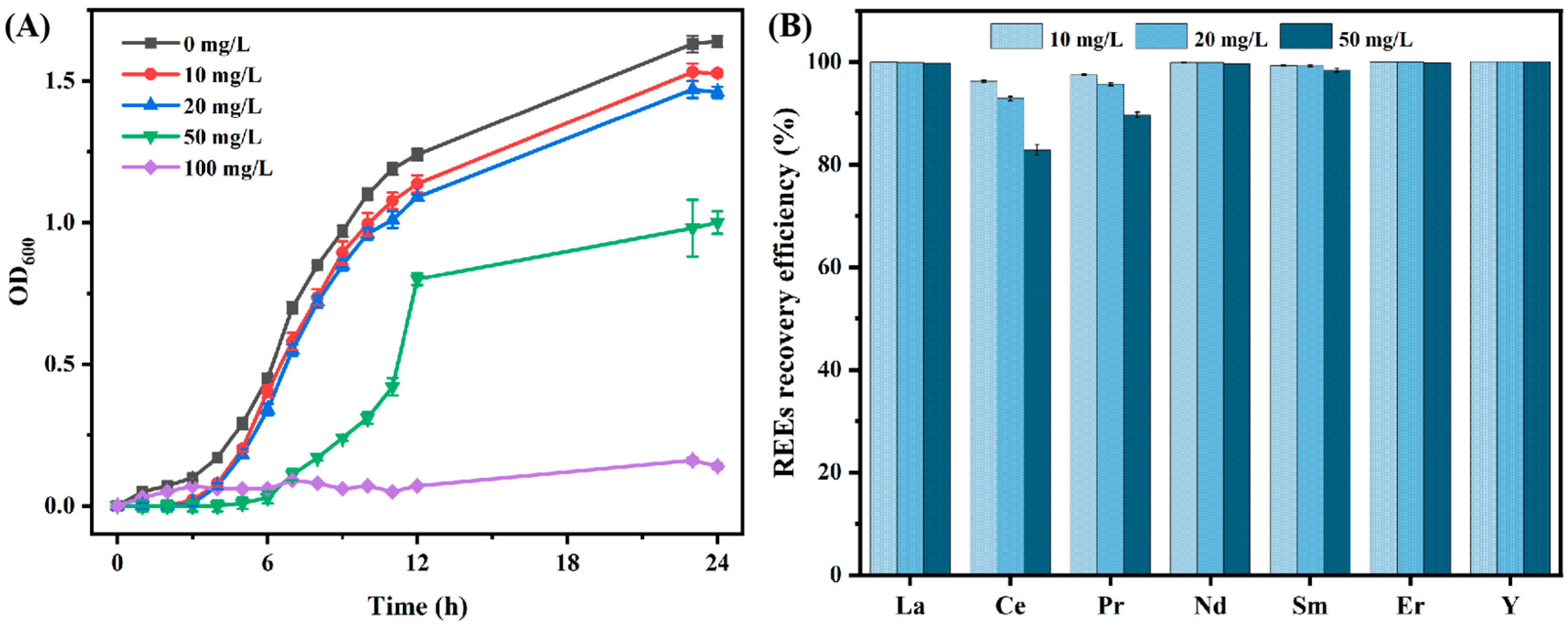
3.1.1. Effect of Different Initial REEs Concentrations on the Recovery Efficiency of REEs by B. cepacia

Figure 1A illustrates the microbial growth curve of B. cepacia under different initial concentrations of REEs, highlighting a preference for survival at a maximum concentration of 50 mg/L (equal to the total REEs concentration of 350 mg/L). Except for a single REE concentration of 100 mg/L, B. cepacia thrived at concentrations ranging from 0 to 50 mg/L. After a 24 h incubation period, the mixed REEs solutions were integrated into the bacterial culture. Figure 1B indicates that the recovery efficiency for La, Nd, Sm, Er, and Y approached nearly 100%. Nevertheless, B. cepacia exhibited a constrained capacity for the recovery of Ce and Pr. As the concentration of REEs increased from 10 mg/L to 50 mg/L, the recovery efficiencies for Ce and Pr correspondingly diminished, recording rates of 96.27%, 92.91%, and 82.88% and 97.56%, 95.7%, and 89.71%, respectively. Elevated concentrations of REEs can negatively impact microbial growth and disrupt metabolic pathways, subsequently leading to the reduced recovery of REEs [32]. The presence of active functional groups on the cell surface and microbial metabolites is significantly conducive to REEs biosorption [33]. Moreover, the intracellular transport of rare earth elements (REEs) may also lead to their accumulation within the cell, particularly when cellular constituents such as proteins, lipids, chlorophyll, and peptides bind to REEs [34].

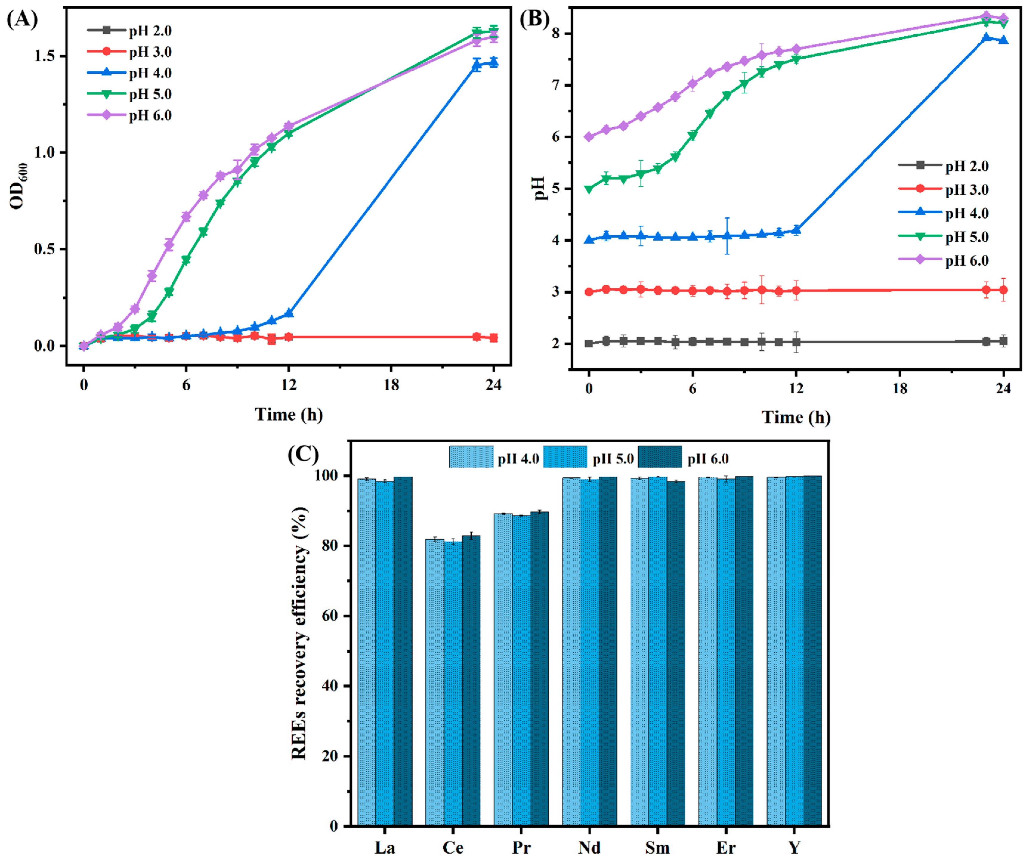
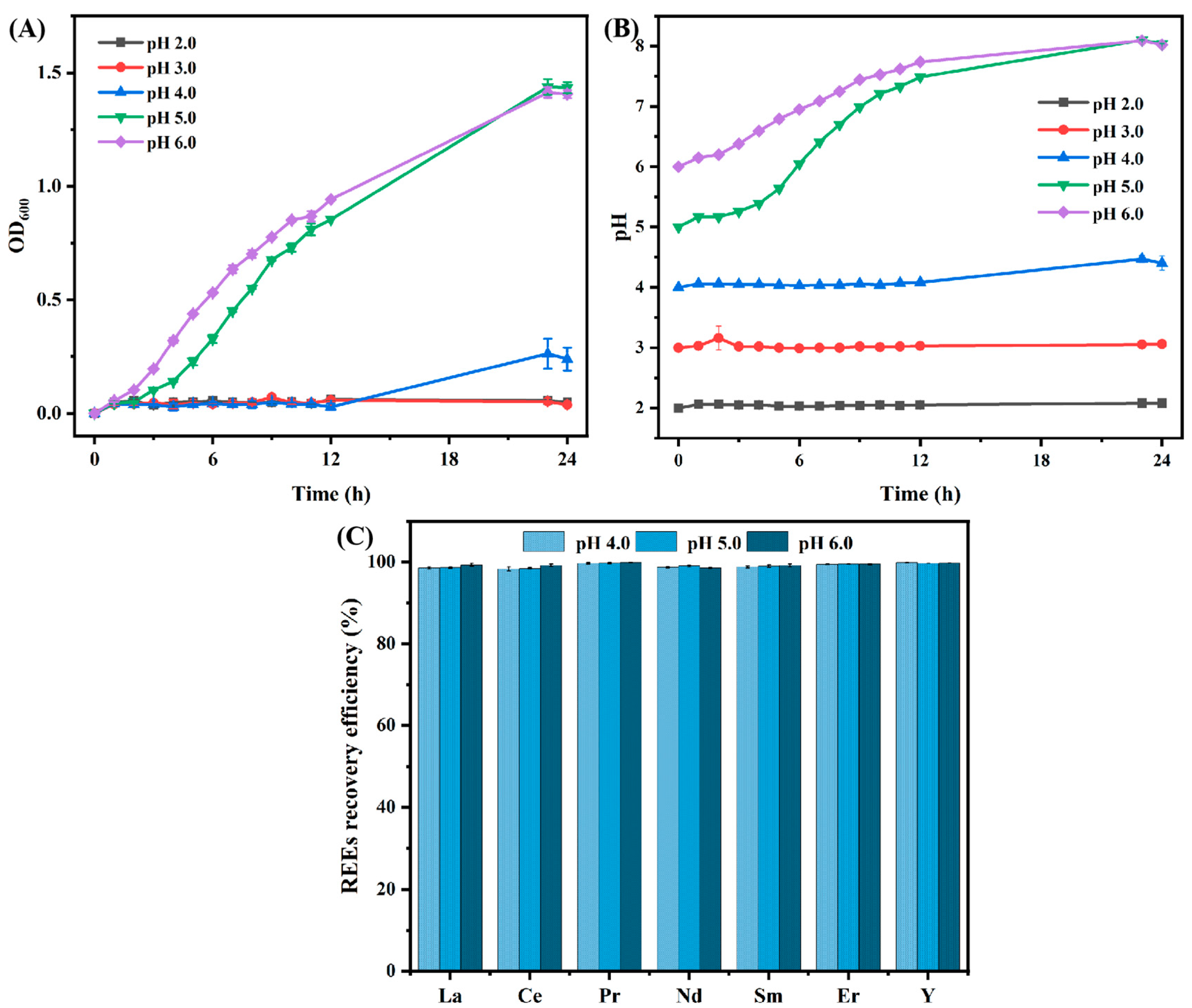
3.1.2. Effect of Different Initial pH Levels on the Recovery Efficiency of REEs by B. cepacia

Based on the aforementioned findings, B. cepacia was inoculated into 100 mL of culture medium with different initial pH levels, and the initial REEs concentration was optimized at 50 mg/L (the total REEs concentration was 350 mg/L). For the growth curves shown in Figure 2A, an obvious lag in the growth of B. cepacia at pH 4.0 was seen, which grew rapidly after 12 h. The results suggested that the growth of B. cepacia was a pH-dependent process, being almost entirely inhibited at lower pH levels of 2.0 and 3.0, yet thriving at pH levels of 5.0 and 6.0. Concurrently, as B. cepacia proliferated, the pH of the medium increased from 4.0, 5.0, and 6.0 to 7.9 ± 0.06, 8.2 ± 0.06, and 8.3 ± 0.1, respectively (Figure 2B). A previous study confirmed that B. cepacia was a phosphate-solubilizing microorganism capable of decomposing phosphate resources for growth, thereby producing a multitude of hydroxide ions [35]. The observed pH increase aligns with microbial growth trends and may also be attributed to the production of certain metabolites by the microorganisms during their growth phase. These alkaline substances also contribute to the recovery of REEs.
However, as shown in Figure 2C, there was no significant variation in REEs recovery efficiency in response to pH changes. The absence of recovery efficiency data for pH levels of 2.0 and 3.0 was due to the inability of B. cepacia to survive in such acidic conditions. Consistently, the recovery efficiency for Ce and Pr was lower compared to other selected REEs (~100%). The recovery efficiency for Ce was 81.86%, 81.22%, and 82.88%, while for Pr it was 89.2%, 88.7%, and 89.71% at pH levels ranging from 4.0 to 6.0, respectively. One possible explanation for the minimal variation in the biosorption ability of B. cepacia at different pH conditions was that the initial 24 h cultivation period allowed for the maturation of bacterial cells, ensuring that all tested pH conditions permitted B. cepacia to reach the logarithmic phase, thus exhibiting consistent biosorption capabilities.

3.2. Recovery of REEs by A. niger

3.2.1. Effect of Different Initial REEs Concentrations on the Recovery Efficiency of REEs by A. niger

Fungal strains, including A. niger, are recognized for their exceptional tolerance to heavy metals by various mechanisms, such as biosorption, complexation, and precipitation [36]. Accordingly, the experiment was designed to evaluate the impact of varying concentrations of REEs on the recovery ability at elevated levels, specifically within the range of 50 to 400 mg/L. Firstly, the tolerance of A. niger to REEs was assessed, with the result displayed in Figure 3A. The introduction of mixed REEs solutions led to a decrease in the pH value of the media, causing the pH to plummet from 2.5 to 1.8. In low REEs concentrations (specifically ranging from 0 to 100 mg/L which corresponded to a total REEs concentration of 0 to 700 mg/L), the medium’s pH observed a significant drop. It decreased from 2.5 to 1.7, from 2.3 to 1.6, and from 2.1 to 1.7, respectively. The pH reduction was likely attributable to the metabolic activity of A. niger, which was able to consume carbon sources to produce organic acids, thereby acidifying the medium [37]. Furthermore, organic acids can be used to improve REEs recovery through complexation, which also mitigates the toxicity impact on microbes [38]. Notably, at the initial REEs concentration of 200 mg/L (the total REEs was 1400 mg/L), the pH value minimally shifted from 1.9 to 1.8. Concurrently, at elevated REEs concentrations ranging from 300 to 400 mg/L (equivalent to total REEs concentrations of 2100 to 2800 mg/L), there was a negligible change in pH.
To assess the biosorption ability of A. niger, the biosorbent was introduced into the mixed REEs solution. Prior to biosorption experiments, A. niger was cultured at 200 rpm at 28 °C for 120 h with an initial medium pH of 2.5. As shown in Figure 3B, A. niger demonstrated significantly varied recovery efficiencies across different concentrations of REEs. At lower REEs concentrations ranging from 50 to 200 mg/L, A. niger achieved recovery efficiencies approaching 80% for elements such as La, Ce, Pr, Nd, and Sm. In contrast, the recovery rates for Er were 52.33%, 38.7%, and 16.89%, and for Y, they were 41.72%, 22.1%, and 5.64%, respectively. However, at higher REEs concentrations of 300 and 400 mg/L, there was a marked decline in the recovery of all REEs. The recovery efficiency for La, Ce, Pr, Nd, and Sm hovered around 50%, while that for Er and Y was less than 5%. The results suggested the REEs recovery efficiency of A. niger was limited, as it failed to achieve complete removal of REEs within the established concentration ranges. Similarly, Zhou et al. [39] demonstrated that A. niger could achieve a maximum adsorption efficiency at a total REEs concentration of 200 mg/L, which is considerably less than the 95% efficiency exhibited by Bacillus sp. Moreover, A. niger had selectivity for REEs, and the adsorption capacity of medium and heavy REEs was weaker than that of light REEs. So, in the later biosorption experiments of A. niger, the initial REE concentrations was adjusted to 20 mg/L, equivalently to the total REEs concentration of 140 mg/L.

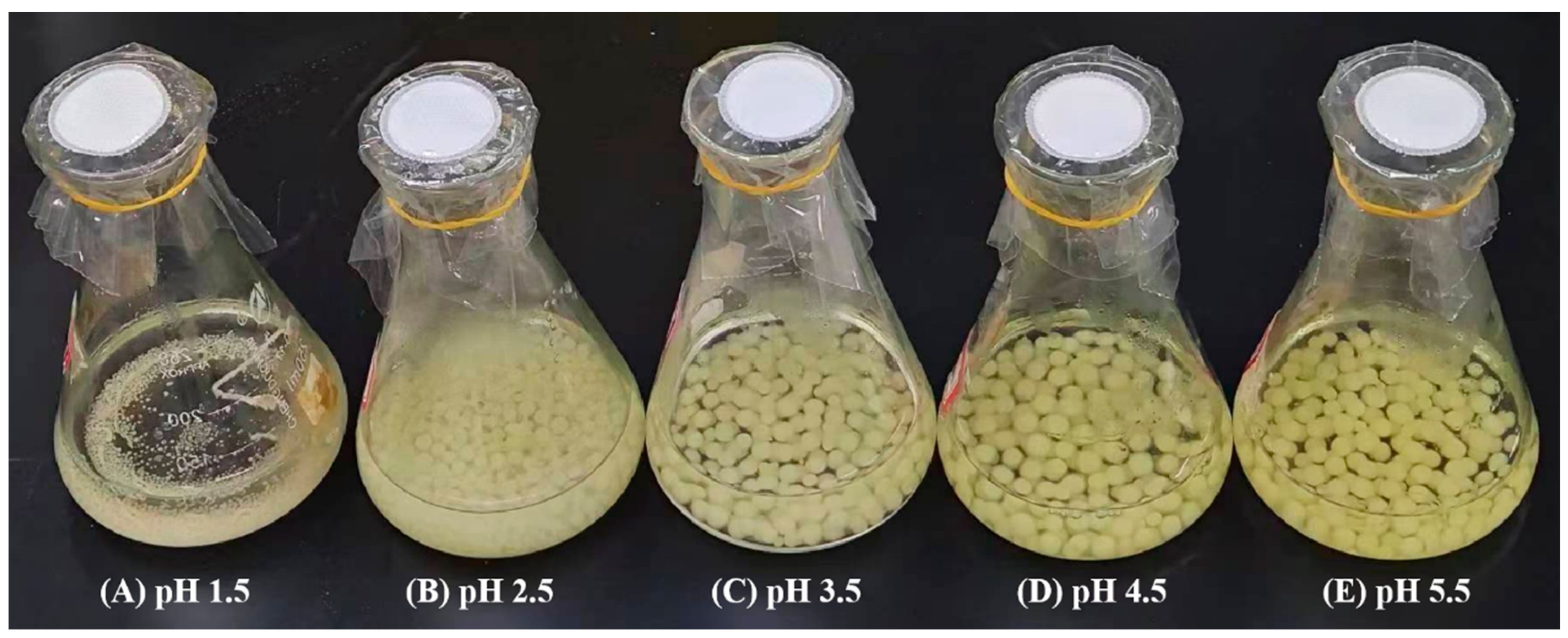
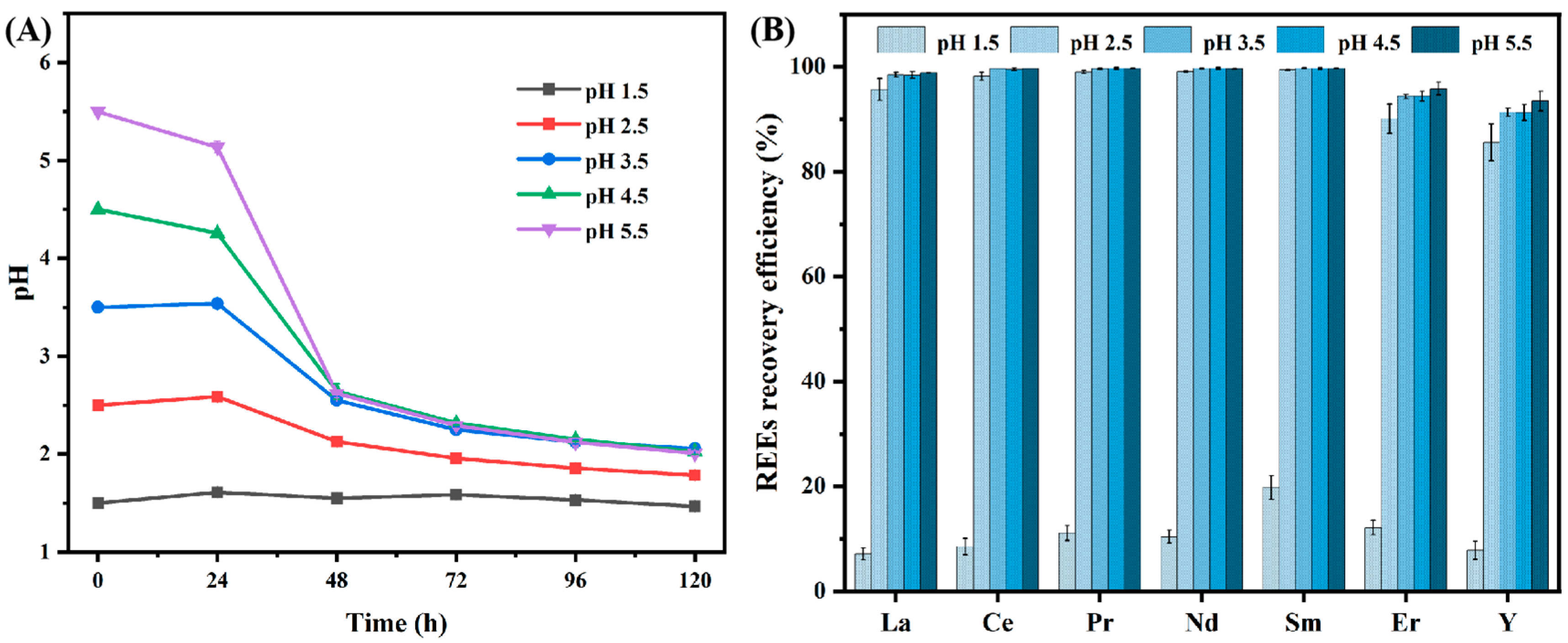
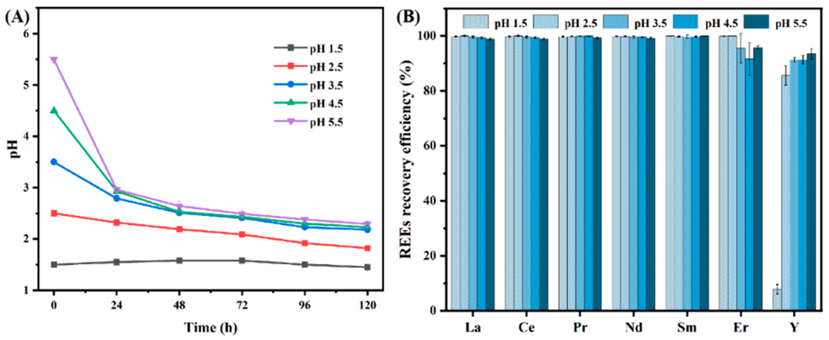
3.2.2. Effect of Different Initial pH Levels on the Recovery Efficiency of REEs by A. niger

pH was an important factor in influencing the growth and metabolism of microorganisms. Under extreme pH conditions, microbial growth may be inhibited. For example, extremely acidic environments may damage cell walls and membranes, interfere with metabolic pathways, and result in insufficient energy production [40]. Similarly, an extremely alkaline environment may affect the structure and function of proteins, which in turn affects the overall metabolism of cells [41]. Drawing from the result of REEs concentrations on the REEs recovery efficiency of A. niger, the REEs concentration was optimized to 20 mg/L to better determine the effect of pH on the biosorption process. The growth morphology of A. niger cultured in different initial pH levels is shown in Figure 4; it was found that the higher pH of the medium, the larger the mycelium pellets size of A. niger. In Figure 4A, in the extremely acidic environment of 1.5, many small particles of A. niger could be observed. Only a few acidophilic microorganisms can grow in such acidic environments, which indicated that A. niger has a good adaptability to the environment [42]. In Figure 4B, the size of microorganisms was moderate, and the most microbial mycelium can be observed, so it was speculated that the medium pH of 2.5 might be more conducive to microbial growth. Within the pH range of 3.5 to 5.5, A. niger exhibited robust growth, characterized by the formation of distinct and expansive mycelium pellets (Figure 4C–E). A larger diameter of mycelium pellets impeded nutrient and oxygen transport, consequently resulting in a diminished microbial density [43]. The above results indicated that the morphology and size of mycelium pellets of A. niger in the incubated solutions were highly correlated with the pH level of the medium, and the appropriate cell size may contribute to the microbial metabolism and may be further conducive to REEs biosorption ability [44].
As shown in Figure 5A, the pH of the media gradually decreased to 1.8~2.0 after 120 h incubation. This might contribute to the production of organic acids, resulting from the consumption of sugar sources during the microbial growth and metabolism process [45,46]. However, the pH of the culture medium remained essentially stable at approximately 1.5, signifying a constrained level of metabolic activity by A. niger. In alignment with the growth morphology of Figure 4A, the mycelium size of A. niger was observed to be quite diminutive. This suggested that the limited adaptive mechanisms available to A. niger were barely sufficient to sustain rudimentary growth and were inadequate for the generation of organic acids and other metabolic byproducts [47]. Then, the cultured mycelium was added to the mixed REEs solution, and the REEs recovery efficiency under different initial medium pH levels ranging from 1.5 to 5.5 is given in Figure 5B. The removal of REEs demonstrated minimal sensitivity to pH variations, and aside from the pH of 1.5, the recovery rates of REEs across other pH conditions were remarkably high. At a pH of 1.5, the REEs recovery efficiency of La, Ce, Pr, Nd, Sm, Er, and Y was 7.06%, 8.52%, 11.05%, 10.36%, 19.78%, 12.12%, and 7.82%, respectively. Notably, both the recovery of Er and Y was significantly influenced by pH. For Er, the recovery efficiency increased from 90.14% (pH 2.5) to 95.88% (pH 5.5), while the recovery efficiency of Y increased from 85.62% (pH 2.5) to 93.49% (pH 5.5), respectively.

3.3. Microgravity Incubation

3.3.1. Effect of Microgravity Incubation on B. cepacia Biosorption Ability

To ascertain the impact of microgravity on both microbial proliferation and biosorption capabilities, B. cepacia was initially cultivated under microgravity conditions for 24 h, after which it was transferred to a fresh medium under normal gravity for further incubation. As shown in Figure 6A, B. cepacia thrived at an initial pH of 2.0 and 3.0 (OD600 = 1.43), yet it was found that a pH of 4.0 was inhospitable to growth (OD600 = 0.26). Compared to growth under normal conditions, B. cepacia also could not survive at low pH conditions (pH of 3.0 and 4.0), and the overall growth also performed markedly worse with a reduced cell density. Within the normal gravity environment, the maximum cell density for B. cepacia was recorded as 1.63 at pH 5.0 and 1.47 at pH 4.0. However, Castro-Wallace et al. [48] demonstrated that microgravity played little effect on the growth and gene expression of Lactobacillus acidophilus ATCC 4356. In contrast, Kim et al. [49] reported that all tested Escherichia coli O157:H7 strains showed more robust growth in the acidified medium when cultivated under a simulated microgravity environment.
Figure 6B depicts the change in pH during the incubation process, where an increase in pH was observed at pH levels of 5.0 and 6.0, attributed to the metabolites produced by B. cepacia. Despite the growth not being as vigorous as in a normal gravity environment, full adsorption of all tested REEs was achieved (Figure 6C). This phenomenon could be ascribed to alterations in the adsorption sites and cellular structures under microgravity conditions, or potentially to the secretion of certain substances that can form stable complexes with REEs, thereby boosting the recovery efficiency.

3.3.2. Effect of Microgravity Incubation on A. niger Biosorption Ability

A. niger was initially cultivated in the microgravity environment for 120 h, after which the harvested solution was inoculated into a 100 mL medium with different initial pH levels at 1% inoculum. The cultures were then allowed to grow for an additional 120 h in an orbital shaker with a speed of 200 rpm at 28 °C. It was taken to evaluate the influence of microgravity conditions on the biosorption process, and the growth morphology is presented in Figure 7. The results revealed that an incubated microgravity seed solution was not conducive to rapid propagation, resulting in the morphology of A. niger changing dramatically. Compared to A. niger cultured under normal gravity, the mycelium exhibited a more irregular growth pattern. At lower pH levels (1.5 and 2.5), the mycelium clumped together forming large masses, with the surrounding hyphae splitting into finer filaments over the course of incubation. Within the pH range that is optimal for A. niger growth (pH of 3.5–5.5), the fungal spores were able to develop into fully formed mycelium. However, they did not maintain the regular spherical shape typically seen under normal gravity conditions but instead adopted an elliptical or elongated strip shape. Different microbes have different adaptability levels to the environment; many microbes such as Streptomyces hygroscopicus and Streptomyces clavuligerus were confirmed to grow more slowly in simulated microgravity compared to a standard atmospheric environment [50,51]. The results of this study were consistent with those reported by Liu et al. [52], who also noted significant morphological changes in the mycelium of Aspergillus brasiliensis when cultivated under a microgravity environment. The observed variation in the mycelium could be attributed to the reduced shear stress and the diminished gravitational forces, which in turn influence the synthesis of the cell wall and the preservation of cellular shape, consequently leading to modifications in cellular morphology [53].
The A. niger culture, which had been incubated under microgravity conditions, was utilized to evaluate the influence of pH on the biosorption efficiency of REEs. As depicted in Figure 8A, a notable reduction in the pH of the media was observed, with declines from 5.5 to 2.3, from 4.5 to 2.2, from 3.5 to 2.2, from 2.5 to 1.8, and from 1.5 to 1.4, respectively. This trend in pH reduction was less pronounced than that observed under normal gravity conditions. Remarkably, the recovery efficiency at the lowest pH of 1.5 demonstrated a marked increase when compared to A. niger cultured under normal gravity. The biosorption recovery for La, Ce, Pr, Nd, Sm, and Er saw a dramatic rise from 7.06%, 8.52%, 11.05%, 10.36%, 19.78%, 12.12%, and 7.82% to nearly 100%, respectively. However, the recovery of Y remained unaffected. These findings implied that microgravity may enhance the REEs recovery efficiency of A. niger, potentially offering a novel strategy for optimizing the recovery of REEs under such conditions.

3.4. Possible Recovery Mechanisms

Based on the results presented, the two microbial strains exhibited distinct adsorption effects on REEs; B. cepacia demonstrated high recovery efficiency and tolerance towards REEs. Upon growth, the pH environment increased for B. cepacia, which suggested that the production of alkaline substances could precipitate REEs, thereby facilitating their removal. Conversely, A. niger’s growth was accompanied by a decrease in pH, indicating that REEs biosorption might be attributed to metabolites that complexed with REEs. Additionally, it was possible that REEs in the solution could accumulate on the cell surface, thereby enhancing the recovery efficiency. Furthermore, under microgravity conditions, there was a noticeable change in the growth morphology and the medium’s pH, which corresponded with an enhancement in REEs recovery. These observations highlighted the influence of microgravity on microbial growth and REEs recovery.

4. Conclusions

The current investigation has shed light on the impact of microgravity on the growth and REEs (La, Ce, Pr, Nd, Sm, Er, and Y) recovery efficiency of B. cepacia and A. niger. Our findings revealed that B. cepacia could tolerate a maximum concentration exceeding 50 mg/L (the total REEs concentration was 350 mg/L), whereas the tolerance of A. niger was lower. After a reaction for 30 min, B. cepacia demonstrated the ability to achieve approximately 100% REEs removal, which may be attributed to the production of alkaline compounds during the metabolic process, as well as the bioaccumulation of REEs within the bacterial cells. In contrast, the biosorption ability of A. niger was far from what was expected due to the acidification of the medium (such as producing organic acids). However, when both strains were subjected to microgravity, significant changes in cell morphology and metabolite production were observed. The complicated adaptations substantially improved REEs recovery under the specified microgravity conditions. This study establishes a foundation for space biomining in the recovery of REEs resources beyond Earth’s atmosphere. Future studies should further explore the relationship between biosorption and microgravity, focusing on clarifying the intrinsic mechanisms of biosorption and preparing for space biomining.

Author Contributions

Writing—original draft preparation, conceptualization, methodology, N.H.; formal analysis, data curation, Z.Z.; writing—review and editing, validation, X.M.; project administration, writing—review and editing, S.D.; supervision, conceptualization, methodology, funding acquisition, H.Z. All authors have read and agreed to the published version of the manuscript.

Funding

This work was supported by the National Key Research and Development Program of China (2022YFE0119600) and the National Natural Science Foundation of China (52222406).

Data Availability Statement

The data are contained within the article.

Conflicts of Interest

The authors declare no conflicts of interest. The funders had no role in the design of the study; in the collection, analyses, or interpretation of the data; in the writing of the manuscript; or in the decision to publish the results.

References

  1. Meng, X.Y.; Zhao, H.B.; Zhang, Y.S.; Shen, L.; Gu, G.H.; Qiu, G.Z.; Zhang, X.G.; Yu, H.; He, X.; Liu, C. Simulated bioleaching of ion-adsorption rare earth ore using metabolites of biosynthetic citrate: An alternative to cation exchange leaching. Miner. Eng. 2022, 189, 107900. [Google Scholar] [CrossRef]
  2. Zhao, Y.; Zhao, H.B.; Shen, L.; Qiu, G.Z.; Wang, Y.Y.; Zhao, Y.; Zhao, H.; Shen, L.; Qiu, G.; Wang, Y. Study on the role of microbial metabolites in in-situ noncontact bioleaching of ion-adsorption rare earth ore. J. Environ. Manag. 2024, 368, 122184. [Google Scholar] [CrossRef]
  3. Brewer, A.; Chang, E.; Park, D.M.; Kou, T.Y.; Li, Y.; Lammers, L.N.; Jiao, Y.Q.; Brewer, A.; Chang, E.; Park, D.M.; et al. Recovery of Rare Earth Elements from Geothermal Fluids through Bacterial Cell Surface Adsorption. Environ. Sci. Technol. 2019, 53, 7714. [Google Scholar] [CrossRef]
  4. Das, N.; Das, D.; Das, N.; Das, D. Recovery of rare earth metals through biosorption: An overview. J. Rare Earths 2013, 31, 933. [Google Scholar] [CrossRef]
  5. Park, D.M.; Brewer, A.; Reed, D.W.; Lammers, L.N.; Jiao, Y.Q. Recovery of Rare Earth Elements from Low-Grade Feedstock Leachates Using Engineered Bacteria. Environ. Sci. Technol. 2017, 51, 13471. [Google Scholar] [CrossRef] [PubMed]
  6. Taggart, R.K.; Hower, J.C.; Hsu-Kim, H. Effects of roasting additives and leaching parameters on the extraction of rare earth elements from coal fly ash. Int. J. Coal Geol. 2018, 196, 106. [Google Scholar] [CrossRef]
  7. Tofan, L.; Bojoaga, C.N.; Paduraru, C.; Tofan, L.; Bojoaga, C.-N.; Paduraru, C. Biosorption the recovery and analysis of rare earth elements and platinum group metals from real samples. A review. Environ. Chem. Lett. 2022, 20, 1225. [Google Scholar] [CrossRef]
  8. Owusu-Fordjour, E.Y.; Yang, X.B. Bioleaching of rare earth elements challenges and opportunities: A critical review. J. Environ. Chem. Eng. 2023, 11, 110413. [Google Scholar] [CrossRef]
  9. Wu, M.X.; Liang, J.J.; Tang, J.; Li, G.; Shan, S.P.; Guo, Z.H.; Deng, L.; Wu, M.; Liang, J.; Tang, J.; et al. Decontamination of multiple heavy metals-containing effluents through microbial biotechnology. J. Hazard. Mater. 2017, 337, 189. [Google Scholar] [CrossRef]
  10. Sethurajan, M.; van Hullebusch, E.D.; Fontana, D.; Akcil, A.; Deveci, H.; Batinic, B.; Leal, J.P.; Gasche, T.A.; Kucuker, M.A.; Kuchta, K.; et al. Recent advances on hydrometallurgical recovery of critical and precious elements from end of life electronic wastes-a review. Crit. Rev. Environ. Sci. Technol. 2019, 49, 212. [Google Scholar] [CrossRef]
  11. Meng, X.Y.; Zhao, H.B.; Zhao, Y.; Shen, L.; Gu, G.H.; Qiu, G.Z.; Meng, X.; Zhao, H.; Zhao, Y.; Shen, L.; et al. Heap leaching of ion adsorption rare earth ores and REEs recovery from leachate with lixiviant regeneration. Sci. Total Environ. 2023, 898, 165417. [Google Scholar] [CrossRef]
  12. Fomina, M.; Gadd, G.M.; Fomina, M.; Gadd, G.M. Biosorption: Current perspectives on concept, definition and application. Bioresour. Technol. 2014, 160, 3. [Google Scholar] [CrossRef] [PubMed]
  13. Jiang, L.; Zhou, W.Z.; Liu, D.S.; Liu, T.; Wang, Z.D.; Jiang, L.; Zhou, W.; Liu, D.; Liu, T.; Wang, Z. Biosorption isotherm study of Cd2+, Pb2+ and Zn2+ biosorption onto marine bacterium Pseudoalteromonas sp SCSE709-6 in multiple systems. J. Mol. Liq. 2017, 247, 230. [Google Scholar] [CrossRef]
  14. Önal, S.; Baysal, S.H.; Ozdemir, G.; Onal, S.; Baysal, S.H.; Ozdemir, G. Studies on the applicability of alginate-entrapped Chryseomonas luteola TEM 05 for heavy metal biosorption. J. Hazard. Mater. 2007, 146, 417. [Google Scholar] [CrossRef]
  15. Nourbakhsh, M.N.; Kiliçarslan, S.; Ilhan, S.; Ozdag, H.; Nourbakhsh, M.N.; Kiliçarslan, S.; Ilhan, S.; Ozdag, H. Biosorption of Cr6+, Pb2+ and Cu2+ ions in industrial waste water on Bacillus sp. Chem. Eng. J. 2002, 85, 351. [Google Scholar] [CrossRef]
  16. Lu, N.Q.; Hu, T.J.; Zhai, Y.B.; Qin, H.Q.; Aliyeva, J.; Zhang, H.; Lu, N.; Hu, T.; Zhai, Y.; Qin, H.; et al. Fungal cell with artificial metal container for heavy metals biosorption: Equilibrium, kinetics study and mechanisms analysis. Environ. Res. 2020, 182, 109061. [Google Scholar] [CrossRef]
  17. Texier, A.C.; Andrès, Y.; Le Cloirec, P.; Texier, A.C.; Andrès, Y.; Le Cloirec, P. Selective biosorption of lanthanide (La, Eu, Yb) ions by Pseudomonas Aeruginosa. Environ. Sci. Technol. 1999, 33, 489. [Google Scholar] [CrossRef]
  18. Jiang, M.Y.; Ohnuki, T.; Tanaka, K.; Kozai, N.; Kamiishi, E.; Utsunomiya, S.; Jiang, M.; Ohnuki, T.; Tanaka, K.; Kozai, N.; et al. Post-adsorption process of Yb phosphate nano-particle formation by Saccharomyces cerevisiae. Geochim. Cosmochim. Acta 2012, 93, 30. [Google Scholar] [CrossRef]
  19. Fein, J.B.; Martin, A.M.; Wightman, P.G.; Fein, J.B.; Martin, A.M.; Wightman, P.G. Metal adsorption onto bacterial surfaces: Development of a predictive approach. Geochim. Cosmochim. Acta 2001, 65, 4267. [Google Scholar] [CrossRef]
  20. Park, D.M.; Reed, D.W.; Yung, M.C.; Eslamimanesh, A.; Lencka, M.M.; Anderko, A.; Fujita, Y.; Riman, R.E.; Navrotsky, A.; Jiao, Y.Q. Bioadsorption of Rare Earth Elements through Cell Surface Display of Lanthanide Binding Tags. Environ. Sci. Technol. 2016, 50, 2735. [Google Scholar] [CrossRef]
  21. Johnson, D.B.; Johnson, D.B. Biomining-biotechnologies for extracting and recovering metals from ores and waste materials. Curr. Opin. Biotechnol. 2014, 30, 24. [Google Scholar] [CrossRef] [PubMed]
  22. Nickerson, C.A.; Ott, C.M.; Wilson, J.W.; Ramamurthy, R.; Pierson, D.L.; Nickerson, C.A.; Ott, C.M.; Wilson, J.W.; Ramamurthy, R.; Pierson, D.L. Microbial responses to microgravity and other low-shear environments. Microbiol. Mol. Biol. Rev. 2004, 68, 345. [Google Scholar] [CrossRef] [PubMed]
  23. Huang, B.; Li, D.G.; Huang, Y.; Liu, C.T.; Huang, B.; Li, D.-G.; Huang, Y.; Liu, C.-T. Effects of spaceflight and simulated microgravity on microbial growth and secondary metabolism. Mil. Med. Res. 2018, 5, 18. [Google Scholar] [CrossRef]
  24. Horneck, G.; Klaus, D.M.; Mancinelli, R.L.; Horneck, G.; Klaus, D.M.; Mancinelli, R.L. Space Microbiology. Microbiol. Mol. Biol. Rev. 2010, 74, 121. [Google Scholar] [CrossRef]
  25. Wilson, J.W.; Ott, C.M.; Bentrup, K.H.Z.; Ramamurthy, R.; Quick, L.; Porwollik, S.; Cheng, P.; McClelland, M.; Tsaprailis, G.; Radabaugh, T.; et al. Space flight alters bacterial gene expression and virulence and reveals a role for global regulator Hfq. Proc. Natl. Acad. Sci. USA 2007, 104, 16299. [Google Scholar] [CrossRef]
  26. Wilson, J.W.; Ramamurthy, R.; Porwollik, S.; McClelland, M.; Hammond, T.; Allen, P.; Ott, C.M.; Pierson, D.L.; Nickerson, C.A. Microarray analysis identifies Salmonella genes belonging to the low-shear modeled microgravity regulon. Proc. Natl. Acad. Sci. USA 2002, 99, 13807. [Google Scholar] [CrossRef] [PubMed]
  27. Vukanti, R.; Model, M.A.; Leff, L.G.; Vukanti, R.; Model, M.A.; Leff, L.G. Effect of modeled reduced gravity conditions on bacterial morphology and physiology. Bmc Microbiol. 2012, 12, 4. [Google Scholar] [CrossRef]
  28. Kacena, M.A.; Merrell, G.A.; Manfredi, B.; Smith, E.E.; Klaus, D.M.; Todd, P.; Kacena, M.A.; Merrell, G.A.; Manfredi, B.; Smith, E.E.; et al. Bacterial growth in space flight: Logistic growth curve parameters for Escherichia coli and Bacillus subtilis. Appl. Microbiol. Biotechnol. 1999, 51, 229. [Google Scholar] [CrossRef]
  29. Benoit, M.R.; Li, W.; Stodieck, L.S.; Lam, K.S.; Winther, C.L.; Roane, T.M.; Klaus, D.M.; Benoit, M.R.; Li, W.; Stodieck, L.S.; et al. Microbial antibiotic production aboard the International Space Station. Appl. Microbiol. Biotechnol. 2006, 70, 403. [Google Scholar] [CrossRef]
  30. Cockell, C.S.; Santomartino, R.; Finster, K.; Waajen, A.C.; Eades, L.J.; Moeller, R.; Rettberg, P.; Fuchs, F.M.; Van Houdt, R.; Leys, N.; et al. Space station biomining experiment demonstrates rare earth element extraction in microgravity and Mars gravity. Nat. Commun. 2020, 11, 5523. [Google Scholar] [CrossRef]
  31. Gumulya, Y.; Zea, L.; Kaksonen, A.H.; Gumulya, Y.; Zea, L.; Kaksonen, A.H. In situ resource utilisation: The potential for space biomining. Miner. Eng. 2022, 176, 107288. [Google Scholar] [CrossRef]
  32. Akberdin, I.R.; Collins, D.A.; Hamilton, R.; Oshchepkov, D.Y.; Shukla, A.K.; Nicora, C.D.; Nakayasu, E.S.; Adkins, J.N.; Kaiyuzhnaya, M.G.; Akberdin, I.R.; et al. Rare Earth Elements Alter Redox Balance in Methylomicrobium alcaliphilum 20ZR. Front. Microbiol. 2018, 9, 2735. [Google Scholar] [CrossRef] [PubMed]
  33. Luo, S.; Li, L.L.; Chen, A.W.; Zeng, Q.R.; Xia, H.; Gu, J.D.; Luo, S.; Li, L.; Chen, A.; Zeng, Q.; et al. Biosorption of diethyl phthalate ester by living and nonliving Burkholderia cepacia and the role of its cell surface components. Chemosphere 2017, 178, 187. [Google Scholar] [CrossRef] [PubMed]
  34. Vo, P.H.N.; Danaee, S.; Hai, H.T.N.; Huy, L.N.; Nguyen, T.A.H.; Nguyen, H.T.M.; Kuzhiumparambil, U.; Kim, M.; Nghiem, L.D.; Ralph, P.J. Biomining for sustainable recovery of rare earth elements from mining waste: A comprehensive review. Sci. Total Environ. 2024, 908, 168210. [Google Scholar] [CrossRef]
  35. He, N.; Ran, M.D.; Hu, L.; Jiang, C.Y.Z.; Liu, Y.Y.; He, N.; Ran, M.; Hu, L.; Jiang, C.; Liu, Y. Periplasmic space is the key location for Pb (II) biomineralization by Burkholderia cepacia. J. Hazard. Mater. 2023, 445, 130465. [Google Scholar] [CrossRef]
  36. Amini, M.; Younesi, H.; Bahramifar, N.; Lorestani, A.A.Z.; Ghorbani, F.; Daneshi, A.; Sharifzadeh, M.; Amini, M.; Younesi, H.; Bahramifar, N.; et al. Application of response surface methodology for optimization of lead biosorption in an aqueous solution by Aspergillus Niger. J. Hazard. Mater. 2008, 154, 694. [Google Scholar] [CrossRef]
  37. Shen, L.; Zhou, H.; Shi, Q.Y.; Meng, X.Y.; Zhao, Y.; Qiu, G.Z.; Zhang, X.G.; Yu, H.; He, X.; He, H.J.; et al. Comparative chemical and non-contact bioleaching of ion-adsorption type rare earth ore using ammonium sulfate and metabolites of Aspergillus niger and Yarrowia lipolytica to rationalise the role of organic acids for sustainable processing. Hydrometallurgy 2023, 216, 106019. [Google Scholar] [CrossRef]
  38. Meng, X.Y.; Zhao, H.B.; Zhao, Y.; Shen, L.; Gu, G.H.; Qiu, G.Z. Effective recovery of rare earth from (bio)leaching solution through precipitation of rare earth-citrate complex. Water Res. 2023, 233, 119752. [Google Scholar] [CrossRef] [PubMed]
  39. Zhou, H.; Wang, J.; Shao, S.; Yu, X.; Kang, J.; Qiu, G.; Chen, Z.; Zhao, H.; Shen, L. Comparison of biosorption behavior and mechanism of La3+, Sm3+, Y3+, Nd3+, Er3+ by Aspergillus niger and Bacillus sp. J. Water Process Eng. 2024, 59, 104965. [Google Scholar] [CrossRef]
  40. Atasoy, M.; Ordóñez, A.A.; Cenian, A.; Djukic-Vukovic, A.; Lund, P.A.; Ozogul, F.; Trcek, J.; Ziv, C.; De Biase, D.; Atasoy, M.; et al. Exploitation of microbial activities at low pH to enhance planetary health. Fems Microbiol. Rev. 2024, 48, fuad062. [Google Scholar] [CrossRef]
  41. Roy, P.K.; Ha, A.J.W.; Mizan, M.F.R.; Hossain, M.I.; Ashrafudoulla, M.; Toushik, S.H.; Nahar, S.; Kim, Y.K.; Ha, S.D.; Roy, P.K.; et al. Effects of environmental conditions (temperature, pH, and glucose) on biofilm formation of Salmonella enterica serotype Kentucky and virulence gene expression. Poultry Science 2021, 100, 101209. [Google Scholar] [CrossRef] [PubMed]
  42. Isobe, K.; Inoue, N.; Takamatsu, Y.; Kamada, K.; Wakao, N.; Isobe, K.; Inoue, N.; Takamatsu, Y.; Kamada, K.; Wakao, N. Production of catalase by fungi growing at low pH and high temperature. J. Biosci. Bioeng. 2006, 101, 73. [Google Scholar] [CrossRef]
  43. Papagianni, M.; Papagianni, M. Fungal morphology and metabolite production in submerged mycelial processes. Biotechnol. Adv. 2004, 22, 189. [Google Scholar] [CrossRef]
  44. Ma, J.J.; Li, S.Y.; Wang, J.X.; Jiang, S.T.; Panchal, B.; Sun, Y.Z. Bioleaching rare earth elements from coal fly ash by Aspergillus Niger. Fuel 2023, 354, 129387. [Google Scholar] [CrossRef]
  45. He, N.; Hu, L.; He, Z.G.; Li, M.K.; Huang, Y.J.; He, N.; Hu, L.; He, Z.; Li, M.; Huang, Y. Mineralization of lead by Phanerochaete chrysosporium microcapsules loaded with hydroxyapatite. J. Hazard. Mater. 2022, 422, 126902. [Google Scholar] [CrossRef] [PubMed]
  46. Shen, L.; Zhou, H.; Qiu, G.Z.; Zhao, H.B.; Shen, L.; Zhou, H.; Qiu, G.; Zhao, H. A review of bioleaching combined with low-cost fermentation and metabolic engineering technology to leach rare earth elements. J. Environ. Chem. Eng. 2024, 12, 112117. [Google Scholar] [CrossRef]
  47. Prusky, D.; McEvoy, J.L.; Saftner, R.; Conway, W.S.; Jones, R.; Prusky, D.; McEvoy, J.L.; Saftner, R.; Conway, W.S.; Jones, R. Relationship between host acidification and virulence of Penicillium spp. on apple and citrus fruit. Phytopathology 2004, 94, 44. [Google Scholar] [CrossRef]
  48. Castro-Wallace, S.; Stahl, S.; Voorhies, A.; Lorenzi, H.; Douglas, G.L.; Castro-Wallace, S.; Stahl, S.; Voorhies, A.; Lorenzi, H.; Douglas, G.L. Response of Lactobacillus acidophilus ATCC 4356 to low-shear modeled microgravity. Acta Astronaut. 2017, 139, 463. [Google Scholar] [CrossRef]
  49. Kim, H.W.; Rhee, M.S.; Kim, H.W.; Rhee, M.S. Low-shear modeled microgravity impacts the acid stress response and post-thermal stress behavior of acid-resistant, adaptable, and sensitive Escherichia coli O157:H7 strains. Food Control 2021, 121, 107603. [Google Scholar] [CrossRef]
  50. Fang, A.; Pierson, D.L.; Mishra, S.K.; Demain, A.L.; Fang, A.; Pierson, D.L.; Mishra, S.K.; Demain, A.L. Growth of Streptomyces hygroscopicus in rotating-wall bioreactor under simulated microgravity inhibits rapamycin production. Appl. Microbiol. Biotechnol. 2000, 54, 33. [Google Scholar] [CrossRef]
  51. Fang, A.; Pierson, D.L.; Mishra, S.K.; Koenig, D.W.; Demain, A.L. Secondary metabolism in simulated microgravity: Beta-lactam production by Streptomyces clavuligerus. J. Ind. Microbiol. Biotechnol. 1997, 18, 22. [Google Scholar] [CrossRef] [PubMed]
  52. Liu, Q.Q.; Zhang, Q.; Dang, L.; Chen, N.A.; Yin, Z.; Ma, L.L.; Feng, Y.L.; Li, W.L.; Wei, Y.L.; Zhang, W.D.; et al. The interaction between Aspergillus brasiliensis and exposed copper circuits in the space microgravity environment. Corros. Sci. 2024, 234, 112132. [Google Scholar] [CrossRef]
  53. Kalpana, D.; Im, C.; Lee, Y.S. Comparative growth, cross stress resistance, transcriptomics of Streptococcus pyogenes cultured under low shear modeled microgravity and normal gravity. Saudi J. Biol. Sci. 2016, 23, 24. [Google Scholar] [CrossRef] [PubMed]
Figure 1. Effect of initial REEs concentration (refers to the concentration of a single REE) on B. cepacia: (A) growth curve and (B) REEs recovery efficiency. Initial pH: 6.0; biosorption reaction time: 30 min.
Minerals 14 01055 g001
Figure 2. Effect of initial pH on B. cepacia: (A) growth curve, (B) pH variation, and (C) REEs recovery efficiency. Initial REEs concentration: 50 mg/L; biosorption reaction time: 30 min.
Minerals 14 01055 g002
Figure 3. Effect of initial REEs concentration on the growth of A. niger: (A) pH variation and (B) REEs recovery efficiency. Initial pH: 2.5; adsorption reaction time: 12 h.
Minerals 14 01055 g003
Figure 4. The growth morphologies of A. niger inoculated in normal gravity at different initial pH levels.
Minerals 14 01055 g004
Figure 5. Effect of initial pH on the growth of A. niger: (A) pH variation and (B) REEs recovery efficiency. Initial REEs concentration: 20 mg/L; biosorption reaction time: 12 h.
Minerals 14 01055 g005
Figure 6. Effect of initial pH on B. cepacia (inoculated with the microgravity-adapted seed solution): (A) growth curve, (B) pH variation, and (C) REEs recovery efficiency. Initial REEs concentration: 50 mg/L; biosorption reaction time: 30 min.
Minerals 14 01055 g006
Figure 7. The growth morphology of A. niger (inoculated with the microgravity-adapted seed solution) cultured under different initial pH conditions: (A) 1.5, (B) 2.5, (C) 3.5, (D) 4.5, and (E) 5.5.
Minerals 14 01055 g007
Figure 8. Effect of inoculating A. niger with the microgravity-adapted seed solution under different initial pH levels: (A) pH variation and (B) REEs recovery efficiency. Initial REEs concentration: 20 mg/L; biosorption reaction time: 12 h.
Minerals 14 01055 g008
Disclaimer/Publisher’s Note: The statements, opinions and data contained in all publications are solely those of the individual author(s) and contributor(s) and not of MDPI and/or the editor(s). MDPI and/or the editor(s) disclaim responsibility for any injury to people or property resulting from any ideas, methods, instructions or products referred to in the content.

