Abstract
The Lower Berezovskaya subformation in the Upper Cretaceous is a complex reservoir with unconventional reservoir rocks located in the north of Western Siberia. The clay–siliceous deposits in the Lower Berezovskaya subformation are represented by various types of silicites that have unique petrophysical and mineralogical characteristics that cause significant difficulties in the development of associated productive layers. The aim of the research is to study their mineral composition, the parameters of the void space structure, and the conditions of formation, as well as to determine the sources of silica and perform a detailed study on the structure of nanoquartz. To study the mineral composition, the structure of the void space, and the genesis, special methodological techniques were applied, and a comprehensive analysis of the results using a wide range of laboratory studies, including optical micro- and stereoscopy, scanning electron microscopy, X-ray diffraction analysis, and microtomography, was performed. Silicites in the Lower Berezovskaya subformation are caused by the complex structure of the void space, which includes a wide range of genetic types of voids ranging from micron and submicron dimensions to fractions of a millimeter (voids confined to the burrowing organisms, intraform voids, interform voids, lenticular voids, cellular voids, and voids confined to microstylolite seams). The most widespread type of void is the interaggregate (lenticular and cellular) void, which is formed by clay (montmorillonite) scales, on the surface of which numerous α-quartz nanocrystals ranging in size from 0.05 to 0.5 microns are noted. The content of such quartz reaches up to 80% of the total volume of the rock in individual samples. The source of siliceous material for nanoquartz crystals was most likely volcanic processes, since the revealed mineral paragenesis of montmorillonite, cristobalite, and zeolites indicates the active transformation of ash material that entered the basin from volcanic formations.
Keywords:
reservoir rocks; voids; silicites; the Upper Cretaceous; nanoquartz; cristobalite; zeolites; montmorillonite 1. Introduction
The clay–siliceous rocks in the Lower Berezovskaya subformation of the Upper Cretaceous of the northern part of Western Siberia are a unique object in oil and gas geology in terms of their lithological characteristics, genesis, mineralogy, and void space structure (Figure 1). Their very high porosity, reaching 40%–45%, and very low permeability, not exceeding 1–2 mD, results in gas resources confined to these deposits being hard-to-recover. The gas potential of the deposits in the Lower Berezovskaya subformation was investigated in the works [1,2,3,4,5,6,7,8]. However, detailed studies of the structure of the void space at the micro- and nanoscale and its mineral crystallization have not been carried out in this region before. In earlier works, questions about the sources of silica for these deposits were also not considered.
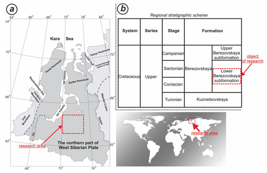
Figure 1.
The research area located within the northern part of Western Siberia (a). Regional stratigraphic scheme for the deposits in the Lower Berezovskaya subformation (b).
The aim of this research is to study their mineral composition, the parameters of the void space structure, and the conditions of formation, as well as to determine the sources of silica and perform a detailed study on the structure of nanoquartz. To study their mineral composition, the parameters of the structure of the void space, and the conditions of formation, special methodological techniques were applied, providing a comprehensive analysis of the results from a wide range of laboratory studies, including optical micro- and stereoscopy, scanning electron microscopy, X-ray diffraction analysis, and microtomography.
The mineral composition of the clay–siliceous rocks in the Lower Berezovskaya subformation is represented by quartz, cristobalite, montmorillonite, and illite. Zeolites (clinoptilolite and heulandite), as well as pyrite, predominate among accessory minerals; potassium feldspar, plagioclases, and anhydrite are less common.
In these rocks, the problem of the source of siliceous material and the mechanism of siliceous mineral formation is quite debatable. It is probable that the source of the siliceous material, which is the main component of rocks, was volcanic and biogenic processes, as well as the introduction of terrigenous material from the continent. The detrital material brought from the continent is present in rocks in extremely small quantities, so it is unlikely that the continental source of silica was the main one. Biogenic silica apparently played a much more significant role. In the rocks, there is a significant amount of the remains of siliceous plankton composed of cristobalite and, less often, opal. However, a larger volume of biogenic silica is concentrated in bacterial formations that transformed dissolved silica into various morphological elements of the rock structure. Bacterial microorganisms transformed the geochemical environment of the diagenesis of the studied deposits. Numerous coccoid forms and trichomes of bacteria are usually mineralized with cristobalite, forming needle-like accretions. There are also numerous bacterial biofilms mineralized with clay minerals, mainly montmorillonite, on the surface of which numerous quartz nanocrystals are noted.
Clay minerals also play a significant role in the mineral composition of the silicites in the Lower Berezovskaya subformation, the content of which varies mainly in the range of 10%–30%, and in some samples up to 40%–45%. They are mainly represented by montmorillonite and, to a lesser extent, illite and chlorite.
In the void space of the studied silicites, autigenic crystals of zeolites are quite often observed, represented mainly by clinoptilolite and heulandite.
According to the results of X-ray diffraction analysis, the quartz content in the rocks under study ranges from 20 to 70%–80%. Of the total amount of quartz, clastic grains account for no more than 5%–7%. It was not possible to diagnose such high quartz contents by optical microscopy methods. However, scanning electron microscopy studies have revealed that the quartz component in the deposits in the Lower Berezovskaya subformation consists of α-quartz, which manifests itself in the form of nanoscale (0.05–0.5 microns) quartz spheres that form clusters on the surfaces of clay flakes.
Quartz nanocrystals were first discovered in the Cretaceous deposits of the Ekofisk deposit [9]. Further studies have shown that nanoquartz is found in various deposits quite often [10,11,12,13,14,15,16]. The proportion of dispersed nanoquartz varies greatly from 10 to more than 80%. The maximum content of nanoquartz in rocks occurs during periods of maximum volcanic activity.
This article describes, in detail, the sources of silica for silicites, the forms of separation of nanoquartz, the patterns of its manifestation along the section, and its association with certain types of void space in the sediments in the Lower Berezovskaya subformation in the north of Western Siberia. The results obtained during the research can be used to determine the regularities of the structure and properties of reservoir rocks in the Lower Berezovskaya subformation in the oil and gas-bearing areas of Western Siberia.
2. Materials and Methods
The work is based on an extensive volume of factual material, including core material for 7 deep drilling wells as well as stock geological and geophysical data. In the work, sedimentological analysis was performed on 829 m of the core; about 770 thinsections were examined with an optical microscope; 305 samples were examined on a scanning electron microscope. Determination of the mineral composition according to X-ray diffractometry was carried out for 305 samples. Three dimensional studies on the structure of the void space were carried out on 23 samples.
The studied samples are evenly distributed over the section and, on average, two-to-three samples were taken from one meter.
The lithological characteristics of rocks were determined using the Axio Imager A2m Carl Zeiss polarizing microscope and the Carl Zeiss Micro Imaging GmbH (Oberkochen, Baden-Württemberg, Germany) stereomicroscope. Transverse (cross-layering) and longitudinal (layering) sandings were generated and, according to the results of stereoscopy, textural components of rocks were identified in them. Painted 50 × 75 mm thin sections were made of synchronous sandings, the size of which practically corresponds to the size of the sanding. This made it possible to synchronize multi-scale studies and give micro-characteristics to macro-textural objects.
The identification of structural and textural features, as well as the distribution of the void space associated with the activity of burrowing organisms, was carried out by computer X-ray tomography using a DeskTop 130 tomograph (RX SOLUTIONS, Chavanod, France).
Detailed studies of the structure of nanoquartz spheres/crystals were performed on a scanning electron microscope JSM 6610 LV (JEOL, Tokyo, Japan), equipped with an energy dispersion spectrometer IE350 (OXFORD INSTRUMENTS, Abingdon, Oxfordshire, England), which was used for point microanalysis of the elemental composition of individual nanocrystals. The manufactured rock chips were glued to the preparation holder with electrically conductive carbon tape before viewing with an electron microscope. Then, to remove the charge formed by the interaction of the electron beam with the sample surface, they were sprayed with a thin conductive platinum coating. The thickness of the platinum coating was 20 nm. The coating was produced in the JFC-1600-JEOL device ((JEOL, Tokyo, Japan). The samples were taken in two modes: the secondary electrons mode (“secondary electron image”, abbreviated “SEI”) and the backscattered electrons mode (“back scattered electrons”, abbreviated “BES”). The observation mode is indicated as a stamp on the pictures. The main shooting mode is the secondary electrons mode. In this mode, the topographic contrast of the sample surface is clearly visible. In this mode, the maximum resolution is reached. In the backscattered (also known as “elastically scattered” in the literature) electrons mode, when studying surfaces with a heterogeneous composition, an additional brightness distribution is superimposed on the topographic image of secondary electrons, which depends on the average atomic number Z of the substance at each micro-stage (the so-called phase composition). This mode was used when it was necessary to show the heterogeneity of the material composition of the surface. The analytical conditions for studying the samples were an accelerating voltage 30 kV, spot size 40–45, and working distance 10–14 mm.
The study of the mineral composition of the samples was carried out using the Rigaku smartLAB X-Ray Diffractometer (Rigaku Corporation, Tokyo, Japan), which is designed for analyzing powders of minerals and rocks. To achieve optimal results, the diffractometric survey was carried out at a rate of 5° per minute in the range from 3° to 70°. The interpretation of the obtained diffractograms is carried out using the Rigaku PDXL2 software Version 2.8.4.0 (Rigaku Corporation, Tokyo, Japan). Using this program, both qualitative and quantitative analysis is carried out. Interpretation is carried out using the ICDD (International Centre for Diffraction Data) and AMCSD (American Mineralogist Crystal Structure Database) databases. The Rietveld method is used to quantify the mineral composition of rocks.
In addition, during the study, surveys were carried out for some samples to identify the mineral composition of the clay component. For this purpose, a special sample preparation was carried out, during which it was possible to produce undirected samples containing a larger amount of the clay component (compared with samples for general mineral analysis). After the diffractometric survey, the samples were saturated with glycerin (to identify minerals of the smectite group in the samples), and then a second survey was carried out. Both diffractometric surveys for the detection of clay minerals were carried out in the range from 3° to 30° at a rate of 5° per minute. Then, the undirected samples were subjected to temperature treatment (at 550 °C), after which another diffractometric survey was carried out, in the range from 3° to 30° at a speed of 5° per minute, to detect kaolinite reflexes.
Petrophysical studies, including the determination of the porosity and permeability of rocks, were performed on a collection of core samples. The samples were cylinders with a diameter of 3 mm. The automated porosity and permeability measurement system “AP-608” manufactured by Coretest Systems USA (Reno, NV, USA) was used for the measurement. The study was carried out using the method of non-stationary gas filtration. The sample was weighed and its length and diameter were determined, after which it was placed in the core holder. The crimping pressure was 3.4 MPa. The gas is nitrogen. Porosity and permeability are measured by the device automatically.
The variety of morphometric characteristics of the voids necessitates the widespread use of methods with different resolutions. Thus, the study of the parameters of voids larger than 0.1 mm and the patterns of their distribution in the rock is most effectively carried out using a stereoscope. Optical microscopy is mainly used to study the distribution of voids in a rock with a size of 0.015–0.1 mm. Scanning electron microscopy makes it possible to study voids from 1 nm to 200 microns. Computed tomography allows us to estimate the parameters of voids up to 5 microns in size and their distribution in the sample. It should be noted that each of these methods works on a limited sample volume. The largest volume can be studied using a stereoscope, where the sample size can reach 15–20 cm.
3. Results
3.1. Lithological Characteristics of the Lower Berezovskaya Subformation
The studied rocks in the Lower Berezovskaya subformation differ in mineral composition, texture, structure, and the composition of rock-forming organisms (Figure 2).
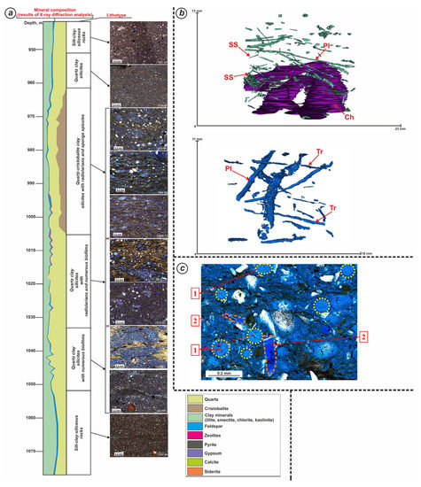
Figure 2.
The graph illustrates the change in the mineral composition (according to the results of X-ray diffraction analysis), the change in the lithotypes of rocks in the section of the Lower Berezovskaya subformation (a). Pyritized Pilichnus isp. (Pl), Chondrites isp. (Ch), Trichichnus isp. (Tr) and sponge spicules (SS). Microphotographic images (b). Fragments of radiolaria (1) and sponge spicules (2) in a petrographic section painted by blue resin (c).
The following lithotypes were identified in the studied sections from the Lower Berezovskaya subformation:
- Silt-clay-siliceous rocks;
- Quartz clay silicites;
- Quartz-cristobalite clay silicites with radiolarians and sponge spicules;
- Quartz clay silicites with radiolarians and numerous biofilms;
- Quartz clay silicites with numerous biofilms.
In silicites, the amount of siliceous matter represented by quartz and cristobalite is more than 50%. The structure of the siliceous substance is a nano-micrograin. It is distributed relatively evenly in the rock. The studied samples also contain a clay component (up to 30% in individual samples). The clay substance has a micro-fine scaled structure. It is represented by montmorillonite and, less often, illite and chlorite. The distribution of the clay component is relatively uniform. It is worth noting that clay minerals are most often mineralized bacterial biofilms.
In silicites, there are skeletons of radiolarian shells, fragments of diatoms, and sponge spicules. The skeletons of radiolarian shells have a rounded and oval shape. Their size is 0.05–0.16 mm. The shells have medium and poor preservation. The mesh structure of the shell skeleton is often visible. They are mineralized with cristobalite and, less often, with clay minerals. Some skeletons of radiolarian shells are mineralized with opal. The spicules of sponges in longitudinal (0.05–0.1 mm in size) and transverse (0.01–0.03 mm in size, rarely up to 0.4 mm) sections have poor preservation. The channels of the sponge spicules are made of cristobalite and, less often, clay minerals and sometimes glauconite.
Single inclusions of siltstone material were found in the studied samples. This material is represented by grains of potassium feldspar and plagioclases.
In addition to the main rock-forming components, pyrite is present in the rock. Pyrite is present in the form of finely dispersed inclusions, which are globules with a size of 0.01–0.1 mm. It replaces radiolarian shells and forms pseudomorphoses along the burrowing organisms (up to 0.85 mm). Its amount is up to 1%. Newly formed zeolite crystals are observed in the voids. Their number varies from 0.5% to 4.2%. There are single grains of glauconite with a rounded, irregular shape and a size of 0.02–0.04 mm.
Silt–clay–siliceous rocks are composed of siliceous and, to a lesser extent, clay material with a slight admixture of detrital material. The siliceous substance of the cryptocrystalline and micrograin structure makes up the majority of the rock—55%–60%. The clay material has a micro-fine scaled structure and a polymineral composition. It is represented mainly by illite and, to a lesser extent, by montmorillonite. The clay component accounts for 35%–40%. The rock also contains a scattered detrital admixture (1%–5%) with a fine aleurite dimension (0.01–0.05 mm) represented by semi-angular, semi-rolled, and angular fragments of plagioclase and potassium feldspar, quartz, and muscovite scales. The distribution of clastics is uniform.
Quartz, cristobalite, opal, and clay minerals are present in the mineral composition of rocks, and zeolites (clinoptilolite and heulandite), pyrite, potassium feldspar, and plagioclases are present as accessory minerals. Gypsum, anhydrite, calcite, and siderite are extremely rare. They are in the form of single crystals.
The textural characteristics of rocks are largely determined by numerous traces of the vital activity of burrowing organisms, among which the ichnofacial complexes Phycosiphon–Pilichnus, Phycosiphon–Nereites (+Paleophycus), and Chondrites–Phycosiphon predominate. Scanning electron microscopy allowed the features of the structure and mineralization of the passages of iloed worms to be revealed. It was found that part of the passages is filled with pyrite globules that have a bacterial nature, as well as cristobalite bacterial spheres. Other types of passages are filled with autigenic grains of glauconite, and detrital grains of quartz and feldspar. In addition, aggregate accumulations of clay minerals are found in the passages of the iloed worms. Some of the traces of burrowing organisms can be made entirely with zeolite crystals. In addition, the walls of burrowing organisms can be made of pyrite, cristobalite, zeolite, anhydrite, and calcite (Figure 3).
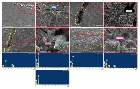
Figure 3.
Partial filling of traces of burrowing organisms (yellow dasehed line) with zeolites, cristobalite, pyrite, and glauconite (SEM images and EDS spectra). Spectra: 1—pyrite; 2—zeolite; 3—cristobalite; 4—zeolite; 5—glauconite. Red squares: enlarged fragments of selected areas of images.
The main rock-forming organisms for the deposits in the Lower Berezovskaya subformation are radiolarians, diatoms, and sponges. The main role in mineral formation was played by various bacterial forms that transformed dissolved silica into various morphological elements in the rock structure. The huge role of bacterial microorganisms consisted of the transformation of the geochemical environment in the diagenesis of the studied deposits.
In the silicites of the Lower Berezovskaya subformation, shaped elements (sponge spicules, fragments of radiolarians, diatoms) are mineralized mainly by cristobalite and, to a lesser, extent quartz, fine-scaled clay mass, and zeolites.
The bulk is composed of bacterial biofilms mineralized with clay minerals, mainly montmorillonite. Quartz nanocrystals with a size of 0.05–0.5 microns are present on the surface of biofilms.
In the section, there is a differentiation in the content of shaped elements and the amount of quartz, cristobalite, zeolites, and montmorillonite (Figure 4). This is due to the differentiation in the sedimentation conditions and the nature of diagenetic changes.
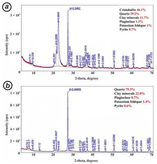
Figure 4.
Diffractogram curves of rock samples from the upper (a) and lower (b) parts of the section. Symbol: d—interplanar spacing, measured in angstroms.
In the upper part of the section, shaped elements, represented by a complex of relics of radiolarians, diatoms, sponges, and bacterial forms, play an essential role in the structure of rocks. It is in bacterial forms that the bulk of cristobalite is concentrated (up to 40%–55%), which is represented in the form of brushes that are present on the walls of trichomes of bacteria, as well as in the form of spherical clusters of coccoid bacteria (Figure 5). The quartz component is represented in rocks by nanocrystals covering the scales of clay minerals. The size of the quartz nanocrystals is no more than 0.5 microns. The identification of the forms of quartz distribution in rocks required their study on a scanning electron microscope with magnifications of 8000–20,000.
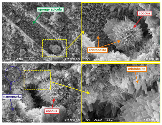
Figure 5.
Coccoid bacteria mineralized by cristobalite in silicates. Microcrystals of cristobalite on the wall of the sponge spicule. Nanoquartz crystals on the surface of clay flakes. Yellow squares: enlarged fragments of selected areas of images.
In the upper part of the section, the porosity varies from 24 to 33%, and the permeability is from 0.01 to 0.16 mD.
The lower part of the section is a cluster of bacterial biofilms containing a significant amount of carbon, which deposited clay minerals on themselves (Figure 6). The quartz content in the lower part of the section is higher (50%–80%). The quartz component of rocks is represented by quartz nanocrystals (0.05–0.5 microns), which grow on clay biofilms (the content of clay minerals is up to 20%–40%). The content of shaped elements is sharply reduced compared to the upper part of the section. Accordingly, the cristobalite component is practically absent here. In general, the content of the clay component increases significantly at the bottom of the section.
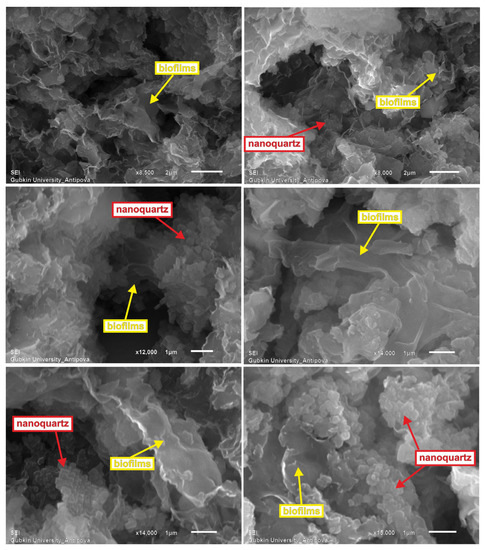
Figure 6.
Bacterial biofilms with quartz nanocrystals on their surface.
In the lower part of the section, the porosity varies in the range of 30%–35%, and the permeability is 0.01–0.5 mD.
The underlying deposits are represented by silt–clay–siliceous rocks.
3.2. Genetic Types of Void Space
The void space of clay–siliceous deposits in the Lower Berezovskaya subformation is a complex and hierarchical system consisting of various genetic types of voids with a wide size range.
Comprehensive studies on the void space made it possible to identify the main genetic types of voids and determine the patterns of their distribution in rocks. Genetic types of voids differ in morphometric parameters, patterns of distribution in the volume of the rock (texture of the void space), and mineralization of the walls of voids.
Based on the integration of different scale research methods, the following genetic types of void space were identified:
- Voids confined to microstylolite seams;
- Voids associated with burrowing organisms’ movements;
- Intraform voids;
- Interform voids;
- Lenticular voids;
- Cellular voids.
Some of these voids are confined to certain lithotypes or their components, and some have a universal character. This type of voids can include voids confined to microstylolite seams. This type of void is quite widespread in the section in various lithotypes. In some samples, approximately 50 of this type of void may occur per 0.5 cm2. They have an elongated shape (0.5–7 mm), and their openness reaches 0.1 mm. The walls of the voids are mineralized with quartz and, in some cases, pyrite or clay minerals.
Voids confined to the burrowing organisms are quite widespread in all lithotypes. The geometry of these voids is largely determined by the species of the certain bioturbator organisms. These voids play a crucial role in creating connectivity channels between different genetic types of voids in the rocks of the Lower Berezovskaya subformation (Figure 7). The individual largest passages are highly porous zones in which voids are concentrated, confined to the intergranular and intercrystalline space. The voidness of this zone can reach up to 50% of the total stroke volume. It should be noted that when studying the burrowing organisms’ passages on an electron microscope, the porous structure of the passage wall is clearly visible. This is a decisive factor for the connectivity of the course with the entire volume of the void space of the rock. The mineralization of the walls of voids confined to the burrowing organisms is extremely diverse. Autigenic crystals of cristobalite, pyrite, zeolites, anhydrite, as well as flakes of clay minerals are noted on the walls of these voids. The size of the zones of increased voidness associated with burrowing organisms’ movements varies greatly. In a cross-section, they vary from tenths of a mm to 1 cm. They can reach several cm in length.
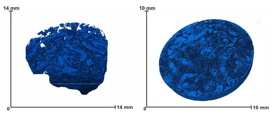
Figure 7.
Microphotographic images of voids confined to the burrowing organisms Phycosiphon isp. The clay cores of the burrows are clearly visible, and the more porous material of the sprites is in their surroundings.
One of the most significant types of voids is the intraform void, which is confined to the relics of rock-forming organisms. A significant part of these voids is associated with the relics of radiolariums, diatoms and spicules. In the sections, voids associated with radiolarian relics are clusters of small pores bounded by the radiolarian skeleton. The size of such sections ranges from 0.05 to 0.18 mm. With the help of SEM, it was possible to estimate the size of these small voids, which are 1–3 microns. A similar size of voids is confined to the relics of diatoms. The size of voids confined to spicules varies across a wide range. In a cross-section, it ranges from 10 microns to several tenths of a millimeter. The longitudinal incision can reach 200–300 microns.
The characteristics of the mineralization of intraform voids are extremely diverse. Numerous spherical accumulations of cristobalite, which has a microbial nature, and crystals of zeolites, pyrite, and clay minerals are noted on the walls of intraform voids (Figure 8).
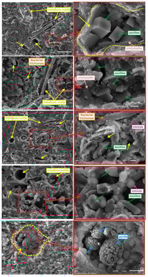
Figure 8.
Cristobalite, zeolites, and pyrite are on the walls of intraform voids. Coccoid bacteria and trichomes of bacteria are on the walls of intraform voids. Red squares: enlarged fragments of selected areas of images.
Often, elongated rod-shaped bacterial formations composed of cristobalite, clay minerals, and zeolites stand out in the inner space of the spicules.
Intraform voids are most common in the upper part of the section.
Interform voids in individual lithotypes can play a significant role. In particular, in one of the sections, a fairly powerful bundle is allocated, in which these voids are the main ones. These voids are confined to the space between coccoid cristobalite bacterial spherical forms (Figure 9). The size of the voids is 1–3 microns. It can be assumed that the connectivity of these voids will be high, which should provide higher permeability values.
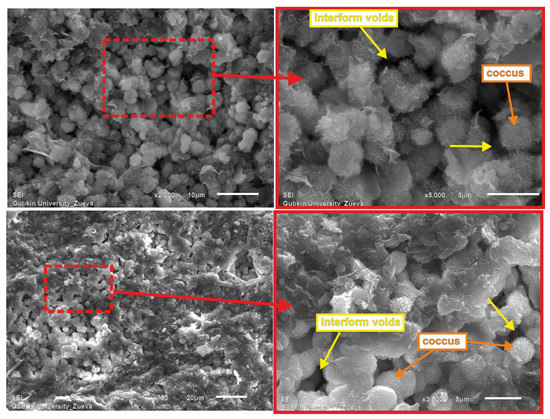
Figure 9.
Interform voids are confined to the space between coccoid cristobalite bacterial spherical forms. Red squares: enlarged fragments of selected areas of images.
The most widespread type of void is the interaggregate (lenticular and cellular) void in the clay mass of the rock. The size of cellular voids, which have mainly isometric outlines, ranges from 1 to 5 microns (Figure 10). Lenticular voids have an elongated shape, a long axis of up to 10 microns, and a short axis ranging from 1 to 5 microns (Figure 11). The walls of voids are often mineralized with quartz nanocrystal; the size of the crystals does not exceed fractions of a micron. In some cases, the interaggregate voids are filled with accumulations of organic matter. In the upper parts of the incision, needle-like crystals of cristobalite sometimes stand out on the walls of the interaggregate voids.
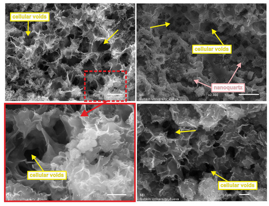
Figure 10.
Uniform distribution of cellular voids with an isometric shape, 1–5 microns in size. Red squares: enlarged fragments of selected areas of images.
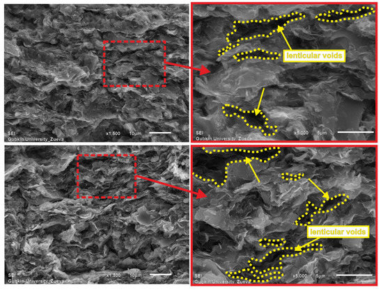
Figure 11.
Lenticular voids with an elongated shape. Their size is up to 10 microns. Red squares: enlarged fragments of selected areas of images.
The distribution of the void space in reservoir rocks is largely determined by their textural characteristics, which are due to the distribution of siliceous, clay components in the volume of the rock and, to a large extent, the type and intensity of bioturbation.
The percentage of different genetic types of voids varies greatly by section. For example, the main porosity of rocks in the upper part of the section is associated with intraform voids, and in the lower part of the section with interaggregate (lenticular and cellular). The voids confined to microstylolite seams and to the burrowing organisms’ passages are distributed relatively evenly across the section.
Due to the fact that the main part of voids in reservoir rocks is from 1 to 5 microns, the permeability value of these rocks does not exceed 1–2 mD on average.
The typification of the void space and the patterns of spatial distribution in the types of voids in the sediments under study should be taken into account when modeling the filtration characteristics of productive formations. The revealed features in the mineralization of the walls of voids should be taken into account when developing methods of impact on the formation.
3.3. Sources of Silica
Numerous studies indicate that there are three different primary sources of silica in marine sedimentary rocks [17,18,19]:
- Biogenic silica, the source of which is siliceous organisms;
- Terrigenous silica, which comes mainly in the form of debris from rivers and Aeolian transport;
- Hydrothermal silica brought by hydrothermal fluids.Secondary quartz can be formed by several mechanisms:
- Dissolution of skeletal grains;
- Pressure dissolution of detrital quartz and possibly other silicate grains;
- Clay transformation.
The transformation of clay minerals is another major source of autigenic quartz. Transformations of smectite into illite and illite into muscovite can result in free silica. In addition, small amounts of silica can be formed as a result of changes in detrital feldspar [20].
Since there are many sources of silica (primary and secondary), quartz from different origins may differ in form and the nature of manifestation.
The question regarding the sources of silica for the silicites in the Lower Berezovskaya subformation has remained unclear and controversial for a long time. Seawater is sharply undersaturated with respect to amorphous silica. However, the Late Cretaceous epoch is associated with the appearance of powerful tropical forests on land with their enormous bio-productivity and biochemical weathering, which could lead to an increase in the intake of dissolved material into the sedimentation basin. Since aluminum oxide and silicon oxide are geochemically related, an increase in the inflow of dissolved silica into the pool can also be assumed.
In addition, ash material and volcanoclastics could also increase the concentrations of dissolved silica in seawater. The bulk of the siliceous material enters the ocean floor after the death of organisms using silica to build their skeletons. Biogenic extraction along with partial dissolution of skeletal material leads to a rapid increase in the concentration of dissolved silica with depth. The main areas of distribution of siliceous silts on the ocean floor correspond to areas of high biological productivity, where nutrient-rich deep waters rise to the warm photic zone. Organisms extract a significant proportion of silica from the ocean, which is many times higher than its intake from all sources, including terrigenous demolition and supply by hydrotherms. Lighter and thinner skeletons dissolve in the upper silica-saturated layers of water masses, in turn, mixing by bottom currents contributes to dissolution in sediments. Active burial by sediments contributes to the preservation of skeletons. Most of the biogenically extracted silica is dissolved again and participates in the cycle. There is no complete analogy of modern biogenic flint sediments with ancient silicites [21,22].
The main silicon-forming organisms of the Upper Cretaceous were radiolarians, which are exclusively marine organisms living in waters with normal salinity and leading a planktonic lifestyle. Radiolarians of the genus Phaseliforma, Prunobrachium, Porodiscus, and Stichocapsa were found in the sediments from the Lower Berezovskaya subformation.
With the appearance of a large number of trace elements in the water masses acting as biostimulators of phytoplankton growth, diatoms actively began their development, which became competitors for radiolarians in the fight for silica. The latter lost, which led to a reduction in species. It was not a catastrophic phenomenon with instant extinction, but a long process.
In the silicites in the Lower Berezovskaya sub-formation, as diagenetic transformations took place, the skeletons of siliceous organisms dissolved and, accordingly, a violation or partial loss of the biomorphic structure is recorded. Bacterial biocenoses played a significant role in the transformation of the skeletons of siliceous organisms and subsequent mineral formation. The transformation of the structure is also accompanied by a change in the material composition due to successive dissolution–precipitation–recrystallization reactions and polymorphic transitions of silica—opal-A to opal-ST (opal–cristobalite–tridymite) [23]. Thus, the source of silica for cristobalite crystals was probably biogenic opal, which was observed in a significant amount in the upper part of the section enriched with siliceous plankton.
Numerous coccoid forms and trichomes of bacteria, as a rule, are mineralized with cristobalite, forming needle-like accretions.
Another primary source of silica for the studied deposits in the Lower Berezovskaya sub-formation was ash material entering the basin from volcanic formations located within the folded eastern border of Western Siberia. This is confirmed by the revealed mineral paragenesis of montmorillonite, cristobalite, and zeolites.
The proportion of detrital quartz in rocks is low and is no more than 5%–7%.
Thus, the intake of silica into the sedimentary basin was controlled by biogenic, volcanogenic, and terrigenous factors (Figure 12).
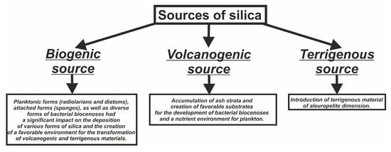
Figure 12.
Lenticular voids with an elongated shape. Their size is up to 10 microns.
3.4. Forms of Nanoquartz and Patterns of Distribution over the Section
When studying rocks with a scanning electron microscope at magnifications from 8000 to 20,000 times, clusters of nanoquartz in the form of spherical particles with a diameter of approximately 0.1–0.5 microns on the surface of clay flakes were detected (Figure 13).
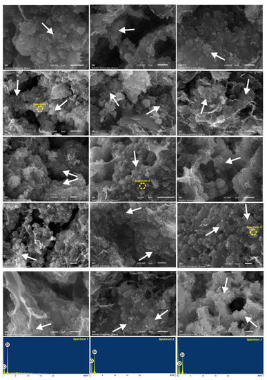
Figure 13.
Nanoquartz crystals (marked with white arrows) are on the surface of montmorillonite flakes (SEM images and EDS spectra).
It should be noted that mainly clusters of nanoquartz were observed on the scales of montmorillonite. The illite particles remain practically free from quartz crusts. Quartz crusts often completely line cellular voids in the clay mass. This is a factor that prevents them from deforming at the stage of late diagenesis.
In general, the section from the Lower Berezovskaya subformation has a thickness of about 60 m and is divided into two parts according to the mineral composition of the rocks. In the upper part of the section, the quartz content ranges from 20 to 40%–45%, and in the lower part, from 50 to 80%. In addition, the content of cristobalite in the upper part of the section is much higher; 55%–80%. The amount of clay component in the section varies little and averages 10%–30%, and in some samples up to 40%. The distribution of shaped elements is also quite indicative. The greatest variety and number of planktonic forms and coccoid bacteria is observed in the upper part of the section. This distribution suggests that different sources of silica dominated the section at different times.
4. Discussion
The formation of sediments in the Lower Berezovskaya subformation occurred in the in the central part of the West Siberian shallow sea basin [24,25,26].
The sources of siliceous material, which is the main component of rocks, were volcanic and biogenic processes, as well as the introduction of terrigenous material from the continent.
The revealed mineral paragenesis of montmorillonite, cristobalite, and zeolites indicates an active transformation of ash material that entered the basin from volcanic formations located in the east of the study area. The transformation of ash into montmorillonite was largely due to the activity of bacterial biocenoses, traces of which are abundantly present in rocks in the Lower Berezovskaya subformation. Apparently, the most active introduction of ash material occurred at the early stages of the formation of deposits in the Lower Berezovskaya subformation, where, in general, there is a slight increase in the number of smectites. In addition, a decrease in biogenic components in the lower part of the section indicates unfavorable conditions for the development of biocenoses.
The analysis of ichofacies showed that during the deposition of the Lower Berezovskaya subformation, disoxic conditions periodically arose. This correlates with the well-known anoxic events of the Late Cretaceous [27].
The differentiation of the lithological characteristics in the lower part of the section was largely determined by the spreading of ash material by underwater currents and its accumulation in certain elements of the seabed relief.
During the formation of the lower part of the section, there was an intensive accumulation of ash material, which was subsequently transformed into montmorillonite with the active participation of bacterial paleocenoses.
During the period of accumulation of deposits in the upper part of the section, as volcanic activity decreased and clay material was introduced from the land, the development of biocenoses formed by bacterial complexes and planktonic forms (radiolaria, diatoms), as well as sponges, began to intensify. This complicated the rock structure and increased the diversity of the void space.
A distinctive feature of the siliceous deposits in the Lower Berezovskaya subformation is the active development of coccoid forms of bacteria. They, feeding on the organic matter of the sediment, created cristobalite mineral formations in the form of balls and crusts. This process is most actively observed in the middle and upper parts of the Lower Berezovskaya subformation. The processes associated with the activity of bacterial biocenoses significantly influenced the structural and textural characteristics of the studied deposits. In the upper part of the section of the deposits in the Lower Berezovskaya subformation, the introduction of clay material from the east apparently intensified. This gradually increased the content of the clay component in the rocks.
In the sections of the deposits in the Lower Berezovskaya subformation, there is a pronounced tendency of zeolization of the void space. Autigenic crystals of zeolites, mainly represented by clinoptilolite and heulandite, are quite often observed in the void space of the studied silicites along almost the entire section. Based on the mineral paragenesis that compose the deposits in the Lower Berezovskaya subformation, zeolites (clinoptilolite) were probably formed to a greater extent due to the diagenetic transformation of biogenic silica, colloids of aluminosilicates, and crystallites of clay minerals. Reservoir rocks in the Lower Berezovskaya subformation contain clinoptilolite in an amount from the 1% to 15% in some interlayers.
It is worth noting that pyroclastic material is present in the sediments in a small amount, which could also contribute to the formation of clinoptilolite due to the diagenetic transformation of volcanic glass under the action of seawater and alkaline pore solutions [28]. Once in an aqueous environment, volcanic glass hydrates and dissolves. If the pH of the water is less than 7.5–8.0, then volcanic glass is converted into clay minerals. Further dissolution of the glass quickly leads to an increase in the pH of the pore solution to 9.0 and higher. As a result, the formation of clay minerals stops, and zeolites are formed. Therefore, for the process of converting volcanic glass into zeolites, its equilibrium with the solution in a closed or semi-closed system in the sediment is necessary with a very weak circulation of pore solutions with a geochemical barrier of a pH value greater than 8.0–8.5. With the constant renewal of the liquid phase in conditions of intensive pore water circulation, the formation of zeolites does not occur, but clay minerals usually appear instead [29].
The source of the siliceous substance for nanoquartz crystals was most likely volcanic processes. The revealed mineral paragenesis of montmorillonite, cristobalite, and zeolites indicates an active transformation of ash material that entered the basin from volcanic formations located within the folded eastern border of Western Siberia.
The formation of α-quartz nanocrystals most likely occurred according to the following scheme. A slight increase in the Si concentration in the medium, associated with frequent volcanic eruptions, led to the crystallization of nanoquartz spheres. Nanoquartz floccules were formed in suspensions with sufficient ionic strength, such as seawater. They were small, loose, flake-like clusters in a suspended state. Nanoquartz spheres have colloidal properties. This determines their specific form of allocation. After crystallization, colloidal quartz spheres were flocculated and deposited on the seabed composed of clay silt. A similar mechanism of nanoquartz formation was considered in [30].
An indirect confirmation of the significant influence of volcanism on sedimentation processes in the Late Cretaceous basin of the north of Western Siberia is the complete absence of coccolith remains in contrast to the Late Cretaceous basin of the Pre-Caucasus. This is probably due to a decrease in pH, which can lead to partial or complete dissolution of coccoliths in the water column. Lowering the pH requires a significant amount of acidifying agent atmospheric CO2. It reduces calcification of marine plankton when mixed with seawater [31,32,33,34,35]. CO2 released in large quantities during volcanic eruptions may be the cause of the dissolution of coccoliths in the Cretaceous deposits of Western Siberia.
5. Conclusions
The deposits in the Lower Berezovskaya subformation are a complex reservoir with unconventional reservoir rocks. The void space in the reservoir rocks of the Lower Berezovskaya subformation is represented by voids of various geneses and sizes, including voids confined to burrowing organisms, intraform voids, interform voids, lenticular voids, cellular voids, and voids confined to microstylolite seams.
The walls of voids confined to microstylolite seams are mineralized with quartz and, in some cases, pyrite or clay substance. The mineralization of the walls of voids confined to the burrowing organisms is extremely diverse. Autigenic crystals of cristobalite, pyrite, zeolites, anhydrite, as well as flakes of clay minerals are noted on the walls of these voids. Numerous spherical accumulations of cristobalite, which have a microbial nature, and crystals of zeolites, pyrite, and clay minerals are noted on the walls of intraform voids. The most widespread type of void is the interaggregate (lenticular and cellular) void, which is formed by clay scales, on the surface of which numerous crystals of α-quartz are observed, manifested in the form of nanoscale (0.05–0.5 microns) quartz spheres. The content of such quartz reaches up to 80% of the total volume of the rock.
The intake of silica during the formation of the deposits in the Lower Berezovskaya subformation was controlled by complex of factors, including biogenic, volcanogenic, terrigenous, and biohemogenic factors.
The biogenic factor was determined by the intensity of the development of planktonic forms (radiolaria and diatoms), attached forms (sponges), as well as diverse forms of bacterial biocenoses. Various kinds of siliceous organisms had a significant impact on the deposition of various forms of silica and on the creation of a favorable environment for the transformation of volcanogenic and terrigenous materials.
The volcanogenic factor exerted a powerful influence, consisting of the accumulation of ash strata and the creation of favorable substrates for the development of bacterial biocenoses and nutrient environments for plankton. In addition, a significant amount of ash material in general could negatively affect the intensity of the development of biocenoses.
In addition, one should not discount the introduction of terrigenous material in the siltstone dimension, which was probably controlled by underwater currents that distributed this material in accordance with the fault–block structure of the seabed.
The source of silica for the formation of quartz nanocrystals on motmorillonite flakes was the products of volcanic eruptions. The source of silica for cristobalite crystals was probably biogenic opal, which was observed in a significant amount in the upper part of the section enriched with siliceous plankton.
Author Contributions
Conceptualization, O.A.Z. and O.V.P.; methodology, A.V.P.; formal analysis, O.A.Z.; investigation, O.A.Z.; resources, O.V.P.; data curation, O.A.Z.; writing—original draft preparation, O.A.Z.; writing—review and editing, O.A.Z.; visualization, O.A.Z.; supervision, O.V.P.; project administration, O.V.P. All authors have read and agreed to the published version of the manuscript.
Funding
This research received no external funding.
Data Availability Statement
The data presented in this study are openly available in article at reference number [4,24].
Conflicts of Interest
The authors declare no conflict of interest. The funders had no role in the design of the study; in the collection, analyses, or interpretation of data; in the writing of the manuscript, or in the decision to publish the results.
References
- Doroshenko, A.A.; Karymova, Y.O. Characteristics of the void space of the flask senon deposits of the north of Western Siberia. Expo. Oil Gas 2017, 6, 23–27. [Google Scholar]
- Rodivilov, D.B.; Kosarev, P.N.; Mamyashev, V.G. Non-traditional reservoir of the Lower Berezovskaya subformaction and its searching criteria. Oil Gas 2018, 3, 37–43. [Google Scholar] [CrossRef][Green Version]
- Karymova, Y.O. Litho-capacitive model of the void space of nano reservoir rocks of the Lower Berezovskaya subformaction of the north of Western Siberia. Expo. Oil Gas 2018, 3, 20–24. [Google Scholar]
- Rybyakov, A.N.; Nersesov, S.V.; Sokolovsky, R.A.; Postnikov, A.V.; Postnikova, O.V.; Zueva, O.A.; Kuznetsov, A.S.; Doroshenko, A.A.; Doroshenko, A.A.; Karymova, Y.O.; et al. The genesis of silicites and void space reservoir rocks of coniacian-santonian gas-bear deposits. Gas Ind. 2020, 8, 54–62. [Google Scholar]
- Cherepanov, V.V.; Menshikov, S.N.; Varyagov, S.A.; Gladkov, D.Y.; Bondarev, V.L.; Peacemaking, M.Y.; Klokov, V.P. Problems of assessing the oil and gas potential of deposits of the Lower Berezovskaya subformaction of the north of Western Siberia Geology. Geophys. Dev. Oil Gas Fields 2015, 2, 11–26. [Google Scholar]
- Novoselova, M.Y.; Agalakov, S.E. Gas content of the Nadsenomanian deposits of Western Siberia. News Univ. Oil Gas 2019, 4, 10–23. [Google Scholar]
- Rodivilov, D.B.; Nersesov, S.V.; Nezhdanov, A.A.; Ogibenin, V.V. Prospects for the development of deposits containing gas hydrates of the Medvezhye deposit (Western Siberia). Gas Ind. 2019, 8, 48–55. [Google Scholar]
- Perezhogin, A.S.; Nezhdanov, A.A.; Smirnov, A.S. Prospects for the development of the cenomanian oil and gas complex in the north of Western Siberia. Exhib. Oil Gas 2016, 6, 42–45. [Google Scholar]
- Lindgreen, H.; Jakobsen, F.; Springer, N. Nano-size quartz accumulation in reservoir chalk, Ekofisk Formation, South Arne Field, North Sea. Clay Miner. 2010, 45, 171–182. [Google Scholar] [CrossRef]
- Calvert, S.E. Deposition and diagenesis of silica in marine sediments. Int. Assoc. Sedimentol. Spec. Publ. 1974, 1, 273–300. [Google Scholar]
- Clayton, C.J. The chemical environment of flint formation in Upper Cretaceous chalk. In The Scientific Study of Flint and Chert; Cambridge University Press: Cambridge, UK, 1986; pp. 43–54. [Google Scholar]
- Jakobsen, F.; Lindgreen, H.; Springer, N. Precipitation and flocculation of spherical nano silica in North Sea chalk. Clay Miner. 2000, 35, 175–184. [Google Scholar] [CrossRef]
- Lindgreen, H.; Drits, V.A.; Salyn, A.L.; Jakobsen, F.; Springer, N. Formation of flint horizons in North Sea chalk through marine sedimentation of nano-quartz. Clay Miner. 2011, 46, 525–537. [Google Scholar] [CrossRef]
- Zijlstra, H.J.P. Early diagenetic silica precipitation, in relation to redox boundaries and bacterial metabolism, in Late Cretaceous chalk of the Maastrichtian type locality. Geol. Mijnb. 1987, 66, 343–355. [Google Scholar]
- Jeans, C.V. Silifications and associated clay assemblages in the Cretaceous marine sediments of Southern England. Clay Miner. 1978, 13, 101–126. [Google Scholar] [CrossRef]
- Littke, R.; Fourtanier, E.; Thurow, J.; Taylor, E. Silica diagenesis and its effects on lithification of Broken ridge deposits, Central Indian Ocean. Proc. Ocean Drill. Program Sci. Results 1991, 121, 121–261. [Google Scholar]
- Beauchamp, B.; Baud, A. Growth and demise of Permian biogenic chert along northwest Pangea: Evidence for end-Permian collapse of thermohaline circulation. Palaeogeogr. Palaeoclimatol. Palaeoecol. 2002, 184, 37–63. [Google Scholar] [CrossRef]
- Lei, Z.; Dashtgard, S.E.; Wang, J.; Li, M.; Feng, Q.; Yu, Q.; Zhao, A.; Du, L. Origin of chert in Lower Silurian Longmaxi Formation: Implications for tectonic evolution of Yangtze Block, South China. Palaeogeogr. Palaeoclimatol. Palaeoecol. 2019, 529, 53–66. [Google Scholar] [CrossRef]
- Frolov, V.T. Lithology, 1st ed.; MSU Press: Moscow, Russia, 1992; pp. 162–171. [Google Scholar]
- Metwally, Y.M.; Chesnokov, E.M. Clay mineral transformation as a major source for authigenic quartz in thermo-mature gas shale. Appl. Clay Sci. 2012, 55, 138–150. [Google Scholar] [CrossRef]
- Yapaskurt, O.V. Lithology, 1st ed.; Publishing House of the Academy: Moscow, Russia, 2008; pp. 217–225. [Google Scholar]
- Agarkov, Y.V. Analysis of the dynamics of the disappearance and appearance of new species of radiolarians in the Cretaceous and Paleogene periods. Sci. J. KubSAU 2017, 132, 1–12. [Google Scholar]
- Smirnov, P.V. Phase transitions of silica in opal-cristobalite rocks as a quality factor of siliceous raw materials. Bull. Tomsk Polytech. Univ. Geo Assets Eng. 2017, 328, 6–15. [Google Scholar]
- Postnikov, A.V.; Postnikova, O.V.; Zueva, O.A.; Kulagina, N.K.; Kitaeva, I.A.; Loshkareva, V.A. Siliceous reservoir rocks of Late Cretaceous deposits of the Nadym-Pur-Taz region. Pustovalov Read. 2022, 110–112. [Google Scholar]
- Vyssotski, A.V.; Vyssotski, V.N.; Nezhdanov, A.A. Evolution of the West Siberian basin. Mar. Petrol. Geol. 2006, 23, 93–126. [Google Scholar] [CrossRef]
- Golbert, A.V.; Markova, L.G.; Polyakova, I.D.; Saks, V.N.; Teslenko, Y.V. West Siberian Paleolandscapes in the Jurassic, Cretaceous, and Paleogene; Nauka: Moscow, Russia, 1968; p. 152. [Google Scholar]
- Konyukhov, A.I. Oceanic anoxic events of the Cretaceous period and their role in the formation of source rocks in the basins of continental margins. Georesources 2017, 19, 43–55. [Google Scholar] [CrossRef]
- Khardikov, A.E.; Boyko, N.I.; Agarkov, Y.V. Zeolites of the Eastern Ciscaucasia and prospects for their practical use. News of higher educational institutions. Geol. Explor. 1992, 6, 86–91. [Google Scholar]
- Konyukhova, T.P.; Distanov, U.G. Technological classification of mixed siliceous zeolite-containing rocks. Explor. Prot. Miner. Resour. 2000, 9, 45–46. [Google Scholar]
- Jakobsen, F.C.; Lindgreen, H. Nano-quartz in North Sea Danian chalk. Geol. Surv. Den. Greenl. 2012, 26, 9–12. [Google Scholar]
- Riebesell, U.; Zondervan, I.; Rost, B.; Tortell, P.D.; Zeebe, R.E.; Morel, F.M. Reduced calcification of marine plankton in response to increase atmospheric CO2. Nature 2000, 407, 364–367. [Google Scholar] [CrossRef]
- Feely, R.A.; Sabine, C.L.; Lee, K.; Berelson, W.; Kleypas, J.; Fabry, V.J.; Millero, F.J. Impact of anthropogenic CO2 on the CaCO3 systemin the oceans. Science 2004, 305, 362–366. [Google Scholar] [CrossRef]
- Holmes, A. Principles of Physical Geology, 2nd ed.; Thomson Learning: Southbank, Australia, 1965; 1288p. [Google Scholar]
- Frondini, F.; Chiodini, G.; Caliro, S.; Cardellini, C.; Granieri, D.; Ventura, G. Diffuse CO2 degassing at Vesuvio, Italy. Bull. Volcanol. 2004, 66, 642–651. [Google Scholar] [CrossRef]
- Schuiling, R.D. Thermal effects of massive CO2 emissions associated with subduction volcanism. C. R. Geosci. 2004, 336, 1053–1059. [Google Scholar] [CrossRef]


Disclaimer/Publisher’s Note: The statements, opinions and data contained in all publications are solely those of the individual author(s) and contributor(s) and not of MDPI and/or the editor(s). MDPI and/or the editor(s) disclaim responsibility for any injury to people or property resulting from any ideas, methods, instructions or products referred to in the content. |
© 2023 by the authors. Licensee MDPI, Basel, Switzerland. This article is an open access article distributed under the terms and conditions of the Creative Commons Attribution (CC BY) license (https://creativecommons.org/licenses/by/4.0/).