1. Introduction
Scheelite is a calcium-containing tungstate with an ideal chemical component of CaWO
4 belonging to a tetragonal crystal system, with a space group of C
6 4h-I4
1/a. The crystal structures contain irregular W–O tetrahedrons that share edges with the Ca–O octahedrons [
1]. The calcium in the scheelite structure is usually replaced by various rare earth elements (REEs), and the tungsten is usually replaced by molybdenum [
2,
3]. Scheelite and scheelite-type crystal materials have important applications in many fields, such as nano-scale scheelite crystals prepared by doping with different REEs can be used as nuclear radiation detection materials due to their strong ability to block high-energy particles and rays; in addition, scheelite-type molybdate thin films can be used as photoelectric functional materials, etc. [
4,
5,
6,
7,
8,
9,
10,
11,
12]. Deposits of scheelite are widely distributed around the world. They can be found in Nevada, California, and New Mexico in America; Kalgoorlie-Norseman in Australia; Nui Phao in northeastern Vietnam; Seoul in South Korea; Colombage-Ara in Sri Lanka; and so on [
13,
14,
15,
16]. In addition, scheelite is produced in Nanni Lake in Henan Province; Persimmon Bamboo Garden in Hunan Province; Nanyang Tian in Yunnan Province; Chahansen in Qinghai Province; Xuebaoding in Sichuan Province; and other regions in China [
17,
18,
19].
The Xuebaoding mineral deposit in Pingwu County, Mianyang City, Sichuan Province, China, is a high-temperature, hydrothermal, greisen vein-type, giant crystal gem-quality deposit with high alkali metal content. Scheelite produced in the Xuebaoding deposit has large grain sizes, perfect crystal shape, a rare color (yellow-orange hue), and is accompanied by tourmaline, muscovite, beryl, feldspar, quartz, cassiterite, etc. According to previous studies, the color and brightness of scheelite usually depend on the composition and content of the REEs inside and have no obvious relationship with the major chemical composition and crystal structure of scheelite [
2]. There is a diversity of REEs in Xuebaoding scheelite, in which their contents are in abundance [
3]. There is little research into the origin of the color of Xuebaoding scheelite; hence, this article takes five faceted scheelite samples from the Xuebaoding deposit as the research objects and discusses their color genesis through Fourier infrared spectroscopy, micro-Raman spectroscopy, ultraviolet-visible spectroscopy, fluorescence spectroscopy, and laser ablation inductively coupled plasma mass spectrometry.
2. Geology
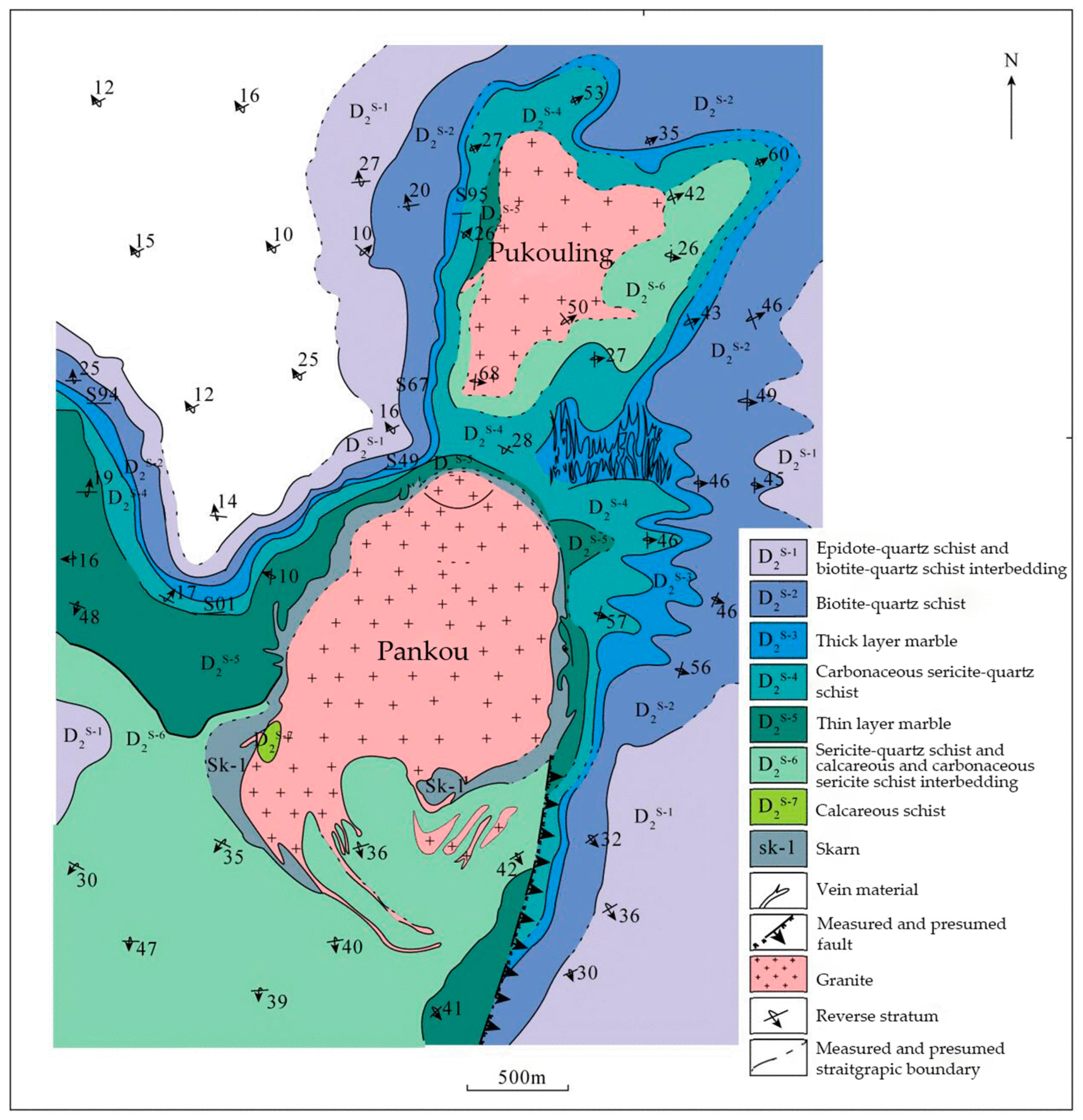
The Xuebaoding deposit is located 14.5 km northwest of Huya Town, Pingwu County, Mianyang City, Sichuan Province. In the 1990s, a plentiful supply of beryl, scheelite, cassiterite, apatite, fluorite, muscovite, and other coarse-grained crystals were discovered in the Xuebaoding deposit. The deposit is located between the Pankou and Pukouling granite bodies. In addition, the gem-grade scheelite is from the Xuebaoding W-Sn-Be deposit, located in the Songpan–Ganzi orogenic belt in southwest China and the secondary structure of the Moziping–Shangnami syncline core in the southeast complex structural belt of the Motianling Zibaishan dome structure. The granite group is obviously controlled by the core of the dome structure and intrudes into the Triassic stratum. The single rock mass is relatively small (the largest Pankou rock mass is about 550 m wide from east to west and 600 m long from south to north) [
20,
21,
22]. The ore veins mainly developed between the Pankou and Pukouling granite bodies, and there is marble around them (
Figure 1).
The ore vein includes the core, mainly composed of quartz, and the edge, mainly composed of coarse-grained beryl, cassiterite, scheelite, feldspar, albite, muscovite, fluorite, and apatite. According to the spatial distribution, wall rock characteristics, and mineral associations, the vein can be roughly divided into three types: (I) the main minerals are mica and beryl, and the wall rock is granite; (II) the main minerals are mica, beryl, and calcite, whereas the wall rock is transitional, from granite to marble; (III) the main minerals are mica, beryl, calcite, scheelite, cassiterite, fluorite, apatite, needle-like tourmaline, and a small amount of quartz, and the wall rock is marble; And type (III) is the main metallogenic type found in the Xuebaoding deposit [
20,
21,
22].
3. Market Value
The scheelite produced in the crystal cavity of the W-Sn-Be ore vein in Pingwu is famous for its large crystals, color, strong luster, high transparency, and rich combinations, and it is one of the best-known species in the mineral world. The rare biconical orange scheelite can co-exist or be associated with light blue hexagonal tabular aquamarine, black and shiny cassiterite, large-crystal white feldspar, colorless or opalescent quartz, scale muscovite, and mushistonite covering kesterite. The mineral crystal cluster formed by these combinations displays bright colors, strong contrasts, and beautiful shapes. It has high ornamental and scientific research value and is loved by collectors all over the world. In addition to the scheelite crystals, there is an increasing amount of faceted scheelite on the market, which is sought after by consumers and mineral collectors.
4. Materials and Methods
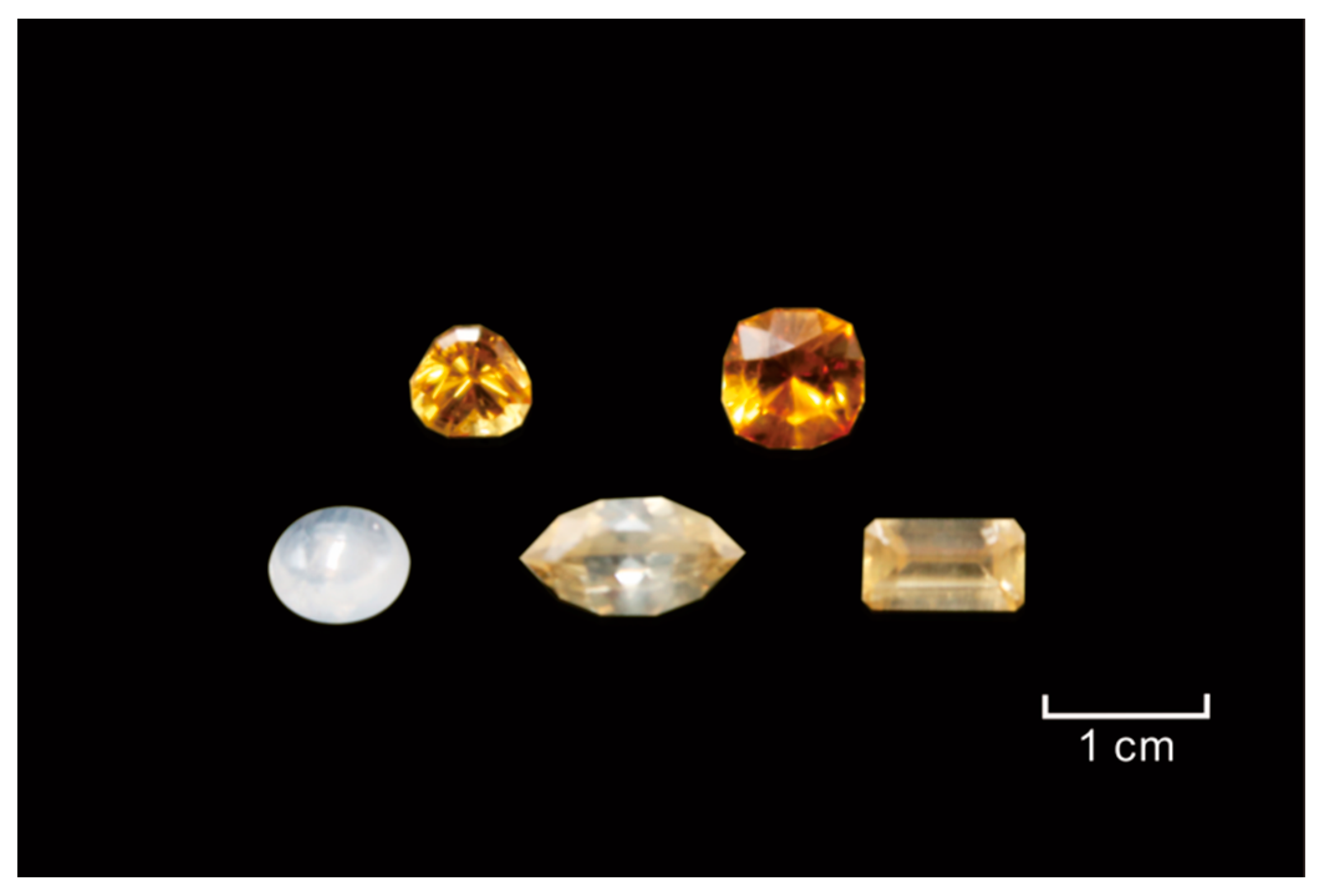
The samples to be tested were collected by the authors in the Xuebaoding mining area, and all samples were processed—4 facets and 1 cabochon (
Figure 2). The sample numbers are BWK1–5, in order. The five samples were divided into three groups according to color: (I) colorless scheelite sample—BWK1; (II) pale-yellow scheelite samples—BWK2-3; (III) orange scheelite samples—BWK4-5. The sample BWK2 features parallel color bands (
Figure 3), which plays an important role in studying the color origin of Xuebaoding scheelite.
All the experiments recorded in this article were carried out in the laboratory at the School of Jewelry, Shanghai Jianqiao University (Shanghai, China), with the exception of the LA-ICP-MS test.
FTIR spectra were acquired from 1500 to 400 cm−1 using a Bruker Tensor-27 Fourier transform infrared spectrometer (Billerica, MA, USA) in reflection mode with the resolution set at 6 cm−1.
UV-Vis-NIR spectra were recorded on a GEM-3000 spectrophotometer (made by Guangzhou Biaoqi Optoelectronic Technology Development Co., Ltd., Guangzhou, China) in the range of 200 to 1100 nm, with a 26 ms integral time, an average number of 100 times, and a smooth width of 6.
Raman spectra were recorded on a Renishaw inVia laser confocal Raman spectrometer in the range of 200 to 1000 cm−1 with an excitation laser wavelength 532 nm, grating 1200 l/mm, laser power 0.05%, micro-aberration objective lens 50×, spectral resolution 1 cm−1, and exposure time 10 s.
Three dimensional fluorescence spectra were recorded on a PerkinElmer fl6500 fluorescence spectrometer (Waltham, MA, USA). The test conditions were prepared as follows: an excitation wavelength of 230 to 290 nm, an emission wavelength of 400 to 650 nm, an excitation slit width of 20 nm, an emission slit width of 2.5 nm, scanning mode with a scanning speed of 1200 nm/min and a photomultiplier voltage of 250 V.
In rare earth elements, measurements were performed using an Aglient 7900ICP-MS (Santa Clara, CA, USA) fitted with an ESL NWR193UC laser ablation system, housed at Shanghai Chemlab Instrument Co., Ltd. (Shanghai, China) In mapping analysis, we use a laser repetition rate of 20 Hz at 3 J/cm2 and beam diameters of ~35 µm. In micro-area analysis, we use a laser repetition rate of 15 Hz at 4 J/cm2 and beam diameters of ~50 µm.
5. Results and Discussion
5.1. Spectroscopic Methods
The [WO
4]
2− pentatom tetrahedron in the scheelite structure is a nonlinear complex anion group. Under ideal conditions, its vibration degrees of freedom are 3 × 5 − 6 = 9. There are four vibrational forms as a result of degeneracy—the ν
1 symmetric stretching vibration, the ν
2 out-of-plane bending vibration, the ν
3 asymmetric stretching vibration, and the ν
4 in-plane bending vibration. In the crystal structure of scheelite, the structure along the L
4 direction (C axis) is squashed and distorted (
Figure 4).
5.1.1. Infrared
Figure 5 and
Table 1 show the absorption peaks of the Xuebaoding scheelite samples in the fingerprint region (1500–400 cm
−1). The results indicate that the absorption range from 900 to 800 cm
−1 is attributed to the ν
3 asymmetric stretching vibration (806, 817, 856, 867 cm
−1) of the [WO
4]
2− tetrahedral group, and the absorption peak at 440 cm
−1 is attributed to the ν
2 out-of-plane bending vibration of the [WO
4]
2− tetrahedral group.
5.1.2. Raman
Figure 6 and
Table 2 show the detailed Raman spectra of the scheelite samples. The weak Raman shift near 211 cm
−1 is caused by the translation mode of (Ca–O) in the crystal structure. The Raman shifts at 332 cm
−1 and near 400 cm
−1, which correspond to the A
g and B
g Raman vibration modes, respectively, are attributed to the ν
2 out-of-plane bending vibration of [WO
4]
2−. As the color deepens, the Raman shift near 400 cm
−1 shifts gradually from 400 to 394 cm
−1, which may be due to the change in the bond length of the tungsten–oxygen tetrahedron as the chromogenic-related REEs are more involved in the lattice. The main peak located at 911 cm
−1 is assigned to the ν
1 symmetric stretching vibration of [WO
4]
2− (A
g Raman vibration mode); the peak located at 797 cm
−1 is assigned to the ν
3 asymmetric stretching vibration of [WO
4]
2− (B
g Raman vibration mode) [
2].
5.1.3. UV-Vis-NIR
The representative UV-Vis-NIR spectra from the scheelite samples with favorable transparency and clarity are shown in
Figure 7.
The colorless sample BWK1 only presented an absorption peak at 317 nm. The colored samples BWK2–5 showed many peaks and complex characteristics in the spectra: the absorption peak intensities of the orange-tone samples BWK4 and BWK5 at 584, 588, 682, 743, 750, 803, and 874 nm were significantly higher than those of the pale-yellow-tone samples BWK2 and BWK3; it is a preliminary inference that these absorption peaks may be related to the origin of the yellow-orange hue of scheelite.
The narrow peaks around 584 and 803 nm indicate the presence of the rare earth element Nd [
29]. Through comparisons and combinations with previous studies, the absorption spectra of scheelite are attributed to the existence of “didymium”, a mixture of the rare earth elements Pr and Nd. Scheelite from other places also has similar spectral characteristics [
30].
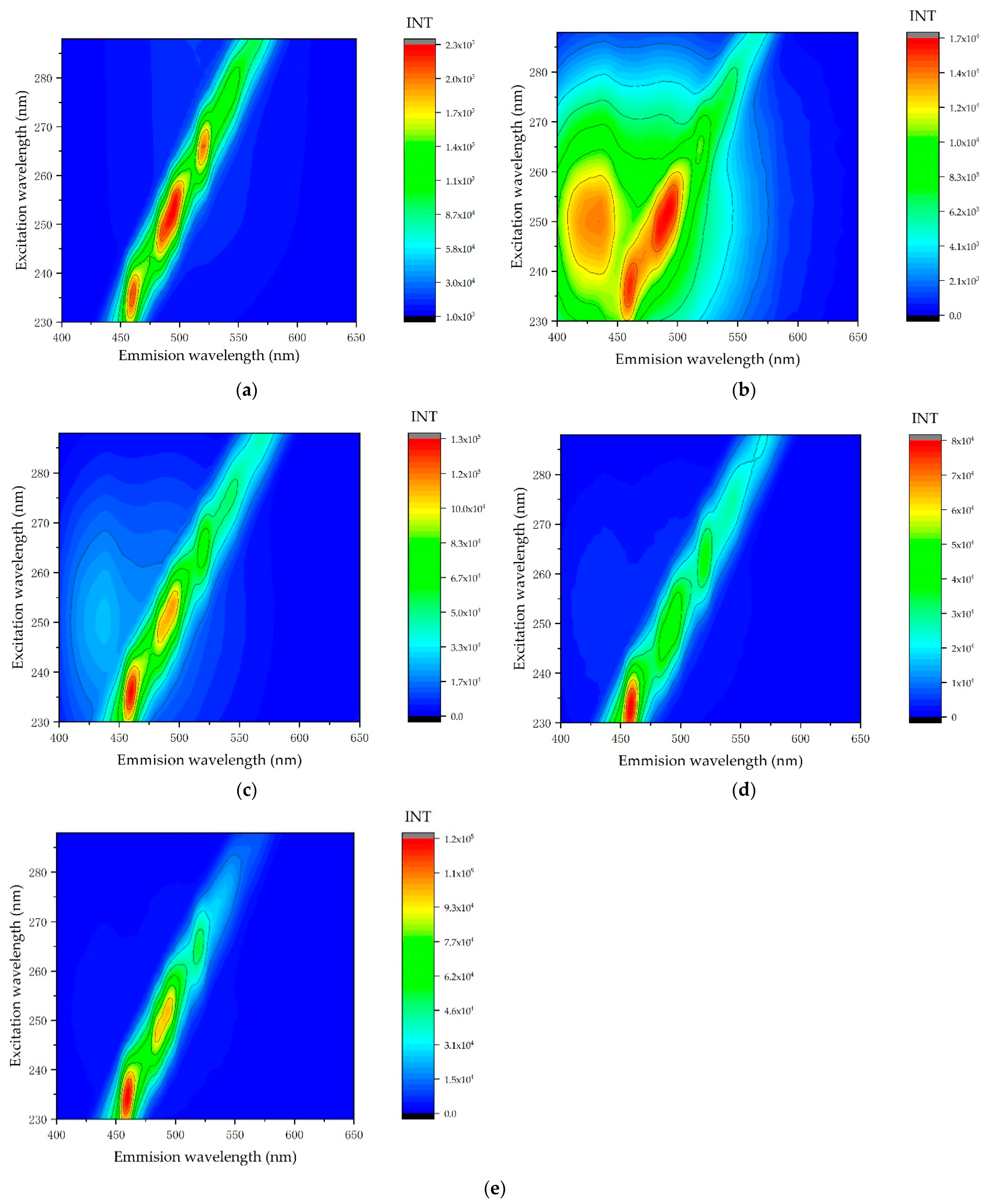
5.1.4. Three-Dimensional Fluorescence
The three-dimensional fluorescence spectra show (
Figure 8) that the number and position of the main fluorescence peaks of the colorless sample BWK1 and the orange scheelite samples BWK4 and BWK5 are similar, and they are all located around λ
ex235 nm/λ
em455 nm, λ
ex250 nm/λ
em490 nm, and λ
ex265 nm/λ
em523 nm. In addition to the above-mentioned main fluorescence peaks, the pale yellow scheelite samples BWK2 and BWK3 also display fluorescence peaks around λ
ex250 nm/λ
em425 nm. These differences may be related to the varying content and types of REEs in the scheelite structure. The isomorphic substitution of Ca
2+ by REEs in the scheelite crystal structure can easily form a luminescent center, but the emission of other intrinsic [WO
4]
2− and the isomorphically doped [MnO
4]
2− is broad and strong, which masks the excitation of REEs in the crystal structure, and this makes the fluorescence spectra of REEs such as Tm, Er, and Ho difficult to observe [
31].
5.1.5. LA-ICP-MS
The LA-ICP-MS test results (
Table 3) show that the concentrations of REEs are extremely low in colorless scheelite sample BWK1; relatively high in the pale yellow scheelite sample BWK3; and high in the orange scheelite samples BWK4–5. Based on these results, it is preliminarily speculated that the concentrations of these fifteen REEs in the Xuebaoding scheelite samples positively correlate with the concentration of color.
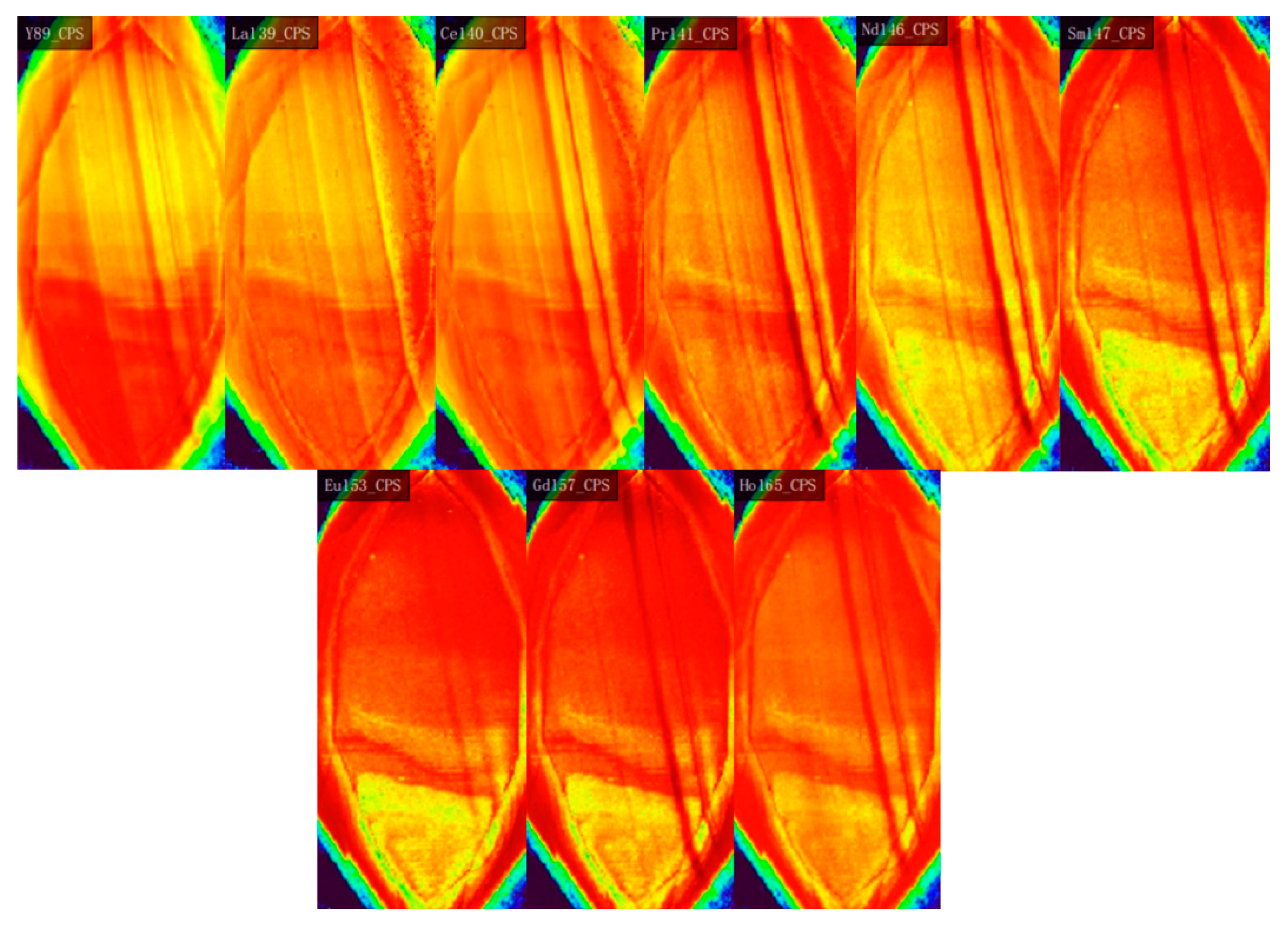
LA-ICP-MS mapping analysis was performed on the pale yellow scheelite sample BWK2 with parallel color bands. The REE enrichment images are shown in
Figure 9. The enrichment degrees of rare earth elements Y, La, Ce, Pr, Nd, Sm, Eu, Gd, and Ho are positively correlated with the degrees of color saturation in the samples.
Based on the results of micro-area analysis and mapping analysis, it is speculated that the yellow-orange hue of Xuebaoding scheelite may be related to the contribution of eight rare earth elements: La, Ce, Pr, Nd, Sm, Eu, Gd, and Ho.
6. Conclusions
This article provides direct evidence relating to the color origins of scheelite from the Xuebaoding deposit, Huya County, Mianyang City, Sichuan Province. The rare earth elements (by LA-ICP-MS) of the Xuebaoding scheelite samples were determined, which provided improved data to support the color origins of scheelite.
The infrared and Raman spectra characteristics of Xuebaoding scheelite are similar to those of the scheelite from other places, such as Colombage-Ara, Sri Lanka. The characteristic absorption peaks related to Nd in the UV-Vis-NIR spectra confirm that the color of Xuebaoding scheelite is related to the isomorphous substitution of Nd ions for Ca ions.
Combined with previous studies and comparative examples of hypersensitive transitions caused by f–f electrons of REE ions (converting the wavelength of absorption light measured by an ultraviolet-visible spectrophotometer into the J-level of REE ion transitions), it can be reasonably inferred that the origin of the yellow-orange hue of Xuebaoding scheelite is closely related to light REEs within its crystal structure, such as La, Ce, Pr, and Nd. The chromogenic mechanisms of other REEs that may contribute to the color of scheelite, such as Sm, EU, Gd, and Ho, require further clarification.
Author Contributions
Conceptualization, X.L. and Q.C.; methodology, X.L.; software, X.L. and J.Y.; validation, X.L. and J.Y.; formal analysis, J.Y.; investigation, X.L.; resources, X.L.; data curation, X.L. and J.Y.; writing—original draft preparation, X.L. and J.Y.; writing—review and editing, X.L. and J.Y.; visualization, X.L.; supervision, Q.C.; project administration, X.L. and Q.C.; funding acquisition, X.L. All authors have read and agreed to the published version of the manuscript.
Funding
This research and the APC. was funded by [Scientific Research Project of Shanghai Jianqiao University] grant number [17001SJQ].
Data Availability Statement
The data presented in this study are available within the article.
Acknowledgments
Thanks to the two reviewers and the editors for their comprehensive and professional suggestions.
Conflicts of Interest
The authors declare no conflict of interest.
References
- Zhang, M.R.; Li, P.J.; Hu, G.Q.; Yin, Z.W. Optical Absorption Edges and their Origin of Scheelite-Structured Tungstate and Molybdate Crystals. Acta Opt. Sin. 1998, 18, 1591–1596. [Google Scholar]
- Liu, Y.; Deng, J.; Xing, Y.Y.; Jiang, S.Q. Vibrational Spectra of Schceelite and Its Color Genesis. Spectrosc. Spectr. Anal. 2008, 28, 121–124. [Google Scholar]
- Liu, Y.; Deng, J.; Li, C.F.; Shi, G.H.; Zheng, A.L. Rare Earth Geochemistry and Sm-Nd Isotope dating of Xuebaoding Scheelite Deposit, Sichuan Province. Chin. Sci. Bull. 2007, 52, 1923–1929. (In Chinese) [Google Scholar]
- Culver, S.P.; Brutchey, R.L. Lanthanide-activated scheelite nanocrystal phosphors prepared by the low-temperature vapor diffusion sol–gel method. Dalton Trans. 2016, 45, 18069–18073. [Google Scholar] [CrossRef]
- Huang, J.B.; LI, Q.F.; Wang, J.; Jin, L.; Tian, B.S.; Li, C.Y.; Shi, Y.R.; Wang, Z.L.; Hao, J.H. Controllable synthesis of lanthanide Yb3+ and Er3+ co-doped AWO4 (A = Ca, Sr, Ba) micro-structured materials: Phase, morphology and up-conversion luminescence enhancement. Dalton Trans. 2018, 47, 8611–8618. [Google Scholar] [CrossRef]
- Bayrakçeken, F.; Demir, O.J.; Karaaslan, İ.Ş. Specific heat functions for the orthorhombic Nd3+ in scheelite type of crystals. Spectrochim. Acta Part A Mol. Biomol. Spectrosc. 2007, 66, 1291–1294. [Google Scholar] [CrossRef]
- Morozov, V.; Arakcheeva, A.; Redkin, B.; Sinitsyn, V.; Khasanov, S.; Kudrenko, E.; Raskina, M.; Lebedev, O.; Tendeloo, G.V. Na2/7Gd4/7MoO4: A Modulated Scheelite-Type Structure and Conductivity Properties. Inorg. Chem. 2012, 51, 5313–5324. [Google Scholar] [CrossRef]
- Ramakrishna, P.V. Cathodoluminescence properties of gadolinium-doped CaMoO4: Eu nanoparticles. Spectrochim. Acta Part A Mol. Biomol. Spectrosc. 2015, 149, 312–316. [Google Scholar] [CrossRef]
- Cornacchia, F.; Lieto, A.D.; Tonelli, M.; Richter, A.; Heumann, E.; Huber, G. Efficient visible laser emission of GaN laser diode pumped Pr-doped fluoride scheelite crystals. Opt. Express 2008, 16, 15932–15941. [Google Scholar] [CrossRef]
- Groń, T.; Karolewicz, M.; Tomaszewicz, E.; Guzik, M.; Oboz, M.; Sawicki, B.; Duda, H.; Kukula, Z. Dielectric and magnetic characteristics of Ca1−xMnxMoO4 (0 ≤ x ≤ 0.15) nanomaterials. J. Nanoparticle Res. 2019, 21, 8–19. [Google Scholar] [CrossRef]
- Wang, H.; Huang, Z.L.; Liu, Y. The preparation and token of the scheelite type of the fluorescent material CaWO4:Ba2+. J. Wuhan Inst. Technol. 2008, 30, 78–80,83. [Google Scholar]
- Gao, D.J.; Xiao, D.Q.; Bi, J.; Yu, P.; Zhang, W.; Zhu, J.G. Study of Preparation Molybdate Films with Structure of Scheelite-type by Electrochemical Deposition Technology. J. Harbin Univ. Sci. Technol. 2002, 7, 60–61. [Google Scholar]
- Cao, F.; Yang, H.P.; Wang, W.; Zhang, L. General Situation and Analysis of Supply and Demand of Global Tungsten Resources. Conserv. Util. Miner. Resour. 2018, 2, 145–150. [Google Scholar]
- Zhu, H.L.; Zhang, L.P.; Du, L.; Sui, Q.L. The geochemical behavior of tungsten and the genesis of tungsten deposits in South China. Acta Petrol. Sin. 2020, 36, 13–22. [Google Scholar]
- Ghaderi, M.; Palin, J.M.; Campbell, I.H.; Sylvester, P.J. Rare earth element systematics in scheelite from hydrothermal gold deposits in the Kalgoorlie-Norseman region, Western Australia. Econ. Geol. 1999, 94, 423–437. [Google Scholar] [CrossRef]
- Nguyen, T.H.; Nevolko, P.A.; Phama, T.D.; Svetlitskaya, T.V.; Tran, T.H.; Shelepaev, R.A.; Fominykh, P.A.; Pham, N.C. Age and genesis of the W-Bi-Cu-F(Au) Nui Phao deposit, Northeast Vietnam: Constrains from U-Pb and Ar-Ar Geochronology, fluid inclusions study, S-O isotope systematic and scheelite geochemistry. Ore Geol. Rev. 2020, 123, 103578. [Google Scholar] [CrossRef]
- Bi, C.S. Basic Geological Characteristics of Skarn-type Scheelite Deposite in China. Acta Geosci. Sin. 1987, 9, 49–64. [Google Scholar]
- Yan, X.G.; Yin, J.W.; Park, J.H.; Kong, D.X.; Zhang, S.T.; Yun, H. The mineralogical characteristics of scheelite of Nannihu Mo-W deposit in Henan. J. Chin. Electron Microsc. Soc. 2013, 32, 318–324. [Google Scholar]
- Han, J.; Li, Y.B.; Yang, H.P.; Zhao, Z.Y. Metallogenic characteristics and prospecting prospective of Chahansen area in Qinhai Province. Miner. Explor. 2019, 10, 525–529. [Google Scholar]
- Li, J.K.; Liu, S.B.; Wang, D.H.; Fu, X.F. Metallogenic epoch of Xuebaoding W-Sn-Be deposit in northwest Sichuan and its tectonic tracing significance. Miner. Depos. 2007, 26, 557–562. [Google Scholar]
- Liu, Y. Mineralogical characteristics and genetic mechanism of the Xuebaoding deposit in northwestern Sichuan Province. Acta Petrol. Mineral. 2017, 36, 549–563. [Google Scholar]
- Liu, Y. Mineralogical Characteristics and Genesis Mechanism of Xuebaoding W-Sn-Be Deposit, NW Sichuan Province. Ph.D. Thesis, China University of Geosciences, Beijing, China, 2010. [Google Scholar]
- Zhou, K.C.; Qi, L.J.; Xiang, C.J.; Feng, Q.M.; Wan, J.S.; Hao, Q. Geologic Characteristic of Forming Beryl Gem Form Pingwu Sichuan. J. Mineral. Petrol. 2002, 22, 1673–1684. [Google Scholar]
- Zhu, X.X.; Liu, Y. Chemical Composition of Minerals in Xuebaoding W-Sn-Be Deposit, Sichuan Province: Constraints on Ore Genesis. Rock Miner. Anal. 2021, 40, 296–305. [Google Scholar]
- Hazen, R.M.; Finger, L.W.; Mariathasan, J.W.E. High-pressure crystal chemistry of scheelite-type tungstates and molybdates. J. Phys. Chem. Solids 1985, 46, 253–263. [Google Scholar] [CrossRef]
- Momma, K.; Izumi, F. VESTA: A three-dimensional visualization system for electronic and structural analysis. J. Appl. Crystallogr. 2008, 41, 653–658. [Google Scholar] [CrossRef]
- Zhou, P.L. Infrared Spectra of Wolframite and Scheelite from Tungsten Ore Deposits in Southern Ganzhou, Jiangxi Province. Acta Mineral. Sinca 1984, 4, 319–322. [Google Scholar] [CrossRef]
- Peng, W.S.; Liu, G.K. Mineral Infrared Spectrum Atlas; Science Press: Beijing, China, 1982; 244p. [Google Scholar]
- An, N.; Guo, Q.F.; Liao, L.B. Gemological and Spectroscopic Characteristics of Scheelite from Xuebaoding, Sichuan, China. Gemol. Technol. 2019, 173–176. [Google Scholar] [CrossRef]
- Gunawardene, M. Colombage-Ara Scheelite. Gems Gemol. 1986, 22, 166–169. [Google Scholar] [CrossRef]
- Brugger, J.; Lahaye, Y.; Costa, S.; Lambert, D.; Bateman, R. Inhomogeneous distribution of REE in scheelite and dynamics of Archaean hydrothermala systems (Mt. Charlotte and Drysdale gold deposits, Western Australia). Contrib. Mineral. Petrol. 2000, 139, 251–264. [Google Scholar] [CrossRef]
| Publisher’s Note: MDPI stays neutral with regard to jurisdictional claims in published maps and institutional affiliations. |
© 2022 by the authors. Licensee MDPI, Basel, Switzerland. This article is an open access article distributed under the terms and conditions of the Creative Commons Attribution (CC BY) license (https://creativecommons.org/licenses/by/4.0/).


