Abstract
Polycrystalline bismuth phosphate BiPO4 was synthesized by solid-state reaction at different temperatures varying from 500 to 900 °C. The samples were characterized by X-ray diffraction (XRD), scanning electron microscopy (SEM), energy dispersive X-ray analysis (EDS) and Raman spectroscopy. The low-temperature phase of BiPO4 has monoclinic structure with a space group P21/n, and was transformed into the monoclinic phase P21/m with a slight distortion of monoclinic lattice when it was heated above 500 °C. The effect of the transformation on the structure, morphology and photocatalytic properties was examined. The photocatalytic activity of each sample, in presence of Rhodamine B (RhB) in aqueous solution, was carried out and analyzed under UV light irradiation. Photoexperiments showed that the material prepared at 500 °C is the best catalyst with degradation efficiency of the order of 96% after 12 min of reaction time under UV light irradiation. This high photocatalytic efficiency could be due to their structural and morphological changes. The photocatalytic degradation mechanism of RhB in the presence of the best photocatalyst BiP-500 °C is proposed. The stability of the catalyst was also examined by carrying out four successive tests of the degradation in the presence of BiP-500 °C. Total organic carbon (TOC) was used to further estimate the rate of mineralization in the presence of BiP-500 °C (83% TOC removal). Photoluminescence experiments performed under UV-laser light irradiation revealed emissions in the green-orange range, with optimal intensities for the mix systems observed at 550 °C.
1. Introduction
The discharge of dye-containing effluents by textile industries is considered as one of the major problems that threatens the aquatic environment [1,2,3]. In addition to the cosmetic problem, the toxicity and carcinogenic properties of most of these dyes are of serious concern [4,5].
Photocatalysis, as an advanced oxidation process, is considered as one of the best alternatives to deal with this issue. Photocatalysis provides a powerful oxidation via the generation of highly oxidizing species, e.g., radicals OH• [6,7,8,9]. These latter are non-selective and can lead to a good removal of all types of non-biodegradable organic contaminants including dyes, pesticides and pharmaceuticals [10,11,12,13,14,15]. Unlike conventional techniques, such as the adsorption method on activated carbon or biological treatments, photocatalysis has been considered as a cost-effective method to completely convert organics into CO2 and H2O without the production of large quantities of sludge [16,17,18,19].
During the last few years, researchers have developed many semiconductors based on oxides [20,21,22,23], hydroxides [24,25], tungstates [26,27,28], molybdates [29] and phosphates [30,31,32,33,34,35,36,37,38] for the decontamination of wastewater. A good photocatalyst should be simple to prepare, nontoxic, cheap and highly effective in utilizing light for the illumination of organic pollutants [39].
BiPO4 is a promising photocatalyst material. It has shown a good performance in photoelectrochemical applications such as water splitting and oxidation of organic pollutants. BiPO4 has three main crystal structures: two monoclinic phases (M1, space group: P21/n and M2, space group: P21/m) and one hexagonal phase (H, space group: P3121). Among them, HBIP, with a band gap of 4.6 eV, shows the lowest photocatalytic activity, while M2 and M1, with band gaps of 4.2 eV and 3.8 eV, respectively, possess higher photocatalytic activity [40]. M1 is reported to have much better photocatalytic activity than that of traditional P25 TiO2 [41,42,43,44]. This high photocatalytic activity was explained by the inductive effect of PO43− leading to a good separation of the electron and hole pairs [45]. For this reason, BiPO4-based photocatalysts have been extensively studied for environmental remediation applications [40,46,47,48,49].
So far, different methods have been used to synthesize BiPO4, including the chemical vapor process [50,51], the hydrothermal process [30], microwave synthesis [51] and the sonochemical method [52]. However, the preparation of BiPO4 materials with controlled phase and morphology is still a challenge for material scientists.
The choice of the synthesis method and the control of the experimental conditions are very important in order to reach the right structural, microstructural, and electrical properties, which are crucial for obtaining the optimum photocatalytic properties to easily degrade organic pollutants. For example, Table 1 summarizes the different preparation methods of BiPO4 particles with their photocatalytic activities towards the degradation of organic pollutants.

Table 1.
Synthesis methods and photocatalytic properties of BiPO4.
In our recent work [61], BiPO4 samples were synthesized from polycrystalline precursors obtained by coprecipitation method, then, thermally treated at temperatures Θ of 200, 400, 600 and 900 °C during 3 h. The progressive formation of three polymorphs, hexagonal phase at 200 °C, monoclinic P21/n at intermediate temperatures and P21/m at 900 °C, allowed observing different behaviors of photocatalytic activities and photoluminescence emissions. The role of thermal decomposition was to modify crystal structures, crystallite dimensions and morphologies to obtain or vary certain properties. The highest photocatalytic efficiencies were observed with large-sized particles of biphasic BiPO4 samples thermally treated at 400 and 600 °C.
Presently, we develop a systematic study of the photocatalytic and photoluminescent properties of BiPO4 polymorphs obtained by the traditional solid-state reaction, at different thermal treatment temperatures ranging from 500 to 900 °C.
2. Experimental Section
2.1. Reagents
Bismuth oxide (Bi2O3) ≥ 99.9% was purchased from Fluka Chemika. Ammonium hydrogen phosphate (NH4)H2PO4 ≥ 98.0% was purchased from ProLabo. The azo dye (Rhodamine B) used in this work was obtained from Sigma-Aldrich. The purity of the dye was greater than 95%. All the reagents were used as received, without further purification.
2.2. Elaboration of Samples
There are several methods for synthesizing phosphate-based materials in powder form. In our previous study concerning BiPO4 [30,42,53], all samples were obtained from coprecipitation technique followed by thermal treatment at various temperatures [61]. In this work, we developed a new route based on solid-state reaction. BiPO4 bismuth phosphate was prepared from polycrystalline Bismuth Bi2O3 oxide (Fluka Chemika > 99.0%) and ammonium hydrogen phosphate (NH4)H2PO4 (ProLabo ≥ 98.0%). Suitable amounts of these starting precursors were ground in an agate mortar and then thermally heated at different temperatures between 500 and 900 °C with a step of 50 °C for 3 h. The samples presently studied at different temperatures of thermal treatment Θ will be noted BiP-Θ with Θ = 500, 550, 600, 650, 700, 750, 800, 850 and 900 °C.
2.3. Characterization Techniques
The identification of the crystalline powders was carried out by X-ray diffraction (XRD). The XRD patterns of the polycrystalline ceramics were recorded at room temperature using an Empyrean Panalytical diffractometer operating at 45 kV/35 mA, using the CuK(α1−α2) radiation (λ = 1.5406 and 1.5444 Å) of copper source with Ni filter, and working in continuous mode with a step size of 0.013°. Scanning electron microscopy (SEM) analysis was used to observe the morphology and the local composition of the crystalline phases. The device used was a Supra 40 VP Column Gemini Zeiss (Zeiss, Iéna, Germany), operated at 20 KeV, coupled with an Energy Dispersive X-rays Spectroscopy (EDXS) type analyzer, allowing us to determine the local elemental composition of our materials.
Raman spectroscopy was used to correlate the vibrational characteristics with the structural analyses from X-ray diffraction. The Raman spectra were registered at room temperature using spectrometer Horiba Jobin-Yvon HR800 LabRam system, with wavenumbers of Raman shifts ranging between 100 and 1500 cm−1. The 633 nm line of an Ar-ion laser was used as the excitation source; the photonic power applied to the samples was limited to 5 µW with an acquisition time of 30 s.
2.4. Photocatalytic Experiments
The photocatalytic reactor consisted of a light source of 5 UV lamps (Puritec lamp, HNS S 7W (Osram, Berlin, Germany), λ = 254.6 nm). In the middle of the reactor, the solution to be irradiated was placed in a beaker in which the homogeneity was ensured by a magnetic stirrer. A cooling system was adapted to avoid the effect of temperature. The temperature of the solution was then maintained between 26 ± 1 °C.
The photocatalytic performances of the photocatalysts were evaluated though the photocatalytic degradation of RhB. Each catalyst (mass of 100 mg) was suspended in 100 mL of RhB solution (5 mg L−1). Before irradiation, the solution was stirred for 1 h inside the photoreactor to achieve the adsorption–desorption equilibrium between the catalyst and RhB. During irradiation, 3 mL solutions were collected at times changed from 2 to 30 min depending on the type of photocatalyst. UV visible JENWAY-6705 spectrometry (Cole-Parmer Ltd., Paris, France) was used to determine the variation of concentration of RhB as a function of irradiation time. To propose a degradation mechanism in the presence of the best catalyst, disodium ethylenediaminetetraacetic acid (EDTA-2Na (Sigma-Aldrich, Saint-Louis, Missouri, États-Unis), a scavenger of h+), isopropanol (IPA (Sigma-Aldrich, Saint-Louis, Missouri, États-Unis), a scavenger of OH) and L-ascorbic acid (Sigma-Aldrich, Saint-Louis, Missouri, États-Unis), a scavenger of O2•−) were used. For each test, certain amounts of scavengers (4 mmol L−1) were added into the rhodamine B solution prior to addition of BiPO4 catalysts. The mineralization of RhB and its intermediates during the photocatalytic reaction was evaluated by the measurement of total organic carbon present in aqueous solution. Analysis was performed with a Shimadzu TOC-5000-A system (Shimadzu, Kyoto, Japan) equipped with a non-dispersive infrared detector (Shimadzu, Kyoto, Japan) and ASI-5000-A auto-sampler (Shimadzu, Kyoto, Japan). The temperature of solution was maintained at 25 ± 4 °C. The photocatalytic efficiency was calculated from the variation of the concentrations of RhB using the intensity Equation (1):
where C0 and C are the initial and time t concentrations of RhB, respectively.
Degradation efficiency (%) = 100 × (C0 − C)/C0
2.5. Photoluminescence Experiments
The device used to perform photoluminescence (PL) tests under UV excitation was a Horiba Jobin-Yvon HR800 LabRam spectrometer (Horiba France SAS, Palaiseau, France) equipped with an argon ionized laser, used as an excitation source with a wavelength of 364.8 nm (3.4 eV), and power fixed at 5 μW. The measurements were recorded in the spectral range between 400 and 900 nm with an acquisition time of 5 s.
3. Results and Discussion
3.1. X-ray Diffraction Analyses
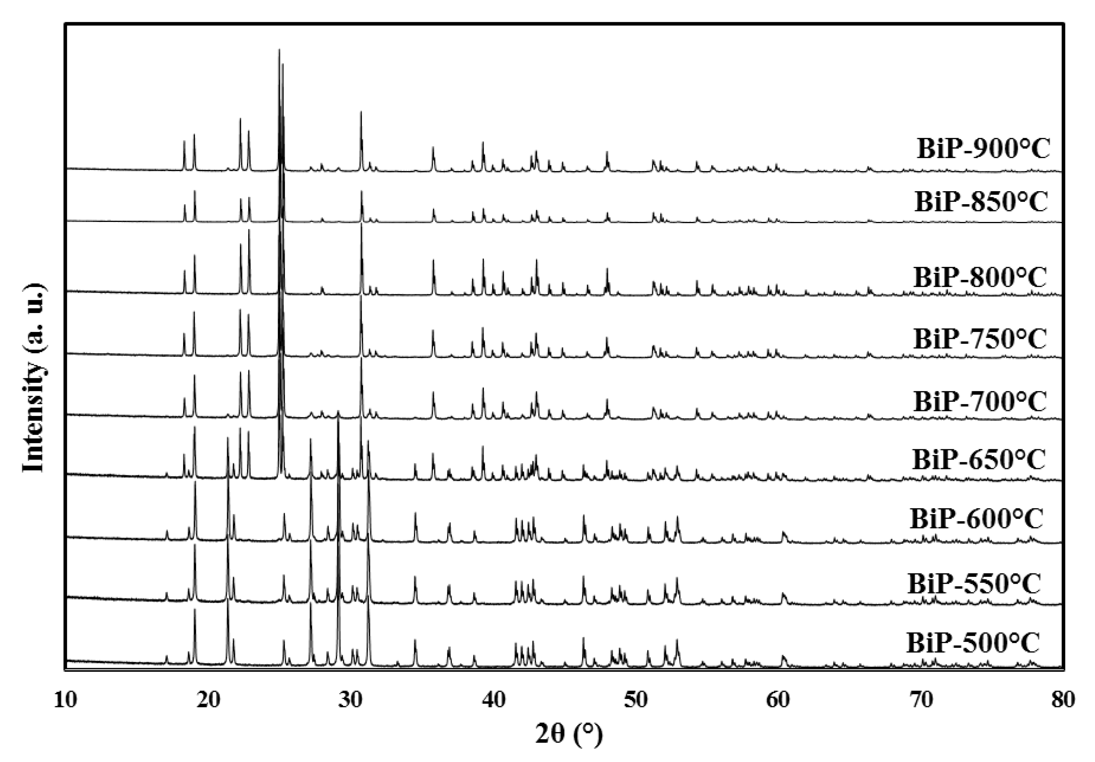
Figure 1 depicts the XRD patterns of the as-synthesized BiPO4 samples obtained by solid-state and calcined at different temperatures changing from 500 to 900 °C. All the detected diffraction peaks for the BiP-500 sample could be indexed into the monoclinic phase with space group P21/n (M1) single phase. When the calcination temperature increases above 550 °C, the nMBIP phase is progressively transformed into the monoclinic phase of BiPO4 with space group P21/m (M2) (Figure 2). In this instance, I crystal phase structure of the prepared BiPO4 samples calcined at 800 °C exhibits the presence of only a single phase of M2. Upon increasing the heat treatment above 850 °C, the appearance of small residues of the M1 phase is shown. To sum up, 500 °C, 800 °C, and other temperatures give rise to a single phase of M1, a single phase of M2 and a mixture of the two phases.

Figure 1.
XRD patterns of bismuth phosphate BiPO4 obtained after different thermal treatments. BiP-500: monoclinic M1, at 500 °C; BiP-550 and 600: monoclinic M1 + traces of monoclinic M2, at 550 and 600 °C; BiP-650: monoclinic M1 + monoclinic M2 at 650 °C; BiP-800: monoclinic M2, at 800 °C; BiP-700, 750, 850 and 900: M2 + traces of M1 at 700, 750, 850 and 900.
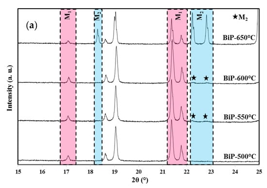

Figure 2.
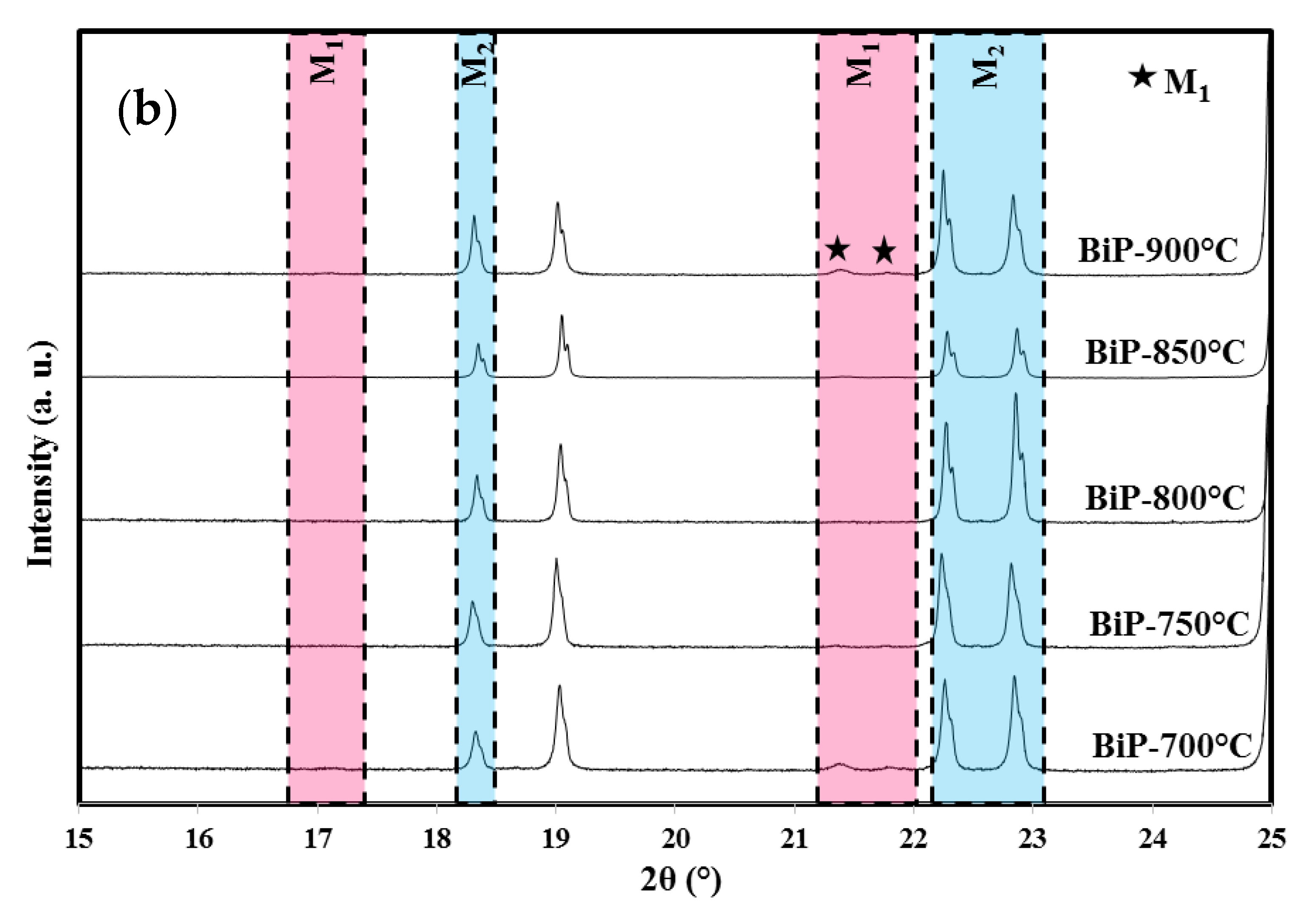
XRD diffraction profiles in the angular range 15° to 25° 2θ of the BiP-Θ samples: the disappearance of the Kα1–α2 doublet of the Bragg peaks for the monoclinic phase M1 from BiP-700 °C, appearance of the Bragg peaks from BiP-550 °C of the monoclinic phase M2. (a) 500, 550, 600 and 650 °C, (b) 700, 750, 800, 850 and 900 °C.
Based on the intensity of the peaks of the two phases using ’X’ Pert High Score Plus software, Version 3.0.5, the portions of the two monoclinic crystal structures of the BiPO4 samples can be calculated and are depicted in Table 2. As can be seen, calcining the example at 550 °C, a small amount of 9 mol. % of the M1 was transformed to M2. Expanding the calcination temperature, the weight level of M2 becomes increasingly important until complete vanishing of the M1 at 800 °C. Above this latter temperature, the opposite is seen; a small amount of the unadulterated M2 has been transformed into M1.
Table 2.
The transformed mole fraction of M1 and M2 samples sintered at various temperatures.
3.2. Scanning Electron Microscopy: Morphology of the Powders
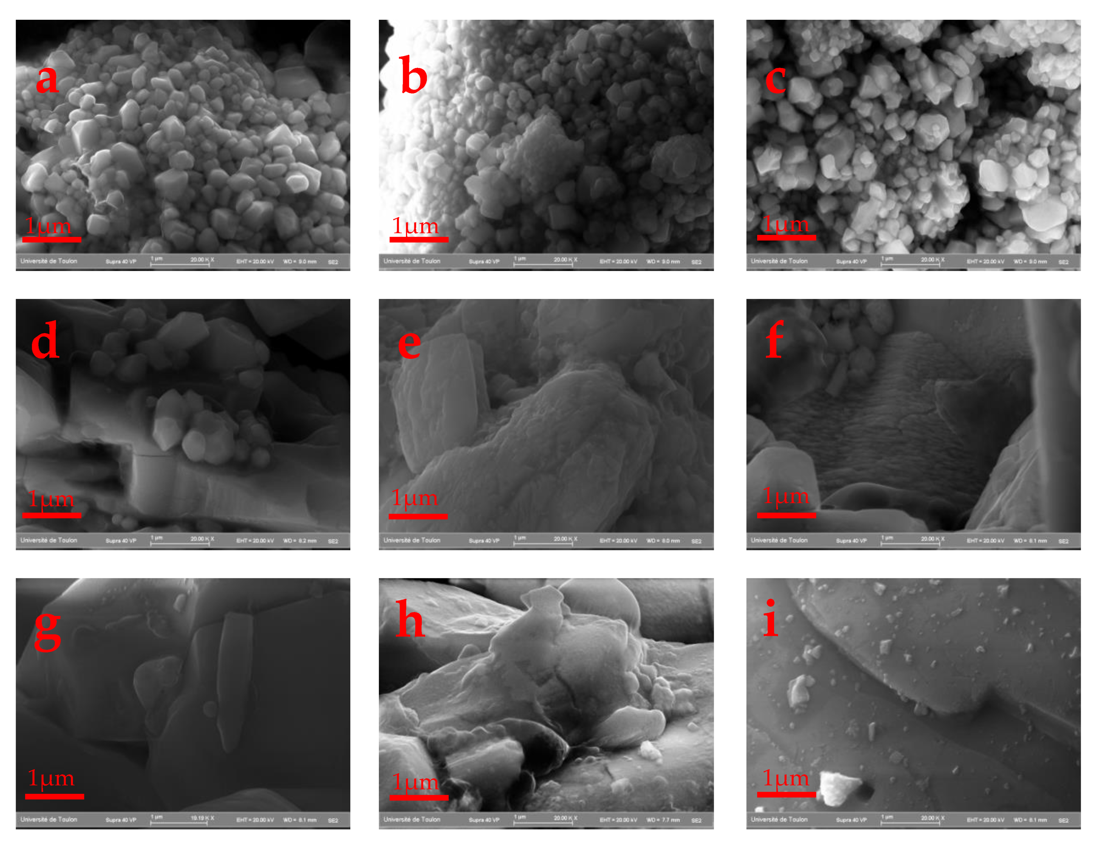
Scanning electron micrographs of the BiPO4 samples obtained at different calcination temperatures changing from 500 to 900 °C show morphological changes. The BiP-500 and 600 samples (Figure 3a–c) show no significant morphology change with the presence of spherical particle shapes with 200–300 nm size distributions. A further increase in the calcination temperature from 650 °C to 900 °C dramatically changed the BiPO4 particles to irregular large particles as they aggregated and bonded with each other to form big clusters (Figure 3d–i). Moreover, it was observed that the initial spherical morphology of BiP-500/600 disappeared, and a new shape with a larger particle size was observed, giving rise to a remarkable decrease in the specific surface areas.
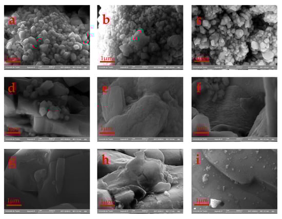
Figure 3.
SEM images of: (a) BiP-500, (b) BiP-550, (c) BiP-600, (d) BiP-650, (e) BiP-700 (f) BiP-750, (g) BiP-800, (h) BiP-850 and (i) BiP-900.
From 700 °C to 900 °C, M2 is the major phase compared to M1. All samples show a high purity, as the EDX analysis in Table 3 shows. Indeed, the atomic ratio Bi/P is equal to 1 in all the elaborated samples, which further confirms the successful synthesis of BiPO4.
Table 3.
EDX analyses for Bi and P atoms.
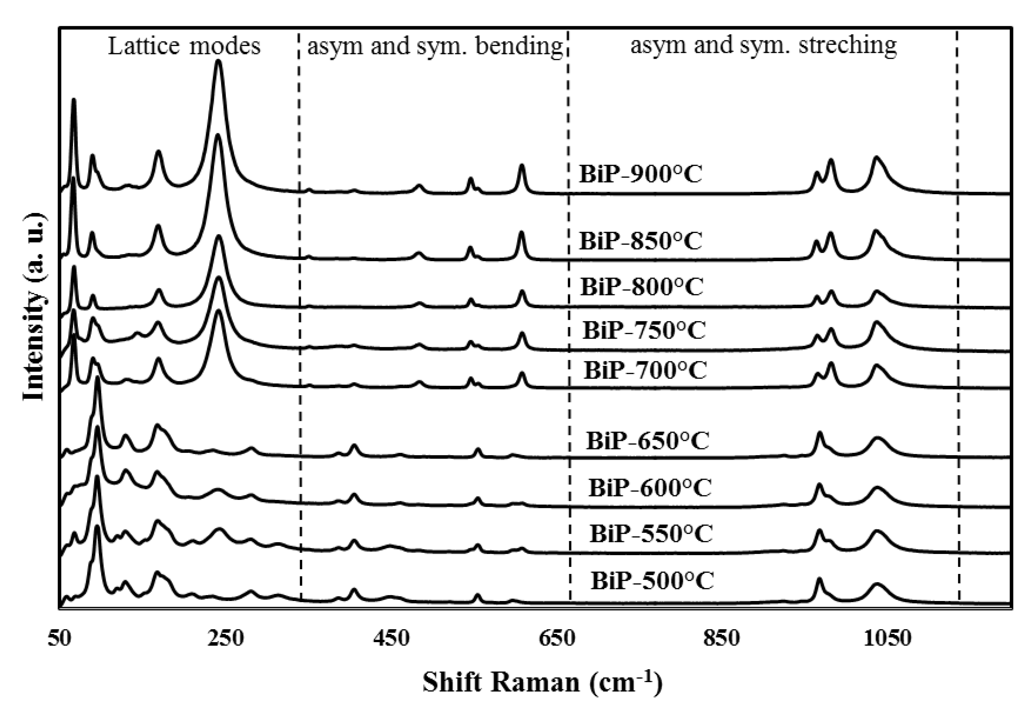
3.3. Raman Spectroscopy Study
Symmetrical changes of PO4 tetrahedra with phase transformation during heat treatment were studied using Raman spectroscopy. As shown in Figure 4, some distinct differences can be clearly seen in their vibrational characteristics compared to phosphate groups [62,63]. Based on the group theory analysis, the isolated PO4 group has a Td symmetry with nine internal modes which can be represented as Equation (2).
Tvib = A1 + E + 2F2

Figure 4.
Raman spectra of bismuth phosphate BiPO4 obtained at different thermal treatments.
It is worth mentioning that the representation E is doubly detached and F2 is triply detached. The symmetrical (υ1) and asymmetrical (υ3) P–O stretching vibrations of the PO4 group correspond to the A1 and one mode of F2 representation, respectively, while the other modes’, F2 and E, representation correspond to the O–P–O flexion of υ2 and υ4, respectively [64,65]. More precisely, the orthophosphate PO4 group isolated in the monoclinic phase P21/n (M1) is characterized by several bands, such as υ1, υ2, υ3 and υ4, which are located at 969, 461, 1039 and 597 cm−1, respectively. Depending on the symmetry around the PO4 phosphate group and the nature of the cation introduced into the matrix, the vibration modes mentioned above undergo a shift either towards blue or towards red. This is the case of the polymorphic change from the monoclinic phase P21/n (M1) to the monoclinic phase P21/m (M2) (Table 4). In other words, the symmetry of the PO4 phosphate group is changed. More precisely, the experimental values of the wavenumbers for υ1, υ2, υ3 and υ4 are, respectively, 965, 485, 1037 and 609 cm−1 [43,64]. In the Raman spectra, the decrease in certain bands and the increase in others show the disappearance of one phase and the disappearance of another, which is confirmed by the fractions of the M1 and M2 phases in each sample, which further confirms the results found by X-ray diffraction.
Table 4.
Raman spectroscopy data and assignments of vibration bands for the monoclinic phases of BiPO4.
4. Photocatalytic Properties
To evaluate and compare the photocatalytic activities of the obtained different phases of BiPO4, the photodecomposition of RhB under UV light irradiation was carried out, and the determination of kinetics constants could be then achieved by following the intensity of the RhB absorption maximal band located at 554 nm.
To determine the kinetic constant, which characterizes the photocatalytic efficiency, a first order kinetics rate law (Langmuir-Hinshelwood model) was proposed as Equation (3) [66]:
Ln (Ct/C0) = −kobs t
Following both the adsorption/desorption (with the catalysts and without UV irradiation) and the photolysis (without the catalysts and under UV irradiation), the BiPO4 catalysts show only 2% degradation after 12 min of reaction time, which shows that the RhB molecule is stable during this reaction time (Figure 5a).
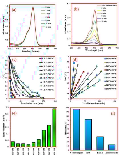
Figure 5.
(a) Stability of RhB under UV light; (b) UV–vis absorption with time irradiation of a solution containing 100 mg of BiP-500 °C and 5 ppm RhB; (c) Degradation efficiencies of the as-prepared samples; (d) Pseudo-first-order kinetics of the photocatalysts; (e) Evolution of the apparent rate constant k as function of photocatalysts; (f) Effect of scavengers on the photocatalytic degradation of RhB by BiP-500.
Figure 5b shows that, under UV irradiation, the intensity of the pollutant absorption band decreases with the irradiation time in the presence of BiPO4 prepared by solid-state reaction and thermally treated at 500 °C. The position of the maximum of this band is shifted from 554 to about 535 nm during decomposition.
Figure 5c shows the kinetics of photocatalytic degradation in the presence of the various photocatalysts. After 12 min of UV irradiation, the photocatalytic efficiencies determined from the ratio Ct/C0 of the BiP-Θ catalysts with Θ = 500, 550, 600, 650, 700, 750, 800, 850 and 900 °C reach the values of 96.6%, 78.6%, 69.6%, 50.6%, 24.4%, 14.3%, 17.4%, 21% and 27.7%, respectively. It should be noted that these observed efficiencies result from both photolysis and photocatalysis effects. In other terms, the highest efficiencies should be obtained from sample BiP-500. The results in Figure 5d show that the photocatalytic reaction follows a pseudo-first-order kinetics rate law.
The observed rate constants kobs are reported in Figure 5e. Their values in min−1 are 0.2893, 0.1648, 0.1264, 0.056, 0.0342, 0.0175, 0.0249, 0.025 and 0.0403, for the BiP-Θ photocatalysts with Θ = 500, 550, 600, 650, 700, 750, 800, 850 and 900 °C, respectively.
The photocatalytic efficiencies of the BiPO4 series produced by solid–solid reaction, and, in particular, the high photocatalytic efficiency of the BiP-500 phase obtained at 500 ° C, appear to be the highest of the series studied, the rate constants being at least 15 times stronger than those of the other photocatalysts, especially compared to BiP-750.
These improved efficiencies could be correlated to the observed specific morphologies characterized by well-crystallized small-sized grains, and also to the presence of the monoclinic phase with space group P21/n [67,68]. It is worth mentioning that the photocatalytic properties of the obtained samples decrease as a function of the heat treatment; therefore, these results are in a good agreement with the XRD and SEM results. Indeed, the M1 amount decreases while that of M2 increases; this latter shows less activity towards RhB degradation, which explains the obtained results.
As we have mentioned above, the photocatalytic degradation of RhB was studied in an aqueous suspension of bismuth phosphates. The discoloration of this pollutant in the presence of the best photocatalyst (BiP-500 °C) is total after 12 min of UV irradiation at an initial concentration of 5 mg.L−1. In order to determine the photocatalytic mechanism and to confirm the main active species in the photocatalytic process, such as hydroxyl radical (OH•), hole (h+) and superoxide radical (O2•−), active species trapping experiments were carried out. More precisely, the photocatalytic degradation of RhB in the presence of scavengers has been studied. Isopropanol alcohol (IPA), disodium ethylenediaminetetraacetic acid (EDTA-2Na) and L-ascorbic acid were used as scavengers of (OH•) species, holes (h+) and (O2•−) species, respectively.
The removal efficiency of RhB onto BiP-500 in the presence of different scavengers (L-ascorbic acid, EDTA-2Na, and IPA) is shown in Figure 5f. The photocatalytic degradation efficiency of RhB is 96.6% in the absence of any scavenger. L-ascorbic acid, EDTA-2Na significantly decrease the degradation of RhB with efficiencies of 12.8% and 31.6%, respectively. We obtained a degradation rate of 72.7% in the presence of IPA. These results, therefore, show that the degradation process is enhanced predominantly by holes (h+) and superoxide radicals (O2•−). The hydroxyl radical plays a minor role in the degradation mechanism since the efficiency of the degradation of was not decreased in a significant way when this radical was inhibited. We can assume that the degradation of RhB can be summarized in the following reactions:
| BiPO4 | → | BiPO4 (h+, e−) |
| BiPO4 (e−) + O2 | → | O2•− |
| BiPO4 (h+) + H2O | → | OH− + H+ |
| BiPO4 (h+) + OH− | → | OH• |
| BiPO4 (h+) + RhB | → | RhB+ |
| RhB + O2− + h+ + OH• | → | CO2 + H2O + simple molecules |
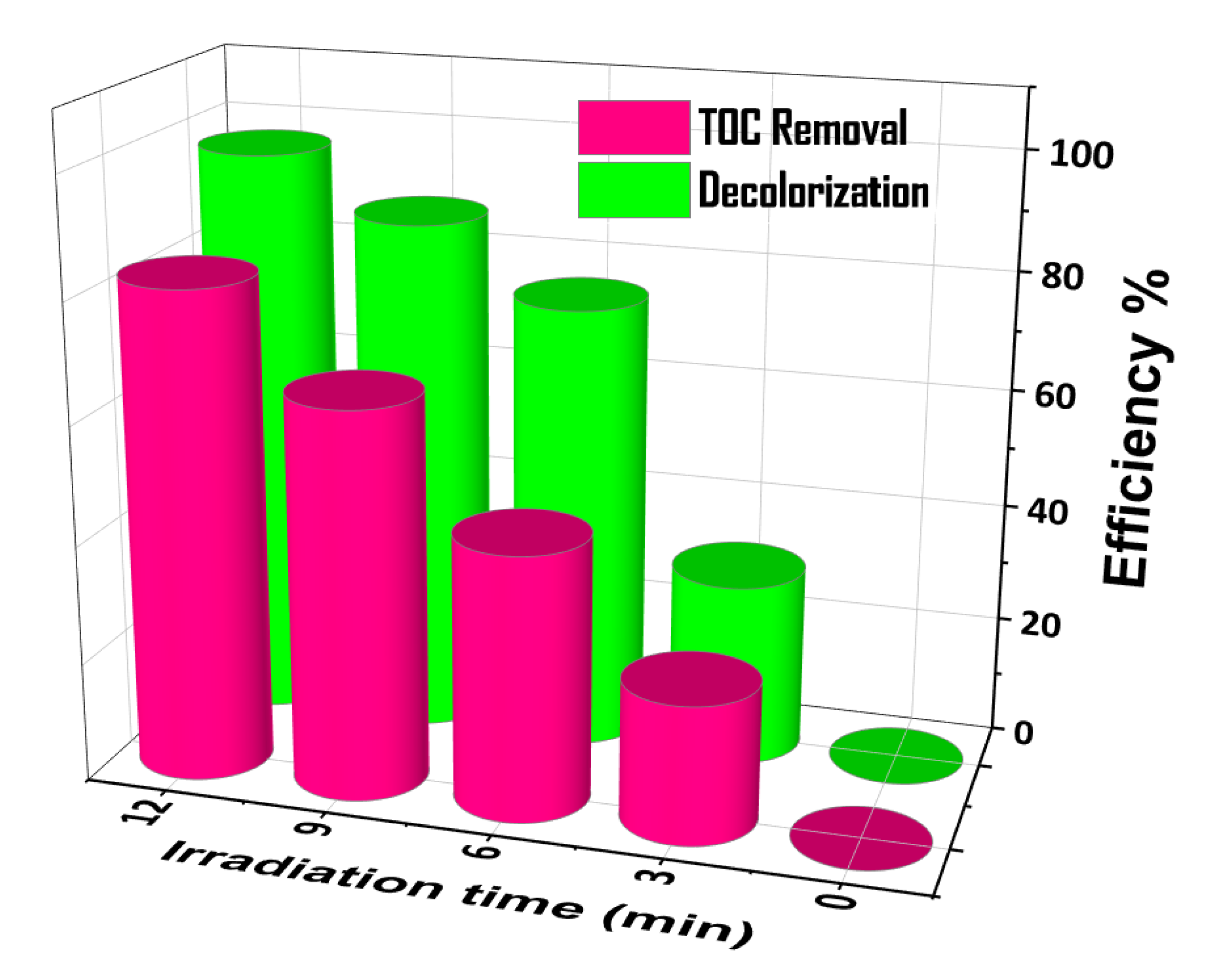
Figure 6 shows the total organic carbon (TOC) removal efficiency using BiP-500 has attained about 83% after 12 min under UV-visible light irradiation. This shows that BiP-500 exhibits important photocatalytic performance for the photodegradation of RhB. Such results also demonstrate that the RhB would be mineralized into CO2 and H2O by the as-prepared samples, which in is favor of its application in the field of the treatment of wastewater.

Figure 6.
TOC removal vs. discoloration of Rhodamine B over as BiP-500 photocatalyst.
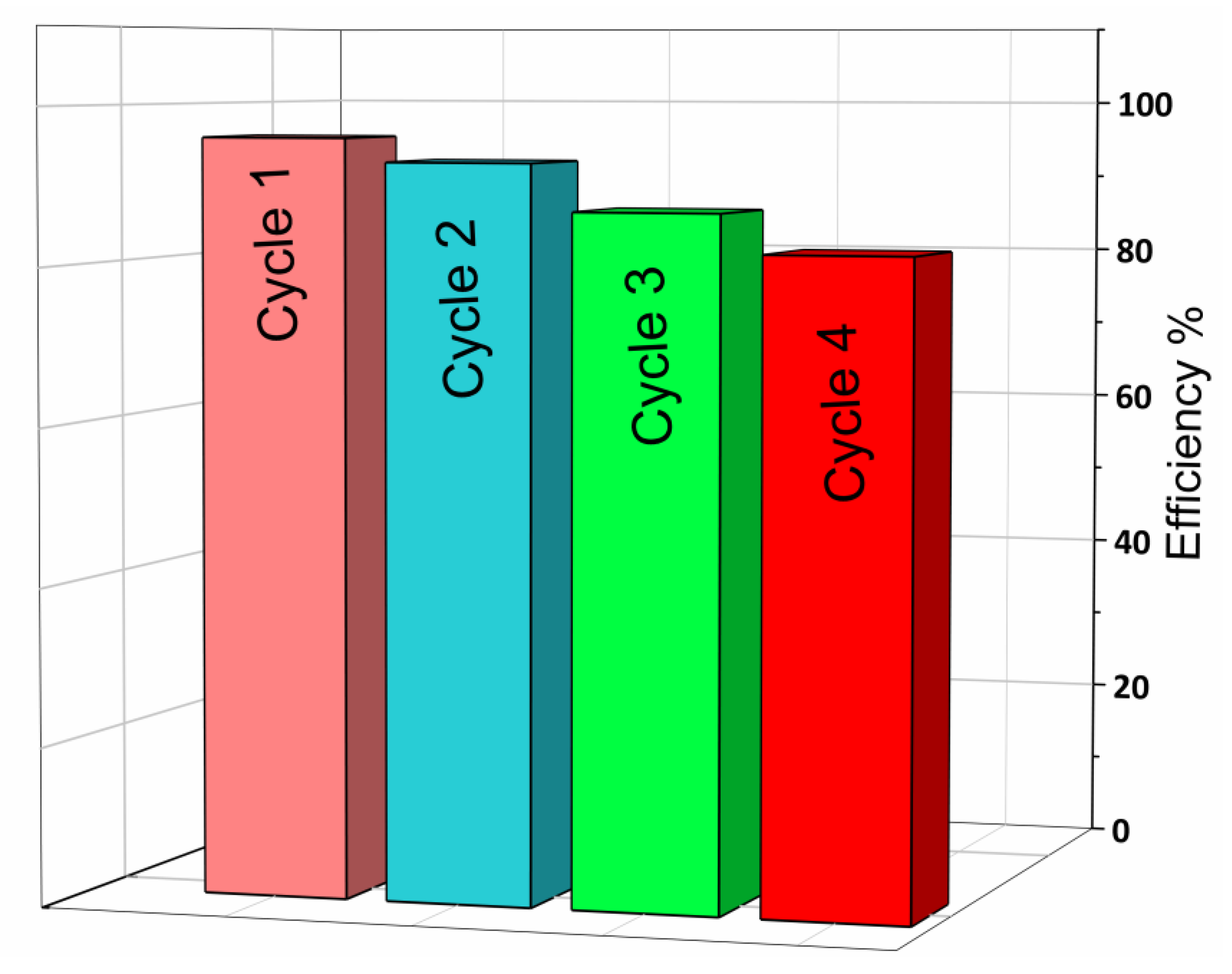
Stability of the BiP-500 °C Catalyst
The photostability and reusability of the photocatalyst activity during the depollution process is an important parameter to consider from an economical point of view. Thus, in order to verify the efficiency of the recycling of the photocatalyst used in this work, four recycling tests were carried out. The sample can be separated easily after each cycle of the photocatalysis reaction. The results of photostability are shown in Figure 7. The initial concentration of RhB was set at 0.01 mmol L–1 in the presence of 100 mg of the catalyst.
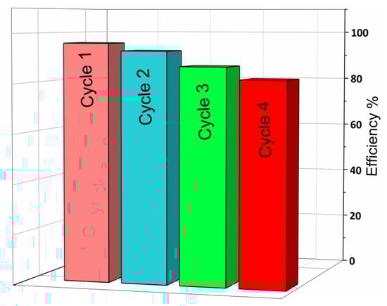
Figure 7.
Discoloration performance of recycled catalyst ([RhB] = 0.01 mM; irradiation time = 12 min; [Catalyst] = 1.0 g/L; room temperature).
For each test, irradiation was carried out for 12 min under UV-visible. After repeating the process four times, the photocatalytic activity slightly decreased from 96% to 82%; this slight decrease may be due to the decrease in catalyst mass and the deactivation of some active sites of our catalyst by the organic pollutant. These results indicate the high stability and reusability of our photocatalyst.
5. Photoluminescent Properties
The photoluminescence spectra of BiP-Θ materials were recorded under UV excitation and are shown in Figure 8a,b. These emission spectra were processed using the Labspec program. The broad emission bands observed in the range 400 to 800 nm in all the powders of the BiP-Θ samples with Θ = 500, 550, 600, 650 and 700 °C (Figure 8a), show similar profiles with a maximum at 515 ± 20 nm. The results in the literature have shown that, in the case of Bi3+ isolated in a solid matrix, the allowed transition of Bi3+ is 3P1→1S0, and that it is characterized by emission spectra with wavelengths close to 470 nm (2.6 eV) [61]. In the case of the study of the three polymorphic varieties of BiPO4, the Bi3+ emissions under excitation at 360 nm were observed at 450 nm (2.8 eV) for the hexagonal and monoclinic M1 phases. Currently, a wide emission band (450 to 700 nm) is systematically observed: the emission profile can be broken down into several emissions comprising at least three components (470, 510–520 and 600–680 nm). Emissions ranging from 500 nm to 700 nm are generally attributed to surface defects. This was also the case for BiPO4 prepared by the coprecipitation method, where a similar broad band was bound to the M1 structure and attributed to transitions associated with changes in Bi3+ environments, due to the presence of oxygen vacancy defects [VO]q° (where q° is the positive electric charge with q = 0, 1 or 2) disrupting the PO4 and BiO8 structural groups of the monoclinic structure. Other defects could also be at the origin of this broadband (Bi vacancies or interstitial sites).
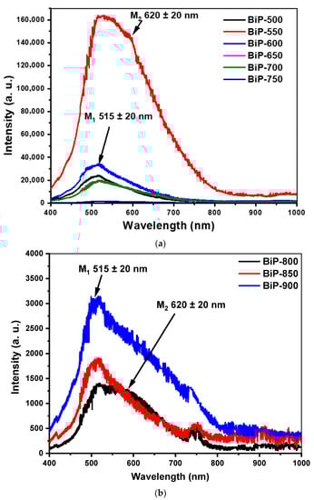
Figure 8.
UV-excited photoluminescence spectra (364 nm) of BiPO4 prepared by solid state reaction and heat treated at (a) 500, 550, 600, 650, 700 and 750 °C, (b) 800, 850 and 900 °C.
The BiP-750 sample, which was characterized by the presence of the majority of the monoclinic M2 phase, showed a single band with a maximum close to (620 ± 20 nm). In the case of the BiP-Θ samples with Θ = 800, 850 and 900 °C (Figure 8b), two characteristic bands of phase M1 (515 ± 20 nm) and M2 (620 ± 20 nm) were observed, with low intensities compared to the other BiP-Θ samples with Θ = 500, 550, 600, 650 and 700 °C.
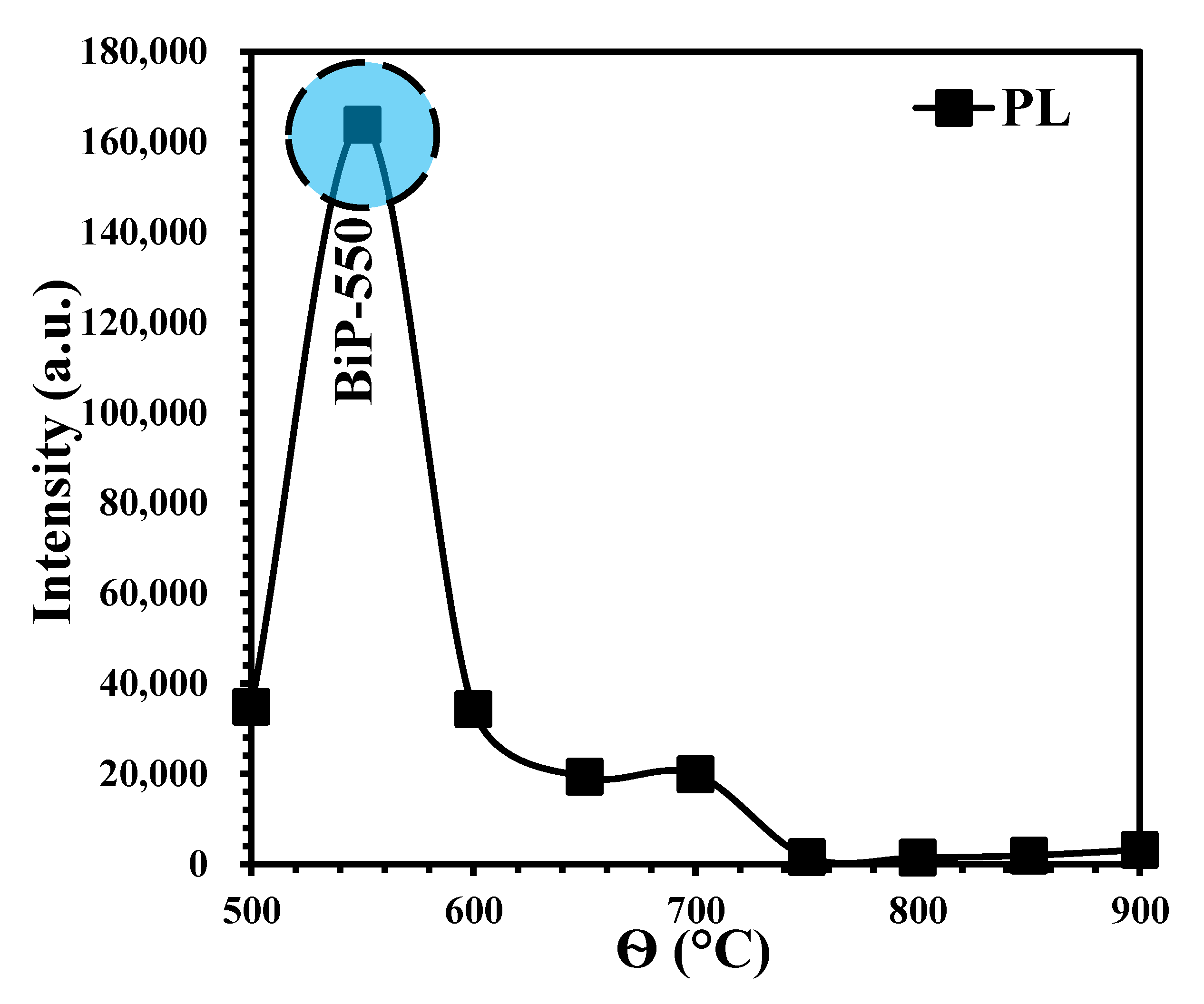
In Figure 9, we have reported the intensities of the maximum bands as a function of the heat treatment temperature Θ. The two characteristic bands of phase M1 (515 ± 20 nm) and M2 (620 ± 20 nm) show almost invariant intensities with Θ. We observed a maximum for the polymorph sample BiP-550 (M1 and traces of M2). The intensity observed for this particular sample is more than five times greater than that observed for the pure phase BiP-500 (monoclinic M1) and one hundred times greater than that of BiP-750 and BiP-800 (monoclinic M2). These results of photoluminescence under UV (364 nm) are in good agreement with the results of the characterization of powders by XRD, more precisely according to the appearance and disappearance of the two monoclinic phases, M1 and M2.

Figure 9.
Variation of intensity of BiP-Θ samples with elaboration temperature.
6. Conclusions
The phase change and photocatalytic properties of BiPO4 ceramics were examined. The monoclinic phase of BiPO4 with space group P21/n (phase stable at low-temperature) was transformed into the monoclinic phase with space group P21/m (phase stable at high-temperature) when it was heated above 500 °C. When the low-temperature phase was thermally heated at 800 °C, the pure phase M2 was obtained perfectly. From heat treatment at 850 to 900 °C of M2, a small fraction of M2 was transformed into M1 again. From a quantitative analysis based on the relative intensities of the two-phase mixture, the amount of transformed phase was calculated and the photocatalytic properties of BiPO4 ceramics with the variation of the fraction of the two phases were studied. The best photocatalytic activity was obtained with the BiP-500/600/650 samples corresponding to the P21/n phase and presenting the smallest grain sizes. The mechanism of photocatalytic degradation and the stability of the best photocatalyst towards the degradation of RhB as a polluting dye have been studied. The mineralization of the pollutant studied in the presence of our photocatalyst was confirmed by carrying out TOC tests.
Author Contributions
A.B. (Abdessalam Bouddouch): Investigation, Writing—original draft, Methodology; E.A.: Discussion, Data curation; B.B.: Supervision, Writing—review and editing; A.T.: Writing—review and editing; F.G.: Supervision, Characterization; S.V.: Supervision, Characterization; J.-R.G.: review and editing; J.-C.V.: Characterization and Data Curation; A.B. (Abdeljalil Benlhachemi): Supervision, Conceptualization. All authors have read and agreed to the published version of the manuscript.
Funding
Financially supported by CAMPUS FRANCE (PHC TOUBKAL 2018 (France–Morocco bilateral program) Grant Number: 38999WE) and PPR project financed by the CNRST under number PPR/2015/32.
Acknowledgments
This work was carried out in the laboratory materials and environment (LME), at the faculty of sciences Agadir, Ibn Zohr University, IM2NP laboratory, University of Toulon.
Conflicts of Interest
The authors declare that they have no known competing financial interests or personal relationships that could have appeared to influence the work reported in this paper.
References
- Al-Mamun, M.; Kader, S.; Islam, M.; Khan, M. Photocatalytic activity improvement and application of UV-TiO2 photocatalysis in textile wastewater treatment: A review. J. Environ. Chem. Eng. 2019, 7, 103248. [Google Scholar] [CrossRef]
- Tounsadi, H.; Metarfi, Y.; Taleb, M.; El Rhazi, K.; Rais, Z. Impact of chemical substances used in textile industry on the employee’s health: Epidemiological study. Ecotoxicol. Environ. Saf. 2020, 197, 110594. [Google Scholar] [CrossRef]
- Haounati, R.; Ouachtak, H.; El Haouti, R.; Akhouairi, S.; Largo, F.; Akbal, F.; Benlhachemi, A.; Jada, A.; Addi, A.A. Elaboration and properties of a new SDS/CTAB@Montmorillonite organoclay composite as a superb adsorbent for the removal of malachite green from aqueous solutions. Sep. Purif. Technol. 2020, 255, 117335. [Google Scholar] [CrossRef]
- Chennah, A.; Naciri, Y.; Taoufyq, A.; Bakiz, B.; Bazzi, L.; Guinneton, F.; Villain, S.; Gavarri, J.R.; Benlhachemi, A. Electrodeposited zinc phosphate hydrate electrodes for electrocatalytic applications. J. Appl. Electrochem. 2018, 49, 163–177. [Google Scholar] [CrossRef]
- Naciri, Y.; Hsini, A.; Ajmal, Z.; Bouddouch, A.; Bakiz, B.; Navío, J.; Albourine, A.; Valmalette, J.-C.; Ezahri, M.; Benlhachemi, A. Influence of Sr-doping on structural, optical and photocatalytic properties of synthesized Ca3(PO4)2. J. Colloid Interface Sci. 2020, 572, 269–280. [Google Scholar] [CrossRef]
- Bouddouch, A.; Amaterz, E.; Taoufyq, A.; Bakiz, B.; Guinneton, F.; Villain, S.; Valmalette, J.; Gavarri, J.; Benlhachemi, A. Photocatalytic and photoluminescent properties of a system based on SmPO4 nanostructure phase. Mater. Today Proc. 2020, 27, 3139–3144. [Google Scholar] [CrossRef]
- Souza, R.P.; Ambrosio, E.; Souza, M.T.F.; Freitas, T.K.F.S.; Ferrari-Lima, A.M.; Garcia, J.C. Solar photocatalytic degradation of textile effluent with TiO2, ZnO, and Nb2O5 catalysts: Assessment of photocatalytic activity and mineralization. Environ. Sci. Pollut. Res. 2017, 24, 12691–12699. [Google Scholar] [CrossRef]
- Maleki, A.; Safari, M.; Shahmoradi, B.; Zandsalimi, Y.; Daraei, H.; Gharibi, F. Photocatalytic degradation of humic substances in aqueous solution using Cu-doped ZnO nanoparticles under natural sunlight irradiation. Environ. Sci. Pollut. Res. 2015, 22, 16875–16880. [Google Scholar] [CrossRef]
- Frederichi, D.; Scaliante, M.H.N.O.; Bergamasco, R. Structured photocatalytic systems: Photocatalytic coatings on low-cost structures for treatment of water contaminated with micropollutants—A short review. Environ. Sci. Pollut. Res. 2021, 28, 23610–23633. [Google Scholar] [CrossRef]
- Herrmann, J.-M. Heterogeneous photocatalysis: State of the art and present applications In honor of Pr. R.L. Burwell Jr. (1912–2003), Former Head of Ipatieff Laboratories, Northwestern University, Evanston (Ill). Top. Catal. 2005, 34, 49–65. [Google Scholar] [CrossRef]
- Im, J.-K.; Son, H.-S.; Kang, Y.-M.; Zoh, K.-D. Carbamazepine Degradation by Photolysis and Titanium Dioxide Photocatalysis. Water Environ. Res. 2012, 84, 554–561. [Google Scholar] [CrossRef]
- Chi, C.; Pan, J.; You, M.; Dong, Z.; Zhao, W.; Song, C.; Zheng, Y.; Li, C. The porous TiO2 nanotubes/Ag3PO4 heterojunction for enhancing sunlight photocatalytic activity. J. Phys. Chem. Solids 2018, 114, 173–178. [Google Scholar] [CrossRef]
- Farbod, M.; Khademalrasool, M. Synthesis of TiO2 nanoparticles by a combined sol–gel ball milling method and investigation of nanoparticle size effect on their photocatalytic activities. Powder Technol. 2011, 214, 344–348. [Google Scholar] [CrossRef]
- Radwan, E.K.; Yu, L.; Achari, G.; Langford, C.H. Photocatalytic ozonation of pesticides in a fixed bed flow through UVA-LED photoreactor. Environ. Sci. Pollut. Res. 2016, 23, 21313–21318. [Google Scholar] [CrossRef] [PubMed]
- Amaterz, E.; Tara, A.; Bouddouch, A.; Taoufyq, A.; Bakiz, B.; Benlhachemi, A.; Jbara, O. Photo-electrochemical degradation of wastewaters containing organics catalysed by phosphate-based materials: A review. Rev. Environ. Sci. Bio/Technol. 2020, 19, 843–872. [Google Scholar] [CrossRef]
- Fu, H.; Pan, C.; Yao, A.W.; Zhu, Y. Visible-Light-Induced Degradation of Rhodamine B by Nanosized Bi2WO6. J. Phys. Chem. B 2005, 109, 22432–22439. [Google Scholar] [CrossRef]
- Hoffmann, M.R.; Martin, S.T.; Choi, W.; Bahnemann, D.W. Environmental Applications of Semiconductor Photocatalysis. Chem. Rev. 1995, 95, 69–96. [Google Scholar] [CrossRef]
- Haounati, R.; El Guerdaoui, A.; Ouachtak, H.; El Haouti, R.; Bouddouch, A.; Hafid, N.; Bakiz, B.; Santos, D.; Taha, M.L.; Jada, A.; et al. Design of direct Z-scheme superb magnetic nanocomposite photocatalyst Fe3O4/Ag3PO4@Sep for hazardous dye degradation. Sep. Purif. Technol. 2021, 277, 119399. [Google Scholar] [CrossRef]
- Ellouzi, I.; Bouddouch, A.; Bakiz, B.; Benlhachemi, A.; Oualid, H.A. Glucose-assisted ball milling preparation of silver-doped biphasic TiO2 for efficient photodegradation of Rhodamine B: Effect of silver-dopant loading. Chem. Phys. Lett. 2021, 770, 138456. [Google Scholar] [CrossRef]
- Jan, T.; Azmat, S.; Wahid, B.; Adil, M.; Alawadhi, H.; Mansoor, Q.; Farooq, Z.; Ilyas, S.; Ahmad, I.; Ismail, M. Chemically synthesized ZnO-Bi2O3 (BZO) nanocomposites with tunable optical, photoluminescence and antibacterial characteristics. Mater. Sci. Semicond. Process. 2018, 84, 71–75. [Google Scholar] [CrossRef]
- Bao, N.; Liu, Y.; Li, Z.-W.; Yu, H.; Bai, H.-T.; Xia, L.; Feng, D.-W.; Zhang, H.-B.; Dong, X.-T.; Wang, T.-Y.; et al. Construction of order mesoporous (Eu–La)/ZnO composite material and its luminescent characters. J. Lumin. 2016, 177, 409–415. [Google Scholar] [CrossRef]
- Ajmal, M.; Ali, T.; Mian, S.A.; Khan, M.A.; Ahmad, S.; Khan, A.A. Effects of Ce3+-doping concentration on the luminescent properties of La2O3: Ce3+ phosphors. Mater. Today Proc. 2017, 4, 4924–4929. [Google Scholar] [CrossRef]
- Amaterz, E.; Bouddouch, A.; Tara, A.; Taoufyq, A.; Bakiz, B.; Benlhachemi, A.; Jbara, O. Correlation between photoluminescence and photoelectrochemical properties of SrHPO4/BaHPO4/FTO anode material. Opt. Mater. 2020, 109, 110268. [Google Scholar] [CrossRef]
- Ansari, A.A.; Aldalbahi, A.; Labis, J.P.; El-Toni, A.M.; Ahamed, M.; Manthrammel, M. Highly biocompatible, monodispersed and mesoporous La(OH)3:Eu@mSiO2 core-shell nanospheres: Synthesis and luminescent properties. Colloids Surf. B Biointerfaces 2018, 163, 133–139. [Google Scholar] [CrossRef]
- García-Murillo, A.; Carrillo-Romo, F.D.J.; Oliva-Uc, J.; Esquivel-Castro, T.A.; de la Torre, S.D. Effects of Eu content on the luminescent properties of Y2O3:Eu3+ aerogels and Y(OH)3/Y2O3:Eu3+@SiO2 glassy aerogels. Ceram. Int. 2017, 43, 12196–12204. [Google Scholar] [CrossRef]
- Taoufyq, A.; Mauroy, V.; Guinneton, F.; Bakiz, B.; Villain, S.; Hallaoui, A.; Benlhachemi, A.; Nolibe, G.; Lyoussi, A.; Gavarri, J.-R. Role of the chemical substitution on the luminescence properties of solid solutions Ca(1 − x)Cd(x)WO4 (0 ≤ x ≤ 1). Mater. Res. Bull. 2015, 70, 40–46. [Google Scholar] [CrossRef]
- Bakiz, B.; Hallaoui, A.; Taoufyq, A.; Benlhachemi, A.; Guinneton, F.; Villain, S.; Ezahri, M.; Valmalette, J.-C.; Arab, M.; Gavarri, J.-R. Luminescent properties under X-ray excitation of Ba(1 − x)PbxWO4 disordered solid solution. J. Solid State Chem. 2018, 258, 146–155. [Google Scholar] [CrossRef]
- Hallaoui, A.; Taoufyq, A.; Arab, M.; Bakiz, B.; Benlhachemi, A.; Bazzi, L.; Villain, S.; Valmalette, J.-C.; Guinneton, F.; Gavarri, J.-R. Influence of chemical substitution on the photoluminescence of Sr(1−x)PbWO4 solid solution. J. Solid State Chem. 2015, 227, 186–195. [Google Scholar] [CrossRef]
- Hallaoui, A.; Taoufyq, A.; Arab, M.; Bakiz, B.; Benlhachemi, A.; Bazzi, L.; Valmalette, J.-C.; Villain, S.; Guinneton, F.; Gavarri, J.-R. Structural, vibrational and photoluminescence properties of Sr(1−x)PbxMoO4 solid solution synthesized by solid state reaction. Mater. Res. Bull. 2016, 79, 121–132. [Google Scholar] [CrossRef]
- Xue, F.; Li, H.; Zhu, Y.; Xiong, S.; Zhang, X.; Wang, T.; Liang, X.; Qian, Y. Solvothermal synthesis and photoluminescence properties of BiPO4 nano-cocoons and nanorods with different phases. J. Solid State Chem. 2009, 182, 1396–1400. [Google Scholar] [CrossRef]
- Du, G.; Kan, X.; Han, Y.; Sun, Z.; Guo, W. Structure and luminescence of YPO4:Dy3+ microflowers. Mater. Lett. 2012, 74, 229–231. [Google Scholar] [CrossRef]
- Yang, J.; Xiong, H.; Dong, J.; Yang, C.; Gan, S.; Zou, L. Facile hydrothermal synthesis and luminescent properties of Sm3+/Eu3+ codoped GdPO4 phosphors. J. Phys. Chem. Solids 2017, 111, 355–363. [Google Scholar] [CrossRef]
- Kirubanithy, M.; Irudayaraj, A.A.; Raj, A.D.; Manikandan, S. Synthesis, Characterization and Photoluminescence Behaviours of CePO4 and Tb-doped CePO4 Nanostructures. Mater. Today Proc. 2015, 2, 4344–4347. [Google Scholar] [CrossRef]
- Zhang, W.; NI, Y.; Huang, W.; Lu, C.; Xu, Z. Hydrothermal synthesis, structure study and luminescent properties of YbPO4:Tb3+ nanoparticles. J. Rare Earths 2010, 28, 299–302. [Google Scholar] [CrossRef]
- Bühler, G.; Feldmann, C. Microwave-Assisted Synthesis of Luminescent LaPO4:Ce, Tb Nanocrystals in Ionic Liquids. Angew. Chem. Int. Ed. 2006, 45, 4864–4867. [Google Scholar] [CrossRef]
- Bouddouch, A.; Amaterz, E.; Bakiz, B.; Taoufyq, A.; Guinneton, F.; Villain, S.; Gavarri, J.-R.; Valmalette, J.-C.; Benlhachemi, A. Customized synthesis of functional bismuth phosphate using different methods: Photocatalytic and photoluminescence properties enhancement. Nanotechnol. Environ. Eng. 2021, 6, 1–12. [Google Scholar] [CrossRef]
- Bouddouch, A.; Amaterz, E.; Bakiz, B.; Taoufyq, A.; Guinneton, F.; Villain, S.; Valmalette, J.; Gavarri, J.; Benlhachemi, A. Photocatalytic and photoluminescence properties of CePO4 nanostructures prepared by coprecipitation method and thermal treatment. Optik 2021, 238, 166683. [Google Scholar] [CrossRef]
- Naciri, Y.; Hsini, A.; Bouziani, A.; Djellabi, R.; Ajmal, Z.; Laabd, M.; Navío, J.A.; Mills, A.; Bianchi, C.L.; Li, H.; et al. Photocatalytic oxidation of pollutants in gas-phase via Ag3PO4-based semiconductor photocatalysts: Recent progress, new trends, and future perspectives. Crit. Rev. Environ. Sci. Technol. 2021, 1–44. [Google Scholar] [CrossRef]
- Binas, V.; Sambani, K.; Maggos, T.; Katsanaki, A.; Kiriakidis, G. Synthesis and photocatalytic activity of Mn-doped TiO2 nanostructured powders under UV and visible light. Appl. Catal. B Environ. 2012, 113–114, 79–86. [Google Scholar] [CrossRef]
- Zhu, Y.; Ling, Q.; Liu, Y.; Wang, H.; Zhu, Y. Photocatalytic performance of BiPO4 nanorods adjusted via defects. Appl. Catal. B Environ. 2016, 187, 204–211. [Google Scholar] [CrossRef] [Green Version]
- Pan, C.; Li, D.; Ma, X.; Chen, Y.; Zhu, Y. Effects of distortion of PO4 tetrahedron on the photocatalytic performances of BiPO4. Catal. Sci. Technol. 2011, 1, 1399–1405. [Google Scholar] [CrossRef]
- Pan, C.; Zhu, Y. A review of BiPO4, a highly efficient oxyacid-type photocatalyst, used for environmental applications. Catal. Sci. Technol. 2015, 5, 3071–3083. [Google Scholar] [CrossRef]
- Zhu, Y.; Liu, Y.; Lv, Y.; Ling, Q.; Liu, D.; Zhu, Y. Enhancement of photocatalytic activity for BiPO4via phase junction. J. Mater. Chem. A 2014, 2, 13041–13048. [Google Scholar] [CrossRef]
- Zhu, Y.; Wang, Y.; Ling, Q.; Zhu, Y. Enhancement of full-spectrum photocatalytic activity over BiPO4/Bi2WO6 composites. Appl. Catal. B Environ. 2017, 200, 222–229. [Google Scholar] [CrossRef] [Green Version]
- Zhang, Y.; Selvaraj, R.; Sillanpää, M.; Kim, Y.; Tai, C.-W. The influence of operating parameters on heterogeneous photocatalytic mineralization of phenol over BiPO4. Chem. Eng. J. 2014, 245, 117–123. [Google Scholar] [CrossRef]
- Zhang, Y.; Sillanpää, M.; Obregón, S.; Colón, G. A novel two-steps solvothermal synthesis of nanosized BiPO4 with enhanced photocatalytic activity. J. Mol. Catal. A Chem. 2015, 402, 92–99. [Google Scholar] [CrossRef]
- Liu, Y.; Zhu, Y.; Xu, J.; Bai, X.; Zong, R.; Zhu, Y. Degradation and mineralization mechanism of phenol by BiPO4 photocatalysis assisted with H2O2. Appl. Catal. B Environ. 2013, 142–143, 561–567. [Google Scholar] [CrossRef]
- Liu, G.; Liu, S.; Lu, Q.; Sun, H.; Xiu, Z. Synthesis of Mesoporous BiPO4 Nanofibers by Electrospinning with Enhanced Photocatalytic Performances. Ind. Eng. Chem. Res. 2014, 53, 13023–13029. [Google Scholar] [CrossRef]
- Lv, Y.; Zhu, Y.; Zhu, Y. Enhanced Photocatalytic Performance for the BiPO4–x Nanorod Induced by Surface Oxygen Vacancy. J. Phys. Chem. C 2013, 117, 18520–18528. [Google Scholar] [CrossRef]
- Lin, Y.-F.; Chang, H.-W.; Lu, S.-Y.; Liu, C.W. Preparation, Characterization, and Electrophysical Properties of Nanostructured BiPO4 and Bi2Se3 Derived from a Structurally Characterized, Single-Source Precursor Bi[Se2P(OiPr)2]3. J. Phys. Chem. C 2007, 111, 18538–18544. [Google Scholar] [CrossRef]
- Li, G.; Ding, Y.; Zhang, Y.; Lu, Z.; Sun, H.; Chen, R. Microwave synthesis of BiPO4 nanostructures and their morphology-dependent photocatalytic performances. J. Colloid Interface Sci. 2011, 363, 497–503. [Google Scholar] [CrossRef] [PubMed]
- Geng, J.; Hou, W.-H.; Lv, Y.-N.; Zhu, J.-J.; Chen, H.-Y. One-Dimensional BiPO4Nanorods and Two-Dimensional BiOCl Lamellae: Fast Low-Temperature Sonochemical Synthesis, Characterization, and Growth Mechanism. Inorg. Chem. 2005, 44, 8503–8509. [Google Scholar] [CrossRef] [PubMed]
- Pan, C.; Xu, J.; Chen, Y.; Zhu, Y. Influence of OH-related defects on the performances of BiPO4 photocatalyst for the degradation of rhodamine B. Appl. Catal. B Environ. 2012, 115–116, 314–319. [Google Scholar] [CrossRef]
- Pan, C.; Zhu, Y. New Type of BiPO4 Oxy-Acid Salt Photocatalyst with High Photocatalytic Activity on Degradation of Dye. Environ. Sci. Technol. 2010, 44, 5570–5574. [Google Scholar] [CrossRef]
- Becerro, A.I.; Criado, J.; Gontard, L.C.; Obregón, S.; Fernández, A.; Colón, G.; Ocaña, M. Bifunctional, Monodisperse BiPO4-Based Nanostars: Photocatalytic Activity and Luminescent Applications. Cryst. Growth Des. 2014, 14, 3319–3326. [Google Scholar] [CrossRef] [Green Version]
- Yan-Yan, Z.; Yan-Fang, L.; Yan-Hui, L.; Hua, W.; Qiang, L.; Yong-Fa, Z. Reflux Preparation and Photocatalytic Performance of Bismuth Phosphate Nanorods. Acta Phys.-Chim. Sin. 2013, 29, 576–584. [Google Scholar] [CrossRef]
- Zhang, Q.; Tian, H.; Li, N.; Chen, M.; Teng, F. Controllable growth of novel BiPO4 dendrites by an innovative approach and high energy facets-dependent photocatalytic activity. CrystEngComm 2014, 16, 8334–8339. [Google Scholar] [CrossRef]
- Li, L.; Xu, J.; Guo, C.; Zhang, Y. Removal of rhodamine B from aqueous solution by BiPO4 hierarchical architecture. Front. Environ. Sci. Eng. 2013, 7, 382–387. [Google Scholar] [CrossRef]
- Ye, H.; Lin, H.; Cao, J.; Chen, S.; Chen, Y. Enhanced visible light photocatalytic activity and mechanism of BiPO4 nanorods modified with AgI nanoparticles. J. Mol. Catal. A Chem. 2014, 397, 85–92. [Google Scholar] [CrossRef]
- Cheng, L.-W.; Tsai, J.-C.; Huang, T.-Y.; Huang, C.-W.; Unnikrishnan, B.; Lin, Y.-W. Controlled synthesis, characterization and photocatalytic activity of BiPO4 nanostructures with different morphologies. Mater. Res. Express 2014, 1. [Google Scholar] [CrossRef]
- Bouddouch, A.; Amaterz, E.; Bakiz, B.; Taoufyq, A.; Guinneton, F.; Villain, S.; Gavarri, J.; Ezahri, M.; Valmalette, J.; Benlhachemi, A. Role of thermal decomposition process in the photocatalytic or photoluminescence properties of BiPO4 polymorphs. Water Environ. Res. 2020, 92, 1874–1887. [Google Scholar] [CrossRef]
- Zhao, M.; Yang, L.; Li, G.; Li, L.; Zheng, J. Exploring the unique electrical properties of metastable BiPO4 through switchable phase transitions. CrystEngComm 2013, 15, 609–615. [Google Scholar] [CrossRef]
- Poloznikova, M.E.; Fomichev, V.V. The vibrational spectra and characteristic features of the orthophosphates of Group I–III elements. Russ. Chem. Rev. 1994, 63, 399–409. [Google Scholar] [CrossRef]
- Silva, E.; Ayala, A.P.; Guedes, I.; Paschoal, C.; Moreira, R.; Loong, C.-K.; Boatner, L. Vibrational spectra of monazite-type rare-earth orthophosphates. Opt. Mater. 2006, 29, 224–230. [Google Scholar] [CrossRef]
- Heuser, J.; Bukaemskiy, A.; Neumeier, S.; Neumann, A.; Bosbach, D. Raman and infrared spectroscopy of monazite-type ceramics used for nuclear waste conditioning. Prog. Nucl. Energy 2014, 72, 149–155. [Google Scholar] [CrossRef]
- Yang, L.-Y.; Dong, S.-Y.; Sun, J.-H.; Feng, J.-L.; Wu, Q.-H.; Sun, S.-P. Microwave-assisted preparation, characterization and photocatalytic properties of a dumbbell-shaped ZnO photocatalyst. J. Hazard. Mater. 2010, 179, 438–443. [Google Scholar] [CrossRef] [PubMed]
- Mclaren, A.; Valdes-Solis, T.; Li, G.; Tsang, S.C. Shape and Size Effects of ZnO Nanocrystals on Photocatalytic Activity. J. Am. Chem. Soc. 2009, 131, 12540–12541. [Google Scholar] [CrossRef]
- Wang, Y.; Li, X.; Wang, N.; Quan, X.; Chen, Y. Controllable synthesis of ZnO nanoflowers and their morphology-dependent photocatalytic activities. Sep. Purif. Technol. 2008, 62, 727–732. [Google Scholar] [CrossRef]


Publisher’s Note: MDPI stays neutral with regard to jurisdictional claims in published maps and institutional affiliations. |
© 2021 by the authors. Licensee MDPI, Basel, Switzerland. This article is an open access article distributed under the terms and conditions of the Creative Commons Attribution (CC BY) license (https://creativecommons.org/licenses/by/4.0/).