Article

Detection of Antibiotic-Resistant Escherichia coli in the Upper Citarum River Using a β-D-Glucuronidase Method

by
Siska Widya Dewi Kusumah
1,
Mochinaga Katsuya
2,
Rifky Rizkullah Fahmi
3,
Peni Astrini Notodarmojo
4,
Ahmad Soleh Setiyawan
4,
Hisashi Satoh
2 and
Herto Dwi Ariesyady
4,*
1
Doctoral Program of Environmental Engineering, Faculty of Civil and Environmental Engineering, Institut Teknologi Bandung, Jl. Ganesha, 10, Bandung 40132, Indonesia
2
Division of Environmental Engineering, Faculty of Engineering, Hokkaido University, North-13, West-8, Sapporo 060-8628, Japan
3
Master’s Program of Environmental Engineering, Faculty of Civil and Environmental Engineering, Institut Teknologi Bandung, Jl. Ganesha, 10, Bandung 40132, Indonesia
4
Faculty of Civil and Environmental Engineering, Institut Teknologi Bandung, Jl. Ganesha, 10, Bandung 40132, Indonesia
*
Author to whom correspondence should be addressed.
Water 2025, 17(18), 2791; https://doi.org/10.3390/w17182791
Submission received: 24 August 2025 / Revised: 17 September 2025 / Accepted: 19 September 2025 / Published: 22 September 2025
(This article belongs to the Section Water Quality and Contamination)

Abstract

Background: Polluted rivers may become reservoirs of antibiotic-resistant Escherichia coli (AREc), raising concerns about environmental health. While monitoring is crucial for recognizing their incidence and evaluating mitigation solutions, current approaches are limited due to high costs, labor-intensive methods, and a lack of standardized indicators. This study aims to identify the priority AREc as the monitoring target and evaluate the applicability of the β-glucuronidase enzyme detection method (MPR Method) as an alternative rapid method for profiling AREc in the Upper Citarum River. Methods: River water sampling was conducted along the river during two periods with varying rainfall levels. Total Escherichia coli (TEc) and twelve types of antibiotic-resistant Escherichia coli (AREc) were measured simultaneously by the Agar Method and the β-D-Glucuronidase detection (MPR Method). Results: Statistical data analyses indicate that Total Escherichia coli (TEc) concentrations in the Upper Citarum River increase during periods of higher rainfall (𝓍 = 2558 ± 360 CFU/mL). Erythromycin-resistant Escherichia coli dominates in both periods (Period I 𝓍 = 57.6 ± 25.9%, Period II 𝓍 = 49.96 ± 29.5%). However, tetracycline-resistant Escherichia coli and Extended-Spectrum β-lactamase-producing Escherichia coli (ESBL-Ec) are the most suitable indicators for AREc concentration due to their consistency and correlation with other AREc types. The MPR method achieved an accuracy of up to 87.2%, a sensitivity of 67.4%, and a specificity of 94%. Conclusion: The MPR Method was considered a better alternative for the AREc screening method, particularly in a high bacterial load aquatic environment.

1. Introduction

Antibiotics are beneficial drugs that treat various bacterial infections in humans and animals [1]. For years, inadequate regulation of the antibiotic supply chain, coupled with limited public awareness, has contributed to the inappropriate use of antibiotics in both humans and animals, thereby accelerating the spread of antibiotic-resistant bacteria, particularly in developing countries [2,3,4,5,6] Antibiotic-resistant bacteria (ARB) have emerged as a significant global health threat, as infections caused by these pathogens are difficult to treat, leading to increased morbidity, mortality, and healthcare burdens [7,8]. Recent studies have shown that antibiotic resistance, which commonly develops in human or animal microbiomes, can also emerge within environmental microbiomes, such as those found in contaminated river systems [9]. In aquatic ecosystems, the presence of antibiotic residues at subtherapeutic levels, along with other pollutants such as heavy metals, nano-polystyrene, and quaternary ammonium compounds, creates continuous selection pressure, promoting the proliferation of resistant strains [10,11,12].
Systematic monitoring of antibiotic-resistant bacteria (ARB) concentrations and their spatial and temporal distribution in river systems is crucial for assessing health risks and informing effective mitigation strategies. WHO has listed antibiotic-resistant Escherichia coli (AREc) as one of the priority pathogens to be monitored closely, as it can spread between human, animal, and environmental matrices, particularly in low- to middle-income countries (LMICs) due to inadequate hygiene and sanitation practices [13,14,15,16]. However, routine monitoring of ARB concentrations in river water remains challenging for developing countries. The lack of specific bacterial targets as indicators for ARB, combined with the reliance on conventional phenotypic detection methods and molecular-based methods, is making the current method of monitoring costly and labor-intensive [14,15,16]. Moreover, while several studies propose antibiotic-resistant Escherichia coli (AREc) as an indicator of antibiotic-resistant bacteria in rivers, resistance patterns of AREc may vary between countries due to variations in antibiotic use and availability [17,18,19,20].
The development of Escherichia coli detection methods in various environments has been continually evolving. Enzyme-based phenotypic detection methods, utilizing agar or liquid media, have been the most widely used for detecting coliforms and Escherichia coli in clean water samples. However, these methods are often labor-intensive and time-consuming in their preparation [21,22,23,24]. Another innovative approach involves the use of microplate readers, which combine enzymatic and fluorescence assays for the simultaneous detection of susceptible Escherichia coli and antibiotic-resistant Escherichia coli (AREc), providing rapid, high-throughput, and sensitive measurements. A recent study by [16] developed a simple phenotypic detection method to enumerate Antibiotic-resistant Escherichia coli in Japan’s rivers and wastewater using a microplate reader. This method, called a microplate reader (MPR) method, utilizes the detection of β-glucuronidase enzyme activity as a proxy for determining the ratio of viable, antibiotic-resistant Escherichia coli (AREc) in river water samples. An antibiotic-resistant Escherichia coli (AREc) can survive and degrade 4-methylumbelliferyl-β-D-glucuronide (MUG) substrate in test solutions medium containing antibiotics. Enzymatic degradation creates a fluorescent intensity measured by a microplate reader, which is directly proportional to the concentration of viable bacterial cells within the sample. The MPR method offers a novel advantage by enabling simultaneous analysis of multiple samples without requiring pre-processing steps such as cell lysis, filtration, or enzyme extraction, and is particularly suitable for monitoring the spatiotemporally heterogeneous distribution of fecal pollution in urban rivers [25]. However, the applicability of the MPR method in complex matrices, such as those found in highly polluted rivers, still needs to be evaluated.
This study was conducted in the Upper Citarum River in West Java, Indonesia, as a representative example of a chronically polluted river from various effluent sources in low— to middle-income countries (LMICs). The Upper Citarum River starts at the Cisanti Spring at the foot of Mount Wayang, flows through four administrative areas, including Bandung, Western Bandung, and Cimahi, and ends at Lake Saguling. It was the primary source of drinking water, sanitation, irrigation, aquaculture, and electricity for the Java regions [26]. Despite its critical importance, the river’s water quality continues to decline due to sedimentation from upstream deforestation; run-off effluents from industrial, agricultural, and domestic activities; excessive solid waste disposal by the community; and siltation due to fisheries activity [27]. In a previous study by [28], wastewater effluents from domestic, farm, pharmaceutical industries and hospitals around the Citarum River contained a notable proportion of antibiotic-resistant Escherichia coli (AREc). Several types of antibiotic-resistant Escherichia coli (AREc) in the river also have been detected, posing a significant health threat to the surrounding communities [28,29]. From a broader perspective, identifying river-specific priority antibiotic-resistant Enterobacteriaceae and adopting simplified yet reliable detection methods can reduce the financial burden of surveillance programs while improving stakeholder compliance and ensuring consistent, long-term monitoring of antibiotic resistance trends, particularly in the Upper Citarum River. Therefore, this study aims to assess the spatiotemporal distribution of antibiotic-resistant Escherichia coli (AREc) and the accuracy of the MPR method for its detection in the Upper Citarum River.

2. Materials and Methods

2.1. River Water Sampling

This study was conducted in the Upper Citarum River, West Java, Indonesia. River water sampling was conducted at 14 locations along the river, approximately after it merged with the river branches. Therefore, the water quality at each sampling point is primarily influenced by runoff from the different land uses within the respective sub-watersheds. The number of sampling locations, as shown in Figure 1, was determined to ensure representation of the major tributaries contributing to the river system. These 14 sites also correspond to the official government river monitoring points. The river water was sampled using the grab sampling technique from three cross-sectional points at two depth variations, without sediment collection, as per SNI 03-7016-2004 [30]. Sediment was excluded because the focus of this study was on microbial contamination in the water column, which more directly reflects recent pollution inputs and human exposure risk. Sampling was conducted in two periods: Period I, February to March 2024 (with an average precipitation of 155.7 mm/month) and Period II, November to December 2024 (with an average precipitation of 302.3 mm/month). Rainfall data in two periods were tested using independent t-test (Welch’s t-test) indicating significant difference in both periods (t = −3.28, p = 0.022). River water samples were carefully placed in 200 mL sterile glass bottles, preserved at 4 °C during transportation and storage, and then processed on the subsequent day. On-site measurements were performed for pH, Total Dissolved Solids, and Temperature using water quality tester device (NF-EZ9909 SP, Noyafa®, Hong Kong, China). Meanwhile, Dissolved Oxygen was measured using Pen DO Meter (PDO-520, Lutron Electronic Enterprise Co., Ltd., Taipei, Taiwan). All samples were delivered to be processed in the Hygiene Industry & Toxicology Laboratory at Institut Teknologi Bandung, Bandung, Indonesia.

2.2. Enumeration of Total Escherichia coli (TEc) and Antibiotic-Resistant Escherichia coli (AREc) Using the Conventional Agar Method

The enumeration of TEc and AREc was performed by direct colony counting on the Chromocult Coliform Agar Merck (CCA). Test plates of CCA agar with antibiotics were prepared as listed in Table 1 to enumerate AREc, whereas test plates of CCA without antibiotics were prepared to enumerate the TEc. The antibiotic types used in this study were selected based on the most widely used antibiotics in the Upper Citarum Watershed and represent AwaRe monitoring categories as designated by the WHO [12]. The concentration of antibiotics in the CCA agar was adjusted to the specific minimum inhibitory concentration (MIC) values [31]
Initially, to quantify TEc, 10–50 μL of the sample was evenly spread on agar without antibiotics using an L-shaped spreader, and direct colony counting was performed after a 24-h incubation period at 37 °C. Viable TEc are identified as single, bluish-purple colonies. In parallel, an equal volume of samples was also inoculated on twelve other CCA Test Plates with antibiotics, and the viable colonies were counted as related antibiotic-resistant Escherichia coli (AREc). All tests are conducted in triplicate. For turbid samples or if the counted colonies are more than 200 CFU, the sample will be diluted by a factor of 10 using 0.9% NaCl solution. This conventional agar method procedure results in the number of TEc and AREc in CFU/mL. Further calculation by dividing each AREc concentration by the TEc concentration in each sample, resulting in the AREc Ratio (%).

2.3. Enumeration of Antibiotic-Resistant Escherichia coli (AREc) Ratio Using the Microplate Reader (MPR Method)

Similar river water samples used in the agar test were also used to measure the ratio of antibiotic-resistant Escherichia coli (AREc) using the 96-well microplate and a microplate reader called MPR method [16]. Initially, 180 μL of samples in three dilution variations with 20 μL of MUG substrate were added to eighteen wells for a calibration curve. Then, the same aliquots of sample and MUG substrate was added to the other six wells, along with 10 μL of antibiotic solutions with MICs. To be comparable, the antibiotics used in this procedure are of the same type and concentration as those used in previous CCA Agar procedures (Table 1). The antibiotic was added to measure the single resistant AREc ratio (No. 2 to No. 11 of Table 1). The antibiotic was added individually, while for Multidrug-resistant Escherichia coli (MDR-Ec), the antibiotics (amoxicillin, cefotaxime, tetracycline, and ciprofloxacin) were homogenized in the wells to reflect the simultaneous antibiotic exposure that commonly occurs in natural aquatic environments. This approach ensured both comparability with the agar-based results and ecological relevance. Six wells containing only 95% NaCl sterile solution and MUG were used as blanks. All blank and sample tests were performed in six replicates. The microplate was inserted into the Microplate Reader Tecan Infinite 200 Pro (Tecan Group Ltd., Männedorf, Switzerland) to measure the fluorescence intensity for 24 h using an absorbance 360-nm excitation filter and a 460-nm fluorescence filter. The resulting graphs were analyzed to determine the Logarithmic Phase Initiation Time (LPIT), which is the time when the fluorescence initially rises, indicating a log phase in a bacterial growth curve. Afterwards, the LPIT for each AREc concentration is with the TEc concentration, resulting in the AREc ratio (%) using the Microplate Reader (MPR), see Figure 2.
Calibration curves were generated from the dilution series for each sample, with linear regression used to relate fluorescence intensity to E. coli concentration. The R2 values ranged from 0.59 to 0.98 (mean = 0.85) in Period I and 0.43 to 0.98 (mean = 0.69) in Period II. The limits of detection (LOD) were 1.67–4.16 CFU/mL (mean = 2.76) and 1.88–6.31 CFU/mL (mean = 3.97) for Periods I and II, respectively. These values were lower than those reported for treated wastewater (22 MPN/mL) and river water (4 MPN/mL) [32,33]. This sample-specific approach reflects the complexity of river water matrices and ensures reproducibility while accommodating variability among samples.

2.4. Stastitical Analysis

The AREc Ratio (%) was calculated using the following equation (Equation (1)):
A R E c   R a t i o   R e s i s t a n t   t o   A n t i b i o t i c   x   % =   E s c h e r i c h i a   c o l i   t o   a n t i b i o t i c   x   i n   s a m p l e T o t a l   E s c h e r i c h i a   c o l i   i n   S a m p l e
The AREc Ratio in each river segment for the two periods was plotted in spatiotemporal graphs to assess the influence of surrounding activity. Further PCA analysis was conducted to select the indicator bacteria in the Upper Citarum River. To be selected as an indicator bacterium, the selected AREc subtype must come from the same contamination source, exhibit the same environmental stability, and have a concentration that correlates with the concentration of the target bacterium (TEc and other AREc subtypes). The analysis continued to determine the accuracy value of the MPR method in measuring AREc concentration by comparing the AREc Ratio from the MPR method to the CCA Agar method using Equations (2)–(4). Pearson Correlation is used to determine the correlation between the AREc Ratio by the CCA Agar Method and the MPR Method.
S e n s i t i v i t y   % T r u e   P o s i t i v e T r u e   P o s i t i v e + F a l s e   N e g a t i v e
S p e c i f i c i t y   % = T r u e   N e g a t i v e T r u e   N e g a t i v e + F a l s e   N e g a t i v e
A c c u r a c y   % = T r u e   P o s i t i v e + T r u e   N e g a t i v e T r u e   N e g a t i v e + T r u e   N e g a t i v e + F a l s e   P o s i t i v e + F a l s e   N e g a t i v e

3. Results and Discussion

3.1. Spatiotemporal Concentration of Total Escherichia coli (TEc) in Upper Citarum River

Anthropogenic activities, land use, the distribution of effluent sources, and climate significantly impact the dynamics of TEc concentration in the Upper Citarum River. Figure 3 shows the TEc in the Upper Citarum River during the two monitoring periods. In general, the TEc concentration increases towards the upstream and has several points of sudden increase in the mid-segment.
In both periods, the lowest TEc concentration was in the R-1 (Cisanti Spring), which was 0 CFU/mL in the low rainfall period and 33 ± 58 CFU/mL in the high rainfall period. Both results met the river water quality requirements for drinking water standards, with a maximum of 100 MPN/100 mL for Fecal Coliform, as per Indonesian government regulation No. 22 of 2021. The detection of TEc in the Cisanti spring water during periods of high rainfall indicates runoff of fecal contamination from wild animals and manure from the surrounding area into the spring pool area. The water quality in the aquifer of Cisanti Spring is still preserved because it is sourced from a deep ground aquifer in the protected catchment area of Mount Wayang. However, agricultural and livestock activities cause pollution of organic and inorganic materials into the pond when water from the aquifer rises to the ground surface [34].
In the following segments, TEc concentrations increased further, exhibiting different patterns of increase above the national water quality standard between high- and low-rainfall period. In the low rainfall period, TEc concentrations gradually increased in the mid-segment before declining downstream. This pattern is influenced by effluent contamination from anthropogenic activities that spill over into the main river body or surrounding river branches. Areas R-2 (Radug) to R-4 (Koyod) are agricultural and livestock areas with few settlements, so that the increase in Escherichia coli concentration is due to runoff of fecal material and manure. The significant increase in R-5 (Sapan) to R-12 (Daraulin) is influenced by the continuous high fecal contamination from dense settlements in the Bandung area (Figure 1). This area is known to have proper sanitation coverage of only 69.12% in 2024 and the rest can drain graywater and blackwater waste directly into the river body [35]. The final segment (R-13 (Nanjung) to R-14 (Batujajar)) is an area with a mix of agricultural, aquaculture, and industrial activities, resulting in less fecal contamination and more inorganic contamination, such as heavy metals.
During the high-rainfall period, TEc concentrations in segments R-3 (Majalaya), R-4 (Koyod), R-9 (Rancamanyar), and R-13 (Nanjung) tended to be lower due to rainwater dilution. However, in several other segments, there was a very significant increase exceeding concentrations in the low rainfall period. The increase in TEc concentrations in R-2 (Radug), R-6 (Cikarees), and R-14 (Batujajar) occurred due to runoff of livestock fecal material and manure used from the agricultural sector. Livestock activities in this area, on average, do not have livestock waste treatment facilities. Fecal material is clogged in waterways or given to farmers as manure [36]. Thus, during the rainy season, fecal material from livestock that is sedimented and becomes fertilizer in gardens or rice fields will run off into river bodies. The increase in TEc concentrations in segments R-6 (Cikarees) and R-11 (Katapang) is influenced by domestic effluent and decaying garbage carried by river currents. The distribution of anthropogenic activities, including settlements, livestock, agriculture, and aquaculture, which can be drivers of TEc contamination, is shown in Figure 4.
In this study, TEc concentrations in the Upper Citarum ranged from 0–3350 CFU/mL (𝓍 = 1621 ± 300 CFU/mL) in period I and 33–10,033 CFU/mL (𝓍 = 2558 ± 360 CFU/mL) in period II. This result is aligned with the Government report from annual monitoring, which states that the TEc concentration in the Upper Citarum River ranges annually from 1 to 16,000 CFU/mL, with a lower concentration in the wet season compared to the dry season due to dilution [37]. The comparisons of TEc in selected rivers are shown in Table 2. TEc concentrations in the Upper Citarum River are relatively higher than those in Japan, Malaysia, and even India, indicating alarming results.

3.2. Spatiotemporal Concentration of Antibiotic-Resistant Escherichia coli (AREc) in the Upper Citarum River

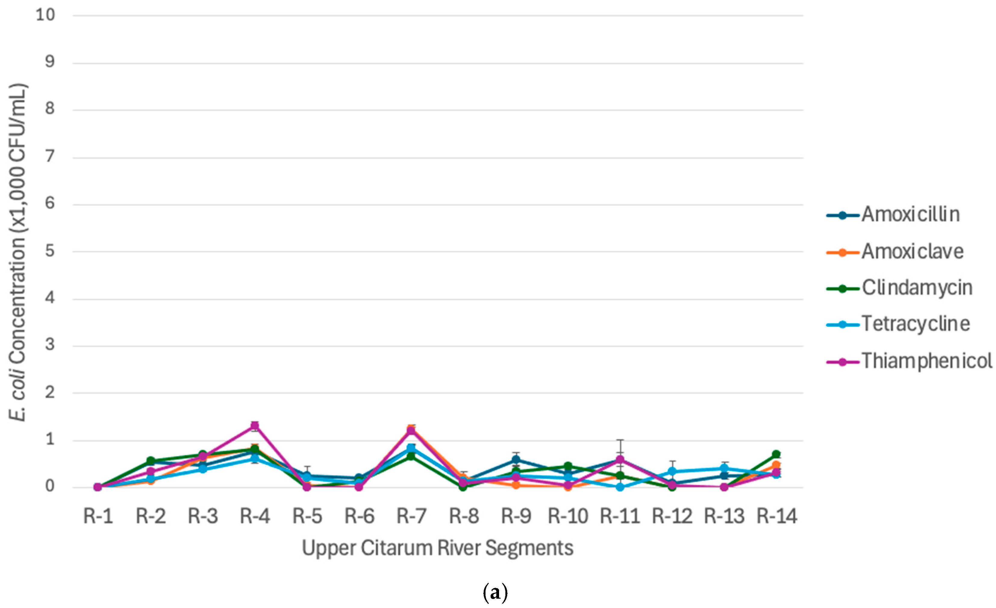
Antibiotic-resistant Escherichia coli (AREc) has been detected in both Period I and Period II in all sampling segments, including Cisanti Spring. Hence, the possibility of a native resistant gene in Escherichia coli needs to be considered. In Figure 5, it can be seen that the AREc concentration increases downstream, starting in the Radug (R-2) segment in both periods. There was also a sudden increase in AREc concentrations in the R-6 (Cikarees), R-11 (Katapang), and R-14 (Batujajar) segments in line with the increase in TEc. This indicates that the primary sources of contamination may be from agriculture, livestock, and domestic activities.
In general, the concentration of AREc that are resistant to access-antibiotics is predominantly higher than those resistant to watch and reserve antibiotics. However, it is notable that AREc resistant to amoxicillin, tetracycline, oxytetracycline, and thiamphenicol showed predominantly high concentrations in all segments, particularly during Period I. The four antibiotics are classified as access and watch category antibiotics; however, in Indonesia, they are widely used as the first-line treatment for infectious diseases caused by Gram-negative bacteria, such as typhoid, and for treating infections in animals, particularly in the fisheries and livestock sectors [47]. Some of these antibiotics are used without a doctor’s prescription and given as supplements to healthy livestock [48,49,50]. Consequently, the high concentration of AREc resistant to these antibiotics indicates a potential reduction in their effectiveness and raises concerns about their continued clinical utility, underscoring the need for parallel evaluation in clinical settings.
The sudden rise of antibiotic-resistant Escherichia coli (AREc) Resistant to watch-category antibiotics in the R-6 (Cikarees), R-11 (Katapang), and R-14 (Batujajar) segments during Period II is related to the high density of domestic settlements and livestock in the area (Figure 5). In a previous study, it was found that residents and livestock used a lot of amoxicillin and tetracycline antibiotics for their activities [27]. In addition, the Koyod segment is a large rice field area with a significant increase in BOD, as seen in both Katapang (7.75 mg/L) and Batujajar (12.6 mg/L) segments, compared to the average BOD in the Upper Citarum River (7.87 mg/L).
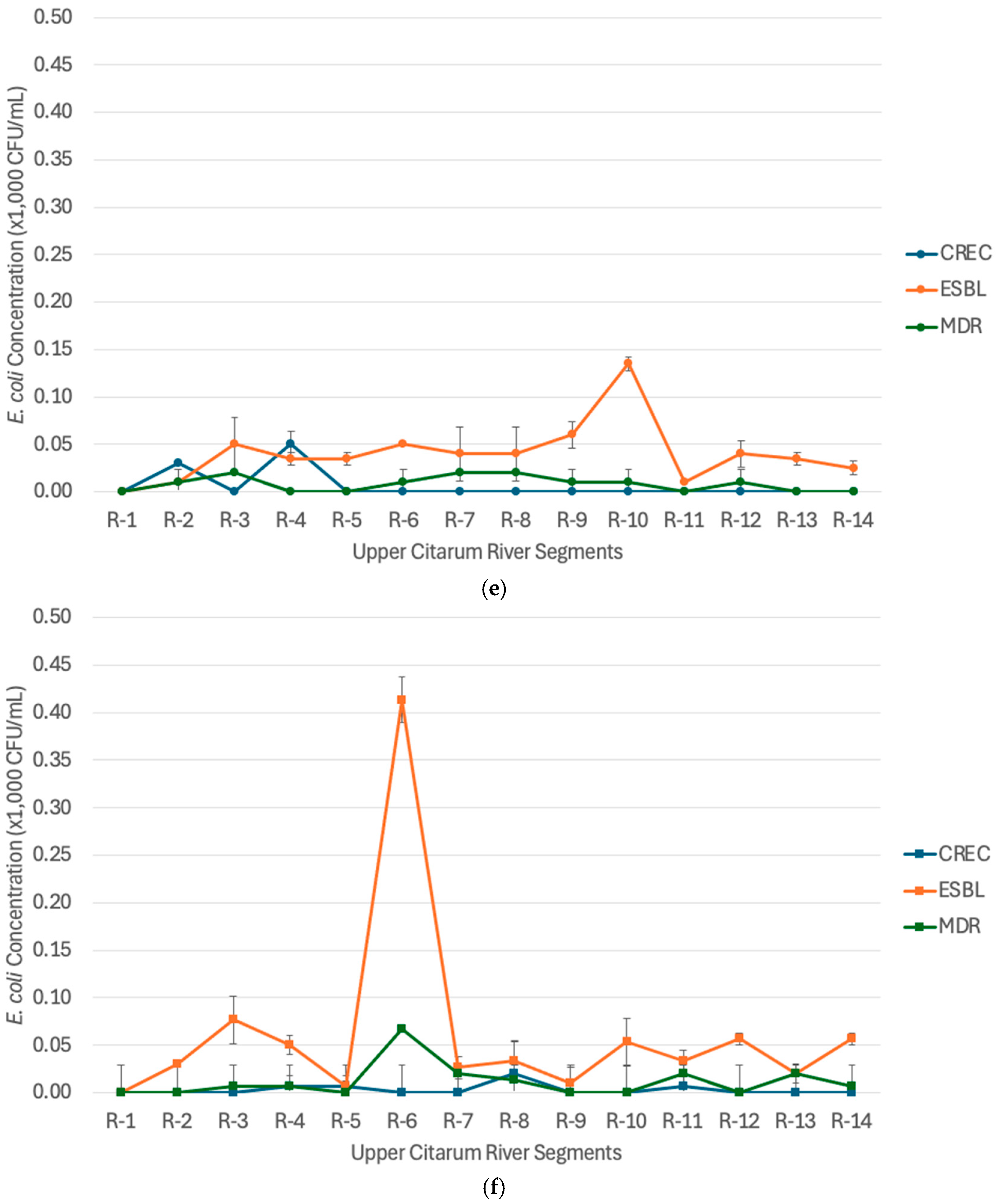
AREc resistant to more than one type of antibiotic, such as ESBL-Ec and MDR-Ec types, have begun to be detected in the Upper Citarum, particularly segment R-2 (Radug) to R-14 (Batujajar), with the concentration of ESBL-Ec 10–135 CFU/mL and MDR-Ec 10–20 CFU/mL in Period I and ESBL-Ec 10–413 CFU/mL and MDR-Ec 6–66 CFU/mL in Period II (Figure 5). This result indicates that the Citarum River may become a reservoir and vector for the spread of Escherichia coli resistance, posing a significant health risk to residents in the Upper Citarum watershed. ESBL-Ec, which is a WHO monitoring priority, has become one of the most highly detected pathogenic bacteria among the three types of AREc in the Upper Citarum River. A study by Cassini [51] estimated that 63.5% of antibiotic-resistant bacterial infections were linked to healthcare settings, which accounted for 72.4% of attributable deaths and 74.9% of disability-adjusted life years (DALYs) per 100,000 population in the European Union. It is stated that Italy and Greece had the highest disease burden, with ESBL-producing Escherichia coli identified as the most prevalent pathogen [48]. A study by Gomi [52] reported that 14.3% of Escherichia coli isolate from the Yamato River were multidrug-resistant. In comparison, other studies found the ratio of multidrug-resistant Escherichia coli to be 49.48% in Ghana’s River, 75% in Portugal’s River, and 34% in the Malaysian River [53,54,55]. However, compared to countries in Europe, such as Greece, where the ratio of ESBL-Ec in the river is up to 60%, the condition of AREc contamination in the Upper Citarum River is better.
The concentration of AREc in a water body indicates the extent of dispersion and the potential health risks due to water usage by its surrounding communities. Figure 6 shows the average concentrations of AREc in all segments of the Upper Citarum River. In general, concentrations of all AREc types were higher in period I when rainfall was lower. A study by [56] demonstrates that slow river flow resulting from low rainfall increases the likelihood of resistant-bacterial gene transfer. The figure also shows that the concentration of AREc resistant to antibiotic erythromycin, amoxicillin, and thiamphenicol is the most dominant in Period I, and AREc resistant to antibiotic erythromycin and thiamphenicol in Period II. In a previous study by [28] AREc resistant to antibiotics, including erythromycin, amoxicillin, and thiamphenicol, was primarily found in the effluent from farms and settlements. AREc resistant to meropenem (reserve category) have been detected in Period I, thus indicating a serious environmental health risk.
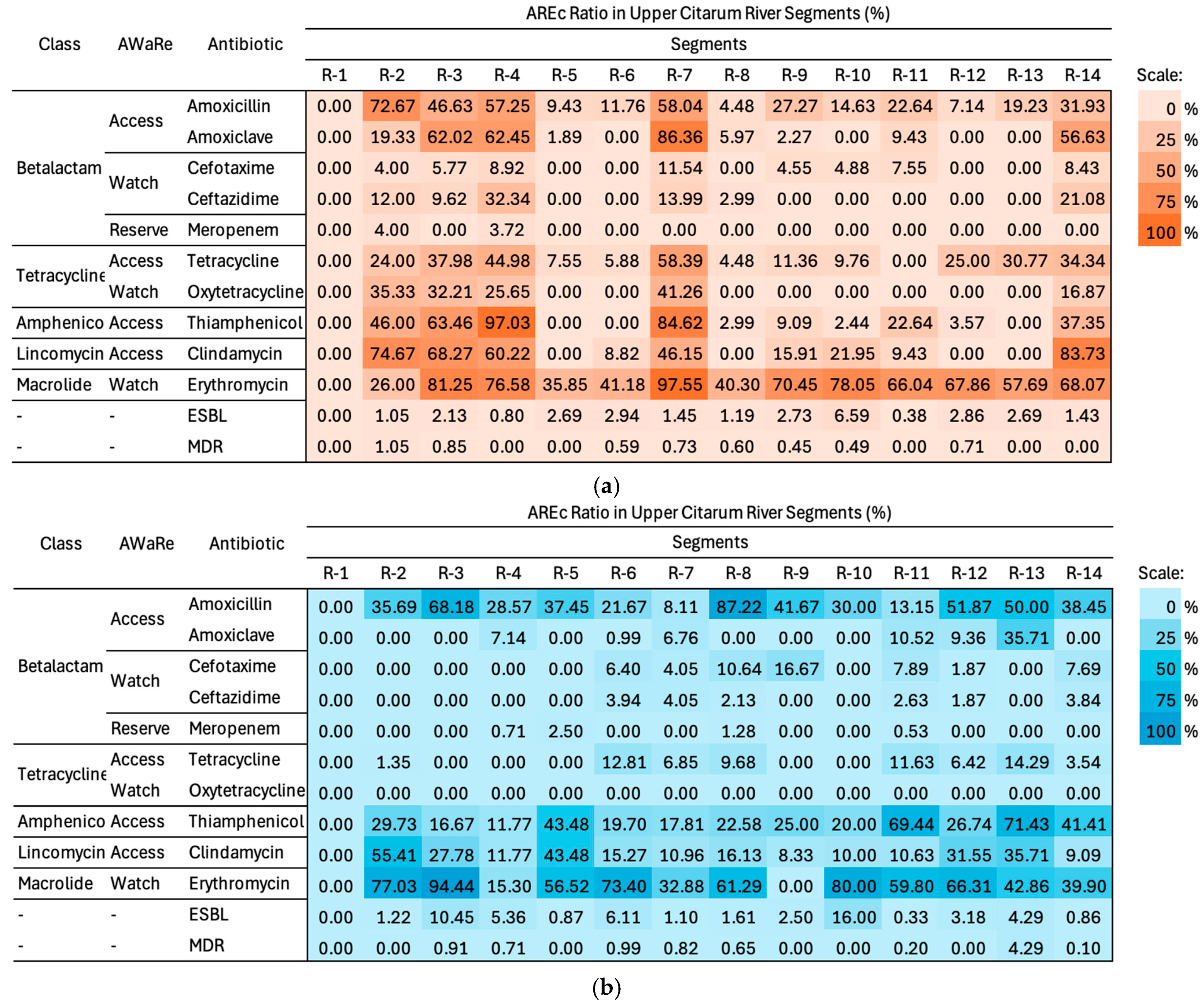
The Ratio of AREc (%) in a water body indicates the source characterization of AREc contamination, which indirectly shows the pattern of irrational consumption of antibiotics in the area. The Ratio of AREc along the Upper Citarum River is shown in Figure 7 below. On average for all river water segments, AREc resistant to erythromycin is the highest AREc ratio in both Period I (𝓍 = 57.6 ± 25.9) and Period II (𝓍 = 49.96 ± 29.5). This condition may be attributed to the runoff of fecal material from livestock waste and the use of fertilizers in the agricultural sector, which enter river water bodies. It is known that erythromycin is more environmentally recalcitrant in sediments, exhibiting a longer half-life of approximately 17 days [57]. AREc resistant to amoxicillin, clindamycin, and thiamphenicol in the Upper Citarum River were also dominant, as well as in both periods. These antibiotics are categorized as access and watch antibiotics, often consumed by surrounding communities as the first line of treatment for infectious diseases, particularly respiratory tract infections, skin infections, and gastrointestinal infections.
Figure 7 also shows that the Ratio of AREc resistant to Amoxiclav, Ceftazidime, Tetracycline, and Oxytetracycline is higher in the first period. Amoxiclav and Ceftazidime are commonly administered by local communities in healthcare facilities. In a previous study, ARECs are highly concentrated in wastewater effluents from hospitals and the pharmaceutical industry. The Ratio of AREc resistant to Amoxiclav and Ceftazidime was higher in period I in the R-2 (Radug) to R-11 (Katapang) segment, indicating that contamination from domestic settlements was more notable in the middle segment, which is the estuary of the Bojongsoang River containing domestic wastewater from Bandung City (see Figure 8). In February and March, the consumption of those antibiotics increased due to the elevated incidence of acute respiratory infections and diarrhea during the transitional season [58].
AREc resistant to tetracycline is notably higher in Period I (𝓍 = 21 ± 18.2), whereas AREc resistant to oxytetracycline was not detected (0%) in Period II. Tetracycline and oxytetracycline is widely used in chicken farms and fisheries in the early segments of the Upper Citarum (R-2 (Radug), R-4 (Koyod) and R-7 (Bojongsoang), where the agricultural and livestock activities are prominent. Even though oxytetracycline has the longest half-life (approximately 77 days) in the aquatic environment, during high rainfall, the effluent from the fisheries sector, which contains AREc-resistant to this antibiotic type, is easily washed out.

3.3. Priority Bacterial Target of Antibiotic-Resistant Escherichia coli (AREc) in the Upper Citarum River

Regular monitoring of AREc in river bodies is crucial for determining the status of environmental health risks, particularly in the Upper Citarum River, where its water is still used as a source of drinking water. Therefore, it is necessary to select priority AREc that can be used as indicators for the presence of other types of AREc bacteria. Thus, monitoring can be carried out effectively and efficiently in terms of time and cost. The criteria of a good bacterial indicator based on Environmental Protection Agency (EPA) are the consistency of presence and concentration with other AREc, the similarity of origin from the same contamination source (e.g., domestic, livestock, or industrial waste), the environmental stability as the target bacteria and the easiness of detection method [59].
A pearson correlation test, as shown in Figure 9, was conducted to assess the correlation among AREc concentrations in the Upper Citarum River (p < 0.01). The results show that AREc resistant to amoxicillin, ceftazidime, oxytetracycline, clindamycin, and tetracycline have a strong correlation (r2 > 0.7, p < 0.05), indicating a genetic linkage [60]. AREc resistant to tetracycline had a high correlation (r2 between 0.39 and 0.75) with other single-resistant AREcs, but a low correlation with MDR-Ec (r2 = 0.202), suggesting that it could be a good indicator only for other single-resistant AREcs. From the previous section (Figure 5), it is known that AREc resistant to tetracycline is consistently measured at a high concentration in both periods due to high contamination from the farm’s wastewater along the river. As for multidrug resistance AREc, the correlation coefficient of ESBL-Ec with other single-resistant AREc varies but has a close correlation with amoxicillin (r2 = 0.571), cefotaxime (r2 = 0.734), and erythromycin (r2 = 0.566). The ESBL-Ec correlates closely with the MDR-Ec (r2 = 0.804). ESBL-Ec is known to have resistance genes towards beta-lactam antibiotics, including penicillin, cephalosporin, and aztreonam [61]. Due to the position of the gene on the plasmid, some ESBL-Ec strains can also be resistant to carbapenem, fluoroquinolone, aminoglycoside, and tetracycline, which is induced by gene mutation [45,46]. This result suggests that ESBL-Ec is the best alternative as a universal indicator bacterium for AREc in the Upper Citarum River, based on Pearson Correlation.
A Kaiser-Meyer-Olkin test was performed on the AREc concentration data, resulting in 0.510 which implies enough significancy for Principal Component Analysis. The Principal Component Analysis results indicate the number of principal components required to explain most of the dataset’s variability, as well as a table displaying the variance explained by each principal component. Tetracycline-resistant AREc had the highest PC1 loading (0.869), as shown in the Supplementary Materials, making it the most influential indicator of variation in the concentrations of other AREc.
The clustering visualization of the PCA analysis in Figure 10 shows that Tetracycline-resistant AREc is in the same cluster as most of the single-resistant AREc, in line with the PC1 calculation results. ESBL-Ec is in the same cluster as TEc and MDR-Ec; therefore, the presence of ESBL-Ec is more significant in terms of environmental health risk. Based on these results, it can be concluded that for monitoring the distribution of single antibiotic-resistant AREc, Tetracycline-resistant AREc is the main priority in the Upper Citarum River. However, ESBL-Ec is more suitable for indicating the multidrug resistance AREc since it has the highest PC1 loading (0.659) amongst MDR-Ec and CREc, and is consistently detected in all river water samples. In many countries, such as the US, India, China, Germany, and Brazil, ESBL-Ec is one of the priority bacteria for monitoring the distribution of AREc in water bodies [50,51]. However, the determination of AREc indicator bacteria should be done regularly, since resistance patterns may vary between countries and change over time, influenced by variations in antibiotic usage practices and local antibiotic availability [17].

3.4. Comparison of Antibiotic-Resistant Escherichia coli Ratio Using the Two Methods

Comparison of the AREc ratio in river water samples measured by the CCA Agar method and the MPR method is shown in Figure 11. The MPR Method has an overall high agreement rate compared to the CCA Agar Method in predicting the AREc Ratio, as indicated by the linearity of the data spread across the midline of the logarithmic scale. AREc ratios obtained through the MPR method had a strong Pearson correlation rate with the CCA Agar method for most single-resistant AREc, particularly AREc resistant to Amoxiclav (r2 = 0.885, p-value < 0.05), Ceftazidime (r2 = 0.868, p-value < 0.05), and Oxytetracycline (r2 = 0.862, p-value < 0.05). The ratios of AREc resistant to erythromycin (r2 = 0.416, p-value < 0.05) and cefotaxime (r2 = 0.411, p-value < 0.05) using the MPR method results tends to have a weaker correlations rate. The type of antibiotic affects the accuracy of the MPR method in predicting the AREc Ratio. Some antibiotic solutions have intrinsic fluorescence activity, such as tetracycline, thiamphenicol, and erythromycin. Moreover, bactericidal antibiotics, such as cefotaxime, tend to lyse the cell wall and release the GUS into the media [62]. Therefore, the AREc Ratio to these antibiotics is higher in the MPR method. AREc Ratio obtained through the MPR method for ESBL-Ec and MDR-Ec had the weakest correlation rate (r2 ESBL-Ec = 0.243, p-value > 0.05 and r2 MDR-Ec = 0.025, p-value > 0.05). This condition is due to a lower AREc Ratio (%) detected by the MPR method, which indicates that more bacteria were eliminated in the MPR Method. This condition may be related to the use of multiple antibiotics in the susceptibility test for ESBL-Ec and MDR-Ec. The homogenization of multiple antibiotics in a liquid medium, as in the MPR method, is slightly better, thus increasing the contact surface area between bacteria and antibiotics. Moreover, the homogenization of multiple antibiotics better simulates real environmental conditions, where bacteria are often exposed to complex mixtures of antibiotics rather than single compounds. In addition, liquid media may reduce interferences or other inhibitions from sample matrices. Therefore, in liquid media, such as the MPR method, the bactericidal effect of antibiotics tends to be higher, and the AREc Ratio will be reduced [63,64]. These results show the benefit of the MPR Method as a more accurate method in the context of multiple-antibiotic susceptibility testing.
In several epidemiological studies, a sample is considered a positive result when it contains Antibiotic-Resistant Escherichia coli (AREc) ratios greater than 20%, and vice versa [38]. This study also uses this value as a threshold for determining accuracy. The accuracy of the MPR method in this study was 87.2%, the sensitivity was 67.4%, and the specificity was 94%. The moderate sensitivity represents a key limitation for the MPR Method. The MPR method tends to underestimate AREc ratios compared to the CCA agar method, leading to potential false negatives. This limitation is partly due to the absence of a functional uidA gene in certain Escherichia coli strains, such as the pathogenic O157:H7 variant, which prevents β-glucuronidase expression. Moreover, environmental factors in complex river water matrices may suppress β-glucuronidase activity and reduce sensitivity [25]. The MPR method is more effective at detecting Escherichia coli in high-concentration samples, as shown in Table 3, where sensitivity increases with bacterial density. However, for low concentrations, the method is less reliable since the fluorogenic assay can only detect E. coli down to ~1 CFU per 100 mL under optimized conditions [24]. These findings highlight that the application of the MPR method may require optimization to minimize false negatives and improve detection in samples with low bacterial loads.
Rainfall significantly affected the sensitivity value, as shown in Table 3, where the accuracy decreases during Period II, when rainfall was higher. High rainfall caused a significant amount of rainwater runoff to flow into the river, increasing the solids and other contaminants in the river water. The presence of these contents in the tested river water samples led to the non-detection of AREc content in the river water samples, thus reducing the sensitivity of the detection method. This finding aligns with research by Satoh [16], which states that disturbances in the environmental matrix, such as the presence of microorganisms or other contaminants, can hinder bacterial development. From these findings, the MPR Method may serve as a time-saving and simple alternative method for characterizing the AREc ratio in less turbid river water with a high bacterial load.
Based on these findings and several limitations of this study, the use of the MPR method should be optimized by reducing inferential environmental matrices, such as solids in the river water samples, to increase its sensitivity. Future lab-scale studies should be conducted to balance the comparisons of the AREc Ratio in high and low bacterial loads using a standard, known bacterial sample. A similar study may also be conducted in a more variable temporal and spatial environment to get a more robust understanding of the environmental factors that may interfere with the MPR performance. The results support the use of ESBL-Ec as a sentinel indicator in annual river water quality monitoring, with the MPR technique offering a practical tool to facilitate routine monitoring. These findings may facilitate the integration of environmental monitoring with antimicrobial resistance (AMR) surveillance in the Upper Citarum River.

4. Conclusions

The Upper Citarum River is a heavily polluted river with a high load of Total Escherichia coli, particularly during high rainfall. TEc concentrations in the Upper Citarum ranged from 0–3350 CFU/mL (𝓍 = 1621 ± 300 CFU/mL) in period I (February to March 2024 with an average precipitation of 155.7 mm/month) and 0–10,033 CFU/mL (𝓍 = 2558 ± 360 CFU/mL) in period II (November to December 2024 with an average precipitation of 302.3 mm/month). AREc resistant to erythromycin as the highest AREc ratio, is prominent in both period (Period I 𝓍 = 57.6 ± 25.9%, Period II 𝓍 = 49.96 ± 29.5%), followed by AREc resistant to amoxicillin (Period I 𝓍 = 27.3 ± 25.9%, Period II 𝓍 = 36.5 ± 23.2%), clindamycin (Period I 𝓍 = 27.7 ± 31.7%, Period II 𝓍 = 20.4 ± 15.8%) and thiamphenicol (Period I 𝓍 = 26.3 ± 33.9%, Period II 𝓍 = 29.6 ± 20.4%). This condition is largely influenced by the runoff of fecal waste from farms and settlements in surrounding areas. Furthermore, ESBL-Ec, MDR-Ec, and carbapenem-resistant Escherichia coli are detected in a low ratio in both periods, indicating a potential environmental health threat. The result of PCA analysis indicates the tetracycline-resistant Escherichia coli as the selected AREc indicator for single resistant AREc in the Upper Citarum River with the highest PC1 loading (0.869). However, in terms of environmental health risk, which primarily considers the potential health hazard to the community, ESBL-Ec is also important (PC1 loading 0.659) due to its close relation to MDR-Ec and Carbapenem-resistance Escherichia coli (CREc). Furthermore, the MPR method in this study achieved an accuracy of up to 87.2%, a sensitivity of 67.4%, and a specificity of 94%, which is suitable for the simple alternative AREc detection method in Upper Citarum River, particularly in a high bacterial load aquatic environment such as Upper Citarum River. Due to the limitations of this study, future research should expand monitoring of the river water to capture broader seasonal variability, include additional sampling locations, incorporate sediment sampling to assess further resistance reservoirs, and validate the MPR method across different matrices. Scaling this approach to other rivers and integrating the findings into national antimicrobial resistance surveillance programs will also be important.

Supplementary Materials

The following supporting information can be downloaded at https://www.mdpi.com/article/10.3390/w17182791/s1: Table S1: Spatiotemporal Variability of Total Escherichia coli Concentration in the Upper Citarum River; Table S2. Spatiotemporal Variability of Antibiotic-Resistant Escherichia coli Concentration in Two Measurement Periods; Table S3. Correlation among TEc and AREc with Environmental Factors; Table S4. PCA Analysis for Determining Priority AREc; Table S5. Log Phase Initial Time Calculation.

Author Contributions

Conceptualization, S.W.D.K. and H.D.A.; methodology, S.W.D.K. and H.S.; formal analysis, S.W.D.K. and R.R.F.; investigation, S.W.D.K. and M.K.; validation, P.A.N. and A.S.S.; data curation, S.W.D.K. and M.K.; writing—original draft preparation, S.W.D.K.; writing—review and editing, H.D.A. and H.S.; supervision, H.D.A. and H.S.; funding acquisition, P.A.N. and A.S.S. All authors have read and agreed to the published version of the manuscript.

Funding

This research was partially funded by Hibah Penerbitan Publikasi Internasional dan Program Penelitian, Pengabdian kepada Masyarakat dan Inovasi (PPMI), FTSL, ITB and Kurita Overseas Research Grant Program 2024, grant number: 24Pid083.

Data Availability Statement

The original contributions presented in this study are included in the article/Supplementary Material. Further inquiries can be directed to the corresponding author.

Acknowledgments

The authors gratefully acknowledge Hokkaido University for providing the laboratory equipment and resources that facilitated this study.

Conflicts of Interest

The authors have declared no conflicts of interest. The authors declare that they have no known competing financial interest or personal relationships that could have appeared to influence the work reported in this paper.

References

  1. Hutchings, M.; Truman, A.; Wilkinson, B. Antibiotics: Past, present and future. Curr. Opin. Microbiol. 2019, 51, 72–80. [Google Scholar] [CrossRef]
  2. Klein, E.Y.; Van Boeckel, T.P.; Martinez, E.M.; Pant, S.; Gandra, S.; Levin, S.A.; Goossens, H.; Laxminarayan, R. Global increase and geographic convergence in antibiotic consumption between 2000 and 2015. Proc. Natl. Acad. Sci. USA 2018, 115, E3463–E3470. [Google Scholar] [CrossRef] [PubMed]
  3. Camcioglu, Y.; Okur, D.S.; Aksaray, N.; Darendeliler, F.; Hasanoglu, E. Factors affecting physicians’ perception of the overuse of antibiotics. Med. Mal. Infect. 2020, 50, 652–657. [Google Scholar] [CrossRef]
  4. Ghosh, S.; Bornman, C.; Zafer, M.M. Antimicrobial Resistance Threats in the emerging COVID-19 pandemic: Where do we stand? J. Infect. Public Health 2021, 14, 555–560. [Google Scholar] [CrossRef] [PubMed]
  5. Mancuso, G.; Midiri, A.; Gerace, E.; Biondo, C. Bacterial Antibiotic Resistance: The Most Critical Pathogens. Pathogens 2021, 10, 1310. [Google Scholar] [CrossRef]
  6. Nastiti, A.; Utami, R.R.; Ramadhina, S.Q.; Fathonah, N.; Yoga, G.P.; Ariesyady, H.D.A.; Kusumah, S.W.D.; Hidayat. Disposal practices, risk perceptions, and quantification of potential active pharmaceutical ingredients (APIs) from used human medicine in Upper Citarum River Basin. Int. J. Environ. Health Res. 2025, 1–18. [Google Scholar] [CrossRef] [PubMed]
  7. Mahoney, A.R.; Safaee, M.M.; Wuest, W.M.; Furst, A.L. The silent pandemic: Emergent antibiotic resistances following the global response to SARS-CoV-2. iScience 2021, 24, 102304. [Google Scholar] [CrossRef] [PubMed]
  8. Giacomini, E.; Perrone, V.; ALessandriani, D.; Paoli, D.; Nappi, C.; Degli, E.L. Evidence of antibiotic resistance from population-based studies: A narrative review. Infect. Drug Resist. 2021, 14, 849. [Google Scholar] [CrossRef]
  9. World Health Organization. GLASS Method for Estimating Attributable Mortality of Antimicrobial Resistant Bloodstream Infections; World Health Organization: Geneva, Switzerland, 2020. [Google Scholar]
  10. Larsson, D.; Flach, C.F. Antibiotic resistance in the environment. Nat. Rev. Microbiol. 2022, 20, 257–269. [Google Scholar] [CrossRef]
  11. Li, H.; Song, R.; Wang, Y.; Zhong, R.; Zhang, Y.; Zhou, J.; Wang, T.; Zhu, L. Simultaneous removal of antibiotic-resistant bacteria and its resistance genes in water by plasma oxidation: Highlights the effects of inorganic ions. Sep. Purif. Technol. 2021, 278, 119672. [Google Scholar] [CrossRef]
  12. Jia, Y.; Wang, Z.; Fang, D.; Yang, B.; Li, R.; Liu, Y. Acetaminophen promotes horizontal transfer of plasmid-borne multiple antibiotic resistance genes. Sci. Total Environ. 2021, 782, 146916. [Google Scholar] [CrossRef]
  13. World Health Organization. WHO Bacterial Priority Pathogens List: Bacterial Pathogens of Public Health Importance to Guide Research, Development and Strategies to Prevent and Control Antimicrobial Resistance. Available online: https://www.who.int/publications/i/item/9789240093461 (accessed on 30 May 2024).
  14. Bengstton-Palme, J.; Larsson, D.G.J. Concentrations of antibiotics predicted to select for resistant bacteria: Proposed limits for environmental regulation. Environ. Int. 2016, 86, 140–149. [Google Scholar] [CrossRef] [PubMed]
  15. Ben, Y.; Fu, C.X.; Hu, M.; Liu, L.; Zheng, C.M. Human health risk assessment of antibiotic resistance associated with antibiotic residues in the environment: A review. Environ. Res. 2019, 169, 483–493. [Google Scholar] [CrossRef] [PubMed]
  16. Satoh, H.; Nagahashi, N.; Kikuchi, K.; Hirano, R. Screening Antibiotic-Resistant Escherichia coli in Wastewater and River Water Using a Novel Simple Phenotypic Antibiotic-Susceptibility Testing Method. ACS EST Water 2022, 2, 1301–1308. [Google Scholar] [CrossRef]
  17. Grenni, P. Antimicrobial resistance in rivers: A review of the genes detected and new challenges. Environ. Toxicol. Chem. 2022, 41, 687–714. [Google Scholar] [CrossRef]
  18. Gekenidis, M.T.; Qi, W.; Hummerjohann, J.; Zbinden, R.; Walsh, F.; Drissner, D. Antibiotic-resistant indicator bacteria in irrigation water: High prevalence of extended-spectrum beta-lactamase (ESBL)-producing Escherichia coli. PLoS ONE 2018, 13, e0207857. [Google Scholar] [CrossRef]
  19. Oliveira, M.; Truchado, P.; Cordero-García, R.; Gil, M.I.; Soler, M.A.; Rancaño, A.; García, F.; Álvarez-Ordóñez, A.; Allende, A. Surveillance on ESBL-Escherichia coli and indicator ARG in wastewater and reclaimed water of four regions of Spain: Impact of different disinfection treatments. Antibiotics 2023, 12, 400. [Google Scholar] [CrossRef]
  20. Hashim, N.H.F.; Yusoff, M.A.M.; Gunggang, R.A.T.; Razak, R.A.; Jaafar, M.Z.; Yahaya, N.K.E. Water Quality and Prevalence of Extended Spectrum Beta Lactamase Producing Escherichia coli (ESBL E. coli) in Sungai Terengganu, Malaysia. Malays. Appl. Biol. 2024, 53, 65–75. [Google Scholar] [CrossRef]
  21. Julian, T.R.; Islam, M.A.; Pickering, A.J.; Roy, S.; Fuhrmeister, E.R.; Ercumen, A.; Harris, A.; Bishai, J.; Schwab, K.J. Genotypic and phenotypic characterization of Escherichia coli isolates from feces, hands, and soils in rural bangladesh via the colilert quanti-tray system. Appl. Environ. Microbiol. 2015, 81, 1735–1743. [Google Scholar] [CrossRef] [PubMed]
  22. Genter, F.; Marks, S.J.; Clair-Caliot, G.; Mugume, D.S.; Johnston, R.B.; Bain, R.E.; Julian, T.R. Evaluation of the novel substrate rugTM for the detection of Escherichia coli in water from temperate (zurich, switzerland) and tropical (bushenyi, uganda) field sites. Environ. Sci. 2019, 5, 1082–1091. [Google Scholar]
  23. Wang, D.; Chen, J.; Nugen, S. Electrochemical detection of Escherichia coli from aqueous samples using engineered phages. Anal. Chem. 2017, 89, 1650–1657. [Google Scholar] [CrossRef]
  24. Zhang, Y.; Hong, P.Y.; LeChevallier, M.W.; Liu, W.T. Phenotypic and phylogenetic identification of coliform bacteria obtained using 12 coliform methods approved by the US Environmental Protection Agency. Appl. Environ. Microbiol. 2015, 81, 6012–6023. [Google Scholar] [CrossRef]
  25. Shayan, M.N.; Tanaka, Y.; Hirano, R.; Nakaya, Y.; Satoh, H. A simple and rapid method for detecting fecal pollution in urban rivers by measuring the intrinsic β-D-glucuronidase activity of Escherichia coli. Water Res. 2023, 246, 120689. [Google Scholar] [CrossRef] [PubMed]
  26. Fadhil, M.Y.; Hidayat, Y.; Murtilaksono, K.; Baskoro, D.P.T. Changes in land use and hydrological characteristics of the Upstream Citarum watershed. J. Ilmu Pertan. Indones. 2021, 26, 213–220. [Google Scholar] [CrossRef]
  27. Fitriana, F.; Yudianto, D.; Sanjaya, S.; Roy, A.F.; Seo, Y.C. The assessment of Citarum river water quality in Majalaya District, Bandung regency. Rekayasa Sipil 2023, 17, 37–46. [Google Scholar] [CrossRef]
  28. Kusumah, S.W.D.; Kandio, N.; Maghfirah, R.; Setiyawan, A.S.; Ariesyady, H.D. The occurrence of Antibiotic-Resistant Escherichia coli in the Upper Citarum River and surrounding wastewater effluents. E3S Web Conf. 2024, 485, 07006. [Google Scholar] [CrossRef]
  29. Andriani, F.; Ariesyady, H.D. Microbiological Source Tracking Bakteri Escherichia coli dengan Metode Antibiotic Resistance Analysis di Sungai Cikapundung. J. Environ. Eng. ITB 2013, 19, 170–176. [Google Scholar] [CrossRef]
  30. Badan Standardisasi Nasional. “SNI 03-7016-2004: Tata Cara Pengambilan Contoh Dalam Rangka Pemantauan Kualitas Air pada Suatu Daerah Pengaliran Sungai,” Jakarta: BSN. Available online: https://perpustakaan.ciptakarya.pu.go.id/opac/index.php?p=show_detail&id=10227 (accessed on 5 January 2025).
  31. Clinical and Laboratory Standards Institute (CLSI). Performance Standards for Antimicrobial Susceptibility Testing, 34th ed.; CLSI Supplement M100; Clinical and Laboratory Standards Institute: Wayne, PA, USA, 2024. [Google Scholar]
  32. Satoh, H.; Kikuchi, K.; Katayose, Y.; Tsuda, S.; Hirano, R.; Hirakata, Y.; Kitajima, M.; Ishii, S.; Oshiki, M.; Hatamoto, M.; et al. Simple and reliable enumeration of Escherichia coli concentrations in wastewater samples by measuring β-d-glucuronidase (GUS) activities via a microplate reader. Sci. Total Environ. 2020, 715, 136928. [Google Scholar] [CrossRef]
  33. Satoh, H.; Katayose, Y.; Hirano, R. Simple enumeration of Escherichia coli concentrations in river water samples by measuring β-D-glucuronidase activities in a microplate reader. Water Sci. Technol. 2021, 83, 1399–1406. [Google Scholar] [CrossRef]
  34. Rahayudin, Y.; Iskandar, I.; Agus, M.; Ramdhan. Hidrogeologi dan Hidrogeokimia Mata Air Danau Cisanti (Hulu Sungai Citarum). Master’s Thesis, Bandung Institute of Technology, Bandung, Indonesia, 2014. [Google Scholar]
  35. BPS Provinsi Jawa Barat. Persentase Rumah Tangga yang Memiliki Akses Terhadap Sanitasi Layak (Persen), 2022–2024. Available online: https://jabar.bps.go.id/id/statistics-table/2/NzI4IzI=/persentase-rumah-tangga-yang-memiliki-akses-terhadap-sanitasi-layak.html (accessed on 5 January 2025).
  36. Zahra, Z.; Indrawaty, L.; Arfines, P.P. Manajemen Pengelolaan Sampah Rumah Tangga Dan Limbah Ternak Di Kawasan Peternakan Daerah Aliran Sungai Citarum Kabupaten Bandung. J. Ekol. Kesehat. 2021, 20, 165–175. [Google Scholar] [CrossRef]
  37. Kementerian Lingkungan Hidup dan Kehutanan Republik Indonesia. “Laporan Kinerja Tahun 2023: Direktorat Pengendalian Pencemaran Air, Direktorat Jenderal Pengendalian Pencemaran dan Kerusakan Lingkungan,” Kementerian Lingkungan Hidup dan Kehutanan Republik Indonesia. Available online: https://ppkl.menlhk.go.id/website/filebox/1194/240118150657LKj%20Direktorat%20PPA%202023.pdf (accessed on 2 August 2025).
  38. Ram, S.; Vajpayee, P.; Shanker, R. Prevalence of multi-antimicrobial-agent resistant, shiga toxin and enterotoxin producing Escherichia coli in surface waters of river ganga. Environ. Sci. Technol. 2007, 41, 7383–7388. [Google Scholar] [CrossRef]
  39. Bowes, M.J.; Read, D.S.; Joshi, H.; Sinha, R.; Ansari, A.; Hazra, M.; Simon, M.; Vishwakarma, R.; Armstrong, L.K.; Nicholls, D.J.; et al. Nutrient and microbial water quality of the upper Ganga River, India: Identification of pollution sources. Environ. Monit. Assess. 2020, 192, 533. [Google Scholar] [CrossRef]
  40. Matta, G.; Naik, P.K.; Machell, J.; Kumar, A.; Gjyli, L.; Tiwari, A.K.; Kumar, A. Comparative study on seasonal variation in hydro-chemical parameters of Ganga River water using comprehensive pollution index (CPI) at Rishikesh (Uttarakhand) India. Desalination Water Treat. 2018, 118, 87–95. [Google Scholar] [CrossRef]
  41. Kumari, A.; Sinha, S.K.; Rani, N.; Sinha, R.K. Assessment of heavy metal pollution in water, sediment, and fish of the river Ganga at Varanasi, India. Arab. J. Geosci. 2021, 14, 2346. [Google Scholar] [CrossRef]
  42. Rohene, A.; Srikaran, P. Modeling the Impact of Animal Farming on Water Quality of Sg. Serin. Ph.D. Thesis, Universiti Malaysia Sarawak, Kota Samarahan, Malaysia, 2009. [Google Scholar]
  43. Yee, L.; Wee, L.; Bilung, L.; Lee, N. Impact of different land uses on the Escherichia coli concentrations, physical and chemical water quality parameters in a tropical stream. Borneo J. Resour. Sci. Technol. 2016, 2, 42–51. [Google Scholar] [CrossRef]
  44. Tachibana, H.; Yamamoto, K.; Yoshizawa, K.; Magara, Y. Non-point pollution of Ishikari river, Hokkaido, Japan. Water Sci. Technol. 2001, 44, 1–8. [Google Scholar] [CrossRef]
  45. Sikder, M.T.; Kihara, Y.; Yasuda, M.; Yustiawati Mihara, Y.; Tanaka, S.; Odgerel, D.; Mijiddorj, B.; Syawal, S.M.; Hosokawa, T.; Saito, T. River Water Pollution in Developed and Developing Countries: Judge and Assessment of Physicochemical Characteristics and Selected Dissolved Metal Concentration. CLEAN Soil Air Water 2013, 41, 60–68. [Google Scholar] [CrossRef]
  46. Negishi, J.N.; Izumi, H.; Wu, J.; Fukui, S.; Koizumi, I. Does winterkill explain contrasting demographics of winter. bioRxiv 2023. 2023.11.07.565987. [Google Scholar] [CrossRef]
  47. Hidayati, N.V.; Syakti, A.D.; Asia, L.; Lebarillier, S.; Khabouchi, I.; Widowati, I.; Sabdono, A.; Piram, A.; Doumenq, P. Emerging contaminants detected in aquaculture sites in Java, Indonesia. Sci. Total Environ. 2021, 773, 145057. [Google Scholar] [CrossRef]
  48. Kurniawan, A.R.; Suryani, E.; Putra, I.M. Antibiotic utilization and its implications among ruminant farmers and stakeholders in Sumbawa Regency, Indonesia. One Health 2017, 7, 122–127. [Google Scholar]
  49. Wanniatie, S.; Furi, M.; Widiasih, D.A. Detection of antibiotic residues in chicken meat and eggs from traditional markets at Yogyakarta City using bioassay method. Vet. World 2019, 12, 1717–1722. [Google Scholar]
  50. Widiasih, D.A.; Pratama, R.P.; Drastini, Y.; Putri, K.; Fatimah, L.N.; Indarjulianto, S. Rapid testing of antibiotic residues to increase food safety awareness of animal origin. Vet. World 2024, 17, 1177–1183. [Google Scholar] [CrossRef]
  51. Cassini, A.; Högberg, L.D.; Plachouras, D.; Quattrocchi, A.; Hoxha, A.; Simonsen, G.S.; Colomb-Cotinat, M.; Kretzschmar, M.E.; Devleesschauwer, B.; Cecchini, M.; et al. Attributable deaths and disability-adjusted life-years caused by infections with antibiotic-resistant bacteria in the EU and the European Economic Area in 2015: A population-level modelling analysis. Lancet Infect. Dis. 2019, 19, 56–66. [Google Scholar] [CrossRef] [PubMed]
  52. Gomi, R.; Matsuda, T.; Matsumura, Y.; Yamamoto, M.; Tanaka, M.; Ichiyama, S.; Yoneda, M. Whole-Genome Analysis of Antimicrobial-Resistant and Extraintestinal Pathogenic Escherichia coli in River Water. Appl. Environ. Microbiol. 2017, 83, e02703. [Google Scholar] [CrossRef] [PubMed]
  53. Bessa, L.J.; Barbosa-Vasconcelos, A.; Mendes, Â.; Vaz-Pires, P.; da Costa, P.M. High prevalence of multidrug-resistant Escherichia coli and Enterococcus spp. in river water, upstream and downstream of a wastewater treatment plant. J. Water Health 2014, 12, 426–435. [Google Scholar] [CrossRef]
  54. Ghaderpour, A.; Ho, W.S.; Chew, L.-L.; Bong, C.W.; Chong, V.C.; Thong, K.-L.; Chai, L.C. Diverse and abundant multi-drug resistant E. coli in Matang mangrove estuaries, Malaysia. Front. Microbiol. 2015, 6, 977. [Google Scholar] [CrossRef]
  55. Odonkor, S.T.; Addo, K.K. Prevalence of multidrug-resistant Escherichia coli isolated from drinking water sources. Int. J. Microbiol. 2018, 1, 7204013. [Google Scholar]
  56. Madsen, J.S.; Burmølle, M.; Hansen, L.H.; Sørensen, S.J. The interconnection between biofilm formation and horizontal gene transfer. FEMS Immunol Med Microbiol. 2012, 65, 183–195. [Google Scholar] [CrossRef]
  57. Felis, E.; Montesissa, C.; Gabbarini, C. Persistence and degradation of erythromycin in sediments and water under laboratory conditions. Environ. Toxicol. Chem. 2012, 31, 847–852. [Google Scholar]
  58. Hidayati, R.; Anggiani, S.T.; Maufikoh, I. Incidence Analysis of an Acute Respiratory Infection due to Climate Conditions and PM10 Concentration. Agromet 2017, 31, 62–70. [Google Scholar] [CrossRef]
  59. Anjum, M.F.; Schmitt, H.; Börjesson, S.; Berendonk, T.U.; Donner, E.; Stehling, E.G.; Boerlin, P.; Topp, E.; Jardine, C.; Li, X.; et al. The potential of using E. coli as an indicator for the surveillance of antimicrobial resistance (AMR) in the environment. Curr. Opin. Microbiol. 2021, 64, 152–158. [Google Scholar] [CrossRef]
  60. Kerek, Á.; Román, I.; Szabó, Á.; Kovács, D.; Kardos, G.; Kovács, L.; Jerzsele, Á. Antibiotic resistance genes in Escherichia coli—Literature review. Crit. Rev. Microbiol. 2025, 1–35. [Google Scholar] [CrossRef]
  61. Paterson, D.L.; Bonomo, R.A. Extended-spectrum beta-lactamases: A clinical update. Clin. Microbiol. Rev. 2005, 18, 657–686. [Google Scholar] [CrossRef] [PubMed]
  62. Pawar, S.P.; Pawar, P.P.; Patil, K.S.; Jadhav, P.V. A fluorescence-based sensor for selective and sensitive detection of amoxicillin in aqueous media. Spectrochim. Acta A Mol. Biomol. Spectrosc. 2017, 179, 108–115. [Google Scholar]
  63. Cansizoglu, M.; Tamer, Y.; Farid, M.; Koh, A.; Toprak, E. Rapid ultrasensitive detection platform for antimicrobial susceptibility testing. PLoS Biol. 2019, 17, e3000291. [Google Scholar] [CrossRef] [PubMed]
  64. Meerwein, M.; Tarnutzer, A.; Böni, M.; Bambeke, F.; Hombach, M.; Zinkernagel, A. Increased azithromycin susceptibility of multidrug-resistant gram-negative bacteria on rpmi-1640 agar assessed by disk diffusion testing. Antibiotics 2020, 9, 218. [Google Scholar] [CrossRef]
Figure 1. Sampling Points.
Water 17 02791 g001
Figure 2. Procedure of MPR Method (In addition to the original, 4 times, and 16 times dilutions without antibiotic, an extra cuvette was prepared with the addition of antibiotic).
Water 17 02791 g002
Figure 3. Concentration of Total Escherichia coli (TEc) in Upper Citarum River Across 14 Sampling Sites during Period I (February–March, Orange Line) and Period II (November–December, Blue Line), Compared with the Class I Water Quality Standard (Green Line).
Water 17 02791 g003
Figure 4. The distribution and number of livestock (cattle and poultry) in the Upper Citarum Watershed. The thick blue line represents the main Upper Citarum River, while the thin blue lines represent the river branches.
Water 17 02791 g004
Figure 5. Concentration of Escherichia coli Resistant to ‘Access’, ‘Watch’, and ‘Reserve’ Antibiotics in Upper Citarum River. (a) AREc Resistant to ‘Access’ Antibiotic on Period I. (b) AREc Resistant to ‘Access’ Antibiotic on Period II. (c) AREc Resistant to ‘Watch’ Antibiotic on Period I. (d) AREc Resistant to ‘Watch’ Antibiotic on Period II. (e) AREc Resistant to ‘Reserve’ Antibiotic on Period I. (f) AREc Resistant to ‘Reserve’ Antibiotic on Period II.
Water 17 02791 g005aWater 17 02791 g005bWater 17 02791 g005c
Figure 6. Concentration average of Antibiotic-Resistant Escherichia coli in nine segments of Upper Citarum River During (a) Period I (February to March 2024, average precipitation: 155.7 mm/month) and (b) Period II (November to December 2024, average precipitation: 302.3 mm/month).
Water 17 02791 g006aWater 17 02791 g006b
Figure 7. Ratio of Antibiotic-Resistant Escherichia coli (%) in nine segments of Upper Citarum River during (a) Period I (February to March 2024, average precipitation: 155.7 mm/month) and (b) Period II (November to December 2024, average precipitation: 302.3 mm/month) on heatmap shading indicating resistance levels from 0% (light) to 100% (dark).
Water 17 02791 g007
Figure 8. The Spatial Distribution of Hospital and Pharmaceutical Industries in the Upper Citarum Watershed. The thick blue line represents the main Upper Citarum River, while the thin blue lines represent the river branches.
Water 17 02791 g008
Figure 9. The Heatmap of Pearson Correlation Coefficient between AREc Types showing the Similarities of AREc’s Concentration Trend in the Upper Citarum River.
Water 17 02791 g009
Figure 10. Component Plot of Principal Component Analysis between AREc Types in the Upper Citarum River with Component 1 (x-axis) explains most of variation and Component 2 (y-axis) distinguishes influential indicators.
Water 17 02791 g010
Figure 11. Comparisons of Antibiotic-Resistant Escherichia coli Ratio (%) Resulted from the Conventional CCA Agar Method and MPR Method. (a) AREc Resistant to Amoxicillin ( R 2 = 0.519, p-value < 0.05). (b) AREc Resistant to Amoxiclav ( R 2 = 0.885, p-value < 0.05). (c) AREc Resistant to Cefotaxime ( R 2 = 0.411, p-value < 0.05). (d) AREc Resistant to Ceftazidime ( R 2 = 0.868, p-value < 0.05). (e) AREc Resistant to Clindamycin ( R 2 = 0.777, p-value < 0.05). (f) AREc Resistant to Erythromycin ( R 2 = 0.416, p-value < 0.05). (g) AREc Resistant to Tetracycline ( R 2 = 0.732, p-value < 0.05). (h) AREc Resistant to Thiamphenicol ( R 2 = 0.667, p-value < 0.05). (i) AREc Resistant to Oxytetracycline ( R 2 = 0.862, p-value < 0.05). (j) MDR-Ec ( R 2 = 0.025, p-value = 0.900). (k) CREc ( R 2 = - , p-value = -). (l) ESBL-Ec ( R 2 = 0.243, p-value = 0.212).
Water 17 02791 g011aWater 17 02791 g011b
Table 1. List of Antibiotic-Resistant Escherichia coli (AREc) identified in this Study and the Antibiotic Category sorted by their Potency.
NoTest Plates for Specific Escherichia coli VariantsAntibiotics Used in the AgarMinimum Inhibitory Concentration (μg/mL)AwaRe Classification for the
Antibiotic
1Total Escherichia coliNone--
2Amoxicillin-resistant Escherichia coliAmoxicillin32Access
3Thiamphenicol- resistant Escherichia coliThiamphenicol32Access
4Tetracycline-resistant Escherichia coliTetracycline16Access
5Clindamycin-resistant Escherichia coliClindamycin32Access
6Amoxiclav-resistant Escherichia coliAmoxiclav16Access
7Oxytetracycline-resistant Escherichia coliOxytetracycline16Watch
8Erythromycin-resistant Escherichia coliErythromycin16Watch
9Cefotaxime-resistant Escherichia coliCefotaxime4Watch
10Ceftazidime-resistant Escherichia coliCeftazidime16Watch
11Carbapenem-resistant Escherichia coliMeropenem4Reserve
12Multidrug-resistant Escherichia coli
(All antibiotics are homogenized in the test tube/plate)
Amoxicillin
Cefotaxime
Tetracycline
Ciprofloxacin
32
4
16
1
Access
Watch
Access
Watch
13ESBL-Escherichia coli
(Using two stages test plate. Viable colonies in Test plate I were tested in Test Plate II)
Test Plate I:
Amoxicillin
Cefotaxime
32
4
Access
Watch
Test Plate II:
Amoxicillin
Cefotaxime
Clavulanic Acid
32
4
16
Access
Watch
Watch
Table 2. Comparisons of Water Quality Parameters and Escherichia coli Concentrations in Selected Rivers.
ParametersDissolved OxygenBODHeavy MetalsTotal Escherichia coliReferences
Citarum River, IndonesiaDry season:
3.1–7.6 mg/L
Rainy season:
2.8–6.4 mg/L
<40 mg/LZinc, Pb, Hg concentration exceeding the WHO standardDry season:
0–3350 CFU/mL
Rainy season:
0–10,330 CFU/mL
This study; [27]
Gangga River, India<5 mg/L<30 mg/LPb, Cd, Cr concentration exceeding the WHO standard7.94 to 501.19 CFU/mL[38,39,40,41]
Serin River, Malaysia3.20–7.65 mg/L1.86–7.99 mg/LNA17.42–1898.82 CFU/mL[42,43]
Ishikari River, Japan3.3–10.6 mg/L1.8–29.8 mg/LMn and Cd exceed WHO standard9–44 CFU/ml[44,45,46]
Table 3. Factors Affecting Accuracy, Sensitivity, and Specificity of the MPR Method.
FactorsVariableAgreement Rate (%)
AccuracySensitivitySpecificity
MatricesPeriod I (Average Rainfall: 155.7 mm/month)89.8%83.6%92.4%
Period II (Average Rainfall: 302.3 mm/month)84.5%45.9%95.4%
Abundance of Bacteria
(Total Escherichia coli in CFU/mL)
0–500 CFU/mL58.34%71.87%56.64%
500–1000 CFU/mL75.86%89.47%50%
1000–1500 CFU/mL92.3%100%50%
>1500 CFU/mL40%100%0%
Disclaimer/Publisher’s Note: The statements, opinions and data contained in all publications are solely those of the individual author(s) and contributor(s) and not of MDPI and/or the editor(s). MDPI and/or the editor(s) disclaim responsibility for any injury to people or property resulting from any ideas, methods, instructions or products referred to in the content.