Article

Efficient Chromium(VI) Removal Through In Situ Nano-Iron Sulfide Formation at the Cathode of Microbial Fuel Cells

College of Biotechnology and Pharmaceutical Engineering, Nanjing Tech University, Nanjing 211816, China
*
Author to whom correspondence should be addressed.
These authors contributed equally to the article.
Water 2025, 17(14), 2073; https://doi.org/10.3390/w17142073
Submission received: 12 June 2025 / Revised: 8 July 2025 / Accepted: 9 July 2025 / Published: 11 July 2025

Abstract

This study introduces an advanced strategy for improving microbial fuel cell (MFC) performance in hexavalent chromium (Cr(VI)) wastewater treatment. A high-performance nano-iron sulfide (nano-FeS) hybridized biocathode was developed by regulating glucose concentration and applying an external voltage. The combination of a glucose concentration of 1000 mg/L and a 0.2 V applied voltage greatly promoted the in situ biosynthesis of nano-FeS, resulting in smaller particle sizes and increased quantities within the biocathode, leading to enhanced electrochemical performance. The MFC with the hybridized biocathode exhibited the highest power density (43.45 ± 1.69 mW/m2) and Cr(VI) removal rate (3.99 ± 0.09 mg/L·h), outperforming the control by 29% and 71%, respectively. The improvements were attributed to the following processes. (1) Nano-FeS provided additional active sites that enhanced electron transfer and electrocatalytic activity, reducing cathode passivation; (2) it protected microorganisms by reducing Cr(VI) toxicity, promoting redox-active substance enrichment and antioxidant enzyme secretion, which maintained microbial activity; (3) the biocathode selectively enriched electroactive and Cr(VI)-reducing bacteria (such as Brucella), fostering a stable and symbiotic microbial community. This study highlights the promising potential of regulating carbon source and external voltage to boost nano-FeS biosynthesis, offering a sustainable and efficient strategy for MFC-based Cr(VI) wastewater treatment with practical implications.

1. Introduction

Chromium and its derivatives are widely employed in various industrial processes, including pigment production, smelting, and leather tanning [1]. However, hexavalent chromium wastewater generated from these industrial activities has caused severe contamination to soil and water sources, particularly because Cr(VI) exhibits high water solubility and mobility, facilitating its rapid diffusion and infiltration [2]. Cr(VI) is highly carcinogenic, teratogenic, and mutagenic, demonstrating resistance to biological degradation and the ability to bioaccumulate, which poses a great threat to the ecological environment and human health [3].
The current strategies for Cr(VI) removal from wastewater mainly include chemical reduction, membrane separation, adsorption, and biological reduction processes [4]. The fundamental process is based on the reduction of hexavalent chromium (Cr(VI)) to trivalent chromium (Cr(III)), which exhibits substantially lower toxicity, improved immobilization, and reduced solubility in aqueous solutions [5,6]. However, chemical treatment processes often generate secondary pollution, while membrane separation suffers from membrane fouling, leading to high maintenance costs. Adsorption materials typically have a narrow applicable pH range and are difficult to recover and reuse. While microbial reduction represents an environmentally benign and safe strategy, Cr(VI) exerts significant bacteriostatic effects, leading to extended remediation durations and constrained applicability for industrial effluents containing elevated Cr(VI) concentrations [7]. Recently, microbial fuel cells (MFCs), which integrate microbial and electrochemical processes, have attracted significant attention in the field of environmental remediation due to their ability to simultaneously degrade pollutants through microbial metabolism and generate electricity in a green and sustainable manner [3]. In the treatment of Cr(VI)-contaminated wastewater, since Tandukar et al. [8] first achieved Cr(VI) reduction in the MFC biocathode, MFC biocathodes have rapidly emerged as a prominent technology for Cr(VI) removal, owing to their low cost, renewable catalytic functionality, and sustainability [9]. However, the performance of biocathodes is still limited by low extracellular electron transfer (EET) efficiency, particularly due to cathode passivation caused by Cr(III) deposition, which poses a major obstacle to continuous Cr(VI) removal [1]. To address these challenges, researchers have suggested multiple approaches to enhance the performance of microbial fuel cell (MFC) biocathodes. For example, Cheng et al. [10] optimized the bidirectional EET efficiency by increasing the intracellular cAMP levels in Shewanella oneidensis MR-1, thereby drastically improving Cr(VI) reduction. Ali et al. [3] enhanced the Cr(VI) reduction efficiency by preparing FeS@rGO nanocomposites to modify graphite felt electrodes, owing to the excellent conductivity and low internal resistance of this composite material. In addition, Yu et al. [11] modified the surface of sediment microbial fuel cell electrodes with polystyrene sulfonic acid and amino carbon nanotubes, which not only ameliorated electron transfer but improved Cr(VI)-reducing bacteria, distinctively improving the Cr(VI) removal rate. While these studies have made positive progress in augmenting electron transfer and Cr(VI) removal efficiency, inefficient EET remains a core challenge in the development of biocathodes. However, strategies involving the combination of nanometal materials and microorganisms have gained recognition as a key direction for enhancing biocathode performance over the past few years [12]. Nanometallic materials not only provide a higher specific surface area and excellent conductivity, but also significantly enhance the electron transfer rate and microbial activity of biocathodes [13,14]. This innovative approach of integrating nanometals with microorganisms shows promising potential to overcome the limitations of conventional MFCs in Cr(VI) removal, offering a more efficient solution for wastewater treatment.
Iron sulfide (FeS) nanoparticles exhibit superior electrical conductivity, high specific surface area, and exceptional biocompatibility, rendering them highly effective for Cr(VI) removal applications [15]. Compared to chemically synthesized nano-FeS, biologically synthesized nano-FeS shows notable superiority in terms of dispersibility and biocompatibility [16]. In natural anaerobic environments, microorganisms such as dissimilatory metal-reducing bacteria (DMRB) and sulfate-reducing bacteria (SRB) can promote the synthesis of nano-FeS [17]. Our prior investigations have established that nano-FeS can be biosynthetically produced in situ in the mixed-culture anode of MFC and forms a three-dimensional network structure when combined with biofilms, effectively enhancing EET and microbial activity [18]. Subsequently, we also achieved in situ biosynthesis of nano-FeS in the MFC cathode for the first time, and the formed heterogeneous biofilm not only enhanced EET and microbial activity, but also promoted the deep reduction of Cr(VI) to Cr(0), achieving a 1.08 times increase in Cr(VI) removal rate compared to the control group [19]. This suggests that, distinct from conventional microbial reduction and MFC bioanode reduction, the nano-FeS generated through MFC biocathode reduction exhibits superior properties. Moreover, the formed hybrid biofilm enables efficient and facile treatment of highly oxidative contaminants at the cathode interface. To enhance the in situ synthesis of nano-FeS within biocathodes and increase the efficiency of MFCs in treating Cr(VI)-contaminated wastewater, it is essential to strengthen both the cathode’s reducing capacity and its biological activity [20]. However, MFC biocathodes typically exhibit limited reducing capacity and low activity, primarily because cathode electrons solely rely on anode-derived electron transfer and the catholyte contains only inorganic carbon sources [9]. Liang et al. [21] significantly enhanced chloramphenicol reduction efficiency by applying an external voltage (0.5 V) to the biocathode along with glucose supplementation. This demonstrates that applied voltage can enhance cathodic reducing power, while organic carbon sources serve as microbial electron donors to further improve both cathodic reduction capacity and cellular activity. Therefore, further optimization of nano-FeS hybridized biofilm formation through applied voltage and organic carbon source supplementation in MFC biocathodes may constitute an effective strategy for enhancing Cr(VI) removal. However, to our knowledge, the efficacy of this approach and its impact on Cr(VI)-reducing biocathodes have not yet been systematically investigated.
This study aimed to enhance the in situ synthesis of nano-FeS at MFC biocathodes through glucose supplementation and micro-voltage application (0.2 V), thereby utilizing the obtained nano-FeS hybridized biocathodes to improve chromium-containing wastewater treatment. The investigation systematically examined (1) the effects of varying glucose concentrations and applied voltage on in situ nano-FeS synthesis and hybridized biofilm formation and (2) the concomitant electricity generation and Cr(VI) removal performance across different biocathode configurations. Through comprehensive characterization of nano-FeS material properties, hybridized biofilm surface morphology/elemental composition, and cathode microbial community dynamics, we elucidate the fundamental mechanisms governing Cr(VI) removal by these in situ fabricated nano-FeS hybridized biofilms. Our findings provide novel insights and strategic approaches for optimizing MFC-based heavy metal wastewater treatment systems.

2. Materials and Methods

2.1. MFC Configuration and Operation

The dual-chamber MFC configuration in this study was constructed following previously established methods [22]. The effective volume of both cathode and anode chambers was 70 mL. An external resistance of 1000 Ω was connected between the electrodes. The complete microbial fuel cell (MFC) system was operated in batch mode under regulated environmental conditions (30 °C, dark) within a biochemical incubator. To ensure experimental reproducibility, each experimental group was conducted in triplicate.

2.2. Experimental Design and Setup

2.2.1. In Situ Self-Assembly of Nano-FeS Hybridized Biocathode

This study established four experimental groups: Control, Glu(500), Glu(1000), and Glu(1000) + 0.2 V, to investigate the in situ self-assembly of the nano-FeS hybridized biocathode. The specific conditions for each group were as follows.
Control Group: Initially, the MFC anode was inoculated to develop a mature biofilm, which was subsequently inverted onto the cathode to facilitate nano-FeS synthesis and the self-assembly of the hybridized biofilm. During anode biofilm acclimation, the anolyte contained phosphate buffer solution (PBS) with 1000 mg/L glucose. The mature bioelectrodes used in this study maintained an initial potential of approximately −0.4 V. The acclimated electrode biofilm was reversed to function as a biocathode in the MFC for nano-FeS synthesis, while the anodic chamber was equipped with a pre-acclimated bioanode. During this process, the anolyte remained a phosphate buffer solution containing 1000 mg/L glucose, and the catholyte consisted of a biocathode solution containing 5 mM FeCl3 and 5 mM Na2S2O3 in phosphate buffer (with NaHCO3 as inorganic carbon source) [9].
Glu(500) Group: All experimental conditions were the same as the control group, except that 500 mg/L glucose was added to the catholyte as an organic carbon source and electron donor during nano-FeS synthesis.
Glu(1000) Group: All experimental conditions were the same as the control group, except that 1000 mg/L glucose was added to the catholyte as an organic carbon source and electron donor during nano-FeS synthesis.
Glu(1000) + 0.2 V Group: All experimental conditions were the same as the control group, except that 1000 mg/L glucose was added to the catholyte as an organic carbon source and electron donor during nano-FeS synthesis; meanwhile, the MFC was applied with 0.2 V external voltage.
The in situ self-assembly experiment of the nano-FeS hybridized biocathode was conducted over 8 cycles (each cycle was 4 days), and fresh anolyte and catholyte was replaced each cycle.

2.2.2. Cr(VI) Removal Experiment by Nano-FeS Hybridized Biocathode

The four different nano-FeS hybridized biocathodes prepared under various synthesis conditions were washed clean to construct biocathode MFCs for Cr(VI) removal experiments. During the Cr(VI) removal experiments, the catholyte in all experimental groups was replaced with synthetic Cr(VI)-containing wastewater containing 40 mg/L Cr(VI), as described in our previous study [23,24], while the other experimental conditions were the same as those described above for the biocathode MFCs. The Cr(VI) removal experiments were performed for three consecutive operational cycles (10 h per cycle), with fresh anolyte and catholyte replaced at the end of each cycle.

2.3. Measurements and Analyses

Voltages were recorded at 10 min intervals with a data acquisition unit (Keithley Instruments, Cleveland, OH, USA). An electrochemical workstation CHI660E (Chenhua, Shanghai, China) was used to measure cyclic voltammetry (CV) curves. The cathode was the working electrode, Ag/AgCl was used as the reference electrode, and the anode was used as the counter electrode. CV was measured in a range from −0.80 to 0.80 V with a scan rate of 10 mV/s; EIS frequency was set to a range from 100 kHz to 5 MHz with a potential amplitude of 10 mV. MFC power density and polarization curves were obtained via linear sweep voltammetry (LSV) in a two-electrode system. The anode was the working electrode, and the cathode and reference electrodes were the counter electrode, with a negative open circuit voltage as the starting point and a termination voltage of 0. The concentrations of total iron, Fe3+, and Fe2+ in the cathodic solution of the MFC were quantified using a colorimetric assay with 5-sulfosalicylic acid [25]. The concentration of S2− in the MFC cathode solution was determined using methylene blue spectrophotometry [26].
At the end of each biocathode synthesis cycle, the black precipitate formed on the cathode was collected by centrifugation and washing as reported in a previous study [18]. The obtained precipitate was subsequently characterized for its morphology, crystal structure, and elemental composition using scanning electron microscopy (SEM), X-ray diffraction (XRD), and X-ray photoelectron spectroscopy (XPS).

2.4. Analysis and Testing of Biocathodes

The effect of Cr(VI) removal on the biocathodes was assessed using confocal laser scanning microscopy (CLSM, Leica TCS SP8, Wetzlar, Germany), along with measurements of extracellular polymeric substances (EPS) content, ATP levels, and the activity of superoxide dismutase (SOD) and catalase (CAT). The biocathode samples before and after Cr(VI) removal were extracted from the total DNA by a DNA extraction kit (PowerSoil® DNA Isolation Kit Components, Mo-Bio, Carlsbad, CA, USA) and then sent to Shanghai Majorbio Bio-Pharm Technology Co., Ltd. (Shanghai, China) for 16S rRNA gene high-throughput sequencing on the Illumina MiSeq platform. rRNA gene high-throughput sequencing (primers: 338F and 806R) was carried out on the Illumina MiSeq platform at Shanghai Meiji Biotechnology Co. (Shanghai, China).

2.5. Data Analysis Methods

Statistical analysis was performed using SPSS software (version 26.0). All experimental results are expressed as the mean ± standard deviation (SD) derived from triplicate independent trials. One-way analysis of variance (ANOVA) was employed to compare intergroup differences, with a p-value < 0.05 considered statistically significant.

3. Results and Discussion

3.1. Effect of Different Conditions on the In Situ Synthesis of Nano-FeS by Biocathode

3.1.1. Synthesis Quantity of Nano-FeS

The in situ biosynthesis of nano-FeS in the biocathode essentially involved the reduction of Fe3+ and sulfur compounds (e.g., sulfate, thiosulfate, and elemental sulfur) to Fe2+ and S2− [17]. Figure 1 illustrates the dynamic changes in Fe2+ and S2− concentrations during the biocathode nano-FeS synthesis. As shown in Figure 1a, no Fe2+ production was observed in the Control group during the first two days of the cycle 1, likely because of the absence of an organic carbon source. Without the organic carbon source, Fe3+ reduction relied solely on electrons supplied by the cathode, leading to limited reducing power of cathode. By the end of cycle 1, the Fe2+ concentration in the Glu(1000) group reached 3.56 mmol/L, which was 23.73 and 2.02 times that of the Control and Glu(500) groups, respectively. The added organic carbon not only enhanced microbial metabolic activity but also acted as an electron donor to promote Fe3+ reduction, thereby increasing Fe2+ production [27]. In the Glu(1000) + 0.2 V group, Fe2+ concentrations maintained consistently low levels throughout the initial three operational cycles, potentially indicating voltage-induced suppression of biofilm metabolic activity. However, the microorganisms’ adaptation to the higher current subsequently improved reduction ability, resulting in the highest total Fe2+ concentration (18.02 mmol/L) and stability across the last four cycles [28]. After eight cycles, the Glu(1000) group achieved the highest Fe3+ removal efficiency (84.15%), followed by the Glu(1000) + 0.2 V (70.63%) and Glu(500) (70.30%) groups, with the control group exhibiting the lowest efficiency (42.35%).
As shown in Figure 1b, the S2− concentration at the end of the cycle 1 was highest in the Glu(1000) + 0.2 V group (1.28 mmol/L), followed by the Glu(1000) group (0.95 mmol/L) and the Glu(500) group (0.42 mmol/L). No S2− was detected in the Control group. Compared to Fe reduction, the microbial sulfur reduction is more complex, as it generates not only S2− but also S0 and other sulfur compounds with varying valence states. The reaction require lower reduction potential and is therefore more difficult to initiate [17,18]. In addition, sulfide generated during biological sulfur reduction can further facilitate the reduction of Fe3+ to Fe2+ [29]. This resulted in significantly lower S2− production compared to Fe2+. The combined effects of the organic carbon source and external voltage in the Glu(1000) + 0.2 V group provided sufficient reducing power to promote microbial sulfur reduction [30]. Studies have shown that nano-FeS synthesis is influenced by sulfur cycling, and the final S2− concentration in the cathode chamber is the key determinant of nano-FeS production [31]. In each synthesis cycle, the Glu(1000) + 0.2 V group consistently achieved the highest S2− concentration, ultimately stabilizing at 4.55 mmol/L, which was 1.57 times as high as that of the control group. These results indicate that sufficient reducing power in the cathode plays an important role in the biosynthesis of nano-FeS.

3.1.2. Physicochemical Analysis of Nano-FeS

In order to identify the successful synthesis of nano-FeS in the biocathode system, the black precipitates collected from each experimental group were tested using SEM, XPS, and XRD. Figure 2a–d displays SEM images, where the control group samples exhibit significant agglomeration and uneven distribution. In contrast, the Glu(500) and Glu(1000) group samples show a more dispersed morphology with a loose and porous structure, while the Glu(1000) + 0.2 V group samples display dense particle distribution. The average particle sizes of the nanoparticles in each group were 23.98 ± 4.53, 23.12 ± 3.81, 16.3 ± 3.67, and 16.02 ± 4.35 nm, respectively. This indicates that the addition of organic carbon source and external voltage provided sufficient reducing power in the cathode, promoting the microbial synthesis of smaller-sized nanoparticles [32].
In the XPS analysis (Figure 2e,f), Fe in different experimental groups primarily exists in four forms: Fe(III-O), Fe(III-S), Fe(II-O), and Fe(II-S) [33]. The Glu(1000) + 0.2 V group exhibited the highest peak area ratio of Fe(II-S) (28.14%), indicating a significant enhancement in Fe reduction under the combined conditions of organic carbon source and applied voltage (Table S1). Additionally, sulfur was present in multiple valence states, including S O 4 2 , S2 O 3 2 , S0, S 2 n and S2− [33], with the highest peak area ratio of S2− peaks found in the Glu(1000) + 0.2 V group (16.21%) (Table S1). This result aligns with the sulfur reduction analysis in Figure 1, confirming that the black precipitate was primarily composed of nano-FeS. This phenomenon may be attributed to the preferential reduction of Fe3+ to FeS through the sulfur-cycle-mediated pathway [34]. In addition to FeS, Fe2+ may also form other iron–sulfur compounds, including FeS2 (pyrite), non-stoichiometric Fe1−xS (pyrrhotite), and Fe1+xS (iron-excess sulfides) [35,36]. The XRD pattern (Figure 2g) provided additional confirmation of the successful synthesis of nano-FeS, as all samples displayed the characteristic diffraction peaks of mackinawite FeS (2θ = 29.9°, 36.9°, and 50.1°). Among them, the control group sample showed the weakest peak intensity and relatively low crystallinity, while the other groups exhibited higher peak intensities and crystallinity, indicating that their synthesized nano-FeS structures were more stable. The enhanced crystallinity of nano-FeS can be attributed to the accelerated electron transfer between Fe and S, facilitated by the presence of a plentiful electron donor. The reduced particle size of nano-FeS provides increased specific surface area and improved catalytic activity, thereby enhancing both adsorptive capacity and reductive transformation of contaminants. These characteristics demonstrate significant potential for environmental remediation applications, particularly in heavy metal removal [32].

3.2. Cr(VI) Removal by Biocathode MFC

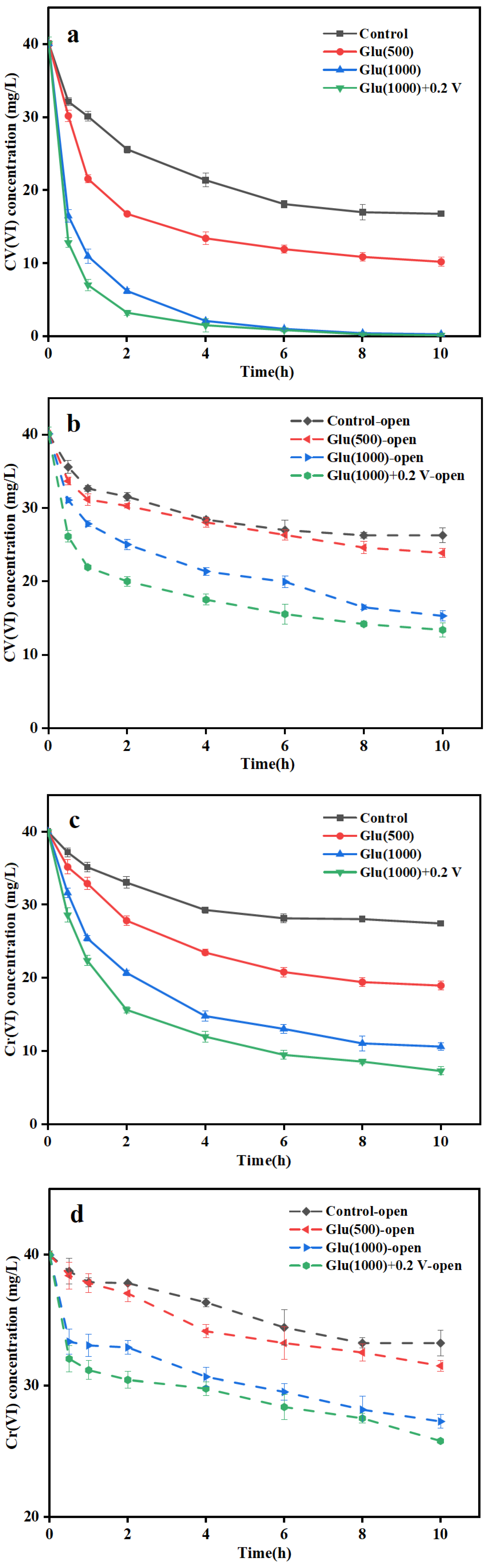
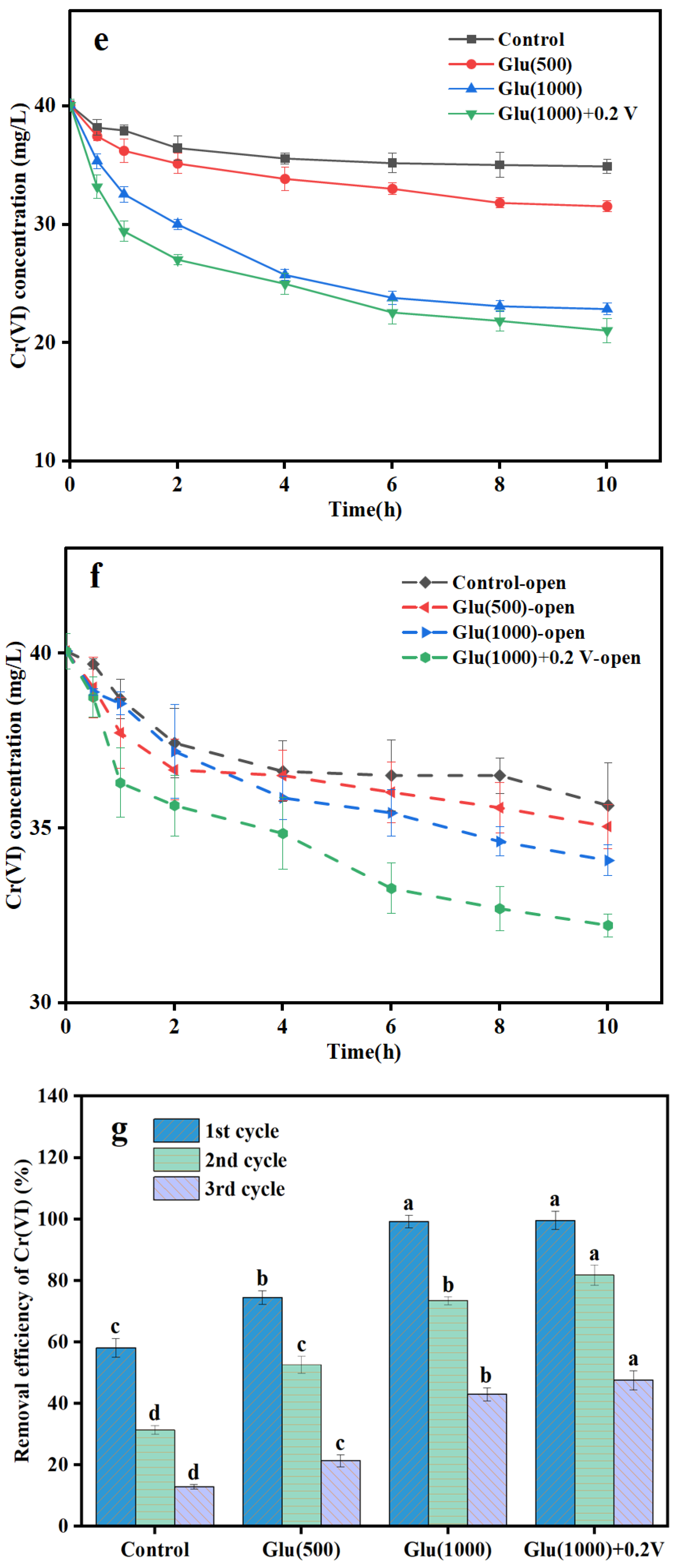
3.2.1. Cr(VI) Removal Performance

Figure 3 illustrates the performance changes of each MFC in different Cr(VI) removal cycles under open-circuit and closed-circuit conditions. The reduction of Cr(VI) concentration under open-circuit conditions was primarily attributed to the adsorption and partial reduction of the nano-FeS biocathode [24]. In the first cycle (Figure 3a,b), Glu(1000) + 0.2 V MFC achieved a Cr(VI) removal rate of 2.67 ± 0.19 mg/L·h under open-circuit conditions (Figure 3a), which was 1.93 times as high as that of the control group. Under closed-circuit conditions (Figure 3b), the Cr(VI) removal rate in the Glu(1000) + 0.2 V group improved to 3.99 ± 0.09 mg/L·h in the first cycle. These results demonstrate that the reduced particle size of nano-FeS substantially improved both the adsorptive capacity and bioelectrochemical reduction efficiency of Cr(VI).
Figure 3c–f show that in the subsequent two cycles, the Cr(VI) removal rate of all MFCs decreased, but the extent of the decrease varied. Notably, the Cr(VI) removal rate in the control group in the third cycle approached that under open-circuit conditions, indicating severe cathode passivation and an almost complete loss of bioelectrochemical reduction capability. In contrast, the Glu(1000) + 0.2 V group showed the smallest decline in Cr(VI) removal rate in the third cycle, with only a 52.27% reduction compared to the first cycle, while the removal rate in the control group decreased by 77.80%.
The Cr(VI) removal efficiency under closed-circuit conditions further confirmed the above conclusion (Figure 3g). During the first cycle, the Cr(VI) removal efficiency in the Glu(1000) + 0.2 V group MFC reached 99.53 ± 2.96%, which was 1.08 times higher than that of the Control group (47.78 ± 1.30%). Across three consecutive operational cycles, the Glu(1000) + 0.2 V treatment group maintained superior Cr(VI) removal efficiency with minimal performance attenuation compared to other experimental groups. This indicated that the larger number and smaller particle size of nano-FeS significantly alleviate cathode passivation, maintain high bioelectrochemical reduction activity of the cathode, and effectively promote the reduction and removal of Cr(VI) [37]. Furthermore, the in situ synthesis of nano-FeS resulted in more active sites on the cathode surface, simultaneously enhancing the conductivity of the cathode, and therefore maintained the stability of the nano-FeS biocathode in long-term operation [17].
As shown in Table 1, compared to the biocathode enhancement strategies employed in similar studies, the in situ modification of the biocathode with nano-FeS in this study achieved superior Cr(VI) removal performance. The novelty of this strategy is primarily reflected in three aspects. In the waste-to-waste approach, organic wastewater is utilized as a carbon source for microorganisms, while sulfur/iron-containing wastewater serves as a source of sulfur and iron. This is coupled with renewable energy sources (wind and solar energy) and the small voltage generated by MFC to in situ synthesize nano-FeS. This sustainable approach effectively reduces both economic and time costs. Regarding long-term stability, under continuous electron supply, microorganisms are able to stably and continuously regenerate nano-FeS in situ [33], forming a stable symbiotic network. This avoids the issues encountered in traditional methods where catalysts are synthesized first and then combined with microorganisms, leading to catalyst pore blockage and reduced efficiency. Performance advantages were noted. The Cr(VI) removal rate in this study increased by only 1.08 times compared to the control group, but the maximum Cr(VI) removal rate (3.99 mg/L·h) surpassed the levels achieved in most similar studies [38,39]. Compared to traditional methods, the approach in this study is simpler to operate, more stable, and demonstrates superior practicality. These results not only verify the high efficiency of nano-FeS but also indicates that high-performance nano-FeS biosynthesized in situ by modulating the carbon source concentration and applied voltage can significantly enhance the power production, chromium removal, and stability performances of biocathodes. This provides new enhancement strategies and design ideas for the application of bioelectrochemical systems in heavy metal wastewater treatment.

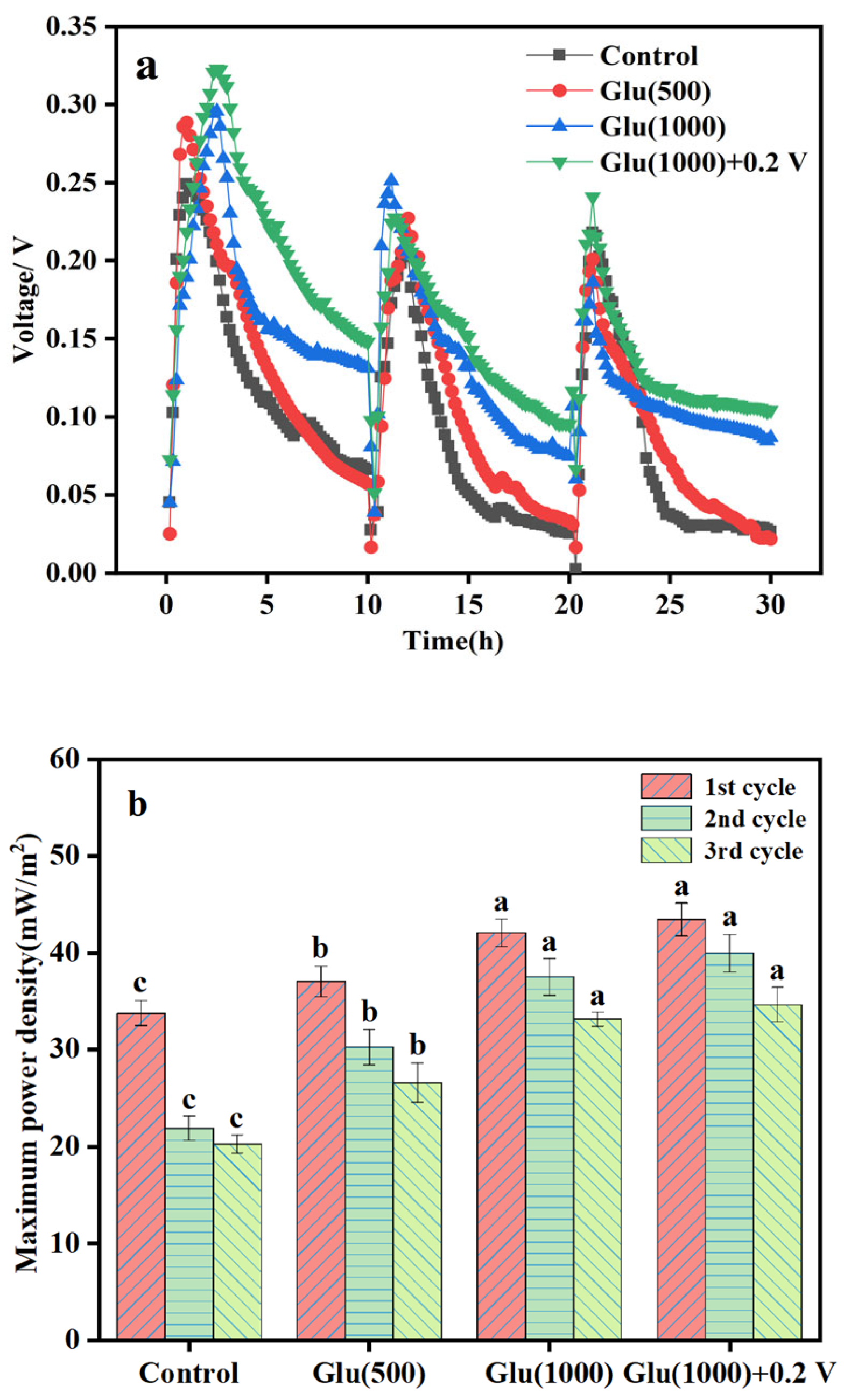
3.2.2. Electricity Generation in Biocathode MFC

As shown in Figure 4a, the nano-FeS biocathodes synthesized under different conditions exhibited varying impacts on electricity generation and Cr(VI) removal by MFCs. The output voltage of each MFCs was as follows: Control < Glu(500) < Glu(1000) < Glu(1000) + 0.2 V. The maximum voltage of the Glu(1000) + 0.2 V group reached 0.32 V, which was 1.28 times as high as that of the control group. Furthermore, during the three consecutive Cr(VI) removal cycles, the Glu(1000) + 0.2 V group consistently maintained higher voltage output, with a significantly slower voltage decline trend compared to the other groups. Figure 4b further illustrates the changes in maximum power density over three consecutive Cr(VI) removal cycles. The Glu(1000) + 0.2 V group exhibited significantly higher maximum power density in each cycle compared to both the control and Glu(500) groups, though the improvement over Glu(1000) alone was not statistically significant. Specifically, in the first cycle, the Glu(1000) + 0.2 V group achieved a maximum power density of 43.45 ± 1.69 mW/m2, representing a 29% enhancement over the ontrol group; in the second cycle, power density declined by only 8.01% (vs. 35.20% in the control); in the third cycle, a 20.21% reduction was observed, markedly lower than the control group’s 39.96% decrease. In addition, it was observed that nano-FeS contributed more substantially to Cr(VI) removal enhancement than to the promotion of EET within the system. Cyclic voltammetry analysis revealed that both the Glu(1000) and Glu(1000) + 0.2 V groups demonstrated significantly enhanced reduction peak currents compared to the control and Glu(500) groups, accompanied by a positive shift in reduction potential from 0.12 V to 0.43 V (Figure S1(a1–a3)). Notably, the reduction peak current of the Glu(1000) + 0.2 V group was 3.64 times that of the Control group. EIS analysis (Figure S1(b1–b3)) showed that the Glu(1000) + 0.2 V group consistently maintained the lowest charge-transfer resistance throughout the three cycles. This may be due to the large number and small particle size of nano-FeS, which significantly improves the electrochemical performance of the Glu(1000) + 0.2 V group biocathode and effectively alleviates biocathode passivation [13]. This result not only validates the high efficiency of nano-FeS but also demonstrates that the in situ synthesis of high-performance nano-FeS, regulated by carbon source concentration and applied voltage, can significantly enhance the electricity generation, Cr(VI) removal, and stability of biocathode MFCs. This provides a novel enhancement strategy and design approach for the application of bioelectrochemical systems in heavy metal wastewater treatment [17].

3.3. Analysis of Nano-FeS Biocathode Before and After Cr(VI) Removal

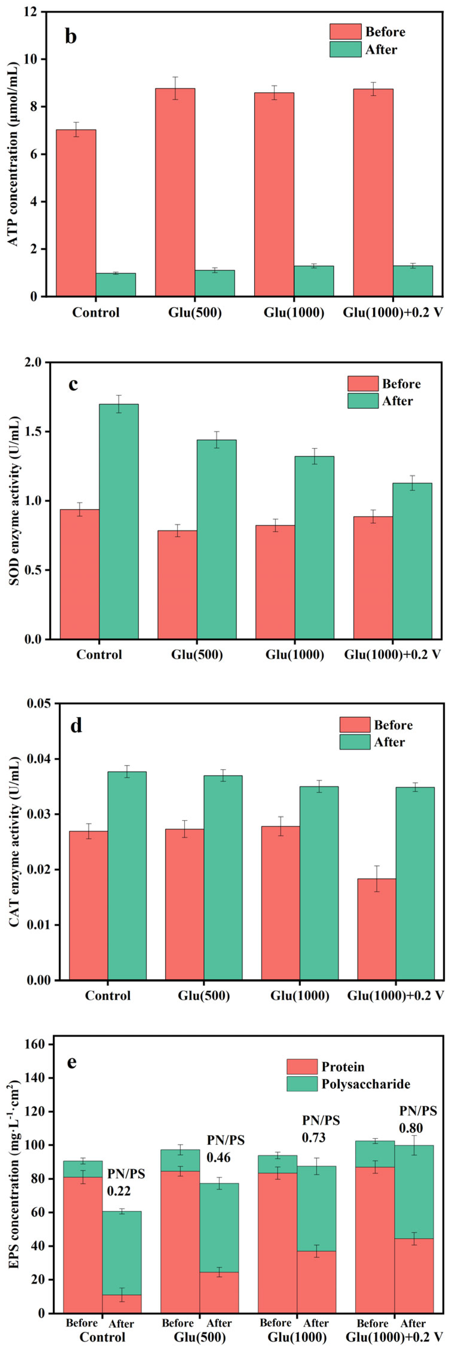
3.3.1. Physiological and Biochemical Analysis of Nano-FeS Biocathodes

Through CLSM analysis (Figure 5a and Figure S2), it was observed that the differences in cellular activity among the different nano-FeS hybridized biocathodes before Cr(VI) removal were not significant. These results suggest that the in situ cathode synthesis conditions maintained biofilm metabolic activity, as evidenced by consistent catalytic performance across experimental groups. Quantitative analysis of the biomass within the biofilms revealed a gradual decrease in biomass from the inner to the outer layers (Figure S3). This distribution characteristic is mainly due to the presence of nano-FeS in the middle and outer layers, which occupy some of the space needed for bacterial growth, thereby constraining the bacterial distribution [18,24]. Cr(VI), as a highly oxidizing pollutant, exerts direct toxicity on bacterial cells, leading to a significant decline in cellular viability [44]. Experimental results showed that the Glu(1000) + 0.2 V electrode maintained the highest cell activity in the biofilm after Cr(VI) removal (Figure S2). This result aligns with the previously mentioned SEM analysis, further highlighting that the exceptional physicochemical properties of nano-FeS are essential in sustaining the cell activity of the biofilm.
For further confirmation the influence of Cr(VI) removal on microbial metabolism, this study analyzed the ATP concentration as well as the SOD and CAT activities in the biocathodes (Figure 5b–d). Before Cr(VI) removal, the ATP concentration in all biocathodes was higher than 7.0 μmol/mL, with no major differences between the groups. Moreover, there were no significant differences in SOD (all less than 1.0 U/mL) and CAT activities (all less than 0.03 U/mL). These results indicate that the biocathodes did not activate significant stress or defense responses in the absence of Cr(VI) stress [45]. ATP, as a key form of energy storage and transfer for bacterial bioremediation, reflects the metabolic activity of microorganisms through its concentration changes [46]. After Cr(VI) removal, the ATP concentrations of all biocathodes significantly decreased. Compared with the control group, the Glu(1000) and Glu(1000) + 0.2 V biocathodes exhibited higher ATP concentrations (about 1.30 μmol/mL), and their ATP levels were increased by 31% compared with the control group (Figure 5b). Additionally, the activities of the SOD and CAT are widely recognized as key indicators of cellular defense mechanisms against toxic pollutants [47]. After Cr(VI) removal, the SOD and CAT activities of all biocathodes significantly increased (Figure 5c,d), demonstrating activation of cellular antioxidant defense system during Cr(VI) removal. The SOD (1.13 U/mL) and CAT activities (0.035 U/mL) of the Glu(1000) + 0.2 V biocathode were lower than those of the Control group (SOD and CAT activities of 1.70 U/mL and 0.038 U/mL, respectively). This protective effect may be attributed to the nano-FeS armor layer, which effectively mitigated Cr(VI) toxicity towards the microbial consortium.
EPS plays a crucial role in protecting microorganisms against exogenous toxic substances, leveraging its strong redox capabilities and three-dimensional structure to shield microbial cells [48]. Analyzing changes in the concentrations of the main EPS components, polysaccharides (PS) and proteins (PN), can reveal the functional mechanisms of EPS during Cr(VI) removal. The findings indicate that there were no notable differences in EPS concentrations across the biocathodes prior to Cr(VI) removal (Figure 5e). Studies indicate that microorganisms secrete large amounts of PS under adverse environmental stress, and high PS concentrations may inhibit EET, thereby negatively affecting electrochemical reactions [49]. After Cr(VI) removal, the PN concentrations of all biocathodes decreased, while the PS concentrations relatively increased. The Glu(1000) + 0.2 V biocathode exhibited the maximum PN/PS ratio (0.80), which was 3.64 times as high as that of the control group. This phenomenon suggests that the Glu(1000) + 0.2 V biocathode contained more electrochemically active substances (PN), which significantly promote redox reactions and electron transfer processes, thereby accelerating Cr(VI) reduction [50]. Additionally, the Glu(1000) + 0.2 V biocathode had the highest EPS concentration (99.90 mg·L−1·cm2), which was 1.65 times that of the control group. This high EPS concentration not only provides a physical barrier to protect microorganisms from Cr(VI) toxicity but also facilitates the immobilization of extracellular redox-active substances, helping to maintain bacterial metabolic activity [51]. These results indicate that smaller-sized nano-FeS can more effectively stimulate EPS secretion and enhance the content of redox-active substances in EPS. The interaction between nano-FeS and EPS enhances both the adsorption and reduction of Cr(VI), while also boosting the metabolic flexibility of microorganisms. The synergistic interaction between nano-FeS and EPS enhances both Cr(VI) adsorption and reduction efficiency and microbial metabolic adaptability [52].

3.3.2. Physicochemical Analysis of Nano-FeS Biocathode

To further physicochemical analysis of different nano-FeS biocathodes before and after Cr(VI) removal, the electrodes were characterized using SEM and XPS. The SEM images (Figure 6(a1–a4)) showed that prior to Cr(VI) removal, the nano-FeS biocathodes exhibited a distinct three-dimensional porous structure, with the Glu(1000) + 0.2 V group being the most prominent, displaying the biofilm surface covered with numerous particles and rod-shaped bacteria. These particles were confirmed to be nano-FeS through subsequent XPS analysis. This aligns with literature reports that nano-FeS forms an efficient electron transfer network between cells through a “bridging” effect [53]. In contrast, the control group biocathode structure was relatively flat and lacked three-dimensional characteristics. After Cr(VI) removal (Figure 6(b1–b4)), all biocathodes showed deposition and flaky substances. However, the extent of cellular damage varied significantly among the biofilms. These findings indicate that the nano-FeS coating functioned as a protective barrier, significantly reducing Cr(VI) cytotoxicity to microbial cells.
The XPS analysis further revealed the mechanism of nano-FeS during Cr(VI) reduction (Figure 6c–e). The Fe 2p spectra (Figure 6c) indicate that the Glu(1000) + 0.2 V group exhibited the highest Fe(II)-S content, with an Fe(II)/Fe(III) ratio of 1.23 (Table S2), indicating the strongest reduction capability [19], excellent dispersion of nano-FeS, and enhanced microbial electrochemical activity. During the Cr(VI) reduction process, Fe(II) is oxidized to Fe(III), while active microorganisms utilize organic carbon sources from the cathode and electrons from the MFC anode to mediate the reduction of Fe(III) back to Fe(II). This cyclic process allows Fe(II) to continuously participate in the in situ synthesis of nano-FeS, preventing electrode passivation and effectively avoiding secondary pollution. Additionally, the S 2p spectra (Figure 6d) showed the presence of significant peaks corresponding to S2−, S0, and S O 4 2 in the Glu(1000) + 0.2 V group biofilm, consistent with the behavior of SRB in synthesizing active sulfur compounds in reducing environments [33]. According to the Cr 2p XPS spectrum (Figure 6e), the reduction products of Cr(VI) include Cr(III) (586.80 eV), generated by biocatalytic reduction of Cr(VI), and Cr(OH)3 (577.41 eV), and Cr2O3 (576.62 eV), which are reduced by nano-FeS. Notably, Cr(III) can be further deeply reduced to Cr(0) (583.01 eV) by nano-FeS within the bio-cathode environment. The characteristic peak of Cr(0) (583.01 eV) was observed in each nano-FeS biocathodes, which is a significant highlight of this technology, demonstrating the achievement of deep reduction of Cr(VI). The Cr(0) peak area of the Glu(1000) + 0.2 V group was 1.98 times that of the control group (Table S3), which could be attributed to the synergistic effect of nano-FeS and electroactive microorganisms enhancing the reduction efficiency from Cr(III) to Cr(0) while lowering the activation energy of the reaction [54]. Furthermore, the bioelectrochemical reduction environment of the cathode facilitated the rapid reduction of Fe(III) to Fe(II), effectively alleviating the consumption of the reducing agent Fe(II), which further enhanced the deep reduction of Cr(VI) in MFC [55]. The Glu(1000) + 0.2 V group markedly improved nano-FeS production and the reducing capacity of the biocathode, driven by the combined effects of organic carbon sources and applied voltage. This approach provides a novel strategy for alleviating cathode passivation and demonstrates its potential practical application in Cr(VI)-containing wastewater.

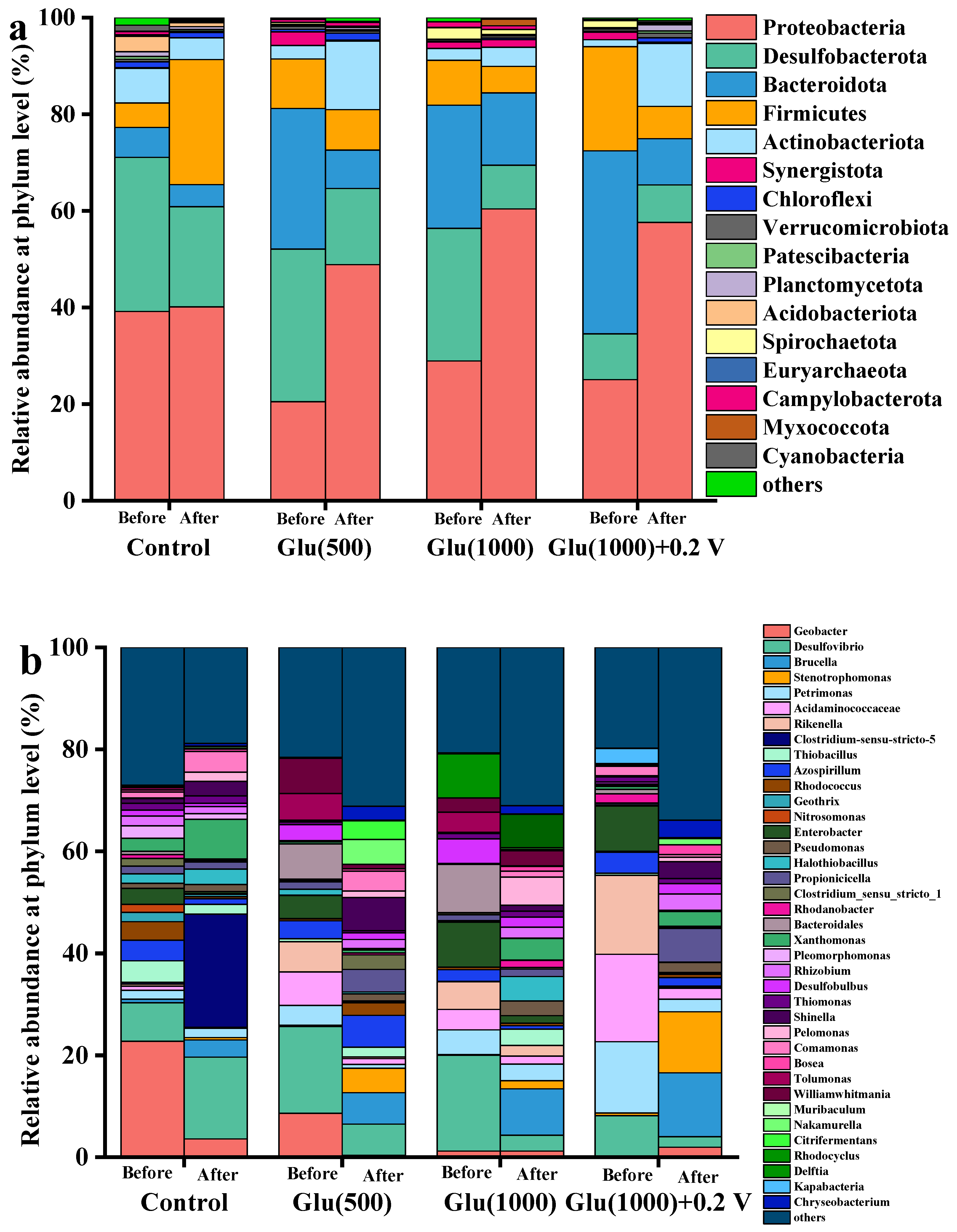
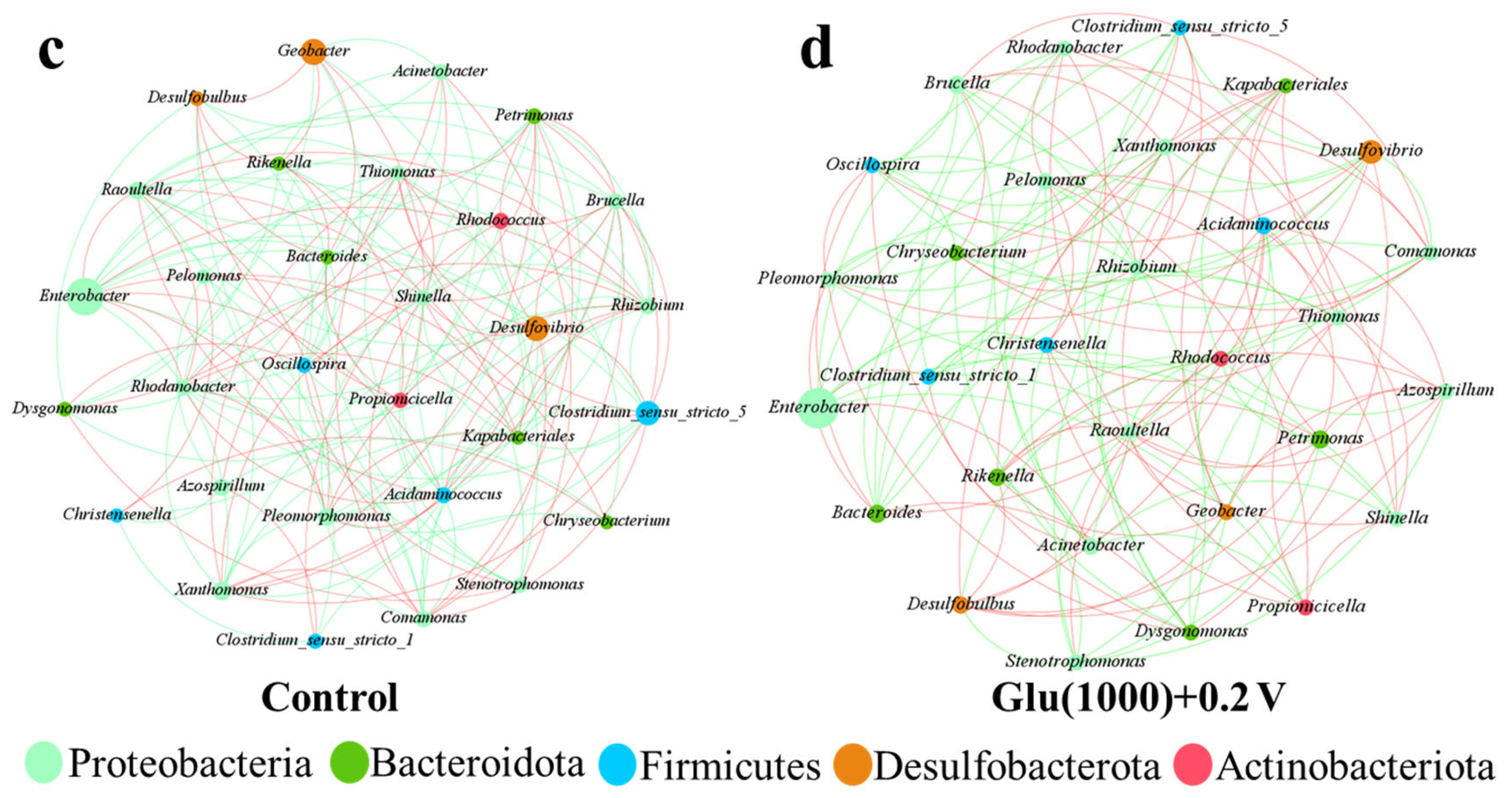
3.3.3. Microbial Community Analysis of Nano-FeS Biocathodes

Figure 7 illustrates the changes in microbial community composition of different biocathodes before and after Cr(VI) removal. At the phylum level (Figure 7a), the microbial communities in all biocathodes were predominantly composed of Proteobacteria, Desulfobacterota, Bacteroidota, and Firmicutes prior to Cr(VI) treatment, though their relative abundances varied significantly among groups. Among these, Proteobacteria and Firmicutes have attracted considerable attention because of their prominent roles in electron transfer [56]. Desulfobacterota and Bacteroidota contain numerous electrochemically active bacteria that facilitate the synthesis of nano-FeS [56,57]. After Cr(VI) removal, the dominant phyla in the biocathodes remained largely consistent with those before removal, but the relative abundance of Proteobacteria significantly increased, further highlighting their critical role in Cr(VI) reduction and electron transfer. In particular, in the Glu(1000) and Glu(1000) + 0.2 V groups, the relative abundances of Proteobacteria reached 60.41% and 57.61%, respectively, drastically higher than those in other groups. Proteobacteria play a pivotal role in electricity generation within MFCs and have also been shown to exhibit significant Cr(VI) tolerance, highlighting their adaptability and functionality in Cr(VI) removal processes. Additionally, Desulfobacterota, Bacteroidota, and Firmicutes have been demonstrated to promote Cr(VI) reduction and electricity generation in MFCs [11,56].
At the genus level (Figure 7b), before Cr(VI) removal, the dominant genera in all biocathodes included Desulfovibrio. Desulfovibrio plays an essential role in dissimilatory sulfate reduction, with its cytochrome c serving a core function in electron transfer processes [58]. These characteristics make Desulfovibrio a selectively enriched genus in the biocathodes during the biosynthesis of nano-FeS. After Cr(VI) removal, the dominant genera underwent significant changes, particularly in the Glu(1000) + 0.2 V group, where the relative abundance of Brucella increased significantly to 12.55%, which was 3.71 times that of the control group. The Cr(VI) reductase secreted by Brucella has a critical function during the Cr(VI) reduction process [59]. Additionally, other dominant genera of the Glu(1000) + 0.2 V biocathode included Stenotrophomonas (11.99%) and Propionicicella (6.59%). Stenotrophomonas demonstrate significant electrochemical activity and play crucial functional roles in MFCs, particularly in wastewater treatment and bioelectricity generation [60]. This electroactivity may enhance the electron transfer process, thereby accelerating Cr(VI) reduction. Propionicicella is associated with the fermentation of complex substrates in polluted wastewater [61]. After Cr(VI) removal, all biocathodes selectively enriched dominant genera closely related to Cr(VI) reduction. The genus level principal coordinate analysis indicated that there was a phenomenon of significant separation between the two clusters formed by the biocathode before and after Cr(VI) removal (Figure S4). This phenomenon of significant segregation indicates that the microbial community structure of the biocathode was significantly influenced during the removal of Cr(VI).
Microbial molecular ecological network analysis revealed that the proportion of positive correlations between microorganisms in the Glu(1000) + 0.2 V biocathode was 52.94%, compared to 45.51% in the Control group (Figure 7c,d). This suggests that microbial positive correlations in the Glu(1000) + 0.2 V biocathode were stronger [62]. Studies indicate that nano-FeS with smaller particle sizes offers better long-term stability [63] and can serve as a slow-release electron donor during microbial activity, thus promoting synergistic effects among microorganisms [64], contributing to the establishment of a stable and mutually beneficial microbial community structure.

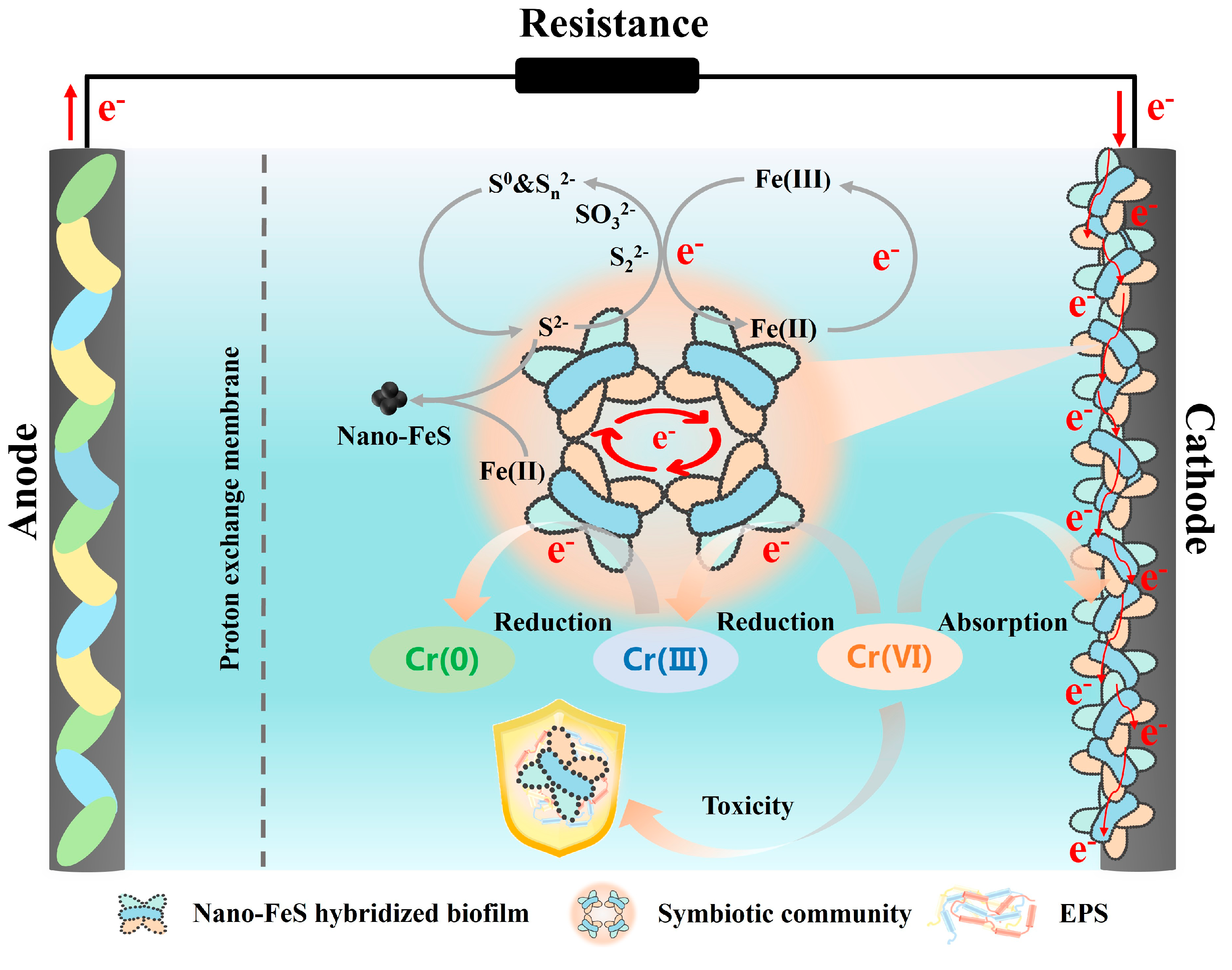
3.4. Enhancement Mechanism of Nano-FeS Biocathode and Practical Implicatons

As shown in Figure 8, the outstanding performance of the Glu(1000) + 0.2 V group in treating Cr(VI)-containing wastewater was primarily driven by the synergistic interaction between microorganisms and high-quality nano-FeS. The combined influence of glucose and applied voltage promoted the efficient in situ synthesis of nano-FeS at the cathode, resulting in smaller particle sizes and increased quantities. This significantly enhanced the biocathode’s electron transfer and electrocatalytic efficiency, providing more active sites for the adsorption and deep reduction of Cr(VI), while mitigating cathode passivation. Nano-FeS acted as a protective “armor” for microbial cells, enriching EPS with redox-active compounds (PN) and antioxidant enzymes (SOD and CAT). This mechanism protected the cells from the Cr(VI)-induced oxidative stress, thereby maintaining high microbial activity and metabolic function. The Glu(1000) + 0.2 V group promoted the selective enrichment and stable succession of dual-functional microbial genera (such as Brucella), which possess both electroactivity and Cr(VI) reduction capabilities. This synergistic interaction promoted the development of a Cr(VI)-resistant microbial consortium with enhanced remediation efficiency.
This study proposes a novel strategy for enhancing the removal of pollutants through in situ biosynthesis of nano-FeS to fabricate hybridized biocathodes. This approach achieves the following dual objectives: (1) optimized nano-FeS synthesis/regeneration via organic wastewater utilization (as a carbon source), combined with self-generated electricity, and renewable energy-powered micro-voltage application (e.g., solar/wind energy), while (2) simultaneously enabling “waste-treats-waste” ecological remediation by employing iron- and sulfur-containing wastewaters as precursor sources [33], which not only effectively reduces raw material and energy costs but also demonstrates significant potential for practical applications. As long as microbial activity and electron supply are maintained, Fe(III) can be reduced to Fe(II), providing the necessary source of iron for the in situ regeneration of nano-FeS, while simultaneously preventing the risk of secondary pollution due to electrode passivation.

4. Conclusions

In this study, high-performance nano-FeS was successfully synthesized in situ and self-assembled into a hybridized biocathode in MFCs by regulating glucose concentration and applying an external voltage. The Glu(1000) + 0.2 V group exhibited the smallest nano-FeS particle size (16.02 ± 4.35 nm) and the largest quantity, significantly boosting the electrochemical performance of the biocathode while alleviating cathode passivation. This biocathode achieved the highest power density (43.45 ± 1.69 mW/m2) and Cr(VI) removal rate (3.99 ± 0.09 mg/L·h) in the MFCs, outperforming the control group by 29% and 71%, respectively. The nano-FeS biofilm selectively enriched electroactive and Cr(VI)-reducing genera (e.g., Brucella), optimized the microbial community structure, and enhanced Cr(VI) tolerance through the protective “armor” layer, ensuring sustained high-efficiency Cr(VI) removal. This study proposes a cost-effective and sustainable intensification strategy for ecological remediation of strongly oxidative pollutants, demonstrating significant potential for practical applications.

Supplementary Materials

The following supporting information can be downloaded at: https://www.mdpi.com/article/10.3390/w17142073/s1. Table S1 Binding energy (BE) and percent peak area for Fe2p3/2 and S2p XPS spectra of different nano-FeS samples. Table S2 Binding energy (BE) and percent peak area for Fe2p3/2 and S2p XPS spectra of nano-FeS hybridized biocathodes before the Cr(VI) removal experiment. Table S3 Binding energy (BE) and percent peak area for Cr2p spectra of nano-FeS hybridized biocathodes after the Cr(VI) removal experiment. Figure S1. The cyclic voltammetry and electrochemical impendence spectroscopy (a1 and b1: 1st cycle; a2 and b2: 2nd cycle; a3 and b3: 3rd cycle) of different nano-FeS biocathodes. Figure S2. The proportion of vital cells in different electrode biofilms before and after the Cr(VI) removal experiment, according to CLSM analysis. Figure S3. The proportion of total cells in different layers of electrode biofilms before the Cr(VI) removal experiment, according to CLSM analysis. Figure S4. The principal coordinate analysis (PCoA) of different nano-FeS biocathodes before and after Cr(VI) removal.

Author Contributions

Y.G.: Writing—original draft, Methodology, Data curation, Validation, Visualization. D.C.: Formal analysis, Methodology, Data curation, Validation, Investigation. S.T.: Validation, Investigation, Software. Y.H.: Project administration, Resources, Formal analysis. W.D.: Methodology, Investigation, Resources, Supervision. X.W.: Conceptualization, Writing—review and editing, Project administration, Funding acquisition, Resources. All authors have read and agreed to the published version of the manuscript.

Funding

This work was financially supported by the National Key Research and Development Program of China (2021YFA0910400), Jiangsu Basic Research Center for Synthetic Biology (BK20233003), the Natural Science Foundation of Jiangsu Province of China for Excellent Young Scholars (BK20211591), the National Natural Science Foundation of China (22478184), the Jiangsu Synergetic Innovation Center for Advanced Bio-Manufacture (XTD2209), the National Students’ Platform for Innovation and Entrepreneurship Training Program (202510291063), and the Provincial Students’ Platform for Innovation and Entrepreneurship Training Program (S202510291142).

Data Availability Statement

Data will be made available on request.

Conflicts of Interest

The authors declare that they have no known competing financial interests or personal relationships that could have appeared to influence the work reported in this paper.

References

  1. Wu, D.; Zhang, B.; Shi, S.; Tang, R.; Qiao, C.; Li, T.; Jia, J.; Yang, M.; Si, X.; Wang, Y.; et al. Engineering extracellular electron transfer to promote simultaneous brewing wastewater treatment and chromium reduction. J. Hazard. Mater. 2024, 465, 133171. [Google Scholar] [CrossRef] [PubMed]
  2. Zhang, X.; Liu, Y.; Li, C. Influence of Cr(VI) concentration on Cr(VI) reduction and electricity production in microbial fuel cell. Environ. Sci. Pollut. Res. 2021, 28, 54170–54176. [Google Scholar] [CrossRef] [PubMed]
  3. Ali, J.; Wang, L.; Waseem, H.; Djellabi, R.; Oladoja, N.A.; Pan, G. FeS@rGO nanocomposites as electrocatalysts for enhanced chromium removal and clean energy generation by microbial fuel cell. Chem. Eng. J. 2020, 384, 123335. [Google Scholar] [CrossRef]
  4. Zheng, Z.; Wang, X.; Wang, M.; Yao, M.; Xue, J. Fabrication of a long-term reductive material by ball milling of mackinawite with sodium bisulfate to remove Cr(VI) in groundwater. J. Environ. Chem. Eng. 2024, 12, 114949. [Google Scholar] [CrossRef]
  5. Zhao, S.; Liu, S.; Sumpradit, T.; Zhou, J.; Qu, J. Magnetic nanoparticles doped biochar cathode in a two-chamber microbial fuel cell for the adsorption-reduction of hexavalent chromium. Int. J. Hydrogen Energy 2024, 63, 163–172. [Google Scholar] [CrossRef]
  6. Kumar, A.; Guo, C.; Sharma, G.; Pathania, D.; Naushad, M.; Kalia, S.; Dhiman, P. Magnetically recoverable ZrO2/Fe3O4/chitosan nanomaterials for enhanced sunlight driven photoreduction of carcinogenic Cr(VI) and dechlorination & mineralization of 4-chlorophenol from simulated waste water. RSC Adv. 2016, 6, 13251–13263. [Google Scholar] [CrossRef]
  7. Huang, L.; Chai, X.; Cheng, S.; Chen, G. Evaluation of carbon-based materials in tubular biocathode microbial fuel cells in terms of hexavalent chromium reduction and electricity generation. Chem. Eng. J. 2011, 166, 652–661. [Google Scholar] [CrossRef]
  8. Tandukar, M.; Huber, S.J.; Onodera, T.; Pavlostathis, S.G. Biological chromium(VI) reduction in the cathode of a microbial fuel cell. Environ. Sci. Technol. 2009, 43, 8159–8165. [Google Scholar] [CrossRef]
  9. Wu, X.; Zhu, X.; Song, T.; Zhang, L.; Jia, H.; Wei, P. Effect of acclimatization on hexavalent chromium reduction in a biocathode microbial fuel cell. Bioresour. Technol. 2015, 180, 185–191. [Google Scholar] [CrossRef]
  10. Cheng, Z.; Xiong, J.; Min, D.; Cheng, L.; Liu, D.; Li, W.; Jin, F.; Yang, M.; Yu, H. Promoting bidirectional extracellular electron transfer of Shewanella oneidensis MR-1 for hexavalent chromium reduction via elevating intracellular cAMP level. Biotechnol. Bioeng. 2020, 117, 1294–1303. [Google Scholar] [CrossRef]
  11. Yu, X.; Guo, T.; Liu, X.; Zhou, B.; Zhai, X.; Yang, J.; Wang, X.; Hou, Y.; Yang, Q. Improving surface properties of cathode and increasing abundance of autotrophic bacteria for chromium reduction with amino functionalized carbon nanotubes. J. Environ. Chem. Eng. 2022, 10, 108005. [Google Scholar] [CrossRef]
  12. Kamali, M.; Aminabhavi, T.M.; Abbassi, R.; Dewil, R.; Appels, L. Engineered nanomaterials in microbial fuel cells—Recent developments, sustainability aspects, and future outlook. Fuel 2022, 310, 122347. [Google Scholar] [CrossRef]
  13. Cheng, C.; Hu, Y.; Shao, S.; Yu, J.; Zhou, W.; Cheng, J.; Chen, Y.; Chen, S.; Chen, J.; Zhang, L. Simultaneous Cr(VI) reduction and electricity generation in Plant-Sediment Microbial Fuel Cells (P-SMFCs): Synthesis of non-bonding Co3O4 nanowires onto cathodes. Environ. Pollut. 2019, 247, 647–657. [Google Scholar] [CrossRef] [PubMed]
  14. He, W.; Li, Y.; Ni, L.; Zhu, W. Effect of stabilizer EDTA on the thermal hazard of green synthesis process of adipic acid and development of microchannel continuous flow process. Emerg. Manag. Sci. Technol. 2023, 3, 22. [Google Scholar] [CrossRef]
  15. Chen, Y.; Liang, W.; Li, Y.; Wu, Y.; Chen, Y.; Xiao, W.; Zhao, L.; Zhang, J.; Li, H. Modification, application and reaction mechanisms of nano-sized iron sulfide particles for pollutant removal from soil and water: A review. Chem. Eng. J. 2019, 362, 144–159. [Google Scholar] [CrossRef]
  16. Huo, Y.; Li, W.; Chen, C.; Li, C.; Zeng, R.; Lau, T.; Huang, T. Biogenic FeS accelerates reductive dechlorination of carbon tetrachloride by Shewanella putrefaciens CN32. Enzym. Microb. Technol. 2016, 95, 236–241. [Google Scholar] [CrossRef]
  17. Yu, Y.; Cheng, Q.; Sha, C.; Chen, Y.; Naraginti, S.; Yong, Y. Size-controlled biosynthesis of FeS nanoparticles for efficient removal of aqueous Cr(VI). Chem. Eng. J. 2020, 379, 122404. [Google Scholar] [CrossRef]
  18. Cui, Y.; Chen, X.; Pan, Z.; Wang, Y.; Xu, Q.; Bai, J.; Jia, H.; Zhou, J.; Yong, X.; Wu, X. Biosynthesized iron sulfide nanoparticles by mixed consortia for enhanced extracellular electron transfer in a microbial fuel cell. Bioresour. Technol. 2020, 318, 124095. [Google Scholar] [CrossRef]
  19. Fan, M.; Zhuang, X.; Gao, Z.; Lv, Z.; Dong, W.; Xin, F.; Chen, Y.; Jia, H.; Wu, X. Electroactive microorganisms synthesizing iron sulfide nanoparticles for enhanced hexavalent chromium removal in microbial fuel cells. Sci. Total Environ. 2023, 889, 164311. [Google Scholar] [CrossRef]
  20. Berner, R.A. Sedimentary pyrite formation: An update. Geochim. Cosmochim. Acta 1984, 48, 605–615. [Google Scholar] [CrossRef]
  21. Liang, B.; Cheng, H.; Kong, D.; Gao, S.; Sun, F.; Cui, D.; Kong, F.; Zhou, A.; Liu, W.; Ren, N.; et al. Accelerated reduction of chlorinated nitroaromatic antibiotic chloramphenicol by biocathode. Environ. Sci. Technol. 2013, 47, 5353–5361. [Google Scholar] [CrossRef] [PubMed]
  22. Wu, X.; Xiong, X.; Owens, G.; Brunetti, G.; Zhou, J.; Yong, X.; Xie, X.; Zhang, L.; Wei, P.; Jia, H. Anode modification by biogenic gold nanoparticles for the improved performance of microbial fuel cells and microbial community shift. Bioresour. Technol. 2018, 270, 11–19. [Google Scholar] [CrossRef] [PubMed]
  23. Wu, X.; Tong, F.; Yong, X.; Zhou, J.; Zhang, L.; Jia, H.; Wei, P. Effect of NaX zeolite-modified graphite felts on hexavalent chromium removal in biocathode microbial fuel cells. J. Hazard. Mater. 2016, 308, 303–311. [Google Scholar] [CrossRef]
  24. Zhuang, X.; Tang, S.; Dong, W.; Xin, F.; Jia, H.; Wu, X. Improved performance of Cr(VI)-reducing microbial fuel cells by nano-FeS hybridized biocathodes. RSC Adv. 2023, 13, 6768–6778. [Google Scholar] [CrossRef] [PubMed]
  25. Karamanev, D.G.; Nikolov, L.N.; Mamatarkova, V. Rapid simultaneous quantitative determination of ferric and ferrous ions in drainage waters and similar solutions. Miner. Eng. 2002, 15, 341–346. [Google Scholar] [CrossRef]
  26. Yu, Y.; Wang, Y.; Fang, Z.; Shi, Y.; Cheng, Q.; Chen, Y.; Shi, W.; Yang, C. Single cell electron collectors for highly efficient wiring-up electronic abiotic/biotic interfaces. Nat. Commun. 2020, 11, 4087. [Google Scholar] [CrossRef]
  27. Hao, L.; Zhang, B.; Cheng, M.; Feng, C. Effects of various organic carbon sources on simultaneous V(V) reduction and bioelectricity generation in single chamber microbial fuel cells. Bioresour. Technol. 2016, 201, 105–110. [Google Scholar] [CrossRef]
  28. Huang, L.; Chai, X.; Chen, G.; Logan, B.E. Effect of set potential on hexavalent chromium reduction and electricity generation from biocathode microbial fuel cells. Environ. Sci. Technol. 2011, 45, 5025–5031. [Google Scholar] [CrossRef]
  29. Hansel, C.M.; Lentini, C.J.; Tang, Y.; Johnston, D.T.; Wankel, S.D.; Jardine, P.M. Dominance of sulfur-fueled iron oxide reduction in low-sulfate freshwater sediments. ISME J. 2015, 9, 2400–2412. [Google Scholar] [CrossRef]
  30. Xia, D.; Yi, X.; Lu, Y.; Huang, W.; Xie, Y.; Ye, H.; Dang, Z.; Tao, X.; Li, L.; Lu, G. Dissimilatory iron and sulfate reduction by native microbial communities using lactate and citrate as carbon sources and electron donors. Ecotoxicol. Environ. Saf. 2019, 174, 524–531. [Google Scholar] [CrossRef]
  31. Zhang, X.; Zhang, D.; Huang, Y.; Zhang, K.; Lu, P. Simultaneous removal of organic matter and iron from hydraulic fracturing flowback water through sulfur cycling in a microbial fuel cell. Water Res. 2018, 147, 461–471. [Google Scholar] [CrossRef] [PubMed]
  32. He, Y.; Fu, Q.; Li, J.; Zhang, L.; Zhu, X.; Liao, Q. In situ biosynthesis of FeS nanoparticles boosts current generation in bioelectrochemical systems through efficient electron transfer. Small 2024, 20, e2309648. [Google Scholar] [CrossRef] [PubMed]
  33. Fu, X.; Wu, J.; Cui, S.; Wang, X.; Liu, H.; He, R.; Yang, C.; Deng, X.; Tan, Z.; Li, W. Self-regenerable bio-hybrid with biogenic ferrous sulfide nanoparticles for treating high-concentration chromium-containing wastewater. Water Res. 2021, 206, 117731. [Google Scholar] [CrossRef] [PubMed]
  34. Wu, D.; Xing, D.; Lu, L.; Wei, M.; Liu, B.; Ren, N. Ferric iron enhances electricity generation by Shewanella oneidensis MR-1 in MFCs. Bioresour. Technol. 2013, 135, 630–634. [Google Scholar] [CrossRef]
  35. Murugan, M.; Miran, W.; Masuda, T.; Lee, D.S.; Okamoto, A. Biosynthesized iron sulfide nanocluster enhanced anodic current generation by sulfate reducing bacteria in microbial fuel cells. ChemElectroChem. 2018, 5, 4015–40220. [Google Scholar] [CrossRef]
  36. Deng, X.; Dohmae, N.; Kaksonen, A.H.; Okamoto, A. Biogenic iron sulfide nanoparticles to enable extracellular electron uptake in sulfate-reducing bacteria. Angew. Chem. Int. Ed. 2020, 59, 5995–5999. [Google Scholar] [CrossRef]
  37. Liu, X.; Chen, M.; Wang, D.; Du, F.; Xu, N.; Sun, W.; Han, Z. Cr(VI) removal during cotransport of nano-iron-particles combined with iron sulfides in groundwater: Effects of D. Vulgaris S. putrefaciens. J. Hazard. Mater. 2024, 472, 134583. [Google Scholar] [CrossRef]
  38. Hou, X.; Huang, L. Synergetic magnetic field and loaded Fe3O4 for simultaneous efficient acetate production and Cr(VI) removal in microbial electrosynthesis systems. Chem. Eng. J. Adv. 2020, 2, 100019. [Google Scholar] [CrossRef]
  39. Ma, L.; Chen, N.; Feng, C.; Yao, Y.; Wang, S.; Wang, G.; Su, Y.; Zhang, Y. Enhanced Cr(VI) reduction in biocathode microbial electrolysis cell using Fenton-derived ferric sludge. Water Res. 2022, 212, 118144. [Google Scholar] [CrossRef]
  40. Wu, Q.; Liu, J.; Mo, W.; Li, Q.; Wan, R.; Peng, S. Simultaneous treatment of chromium-containing wastewater and electricity generation using a plant cathode-sediment microbial fuel cell: Investigation of associated mechanism and influencing factors. Environ. Sci. Pollut. Res. 2023, 30, 41159–41171. [Google Scholar] [CrossRef]
  41. Chen, X.; Wang, Y.; Mamathaxim, N.; Habibul, N.; Hu, Y. Simultaneous sulfamethazine and Cr(VI) removal in lab-scale microbial fuel cell-constructed wetland. J. Environ. Sci. 2025, 154, 91–101. [Google Scholar] [CrossRef] [PubMed]
  42. Fei, K.; Song, T.; Wang, H.; Zhang, D.; Tao, R.; Xie, J. Electrophoretic deposition of carbon nanotube on reticulated vitreous carbon for hexavalent chromium removal in a biocathode microbial fuel cell. R. Soc. Open Sci. 2017, 4, 170798. [Google Scholar] [CrossRef]
  43. Du, J.; Bao, J.; Lu, C.; Werner, D. Reductive sequestration of chromate by hierarchical FeS@Fe0 particles. Water Res. 2016, 102, 73–81. [Google Scholar] [CrossRef] [PubMed]
  44. Wu, M.; Li, Y.; Li, J.; Wang, Y.; Xu, H.; Zhao, Y. Bioreduction of hexavalent chromium using a novel strain CRB-7 immobilized on multiple materials. J. Hazard. Mater. 2019, 368, 412–420. [Google Scholar] [CrossRef] [PubMed]
  45. Wang, Q.; Zhao, Y.; Zhang, C.; Zhao, M.; Jia, X.; Mutabazi, E.; Liu, Y. New insights into hexavalent chromium exposure in electron donor limited denitrification: Bio-electron behavior. Bioresour. Technol. 2023, 380, 129088. [Google Scholar] [CrossRef]
  46. Feng, H.J.; Chen, L.; Ding, Y.C.; Ma, X.J.; How, S.W.; Wu, D. Mechanism on the microbial salt tolerance enhancement by electrical stimulation. Bioelectrochemistry 2022, 147, 108206. [Google Scholar] [CrossRef]
  47. Aliko, V.; Qirjo, M.; Sula, E.; Morina, V.; Faggio, C. Antioxidant defense system, immune response and erythron profile modulation in gold fish, Carassius auratus, after acute manganese treatment. Fish Shellfish Immunol. 2018, 76, 101–109. [Google Scholar] [CrossRef]
  48. Hou, R.; Luo, C.; Zhou, S.; Wang, Y.; Yuan, Y.; Zhou, S. Anode potential-dependent protection of electroactive biofilms against metal ion shock via regulating extracellular polymeric substances. Water Res. 2020, 178, 115845. [Google Scholar] [CrossRef]
  49. Cai, T.; Zhang, Y.; Wang, N.; Zhang, Z.; Lu, X.; Zhen, G. Electrochemically active microorganisms sense charge transfer resistance for regulating biofilm electroactivity, spatio-temporal distribution, and catabolic pathway. Chem. Eng. J. 2022, 442, 136248. [Google Scholar] [CrossRef]
  50. Akhzari, F.; Naseri, T.; Mousavi, S.M.; Khosravi-Darani, K. A sustainable solution for alleviating hexavalent chromium from water streams using Lactococcus lactis AM99 as a novel Cr(VI)-reducing bacterium. J. Environ. Manag. 2024, 353, 120190. [Google Scholar] [CrossRef]
  51. Zhuang, Z.; Yang, G.; Mai, Q.; Guo, J.; Liu, X.; Zhuang, L. Physiological potential of extracellular polysaccharide in promoting Geobacter biofilm formation and extracellular electron transfer. Sci. Total Environ. 2020, 741, 140365. [Google Scholar] [CrossRef] [PubMed]
  52. Song, T.; Jin, Y.; Bao, J.; Kang, D.; Xie, J. Graphene/biofilm composites for enhancement of hexavalent chromium reduction and electricity production in a biocathode microbial fuel cell. J. Hazard. Mater. 2016, 317, 73–80. [Google Scholar] [CrossRef] [PubMed]
  53. Jiang, X.; Hu, J.; Lieber, A.M.; Jackan, C.S.; Biffinger, J.C.; Fitzgerald, L.A.; Ringeisen, B.R.; Lieber, C.M. Nanoparticle facilitated extracellular electron transfer in microbial fuel cells. Nano Lett. 2014, 14, 6737–6742. [Google Scholar] [CrossRef] [PubMed]
  54. Cao, Y.; Zhang, Y.; Yang, L.; Zhu, K.; Yuan, Y.; Li, G.; Yuan, Y.; Zhang, Q.; Bai, Z. Boosting oxygen reduction reaction kinetics through perturbating electronic structure of single-atom Fe-N3S1 catalyst with sub-nano FeS cluster. J. Colloid Interface Sci. 2023, 650, 924–933. [Google Scholar] [CrossRef]
  55. Zhang, P.; Xu, X.; Zhang, X.; Zou, K.; Liu, B.; Qing, T.; Feng, B. Nanoparticles-EPS corona increases the accumulation of heavy metals and biotoxicity of nanoparticles. J. Hazard. Mater. 2021, 409, 124526. [Google Scholar] [CrossRef]
  56. Amanze, C.; Zheng, X.; Anaman, R.; Wu, X.; Fosua, B.A.; Xiao, S.; Xia, M.; Ai, C.; Yu, R.; Wu, X.; et al. Effect of nickel (II) on the performance of anodic electroactive biofilms in bioelectrochemical systems. Water Res. 2022, 222, 118889. [Google Scholar] [CrossRef]
  57. Fu, X.; Wu, J.; Li, J.; Ding, J.; Cui, S.; Wang, X.; Wang, Y.; Liu, H.; Deng, X.; Liu, D.; et al. Heavy-metal resistant bio-hybrid with biogenic ferrous sulfide nanoparticles: pH-regulated self-assembly and wastewater treatment application. J. Hazard. Mater. 2023, 446, 130667. [Google Scholar] [CrossRef]
  58. Kumar, S.S.; Kumar, V.; Gude, V.G.; Malyan, S.K.; Pugazhendhi, A. Alkalinity and salinity favor bioelectricity generation potential of Clostridium, Tetrathiobacter and Desulfovibrio consortium in Microbial Fuel Cells (MFC) treating sulfate-laden wastewater. Bioresour. Technol. 2020, 306, 123110. [Google Scholar] [CrossRef]
  59. Thacker, U.; Parikh, R.; Shouche, Y.; Madamwar, D. Reduction of chromate by cell-free extract of Brucella sp. isolated from Cr (VI) contaminated sites. Bioresour. Technol. 2007, 98, 1541–1547. [Google Scholar] [CrossRef]
  60. Galai, S.; de los Ríos, A.P.; Hernández-Fernández, F.J.; Kacem, S.H.; Ramírez, F.M.; Quesada-Medina, J. Microbial fuel cell application for azoic dye decolorization with simultaneous bioenergy production using stenotrophomonas sp. Chem. Eng. Technol. 2015, 38, 1511–1518. [Google Scholar] [CrossRef]
  61. Salvian, A.; Farkas, D.; Ramirez-Moreno, M.; Torruella-Salas, D.; Berna, A.; Avignone-Rossa, C.; Varcoe, J.R.; Esteve-Nunez, A.; Gadkari, S. Resilience of anodic biofilm in microbial fuel cell biosensor for BOD monitoring of urban wastewater. npj Clean Water 2024, 7, 53. [Google Scholar] [CrossRef]
  62. Lu, S.; Zhang, Y.; Liu, X.; Xu, J.; Liu, Y.; Guo, W.; Liu, X.; Chen, J. Effects of sulfamethoxazole on nitrogen removal and molecular ecological network in integrated vertical-flow constructed wetland. Ecotoxicol. Environ. Saf. 2021, 219, 112292. [Google Scholar] [CrossRef] [PubMed]
  63. Liu, Y.; Xiao, W.; Wang, J.; Mirza, Z.A.; Wang, T. Optimized synthesis of FeS nanoparticles with a high Cr(VI) removal capability. J. Nanomater. 2016, 2016, 7817296. [Google Scholar] [CrossRef]
  64. Zhang, D.; Wei, Y.; Zhang, M.; Wu, S.; Zhou, L. A collaborative strategy for enhanced anaerobic co-digestion of food waste and waste activated sludge by using zero valent iron and ferrous sulfide. Bioresour. Technol. 2022, 347, 126420. [Google Scholar] [CrossRef]
Figure 1. The Fe2+ (a) and S2− (b) concentrations in different MFC cathode biosynthesis groups during the whole 8 cycles (4 days per cycle); Previous 2 days: the first 2 days of each cathode biosynthesis cycle; Later 2 days: the last 2 days of each cathode biosynthesis cycle.
Water 17 02073 g001
Figure 2. The SEM images ((a): Control; (b): Glu(500); (c): Glu(1000); (d): Glu(1000) + 0.2 V), XPS spectra ((e): Fe2p3/2; (f): S2p), and XRD patterns (g) of different precipitate samples (the red line represents the fitted line, while the black line represents the baseline).
Water 17 02073 g002
Figure 3. Changes in Cr(VI) concentration ((a,b): 1st cycle; (c,d): 2nd cycle; (e,f): 3rd cycle) and Cr(VI) removal efficiency (g) in different nano-FeS biocathode MFCs; the lowercase letters a, b, c and d denote statistically significant differences (p < 0.05) among experimental groups.
Water 17 02073 g003aWater 17 02073 g003b
Figure 4. The voltage-time curves (a) and maximum power densities (b) of different Nano-FeS biocathode MFCs for Cr(VI) removal; the lowercase letters a, b, and c denote statistically significant differences (p < 0.05) among experimental groups.
Water 17 02073 g004
Figure 5. The CLSM images (a1a8), ATP concentration (b), SOD (c), and CAT enzyme activities (d), and EPS concentration (e) of different Nano-FeS biocathodes before and after Cr(VI) removal.
Water 17 02073 g005aWater 17 02073 g005b
Figure 6. The SEM images of different nano-FeS biocathodes before (a1a4) and after (b1b4) Cr(VI) removal; the XPS spectra of different biocathodes before (c,d) and after (e) Cr(VI) removal (the red line represents the fitted line, while the black line represents the baseline).
Water 17 02073 g006
Figure 7. The microbial community compositions ((a) phylum level; (b) genus level) of different nano-FeS biocathodes before and after Cr(VI) removal; the molecular ecological network analysis based on microbial community in Control (c) and Glu(1000) + 0.2 V (d) biocathodes. Nodes are colored by the microbial phylum, and their size is proportional to the relative abundance of the microbial community; the red and green lines between two nodes indicate positive and negative correlations, respectively.
Water 17 02073 g007aWater 17 02073 g007b
Figure 8. The synergistic mechanism between nano-FeS and microorganisms in enhancing Cr(VI) removal via MFCs.
Water 17 02073 g008
Table 1. Comparison of relevant studies using biocathode bioelectrochemical systems (BES) for Cr(VI) removal.
BES TypeBiocathode ConditionCr(VI)
Concentration (mg/L)
Maximum Cr(VI)
Removal Rate (mg/L·h)
Reaction
Time (h)
Reference
Sediment microbial fuel cell (SMFC)Plant biocathode1080.19624[40]
Microbial electrosynthesis system (MES)Nano-Fe3O4 hybridized Serratia marcescens Q1-biocathode602.3024[38]
Microbial electrolysis cell (MEC)Fenton ferric sludge was incorporated into the mixed culture biocathode102.504[39]
Constructed wetland–microbial fuel cell (CW-MFC)Plant biocathode100.1760[41]
MFCMixed culture biocathode (Carbon nanotubes modified vitreous carbon as cathode electrode)200.7848[42]
MFCGraphene hybridized mixed culture–biocathode400.8348[43]
MFCNano-FeS hybridized mixed culture–biocathode403.9910This work
Disclaimer/Publisher’s Note: The statements, opinions and data contained in all publications are solely those of the individual author(s) and contributor(s) and not of MDPI and/or the editor(s). MDPI and/or the editor(s) disclaim responsibility for any injury to people or property resulting from any ideas, methods, instructions or products referred to in the content.

