Article

Enhanced Separation Performance of Graphene Oxide Membrane through Modification with Graphitic Carbon Nitride

China Institute of Atomic Energy, 1 Sanqiang Road, Fangshan District, Beijing 102413, China
*
Author to whom correspondence should be addressed.
Water 2024, 16(7), 967; https://doi.org/10.3390/w16070967
Submission received: 1 March 2024 / Revised: 24 March 2024 / Accepted: 25 March 2024 / Published: 27 March 2024
(This article belongs to the Section Wastewater Treatment and Reuse)

Abstract

:
The treatment of tritiated nuclear wastewater is facing greater challenges with the continuous expansion of the nuclear industry. The key to solving the issue of detritium in low-abundance tritium water lies in developing highly efficient and cost-effective hydrogen isotope separation technology. Graphene oxide (GO) membrane separation method exhibits greater potential compared to other existing energy-intensive technologies for the challenging task of hydrogen isotope separation in nuclear wastewater. In recent years, researchers have explored few strategies to enhance the performance of graphene oxide (GO) membranes in hydrogen isotope water treatment, recognizing the current limitations in separation efficiency. In this study, the GO/g-C3N4 composite membrane has been successfully employed for the first time in the separation of hydrogen isotopes in water. A series of GO membranes were prepared and their performances were tested by a self-made experimental device. As a result, the separation performance of the GO membrane was enhanced by the modification with graphitic carbon nitride (g-C3N4). The permeation rate of the GO/g-C3N4 membrane was higher than that of the GO membrane, while maintaining a high separation factor. Our study also demonstrated that this phenomenon can be attributed to the changes in membrane structure at the microscopic scale. The H/D separation factor and the permeate flux of the composite membrane containing g-C3N4 of 6.7% by mass were 1.10 and 7.2 × 10⁻⁵ g·min−1·cm−2 are both higher than that of the GO membrane under the same experimental conditions, which is promising for the isotope treatment.

1. Introduction

Tritium (T) is a radioactive isotope of hydrogen that can enter the human body through inhalation and skin absorption, posing internal radiation hazards [1]. It occurs in minute quantities in nature and is primarily generated through nuclear reactions, particularly in heavy water reactors (HWRs) where heavy water is used for moderation and cooling purposes [2]. The current primary approach for tritium management in nuclear power plants involves its controlled release into the environment, such as discharging into the sea after dilution with a significant volume of seawater [3].
In the foreseeable future, the expansion of the nuclear industry presents significant challenges regarding the management and treatment of tritium-containing nuclear wastewater. Therefore, the application of tritium extraction or tritium separation technology is imperative in the treatment of nuclear wastewater containing tritium. However, the existing tritium separation techniques such as cryogenic distillation [4] and combined electrolysis catalytic exchange [5,6] are relatively energy-intensive, complex, and unfriendly to the environment. On the contrary, the membrane separation process exhibits significant potential for tritium treatment owing to its low energy consumption, exceptional selectivity, and high operational efficiency. Moreover, it has already gained widespread application in the field of industrial wastewater treatment [7]. As a result, it is necessary to explore an appropriate separation membrane and analyze its working principle and feasibility before designing a complete industrial facility.
Two-dimensional materials have gained significant attention from the public since the discovery of graphene [8]. Graphene’s unique atomic-level thickness, high mechanical strength, stable physical and chemical properties, and impermeability caused by dense atomic arrangement make its derivatives worthy of attention as membrane separation materials, such as porous graphene and graphene oxide [9,10]. In the past decade, numerous researchers have dedicated their efforts to investigating the preparation methods [11], mass transfer mechanisms [12], and separation performance [13,14] of graphene-based two-dimensional separation membranes. These endeavors have greatly advanced scientific progress in this field.
Porous graphene can be prepared by focused ion beam and can be used for hydrogen purification [15,16]. However, the filtration performance of this membrane entirely depends on the pore size, and since the kinetic diameters of different hydrogen isotope molecules vary little, and porous graphene films are not suitable for hydrogen isotope separation. Graphene oxide has become the most promising material for two-dimensional hydrogen isotope separation membranes due to its strong mechanical strength, excellent purification and separation performance, and suitability for large-scale production [17]. Laminar GO membrane contains hydroxyl and epoxy groups that maintain a greater spacing between the graphene layers in the unoxidized regions of the film [18]. Moreover, these groups tend to cluster together, creating a large network of unoxidized capillary channels for particles to pass through. Due to the unique hydrophilicity of oxygen-containing groups and capillary pressure generated by graphene’s hydrophobic layer, water molecules can rapidly penetrate through the capillaries network, achieving a perfect GO membrane penetration of water [19].
Currently, extensive research has been conducted to investigate the transport mechanism of gas, water, and solution ions across the GO membrane [20,21], as well as the water molecular arrangement between the interfaces of two graphene layers, particularly within the capillaries in the GO membrane [22]. Water is transported in the form of double-layer ice in the unoxidized region between the two nanosheets in the GO film, that is, inside the nanoscale capillary [23]. It is destroyed (melted) as it passes through the oxygen-containing groups at the edge of the nanosheet, after which the water molecules penetrate into the next capillary layer and recombine into a double-layer ice structure to continue its transport [24]. The GO membrane exhibits a remarkable ion screening capability in solution desalination experiments that smaller ions (with a hydration radius of less than 4.5 A) can rapidly permeate through the membrane, while larger and organic molecules are virtually impermeable [18].
The hydrophilic nature of the GO membrane makes it a promising candidate for isotope water separation technology. An experiment proved that the content of heavy isotopes (deuterium and tritium) would be significantly reduced after hydrogen isotope water passed through the GO membrane [25]. In recent years, a large number of researchers have invested in the study of the optimization of the separation performance of GO membranes. Compared to GO membranes, various separation membranes optimized based on GO materials show better separation properties, such as higher penetration rates or higher separation factors. These modification methods of the GO membrane include the reduction method [26], surface modification method [27], intercalation method [28], and so on. RGO membranes exhibited a comparable D2O blocking rate to GO membranes while demonstrating superior mechanical stability and serving as an effective material for the separation of hydrogen isotope water mixtures [29]. A research team employed perfluoroalkyl substances [30] or fluorinated silica nanoparticles [31] to modify one side of the GO membrane forming a hydrophobic or super-hydrophobic surface, resulting in modified GO membranes exhibiting enhanced separation factors.
It is worth mentioning that researchers commonly employ heavy water instead of tritium to analyze the separation of hydrogen isotopes in water, primarily due to the potential radiation hazards involved. The H/D separation factors can reflect the H/T separation factors with some theoretical calculations [25].
In this study, GO membranes were modified with graphitic-phase carbon nitride (g-C3N4) and tested using self-made distillation equipment to demonstrate that this intercalation method can enhance the performance of the GO membranes in separating hydrogen isotopes. The impact of intercalating different quantities of g-C3N4 on the selective separation of GO membranes was also investigated.

2. Materials and Methods

2.1. Materials

The nature graphite powder was purchased from Tianjin Damao Co., Ltd. (Tianjin, China). The g-C3N4 dispersion was supplied by Nanjing XFNANO Technology Co., Ltd. (Nanjing, China). Potassium permanganate (KMnO4), concentrated sulfuric acid (H2SO4, 98%), hydrogen peroxide (H2O2, 30%), and hydrochloric acid (HCl, 37%) were supplied from Shanghai Aladdin Biochemical Technology Co., Ltd. (Shanghai, China). PC (polycarbonate) membranes and AAO (anodic aluminum oxide) membranes with 0.22 μm pore size were obtained from Whatman Co., Maidstone, UK. Hydrophobic PTFE membranes (tetrafluoroethylene, ϕ 47 mm, pore size ~1 μm, Millipore Co., Billerica, MA, USA) were used as the support in the distillation tests. The heavy water (99.9 at. % D) was purchased from Energy Chemical Co., Ltd. (Anqing, China). Deionized water (18.25 MΩ) was produced by Ultra-pure Water Purifiers (Ulupure UPR-Ⅱ, Chengdu, China) in the lab.

2.2. Fabrication of Membranes

The GO nanosheets were synthesized from graphite powder using the improved Hummers method [32]. GO dispersion solution was prepared by the ultrasonic dispersing of dry GO in deionized water. The GO membrane was fabricated on a PC substrate through vacuum filtration. In this study, 0.2 g of dried GO solid was sonicated in 100 mL of deionized water to make 2 mg/mL of GO dispersion. After that, 5 mL, 7.5 mL, 10 mL, and 15 mL GO dispersion solutions were taken and prepared into GO membranes of varying thicknesses by vacuum filtration.
The preparation of the GO/g-C3N4 composite membrane followed a similar procedure as that of the GO membrane. The mass concentrations of GO and g-C3N4 suspensions were 2 mg/mL and 0.18 mg/mL, respectively. In this work, 2 mL, 4 mL, 6 mL, 8 mL, and 10 mL g-C3N4 suspensions were added into 5 mL GO suspensions. The suspension of GO and g-C3N4 was intensively mixed by ultrasonication for 2 h and then filtered on the AAO substrate to form a membrane. Following a 24 h drying period at room temperature, the independent membrane could be easily detached from the substrate.

2.3. Characterization of the Membranes

The surface and cross-section morphology of membranes were studied by a field emission scanning electron microscope (FE-SEM, JEOL JSM-7900F, Yamagata, Japan). The gold-coated membranes were soaked in liquid nitrogen to make them easier to tear. The observation of GO nanosheets was supplied by a tunneling electron microscope (TEM, Hitachi H-7650, Marunouchi, Japan). X-ray diffraction (XRD, Bruker D8 ADVANCE, Karlsruhe, Germany) equipped with Cu Kα radiation (λ = 0.154 nm) was applied to measure the d-spacing of the prepared membranes. X-ray photoelectron spectroscopy (XPS, Thermo Scientific ESCALAB QXi, Morecambe, UK) and Raman spectra (Horiba Jobin Yvon LabRam HR800, Palaiseau, France) were used to recognize the chemical composition of the membrane surface.

2.4. Selective Separation Tests

2.4.1. Static Diffusion Experiment

The static diffusion experiment used a 50 mL glass sample bottle, which was filled with 20 mL H2O or D2O (99.9%) liquid. The GO membrane was covered at the opening of the sample bottle, and the place where the mouth of the bottle contacted the membrane was sealed using an O-type silicone ring. The experiment was carried out at room temperature, and the weight of bottles was weighed every 24 h, based on which the amount of liquid lost was calculated. The experiment was conducted for a total of 43 days. The permeate flux (J) was calculated by equation:
J i = m i A t
where m, A, and t represent the mass loss of liquid in the bottle, the membrane area, and the experiment time.

2.4.2. Distillation Tests

To analyze the differences in the performance of GO membranes with different properties in the separation of hydrogen isotope water, a custom-made experimental device for the separation of hydrogen isotope water was established. As shown in Figure S1, the device is divided into three sections: the feed section consists of an evaporation chamber with a capacity of approximately 100 mL and a pressure-reducing valve; the middle section comprises a membrane carrier; and the collect section is connected to a sample bottle for collecting the filtered liquid.
In the test, the evaporation chamber was loaded into a resistance oven and insulated with thermal insulation cotton. All stainless steel components, except for the evaporation chamber and pressure-reducing valve, were covered with a glass fiber heating sleeve to prevent the condensation of water vapor and heavy water vapor within the apparatus. Two temperature controllers were used to regulate the temperature of the resistance furnace and heating sleeve, respectively. A sample collection bottle was positioned on the collection side and connected to the stainless steel tube via a high-temperature-resistant silicone hose. The GO membrane was sealed in the middle section between the silicone and stainless steel gaskets to make sure the whole device was airtight during the process. Considering the vapor pressure on the feed side can reach 0.14 MPa at 110 °C, a support PTFE membrane was attached to the permeate side of the GO membrane to prevent it from being ruptured by the high pressure.
The molar concentration of heavy water in the chamber of 10% was achieved by mixing pure D2O with deionized water. The deuterium and hydrogen abundance of the permeated water were measured by NMR (Bruker AVANCE III 400 MHz, Karlsruhe, Germany). The heavy water samples, each with concentrations of 6%, 8%, 10%, 12%, and 14%, were combined in a 1:1 ratio with pure acetone solution. A small quantity of the resulting mixture was then transferred into an NMR tube for analysis. Two distinct peaks would appear in the 1H NMR spectrum, namely the water peak caused by the vibrations of hydrogen in water and the acetone peak caused by vibrations of hydrogen in methyl groups in acetone. Figure S2 shows a 1H NMR spectroscopy of a mixture of 10% heavy water and acetone with a 1:1 volume ratio. The left peak represents the water peak and the right peak represents the acetone peak, respectively. The area ratio of the two peaks is IH2O/Iacetone = 1.182. The area ratio of the two peaks was positively correlated with their contents. Finally, a graphical representation illustrating the relationship between the concentration of heavy water and the corresponding ratio is presented, revealing a linear correlation upon fitting, thus establishing a standard curve, as shown in Figure S3. To obtain the concentrations of heavy water in distillation tests, the unknown concentration of heavy water to be analyzed was mixed with an acetone solution in a 1:1 ratio, and a portion of the resulting mixture was introduced into the NMR tube. By comparing the peak area ratio of the two observed peaks in the test results with a standard curve, the heavy water concentration of the sample under investigation was determined.
The separation factor q can be calculated by equation:
q = C H / C D C H / C D
where CH and CD represent the hydrogen and deuterium abundance of permeated water, and CH′ and CD′ denote the hydrogen and deuterium abundance of feed water. The deuterium abundance CD is equal to the concentration of heavy water which is given by NMR results and the hydrogen abundance is equal to 1-CD.

3. Results

3.1. The Characteristics of the Membranes

The GO membrane was peeled off from the AAO base membrane, immersed in liquid nitrogen, fractured, and affixed onto the front or side of the sample stage. Subsequently, it was subjected to SEM characterization following gold spraying. As depicted in Figure 1a,b, a disparity in flatness is observed between the top and bottom surfaces of the GO membrane. Herein, the top surface denotes the side exposed to air during vacuum filtration for membrane formation, while the bottom surface refers to its contact with the AAO substrate. The image reveals that the upper surface exhibits roughness and more pronounced wrinkling compared to its relatively flat counterpart on the lower surface. This phenomenon can be attributed to self-assembly occurring primarily in proximity to the AAO filter membrane on its bottom surface, resulting in a highly ordered stratified structure conducive to a flatter morphology. Conversely, due to solution evaporation effects during exposure to air, inadequate arrangement leads to wrinkling [33].
The clear laminated structure can be seen in the cross-section SEM image of the GO membrane, as shown in Figure 1c. When the amount of GO is 10 mg, the average thickness of the GO membrane is 4 μm, and the surface area of the membrane is 12.56 cm2. The relationship between the amount of GO and the membrane thickness in this experiment is 2 g/cm3. Compared with the GO membrane, the GO/g-C3N4 (the mass of GO and g-C3N4 was 10 mg and 0.9 mg) membrane has more bulges on the top surface (Figure 1d); correspondingly, more particles can be observed on the bottom surface (Figure 1e), and the presence of these particles increases the local roughness. In the cross-sectional SEM image of the GO/g-C3N4 membrane, as shown in Figure 1f, a smaller particle is observed within the interlayer region. This observation is absent in the GO membranes, suggesting the inadequate stripping of small g-C3N4 bulks during membrane fabrication.
The 10 mg/mL GO dispersion was diluted by a factor of 100 and subsequently dropped onto the copper net for transmission electron microscopy (TEM) characterization following a 15 min ultrasound treatment. At the edge of the copper mesh, single- or few-layer GO nanosheets were observed. Figure 2 shows a single-layer GO nanosheet whose size is about 0.2 μm in the lateral dimension, which can prove that the GO nanosheets can be striped well by the ultrasound process.
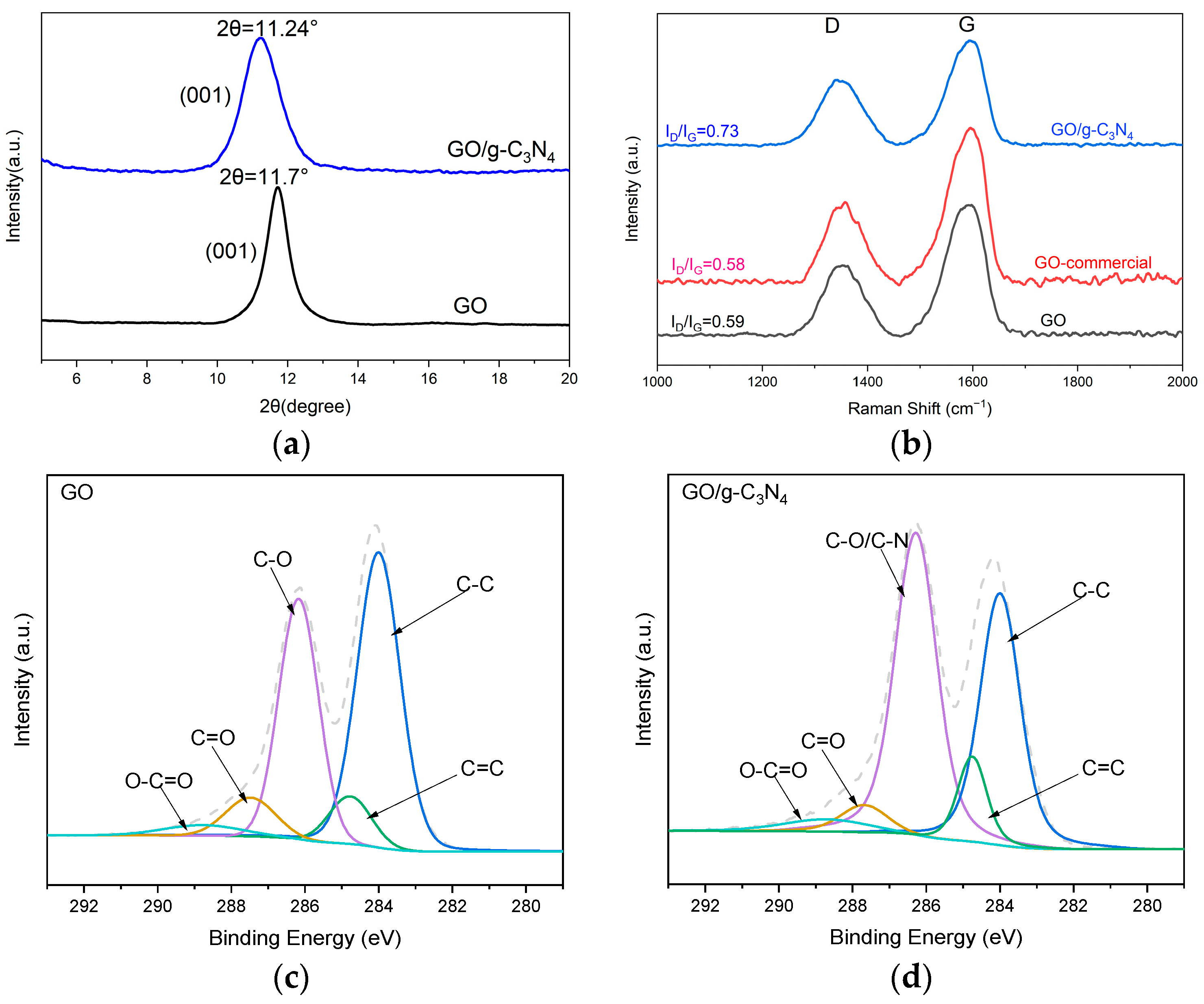
The XRD was used to verify the effect of the intercalation of g-C3N4 on the structure of GO, the peaks correspond to the conjugated aromatic (001) planes. As shown in Figure 2, the characteristic peak of the GO appears at 2θ = 11.7°, corresponding to 7.6 Å d-spacing. The characteristic peak of the GO/g-C3N4 appears at 2θ = 11.24°, corresponding to a d-spacing of 7.9 Å. The increase in the d-spacing was attributed to the intercalation of g-C3N4 nanosheets into the GO nanosheets and caused the peaks shifting in XRD.
The structural transformation of the GO membrane was confirmed by Raman analysis after modification with g-C3N4. GO exhibits two absorption peaks at 1340 cm−1 and 1580 cm−1, corresponding to the presence of disordered carbon and sp2 hybridized planar carbon in graphene or graphite, respectively [34]. The peak at 1340 cm−1 (peak D) represents the vibrational disorder of carbon atoms, indicating structural defects in the aromatic ring where some sp2 carbon atoms are transformed into sp3 hybrid structures. Conversely, the peak at 1580 cm−1 (peak G) arises from the in-plane vibrations of sp2 carbon atoms, symbolizing the symmetry and order of the aromatic ring structure. Furthermore, the ID/IG ratio can be employed to characterize the proportion of disordered carbon to sp2 carbon atoms and objectively evaluate the degree of graphitization in graphene samples. The ID/IG ratios for the GO/g-C3N4 membrane, commercial GO membrane (the GO dispersion was purchased from XFNANO), and lab-made GO membrane were determined as 0.73, 0.58, and 0.59, respectively. These results indicate a lower abundance of perfect graphene layers (sp2 hybridized carbon structure) in the GO/g-C3N4 membrane than that of the GO membranes.
The C1s XPS spectra of the GO membrane and GO/g-C3N4 membrane are shown in Figure 3c and Figure 3d, respectively. There are five main peaks: sp2 carbon (C–C bond) at 284 eV, sp3 carbon (C=C bond) at 284.77 eV, hydroxyl or epoxy groups (C–O bond) at 286.17 eV, carbonyl groups (C=O bond), at 287.48 eV, carboxyl groups (O–C=O bond) at 288.77 eV [35]. The C–O/C–N peak at 286.17 eV in the GO/g-C3N4 spectrum is significantly higher compared to that of the GO spectrum, indicating the successful intercalation of g-C3N4 into the GO membrane. Meanwhile, the combined area proportion of sp2 and sp3 peaks in GO/g-C3N4 is 45.6%, whereas it reaches 53% in the GO spectrum.

3.2. Static Diffusion Experiment

In the previously described static diffusion experiment, GO membranes of varying thicknesses were prepared by the vacuum filtration of 5 mL, 7.5 mL, 10 mL, and 15 mL GO dispersions (2 mg/mL) on PC substrate membranes. The mass of GO used was 10 mg, 15 mg, 20 mg, and 30 mg, respectively, resulting in membrane thicknesses of 4 μm, 6 μm, 8 μm, and 12 μm. The area of the membranes utilized in this experiment is approximately 2 cm2. The experiment was conducted at room temperature for 43 days, the relationship between the mass loss of the samples and time is shown in Figure S4, and the evident correlation between the two variables is readily apparent. Figure 4 illustrates the permeate flux of H2O or D2O through these membranes. It should be noted that the data for the membrane with no added GO (0 mg) represents a control where only the PC substrate membrane was sealed at the top of the bottle. The trend evidence that the permeability of both water and heavy water decreases as the membrane thickness increases. As shown in Table 1, for the PC membrane, the D2O permeate flux was 10.22% lower than that of H2O, which was less than any other GO membrane. The most significant disparity was observed at a membrane thickness of 4 μm when the amount of GO was 10 mg and the permeance of D2O was 14.73% less than that of H2O.

3.3. Distillation Tests

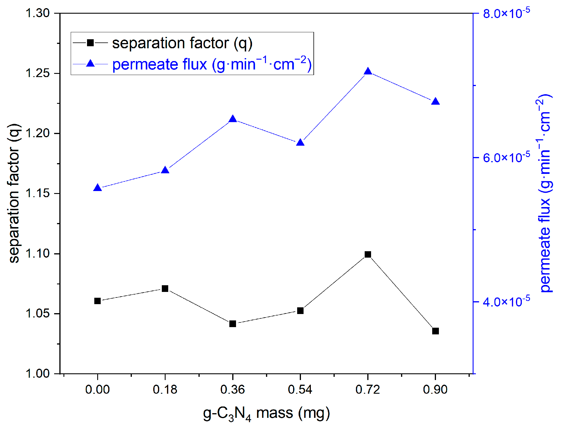
For the distillation tests, the mixed suspensions of GO/g-C3N4 for vacuum filtration were prepared by combining 2 mg/mL dispersions of GO with 0.18 mg/mL g-C3N4 dispersions, followed by subjecting them to ultrasound treatment for 2 h. The volume of g-C3N4 dispersion was 2 mL, 4 mL, 6 mL, 8 mL, and 10 mL, corresponding to the mass of g-C3N4 intercalated in the GO membrane as follows: 0.18 mg, 0.36 mg, 0.54 mg, 0.72 mg, and 0.9 mg shown on the X axis in Figure 5. During the experiments, 10% D2O was evaporated by heating the chamber to 110 °C and subsequently permeating through the membrane. The results of the distillation tests was listed in Table 2. The mass increase in the collected sample bottle was measured, and the permeate flux was calculated using Equation (1), the area of membrane in the tests is about 2 cm2 and the test time is 2880 min. The integral area ratio was given by the NMR spectrum and the deuterium concentration can be obtained by comparing with standard curve shown in Figure S3. The separation factor was calculated by Equation (2).
The separation factor and permeate flux for the GO membrane, as shown in Table 2 with g-C3N4 mass of 0, is 1.06 and 5.6 × 10⁻⁵ g·min−1·cm−2. Additionally, the PTFE support membrane, an advanced commercial micro-filtration membrane with 0.22 μm pore size, was also tested and the separation factor is 1.02. The separation factor of GO/g-C3N4 composite membranes ranges from 1.04 to 1.1, which exhibits a comparable performance to that of the GO membrane. The permeability of all the composite membranes is larger than that of the GO membrane. The optimal performance is achieved with a mass of g-C3N4 at 0.72 mg, accounting for 6.7%. The permeate flux reaches up to 7.2 × 10⁻⁵ g·min−1·cm−2, which is 29% higher than that of the GO membrane, while maintaining a separation factor of 1.1.

4. Discussion

The separation factors of the GO/g-C3N4 membranes were similar to the GO membrane. The collective migration of water molecules in the GO membrane and the resulting hydrogen isotope separation performance are attributed to the unique structure of the GO membrane. The oxygen-containing groups located at the edges of GO nanosheets provide support for interlayer spacing, allowing water molecules to pass through and enter subsequent layers via structural defects within or at the edge of the membrane [36]. Due to its triazine or heptazine structure [37], two-dimensional g-C3N4 possesses an ample number of molecular-level pores for water molecules to pass through, while amino groups are present at the edges, resembling oxygen-containing groups in GO and capable of forming hydrogen bonds with water. The chemical properties of carbon nitride nanosheets closely resemble those of GO nanosheets, thereby preserving the separation efficiency of GO membranes towards hydrogen isotope water.
The permeate flux of the composite membranes exhibited an obvious increase after being modified with g-C3N4. Three contributing factors are postulated to account for this result:
  • Large barely stripped g-C3N4 bulks are first deposited at the bottom of the membrane in the vacuum filtration process, as shown in Figure 1d. These particles contribute to an increased surface roughness of the membrane. A study has demonstrated that the high permeability of GO membranes observed may be attributed to the disordered microstructure of the membrane [38];
  • Smaller particles of partially stripped g-C3N4 with a transverse size of 10 μm and height of 3 μm can be observed to be inserted between the laminates, as illustrated in Figure 1f. The ID/IG ratio of the GO/g-C3N4 membrane is larger than the GO membrane in the Raman analysis, indicating that the interlayer particles disrupt the originally relatively complete structure and result in more defects within the composite membrane.
  • The shift of the peak in the XRD pattern can prove that single or fewer layers of g-C3N4 nanosheets are inserted between the layers of GO nanosheets and the layer d-spacing of the GO membrane is modified [37]. The increase in d-spacing from 7.6 Å to 7.9 Å contributes to the higher permeability [39].
Moreover, the static diffusion test was carried out at room temperature for 43 days, during which the permeation rate of the membranes was maintained as time went by. In the distillation experiment, the temperature of the film is maintained at 110 °C under the action of the glass fiber heating jacket. At this temperature, the permeability rate of the GO membrane will decrease with the increase in time [29], which may be attributed to the membrane being reduced at high temperatures. This results in a slightly lower permeation rate of the GO membrane in the distillation experiment than in the static diffusion experiment performed at room temperature.
The outstanding performance of the GO/g-C3N4 membrane in heavy water separation suggests its potential application in the treatment of tritiated wastewater in the future nuclear industry.

5. Conclusions

In this work, we have successfully fabricated a GO membrane by the Hummers method and modified it with g-C3N4. Static diffusion experiments have proved that there is a difference in the penetration rate between water and heavy water through GO membrane which indicate that the GO membrane separation technology is a possible application used in a stage of nuclear wastewater treatment. A device was designed and customized to make the heavy water vapor permeate the membrane. A series of repeatable experiments were conducted to investigate the impact of the intercalation of g-C3N4 in the GO membrane on the separation performance. The result is the permeation rate of the GO membrane, which was modified with g-C3N4, which is higher than that of the GO membrane, while maintaining a high separation factor. This improvement may be attributed to changes in the membrane structure at the microscopic scale. Based on the results of the characterization experiments, we presented a comprehensive analysis from three perspectives; however, the primary determinant remains inconclusive. The mechanism remains to be fully elucidated through simulation experiments. Compared to energy-intensive technologies, GO membranes have the potential to purify low-concentration tritiated wastewater. This study demonstrates that modifying the membrane through intercalation is a feasible approach. In the future, further investigation into the different intercalating materials should be conducted to enhance the membrane separation performance and make the utilization of GO membranes in industrial-scale nuclear wastewater treatment possible.

Supplementary Materials

The following supporting information can be downloaded at: https://www.mdpi.com/article/10.3390/w16070967/s1, Figure S1: The custom-made device for the isotope separation experiment; Figure S2: The 1H NMR spectroscopy of a mixture of 10% heavy water and acetone by 1:1 volume ratio; Figure S3: The linear relation between the area ratio of the two peaks in the NMR spectra and the heavy water concentration; Figure S4: The relationship between the mass loss of H2O (a) or D2O (b) and time.

Author Contributions

Conceptualization, Y.H.; investigation, Z.L. and R.F.; methodology, L.C.; data curation, S.L. and X.L.; writing—original draft preparation, Z.L.; writing—review and editing, Y.H. All authors have read and agreed to the published version of the manuscript.

Funding

This research was funded by China Institute of Atomic Energy, grant number YZ232505001002.

Data Availability Statement

All relevant data are included in the paper or its Supplementary Materials.

Conflicts of Interest

The authors declare that they have no known competing financial interests or personal relationships that could have appeared to influence the work reported in this paper.

References

  1. Smith, J.; Marks, N.; Irwin, T. The risks of radioactive wastewater release. Science 2023, 382, 31–33. [Google Scholar] [CrossRef]
  2. Han, Y.; Zhong, X. An overview of heavy water reactors. Nuclear Power Reactor Designs 2024, 351–363. [Google Scholar]
  3. Buesseler, K.; Aoyama, M.; Fukasawa, M. Impacts of the Fukushima nuclear power plants on marine radioactivity. Environ. Sci. Technol. 2011, 45, 9931–9935. [Google Scholar] [CrossRef] [PubMed]
  4. Cristescu, I.; Cristescu, I.R.; Dörr, L.; Glugla, M.; Hellriegel, G.; Michling, R.; Murdoch, D.; Schäfer, P.; Welte, S.; Wurster, W. Commissioning of water detritiation and cryogenic distillation systems at TLK in view of ITER design. Fusion Eng. Des. 2007, 82, 2126–2132. [Google Scholar] [CrossRef]
  5. Alekseev, I.A.; Bondarenko, S.D.; Fedorchenko, O.A.; Vasyanina, T.V.; Konoplev, K.A.; Arkhipov, E.A.; Voronina, T.V.; Grushko, A.I.; Tchijov, A.S.; Uborsky, V.V. Heavy water detritiation by combined electrolysis catalytic exchange at the experimental industrial plant. Fusion Eng. Des. 2003, 69, 33–37. [Google Scholar] [CrossRef]
  6. Miller, J.M.; Graham, W.R.C.; Celovsky, S.L.; Tremblay, J.R.R.; Everatt, A.E. Design and operational experience with a pilot-scale CECE detritiation process. Fusion Sci. Technol. 2002, 41, 1077–1081. [Google Scholar] [CrossRef]
  7. Goh, P.S.; Ismail, A.F.; Ng, B.C.; Abdullah, M.S. Recent progresses of forward osmosis membranes formulation and design for wastewater treatment. Water 2019, 11, 2043. [Google Scholar] [CrossRef]
  8. Novoselov, K.S.; Geim, A.K.; Morozov, S.V.; Jiang, D.E.; Zhang, Y.; Dubonos, S.V.; Grigorieva, I.V.; Firsov, A.A. Electric field effect in atomically thin carbon films. Science 2004, 306, 666–669. [Google Scholar] [CrossRef] [PubMed]
  9. Xu, P.T.; Yang, J.X.; Wang, K.S.; Zhou, Z.; Shen, P. Porous graphene: Properties, preparation, and potential applications. Chin. Sci. Bull. 2012, 57, 2948–2955. [Google Scholar] [CrossRef]
  10. Huang, L.; Zhang, M.; Li, C.; Shi, G. Graphene-based membranes for molecular separation. J. Phys. Chem. Lett. 2015, 6, 2806–2815. [Google Scholar] [CrossRef]
  11. Kovtyukhova, N.I.; Ollivier, P.J.; Martin, B.R.; Mallouk, T.E.; Chizhik, S.A.; Buzaneva, E.V.; Gorchinskiy, A.D. Layer-by-layer assembly of ultrathin composite films from micron-sized graphite oxide sheets and polycations. Chem. Mater. 1999, 11, 771–778. [Google Scholar] [CrossRef]
  12. Nair, R.R.; Wu, H.A.; Jayaram, P.N.; Grigorieva, I.V.; Geim, A.K. Unimpeded permeation of water through helium-leak–tight graphene-based membranes. Science 2012, 335, 442–444. [Google Scholar] [CrossRef]
  13. Li, H.; Song, Z.; Zhang, X.; Huang, Y.; Li, S.; Mao, Y.; Ploehn, H.J.; Bao, Y.; Yu, M. Ultrathin, molecular-sieving graphene oxide membranes for selective hydrogen separation. Science 2013, 342, 95–98. [Google Scholar] [CrossRef] [PubMed]
  14. Sun, P.; Wang, K.; Zhu, H. Recent developments in graphene-based membranes: Structure, mass-transport mechanism and potential applications. Adv. Mater. 2016, 28, 2287–2310. [Google Scholar] [CrossRef] [PubMed]
  15. Li, Y.; Zhou, Z.; Shen, P.; Chen, Z. Two-dimensional polyphenylene: Experimentally available porous graphene as a hydrogen purification membrane. Chem. Commun. 2010, 46, 3672–3674. [Google Scholar] [CrossRef]
  16. Celebi, K.; Buchheim, J.; Wyss, R.M.; Droudian, A.; Gasser, P.; Shorubalko, I.; Kye, J.I.; Lee, C.; Park, H.G. Ultimate permeation across atomically thin porous graphene. Science 2014, 344, 289–292. [Google Scholar] [CrossRef]
  17. Liu, G.; Jin, W.; Xu, N. Graphene-based membranes. Chem. Soc. Rev. 2015, 44, 5016–5030. [Google Scholar] [CrossRef]
  18. Joshi, R.K.; Carbone, P.; Wang, F.C.; Kravets, V.G.; Su, Y.; Grigorieva, I.V.; Wu, H.A.; Geim, A.K.; Nair, R.R. Precise and ultrafast molecular sieving through graphene oxide membranes. Science 2014, 343, 752–754. [Google Scholar] [CrossRef] [PubMed]
  19. Radha, B.; Esfandiar, A.; Wang, F.C.; Rooney, A.P.; Gopinadhan, K.; Keerthi, A.; Mishchenko, A.; Janardanan, A.; Blake, P.; Fumagalli, L.; et al. Molecular transport through capillaries made with atomic-scale precision. Nature 2016, 538, 222–225. [Google Scholar] [CrossRef]
  20. Boukhvalov, D.W.; Katsnelson, M.I.; Son, Y.W. Origin of anomalous water permeation through graphene oxide membrane. Nano Lett. 2013, 13, 3930–3935. [Google Scholar] [CrossRef]
  21. Abraham, J.; Vasu, K.S.; Williams, C.D.; Gopinadhan, K.; Su, Y.; Cherian, C.T.; Dix, J.; Prestat, E.; Haigh, S.J.; Grigorieva, I.V.; et al. Tunable sieving of ions using graphene oxide membranes. Nat. Nanotechnol. 2017, 12, 546–550. [Google Scholar] [CrossRef]
  22. Algara-Siller, G.; Lehtinen, O.; Wang, F.C.; Nair, R.R.; Kaiser, U.; Wu, H.A.; Geim, A.K.; Grigorieva, I.V. Square ice in graphene nanocapillaries. Nature 2015, 519, 443–445. [Google Scholar] [CrossRef] [PubMed]
  23. Mario, M.S.F.; Neek-Amal, M.; Peeters, F.M. AA-stacked bilayer square ice between graphene layers. Phys. Rev. B 2015, 92, 245428. [Google Scholar] [CrossRef]
  24. Zhu, W.; Zhu, Y.B.; Wang, L.; Zhu, Q.; Zhao, W.H.; Zhu, C.; Bai, J.; Yang, J.; Yuan, L.F.; Wu, H.; et al. Water confined in nanocapillaries: Two-dimensional bilayer squarelike ice and associated solid–liquid–solid transition. J. Phys. Chem. C 2018, 122, 6704–6712. [Google Scholar] [CrossRef]
  25. Sevigny, G.J.; Motkuri, R.K.; Gotthold, D.W.; Fifield, L.S.; Frost, A.P.; Bratton, W. Separation of Tritiated Water Using Graphene Oxide Membrane; Pacific Northwest National Lab. (PNNL): Richland, WA, USA, 2015. [Google Scholar]
  26. Liu, H.; Wang, H.; Zhang, X. Facile fabrication of freestanding ultrathin reduced graphene oxide membranes for water purification. Adv. Mater. 2015, 27, 249–254. [Google Scholar] [CrossRef] [PubMed]
  27. Joshi, D.J.; Koduru, J.R.; Malek, N.I.; Hussain, C.M.; Kailasa, S.K. Surface modifications and analytical applications of graphene oxide: A review. TrAC Trends Anal. Chem. 2021, 144, 116448. [Google Scholar] [CrossRef]
  28. Chen, L.; Li, N.; Wen, Z.; Zhang, L.; Chen, Q.; Chen, L.; Si, P.; Feng, J.; Li, Y.; Lou, J.; et al. Graphene oxide based membrane intercalated by nanoparticles for high performance nanofiltration application. Chem. Eng. J. 2018, 347, 12–18. [Google Scholar] [CrossRef]
  29. Mohammadi, A.; Daymond, M.R.; Docoslis, A. New insights into the structure and chemical reduction of graphene oxide membranes for use in isotopic water separations. J. Membr. Sci. 2022, 659, 120785. [Google Scholar] [CrossRef]
  30. Wen, M.; Chen, M.; Ren, G.K.; Li, P.L.; Lv, C.; Yao, Y.; Liu, Y.K.; Deng, S.J.; Zheng, Z.; Xu, C.G.; et al. Enhancing the selectivity of hydrogen isotopic water in membrane distillation by using graphene oxide. J. Membr. Sci. 2020, 610, 118237. [Google Scholar] [CrossRef]
  31. Wen, M.; Chen, M.; Chen, K.; Li, P.L.; Lv, C.; Zhang, X.; Yao, Y.; Yang, W.; Huang, G.; Ren, G.K.; et al. Superhydrophobic composite graphene oxide membrane coated with fluorinated silica nanoparticles for hydrogen isotopic water separation in membrane distillation. J. Membr. Sci. 2021, 626, 119136. [Google Scholar] [CrossRef]
  32. Chen, J.; Yao, B.; Li, C.; Shi, G. An improved Hummers method for eco-friendly synthesis of graphene oxide. Carbon 2013, 64, 225–229. [Google Scholar] [CrossRef]
  33. Tsou, C.H.; An, Q.F.; Lo, S.C.; De Guzman, M.; Hung, W.S.; Hu, C.C.; Lee, K.R.; Lai, J.Y. Effect of microstructure of graphene oxide fabricated through different self-assembly techniques on 1-butanol dehydration. J. Membr. Sci. 2015, 477, 93–100. [Google Scholar] [CrossRef]
  34. An, D.; Yang, L.; Wang, T.J.; Liu, B. Separation performance of graphene oxide membrane in aqueous solution. Ind. Eng. Chem. Res. 2016, 55, 4803–4810. [Google Scholar] [CrossRef]
  35. Liu, L.; Zhou, Y.; Xue, J.; Wang, H. Enhanced antipressure ability through graphene oxide membrane by intercalating g-C3N4 nanosheets for water purification. AIChE J. 2019, 65, e16699. [Google Scholar] [CrossRef]
  36. Muscatello, J.; Jaeger, F.; Matar, O.K.; Müller, E.A. Optimizing water transport through graphene-based membranes: Insights from nonequilibrium molecular dynamics. ACS Appl. Mater. Interfaces 2016, 8, 12330–12336. [Google Scholar] [CrossRef] [PubMed]
  37. Wang, Y.; Li, L.; Wei, Y.; Xue, J.; Chen, H.; Ding, L.; Caro, J.; Wang, H. Water transport with ultralow friction through partially exfoliated g-C3N4 nanosheet membranes with self-supporting spacers. Angew. Chem. Int. Ed. 2017, 56, 8974–8980. [Google Scholar] [CrossRef] [PubMed]
  38. Chong, J.Y.; Wang, B.; Mattevi, C.; Li, K. Dynamic microstructure of graphene oxide membranes and the permeation flux. J. Membr. Sci. 2018, 549, 385–392. [Google Scholar] [CrossRef]
  39. Chen, X.; Ching, K.; Rawal, A.; Lawes, D.J.; Tajik, M.; Donald, W.A.; Zhao, C.; Lee, S.H.; Ruoff, R.S. Stage-1 cationic C60 intercalated graphene oxide films. Carbon 2021, 175, 131–140. [Google Scholar] [CrossRef]
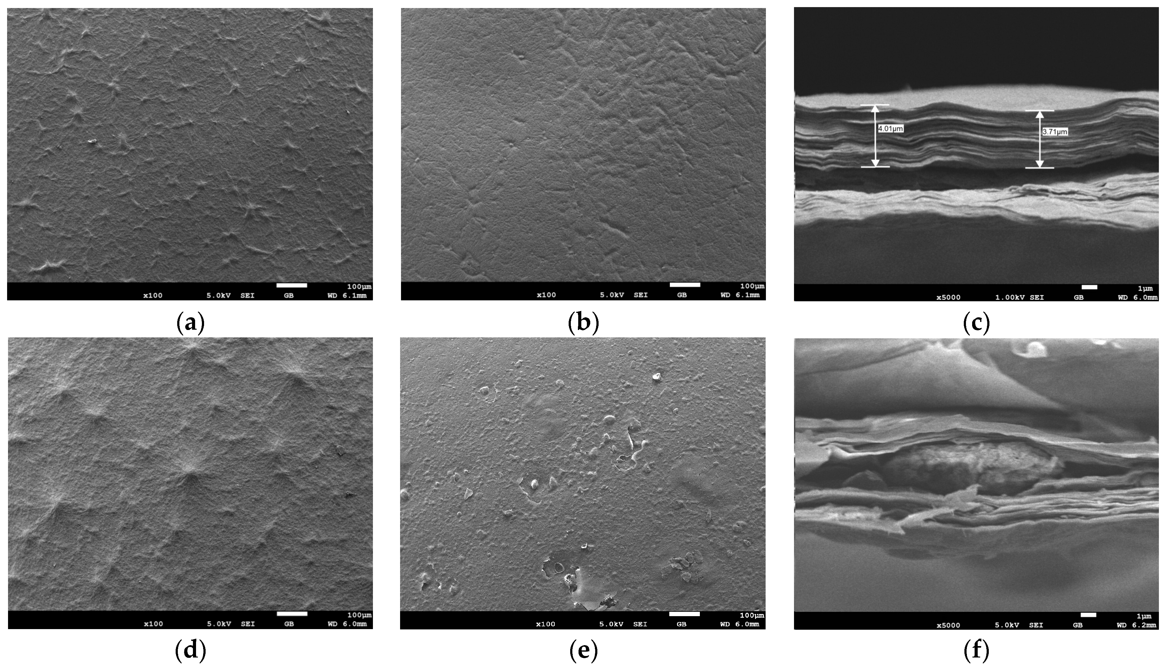
Figure 1. SEM images of (a) the top surface of the GO membrane, (b) the bottom surface of the GO membrane, (c) the cross-section of the GO membrane prepared by 10 mg dried GO, (d) the top surface of the GO/g-C3N4 membrane, (e) the bottom surface of the GO/g-C3N4 membrane, and (f) the cross-section of the GO/g-C3N4 membrane.
Water 16 00967 g001
Figure 2. TEM image of a GO nanosheet.
Water 16 00967 g002
Figure 3. (a) XRD spectra of GO and GO/g-C3N4; (b) Raman spectra of GO, GO-commercial, and GO/g-C3N4; C1s XPS spectrum of (c) GO membrane and (d) GO/g-C3N4 membrane. The grey dashed lines represent the XPS spectral lines.
Water 16 00967 g003
Figure 4. Permeance of H2O and D2O for different amounts of GO used in the fabrication of membranes in the static diffusion experiment.
Water 16 00967 g004
Figure 5. The separation factor and permeate flux of GO membranes with varying masses of g-C3N4 intercalation in the distillation tests.
Water 16 00967 g005
Table 1. The results of the static diffusion experiment.
GO (mg)H2O Permeance
(10−5 g/min·cm2)
D2O Permeance
(10−5 g/min·cm2)
Difference
(10−5 g/min·cm2)
Reduction
07.456.690.76110.22%
107.306.221.0714.73%
157.056.300.73810.48%
207.136.220.90012.64%
306.715.800.90513.48%
Table 2. The results of the distillation tests.
g-C3N4 Mass (mg)Integral Area Ratio in NMRDeuterium ConcentrationSeparation FactorsPermeate Mass (mg)Permeate Flux (10−5 g·min−1·cm−2)
01.1899.48%1.060.3215.57
0.181.1919.40%1.070.3355.82
0.361.1879.64%1.040.3766.53
0.541.1899.55%1.050.3576.20
0.721.1949.18%1.100.4147.19
0.91.1879.69%1.040.3906.77
Disclaimer/Publisher’s Note: The statements, opinions and data contained in all publications are solely those of the individual author(s) and contributor(s) and not of MDPI and/or the editor(s). MDPI and/or the editor(s) disclaim responsibility for any injury to people or property resulting from any ideas, methods, instructions or products referred to in the content.