Article

Effects of Brookite TiO2/CeO2 Nanocomposite on Artemia salina: Induction of Oxidative Stress and Apoptosis Assessment

1
Department of Biological, Geological and Environmental Sciences, University of Catania, Via Androne 81, 95124 Catania, Italy
2
Experimental Zooprophylactic Institute of Sicily “A. Mirri”, Via Gino Marinuzzi, 3, 90129 Palermo, Italy
3
Department of Chemical Sciences, University of Catania, Viale A. Doria 6, 95125 Catania, Italy
*
Author to whom correspondence should be addressed.
Water 2024, 16(14), 1946; https://doi.org/10.3390/w16141946
Submission received: 29 May 2024 / Revised: 5 July 2024 / Accepted: 8 July 2024 / Published: 10 July 2024
(This article belongs to the Section Wastewater Treatment and Reuse)

Abstract

:
TiO2 and CeO2 NPs are widely used in the medical field, for drug administration, oncological therapies, disinfection or diagnostic imaging. To date, their effects on aquatic ecosystems and their impacts on human health are little known but several scientific evidences show how CeO2 NPs are able to store and release oxygen, giving them antioxidant activity against oxidative stress while TiO2 NPs seem to be responsible for cytotoxicity and genotoxicity. In this study, the effects of combined nanoparticles, brookite TiO2/CeO2 nanocomposites, on A. salina nauplii following acute exposure were evaluated. Although the cytotoxicity of nanoparticles is generally dose-dependent and time-dependent, in the following investigation, exposure to combined nanoparticles, brookite TiO2/CeO2 nanocomposites, in A. salina nauplii would appear not to have had a statistically significant negative impact on the viability of the specimens. One of the mechanisms by which NPs exert toxic effects is the induction of oxidative stress. In this study, an assessment of potential oxidative stress and apoptotic damage on exposed organisms was conducted through the use of the DCFH2DA probe and acridine orange. The results obtained suggest that although acute exposure to different concentrations of brookite TiO2/CeO2 nanocomposites did not have lethal effects, the positivity to DCFH2DA and acridine orange observed does not exclude potential sublethal effects.

1. Introduction

Currently, nanotechnology plays a role of great importance especially in the development of nanomaterials useful in physical, biological, biomedical and pharmaceutical applications [1,2,3]. Due to their peculiar properties, nanoparticles can be utilized as a selective drug delivery system in cancer therapy, especially metal oxides able to generate oxidative stress on tumor cells [4,5], but also they can be used in dressings, water disinfection and food packaging. On the other hand, their nano size favors interaction with biological systems, because they are able to overcome biological membranes, reach different organs by the bloodstream [6] and exert in situ effects. Scientific evidences suggest that the main mechanism by which NPs act, especially metal oxide nanoparticles, is oxidative stress [7] and that nano-toxicity is due to the presence of pro-oxidant functional groups placed on their reactive surface or due to NP–cell interactions [8] which cause an imbalance in the redox state of the cell. An increase in oxidative stress seems to induce serious damage to cellular macromolecules such as proteins, lipids and DNA, with deleterious effects on cells. Moreover, under stress conditions, cells activate several processes important for adaptation to adverse conditions or mechanisms of cell death, such as apoptosis or necrosis [9].
TiO2 is commonly used in paints, varnishes, lacquers, paper, plastics, ceramics, catalysts, textiles, cosmetics, sunscreens and pharmaceutical products [10], or in food dyes, glassware, rubber tires, bone implants and in the production of electronic components. Thanks to its photocatalytic properties [11], TiO2 can disintegrate harmful substances, remove stains and kill pathogenic microbes thus constituting a useful tool in self-sterilization and maintaining a clean and safe living environment [12,13], including the removal of air and water pollutants [14,15,16,17,18,19]. Many of the toxic effects are due to the formation of anatase, which seems to induce cyto- and geno-toxicity, increasing inflammatory indices, increased ROS, programmed cell death, micronuclei formation or presence of multinucleated cells [20,21] and DNA damage [22,23,24,25].
CeO2 NPs have attracted considerable scientific interest for their peculiar chemical, physical and biological properties. Cerium is the second member of the series of lanthanides or rare earth elements that can be found in two oxidation states, in the form of ceric ion (Ce4+) and waxy ion (Ce3+), which are responsible for its powerful reduction/oxidation behavior. The thermodynamic efficiency of the redox cycle between 3+ and 4+ on its surface [26] and the characteristic of oxygen absorption and release [27] makes CeO2 NPs capable, both in vivo and in vitro, of eliminating reactive oxygen species, endowing these nanoparticles with a great potential in suppressing inflammation and exerting a cytoprotective effect in different types of mammalian cells, such as neural cells [28], retinal [29], hepatic [30], cardiac [31] and cartilage [32], oxidative stress and inflammatory responses [33].
To monitor the environmental impact of contaminants in the aquatic environment, in vivo ecotoxicological analyses have been conducted. Artemia salina represents one of the most suitable animal models for studying the ecotoxicity of nanomaterials in aquatic ecosystems due to its simplicity, short life cycle and high reproduction rate [34]. A. salina is often used as a bio-indicator [35] being a non-selective filter feeder, capable of accumulating metals and being particularly sensitive to environmental pollutants. Even though several studies have been performed to establish the toxicity of these two metal oxide nanoparticles in vitro, less is known about in vivo toxicity; thus, this study aims to evaluate the effects induced by single TiO2 NPs, single CeO2 NPs and moreover a nanocomposite of brookite TiO2 and CeO2 called hereafter brookite TiO2/CeO2 nanocomposite by an acute exposure on A. salina larvae called nauplii. The crystalline phase of titanium dioxide used is brookite, the less common and less investigated form of TiO2.

2. Materials and Methods

2.1. Synthesis and Characterization of Brookite TiO2/CeO2 Nanocomposites, CeO2 NPs and Brookite TiO2 NPs

Brookite TiO2 was prepared through the thermohydrolysis of TiCl4 in a HCl-concentrated solution following the procedure reported in the ref. [36].
Specifically, 5 mL of titanium tetrachloride (98% Merck, Darmstadt, Germany) was slowly added to 200 mL of demineralized water and 80 mL of HCl. The resultant solution was put in a closed reactor and heated at 100 °C for 48 h in an oven. Afterwards, the formed brookite–rutile mixture was washed to remove the supernatant and to separate the brookite phase. The obtained brookite NPs were dried under vacuum at 55 °C overnight. The formation of the brookite TiO2 crystalline phase was verified by Raman and XRD measurements.
Wetness impregnation was instead employed to obtain the composites with CeO2 (3 wt%). This amount of cerium oxide was the optimal to obtain a good interaction with the brookite TiO2 [37,38]. In detail, a solution of Ce(NO3)3 · 6H2O (Merck) was impregnated to the brookite, and the formed slurry was dried at 120 °C overnight and subsequently calcined at 350 °C for 4 h. The CeO2 NPs were prepared by chemical precipitation with KOH (Merck); the details of the synthesis are reported in [39]. Also, for this sample, drying at 120 °C and calcination at 350 °C for 4 h were applied. The size of the obtained nanoparticles was in the range of 6–10 nm [38].

2.2. Aquatic Organisms

The nauplii (larvae) used for the ecotoxicological assay were provided from dehydrated cysts (JBL Artemio Pur® & Co. KG, Neuhofen, Germany). According to Pecoraro et al. [40], 1 g of cysts were incubated in 1 L of a salt solution called ASPM in a beaker. ASPM water was prepared by adding seven different salts in 1 L of a distilled water and according to Manzini et al. [41] incubation conditions at room temperature (26 ± 1 °C), continuous lighting and ventilation were maintained in order to achieve hatching (within 48 h).

2.3. Preparation of Nanoparticles Solutions

Brookite TiO2/CeO2 nanocomposite, brookite TiO2 NPs and CeO2 NPs were obtained from the Department of Chemical Sciences of the University of Catania. Based on literature data [42], a stock solution of brookite TiO2/CeO2 nanocomposites (100 mg/L), brookite TiO2 NPs (100 mg/L) and CeO2 NPs (100 mg/L), diluted in ASPM solution was prepared. Fresh suspensions with different concentrations of NPs (0.25 mg/L, 0.50 mg/L, 1.0 mg/L, 2.0 mg/L and 4.0 mg/L) were made from the stock suspensions of NPs diluted in ASPM water. Each solution was vortexed for 30 s, and then sonicated using a probe sonicator (Bandelin Sonopuls, Berlin, Germany) to allow their immediate dispersion before use.

2.4. Acute Toxicity Assessment

According to Ignoto et al. [43], after the hatching of the cysts (which occurred between 24 and 48 h) nauplii at stages II and III were selected and transferred to 96-multiwell plates. An amount of 200 μL of NP solution containing the different concentrations was added to the 96-well microplates (the controls were exposed only to ASPM water) and at the end incubated at 26 °C. The numbers of surviving nauplii in each well were counted under a stereomicroscope (Leica EZ4, Buccinasco, Italy) after 24 and 48 h. After the exposition, the percentage of vitality was calculated by the following formula (keeping control samples as a reference):
% Vitality = number   of   live   Artemia   nauplii number   of   live   Artemia   nauplii + number   of   dead   Artemia   nauplii × 100

2.5. Optical Microscopy

In order to assess any morphological damage on nauplii after 48 h exposure, the control and treated nauplii were placed on a glass slide and observed under an optical microscope (Nikon Eclipse E200, Amstelveen, The Netherlands) connected to a digital camera (CMOS Nikon, Amsterdam, The Netherlands).Their images were captured.

2.6. Reactive Oxygen Species Generation (ROS)

According to a study conducted by Ulm et al. [44], the production of reactive oxygen species was identified using DCFH2-DA (2′,7′-Dichlorodihydrofluorescein diacetate). Nauplii treated with NPs were collected and transferred to Eppendorf tubes containing DCFH2-DA probe (10 μM) and incubated for 30 min at 37 °C. After incubation, nauplii were washed twice in PBS (Phosphate Buffer Saline, 1X, pH 7.4) and then ROS positivity was observed by fluorescence microscope (Nikon Eclipse Ci, Amstelveen, The Netherlands).

2.7. Acridine Orange Staining

According to Arulvasu et al. [45], A. salina nauplii treated with various concentrations of NPs were incubated with 10 µL of acridine orange (from stock of 5 µg/mL) for 20 min at room temperature and in the dark. Afterwards, the nauplii were washed in PBS (1X, pH 7.4) once and placed on a slide and observed under a fluorescence microscope (Nikon Eclipse Ci, Amstelveen, The Netherlands).

2.8. Statistical Analysis

All tests were performed for the three types of NPs (brookite TiO2/CeO2 nanocomposite, brookite TiO2 NPs and CeO2 NPs).Two replicates were performed; a total of 144 nauplii were exposed to each concentration of NPs tested and also 144 nauplii formed the control group. Findings were recorded as mean ± standard deviation. The ImageJ software (Fiji 1.46) was used to assess the intensity fluorescence of each image obtained by fluorescence microscope. The data obtained were compared using one-way analysis of variance (ANOVA) by GraphPad Prism software (Edition 9).

3. Results

3.1. Toxicity towards Artemia sp.

After 24 h from exposure, no change was observed in the vitality percentage of treated nauplii compared to controls (around 100%) for brookite TiO2/CeO2 nanocomposite. Even after 48 h from exposure, a decrease in vitality percentage of A. salina’s nauplii was shown for 0.25, 0.50, 1.0 and 2.0 mg/L compared to control, while for 4 mg/L it remained unchanged (Table 1). As regards brookite TiO2 NPs, vitality percentages at 24 h were similar to 48 h, whereas for CeO2 NPs a decrease appeared from 0.25 to 2 mg/L, especially for the higher concentration tested (4 mg/L). However, in all data analyses, statistical analysis did not show a significant difference in the vitality of A. salina nauplii exposed to the different concentrations of brookite TiO2/CeO2 nanocomposite and single nanoparticles (brookite TiO2 and CeO2 NPs) tested (p-value > 0.05). The values of vitality (presented as mean ± SD) for brookite TiO2 and CeO2 NPs are available in Supplementary Information Text S1.

3.2. Optical Microscopy

At 48 h from exposure, fresh observations under the light microscope did not show any morphological and structural damage of nauplii exposed to different concentrations compared to control for brookite TiO2/CeO2 nanocomposite and also brookite TiO2 NPs and CeO2 NPs. However, an accumulation of brookite TiO2/CeO2 nanocomposite was highlighted, mainly in the gut, for 2.0 mg/L and 4.0 mg/L (Figure 1e,f). Also, for nauplii exposed to single brookite TiO2 and CeO2 nanoparticles, an accumulation appeared in the gut (Supplementary Information Text S2).

3.3. Reactive Oxygen Species Generation (ROS)

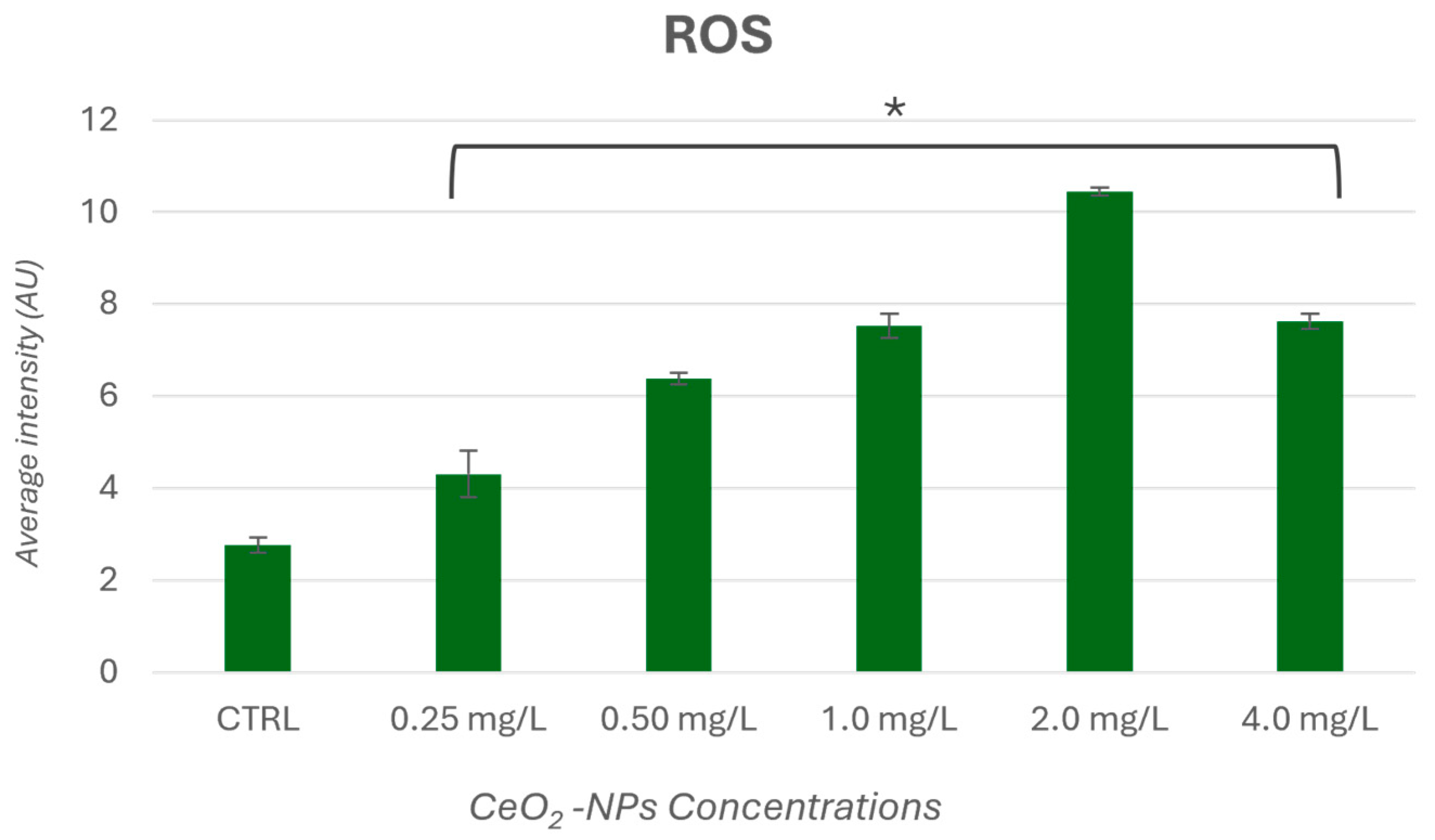
ROS generation in A. salina nauplii after exposure to different NP concentrations of brookite TiO2/CeO2 nanocomposite is shown in Figure 2, in which green fluorescence due to the generation of intracellular reactive oxygen species appears. Positivity to DCFH2-DA was found at the level of the swimming setae with a statistical significance for all concentrations tested compared to control (p < 0.05) (Figure 3). Similarly, this was observed in larvae exposed to different concentrations of CeO2 NPs, while no fluorescence was observed for those exposed to brookite TiO2 NPs (Supplementary Information Text S3).

3.4. Acridine Orange Staining

The observation under a fluorescence microscope allowed us to understand if, after exposure, the nauplii of A. salina showed cellular damage and if this was directly proportional to the different concentrations of brookite TiO2/CeO2 nanocomposite tested. The analysis, through the use of acridine orange, allows detection of the apoptosis of the cells by the emission of green fluorescence. Small fluorescence spots were observed at the level of the appendages and on the head, particularly around the eye, especially for A. salina nauplii exposed to the maximum concentration (4.0 mg/L) with a statistical significance compared to control (p < 0.05) (Figure 4). The analysis was also performed on A. salina nauplii exposed to single nanoparticles. In A. salina nauplii exposed to TiO2-brookite NPs, no fluorescence emission was observed, while for A. salina exposures to CeO2 NP, bright fluorescence spots were observed throughout the animal’s body, particularly on the head, appendages and chest (Supplementary Information Text S4).

4. Discussion

The aim of this study was to analyze the toxic effects of combined brookite TiO2 and CeO2 NPs in A. salina larvae. Today, the wide use of engineered nanomaterials in different fields such as biology, pharmacology and medicine may lead to an increased risk of exposure to NPs. Several evidences show how nanoparticles, thanks to their nano-size, are able to overcome biological membranes, spreading and reaching different organs, where different effects will accumulate and be exerted.
Nanotoxicity depends on the properties of NPs [46]. As previously mentioned, CeO2 NPs seem to act as antioxidants against oxidative stress, storing and releasing oxygen, while TiO2 NPs can cause cyto- and genotoxic effects.
Due to different effects associated with exposure to individual NPs, the purpose of this study is to analyze the actions induced by the combined brookite TiO2/CeO2 nanocomposite on a saltwater organism, A. salina, following acute exposure in order to compare the effect between the combined nanoparticles and the single NPs (brookite TiO2 and CeO2 NPs).
Scientific evidences have shown that the cytotoxic effect of nanoparticles is strongly dose- and time-dependent. In a study performed by Ates et colleagues [47], A. salina nauplii exposed to different sizes of metal nanoparticles showed mortality values increased significantly as exposure time increased.
Another study [48] investigated the nano and bulk toxicity of CeO2 on A. salina. The hatching rate of brine shrimp cysts was inhibited by both CeO2 particles used. Nano CeO2 was shown to be more toxic to the organism when compared to bulk CeO2. Moreover, a higher oxidative stress was found on nauplii treated with CeO2NPs than those exposed to the bulk form [48].
Mortality rates were found to be significantly altered in nauplii exposed to CeO2 and also the swimming speed was significantly inhibited in larvae exposed to 0.1 and 1.0 mg/mL CeO2 NPs [49]. The toxic effects of the exposure to Ce NPs was investigated during larval development in term of variations in the ability to reach adult stage. The results showed no significant toxic effects suggesting that CeO2NPs are not toxic in D. melanogaster under the testing conditions used [50]. Mortality, growth inhibition and genotoxic effects of two types of CeO2 NPs were also evaluated in two species of amphibian larvae. Results show there was no acute toxicity on the specimens after short exposures, even at the highest concentrations. Mortality (35%) was detected on Xenopus larvae after 12 d of exposure at the highest concentration of one type of CeO2 NP. Xenopus larvae growth was negatively affected from 1 mg/L of both CeO2 NPs used while growth inhibition was observed on Pleurodeles larvae only at the highest concentration of one type of NP. No genotoxicity was observed on Xenopus larvae but Pleurodeles exhibited dose-dependent genotoxic effects when exposed to one type of NP [51]. Moreover, TiO2 NPs had some effects on hatching rates and survival after hatching in a concentration-dependent manner [52]. TiO2 NPs’ toxicity increased also with duration of exposure [47,53] and they can lead to changes in eye formation, including changes in the eyeball shape, eyeball shrinking and fading of the iris [53]. In our previous study, where the impact of TiO2-CeO2 nanocomposites was investigated on Danio rerio embryos and larvae, it has been demonstrated that co-exposure did not affect the embryos’ development [54]. After 96 h of exposure, there was no significant mortality and no sublethal effects such as hatching delay, heartbeat alteration, or malformation in embryonic development compared with the control group. According to this, a combination of two type of NPs to obtain brookite TiO2/CeO2 nanocomposite has not caused a statistically significant mortality on A. salina nauplii with increasing of exposure and concentrations tested, even if thanks to the light microscope observations an accumulation of nanoparticles was highlighted mainly in the intestine, especially for the test concentrations of 2.0 mg/L and 4.0 mg/L. Similarly, at the higher concentrations of TiO2 NPs and CeO2 NPs their accumulation has been observed in Artemia’s intestine. Therefore, the uptake of NPs changed with the concentration, as observed by previous studies [53,55]. The presence of brookite TiO2/CeO2 nanocomposite inside the Artemia’s gut did not lead to changes such as enlargement of the intestine, but induced an oxidative condition as suggested by ROS analysis. A fluorescence spots appeared on the bristles of the appendages as well as in nauplii exposed to CeO2 NPs while no fluorescence emission appeared in the groups exposed toTiO2 NPs. Also, cellular damage by exposure to brookite TiO2/CeO2 nanocomposite was confirmed, thanks to acridine orange staining, at the maximum concentration on the head, appendages and chest of the nauplii as for the CeO2 NPs after 48 h of exposure.
Although data in the literature consider CeO2 NPs a promising metal oxide nanomaterial with the ability to scavenge free radicals, thus preventing cell death in oxidative stress [56,57,58,59,60,61], it is evident that CeO2 NPs alone and in combination with TiO2 NPs are able to induce an oxidative condition that is linked to time and concentration of exposure. Therefore, there is an urgent need to develop strategies that lead to improvements in the performance of CeO2 NPs.

5. Conclusions

Although the nanotechnology sector is booming, evidence on the potential impacts on humans and aquatic ecosystems is still limited. Several studies report induced effects certainly linked to the dimensions of nanoparticles and their peculiar properties which may on the one hand make them useful in different sectors, but on the other hand it cannot be excluded that their uncontrolled release into the external environment may bring adverse effects, following chronic exposure. To date, knowledge of the mechanisms by which nanoparticles act or interact with biological systems is still unclear and further studies would be needed to more clearly define the benefits and potential risks. However, according to some studies, one of the main mechanisms of action by which they act is the overproduction of reactive oxygen species (ROS).
Although, in this study, it was possible to define how brookite TiO2/CeO2 nanocomposite does not entail toxic effects in terms of lethality on exposed organisms, the short exposure time would be enough to induce oxidative stress in exposed A. salina nauplii. The results obtained in this study represent preliminary data and could be a useful tool for the evaluation and identification of critical aspects that would deserve great attention in future research.

Supplementary Materials

The following supporting information can be downloaded at: https://www.mdpi.com/article/10.3390/w16141946/s1. Text S1: Vitality rate (24 and 48 h) of A. salina nauplii treated with brookite TiO2 and CeO2 NPs; Text S2: Optical images of A. salina nauplii treated with brookite TiO2 and CeO2 NPs; Text S3: Reactive oxygen species generation (ROS) on A. salina nauplii treated with brookite TiO2 and CeO2 NPs; Text S4: Acridine orange Staining on A. salina nauplii treated with brookite TiO2 and CeO2 NPs.

Author Contributions

Conceptualization, S.I.; methodology, S.I. and G.C.; validation, S.I. and M.V.B.; investigation, S.I., G.C., S.C., R.L.F. and A.S. (Agata Scalisi); resources, R.F.; data curation, S.I., R.P. and E.M.S.; writing—original draft preparation, S.I.; writing—review and editing, S.I., R.P., E.M.S., R.F., A.S. (Antonio Salvaggio) and M.V.B.; visualization, A.S. (Antonio Salvaggio) and M.V.B.; supervision, M.V.B. All authors have read and agreed to the published version of the manuscript.

Funding

This research received no external funding.

Data Availability Statement

Original data are available on request.

Acknowledgments

S.I. thanks the University of Catania, Italy, PhD course XXXIX cycle.

Conflicts of Interest

The authors declare no conflicts of interest.

References

  1. Martis, E.; Badve, R.; Degwekar, M. Nanotechnology based devices and applications in medicine: An overview. Chron. Young Sci. 2012, 3, 68. [Google Scholar] [CrossRef]
  2. Nikalje, A.P. Nanotechnology and its Applications in Medicine. Med. Chem. 2015, 5, 81–89. [Google Scholar] [CrossRef]
  3. Loureiro, A.; Azoia, N.G.; Gomes, A.C.; Cavaco, A.P. Albumin-Based Nanodevices as Drug Carriers. Curr. Pharm. Des. 2016, 22, 1371–1390. [Google Scholar] [CrossRef]
  4. Rasmussen, J.W.; Martinez, E.; Louka, P.; Wingett, D.G. Zinc Oxide Nanoparticles for Selective Destruction of Tumor Cells and Potential for Drug Delivery Applications. Expert Opin. Drug Deliv. 2010, 7, 1063–1077. [Google Scholar] [CrossRef]
  5. Loutfy, S.A.; Al-Ansary, N.A.; Abdel-Ghani, N.T.; Hamed, A.R.; Mohamed, M.B.; Craik, J.D.; Eldin, T.A.S.; Abdellah, A.M.; Hussein, Y.; Hasanin, M.; et al. Anti-proliferative Activities of Metallic Nanoparticles in an In Vitro Breast Cancer Model. Asian Pac. J. Cancer Prev. 2015, 16, 6039–6046. [Google Scholar] [CrossRef]
  6. Pourmand, A.; Abdollahi, M. Current opinion on nanotoxicology. DARU J. Pharm. Sci. 2012, 20, 95. [Google Scholar] [CrossRef]
  7. Santonastaso, M.; Mottola, F.; Colacurci, N.; Iovine, C.; Pacifico, S.; Cammarota, M.; Cesaroni, F.; Rocco, L. In vitro genotoxic effects of titanium dioxide nanoparticles (nTiO2) in human sperm cells. Mol. Reprod. Dev. 2019, 86, 1369–1377. [Google Scholar] [CrossRef]
  8. Khanna, P.; Ong, C.; Bay, B.H.; Baeg, G.H. Nanotoxicity: An Interplay of Oxidative Stress, Inflammation and Cell Death. Nanomaterials 2015, 5, 1163–1180. [Google Scholar] [CrossRef]
  9. Vassal, M.; Rebelo, S.; Pereira, M.D.L. Metal Oxide Nanoparticles: Evidence of Adverse Effects on the Male Reproductive System. Int. J. Mol. Sci. 2021, 22, 8061. [Google Scholar] [CrossRef]
  10. Behnam, M.A.; Emami, F.; Sobhani, Z.; Dehghanian, A.R. The application of titanium dioxide (TiO2) nanoparticles in the photo-thermal therapy of melanoma cancer model. Iran J. Basic Med. Sci. 2018, 21, 1133–1139. [Google Scholar] [CrossRef] [PubMed]
  11. Lagopati, N.; Evangelou, K.; Falaras, P.; Tsilibary, E.C.; Vasileiou, P.V.S.; Havaki, S.; Angelopoulou, A.; Pavlatou, E.A.; Gorgoulis, V.G. Nanomedicine: Photo-activated nanostructured titanium dioxide, as a promising anticancer agent. Pharmacol. Ther. 2021, 222, 107795. [Google Scholar] [CrossRef] [PubMed]
  12. Hashimoto, K.; Irie, H.; Fujishima, A. TiO2 photocatalysis: A historical overview and future prospects. Jpn. J. Appl. Phys. 2005, 44, 8269–8285. [Google Scholar] [CrossRef]
  13. Binas, V.; Venieri, D.; Kotzias, D.; Kiriakidis, G. Modified TiO2 based photocatalysts for improved air and health quality. J. Materiomics 2017, 3, 3–16. [Google Scholar] [CrossRef]
  14. Choi, H.; Stathatos, E.; Dionysiou, D.D. Sol–gel preparation of mesoporous photocatalytic TiO2 films and TiO2/Al2O3 composite membranes for environmental applications. Appl. Catal. B Environ. 2006, 63, 60–67. [Google Scholar] [CrossRef]
  15. Burton, A. Titanium dioxide photocleans polluted air. Environ. Health Perspect. 2012, 120, A229. [Google Scholar] [CrossRef] [PubMed]
  16. Das, R. Application photocatalysis for treatment of industrial waste water—A short review. OALib J. 2014, 1, 1–17. [Google Scholar] [CrossRef]
  17. Muhd Julkapli, N.; Bagheri, S.; Bee Abd Hamid, S. Recent advances in heterogeneous photocatalytic decolorization of synthetic dyes. Sci. World J. 2014, 2014, 692307. [Google Scholar] [CrossRef] [PubMed]
  18. Bratovčić, A. Photocatalytic degradation of organic compounds in wastewaters. Technol. Acta 2019, 11, 17–23. [Google Scholar] [CrossRef]
  19. Zhang, F.; Wang, X.; Liu, H.; Liu, C.; Wan, Y.; Long, Y.; Cai, Z. Recent advances and applications of semiconductor photocatalytic technology. Appl. Sci. 2019, 9, 2489. [Google Scholar] [CrossRef]
  20. Wang, J.J.; Sanderson, B.J.; Wang, H. Cyto-and genotoxicity of ultrafine TiO2 particles in cultured human lymphoblastoid cells. Mutat. Res. 2007, 628, 99–106. [Google Scholar] [CrossRef]
  21. Onuma, K.; Sato, Y.; Ogawara, S.; Shirasawa, N.; Kobayashi, M.; Yoshitake, J.; Yoshimura, T.; Iigo, M.; Fujii, J.; Okada, F. Nano-scaled particles of titanium dioxide convert benign mouse fibrosarcoma cells into aggressive tumor cells. Am. J. Pathol. 2009, 175, 2171–2183. [Google Scholar] [CrossRef] [PubMed]
  22. Di Virgilio, A.L.; Reigosa, M.; de Mele, M.F. Response of UMR 106 cells exposed to titanium oxide and aluminum oxide nanoparticles. J. Biomed. Mater. Res. A 2010, 92, 80–86. [Google Scholar] [CrossRef] [PubMed]
  23. Carriere, M.; Arnal, M.E.; Douki, T. TiO2 genotoxicity: An update of the results published over the last six years. Mutat. Res. Toxicol. Environ. Mutagen. 2020, 854–855, 503198. [Google Scholar] [CrossRef] [PubMed]
  24. Rashid, M.M.; Forte Tavčer, P.; Tomšič, B. Influence of Titanium Dioxide Nanoparticles on Human Health and the Environment. Nanomaterials 2021, 11, 2354. [Google Scholar] [CrossRef] [PubMed]
  25. Scalisi, E.M.; Pecoraro, R.; Salvaggio, A.; Capparucci, F.; Fortuna, C.G.; Zimbone, M.; Impellizzeri, G.; Brundo, M.V. Titanium Dioxide Nanoparticles: Effects on Development and Male Reproductive System. Nanomaterials 2023, 13, 1783. [Google Scholar] [CrossRef]
  26. Korsvik, C.; Patil, S.; Seal, S.; Self, W.T. Superoxide dismutase mimetic properties exhibited by vacancy engineered ceria nanoparticles. Chem. Commun. 2007, 14, 1056–1058. [Google Scholar] [CrossRef] [PubMed]
  27. Deshpande, S.; Patil, S.; Kuchibhatla, S.V.; Seal, S. Size dependency variation in lattice parameter and valency states in nanocrystalline cerium oxide. Appl. Phys. Lett. 2005, 87, 133113. [Google Scholar] [CrossRef]
  28. Das, M.; Patil, S.; Bhargava, N.; Kang, J.F.; Riedel, L.M.; Seal, S.; Hickman, J.J. Auto-catalytic ceria nanoparticles offer neuroprotection to adult rat spinal cord neurons. Biomaterials 2007, 28, 1918–1925. [Google Scholar] [CrossRef]
  29. Chen, J.; Patil, S.; Seal, S.; McGinnis, J.F. Rare earth nanoparticles prevent retinal degeneration induced by intracellular peroxides. In Nano-Enabled Medical Applications, 1st ed.; Balogh, L.P., Ed.; Jenny Stanford Publishing: New York, NY, USA, 2020; Volume 2, pp. 525–546. [Google Scholar]
  30. Amin, K.A.; Hassan, M.S.; Awad, E.-S.T.; Hashem, K.S. The protective effects of cerium oxide nanoparticles against hepatic oxidative damage induced by monocrotaline. Int. J. Nanomed. 2011, 6, 143–149. [Google Scholar] [CrossRef]
  31. Niu, J.; Azfer, A.; Rogers, L.M.; Wang, X.; Kolattukudy, P.E. Cardioprotective effects of cerium oxide nanoparticles in a transgenic murine model of cardiomyopathy. Cardiovasc. Res. 2007, 73, 549–559. [Google Scholar] [CrossRef]
  32. Ponnurangam, S.; O’Connell, G.D.; Chernyshova, I.V.; Wood, K.; Hung, C.T.; Somasundaran, P. Beneficial effects of cerium oxide nanoparticles in development of chondrocyte-seeded hydrogel constructs and cellular response to interleukin insults. Tissue Eng. Part A 2014, 20, 2908–2919. [Google Scholar] [CrossRef] [PubMed]
  33. Horie, M.; Nishio, K.; Kato, H.; Fujita, K.; Endoh, S.; Nakamura, A.; Miyauchi, A.; Kinugasa, S.; Yamamoto, K.; Niki, E.; et al. Cellular responses induced by cerium oxide nanoparticles: Induction of intracellular calcium level and oxidative stress on culture cells. J. Biochem. 2011, 150, 461–471. [Google Scholar] [CrossRef] [PubMed]
  34. Manfra, L.; Tornambè, A.; Savorelli, F.; Rotini, A.; Canepa, S.; Mannozzi, M.; Cicero, A.M. Ecotoxicity of diethylene glycol and risk assessment for marine environment. J. Hazard. Mater. 2015, 284, 130–135. [Google Scholar] [CrossRef] [PubMed]
  35. Viarengo, A.; Lowe, D.; Bolognesi, C.; Fabbri, E.; Koehler, A. The use of biomarkers in biomonitoring: A 2-tier approach assessing the level of pollutant-induced stress syndrome in sentinel organisms. Comp. Biochem. Physiol. Part C Toxicol. Pharmacol. 2007, 146, 281–300. [Google Scholar] [CrossRef] [PubMed]
  36. Bellardita, M.; Fiorenza, R.; D’Urso, L.; Spitaleri, L.; Gulino, A.; Compagnini, G.; Scirè, S.; Palmisano, L. Exploring the photothermo-catalytic performance of Brookite TiO2-CeO2 composites. Catalysts 2020, 10, 765. [Google Scholar] [CrossRef]
  37. Fiorenza, R.; Bellardita, M.; Balsamo, S.A.; Spitaleri, L.; Gulino, A.; Condorelli, M.; D’Urso, L.; Scirè, S.; Palmisano, L. A solar photothermocatalytic approach for the CO2 conversion: Investigation of different synergisms on CoO-CuO/brookite TiO2-CeO2 catalysts. Chem. Eng. J. 2022, 428, 131249. [Google Scholar] [CrossRef]
  38. Fiorenza, R.; Bellardita, M.; Balsamo, S.A.; Gulino, A.; Condorelli, M.; Compagnini, G.; Scirè, S.; Palmisano, L. A solar photothermo-catalytic combined process for the VOCs combustion and the subsequent CO2 valorization using noble metal-free catalysts. Catal. Today 2023, 413, 113949. [Google Scholar] [CrossRef]
  39. Fiorenza, R.; Spitaleri, L.; Gulino, A.; Sciré, S. High-Performing Au-Ag Bimetallic Catalysts Supported on Macro-Mesoporous CeO2 for Preferential Oxidation of CO in H2-Rich Gases. Catalysts 2020, 10, 49. [Google Scholar] [CrossRef]
  40. Pecoraro, R.; Scalisi, E.M.; Messina, G.; Fragalà, G.; Ignoto, S.; Salvaggio, A.; Zimbone, M.; Impellizzeri, G.; Brundo, M.V. Artemia salina: A microcrustacean to assess engineered nanoparticles toxicity. Microsc. Res. Tech. 2021, 84, 531–536. [Google Scholar] [CrossRef]
  41. Manzini, P.; Azzoni, R.; Sansoni, G.; Spaggiari, R.; Baldaccini, G.N.; Fochetti, R.; Mancini, L. Saggio di tossicità acuta con Artemia sp. In Biologia Ambientale; Centro Italiano Studi di Biologia Ambientale (C.I.S.B.A.) Bollettino, Ed.; Bollettino CISBA: Milano, Italy, 1997; Volume 1, pp. 4–9. [Google Scholar]
  42. Thiagarajana, V.; Seenivasana, R.; Jenkinsb, D.; Chandrasekarana, N.; Mukherjee, A. Combined effects of nano-TiO2 and hexavalent chromium towards marine crustacean Artemia salina. Aquat. Toxicol. 2020, 225, 105541. [Google Scholar] [CrossRef]
  43. Ignoto, S.; Pecoraro, R.; Scalisi, E.M.; Buttigé, S.E.; Contino, M.; Ferruggia, G.; Salvaggio, A.; Brundo, M.V. Acute Toxicity of a Marine Emerging Pollutant (Promethazine Hydrochloride) on Artemia sp. ACS Omega 2022, 7, 39619–39623. [Google Scholar] [CrossRef] [PubMed]
  44. Ulm, L.; Krivohlavek, A.; Jurašin, D.; Ljubojević, M.; Šinko, G.; Crnković, T.; Žuntar, I.; Šikić, S.; Vrček, I.V. Response of biochemical biomarkers in the aquatic crustacean Daphnia magna exposed to silver nanoparticles. Environ. Sci. Pollut. Res. 2015, 22, 19990–19999. [Google Scholar] [CrossRef] [PubMed]
  45. Arulvasu, C.; Jennifer, S.M.; Prabhu, D.; Chandhirasekar, D. Toxicity Effect of Silver Nanoparticles in Brine Shrimp Artemia. Sci. World J. 2014, 2014, 256919. [Google Scholar] [CrossRef] [PubMed]
  46. Sarkheil, M.; Johari, S.A.; An, H.J.; Asghari, S.; Park, H.S.; Sohn, E.K.; Yu, I.J. Acute toxicity, uptake, and elimination of zinc oxide nanoparticles (ZnO NPs) using saltwater microcrustacean, Artemia franciscana. Environ. Toxicol. Pharmacol. 2018, 57, 181–188. [Google Scholar] [CrossRef] [PubMed]
  47. Ates, M.; Daniels, J.; Arslan, Z.; Farah, I.O.; Rivera, H.F. Comparative evaluation of impact of Zn and ZnO nanoparticles on brine shrimp (Artemia salina) larvae: Effects of particle size and solubility on toxicity. Environ. Sci. Process. Impacts 2013, 15, 225–233. [Google Scholar] [CrossRef]
  48. Sugantharaj David, E.M.D.; Madurantakam Royam, M.; Rajamani Sekar, S.K.; Manivannan, B.; Jalaja Soman, S.; Mukherjee, A.; Natarajan, C. Toxicity, uptake, and accumulation of nano and bulk cerium oxide particles in Artemia salina. Environ. Sci. Pollut. Res. 2017, 24, 24187–24200. [Google Scholar] [CrossRef] [PubMed]
  49. Gambardella, C.; Mesarič, T.; Milivojević, T.; Sepčić, K.; Gallus, L.; Carbone, S.; Ferrando, S.; Faimali, M. Effects of selected metal oxide nanoparticles on Artemia salina larvae: Evaluation of mortality and behavioural and biochemical responses. Environ. Monit. Assess. 2014, 186, 4249–4259. [Google Scholar] [CrossRef]
  50. Alaraby, M.; Hernández, A.; Annangi, B.; Demir, E.; Bach, J.; Rubio, L.; Creus, A.; Marcos, R. Antioxidant and antigenotoxic properties of CeO2 NPs and cerium sulphate: Studies with Drosophila melanogaster as a promising in vivo model. Nanotoxicology 2015, 9, 749–759. [Google Scholar] [CrossRef]
  51. Bour, A.; Mouchet, F.; Verneuil, L.; Evariste, L.; Silvestre, J.; Pinelli, E.; Gauthier, L. Toxicity of CeO2 nanoparticles at different trophic levels–effects on diatoms, chironomids and amphibians. Chemosphere 2015, 120, 230–236. [Google Scholar] [CrossRef]
  52. Rekulapally, R.; Murthy Chavali, L.N.; Idris, M.M.; Singh, S. Toxicity of TiO2, SiO2, ZnO, CuO, Au and Ag engineered nanoparticles on hatching and early nauplii of Artemia sp. PeerJ 2019, 6, e6138. [Google Scholar] [CrossRef]
  53. Ozkan, Y.; Altinok, I.; Ilhan, H.; Sokmen, M. Determination of TiO2 and AgTiO2 nanoparticles in Artemia salina: Toxicity, morphological changes, uptake and depuration. Bull. Environ. Contam. Toxicol. 2016, 96, 36–42. [Google Scholar] [CrossRef]
  54. Pecoraro, R.; Scalisi, E.M.; Indelicato, S.; Contino, M.; Coco, G.; Stancanelli, I.; Capparucci, F.; Fiorenza, R.; Brundo, M.V. Toxicity of Titanium Dioxide–Cerium Oxide Nanocomposites to Zebrafish Embryos: A Preliminary Evaluation. Toxics 2023, 11, 994. [Google Scholar] [CrossRef] [PubMed]
  55. Bhuvaneshwari, M.; Thiagarajan, V.; Nemade, P.; Chandrasekaran, N.; Mukherjee, A. Toxicity and trophic transfer of P25 TiO2 NPs from Dunaliella salina to Artemia salina: Effect of dietary and waterborne exposure. Environ. Res. 2018, 160, 39–46. [Google Scholar] [CrossRef] [PubMed]
  56. Lord, M.S.; Jung, M.; Teoh, W.Y.; Gunawan, C.; Vassie, J.A.; Amal, R.; Whitelock, J.M. Cellular uptake and reactive oxygen species modulation of cerium oxide nanoparticles in human monocyte cell line U937. Biomaterials 2012, 33, 7915–7924. [Google Scholar] [CrossRef] [PubMed]
  57. Rosenkranz, P.; Fernández-Cruz, M.L.; Conde, E.; Ramírez-Fernández, M.B.; Flores, J.C.; Fernández, M.; Navas, J.M. Effects of cerium oxide nanoparticles to fish and mammalian cell lines: An assessment of cytotoxicity and methodology. Toxicol. Vitr. 2012, 26, 888–896. [Google Scholar] [CrossRef] [PubMed]
  58. Mittal, S.; Pandey, A.K. Cerium oxide nanoparticles induced toxicity in human lung cells: Role of ROS mediated DNA damage and apoptosis. Biomed. Res. Int. 2014, 2891934. [Google Scholar] [CrossRef]
  59. Popov, A.L.; Popova, N.R.; Selezneva, I.I.; Akkizov, A.Y.; Ivanov, V.K. Cerium oxide nanoparticles stimulate proliferation of primary mouse embryonic fibroblasts in vitro. Mater. Sci. Eng. C Mater. Biol. Appl. 2016, 68, 406–413. [Google Scholar] [CrossRef]
  60. Hosseini, M.; Mozafari, M. Cerium Oxide Nanoparticles: Recent Advances in Tissue Engineering. Materials 2020, 13, 3072. [Google Scholar] [CrossRef]
  61. Shcherbakov, A.B.; Zholobak, N.M.; Ivanov, V.K. Biological, biomedical and pharmaceutical applications of cerium oxide. In Cerium Oxide (CeO2): Synthesis, Properties and Applications; Scirè, S., Palmisano, L., Eds.; Elsevier: Amsterdam, The Netherlands, 2020; pp. 279–358. [Google Scholar] [CrossRef]
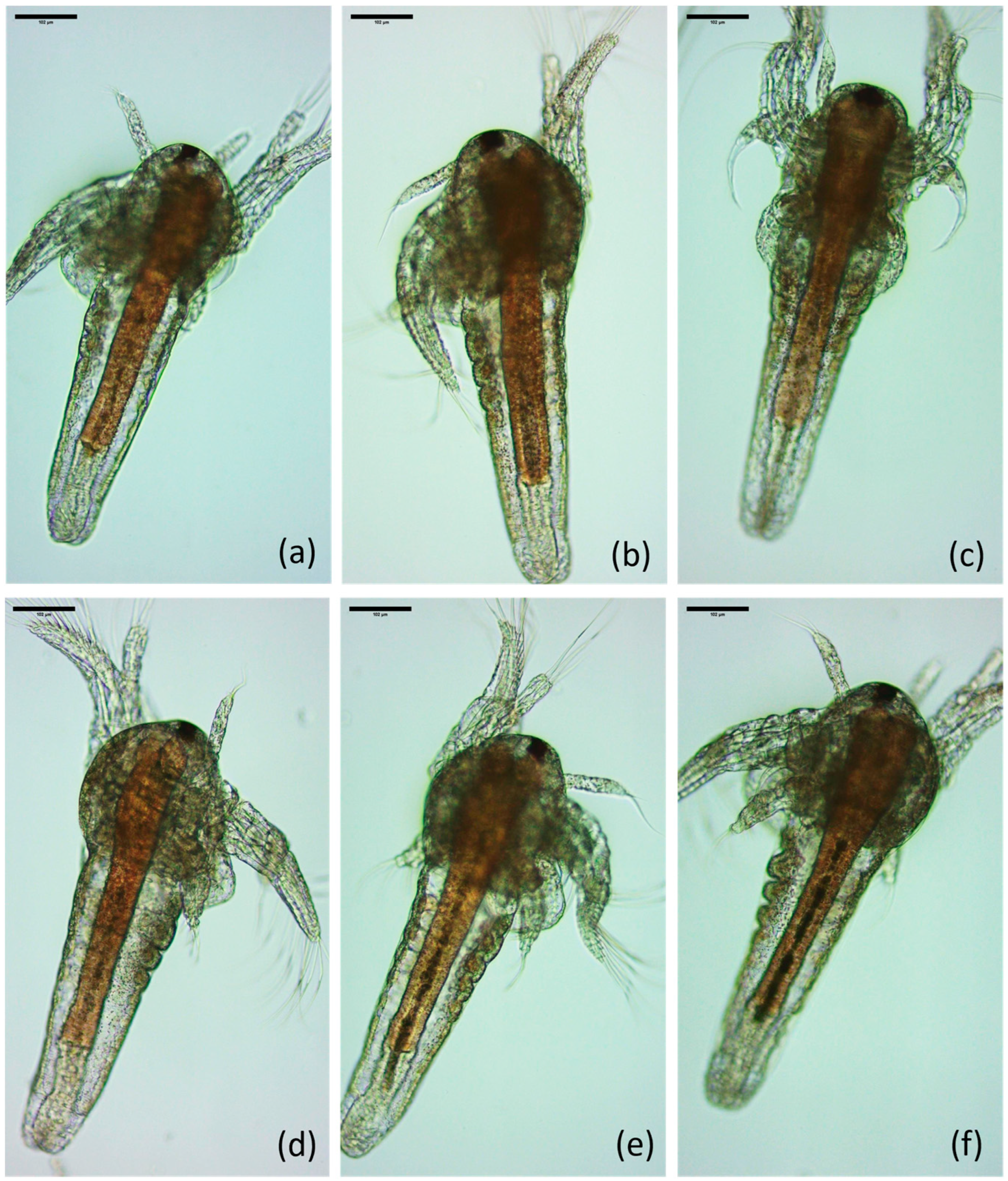
Figure 1. A. salina nauplii, exposed to different concentrations of brookite TiO2/CeO2 nanocomposite within 48 h of exposure. (a) A. Salina control; (b) A. salina nauplii exposed to 0.25 mg/L of brookite TiO2/CeO2 nanocomposite; (c) A. salina nauplii exposed to 0.50 mg/L of brookite TiO2/CeO2 nanocomposite; (d) A. salina nauplii exposed to 1.0 mg/L of brookite TiO2/CeO2 nanocomposite; (e) A. salina nauplii exposed to 2.0 mg/L of brookite TiO2/CeO2 nanocomposite; (f) A. salina nauplii exposed to 4.0 mg/L of brookite TiO2/CeO2 nanocomposite. Scale bar: 102 µm.
Water 16 01946 g001
Figure 2. Fluorescence in A. salina nauplii exposed to different concentrations of brookite TiO2/CeO2 nanocomposite within 48 h of exposure to evaluate ROS generation. (a) A. salina control; (b) Nauplii exposed to 0.25 mg/L of brookite TiO2/CeO2 nanocomposite; (c) Nauplii exposed to 0.50 mg/L of brookite TiO2/CeO2 NP; (d) A. salina nauplii exposed to 1.0 mg/L of brookite TiO2/CeO2 nanocomposite; (e) A. salina nauplii exposed to 2.0 mg/L of brookite TiO2/CeO2 nanocomposite; (f) Nauplii exposed to 4.0 mg/L of brookite TiO2/CeO2 nanocomposite. Scale bar: 47 µm.
Water 16 01946 g002
Figure 3. The histogram represents the average fluorescence intensity (AU) of the reactive oxygen species (ROS) at all concentrations of brookite TiO2/CeO2 nanocomposite tested. p < 0.05 (*).
Water 16 01946 g003
Figure 4. Fluorescence images of A. salina exposed to different concentrations of brookite TiO2/CeO2 nanocomposite within 48 h of exposure to evaluate apoptotic damage: (a) A. salina control; (b) A. salina nauplii exposed to 4.0 mg/L of brookite TiO2/CeO2 nanocomposites. Scale bar 49 µm. The histogram represents the average fluorescence intensity (AU) of the acridine orange (A.O.) for the control group and maximum concentration (4.0 mg/L) of brookite TiO2/CeO2 nanocomposite tested. A p < 0.05 (*).
Water 16 01946 g004
Table 1. Vitality rate (24 and 48 h) of A. salina nauplii treated with various concentrations of brookite TiO2/CeO2 nanocomposite.
Treatments% Vitality to 24 h
(Means ± SD)
% Vitality to 48 h
(Means ± SD)
CTRL98% ± 0.03098% ± 0.064
0.25 mg/L98.96% ± 0.02587.72% ± 0.078
0.50 mg/L97% ± 0.07288.85% ± 0.129
1.0 mg/L99% ± 0.02591.72% ± 0.071
2.0 mg/L99% ± 0.02792.59% ± 0.098
4.0 mg/L100% ± 0.00098% ± 0.051
Disclaimer/Publisher’s Note: The statements, opinions and data contained in all publications are solely those of the individual author(s) and contributor(s) and not of MDPI and/or the editor(s). MDPI and/or the editor(s) disclaim responsibility for any injury to people or property resulting from any ideas, methods, instructions or products referred to in the content.