Article

The Algicidal Potential of a Floating-Bed System against Microcystis aeruginosa in Laboratory Conditions

1
Intelligent Construction Technology Application Service Center, School of Architecture and Engineering, Chongqing City Vocational College, Chongqing 402160, China
2
School of Architecture and Engineering, Chongqing University of Science and Technology, Chongqing 401331, China
3
College of River and Ocean Engineering, Chongqing Jiaotong University, Chongqing 400074, China
4
Chongqing Academy of Science and Technology, Chongqing 401123, China
5
National & Local Joint Engineering Research Center for Ecological Treatment Technology of Urban Water, Zhejiang Provincial Key Laboratory for Water Environment and Marine Biological Resources, Wenzhou University, Wenzhou 325035, China
*
Authors to whom correspondence should be addressed.
Water 2023, 15(20), 3607; https://doi.org/10.3390/w15203607
Submission received: 21 September 2023 / Revised: 10 October 2023 / Accepted: 12 October 2023 / Published: 16 October 2023

Abstract

:
Harmful cyanobacterial blooms pose a major threat. Among them, Microcystis aeruginosa has raised serious concerns for human health due to its frequent occurrence. In this study, an ecological floating-bed system consisting of activated carbon fibers, aquatic plants (Ipomoea aquatica Forsskal), animals (Daphnia), and a solar-powered ultrasonic device was designed. The algae-killing efficiency, removal mechanism, and toxicological effects of the floating-bed system on Microcystis aeruginosa were determined under different conditions. The results showed that the average activity of algal cell dehydrogenase (DHA) was reduced by 64.09%, the average malondialdehyde (MDA) content was reduced to 0.125 μmol/L, and the average removal rate of soluble protein was 57.22% under optimal conditions (pH = 7, temperature = 25 °C, dissolved oxygen concentration = 5 mg/L, and hydraulic retention time = 36 h). Scanning electron microscopy (SEM) analysis showed that the structure within the cells of Microcystis aeruginosa was severely damaged after treatment with the solar-powered ultrasonic carbon fiber eco-floating-bed system. Fourier transform infrared (FTIR) spectroscopy analysis showed that the pyrrole ring of chlorophyll-a was degraded. In addition, a tadpole-based micronucleus test and a comet assay were conducted to assess cell viability and DNA damage in water samples treated with the floating-bed system, and the results confirmed that chromosome damage and genotoxicity were significantly reduced. These findings suggest that the floating-bed system is effective in destroying algal cells, leading to massive algal cell death and reducing the risk of secondary contamination. This study provides a new perspective for further research on ecological floating-bed technology, demonstrating its potential practical application in the prevention and control of cyanobacterial blooms.

1. Introduction

The excessive accumulation of nutrients such as nitrogen and phosphorus in water leads to the overgrowth of aquatic organisms, which is called eutrophication. After these nutrients enter the water body, they will promote the rapid reproduction of algae and other phytoplankton, thus forming a large area of algal blooms [1,2]. It is considered to be one of the most serious environmental problems in lakes, reservoirs, and other natural water bodies [3,4,5]. The existence of algal blooms leads to the deterioration of water quality, interferes with the physiological processes of aquatic organisms, destroys the structure of aquatic ecosystems, and harms the functions of water bodies and ecological environments [1,6,7]. In addition, algal blooms have a serious impact on the utilization of water resources such as industry, agriculture, aquaculture, and water transportation, and pose a potential threat to animals, plants, and humans [8,9].
Many studies have been devoted to exploring physical and chemical methods for inhibiting algal blooms [10,11,12]. However, physical methods have limitations in eliminating algal blooms and are costly [13]. At the same time, chemical methods often cause secondary pollution and have safety problems [14]. Therefore, low-cost and safe biological methods are considered to be a promising means of controlling algal blooms [15,16]. In the biological method, ecological floating-bed technology is widely used because of its advantages of strong operability, low operating cost, easy maintenance, a good landscape effect, and a lack of land occupation [17]. The traditional ecological floating bed is mainly composed of floating-bed plants, floating-bed frames and fixed plant substrates. It relies on floating-bed plants and microorganisms attached to their roots to purify the water body. The design is simple and flexible and has high removal efficiency. However, this method has some limitations. If plant growth is inhibited, the removal efficiency of pollutants will be affected [17].
Because of the limitations of the above floating-bed technology, this study combines activated carbon fiber, solar ultrasonic devices, and aquatic plants and aquatic animals to construct a solar ultrasonic carbon fiber ecological floating-bed system. This system takes Microcystis aeruginosa, a common algal strain in cyanobacterial blooms, as the research object, and aims to comprehensively investigate the algae removal efficiency, physiological properties, spatial structure, and changes in the functional groups inside algal cells of Microcystis aeruginosa during algal cell lysis. In addition, the toxicity of water samples after the treatment of Microcystis aeruginosa with the ecological floating-bed system is also studied.

2. Experimental Methods

2.1. Design of Ecological Floating Bed

The algae control device is constructed using organic glass, making it a durable and transparent device (Figure 1). The total height of the device measures 150 cm, with an internal height of 120 cm. The coverage rate of the floating bed (surface area of the floating bed/water surface area of the experimental tank) is 11.6%. The device is divided into three distinct layers—the upper layer, middle layer, and lower layer (which are composed of three device combinations: plant area, animal area, and biofilm carrier zone). These layers are vertically arranged in sequential order, allowing for efficient space utilization and promoting interactions and treatment processes within each layer. By dividing the device into distinct layers, it becomes possible to optimize and enhance the performance of each component, contributing to the overall effectiveness of the ecological floating-bed system.
The upper layer of the ecological floating-bed system is designated as the area for aquatic plants and solar ultrasonic devices. Within this region, we used ultrasound as a treatment method for specific algae. Specifically, during the ultrasonic treatment, we used an ultrasonic frequency of 1000 Hz and an output power of 80 watts. In addition, we specifically chose a widely consumed vegetable, water spinach (Ipomoea aquatica Forsskal) [18,19]. The design of the system incorporates a ‘felt pad’ structure formed by appropriately spacing the root zones of these plants. This configuration aims to enhance their absorption capacity and their ability to intercept particulate pollutants and algae in the water column, and by optimizing the root zone space, the water spinach plants form an efficient structure for nutrient uptake and pollutant removal. The strategic placement of this vegetation also enables it to efficiently capture suspended particulate matter and algae, thus contributing to the overall water purification process. It is important to mention that the effective height of this particular region was set at 40 cm, ensuring a suitable depth for the water spinach to thrive and perform its intended ecological functions effectively.
The middle layer corresponds to the animal area within the ecological floating-bed system. In this particular area, a species of small crustacean, the water flea (Daphnia magna), was selected as the resident organism. Water fleas (Daphnia), which primarily feed on bacteria, algae, and organic debris, play a crucial role in the ecological function of the floating bed. Utilizing their unique filtration mechanism, daphnia actively contribute to the removal of pollutants and enhance the biodegradability of organic matter through digestion processes [20,21]. To ensure the proper accommodation of the animals, a fine nylon mesh with a mesh size of 1 mm was employed in this region of the floating-bed system. This mesh effectively prevents the escape of daphnia or any unintended intrusion. Furthermore, it maintains a suitable environment for the animals while allowing the passage of necessary nutrients and dissolved substances. It is worth mentioning that the height of this animal area was set at 40 cm, providing a sufficient depth for daphnia to carry out their ecological functions effectively.
The lower layer of the ecological floating-bed system is designated as the bioaugmented zone. In this zone, soft and semi-soft carbon fiber carrier fillers are suspended to serve as substrates for enriching microorganisms and facilitating the formation of an efficient biofilm purification zone. The height of this region was set at 70 cm, providing ample space for the bioaugmentation process, to create an optimal environment for the growth of microorganisms, the effective volume of the floating-bed system was set at 80 L, while maintaining a biofilm density of 30%. This allows for the colonization of a diverse microbial community, which aids in the efficient degradation and removal of pollutants from the water.
Furthermore, a constant heating temperature device is integrated into the system. This ensures that the floating-bed system maintains a consistent temperature range of 15–30 °C, which is conducive to favorable microbial activity. The adopted intermittent operation mode of “water inlet-reaction-water outlet” promotes efficient water flow and treatment processes within the floating-bed system.

2.2. Experimental Water Samples

The experimental water samples were obtained from Longtanzi Reservoir (longitude 106°38′–106°51′, latitude 29°73′–29°93′), with a total capacity of 3.65 × 10⁴ m3, which is a heavily eutrophic area in Beibei, Chongqing.

2.3. Algae Strains and Culture Conditions

Microcystis aeruginosa was used as the algal species for testing, algae species number FACHB-913, and was purchased from the Algal Species Collection Center, Institute of Hydrobiology, Chinese Academy of Sciences. The culture medium was provided by the Algal Species Collection Center, Institute of Hydrobiology, Chinese Academy of Sciences.
To study the effect of Microcystis aeruginosa on the water body, Microcystis aeruginosa was added into 500 mL conical flasks containing water samples from Longtanzi Reservoir; placed in a light incubator at a temperature of (25 ± 1) °C (MGC-250BP-2, Yiheng Scientific Instrument, Shanghai, China), with a light intensity of 2000 Lx~2500 Lx and a light/dark ratio of 12 h:12 h; and incubated in a static state, with shaking three times per day at regular intervals. Under different chlorophyll-a levels, each water sample was sampled after 7 days of action, and the algal effect of Microcystis aeruginosa on the water body of Longtanzi Reservoir was determined by measuring the changes in chlorophyll-a content. Each experiment was performed three times and the average value was taken. The blank control was the water body in Longtanzi Reservoir without Microcystis aeruginosa in a 500 mL conical flask.

2.4. Experimental Schemes

After the formation and domestication of the biological membrane in the system, the effects of pH values (pH = 5, 6, 7, 8, 9), dissolved oxygen levels (DO = 2, 3, 4, 5, 6 mg/L), hydraulic retention time (12, 24, 36, 48, 60 h), and temperature (15, 20, 25, 30 °C) on the algae’s removal efficiency were investigated, with a chlorophyll concentration of 143.79–148.87 μg/(L·d). Furthermore, dehydrogenase activity, soluble protein, and malondialdehyde content were determined using the optimal factors under the aforementioned conditions (shown in Tables S1–S5).

2.5. Experimental Instruments

In this study, a centrifuge (GL-22MS, Shanghai Xiang Yi, Shanghai, China), a ultrasonic cell grinder (BILON-650Y, Shanghai Bilon, Shanghai, China), an XSopt microscope (XSZ-800B, Ningbo Xinsheng Optical Instruments, Ningbo, China), a scanning electron microscope (XL-30-ESEM, Philips, Amsterdam, The Netherlands), and a spectrophotometer (756PC, Shanghai Hengping, Shanghai, China) were used.

2.6. Analysis Indicator and Method

2.6.1. Method for Extracting Crude Enzyme Solution of Algae

Firstly, 25 mL of the algae solution was put into a centrifuge tube and centrifuged at 6000 r/min for 15 min. The supernatant was discarded, 8 mL of pre-chilled 0.05 mol/L, pH = 7.8 phosphate-buffered solution was added to the precipitate, and then, the cells were crushed under ice bath conditions using an ultrasonic cell crusher with a power of 500 W, an interval of 3 s, a crushing time of 6 s, and a number of ultrasonic operations of 60, and no intact cells were examined microscopically. Finally, the crushing solution was centrifuged at 8000 r/min for 20 min to obtain the supernatant, which is the crude enzyme solution, refrigerated at 4 °C and stored for later use.

2.6.2. Measurement of Dehydrogenase Activity (DHA) in Algae Solution

Refer to the triphenyl-tetrazolium chloride-dehydrogenase reduction method to determine the dehydrogenase activity of algae cells [22]. Simply put, the test steps are as follows:
A total of 15 mL of the sample solution to be tested was filtered through a 0.25 μm microporous membrane. Tris-HCl (pH = 7.5) and TTC were added to react with algal cells in a constant-temperature water bath at 35 ± 1 °C for 60 min. We added formaldehyde to stop the reaction. TPF was extracted with acetone and petroleum ether, and the absorbance of petroleum ether extract at 492 nm was determined. The dehydrogenase activity of algal cells was calculated according to the TPF value (expressed as DHA, in μgTPF/mL·h). The experiment was repeated three times.

2.6.3. Determination of Soluble Protein in Algae Solution

The Coomassie Brilliant Blue G250 method was used [23], and bovine serum protein was used as the standard. Measuring steps: Take 1 mL of extracted enzyme solution (control group was 1 mL of distilled water) and add 5 mL of Coomassie Brilliant Blue G-250 protein reagent; then, leave it for 5 min to measure the absorbance value of the reaction solution at 595 nm and find out the content of soluble protein (μg) in the extracted enzyme solution via a standard curve.

2.6.4. Determination of Malondialdehyde Content (MDA) in Algae Solution

Referring to the determination of the thiobarbituric acid (TBA) method [24], MDA reacts with TBA under high temperature and acidic conditions to form a colored substance with maximum absorbance at a wavelength of 532 nm. The specific measurement steps were as follows:
(1)
Draw 2 mL of crude enzyme solution, and 2 mL of distilled water for the control group, add 2 mL of 0.5% (w/v) trichloroacetic acid solution, and then, add 2 mL of 0.67% (w/v) thiobarbituric acid solution. Shake well until uniform.
(2)
Put it in a boiling water bath and boil for 15 min to fully react.
(3)
After the time is over, immediately remove the test tube and put it in cold water for cooling. After cooling, centrifuged it for 20 min at 4000 r/min. Take the liquid at wavelengths of 600 nm, 532 nm, and 450 nm, respectively. Calculate according to Formulas (1) and (2).
MDA concentration (μmol/L) = 6.45 × (A532 − A600) − 0.56 × A450
MDA   content   μ mol / L = MDAconcentration × Crude   enzyme   solution   volume Mixed   liquid   volume
In the formula, A450, A532, and A600 represent the absorbance values at 450 nm, 532 nm, and 600 nm, respectively.

2.7. Determination of Algae Chlorophyll-a

A cellulose acetate membrane with a pore size of 0.45 μm was utilized to filter 100 mL of the algae-containing water sample. Subsequently, the filtered membrane, with algae loaded on top, was folded multiple times and placed into a 15 mL centrifuge tube, which was then inserted into a black plastic bag. The membrane-containing centrifuge tube inside the black plastic bag was subjected to 3–5 cycles of repeated freezing and thawing (frozen for approximately 20 min and thawed for approximately 5 min), and then, dissolved in 10 mL of 90% acetone. Following this, the centrifuge tube containing the dissolved membrane was placed in a refrigerator at 4 °C for extraction, with the extraction duration being 5 h. During the extraction process, the tube was gently shaken in darkness every 1–2 h. Finally, the absorbance of the supernatant from the centrifuge tube was measured at wavelengths of 630 nm, 645 nm, 663 nm, and 750 nm.
Chl a   μ g / L = 11.64 × D 663 D 750 2.16 × D 645 D 750 + 0.10 × D 630 D 750 V 1 / V / δ
In the formula, D is the absorbance, V is the volume of the water sample (L), V1 is the volume of the extraction solution (mL), and δ is the light path of the cuvette (cm).

2.8. Algae Cell Characterization

In this study, we used an ESEM (XL-30, Philips, Amsterdam, The Netherlands) scanning electron microscope to observe the structure of the samples. At the same time, a Fourier transform infrared spectrometer (Avatar 370DTGS, Thermo Nicolet, Madison, WI, USA) was used to measure the infrared absorption spectra of the experimental group and the control group. The scanning range of Fourier transform infrared spectroscopy is 400 cm−1~4000 cm−1.

2.9. Ecotoxicological Analysis of Water Samples after Treatment with an Ecological Floating-Bed System

In this study, a micronucleus test and comet assay were used to evaluate the genotoxicity of tadpoles collected in actual water [25,26]. Tadpoles growing in the Longtanzi reservoir were captured on-site as experimental subjects. Tadpoles with a body length of 25.00 ± 2 mm, a body weight of 0.2 ± 0.05 g, and a healthy and uniform physique were selected for this study. The specific experimental procedures were as follows: The water samples containing algae were divided into control and experimental groups, with the experimental group treated with water samples processed in an ecological floating-bed system, while the control group received untreated original algal water samples. Each group consisted of 25 tadpoles, and no feeding was conducted during the experiment. Sampling was carried out at intervals of 1, 3, 5, and 7 days, with three randomly selected tadpoles from each group used to prepare blood smears. The process involved drying the surface moisture of the tadpoles with filter paper, swiftly tail-severing to obtain blood samples, and preparing blood smears. After natural air-drying, the blood smears were placed in a dark area and fixed with a methanol solution for 15 min. Subsequently, they were stained with Wright’s stain for 15–20 min, and then, rinsed three times with PBS solution (pH 6.8). Finally, Gimsa stain was added for 25 min. After the completion of staining, rinsing with distilled water was performed, followed by natural drying. Lastly, the prepared blood smears were observed under a fluorescence microscope, and 1000 red blood cells were randomly observed in each smear to record the number of micronuclei and nuclear abnormalities in the tadpole red blood cells. The micronucleus rate and nuclear abnormality rate were expressed as per mill (‰).
The comet assay was also performed on the tadpoles [27,28], using the same specimens as the micronucleus assay. The specific procedures were as follows: Firstly, the water samples were divided into blank and experimental groups. The experimental group consisted of water treated in an ecological floating-bed system, while the blank group used the untreated original algal water sample. Each group contained 30 tadpoles, and no feeding was provided during the experiment. Sampling was conducted on the 1st, 3rd, 5th, and 7th days after feeding. Five tadpoles were randomly selected from each group for the comet assay. Subsequently, blood samples were collected from the surface of tadpoles by wiping them with filter paper. After that, the filter papers were used to collect the blood samples and diluted appropriately with PBS solution (pH 7.2). Under a microscope, 5–8 dispersed cells were observed for each sample. Next, double-layer gel slides were prepared by pouring 0.7% normal melting agarose (90 μL) onto the frosted surface of a glass slide, which was then refrigerated at 4 °C for 25–30 min. After solidification, the cover glass was removed. Then, 70 μL of 0.7% low-melting agarose containing blood cells was added as the upper-layer gel on the solidified gel. The slide was covered with a cover glass and refrigerated again at 4 °C for 20–30 min, and then, the cover glass was removed. Subsequently, the gel slides containing blood cells were placed in a freshly prepared cell lysis solution (3.5 mol/L NaCl, 100 mmol/L EDTA, 10 mmol/L Tris, 1% Triton X-100, 10% DMSO, pH 10) and stored in a light-protected environment at 4 °C for 1.5–2.0 h. After lysis, the slides were rinsed 3–5 times with distilled water at 4 °C, and then, immersed in light-protected unwinding solution (10 mmol/L NaOH, 150 mmol/L EDTA, pH = 11) for 30 min. Subsequently, the voltage was adjusted to 160–200 V, and the electric current was set at 200 mA for electrophoresis, with an electrophoresis time of 10–20 min. Afterward, the slides were soaked in 0.4M Tris-HCl (pH 7.5) 3–5 times to neutralize. Finally, the slides were stained with Goldview for 20 min. Under a fluorescence microscope with a wavelength of 525 nm, 100 cells were randomly observed on each slide to calculate the tail intensity and cell damage rate.

3. Results and Discussion

3.1. The Influencing Factors of Solar Ultrasonic Carbon Fiber Ecological Floating-Bed System Algae-Lysing

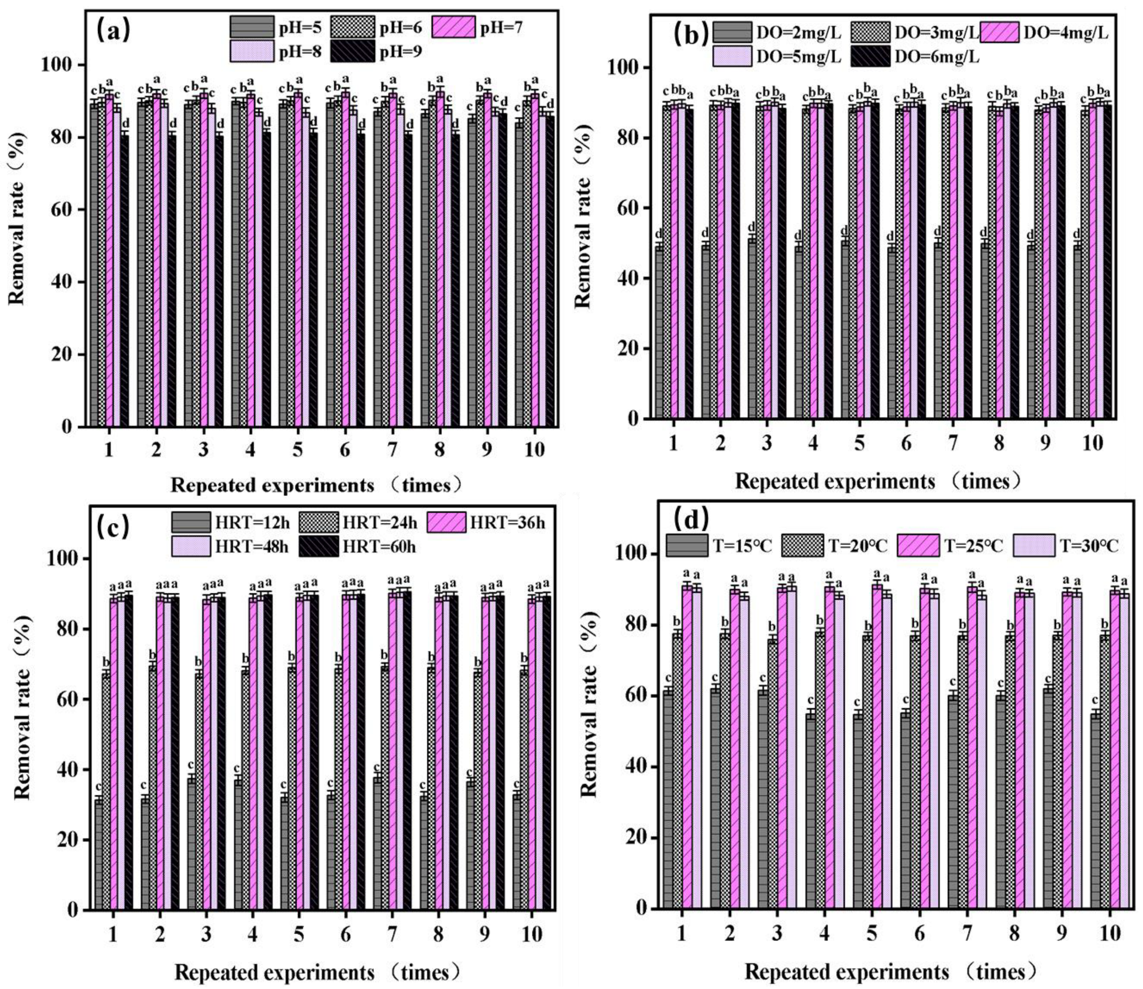
3.1.1. The Effect of Influent pH on the Variation in Chlorophyll-a Absorbance

As shown by the results of Figure 2a, the algae-lysing effect of the solar ultrasonic carbon fiber ecological floating-bed technology system on Microcystis aeruginosa follows the order of pH 7 > pH 6 > pH 5 > pH 8 > pH 9. The system exhibits pronounced algae solubilization at influent pH values of 6 and 7. Under acidic conditions, the removal rate of chlorophyll-a (Chl-a) increases with increasing pH and reaches its maximum value at pH 7, with an average removal rate of 92.07%. However, beyond pH 7, the removal rate of Chl-a decreases significantly. At pH 9, the average removal rate of Chl-a is 81.80%. In the pH range of 5–9, the removal rate of Chl-a can exceed 80%. These results indicate that the solar ultrasonic carbon fiber ecological floating-bed technology can achieve effective algae removal across a wide pH range, with pH 7 being the optimal condition.
This observation aligns with the findings of Chen et al. [29], who also reported that the removal rate of Chl-a increases with the pH of the raw water, but decreases when the pH of the raw water exceeds 7. The analysis suggests that the secretion of certain enzymes by algae-lysing microorganisms during algae lysis process may be more efficient in neutral and acidic environments compared to alkaline conditions, thereby affecting the effectiveness of algae lysis.

3.1.2. The Impact of Dissolved Oxygen (DO) on the Variation in Chlorophyll-a Absorbance

From Figure 2b, it can be seen that the removal of Chl-a increases with the increase in DO. This is in agreement with the findings of Tanaka et al. [30] that Chl-a is almost proportional to DO consumption. When DO increased from 2.0 mg/L to 5.0 mg/L, the removal rate of Chl-a stabilized as DO increased. However, the increase in Chl-a removal rate was not significant with a continued increase in DO. Higher DO levels have a significant impact on the metabolic activities of algae. Adequate oxygen supply facilitates efficient metabolism in algae, enabling them to photosynthesize more effectively. During photosynthesis, algae take in carbon dioxide from the water column and release oxygen, leading to a reduction in chlorophyll-a concentrations [31,32]. However, lower levels of dissolved oxygen can impede the photosynthetic capacity of algae, resulting in diminished chlorophyll-a removal. Algae are less efficient at photosynthesis under low-oxygen conditions, hindering their ability to utilize light energy for the reduction of chlorophyll-a concentrations. This limitation may stem from the constrained growth, metabolism, and photosynthetic activity of algae due to insufficient dissolved oxygen levels [33,34]. Therefore, both excessively low and high dissolved oxygen concentrations are detrimental to the effectiveness of solar-powered ultrasonic carbon fiber ecological floating-bed systems in algae removal. Thus, considering operational costs, a dissolved oxygen concentration of 5.0 mg/L is recommended.

3.1.3. The Influence of Hydraulic Retention Time (HRT) on the Variation in Chlorophyll-a Absorbance

Based on the data depicted in Figure 2c, it can be observed that the removal efficiency of chlorophyll gradually increases at hydraulic retention times (HRT) of 12 h and 24 h, measuring 35.51% and 67.81%, respectively. However, when the HRT is extended to 36 h, the removal rate stabilizes at 88.87%, indicating that further increasing the HRT does not significantly enhance the removal efficiency of chlorophyll. This finding aligns with the research conducted by Solmaz [35] et al. They discovered that shorter HRT and higher flow rates can promote the growth of algae, resulting in increased chlorophyll-a content. The potential underlying reason for this phenomenon is that as the reaction time extends, the nutrients in the water gradually deplete, and the flow carries away the accumulated nutrients in the cells of algae [36]. This flushing effect greatly reduces the possibility of nutrient overload in the water, thereby improving the self-purification capacity of the continuous-flow water system. Additionally, the self-purification ability of the water body is closely linked to the hydraulic retention time [37]. Moreover, the continuous-flow water system prevents the aggregation of microalgae, which could otherwise impact the algae-lysing effect of the solar ultrasonic carbon fiber ecological floating-bed system. Therefore, considering operational costs, it is recommended to set the HRT at 36 h.

3.1.4. The Effect of Temperature on the Variation in Chlorophyll-a Absorbance

The above experimental results show that the algae dissolving effect of the solar ultrasonic carbon fiber ecological floating-bed technology system is closely related to the ambient temperature shown in Figure 2d. Under the conditions of 15 °C, 20 °C, 25 °C, and 30 °C, the algicidal effects of the solar ultrasonic carbon fiber ecological floating-bed technology system on Microcystis aeruginosa are 25 °C > 30 °C > 20 °C > 15 °C, respectively. At 15 °C, the algae dissolving effect of this system is not apparent, which may be due to the influence of low temperature on the secretion of enzymes by microorganisms, which leads to a reduction in its algae dissolving effect. At higher temperatures, the activity of enzymes secreted by microorganisms is enhanced, and their algae-dissolving effect is also enhanced. At 25 °C, the activity of enzymes secreted by microorganisms is the strongest. Therefore, its algicidal effect is greatly enhanced, and its algicidal rate against Microcystis aeruginosa is the highest. Therefore, 25 °C is the best temperature. The experimental results of Yin [38] et al. also pointed out that the cell density and Chl-a concentration increased significantly at 25 °C. However, Microcystis aeruginosa was inhibited at low temperatures and its photosynthesis was affected at high temperatures.
Based on the aforementioned experimental findings, the optimal parameters for effective algae lysis in the solar ultrasonic carbon fiber ecological floating-bed technology system were determined through a single-factor test. These parameters include a pH value of 7, a dissolved oxygen concentration of 5 mg/L, a hydraulic retention time of 36 h, and a temperature of 25 °C.

3.2. Removal Mechanism of Solar Ultrasonic Carbon Fiber Ecological Floating Bed

3.2.1. The Effect of Optimal Conditions on Microcystis aeruginosa Treatment

(1)
Variation in DHA during operation of Microcystis aeruginosa under optimal conditions
Dehydrogenase activity (DHA) is an important indicator reflecting the activity of cells and organisms. It can not only be used for the determination of cell viability in animals and plants, but also reflect the current growth and metabolic activity of organisms to a large extent [39]. It can be seen from the above experimental results that the DHA content of algal cells decreased by 64.09% on average (Figure 3A). The reason may be that the biological activity and physiological metabolic activity of algal cells were inhibited after the algal cells were treated with the solar ultrasonic carbon fiber ecological floating bed, and then, the algal cells died or stopped growing, thus reducing the DHA content of the algal cells.
(2)
Changes in Soluble Protein During Operation of Microcystis aeruginosa under Optimal Conditions
Soluble protein serves as a crucial indicator for assessing the activity of enzymes involved in various plant metabolism processes. Determining the changes in soluble protein content not only helps researchers in comprehending the overall metabolism of plants, but also reflects the intensity of external threats imposed on plants, leading to disturbances in their growth, development, and reproduction [40]. Our experimental results, presented in Figure 3B, demonstrate an average reduction of 57.22% in soluble protein content. This decrease can be attributed to the application of solar ultrasonic carbon fiber ecological floating-bed technology for treating algal cells, which results in inadequate nutrient supply for algal growth and reproduction. Consequently, the reduced protein synthesis adversely affects the cellular growth and metabolic activities of algae, ultimately inhibiting their growth.
(3)
Variation in MDA during operation of Microcystis aeruginosa under optimal conditions
Malondialdehyde (MDA) is the ultimate product of lipid peroxidation in cell membranes. The changes in MDA content not only reflect the degree of oxidative damage to cell membranes but also indicate the extent of cellular injury under environmental changes in plant cells [41,42]. MDA exhibits certain toxicity towards cells, causing disruption and damage to cell membrane functions, as well as severe degradation of various functional molecules, thereby directly impairing plant cell tissue.
The experimental results, as illustrated in Figure 3C, show a significant average increase in MDA content to 0.125 μmol/L. The analysis suggests that this could be attributed to the application of solar ultrasonic carbon fiber ecological floating-bed technology for treating algal cells, which triggers peroxidation reactions in the membrane system lipids. Consequently, the membrane structures of algal cells, including chloroplasts, are disrupted, leading to a significant rise in MDA content. This inhibits photosynthesis and interferes with the nutritional and energy metabolism of algal cells, thereby impacting a series of physiological and biochemical reactions.
According to the above experimental results, after being treated with the solar ultrasonic carbon fiber ecological floating-bed technology system, the average DHA content of algae cells decreased by 64.09%, the average soluble protein content decreased by 57.22%, and the average MDA content increased to 0.125 μmol/L. This shows that the solar ultrasonic carbon fiber ecological floating-bed technology system has a certain algicidal effect on Microcystis aeruginosa under optimal conditions (pH = 7, DO = 5 mg/L, HRT = 36 h, temperature = 25 °C).

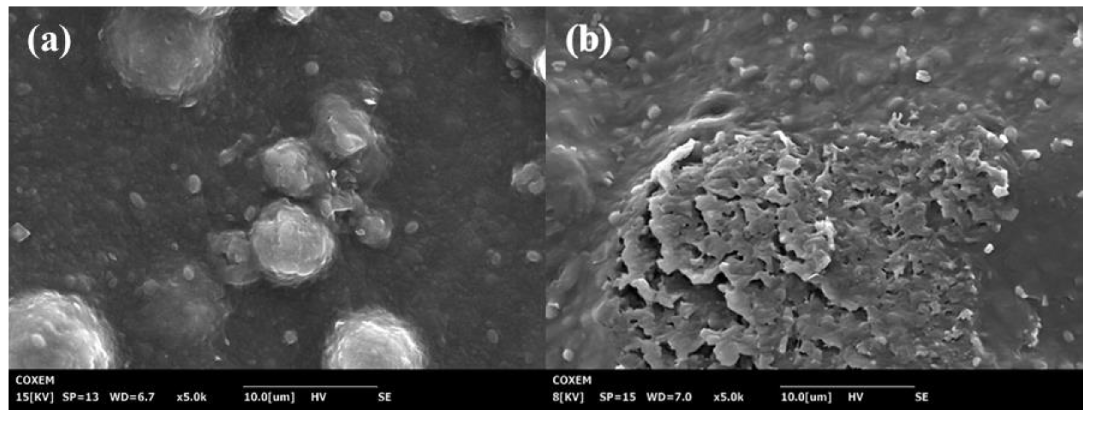
3.2.2. Effect of Optimal Working Conditions on Spatial Structure of Microcystis aeruginosa

It can be seen from the SEM results that before being treated with this system (Figure 4), the structure of Microcystis aeruginosa cells in water samples is complete, the surface is undamaged, the cells thrive, and no intracellular substances are released. However, after being treated with this algae control system, the spatial structure of Microcystis aeruginosa cells is obviously destroyed, the complete structure of Microcystis aeruginosa cells is seriously damaged, Microcystis aeruginosa cells are almost entirely decomposed, and microorganisms tightly wrap the Microcystis aeruginosa cells.
A large amount of the contents of algae cells was released, and the nutrients in Microcystis aeruginosa cells were exhausted by microorganisms. Finally, the growth of Microcystis aeruginosa cells was greatly affected, and many algae cells stopped growing or died.

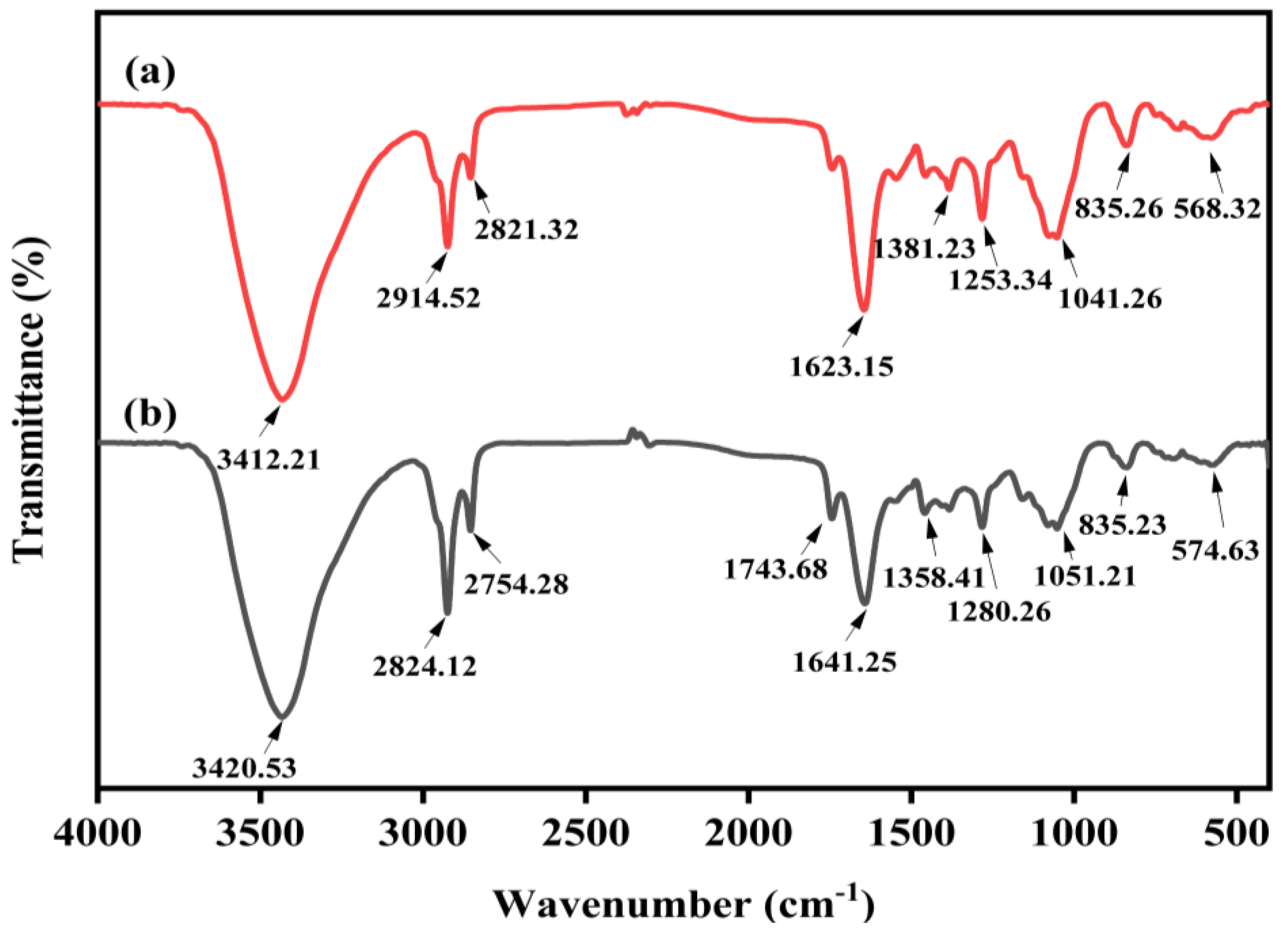
3.2.3. The Effect of Optimal Conditions on the Functional Groups of Microcystis aeruginosa

The normal Microcystis aeruginosa and the algal cells treated with the solar ultrasonic carbon fiber ecological floating bed were analyzed via Fourier transform infrared spectroscopy (FT-IR), show in Figure 5. The results revealed distinct infrared absorption peaks in the regions of 3700–3000 cm−1, 3000–2700 cm−1, 2300–2900 cm−1, and 1800–800 cm−1, representing the major functional groups on the surface of algal cells.
The minor absorption peaks were not extensively analyzed. Based on the OMNIC database and other studies [43,44], the main regions where functional groups changed included: the CH deformation of alkyl groups (1300–1400 cm−1), the stretching vibrations of C=O bonds in protein compounds (1600–1700 cm−1), the stretching vibrations of C-H (2850–2960 cm−1), and the stretching vibrations of -OH in carbohydrates (3000–3700 cm−1). Comparing the FT-IR spectra of normal and treated algal cells, it was observed that the changes in functional groups on the surface of algal cells were not significant, but certain functional groups exhibited shifts and intensity variations in their absorption peaks. Specifically, consistent with previous studies [45], the most notable changes were observed in the C-H region (2850–2960 cm−1), indicating the stretching vibrations of saturated C-H bonds in the samples. Moreover, the absorption peak intensity of the treated Microcystis aeruginosa notably decreased, primarily due to the degradation of carbon–hydrogen bonds in the protein structure of algal cells. The changes in other functional groups were relatively minor. In the range of 3700–3000 cm−1, the infrared absorption peaks exhibited slight stretching vibrations, and the absorption peak shifted from 3420.53 cm−1 to 3412.21 cm−1, which may be related to the secretion of carbohydrates. In the range of 1300–1700 cm−1, significant changes were observed in the amide C=O region, as the absorption peak intensity decreased from 1641.25 cm−1 to 1623.15 cm−1, indicating a certain level of damage to protein amide bonds in algal cells.

3.3. Study on Algae-Lysing Toxicity of Solar Ultrasonic Carbon Fiber Ecological Floating Bed

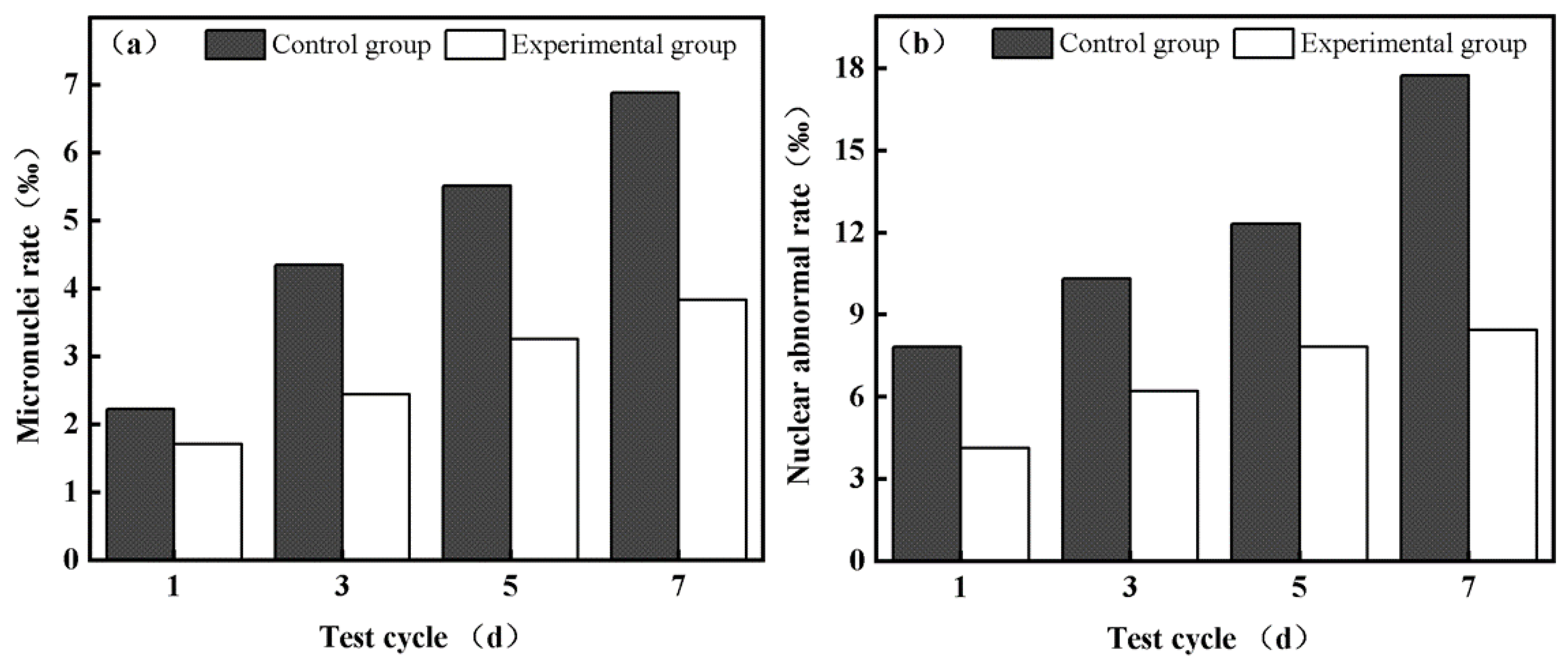
3.3.1. Analysis of Micronucleus Test Results

After the detailed data statistics of the micronucleus rate and nuclear abnormality rate of the tadpole red blood cells treated with the experimental group and the control group for ld, 3d, 5d, and 7d, respectively, it was found that the micronucleus rate and nuclear abnormality rate of the tadpole red blood cells treated with the carbon fiber ecological floating-bed algae control system decreased significantly. The specific data are shown in Figure 6. The results showed that the micronucleus rate of tadpole red blood cells treated with this system was 3.83‰, and the nuclear abnormality rate was 8.45‰. In contrast, during the feeding period of the experiment, the micronucleus rate and nuclear abnormality rate of most tadpoles in the control group showed a significant upward trend, and were 6.88‰ and 17.73‰, respectively. This finding strongly suggests that the content of toxic substances in water samples treated with the carbon fiber ecological floating-bed algae control system is significantly lower than that of untreated algae cells. The toxicological damage to the red blood cells of tadpoles was significantly reduced, which indicates that the toxins of organisms were degraded to a certain extent in the ecological floating bed.
These results further strengthen the importance and application potential of carbon fiber ecological floating-bed algae control systems in the field of toxin treatment and environmental protection. By reducing the content of toxic substances in water samples, the system can effectively reduce the toxic effects on ecosystems and protect biodiversity and ecological balance.

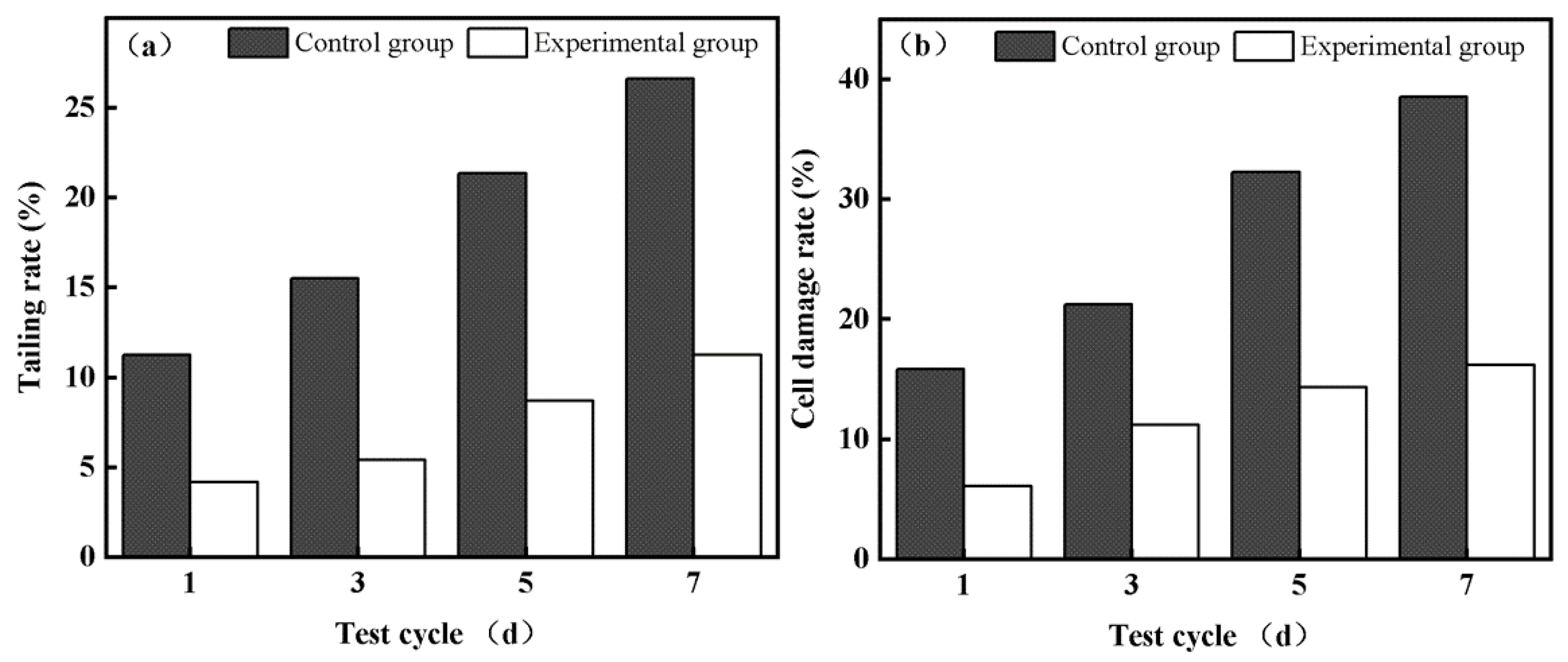
3.3.2. Analysis of Comet Experiment Results

The tailing rate and cell damage rate of tadpole blood cells treated with the experimental group and the control group for ld, 3d, 5d, and 7d were analyzed in detail using statistical software. The specific data can be seen in Figure 7. It can be observed from the above results that in the experimental group, only a small number of cells showed DNA migration, with a tailing rate of 11.27% and a cell damage rate of 16.21%. In contrast, the cells in the control group showed a higher level of DNA migration, with a tailing rate of 26.59% and a cell damage rate of 38.53%.
These findings indicate that the tadpole blood cells underwent obvious DNA breakage after being affected by foreign substances. However, the degree of DNA damage in tadpole blood cells was significantly reduced after treatment with the carbon fiber ecological floating-bed algae control system. This further led to a decrease in mutation rate and mortality. The results showed that after the treatment with the carbon fiber ecological floating-bed algae control system, the ecotoxicology of the water samples and the level of biotoxin in the algae cells were significantly lower than those in the control group.
Based on the above research results, it can be seen that the carbon fiber ecological floating-bed algae control system can significantly reduce the DNA damage of tadpole blood cells and effectively reduce the variation rate and mortality rate of tadpole blood cells during a long period of treatment. In addition, the system can also effectively reduce the ecotoxicology of water samples and reduce the content of biotoxins in algal cells, which is particularly important in reducing the toxic pressure of water environments. It is worth emphasizing that the treatment process of the carbon fiber ecological floating-bed algae control system does not easily cause secondary pollution to the water environment, which further strengthens its application potential in the field of environmental protection.

4. Conclusions

The ecological floating-bed system, consisting of plants (Ipomoea aquatica Forsskal), animals (Daphnia), a biofilm carrier (activated carbon fiber), and a solar-powered ultrasonic device, demonstrated effective removal of Microcystis aeruginosa from water through single-factor experiments. Additionally, the study of algal cell FTIR, SEM, and physiological characteristics under optimal conditions revealed changes in functional groups before and after the treatment of algal cells, significantly affecting their growth. Furthermore, notable oxidative damage was observed in the algal cells, ultimately leading to growth cessation or cell death. Moreover, micronucleus and comet assays were utilized to verify the water samples after the ecological floating-bed system treatment, and the results indicate a certain degree of degradation in genetic toxicity and chromosomal damage associated with algae-contaminated water samples, thereby reducing the risk of secondary pollution. In the future, our research will focus on studying the treatment effects of the solar-powered ultrasonic ecological floating-bed system in eutrophic water bodies. We will also pay attention to changes in various layers, such as changes in plants and the relationship with water fleas during the water purification process. Additionally, the feasibility of implementing this ecological floating-bed system in actual water bodies will be explored.

Supplementary Materials

The following supporting information can be downloaded at: https://www.mdpi.com/article/10.3390/w15203607/s1, Table S1: Operating conditions of floating bed system with pH value as variable.; Table S2: Floating bed system operating condition with DO as variable. Table S3: Floating bed system operating condition with reaction time as variable. Table S4: Floating bed system operating condition with temperature as variable. Table S5: Operating conditions of floating bed system treatment. Table S6: Changes in chlorophyll a concentration in water before and after treatment in a floating bed system under the factor of pH change. Table S7: Changes in removal rates after treatment in a floating bed system under factors varying in pH. Table S8: Changes in chlorophyll a concentration in water before and after treatment in a floating bed system with varying factors of DO values. Table S9: Changes in post-treatment removal rates in floating bed systems for factors that vary in DO. Table S10: Changes in chlorophyll a concentration in water before and after treatment in a floating bed system under the HRT change factor. Table S11: Changes in post-treatment removal rates in floating bed systems under the factor of HRT variation. Table S12: Changes in chlorophyll a concentration in water before and after treatment in a floating bed system as a function of temperature. Table S13: Changes in post-treatment removal rates in floating-bed systems for factors that vary in temperature. Table S14: Changes of erythrocyte micronucleus in tadpoles before and after treatment of Microcystis aeruginosa by reactor. Table S15: The changes of DNA tailing rate and cell damage of red blood cells in tadpoles after treatment of Microcystis aeruginosa water samples in the reactor.

Author Contributions

Conceptualization, D.L. and G. Z; methodology, D.L.; software, C.T.; validation, C.T.; formal analysis, D.L.; investigation, C.T.; resources, G.Z.; data curation, Y.H.; writing—original draft preparation, D.L.; writing—review and editing, Y.H.; visualization, D.L.; supervision, D.S.; project administration, G.Z.; funding acquisition, D.S. All authors have read and agreed to the published version of the manuscript.

Funding

This work was supported by the National Natural Science Foundation of China, grant number (51808086); the Natural Science Foundation of Chongqing, China (CSTB2022NSCQ-MSX1145); the Natural Science Foundation of Yongchuan District Science and Technology Bureau (2023yc-jckx20088); and the Chongqing Bayu Scholars Young Scholars Project (YS2021089); Chongqing Technology Innovation and Application Development Project (CSTB2022TIAD-KPX0133). The scientific research project of Chongqing Municipal Urban Management, "Research on Inno-vative Technology of Microbial Ecological Management of Eutrophic Water Bodies in Chongqing" [Urban Management Ke Zi 2023 No. 02].

Data Availability Statement

Data sharing is not applicable to this article. All data are contained within this manuscript.

Conflicts of Interest

The authors declare no conflict of interest.

References

  1. Paerl, H.W.; Fulton, R.S.; Moisander, P.H.; Dyble, J. Harmful freshwater algal blooms, with an emphasis on cyanobacteria. Sci. World J. 2001, 1, 76–113. [Google Scholar] [CrossRef] [PubMed]
  2. Zhou, B.; Cai, X.; Wang, S.; Yang, X. Analysis of the causes of cyanobacteria bloom: A review. J. Resour. Ecol. 2020, 11, 405–413. [Google Scholar]
  3. Griffith, A.W.; Gobler, C.J. Harmful algal blooms: A climate change co-stressor in marine and freshwater ecosystems. Harmful Algae 2020, 91, 101590. [Google Scholar] [CrossRef]
  4. Landsberg, J.H. The effects of harmful algal blooms on aquatic organisms. Rev. Fish. Sci. 2002, 10, 113–390. [Google Scholar] [CrossRef]
  5. Neves, R.A.; Nascimento, S.M.; Santos, L.N. Harmful algal blooms and shellfish in the marine environment: An overview of the main molluscan responses, toxin dynamics, and risks for human health. Environ. Sci. Pollut. Res. 2021, 28, 55846–55868. [Google Scholar] [CrossRef] [PubMed]
  6. Amorim, C.A.; do Nascimento Moura, A. Ecological impacts of freshwater algal blooms on water quality, plankton biodiversity, structure, and ecosystem functioning. Sci. Total Environ. 2021, 758, 143605. [Google Scholar] [CrossRef] [PubMed]
  7. Reid, A.J.; Carlson, A.K.; Creed, I.F.; Eliason, E.J.; Gell, P.A.; Johnson, P.T.; Kidd, K.A.; MacCormack, T.J.; Olden, J.D.; Ormerod, S.J. Emerging threats and persistent conservation challenges for freshwater biodiversity. Biol. Rev. 2019, 94, 849–873. [Google Scholar] [CrossRef]
  8. Alonso-Rodrıguez, R.; Páez-Osuna, F. Nutrients, phytoplankton and harmful algal blooms in shrimp ponds: A review with special reference to the situation in the Gulf of California. Aquaculture 2003, 219, 317–336. [Google Scholar] [CrossRef]
  9. Yan, T.; Li, X.-D.; Tan, Z.-J.; Yu, R.-C.; Zou, J.-Z. Toxic effects, mechanisms, and ecological impacts of harmful algal blooms in China. Harmful Algae 2022, 111, 102148. [Google Scholar] [CrossRef]
  10. Sun, R.; Sun, P.; Zhang, J.; Esquivel-Elizondo, S.; Wu, Y. Microorganisms-based methods for harmful algal blooms control: A review. Bioresour. Technol. 2018, 248, 12–20. [Google Scholar] [CrossRef]
  11. Zeng, G.; Wang, P.; Wang, Y. Algicidal efficiency and mechanism of Phanerochaete chrysosporium against harmful algal bloom species. Algal Res. 2015, 12, 182–190. [Google Scholar] [CrossRef]
  12. Rajasekhar, P.; Fan, L.; Nguyen, T.; Roddick, F.A. A review of the use of sonication to control cyanobacterial blooms. Water Res. 2012, 46, 4319–4329. [Google Scholar] [CrossRef]
  13. Brown, A.R.; Lilley, M.; Shutler, J.; Lowe, C.; Artioli, Y.; Torres, R.; Berdalet, E.; Tyler, C.R. Assessing risks and mitigating impacts of harmful algal blooms on mariculture and marine fisheries. Rev. Aquac. 2020, 12, 1663–1688. [Google Scholar] [CrossRef]
  14. Kibuye, F.A.; Zamyadi, A.; Wert, E.C. A critical review on operation and performance of source water control strategies for cyanobacterial blooms: Part I-chemical control methods. Harmful Algae 2021, 109, 102099. [Google Scholar] [CrossRef]
  15. Balaji-Prasath, B.; Wang, Y.; Su, Y.P.; Hamilton, D.P.; Lin, H.; Zheng, L.; Zhang, Y. Methods to control harmful algal blooms: A review. Environ. Chem. Lett. 2022, 20, 3133–3152. [Google Scholar] [CrossRef]
  16. Xu, S.; Lyu, P.; Zheng, X.; Yang, H.; Xia, B.; Li, H.; Zhang, H.; Ma, S. Monitoring and control methods of harmful algal blooms in Chinese freshwater system: A review. Environ. Sci. Pollut. Res. 2022, 29, 56908–56927. [Google Scholar] [CrossRef] [PubMed]
  17. Wang, W.-H.; Wang, Y.; Sun, L.-Q.; Zheng, Y.-C.; Zhao, J.-C. Research and application status of ecological floating bed in eutrophic landscape water restoration. Sci. Total Environ. 2020, 704, 135434. [Google Scholar] [CrossRef] [PubMed]
  18. Hu, M.; Ao, Y.; Yang, X.; Li, T. Treating eutrophic water for nutrient reduction using an aquatic macrophyte (Ipomoea aquatica Forsskal) in a deep flow technique system. Agric. Water Manag. 2008, 95, 607–615. [Google Scholar] [CrossRef]
  19. Li, X.-N.; Song, H.-L.; Li, W.; Lu, X.-W.; Nishimura, O. An integrated ecological floating-bed employing plant, freshwater clam and biofilm carrier for purification of eutrophic water. Ecol. Eng. 2010, 36, 382–390. [Google Scholar] [CrossRef]
  20. Freese, H.M.; Schink, B. Composition and stability of the microbial community inside the digestive tract of the aquatic crustacean Daphnia magna. Microb. Ecol. 2011, 62, 882–894. [Google Scholar] [CrossRef]
  21. Hiltunen, M.; Honkanen, M.; Taipale, S.; Strandberg, U.; Kankaala, P. Trophic upgrading via the microbial food web may link terrestrial dissolved organic matter to Daphnia. J. Plankton Res. 2017, 39, 861–869. [Google Scholar] [CrossRef]
  22. Zhang, R.; Jin, R.; Liu, G.; Zhou, J.; Li, C.L. Study on nitrogen removal performance of sequencing batch reactor enhanced by low intensity ultrasound. Bioresour. Technol. 2011, 102, 5717–5721. [Google Scholar] [CrossRef]
  23. Yang, M.; Shi, D.; Wang, Y.; Ebadi, A.G.; Toughani, M. Study on Interaction of Coomassie Brilliant Blue G-250 with Bovine Serum Albumin by Multispectroscopic. Int. J. Pept. Res. Ther. 2021, 27, 421–431. [Google Scholar] [CrossRef]
  24. Draper, H.; Squires, E.; Mahmoodi, H.; Wu, J.; Agarwal, S.; Hadley, M. A comparative evaluation of thiobarbituric acid methods for the determination of malondialdehyde in biological materials. Free Radic. Biol. Med. 1993, 15, 353–363. [Google Scholar] [CrossRef] [PubMed]
  25. Mouchet, F.; Gauthier, L.; Baudrimont, M.; Gonzalez, P.; Mailhes, C.; Ferrier, V.; Devaux, A. Comparative evaluation of the toxicity and genotoxicity of cadmium in amphibian larvae (Xenopus laevis and Pleurodeles waltl) using the comet assay and the micronucleus test. Environ. Toxicol. Int. J. 2007, 22, 422–435. [Google Scholar] [CrossRef] [PubMed]
  26. Poletta, G.; Larriera, A.; Kleinsorge, E.; Mudry, M. Genotoxicity of the herbicide formulation Roundup ®(glyphosate) in broad-snouted caiman (Caiman latirostris) evidenced by the Comet assay and the Micronucleus test. Mutat. Res. Genet. Toxicol. Environ. Mutagen. 2009, 672, 95–102. [Google Scholar] [CrossRef]
  27. de Oliveira, E.M.; Suzuki, M.F.; do Nascimento, P.A.; da Silva, M.A.; Okazaki, K. Evaluation of the effect of 90Sr β-radiation on human blood cells by chromosome aberration and single cell gel electrophoresis (comet assay) analysis. Mutat. Res. Fundam. Mol. Mech. Mutagen. 2001, 476, 109–121. [Google Scholar] [CrossRef] [PubMed]
  28. Cavas, T. In vivo genotoxicity evaluation of atrazine and atrazine–based herbicide on fish Carassius auratus using the micronucleus test and the comet assay. Food Chem. Toxicol. 2011, 49, 1431–1435. [Google Scholar] [CrossRef]
  29. Chen, L.; Liu, C.; Sun, Y.; Sun, W.; Xu, Y.; Zheng, H. Synthesis and characterization of ampholytic flocculant CPCTS-gP (CTA-DMDAAC) and its flocculation properties for microcystis aeruginosa removal. Processes 2018, 6, 54. [Google Scholar] [CrossRef]
  30. Tanaka, T.; Tsuzuki, K.; Nishijima, N.; Takagi, T. Algae-removal performance of a fluidized-bed biofilm reactor system for lake water treatment. Water Sci. Technol. 2001, 43, 277–283. [Google Scholar] [CrossRef]
  31. Li, J.; Wu, L.; Dai, Y.; Wang, Q.; Wang, S.; Zhang, L. Effects of different nitrogen–phosphorus ratio on the freshwater phytoplankton growth and the variations of environmental factors. Ecol. Environ. 2007, 16, 342–346. [Google Scholar]
  32. You, L.; Cui, L.; Liu, Z.; Yang, B.; Huang, Z. Correlation analysis of parameters in algal growth. Environ. Sci. Technol. 2007, 30, 42–44. [Google Scholar]
  33. Kibuye, F.A.; Zamyadi, A.; Wert, E.C. A critical review on operation and performance of source water control strategies for cyanobacterial blooms: Part II-mechanical and biological control methods. Harmful Algae 2021, 109, 102119. [Google Scholar] [CrossRef] [PubMed]
  34. Ruan, X.; Shi, X.; Zhao, Z.; Ni, L.; Wu, Y.; Jiao, T. Correlation between chlorophyll a concentration and environmental factors in shallow lakes in plain river network areas of Suzhou. J. Lake Sci. 2008, 20, 556–562. [Google Scholar]
  35. Solmaz, A.; Işık, M. Optimization of membrane photobioreactor; the effect of hydraulic retention time on biomass production and nutrient removal by mixed microalgae culture. Biomass Bioenergy 2020, 142, 105809. [Google Scholar] [CrossRef]
  36. Fernandez-Marchante, C.; Asensio, Y.; Lobato, J.; Villaseñor, J.; Cañizares, P.; Rodrigo, M. Influence of hydraulic retention time and carbon loading rate on the production of algae. J. Biotechnol. 2018, 282, 70–79. [Google Scholar] [CrossRef]
  37. Silveira, C.F.; de Assis, L.R.; de Sousa Oliveira, A.P.; Calijuri, M.L. Valorization of swine wastewater in a circular economy approach: Effects of hydraulic retention time on microalgae cultivation. Sci. Total Environ. 2021, 789, 147861. [Google Scholar] [CrossRef]
  38. Yin, Y.; Zhang, Y.; Wang, M.; Shi, K. Effects of temperature on the optical properties of Microcystis aeruginosa and Scenedesmus obliquus. J. Freshw. Ecol. 2016, 31, 361–375. [Google Scholar] [CrossRef]
  39. Chettri, D.; Sharma, B.; Verma, A.K.; Verma, A.K. Significance of Microbial Enzyme Activities in Agriculture. Microbiol. Act. Soil Plant Health Manag. 2021, 351–373. [Google Scholar] [CrossRef]
  40. Schwenzfeier, A.; Wierenga, P.A.; Gruppen, H. Isolation and characterization of soluble protein from the green microalgae Tetraselmis sp. Bioresour. Technol. 2011, 102, 9121–9127. [Google Scholar] [CrossRef]
  41. Yan, Z.; Xu, L.; Zhang, W.; Yang, G.; Zhao, Z.; Wang, Y.; Li, X. Comparative toxic effects of microplastics and nanoplastics on Chlamydomonas reinhardtii: Growth inhibition, oxidative stress, and cell morphology. J. Water Process Eng. 2021, 43, 102291. [Google Scholar] [CrossRef]
  42. Zhang, F.; Yao, X.; Sun, S.; Wang, L.; Liu, W.; Jiang, X.; Wang, J. Effects of mesotrione on oxidative stress, subcellular structure, and membrane integrity in Chlorella vulgaris. Chemosphere 2020, 247, 125668. [Google Scholar] [CrossRef] [PubMed]
  43. Mantsch, H.; McElhaney, R. Phospholipid phase transitions in model and biological membranes as studied by infrared spectroscopy. Chem. Phys. Lipids 1991, 57, 213–226. [Google Scholar] [CrossRef]
  44. Theerawitayaart, W.; Prodpran, T.; Benjakul, S. Enhancement of hydrophobicity of fish skin gelatin via molecular modification with oxidized linoleic acid. J. Chem. 2019, 2019, 5462471. [Google Scholar] [CrossRef]
  45. Wang, J.; Zeng, G.; Wang, F.; Huang, X.; Li, Y.; Liang, D.; Zhang, M.; Sun, D. Study on the Algae Lysis Method of White Rot Fungi Algae Control System. Water 2022, 14, 903. [Google Scholar] [CrossRef]
Figure 1. Solar ultrasonic carbon fiber ecological floating bed.
Water 15 03607 g001
Figure 2. (a) Effect of influent pH on algae-lysing effect; (b) effect of dissolved oxygen on algae–lysing; (c) effect of hydraulic retention time on algae-lysing effect; (d) effect of temperature on algae-lysing effect. Data are expressed as mean ± SD (n = 3). Different letters in the figure represent significant differences (p < 0.05).
Water 15 03607 g002
Figure 3. The changes in (A) algal cell dehydrogenase activity, (B) soluble protein content, and (C) malondialdehyde content during the operation of the floating-bed system under optimal conditions and blank control group were analyzed.
Water 15 03607 g003
Figure 4. (a) Original sample of Microcystis aeruginosa; (b) Microcystis aeruginosa cells after ecological floating-bed treatment.
Water 15 03607 g004
Figure 5. (a) Infrared spectra of algae cells in water samples treated with solar ultrasonic carbon fiber ecological floating bed and (b) infrared spectra of algae cells in original water samples.
Water 15 03607 g005
Figure 6. Carbon fiber ecological floating-bed treatment of Microcystis aeruginosa water samples: (a) tadpole red blood cell micronucleus rate changes and (b) tadpole red blood cell nucleus abnormal rate changes.
Water 15 03607 g006
Figure 7. Carbon fiber ecological floating-bed treatment of Microcystis aeruginosa water samples: (a) tadpole red blood cell tailing rate and (b) tadpole red blood cell damage changes.
Water 15 03607 g007
Disclaimer/Publisher’s Note: The statements, opinions and data contained in all publications are solely those of the individual author(s) and contributor(s) and not of MDPI and/or the editor(s). MDPI and/or the editor(s) disclaim responsibility for any injury to people or property resulting from any ideas, methods, instructions or products referred to in the content.

