Article

Copper Bioremediation Ability of Ciliate Paramecium multimicronucleatum Isolated from Industrial Wastewater

1
Institute of Zoology, University of the Punjab, New Campus, Lahore 54590, Pakistan
2
Biofuels Institute, School of Environment and Safety Engineering, Jiangsu University, 301 Xuefu Road, Zhenjiang 212013, China
3
Chemistry Department, College of Science, King Saud University, Riyadh 11451, Saudi Arabia
4
Division of Pharmacy and Optometry, School of Health Sciences, Faculty of Biology, Medicine and Health, University of Manchester, Manchester M13 9PL, UK
5
Division of Environmental Technology, School of Industrial Technology, Universiti Sains Malaysia, Penang 11800, Malaysia
6
School of Biological Sciences, University of the Punjab, New Campus, Lahore 54590, Pakistan
*
Authors to whom correspondence should be addressed.
Water 2022, 14(9), 1419; https://doi.org/10.3390/w14091419
Submission received: 21 March 2022 / Revised: 23 April 2022 / Accepted: 27 April 2022 / Published: 29 April 2022
(This article belongs to the Special Issue Solid/Liquid Adsorption in Water and Wastewater Treatment)

Abstract

:
The growing problems of environmental damage have been caused by the continuous outrush of heavy metals from industrial wastewater. To resolve this issue, bioremediation is playing a safe and eco-friendly role in the removal of these heavy metals from environmental wastewater bodies. It has provoked demand with regard to understanding the mechanisms of bioaccumulation and detoxification developed by the organisms living in the heavy metal-exposed industrial wastewater. The present investigation focuses on Paramecium multimicronucleatum, a ciliated protozoan isolated from industrial wastewater, with the objective of assessing its capabilities as an environmental bioremediator. Purified cell culture was maintained in bold basal salt medium and optimum growth conditions were determined. A maximum growth rate of 6.0–9.0 × 10³ cells/mL at 25–30 °C and pH 7.0 was observed, and therefore revealed to be the optimal growth conditions for this species. It can tolerate 40–50 µg/mL of copper ion stress with little effect on growth rate as compared to control. It is able to uptake more than 80% of copper ions from the medium in 96 h. A significant twofold rise in glutathione content and non-protein thiols was recorded as an indication of a defensive mechanism in place to fight against the oxidative stress caused by the copper treatment. A notable increase of 50–70 µg/mL in total protein content of stressed cells in comparison to non-stressed was also observed as potential induction of some particular proteins for the purpose of resistance against copper stress.

1. Introduction

Water and soil contamination with heavy metals is a prime environmental issue these days. Through consideration of the technological worth of heavy metals, their use has increased in many industries over the years. Humans and their environment are at permanent risk towards the toxic effects of effluents released by these industries. Presence of heavy metals in industrial wastewater, even at lower concentrations, is toxic, as they are non-biodegradable and accumulate in living organisms. They can cause severe health problems, nervous system damage, kidney malfunction, and even death at higher concentrations [1]. Utilization of water and soil containing such metal impurities for food production ultimately allows their entry into the food chain, which adversely affects the health of many living organisms [2,3].
A pivotal role is played by many metal ions in the regulation of various metabolic pathways among living organisms. However, elevated levels of even essential metals like copper is harmful [4]. Among all the heavy metals, copper is most frequently used metal in industrial applications such as electroplating, plastics, and metal finishing. The United States Environmental Protection Agency (USEPA) and World Health Organization (WHO) have declared tolerable amounts of copper ions in industrial wastewater to be 1.3 mg/L and 2 mg/L, respectively, and this level must not be exceeded in drinking water. Removal of copper ions from industrial wastewater is performed by various techniques and methods, such as membrane filtration, cementation, photocatalysis, and adsorption [1].
However, these conventional methods, which are used for the elimination of copper ions from polluted water, are costly and applicable to small areas only. For the treatment of wastewater, the latest research is continuously in the process of developing and applying the latest cheap technologies by using biomass, live plants, and microorganisms [2,5,6,7,8]. Such methods of bioremediation are observed as eco-friendly and useful methods for large scale application in industrial wastewater treatment.
There is a variety of ecological communities in industrial wastewater, such as protozoans, yeast, bacteria, and algae, which are surviving and growing well due to their abilities of tolerance, resistance, detoxification, and metabolization of harmful substances [9]. The protozoan community includes organisms which maintain the quality of the effluents by predation on bacterial populations and tolerance to heavy metals [10]. Protozoans are well known for their efficient resistance and prolonged survival in contaminated wastewater, having high concentrations of metal ions. They have developed mechanisms to metabolize and detoxify toxic metal ions, including Hg2+, Ni2+, Zn2+, Cu2+, Pb2+, and Cd2+ [3].
Availability of optimized culture conditions such as temperature and pH are crucial physiochemical parameters required for the bioremediation abilities of ciliates. Bioavailability, solubility, change in ionization of biomolecules, and toxicity of heavy metals towards these organisms is influenced by the available pH. Moreover, a suitable temperature is of prime importance for the cellular activities of these bioremediators [11]. Among the defensive strategies against heavy metal exposure, oxidized glutathione (GSH) plays a diverse part as an antioxidant enzyme and signaling component for metal homoeostasis [10]. A balanced ratio between reduced and oxidized glutathione is required for the regulation of cellular activities [12]. This tripeptide enzyme contains cysteine as one of the amino acid components with sulfhydryl groups and deals with the oxidative stress caused by excessive generation of reactive oxygen species [13]. Induction of protein synthesis against metal stress for the binding of metal ions is also considered a bioremediation ability of ciliates [11].
Tetrahymena, Stylonchia, and Euplotes have already been employed in research investigation regarding nickel, zinc, cadmium, and copper [4,14,15]. Research data related to Paramecium species for their bioaccumulation studies and defensive mechanisms is not available. This invokes the need for the current research, which focuses on and reveals new directions in terms of bioremediation using isolated Paramecium sp. from industrial wastewater.

2. Materials and Methods

2.1. Sampling of Wastewater

Wastewater samples were collected from industrial areas of Lahore, Faisalabad and Okara city (Samadpura) in sterilized falcon tubes during the months of March-May and September-November and kept at room temperature (25 °C–28 °C) until being checked under microscope on the same day.

2.2. Purification and Isolation of Paramecium sp.

Each sample was checked under the microscope for the presence of ciliates, and the samples containing Paramecium along with other protozoans and rotifers were inoculated in bold basal salt medium. Bold basal salt medium contained the following components: NaNO3, 250 mg/L; CaCl2.H2O, 25 mg/L; MgSO4.7H2O, 75 mg/L; K2HPO4, 75 mg/L; KH2PO4, 175 mg/L; NaCl, 25 mg/L; EDTA, 50 mg/L; KOH, 31 mg/L; FeSO4.7H2O, 4 mg/L; H2SO4, 0.001 M; H3BO3, 114.2 mg/L; ZnSO4.7H2O, 8.81 mg/L; MnCl2.4H2O, 1.44 mg/L; MoO3, 0.71 mg/L; CuSO4.5H2O, 1.57 mg/L; and Co(NO3)2.6H2O, 0.49 mg/L, along with 8–12 boiled grains of wheat. The pH of the medium was adjusted to 7.0–7.1 and kept at room temperature. To obtain the purified culture of Paramecium, a single cell was isolated from the inoculated medium by drop dilution method under the microscope. This single cell of Paramecium was then inoculated in a separate 25 mL of pH-adjusted bold basal salt medium along with 3–4 boiled wheat grains to obtain the axenic culture [16,17].

2.3. Selection of Copper-Resistant Paramecium sp.

For the selection of a copper resistant Paramecium species, 1 µg/mL/day and 5 µg/mL/day copper stress were applied to three Paramecium isolates in 25 mL of bold basal salt medium inoculated with 15–20 cells from the pure culture. After 48 h of stress, the numbers of cells were counted to check the effect of Cu on the growth of Paramecium isolates. One ciliate isolate showed maximum resistance under copper stress by showing the cell movement under microscope and dividing number of cells as signs of viability. This species was selected for further bioremediation experiments.

2.4. Identification of Copper-Resistant Paramecium sp. Using Eukaryotic 18S rRNA Gene Primers

Genomic DNA was isolated from the log phase cells of copper resistant Paramecium sp. by phenol-chloroform method using lysis buffer (42% urea, 0.30 M NaCl, 10 mM EDTA, 10 mM tris-HCL, 2% SDS). 18S rRNA gene was amplified using primers EukF (5′-AATATGGTTGATCCTGCCAGT-3′) and EukR (5′-TGATCCTTCTGCAGGTTCACCTAC-3′) [18]. The PCR product was extracted from agarose gel with GeneJet PCR purification kit (K0701) and sent to First Base Laboratories Malaysia for nucleotide sequencing. The sequence was BLAST and species was identified by checking the homology percentage with data from other species present in the NCBI databases.

2.5. Growth Assay under Copper Stress

Identified Paramecium sp. cells (8–12) were inoculated in 25 mL of bold basal salt medium and copper stress was given in the form of copper sulphate solution with varying concentrations (10, 20, 30, 40, and 50 µg/mL) and kept at its optimum temperature. Morphological changes due to metal stress were observed and the number of dividing cells, by taking drops of 5 µL in triplicates from each flask, were counted under the microscope on a daily basis for four weeks. The growth curve of stressed (copper-treated) and non-stressed (control) culture was plotted with mean values estimated for 1 mL of the medium.

2.6. Optimization of Temperature and pH for Paramecium Growth

Temperature and pH play vital roles in enhancing the growth rate of Paramecium cells, which contributes to their uptake ability. It was determined that 25 mL of culture medium would be inoculated with 8–10 purified cells of Paramecium sp. in triplicates for each temperature. Flasks were kept at different temperatures (20, 25, 30, and 35 °C) in the incubator. Similarly, to determine the optimum pH, the medium was prepared and various pHs (5, 6, 7, 7.5, 8, and 9) were adjusted with the pH meter in triplicates and inoculated with Paramecium cells. The growth rate in the form of increasing number of cells was observed for 10–12 days. Observations were plotted using mean values.

2.7. Estimation of Copper Accumulation and Uptake Ability

Log phase cells of Paramecium species were exposed to different concentrations of copper ions (10, 20, 30, 40, and 50 µg/mL). Positive controls with metal stress only and negative controls with non-stressed Paramecium cells were also prepared for comparison. Samples were collected after 30 min, 24, 48, 72 and 96 h and cell pellets were obtained after centrifugation at 4032× g value for 10 min and washed with 0.9% saline solution. Air-dried pellets were digested in 50 µL of concentrated HNO3 and diluted according to requirement. A supernatant was used to calculate the remaining amount of copper ions in the medium. Copper accumulation and uptake ability was measured by Thermo Unicam-SOLAAR atomic absorption spectrophotometry at a wavelength of 324.8 nm.

2.8. Assessment of Glutathione Levels as Antioxidant

Reduced glutathione possesses a major role as a cellular defense mechanism against induced metal stress and deals with reactive oxygen species which cause oxidative stress in the cells. This tripeptide works as chelating agent and antioxidant due to the presence of the sulfhydryl group in cysteine, which immediately maintains the reduced and oxidized glutathione ratio in the cell for survival.
Levels of reduced glutathione (GSH), oxidized glutathione (GSSG), and total glutathione were estimated among control and copper-treated cells of Paramecium by the recycling method [19]. Cells were harvested after 24 and 48 h of copper stress by centrifugation at 4032× g for 10 min, from both control and experimental culture. Pellets produced were washed with 1 mM phosphate buffer saline and dissolved in 3 mL of 5% sulfosalicyclic acid under cold conditions. This homogenate was centrifuged at 11,200× g for 10 min and 0.5 mL of supernatant was taken and added to 0.5 mL reaction buffer (0.1 M Phosphate buffer saline and 0.5 mM Ethylenediamine tetraacetic acid) along with 50 µL of 3 mM 5,5’-Dithio-Bis (2-Nitrobenzoic Acid) (DTNB), used for estimation of reduced glutathione (GSH) content. The reaction was allowed to incubate for 5 min at 30 °C and absorbance was taken at 412 nm using spectrophotometer. In the same reaction tube, 100 µL of 0.4 mM NADPH and 2 µL of glutathione reductase enzyme was added and incubated for 20 min to obtain the values for total glutathione content. Observations of oxidized glutathione (GSSG) were taken by subtracting values of reduced content from total glutathione. Values were compared by preparing the standard curve of glutathione with varying concentrations.

2.9. Estimation of Other Non-Protein Thiols from Copper-Resistant Paramecium Culture

Other non-protein thiols were estimated in copper-treated and non-treated cells after 24 and 48 h by Israr et al., 2006 [20]. A standard curve for cysteine with varying concentrations was prepared to calculate the content in the sample.

2.10. Estimation of Total Protein Contents from Copper-Resistant Paramecium Culture

The combined or individual roles of some proteins also support resistance to copper in ciliates. The change in levels of total protein content of cells was quantified by the Bradford method. For protein extraction, cells exposed to copper stress and non-stressed cells were centrifuged at 9803× g (4 °C) for 10 min. The pellet obtained was washed with 1.5 mL chilled 10 mM Tris-HCl (pH 7.5) and dissolved in 50 µL of the same solution. To this mixture, 500 µL of triple detergent lysis buffer (50 mM Tris-HCL pH 7.5, 1% Triton, 0.5% Na deoxycholate, 5% SDS and 0.01% β- mercaptethanol) was added, vortexed and provided heat shock for 5 min in a boiling water bath. The tubes were transferred to an ice bucket immediately. After 5 min, tubes were again centrifuged at 9803× g and 4 °C for 15 min. The supernatant (500 µL) was taken and mixed with 5 mL freshly prepared Bradford reagent to obtain the optical density at 595 nm for control and copper-treated samples. A bovine serum albumin (BSA) standard curve with various concentrations was prepared for the comparison.

2.11. Statistical Analysis

Observations were recorded for three independent replicates and mean values were displayed in tables. Student’s t-test (SPSS 16.0, SPSS Inc., IBM, Armonk, NY, USA) was applied in each copper-treated versus non-treated (control) culture analysis to obtain the significant differences.

3. Results

3.1. Copper-Resistant Paramecium Species

Culture for a copper-resistant Paramecium isolate, which shows maximum numbers of diving cells of more than 4.0 × 10² cells/mL when observed under microscope against tested copper stress (1 µg/mL/day and 5 µg/mL/day), was maintained in the lab at its optimum conditions in bold basal salt medium. This species was identified as Paramecium multimicronucleatum (MW066476) through amplification and nucleotide sequencing of the conserved 18S rRNA gene.

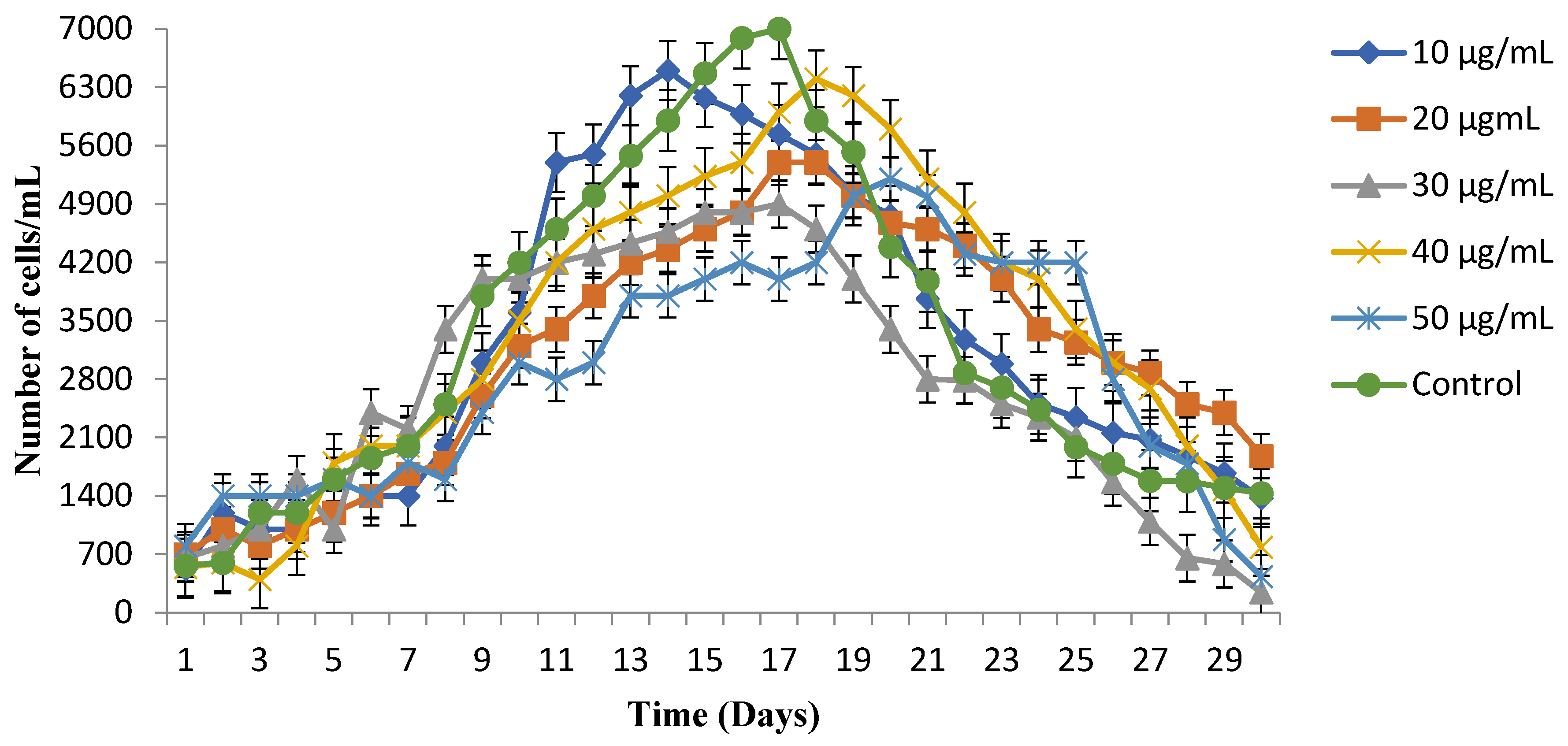
3.2. Growth Observations under Copper Stress

Growth curves exhibit maximum resistance of P. multimicronucleatum against lower concentrations of copper. The observed growth rate reached 5.9 × 10³ and 6.4 × 10³ cells/mL after two weeks of exposure for 10 µg/mL and 40 µg/mL of copper stress, respectively (Figure 1). Control culture displayed increased growth rate of 7.0 × 10³ cells/mL in comparison to copper treated culture for P. multimicronucleatum after two weeks of trial. A decrease in the rate of diving cells was observed after 20 days and reached 1.4 × 10³ cells/mL at the end of the fourth week of the experiment.

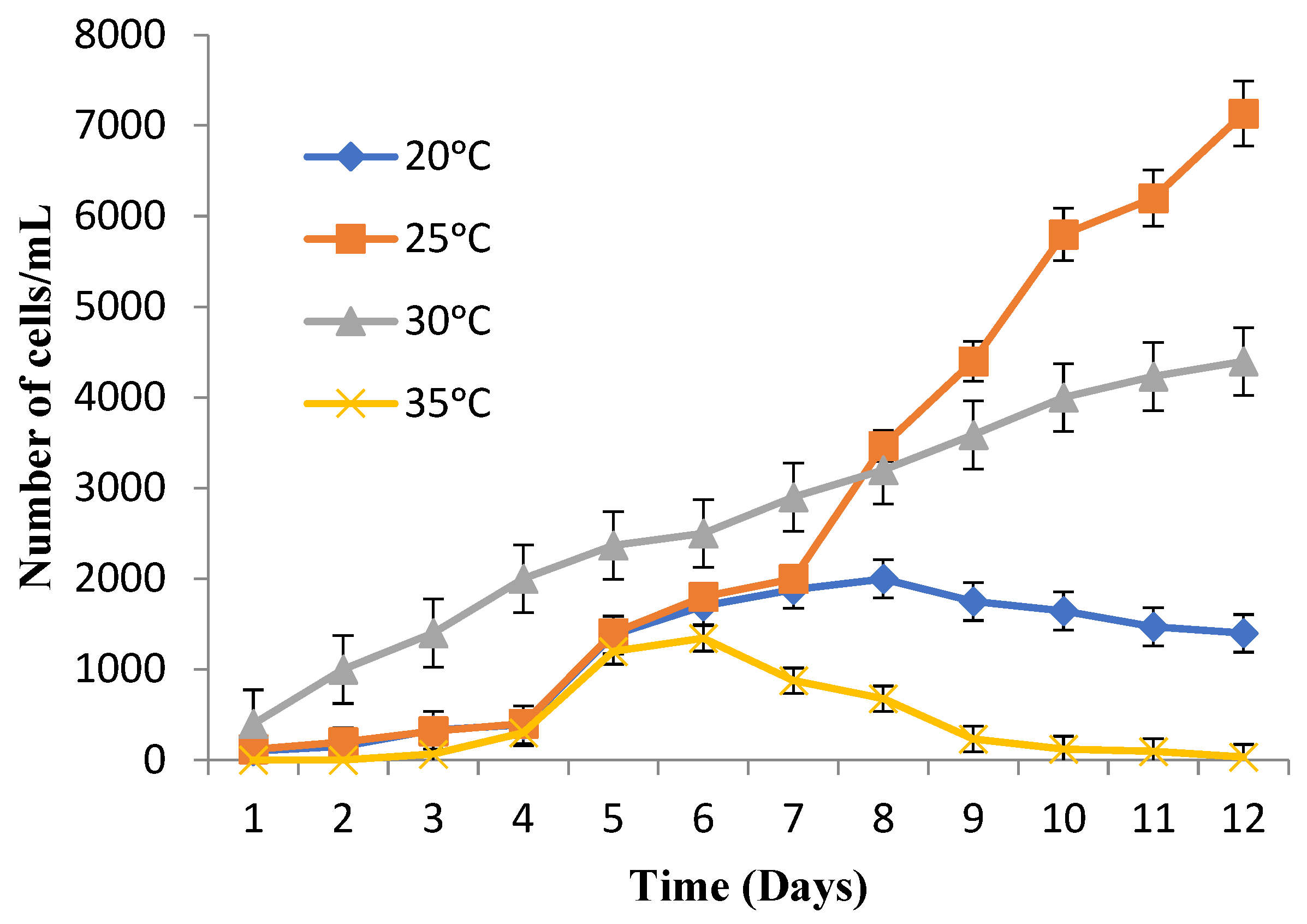
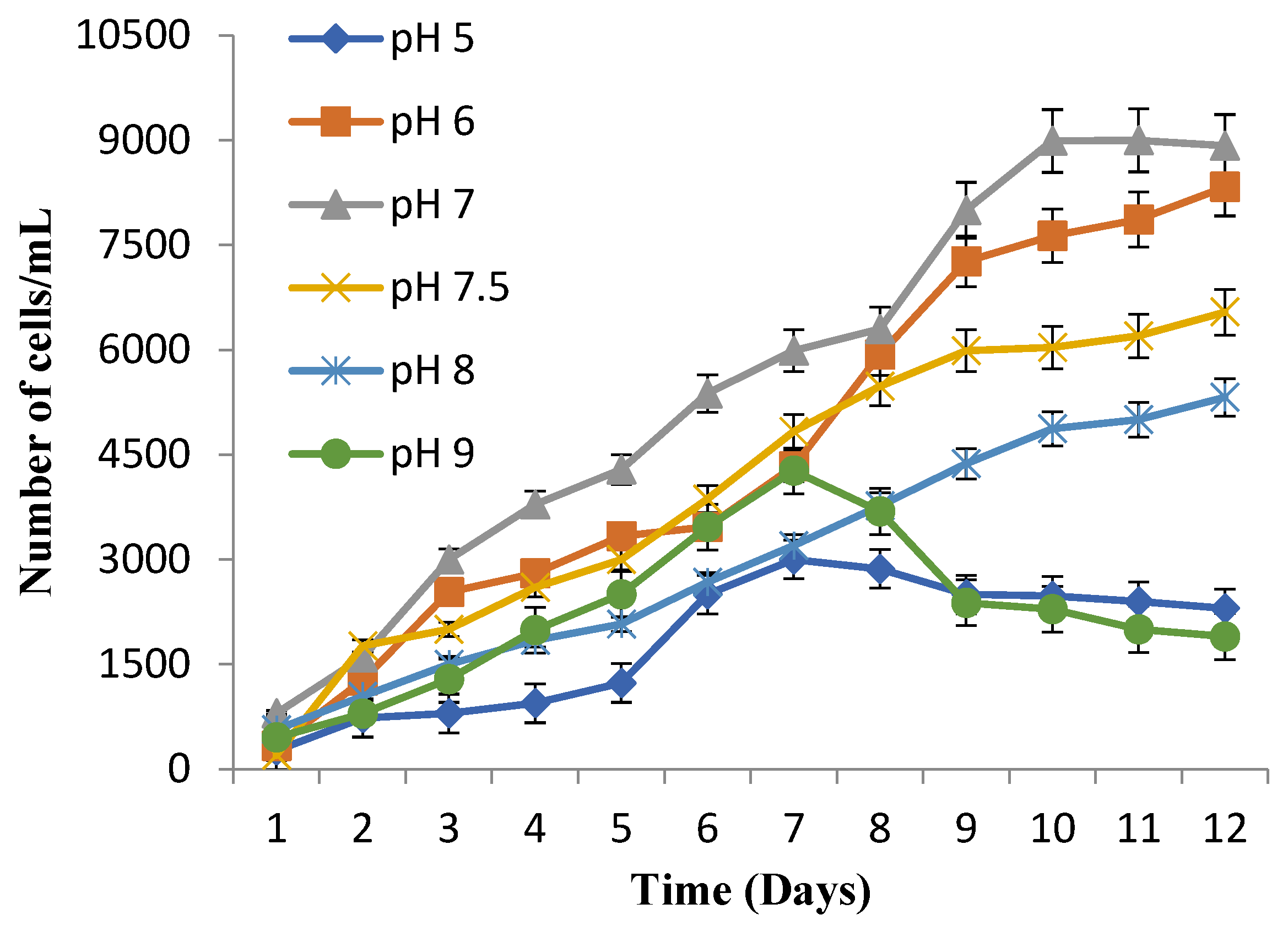
3.3. Suitable Temperature and pH for Maximum Cell Growth

The optimum temperature favoring the maximal growth rate was recorded at 25 °C for P. multimicronucleatum with a significant number of cells of 7.1 × 10³ cells/mL (Figure 2).
In comparison, a reduced growth rate with fewer cells of 4.4 × 10³ cells/mL at 30 °C was observed. While the number of diving cells reached 2.0 × 10³ and 1.3 × 10³ cells/mL at 20 °C and 35 °C, respectively, and then started to decline. At optimum pH, the highest growth rate, with 8.9 × 10³ cells/mL for P. multimicronucleatum, was recorded at neutral pH 7.0 but a good number of cells, 8.3 × 10³ cells/mL, were also recorded at pH 6 (Figure 3). At pH 7.5 and 8, decrease in cell growth with 6.5 × 10³ and 5.3 × 10³ cells/mL, respectively, was noted. Significant growth was not observed at more acidic pH 5 and more basic pH 9 for this species.

3.4. Copper Accumulation and Uptake Ability of Paramecium Cells

The copper-treated cell population of Paramecium multimicronucleatum accumulated and removed copper ions from the culture medium when provided with minimum concentrations. P. multimicronucleatum removed a maximum of 88% of copper ions at the concentrations of 30 µg/mL (Figure 4C) after 96 h of exposure. Other than this highest removal rate, P. multimicronucleatum also showed best results with 81% uptake when treated with 10 µg/mL copper concentration after 96 h. Moreover, it can uptake 75% of copper ions at 20 µg/mL within 48 h, while at 40 and 50 µg/mL of copper, more than 50% and less than 40% uptake was recorded after 24 and 72 h, respectively, and then a gradual decrease was observed (Figure 4D,E). Descending curves for the supernatant represent the remaining amount of copper ions in the culture medium at each concentration.

3.5. GSH Response and Non-Protein Thiols

Copper stress triggers the ability of cells to maintain the levels of reduced glutathione (GSH) in the cell to deal with the oxidative stress. P. multimicronucleatum showed a 1.7-fold significant increase (p < 0.005) in GSH levels when compared with control (24 h) initial level and copper-treated (48 h) final level (Table 1).
This shows that the content of reduced glutathione (GSH) seemed to be enhanced with time of exposure, fighting against the reactive oxygen species produced by copper treatment. A rise in oxidized glutathione (GSSG), by more than 2-fold after 24 and 48 h in copper-treated cells as compared to control, shows the stress situation of the cells. Significant decrease in GSH/GSSG ratio indicates loss of required balance between the reduced and oxidized state of glutathione for healthy cells. When boosting levels of GSH, enhanced content of other non-protein thiols, with 1.2 and 1.4-fold significant increase (p < 0.05) in P. multimicronucleatum after 24 and 48 h, respectively, was also recorded.

3.6. Protein Content

P. multimicronucleatum presented a rise in total protein content with copper treatment as compared to control. A significant change of 1.17-fold was noticed in the cells within 24 h of copper stress (Table 2).

4. Discussion

The emerging trend of industrialization and consumption of heavy metals has triggered the need to identify and understand the defensive mechanisms of organisms living in their wastewaters [21]. Among eukaryotes, ciliates have an ability to detoxify heavy metals and their survival in higher metal concentrations makes them resistant [22]. Tetrahymena, Stylonychia and Euplotes are the major reported ciliate species which can not only survive in a heavy metal-polluted environment but have developed cellular and molecular defensive mechanisms against these pollutants [15,23].
Biosorption and bioaccumulation is a new trend to deal with the toxicity produced by the release of heavy metals in the environment [24]. Protozoans, especially populations of Paramecium, are highly sensitive to the environmental toxic compounds, due to their absence of cell wall, and provide quick responses against them [25,26]. Mechanisms adopted by them involve adsorption, methylation, uptake, production of antioxidant enzymes, and changes at molecular level [27]. This makes them potentially excellent models for toxicological research studies with a potential use in decontaminating wastewater from heavy metals [16,28].
Keeping in view the importance of these organisms, the Paramecium species has been isolated from samples of industrial wastewater polluted with heavy metals. The copper tolerance of five isolates was checked and one species showed maximum resistance, which makes it suitable for this study. Moreover, morphological observations and ribotyping further confirms the species to be Paramecium multimicronuleatum. Ciliates like Stylonychia mytilus, Tetrahymena tropicalis, Oxytricha fallax and Paramecium caudatum have been reported to be isolated from metal-contaminated wastewaters and used for bioaccumulation [3,29,30].
The medium used for lab culture maintenance was bold basal salt medium as it supports maximum growth and increased life span for more than 6 weeks and is reported to be utilized with metal-resistant ciliates [14]. Ciliates’ bacterivorous behavior requires bacterial cells for their nutrition, and the bold basal salt medium provides plenty of bacterial production as it contains a variety of salts, trace metal ions, and grains of wheat. This not only fulfills their feeding requirement but also makes them resistant to dealing with environmental stress [31].
Effect on growth rate with copper exposure was observed for four weeks with daily monitoring. P. multimicronucleatum showed tolerance to each provided concentration of copper but showed maximum resistance against 10–40 µg/mL, with minor effects on morphology, growth rate and movement. With increased copper stress, slow movement, more vacuole formation, loss of typical cell shape, and cell inflammation were observed under microscope. Cell growth decreased to 2.0 × 10³ in copper-treated cells as compared to control (6.0 × 10³ cells/mL) after two weeks (Figure 1). Effect on growth rate along with resistance to copper has been reported in four ciliate species, Colpidium colpoda, Dexiotricha granulosa, Euplotes aediculatus, and Halteria grandinella [32].
Providing different temperature and pH for optimizing the suitable growth conditions revealed that P. multimicronuleatum is compatible with pH 7 (Figure 3) and temperature of 25 °C–30 °C and showed a growth range of 5.0 × 10³–8.0 × 10³ cells/mL (Figure 2). Zahid et al., 2018 [11] describes the same growth characteristics for the ciliate Tetrahymena themophilia with the maximum number of cells. The range of the pH in most of the environmental water bodies and wastewaters is alkaline, which makes these organisms suitable for growing in them for longer periods of time. The maximum growth of Paramecium species requires low temperatures as they are mostly present in the lower or deeper layers of environmental reservoirs [33].
The copper accumulation and uptake ability of P. multimicronuleatum was promising, with more than 80% removal of copper ions from the medium at 10 and 30 µg/mL concentrations within 96 h (Figure 4A,B). However, more than 50% uptake has also been recorded at 40 µg/mL concentration of copper after 72 h. Wastewater-isolated species of Tetrahymena showed more than 50% of copper uptake when treated with 10 μg/mL after 96 h of exposure, but uptake was decreased at higher concentrations of copper [11,34]. Similarly, in this study the Paramecium species did not display much uptake at 50 µg/mL of copper stress, which indicates tissue damage and induction of cell death by apoptosis.
Among antioxidant enzymes, reduced glutathione plays a major role as a defensive mechanism against metal stress. Increased rates of GSH and other non-protein thiols, with more than 1.5-fold enhancement (p < 0.005), was measured for this Paramecium species when provided with 5 μg/mL of copper stress as compared to control. Metal and herbicide-exposed ciliates Stylonychia mytilus and Paramecium tetraurelia showed levels of GSH and other non-protein thiols elevated by 148 and 49% to deal with the stress situation [19,31]. The content of oxidized glutathione was also increased due to oxidative stress caused by reactive oxygen species. Redox activity of copper is involved in production of reactive oxygen species by Fenton/Haber–Weiss reaction and autoxidation. Increased GSH production means increased levels of other non-protein thiol content present in the form of cysteine, which empowers the cells to deal with oxidative stress and allows metal chelation [12]. A more than twofold decrease in GSH/GSSG ratio represents increasing oxidative stress in the cell population after 48 h of exposure. This justifies the production of more reduced glutathione to keep the balanced ratio for healthy cell activities and metabolism [35]. Copper exposure causes significant fold change in the cellular total protein content of P. multimicronucleatum. Similar changes in total protein content and expression is reported in S. mytilus with nickel and zinc treatment [31].
Outcomes from these kinds of toxicological and bioremediation studies would be helpful in dealing with the alarming situations of severe health risks and damaged aquatic ecosystems caused by the huge amounts of heavy metals released in environmental water bodies. Environmentally friendly and sensitive organisms like eukaryotic ciliates (Paramecium) are the best models not only to study the toxicity of heavy metals but also for use in clean-up operations in the environment. They can be exploited for large scale research studies due to their ease of availability in the environment, cheap growing medium, simple handling, and safe culture maintenance. With advanced sequencing and bioinformatic techniques, molecular details as to their tolerance mechanisms against metals can be studied and desired proteins can be overexpressed for biotechnological applications and proposing models for whole cell biosensors.

5. Conclusions

This study focused on the investigation of bioremediation abilities and the required optimum growth conditions of the ciliated protozoan Paramecium multimicronuleatum against copper ions. This organism is sensitive to lower concentrations of copper ions and can be used for bioaccumulation of this heavy metal. The functioning of bioremediation requires suitable growth conditions for Paramecium cells. The antioxidant enzyme, reduced glutathione, and protein synthesis are also influenced by copper exposure and play roles in cellular activities necessary for the detoxification of copper ions.

Author Contributions

Conceptualization, A.L., F.R.S. and A.R.S.; methodology, A.L. and I.Z.; investigation, A.L., I.Z. and F.R.S.; writing—original draft preparation, A.L., I.Z. and S.Z.A. writing—review and editing, S.M.W., G.E.E., M.A.I. and M.R.; supervision, F.R.S. and A.R.S.; funding acquisition, S.M.W. and M.R. All authors have read and agreed to the published version of the manuscript.

Funding

The authors are grateful to the Researchers Supporting Project No. (RSP-2021/161), King Saud University, Riyadh, Saudi Arabia.

Data Availability Statement

The data that support the findings of this study are available from the corresponding author upon reasonable request.

Acknowledgments

Authors are grateful to the Researchers Supporting Project No. (RSP-2021/161), King Saud University, Riyadh, Saudi Arabia. The authors would also like to acknowledge the lab facilities provided by both the Institute of Zoology and School of Biological Sciences, University of the Punjab, Lahore, Pakistan. We would also like to thank the Higher Education Commission for financial support through the National Research Program for Universities.

Conflicts of Interest

The authors declare that there is no conflict of interest.

References

  1. Al-Saydeh, S.A.; El-Naas, M.H.; Zaidi, S.J. Copper removal from industrial wastewater: A comprehensive review. J. Ind. Eng. Chem. 2017, 56, 35–44. [Google Scholar] [CrossRef]
  2. Rehman, A.; Shakoori, F.R.; Shakoori, A.R. Heavy metal resistant Distigma proteus (Euglenophyta) isolated from industrial effluents and its possible role in bioremediation of contaminated wastewaters. World J. Microbiol. Biotechnol. 2007, 23, 753–758. [Google Scholar] [CrossRef]
  3. Rehman, A.; Shakoori, F.R.; Shakoori, A.R. Heavy metal resistant freshwater ciliate, Euplotes mutabilis, isolated from industrial effluents has potential to decontaminate wastewater of toxic metals. Bioresour. Technol. 2008, 99, 3890–3895. [Google Scholar] [CrossRef] [PubMed]
  4. Boldrin, F.; Santovito, G.; Formigari, A.; Bisharyan, Y.; Cassidy-Hanley, D.; Clark, T.G.; Piccinni, E. MTT2, a copper-inducible metallothionein gene from Tetrahymena thermophile. Comp. Biochem. Physiol. C Toxicol. Pharmacol. 2008, 147, 232–240. [Google Scholar] [CrossRef]
  5. Sharma, P.; Singh, S.P.; Parakh, S.K.; Tong, Y.W. Health hazards of hexavalent chromium (Cr (VI)) and its microbial reduction. Bioengineered 2022, 13, 4923–4938. [Google Scholar] [CrossRef]
  6. Rehman, A.; Shakoori, A.R. Heavy metal resistant Chlorella spp., isolated from tannery effluents, and their role in remediation of hexavalent chromium in industrial wastewater. Bull. Environ. Contam. Toxicol. 2001, 66, 542–547. [Google Scholar] [CrossRef]
  7. Pavasant, P.; Apiratikul, R.; Sungkhum, V.; Suthiparinyanont, P.; Wattanachira, S.; Marhaba, T.F. Biosorption of Cu2+, Cd2+, Pb2+, and Zn2+ using dried marine green macroalga Caulerpa lentillifera. Bioresour. Technol. 2006, 97, 2321–2329. [Google Scholar] [CrossRef]
  8. Karthikeyan, S.; Balasubramanian, R.; Iyer, C.S.P. Evaluation of the marine algae Ulva fasciata and Sargassum sp. for the biosorption of Cu (II) from aqueous solutions. Bioresour. Technol. 2007, 98, 452–455. [Google Scholar] [CrossRef]
  9. Selonen, S.; Setala, H. Nutrient leaching, soil pH and changes in microbial community increase with time in lead-contaminated boreal forest soil at a shooting range area. Environ. Sci. Pollut. Res. Int. 2017, 24, 5415–5425. [Google Scholar] [CrossRef]
  10. Madoni, P. Protozoa in wastewater treatment processes: A minireview. Ital. J. Zool. 2011, 78, 3–11. [Google Scholar] [CrossRef]
  11. Zahid, M.T.; Shakoori, F.R.; Zulfiqar, S.; Al-Ghanim, K.A.; Shakoori, A.R. Growth characteristics, metal uptake and expression analysis of copper metallothionein in a newly reported ciliate, Tetrahymena farahensis 2018. Pak. J. Zool. 2018, 50, 1171–1181. [Google Scholar] [CrossRef]
  12. Jozefczak, M.; Remans, T.; Vangronsveld, J.; Cuypers, A. Glutathione is a key player in metal induced oxidative stress defenses. Int. J. Mol. Sci. 2012, 13, 3145–3175. [Google Scholar] [CrossRef] [Green Version]
  13. Forman, H.J.; Zhang, H.; Rinna, A. Glutathione: Overview of its protective roles, measurement, and biosynthesis. Mol. Asp. Med. 2009, 30, 1–12. [Google Scholar] [CrossRef] [Green Version]
  14. Pizzorno, J. Glutathione! Integr. Med. 2014, 13, 8–12. [Google Scholar]
  15. Kamika, I.; Momba, M.N.B. Effect of nickel on nutrient removal by selected indigenous protozoan species in wastewater systems. Saudi J. Biol. Sci. 2015, 22, 147–156. [Google Scholar] [CrossRef] [Green Version]
  16. Rehman, A.; Shakoori, F.R.; Shakoori, A.R. Multiple metal resistance and uptake by a ciliate, Stylonychia mytilus, isolated from industrial effluents and its possible use in wastewater treatment. Bull. Environ. Contam. Toxicol. 2007, 79, 410–414. [Google Scholar] [CrossRef]
  17. Shakoori, A.R.; Rehman, A.; Haq, R.U. Multiple metal resistance in the ciliate protozoan, Vorticella microstoma, isolated from industrial effluents and its potential in bioremediation of toxic wastes. Bull. Environ. Contam. Toxicol. 2004, 72, 1046–1051. [Google Scholar] [CrossRef]
  18. Regensbogenova, M.; Pristas, P.; Javorsky, P.; Moon-van der Stay, S.Y.; Van der Stay, G.W.M.; Hackstein, J.H.P.; Newbold, C.J.; McEwan, N.R. Assessment of ciliates in the sheep rumen by DGGE. Lett. Appl. Microbiol. 2004, 39, 144–147. [Google Scholar] [CrossRef]
  19. Moumeni, O.; Berrebbah, H.; Azzouz, Z.; Amamra, R.; Otmani, H.; Alayat, A.; Benosmane, S.; Djebar, M.R. Effects of cycloxydim on population growth, phagocytosis, contractile vacuole activity and antioxidant responses in the freshwater ciliates (Paramecium tetraurelia). Res. J. Environ. Toxicol. 2016, 10, 115–125. [Google Scholar] [CrossRef] [Green Version]
  20. Israr, M.; Sahi, S.V.J.; Jain, J. Cadmium accumulation and antioxidant responses in the Sesbania drummondii callus. Arch. Environ. Contam. Toxicol. 2006, 50, 121. [Google Scholar] [CrossRef]
  21. Boldrin, F.; Santovito, G.; Irato, P.; Piccinni, E. Metal interaction and regulation of Tetrahymena pigmentosa metallothionein genes. Protist 2002, 153, 283–291. [Google Scholar] [CrossRef]
  22. Vieira, R.H.S.F.; Volesky, B. Biosorption: A solution to pollution? Int. Microbiol. 2000, 3, 17–24. [Google Scholar]
  23. Rehman, A.; Ashraf, S.; Qazi, J.I.; Shakoori, A.R. Uptake of lead by a ciliate, Stylonychia mytilus, isolated from industrial effluents: Potential use in bioremediation of wastewater. Bull. Environ. Contam. Toxicol. 2005, 75, 290–296. [Google Scholar] [CrossRef]
  24. Gadd, G.M. Heavy metal accumulation by bacteria and other microorganisms. Experientia 1990, 13, 273–280. [Google Scholar] [CrossRef]
  25. Francisco, R.; Alpoim, M.C.; Morais, P.V. Diversity of chromium resistant and reducing bacteria in a chromium-contaminated activated sludge. J. Appl. Microbiol. 2002, 92, 837–843. [Google Scholar] [CrossRef] [PubMed] [Green Version]
  26. Gutiérrez, J.C.; Martín-González, A.; Díaz, S.; Ortega, R. Ciliates as a potential source of cellular and molecular biomarkers/biosensors for heavy metal pollution. Eur. J. Protistol. 2003, 39, 461–467. [Google Scholar] [CrossRef]
  27. Haq, R.U.; Rehman, A.; Shakoori, A.R. Effect of dichromate on population and growth of various protozoa isolated from industrial effluents. Folia Microbiol. 2000, 45, 275–278. [Google Scholar] [CrossRef] [PubMed]
  28. Gutierrez, J.C.; Martin-Gonzalez, A.; Diaz, S.; Amaro, F.; Ortega, R.; Gallego, A.; De Lucas, M.P. Ciliates as cellular tools to study the eukaryotic cell–heavy metal interactions. In Heavy Metal Pollution; Brown, S.E., Welton, W.C., Eds.; Nova Science: New York, NY, USA, 2008; pp. 1–44. [Google Scholar]
  29. Rehman, A.; Shakoori, F.R.; Shakoori, A.R. Multiple heavy metal tolerant ciliates, Oxytricha fallax and Paramecium caudatum, isolated fom industrial effluents and their potential use in wastewater treatment. Pak. J. Zool. 2010, 42, 301–309. [Google Scholar]
  30. Chaudhry, R.; Chaudhry, M.T.; Al-Ghanim, K.; Meboob, S.; Shakoori, A.R. Structure modeling of small subunit ribosomal RNA (SSU rRNA) of copper resistant ciliates. Pak. J. Zool. 2014, 46, 1625–1632. [Google Scholar]
  31. Shakoori, F.R.; Zafar, M.F.; Fatehullah, A.; Rehman, A.; Shakoori, A.R. Response of glutathione level in a protozoan ciliate, Stylonychia mytilus, to increasing uptake of and tolerance to nickel and zinc in the medium. Pak. J. Zool. 2011, 43, 569–574. [Google Scholar]
  32. Madoni, P.; Romeo, M.G. Acute toxicity of heavy metals towards freshwater ciliated protists. Environ. Pollut. 2006, 141, 1–7. [Google Scholar] [CrossRef]
  33. Martin-Gonzalez, A.; Dias, S.; Borniquel, S.; Gallego, A.; Gutierrez, J.C. Cytotoxicity and bioaccumulation of heavy metals by ciliated protozoa isolated from urban wastewater treatment plants. Res. Microbiol. 2006, 157, 108–118. [Google Scholar] [CrossRef]
  34. Chaudhry, R.; Shakoori, A.R. Identification of a new copper resistant protist isolate Tetrahymena RT-1 subsp. nov. Pak. J. Zool. 2011, 43, 781–788. [Google Scholar]
  35. Cuypers, A.; Smeets, K.; Ruytinx, J.; Opdenakker, K.; Keunen, E.; Remans, T.; Horemans, N.; Vanhoudt, N.; Van Sanden, S.; Van Belleghem, F.; et al. The cellular redox state as a modulator in cadmium and copper responses in Arabidopsis thaliana seedlings. J. Plant Physiol. 2011, 168, 309–331. [Google Scholar] [CrossRef]
Figure 1. Growth curves of Paramecium multimicronucleatum under different concentrations of copper along with control.
Water 14 01419 g001
Figure 2. Growth observations of Paramecium multimicronucleatum culture provided with different temperatures.
Water 14 01419 g002
Figure 3. Growth observations of Paramecium multimicronucleatum culture provided with different pH.
Water 14 01419 g003
Figure 4. Uptake of copper ions from culture medium by Paramecium multimicronucleatum at concentrations of (A) 10 µg/mL, (B) 20 µg/mL, (C) 30 µg/mL, (D) 40 µg/mL, and (E) 50 µg/mL.
Water 14 01419 g004aWater 14 01419 g004b
Table 1. Change in GSH and other non-protein thiol content in control and copper-exposed P. multimicronucleatum culture after 24 and 48 h.
24-h48-h
P. multimicronucleatum
(Control)
P. multimicronucleatum
(Cu Exposed)
P. multimicronucleatum
(Control)
P. multimicronucleatum
(Cu Exposed)
GSH (mM/g)21.99 ± 0.622.86 ± 0.427.14 ± 0.937.54 ± 0.8 **
GSSG (mM/g)9.43 ± 1.019.83 ± 0.7 *13.35 ± 0.936.75 ± 0.3 **
GSH + GSSG (mM/g)31.43 ± 0.542.70 ± 0.3 *40.50 ± 0.274.30 ± 0.6 **
GSH/GSSG ratio2.43 ± 0.31.16 ± 0.062.08 ± 0.21.02 ± 0.03
Non-protein thiols16.75 ± 0.121.75 ± 0.5 *15.59 ± 0.122.20 ± 0.2 **
Mean ± SEM (n = 3); * (Control vs. Copper after 24 and 48 h; p < 0.05) ** Both (Control vs. Copper after 24 and 48 h + Control 24 h vs. Copper 48 h; p <0.005).
Table 2. Change in total protein content in control and copper-treated P. multimicronucleatum culture after 24 h.
Protein Content (µg/mL)
Control35.43 ± 0.4
Cu Exposed77.04 ± 6.0 *
Mean ± SEM (n = 3), * (p < 0.05).
Publisher’s Note: MDPI stays neutral with regard to jurisdictional claims in published maps and institutional affiliations.

