Article

Assessment of Bilge Water Degradation by Isolated Citrobacter sp. and Two Indigenous Strains and Identification of Organic Content by GC-MS

Department of Chemical Engineering, Cyprus University of Technology, Anexartisias 57 Str., Lemesos 3036, Cyprus
*
Author to whom correspondence should be addressed.
Water 2022, 14(9), 1350; https://doi.org/10.3390/w14091350
Submission received: 3 March 2022 / Revised: 15 April 2022 / Accepted: 19 April 2022 / Published: 21 April 2022
(This article belongs to the Section Wastewater Treatment and Reuse)

Abstract

:
Bilge water is oily saline wastewater accumulated on the hull at the bottom of a vessel, generated from leakage from pipes and engines and wash-down freshwater containing cleaning solvents. The present study focused on isolating microorganisms from oil-contaminated sites and indigenous species from raw bilge water and assessment of their ability to biodegrade bilge water. Using phenanthrene as a carbon source Citrobacter species was isolated from oil-contaminated sites and its optimum growth condition was found. The results indicated significant tolerance of the bacterium which presented great biodegradation ability for the tested carbon source. At high salinity (33 g L−1 of NaCl), sufficient phenathrene removal was achieved (81%), whereas variation of pH from 5 to 10 did not affected the survival of the microorganism. Regarding the effect of temperature and nutrients, Citrobacter sp. was better adapted at 30 °C, while lack of nutrients presented a negative impact on its growth. Halomonas and Exiguobacterium sp. were isolated from real bilge water using phenanthrene and phenol as a carbon source. The isolated strains independently exposed to high and low range bilge water pointed out around 83% and 53% chemical oxygen demand (COD) removal, respectively. Analysis of untreated bilge water by gas chromatography-mass spectrometry (GC-MS) was carried out, and the results confirmed the presence of organic compounds having a high similarity with Heptane, N-hexadecanoic acid, Methyl isobutyl Ketone and 1-butoxy-2-propanol. Chromatographic analysis of treated bilge water after exposure to isolated strains indicated the existence of new compounds. These metabolites presented high similarity with N-hexadecanoic, methyl ester, N-hexadecanoic and Octadecanoic acid methyl ester.

Graphical Abstract

1. Introduction

Bilge water (BW) is the main pollutant of shipboard wastewater, and it can be defined as saline, oily and greasy wastewater with high COD (>3–15 gCOD L−1) containing lubricating oil, cleaning diesel oil, spills from the engine room, oily sludge, water leaks from internal pipes, and seawater filtrations [1]. According to the International Maritime Organization (IMO) regulations [2] and the European directive 2000/59/EC the discharge of oil residues to marine environments is prohibited. Therefore, bilge water is either treated enroute in an oil separation system before being discharged to the sea or deposited at reception facilities on land.
According to the literature, mainly physicochemical processes have been applied for BW treatment like coagulation/flocculation, electrochemical demulsification, membrane separation, etc. [3,4,5,6,7]. However, such methods often imply high operational costs. Moreover, the use of chemicals [8] for solving emulsification problems increases the risk towards living organisms. Thus, additional treatments are needed for the remaining water before its release into the environment [8]. As a result, the development of new cost-effective, fast, environmentally friendly and sustainable treatment technologies is of high importance to comply with the increasingly stringent requirements of international regulations.
On the contrary, biological methods, progressively gain interest in treating saline wastewaters rich in organic content and petroleum hydrocarbons [9] using microorganisms able to consume such compounds [10]. However, biological activity may be strongly inhibited by the hostile nature of the recalcitrant wastes such as BW [11]. Therefore, the use of indigenous species or species adapted to recalcitrant wastewater is proved more advantageous over non-native micro-organisms for achieving better results. Autochthonous microbial isolates are already adapted to thrive in the complex toxic environment and can act synergistically [12]. Thus, bioremediation is considered as more efficient and cost-effective green alternative process [13].
Numerous studies have described the isolation of indigenous species from oil contaminated samples [14,15,16] and as reported there is a possible correlation between hydrocarbon degradation and biosurfactant production by microorganisms which enhances the elimination of organic compounds [17,18,19]. Furthermore, there are several researchers investigated the degradation of crude and diesel oil or petroleum hydrocarbons by native species isolated from oil-contaminated substrates [20,21]. Specifically, Olivera et al. [22], Santisi et al. [23], Nievas et al. [24] and Sivaraman et al. [25] have identified several cultivable oil-degrading bacteria from bilge samples, being found mainly members of the genus Pseudomonas. However, Nisenbaum et al. [9] took sample from oily bilge water from ship and exposed this for a long period to bilge water. The most abundant bacterial species detected were: Marinobacter spp. 20.7%, Alcanivorax spp. 17.3%, Parvibaculum spp. 13.3%, Flavobacteriaceae 10.1%, Gammaproteobacteria PYR10d3 8.4%, Novispirillum spp. 5.0% and Xanthomonadaceae 4.9%. Mazioti et al. [14] isolated Halomonas alkaliphila S5a, from raw bilge wastewater and examined the COD removal from raw bilge water. In addition, Cappello et al. [26] identified Acinetobacter, and Rhodococcus from bilge waster. Corti-Monzón [27] examined the diversity and functionality of the microorganisms occurring in oily bilge wastes. Alkane degradation marker gene alkB PCR-DGGE fingerprinting showed distinct diversity patterns among the samples. Alcanivorax, Marinobacter, and Pseudomonas were highly abundant in bilge samples, suggesting that alkB genes would be hosted by members of these genera.
However, there is scarce information regarding the biodegradation of raw real BW by indigenous and non-indigenous species, as well as the specific organic compounds that are removed due to their action.
Therefore, the objectives of the current study were to (i) isolate and identify microbial strains from oil-contaminated sites and indigenous strains from real bilge water, (ii) investigate the tolerance of isolated species in various extreme environmental conditions: different salinities, temperatures, pH values and nutrients concentrations, (iii) examine the ability of isolated species to eliminate specific hydrocarbons and biodegrade BW resulting substantially decrease of COD removal and (iv) get an overview of the organic compounds present in treated bilge water using GC-MS analysis.

2. Material and Methods

2.1. Collection of Real Bilge Water

Samples of real bilge water were taken from EcoFuel Limited Company (Ecofuel Ltd., Limassol, Cyprus), which collects and treats this type of wastewater at Zygi (Cyprus). The physicochemical properties of bilge water from three batches received in the treatment plant facilities (data are obtained from EcoFuel Ltd. database) are given in Table S1. The extractable total hydrocarbons of BW from a specific batch sample mainly consisted of organic compounds with 6 to 35 carbons in their molecule (C6 to C35). As shown in Table S2, mono-aromatic, aliphatic and polycyclic aromatic hydrocarbons (PAHs), phenol derivatives as well as surfactant related compounds, such as dodecanol, were present. The volatile hydrocarbon content was composed mainly by gasoline components, such as benzene, toluene, ethylbenzene and xylenes (BTEX) phenol and naphthalene derivatives.

2.2. Isolation of Pure Strains from Environmental Samples and Identification of Indigenous Bacterial Strains

Different samples from oil-contaminated soil nearby Ecofuel (Cyprus) Ltd., E107, Mari (https://www.ecofuelcy.com/; accessed on 15 March 2022) and real BW were used to isolate bacterial strains and investigate their tolerance and ability to biodegrade phenanthrene. This compound was selected as a representative one since many aromatic hydrocarbon groups are often detected in BW [1,28].
Briefly, 100 mL conical flasks were filled under sterile conditions with 25 mL modified sterilized mineral salts (Merck, Germany) medium (MSM) containing 50 mg L−1 of phenanthrene (Merck, Germany) and 5 g of contaminated solid sample or 5 mL of liquid sample. After pH adjustment to 7, the flasks were placed in a shaking incubator at 30 °C and agitated at 100 rpm for a period of 10 days until visible turbidity within the liquid phase was observed.
Isolation and identification of each bacterial strain was performed by the removal of 0.1 mL of sample from each flask that exhibited high turbidity and serially diluted by a factor from 10−1 to 10−10. The diluted samples were plated by the streak method on Petri dishes containing a mixture of MSM-agar and 50 mg L−1 of phenanthrene. Afterwards, the inoculated Petri dishes were sealed and incubated up to 10 days (30 °C). Following the incubation period, single colonies were picked and re-cultivated in MSM-agar media following the same procedure as stated above. This procedure was repeated several times until pure identical colonies were observed. Finally, pure cultures of the isolated microorganisms were subjected to 16S rRNA sequence analysis for identification [29].
One difficulty of using phenanthrene was its dissolution in the aqueous MSM medium. Water solubility of phenanthrene is 1.29 mg L−1, which is low characterizing this compound as an insoluble organic component. Thus, ethanol was chosen as the carrier solvent because of its high vapor pressure. Solution of the compound in ethanol was poured in the conical flasks, and it was allowed to stand at the fume hood overnight for complete evaporation of the organic solvent. During the experiments, culture samples (2 mL) were extracted by liquid–liquid extraction and analyzed by high performance liquid chromatography (HPLC)-UV/VIS detector in order to know the accurate dissolved concentration of phenanthrene. The extraction was performed with 2.5 mL of acetonitrile by vortex for 1 min. Afterwards, 2 mL of the supernatant were centrifuged (13000 r.p.m, 10 min), filtered (0.25 μm) for the removal of microbial cells and finally subjected to HPLC analysis.
For isolation of indigenous samples from BW, samples were collected and diluted (10−2 to 10−10). The diluted BW was transferred onto MSM-agar plates containing phenol 0.5 g L−1 and phenanthrene 0.5 g L−1. Salinity was adjusted to 35 g L−1 of NaCl, in order to have same salinity as the sea water. Repeated striking process onto agar plates was performed and after 5 days of cultivation pure cultures were observed. Red/orange-colored and white colonies appeared onto the surface of the agar plates and 2–3 colonies were collected for identification.

2.3. Strain Characterization by 16S rRNA Sequence Analysis

Identification of isolated microorganisms was performed based on [29]. Briefly, Polymerase Chain Reaction (PCR) was accomplished using the following primers: (i) 8f: 5′-AGA GTT TGA TCC TGG CTC AG-3′ and (ii) 1542R: 5′-AAG GAG GTG ATC CAG CCG CA 3′. The reaction was performed as follows: 94 °C (2 min) followed by 33 cycles consisting of 94 °C (1 min), 56 °C (1 min), 72 °C (2 min) and finally 72 °C (7 min). PCR-amplified 16S rRNA genes were purified using the NucleoFast® 96 PCRCleanup Kit (Macherey-Nagel, Düren, Germany). Sequencing alignment was performed by Macrogene (Netherlands) and the resulting alignment of the DNA sequences of the gene encoding 16S rRNA was compared for homology in the NCBI database by BLASTn nucleotide tool analysis. The phylogenetic tree diagram was developed through the neighbor-joining method applying Mega 6.0 software and the most similar sequences according to the NCBI database by BLASTn and boot strapping similar to Koutinas et al. [30].

2.4. Cultivation of Bacterial Strain under Different Environmental Conditions

After selecting the optimal initial concentration of phenanthrene, the effect of temperature, pH, salinity and nutrients on the degradation of the compound was further investigated by the isolated strain Citrobacter sp. D2. Initially, the microorganism was pre-cultivated for 24 h in 100 mL MSM medium containing 150 mg L−1 of phenanthrene and 0.5 g L−1 NaCl. 5 mL of adapted culture was transferred onto conical flasks of 100 mL (25 mL working volume) containing MSM medium under various conditions for a period of 96 h at 30 °C. The various parameters were tested at several levels: salinity (NaCl: 0.5, 15, 20, 33 g L−1), pH (5.0, 7.0, 8.5, 10), temperature (22, 30 and 40 °C) and absence of nutrients (absence of phosphorous, nitrogen or both). Temperature and pH were measured using portable instruments. Optical density of the liquid cultures was measurement at 600 nm at different time intervals using a HACH UV/VIS scanning spectrophotometer (JENWAY 7315, Staffordshire, UK).

2.5. Biodegradation of Real Bilge Water by Different Isolated Bacterial Strains

Each isolated microbial strain was tested for its ability to biodegrade real BW. Firstly, the microbial strains were activated in MSM medium containing phenol (0.5 g L−1) overnight at 30 °C. Then, 0.2% v/v inoculums were transferred in 100 mL conical flasks (25 mL working volume) containing real undiluted BW (100%). The pH of the cultures was adjusted to 7.0 and the flasks were placed into shaking incubator (30 °C, 100 rpm) for a period of 9 days. Liquid samples were collected at different time intervals for COD analysis according to Standard Methods [31]. Specifically, the closed reflux colorimetric method was used for COD. Before spectrophotometric analyses, samples were centrifuged and filtered for removing color and solids. Thereafter, 2.0 mL of the sample was added into glass tubes and digestion solution (1.2 mL) together with sulphuric acid reagent (2.8 mL) were added. The glass tubes were closed tightly, mixed properly and refluxed in a Hach COD reflux reactor (150 °C for 2 h). After cooling to room temperature, the COD was determined by HACH UV/VIS scanning spectrophotometer (JENWAY 7315, Staffordshire, UK) at 620 nm.

2.6. HPLC-UV/VIS Analysis

All samples for the determination of real concentration of phenanthrene in the cultures were analyzed by High Performance Liquid Chromatography (HPLC)-UV/VIS. The system consisted of a LC-20AD pump equipped with a Shimadzu SPD-20A UV/VIS detector and a SIL-20A HT auto sampler (Shimadzu, Milton Keynes, UK). The column was C18 (Thermo Scientific, Waltham, MA, USA) and was heated at 40 °C (column oven CTO-10AS VP, Shimadzu). The detector was set at 254 nm, the injection volume at 20 μL. Isocratic analysis (45 min) was performed using mixture of acetonitrile-ultrapure water (70:30) as a mobile phase solution at a flow rate of 1 mL min−1.
Sample volume of 2 mL was transferred into test tubes (10 mL) and 2.5 mL of acetonitrile were added. The mixtures were placed on vortex for 1 min, so as the extraction of phenanthrene can be achieved by the organic solvent. Then, 2 mL of the supernatant were first centrifuged (13000 r.p.m) and then was passed through filter (0.25 μm) for the removal of microbial cells. Finally, the filtered samples were subjected to HPLC-UV/VIS analysis.

2.7. GC-MS Analysis

Bilge hydrocarbons were extracted according to the main steps of ISO 9377-2:2000 as described by [32] in order to identify the major organic compounds found in BW. Briefly, 9 g of MgSO4·7H2O were added to 100 mL of sample and the pH was adjusted to pH 2 by adding hydrochloric acid. Thereafter, the sample was extracted with 50 mL of hexane (Merck, Germany) for 100 mL of sample (50 mL hexane per 50 mL sample each time) after rigorous stirring for 30 min. The extracts were collected and stored (−18 °C) in a 65 mL serum bottles sealed with Teflon cups until GC-MS analysis. Chromatographic analysis was performed using a Hewlett Packard Gas Chromatograph 5890 Series II connected to a Hewlett Packard Mass Spectrometer HP5971 MSD (Palo Alto, California, CA, USA). The separation of the compounds was achieved using a DB5MS capillary column (60 m × 0.32 mM × 0.25 mm, Supelco, Burlington, MA, USA). The carrier gas was helium (0.9 mL min−1). A sample volume of 1 μL was injected in splitless mode at an inlet temperature of 250 °C. The initial column temperature was held for 5 min at 45 °C, programmed at 5 °C min−1 to a final temperature of 260 °C which was held for 12 min. The MS transfer line temperature was maintained at 280 °C, whereas the ion source temperature was 180 °C. For the qualitative analysis, the mass spectrometer was operated in total ion current mode and identification of compounds was made by comparison of a high probability match with mass spectra reported in the library of the National Institute of Standards and Technology (NIST 5, U.S.A. Department of Commerce).

3. Results and Discussion

3.1. Isolation and Characterization of Pure Bacterial Strains

Two bacterial strains were isolated from soil samples contaminated by oil using phenanthrene as a carbon source. The two PAH biodegrading microorganisms, named D1 and D2, formed white round colonies in all tested concentrations (Figure S1). As the concentration of phenanthrene increased, smaller and lesser colonies were observed, indicating a negative effect on the microbial population with D2 proved to be more tolerant than D1. Measurements of optical density (OD600) at D2 cultures confirmed biomass production from the beginning and followed a rising trend up to the end of cultivation (Figure S2).
Since D2 showed the highest growth and tolerance to phenanthrene, it was selected for further experiments. BLASTn tool analysis was used for the comparison of the generated nucleotide sequence that was received using the 16S rRNA sequencing with other bacterial strains. The results showed that the isolate was similar to the genus Citrobacter and had 99.7% (supplementary information) similarity with Citrobacter freundii and 99.5% similarity with Citrobacter portucalensis. This is also shown in the phylogenetic tree diagram (Figure S3).
Isolation of indigenous bacterial strains from raw BW resulted in two isolates. After five days of cultivation, red/orange-colored and white colonies appeared on the surface of the agar plates. Characterization of the isolated strains resulted in 99.0% homology with Exiguobacterium and Halomonas species and was named Ex-Ind2 and Hal-CG, respectively. More specifically, Exiguobacterium Ex-Ind2 was found to be 99.3% similar with both Exiguobacterium indicum and Exiguobacterium acetilicum, while Halomonas Hal-CG presented 99.7 and 99.3% similarity with Halomonas boliviensis and Halomonas titanicae, respectively (Phylogenic diagram Figures S4 and S5). Mazioti et al. [14] and Santisi et al. [23] also isolated halotolerant Halomonas species from bilge water.

3.2. Biodegradation of Phenanthrene by Citrobacter sp. D2

Growth of Citrobacter sp. D2 was investigated under different environmental conditions. As shown in Figure 1, the microorganism grew and metabolized phenanthrene up to 33 g L−1 of NaCl since visible turbidity was exhibited in all flasks. At 0.5 g L−1 of NaCl, the growth reached the stationary phase almost immediately, from the first day. When 15 g L−1 of NaCl were used, the highest OD600 was obtained, and exponential growth was observed until the end of the experiment. A similar growth trend was also observed at the next concentration level (20 g L−1), but with lower biomass production.
Nevertheless, the increasing trend indicated a potential growth of Citrobacter sp. D2 beyond 4d of cultivation at these two levels. At concentration 33 g L−1 of NaCl, limited growth of the microorganism was achieved. Although, the observed turbidity in the cultures demonstrated that despite its slower growth rate, Citrobacter sp. D2 had the ability not only to adapt, but also to continue biodegrade phenanthrene. Indeed, as shown in Figure 2, the concentration of phenanthrene was reduced at all examined salinity levels during 23 days of incubation. At the end of the experiment, removal reached 81% in the cultures containing 33 g L−1 NaCl.
The Citrobacter genus members’ ability to degrade organic compounds in saline conditions is in line with other studies. Deng et al. [33] indicated that a strain named Citrobacter HP-1 could tolerate salinity less than 10% and altogether remove over 400 mg L−1 of phenol within 20 h.
HPLC analysis of culture samples indicated that the initial concentration of phenanthrene was altered significantly when the compound was exposed to the liquid phase of MSM medium probably due to its low water solubility. Indeed, at the beginning of the experiment, the concentration of phenanthrene in the liquid phase was remarkably low (Figure S6) indicating its low bioavailability. However, during the exponential and early stationary phase of Citrobacter D2 (from 24 to 48 h), the dissolved phenanthrene was increased and finally reached its initial nominal concentration (250 mg L−1). After that, its concentration started to decrease gradually, reaching a final value of 5.37 mg L−1 at the end of the experiment (day 13). As the concentration of phenanthrene was decreased the turbidity of the cultures became more intense implying that the bacteria were able to progressively metabolize and effectively eliminate the compound resulting in almost 97.8% removal at 13d. An increase of phenanthrene’s concentration in the aqueous phase could be attributed to the possible production of biosurfactants by Citrobacter D2, which helped overcoming the low solubility. Adebajo et al. [34] in a recent study reported that Citrobacter sp. can exhibit an emulsification potential as high as 40.7% after 24 h, while other researchers highlighted the production of biosurfactants by this genus as well [35]. Biosurfactants are metabolic products of microorganisms consisting of a hydrophilic head and a hydrophobic tail in the form of the fatty acid associated with the polar head. Their existence results in increase of both compound’s solubility and bioavailability due to the reduction of the interfacial tension and the form of micelles [36]. Furthermore, another remarkable advantage of these amphiphilic molecules is their excellent environmental compatibility and high activity at extreme temperature, pH and salinity [37].
As for pH effect, Citrobacter sp. D2 at (15 g L−1) could survive in all tested levels from 5 to 10 using phenanthrene as a carbon source (Figure S7). Interestingly, the highest value of OD600 nm was observed at pH 8.5 on 3d. However, after the peak was observed, the growth of the microorganism was decreased about 29% indicating a possible inability to survive in such pH values for long time. On the contrary, at pH 7 the growth of Citrobacter sp. D2 followed a steady increase from the beginning until the end of the experiment reaching a final value of OD 600 nm equal to 0.23, demonstrating a better adaptation of the bacterium at neutral pH.
Regarding the temperature impact, the results revealed that the bacterium using phenanthrene as a carbon source was better adapted at 30 °C (Figure S8) despite the lack of acclimatization at saline conditions (15 g L−1 of NaCl). At the other two levels, colonies were still observed; however, their size and abundance were found to be quite limited, indicating a significant suppression of their growth. Indeed, after four days of incubation the OD 600 nm at 30 °C was found to be 0.44 and was about 3.1 and 14.6 higher than the measured values at 22 and 40 °C, respectively.
Lack of nutrients had an obvious adverse effect on the growth of Citrobacter sp. (Figure S9). When the cultures contained phosphorus and nitrate, the measured OD600 reached 0.44 after 4d of cultivation. On the contrary, it found to be about 5.6 times lower in the absence of nutrients. Between the two nutrients, phosphorus seemed to be a more significant factor than nitrates, causing almost 2.5 greater OD 600 nm. The above results are in line with other studies that highlight the substantial impact of nutrients on microorganism’s proliferation and demonstrate that their addition during treatment offers a beneficial alternative in terms of degradation efficiency of hydrocarbons contained in BW [4].

3.3. Biodegradation of Bilge Water by Isolated Strains

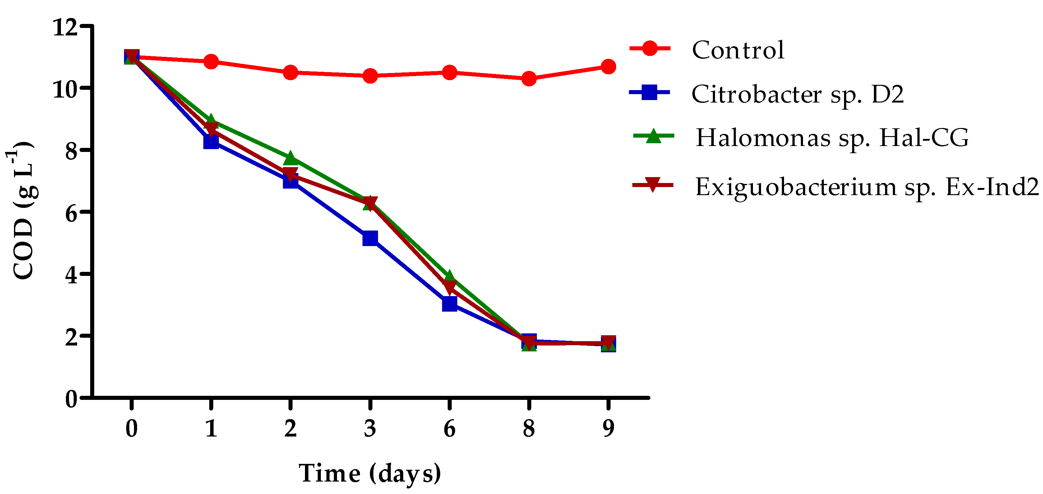
Further experiments were performed using raw BW as the substrate to investigate if the identified microorganisms could survive under this hostile environment. As shown in Figure 3, isolated strains in BW (initial 11 gCOD L−1) resulted in COD removal of around 84% after 11 days in all cultures. This result highlights the potential use of the isolated microorganisms for the effective removal of high range organic load.
However, when the initial COD of the BW was decreased (3.7 gCOD L−1) the COD removal by the three isolated strains was around 52–55% (Figure 4), indicating that the high range BW may contain greater fraction of easily biodegradable organic compounds than the low range BW.
Mazioti et al. [14] noted that isolated strains Halomonas alkaliphila S5a, Vibrio antiquarius S8ib and Pseudodonghicola xiamenensis S8iia had a COD removal around 61–77% after ten days, but the biodegradation was severely dependent on the initial COD of the BW. Their treatment efficiency decreased when the initial COD of BW was relatively low and this result is in line with the findings of the current study. In addition, Mazioti et al. [14] found that under sterile conditions, no significant decrease of the COD occurred over time in the control sample (raw bilge), while under nonsterile conditions, the COD decreased importantly due to biodegradation by indigenous microbes in BW. Therefore, the biodegradation potential also depends on the ability of the indigenous microbial community to remove the COD.

3.4. GC-MS Analysis of Real Bilge Water

GC-MS analysis was performed in order to identify the major organic compounds that can be removed by native isolated strains when raw BW was used as the sole carbon source. According to NIST database results, the detected compounds revealed various similarities, but only highly probability compounds are given in Table 1. Alkenes, brunched alkenes, aromatic molecules, ketones, solvents, etc., are only some examples. The results are consistent with our previous study on BW [32].
Methyl Isobutyl Ketone and heptane found in untreated bilge water (Table 1) were also identified by Vyrides et al. [32] in the bilge. From bilge sample, McLaughlin et al. [1] identified methyl ethyl ketone and Cazoir et al. [38] identified hexadecane. These compounds were similar to Methyl isobutyl Ketone and N-hexadecanoic acid that were identified in the current study. N-hexadecanoic acid was also found in the BW presenting high similarity, in accordance to other studies [39]. The detection of 2-Butoxyethanol (2-BE) found in BW (Table 1), is mainly used as a solvent in surface coatings, paints, varnishes and lubricants, oils, and dyes [40]. Woiski et al. [40] isolated 11 strains capable of entirely degrading 2-butoxyethanol (2-BE) isolated from forest soil, a biotrickling filter, a bioscrubber, and activated sludge. Eight of these strains belong to the genus Pseudomonas.
Figure 5b shows a significant area reduction in all the peaks due to the bilge biodegradation by Citrobacter sp. D2. As shown from Table 2 and Figure 5b, heptane was detected at high concentration in the influent BW, whereas after seven day was not detected to effluent sample. Moreover, several from the compounds identified on day seven were not detected in the influent BW, indicating that these products were the results of raw bilge biodegradation. Several of these compounds were octadecanoic acid, methyl ester and n-Hexadecanoic acid (Table 2). The major peaks (between Rt 35 and 55 min) were reduced to one third from the initial concentration. Compounds with Rt greater than 35 min usually belong to long-chain alkanes and PAHs. Furthermore, the compounds with Rt 10 min and 18.5 min decreased ten times from their initial concentration, indicating the removal of monoaromatics and VOC (i.e., heptane) from influent BW.
The chromatographs (Figure 5c) for Halomonas sp. Hal-CG show an overall reduction in all the peak areas and a reduction in the number of the detected compounds. Heptane was detected in both influent and effluent samples; however, in the effluent was detected a much lower concentration (approximately 10-fold reduction). In addition, Hexadecanoic acid, methyl ester and 2-Propanol, 1-butoxy- were only detected after seven days of exposure to the bilge to Halomonas sp Hal-CG. Those two compounds are usually detected after biological activity, as they are intermediate products of microbial metabolism.
Figure 5d shows biodegradation of Exiguobacterium sp. Ex-Ind2 when exposed to the bilge for seven days. The compound with Rt 37 min was reduced around 40.4%, whereas that at 44 min was presented around 28.9% removal. Moreover, new compounds were detected in the effluent BW, which were not present in the influent indicating degradation of compounds into other by-products.
On the one hand, the above findings highlighted the ability of isolated strains to fruitfully remove different chemicals form BW. On the other hand, it became evident that different strains present different preferences for hydrocarbons. In the case of Citrobacter sp D2, for example, GC-MS analysis indicated that major peaks having retention times between 10 and 18.5 min decreased about ten times compared to their initial concentrations (Figure 5a). In this time interval, monoaromatics and volatile organic compounds (i.e., heptane) are eluted. Additionally, from 35 to 55 min, peaks were reduced to about one third and these compounds usually belong to long-chain alkanes and PAHs.
Furthermore, identification of compounds only in culture samples revealed that by-products can be formed possibly due to breakdown of parent compounds or were solubilized due the biosurfactant production by microbes in BW.
The present results are in line with Vyrides et al. [25], who also detected new compounds after bilge water treatment using MBBRs inoculated with aerobic microbial consortium. The aerobic metabolism of hydrocarbons is typically initiated with the addition of oxygen to generate alcohol through an oxygenase enzyme. Depending on the position of the hydroxyl group, the alcohol is transformed into either an aldehyde or a ketone. Aldehydes are converted to acids through the addition of water across the carbon–oxygen double bond. Ketones can be oxidized to acetyl esters through the action of another oxygenase enzyme, while hydrolysis of the ester results in an acid and an alcohol [41]. Undoubtedly, future research is needed to investigate the metabolic pathways and identify the produced biotransformation products. This is the first study that pointed out that Citrobacter species could biodegrade bilge water. In addition, it shows the biodegradation potential of two indigenous species (Halomonas and Exiguobacterium sp). Furthermore, the performance of those strains in bilge water was investigated using GC-MS analysis before and after biodegradation.

4. Conclusions

This study isolation of Citrobacter species from oil-contaminated sites Halomonas and Exiguobacterium sp from BW was achieved using mainly phenanthrene as a carbon source. Bilge contains high aromatic compounds, and for this reason, phenanthrene was used as a representative compound for the isolation procedure. The isolated strains independently exposed to high range (11 gCOD L−1) and low range bilge water (3.7 gCOD L−1) pointed out around 83% and 53% COD removal, respectively. GC-MS analysis of untreated bilge water confirmed the presence of compounds that had a high similarity with Heptane, N-hexadecanoic acid, Methyl isobutyl Ketone, 1-butoxy- 2-propanol. When BW was exposed independently to isolated strains, the above compounds were reduced, and new compounds were detected that had a high similarity with N-hexadecanoic, methyl ester, N-hexadecanoic, ethyl ester, Octadecanoic acid methyl ester. The results of the present study indicated that the isolated strains might be promising for treating saline wastewaters of high COD rich in hydrocarbons.

Supplementary Materials

The following supporting information can be downloaded at: https://www.mdpi.com/article/10.3390/w14091350/s1, Table S1. Physicochemical properties of real bilge water before treatment; Table S2. Hydrocarbon index of bilge water before treatment; Table S3: BLASTn sequence alignment results for Citrobacter sp. D2; Table S4: BLASTn sequence alignment results for Citrobacter sp. S1; Table S5: BLASTn sequence alignments results Halomonas sp. Hal-CG; Table S6: BLASTn sequence alignment results for Halomonas sp. Hal-CG; Figure S1. Colony formation by Citrobacter sp. D2 strain in the presence of phenanthrene (150 mg L−1); Figure S2. Growth curve of Citrobacter sp. D2 in MSM medium containing phenanthrene (150 mg L−1); Figure S3. Phylogenetic tree diagram for Isolate D1 and D2. The evolutionary history was inferred using the Neighbor-Joining method [42]. The optimal tree is shown. The percentage of replicate trees in which the associated taxa clustered together in the bootstrap test (10,000 replicates) are shown next to the branches [43]. The tree is drawn to scale, with branch lengths in the same units as those of the evolutionary distances used to infer the phylogenetic tree. The evolutionary distances were computed using the Maximum Composite Likelihood method [44] and are in the units of the number of base substitutions per site. This analysis involved 9 nucleotide sequences. Codon positions included were 1st + 2nd + 3rd + Noncoding. All ambiguous positions were removed for each sequence pair (pairwise deletion option). There were a total of 791 positions in the final dataset. Evolutionary analyses were conducted in MEGA11 [45]; Figure S4. Phylogenetic tree diagram for islolate Hal-CG. The evolutionary history was inferred using the Neighbor-Joining method [42]. The optimal tree is shown. The percentage of replicate trees in which the associated taxa clustered together in the bootstrap test (10,000 replicates) are shown next to the branches [43]. The tree is drawn to scale, with branch lengths in the same units as those of the evolutionary distances used to infer the phylogenetic tree. The evolutionary distances were computed using the Maximum Composite Likelihood method [44] and are in the units of the number of base substitutions per site. This analysis involved 7 nucleotide sequences. Codon positions included were 1st + 2nd + 3rd + Noncoding. All ambiguous positions were removed for each sequence pair (pairwise deletion option). There were a total of 330 positions in the final dataset. Evolutionary analyses were conducted in MEGA11 [45]; Figure S5. Phylogenetic tree diagram for islolate Ex-Ind2. The evolutionary history was inferred using the Neighbor-Joining method [42]. The optimal tree is shown. The percentage of replicate trees in which the associated taxa clustered together in the bootstrap test (10,000 replicates) are shown next to the branches [43]. The tree is drawn to scale, with branch lengths in the same units as those of the evolutionary distances used to infer the phylogenetic tree. The evolutionary distances were computed using the Maximum Composite Likelihood method [44] and are in the units of the number of base substitutions per site. This analysis involved 7 nucleotide sequences. Codon positions included were 1st + 2nd + 3rd + Noncoding. All ambiguous positions were removed for each sequence pair (pairwise deletion option). There were a total of 330 positions in the final dataset. Evolutionary analyses were conducted in MEGA11 [45]; Figure S6. Concentration of dissolved phenanthrene during its biodegradation by Citrobacter sp. D2; Figure S7. Growth of Citrobacter sp. D2 under different pH conditions in the presence of phenanthrene (150 mg L−1) and NaCl (15 g L−1) at 30 °C; Figure S8. Growth of Citrobacter sp. D2 under different temperatures in the presence of phenanthrene (150 mg L−1) and NaCl (15 g L−1); Figure S9. Growth of Citrobacter sp. D2 under different nutrient conditions in the presence of phenanthrene (150 mg L−1) and NaCl (15 g L−1).

Author Contributions

G.G.: Conceptualization, investigation, methodology, writing—original draft. E.-M.D.: Investigation. I.V.: Conceptualization, project administration, writing—review and editing. All authors have read and agreed to the published version of the manuscript.

Funding

The study has received funding from the European Union’s Horizon 2020 research and innovation programme under the Marie Sklodowska-Curie grant agreement No 841797.

Institutional Review Board Statement

Not applicable.

Informed Consent Statement

Not applicable.

Data Availability Statement

The datasets used and/or analyzed during the current study are available from the corresponding author on reasonable request.

Acknowledgments

The authors would like to thank Ecofuel LTD for the provision of the wastewater.

Conflicts of Interest

The authors declare no conflict of interest. The funders had no role in the design of the study, in the collection, analyses, or interpretation of data, in the writing of the manuscript, or in the decision to publish the results.

References

  1. McLaughlin, C.; Falatko, D.; Danesi, R.; Albert, R. Characterizing shipboard bilgewater effluent before and after treatment. Environ. Sci. Pollut. Res. 2014, 21, 5637–5652. [Google Scholar] [CrossRef] [PubMed]
  2. IMO. International Convention for the Prevention of Pollution from Ships (MARPOL). 1973. Available online: https://www.imo.org/en/About/Conventions/Pages/International-Convention-for-the-Prevention-of-Pollution-from-Ships-(MARPOL).aspx (accessed on 15 March 2022).
  3. Gryta, M. Separation of saline oily wastewater by membrane distillation. Chem. Pap. 2020, 74, 2277–2286. [Google Scholar] [CrossRef] [Green Version]
  4. Han, M.; Zhang, J.; Chu, W.; Chen, J.; Zhou, G. Research progress and prospects of marine oily wastewater treatment: A review. Water 2019, 11, 2517. [Google Scholar] [CrossRef] [Green Version]
  5. Church, J.; Lundin, J.G.; Diaz, D.; Mercado, D.; Willner, M.R.; Lee, W.H.; Paynter, D.M. Identification and characterization of bilgewater emulsions. Sci. Total Environ. 2019, 691, 981–995. [Google Scholar] [CrossRef] [PubMed]
  6. Tummons, E.N.; Tarabara, V.V.; Chew, J.W.; Fane, A.G. Behavior of oil droplets at the membrane surface during crossflow microfiltration of oil-water emulsions. J. Membr. Sci. 2016, 500, 211–224. [Google Scholar] [CrossRef]
  7. Rincón, G.J.; La Motta, E.J. Simultaneous removal of oil and grease, and heavy metals from artificial bilge water using electro-coagulation/flotation. J. Environ. Manag. 2014, 144, 42–50. [Google Scholar] [CrossRef]
  8. Corti-Monzón, G.; Nisenbaum, M.; Villegas-Plazas, M.; Junca, H.; Murialdo, S. Enrichment and characterization of a bilge microbial consortium with oil in water-emulsions breaking ability for oily wastewater treatment. Biodegradation 2020, 31, 57–72. [Google Scholar] [CrossRef]
  9. Nisenbaum, M.; Corti-Monzón, G.; Villegas-Plazas, M.; Junca, H.; Mangani, A.; Patat, M.L.; Gonzá, J.F.; Murialdo, S.E. Enrichment and key features of a robust and consistent indigenous marine-cognate microbial consortium growing on oily bilge wastewaters. Biodegradation 2020, 31, 91–108. [Google Scholar] [CrossRef]
  10. Fowler, S.J.; Toth, C.R.A.; Gieg, L.M. Community structure in methanogenic enrichments provides insight into syntrophic interactions in hydrocarbon-impacted environments. Front. Microbiol. 2016, 7, 1–13. [Google Scholar] [CrossRef] [Green Version]
  11. Mazioti, A.A.; Notarides, G.; Symeou, G.; Vyrides, I. Improving biological treatment of real bilge wastewater with zero valent iron and activated charcoal addition. Front. Bioeng. Biotechnol. 2020, 8, 1462. [Google Scholar] [CrossRef]
  12. Gouveia, V.; Almeida, C.M.R.; Almeida, T.; Teixeira, C.; Mucha, A.P. Indigenous microbial communities along the NW Portuguese Coast: Potential for hydrocarbons degradation and relation with sediment contamination. Mar. Pollut. Bull. 2018, 131, 620–632. [Google Scholar] [CrossRef] [PubMed]
  13. Lee, D.W.; Lee, H.; Kwon, B.-O.; Khim, J.S.; Yim, U.H.; Kim, B.S.; Kim, J.J. Biosurfactant-assisted bioremediation of crude oil by indigenous bacteria isolated from Taean beach sediment. Environ. Pollut. 2018, 241, 254–264. [Google Scholar] [CrossRef] [PubMed]
  14. Mazioti, A.A.; Vasquez, M.I.; Vyrides, I. Comparison of different cultures and culturing conditions for the biological deterioration of organic load from real saline bilge wastewater: Microbial diversity insights and ecotoxicity assessment. Environ. Sci. Pollut. Res. 2021, 28, 36506–36522. [Google Scholar] [CrossRef] [PubMed]
  15. Uma, V.; Gandhimathi, R. Organic removal and synthesis of biopolymer from synthetic oily bilge water using the novel mixed bacterial consortium. Bioresour. Technol. 2019, 273, 169–176. [Google Scholar] [CrossRef]
  16. Feknous, N.; Branes, Z.; Rouabhia, K.; Batisson, I.; Amblard, C. Isolation characterization and growth of locally isolated hydrocarbonoclastic marine bacteria (Eastern Algerian Coast). Environ. Monit. Assess. 2017, 189, 49. [Google Scholar] [CrossRef]
  17. Primeia, S.; Inoue, C.; Chien, M.F. Potential of biosurfactants’ production on degrading heavy oil by bacterial consortia obtained from tsunami-induced oil-spilled beach areas in Miyagi, Japan. J. Mar. Sci. Eng. 2020, 8, 577. [Google Scholar] [CrossRef]
  18. Rizzo, C.; Rappazzo, A.C.; Michaud, L.; De Domenico, E.; Rochera, C.; Camacho, A.; Lo Giudice, A. Efficiency in hydrocarbon degradation and biosurfactant production by Joostella sp. A8 when grown in pure culture and consortia. J. Environ. Sci. 2018, 67, 115–126. [Google Scholar] [CrossRef]
  19. Boraha, D.; Yadav, R.N.S. Bioremediation of petroleum based contaminants with biosurfactant produced by a newly isolated petroleum oil degrading bacterial strain. Egypt. J. Pet. 2017, 26, 181–188. [Google Scholar] [CrossRef] [Green Version]
  20. Liu, H.; Yang, G.; Jia, H.; Sun, B. Crude Oil Degradation by a Novel Strain Pseudomonas aeruginosa AQNU-1 Isolated from an Oil-Contaminated Lake Wetland. Processes 2022, 10, 307. [Google Scholar] [CrossRef]
  21. Lenchi, N.; Kebbouche-Gana, S.; Servais, P.; Gana, M.L.; Lliros, M. Diesel biodegradation capacities and biosurfactant production in saline-alkaline conditions by Delftia sp. NL1, Isolated from an Algerian Oilfield. Geomicrobiol. J. 2020, 37, 454–466. [Google Scholar] [CrossRef]
  22. Olivera, N.L.; Commendatore, M.G.; Delgado, O.; Esteves, J.L. Microbial characterization and hydrocarbon biodegradation potential of natural bilge waste microflora. J. Ind. Microbiol. Biotechnol. 2003, 30, 542–548. [Google Scholar] [CrossRef] [PubMed]
  23. Santisi, S.; Gentile, G.; Volta, A.; Bonsignore, M.; Mancini, G.; Quatrini, P.; Cappello, S. Isolation and Characterization of Oil-Degrading Bacteria from Bilge Water. Int. J. Microbiol. Appl. 2015, 2, 45–49. [Google Scholar]
  24. Nievas, M.L.; Commendatore, M.G.; Olivera, N.L.; Esteves, J.L.; Bucala, V. Biodegradation of bilge waste from Patagonia with an indigenous microbial Community. Bioresour. Technol. 2006, 97, 2280–2290. [Google Scholar] [CrossRef] [PubMed]
  25. Sivaraman, C.; Ganguly, A.; Nikolausz, M.; Mutnuri, S. Isolation of hydrocarbonoclastic bacteria from bilge oil contaminated water. Int. J. Environ. Sci. Technol. 2011, 8, 461–470. [Google Scholar] [CrossRef] [Green Version]
  26. Cappello, S.; Santisi, S.; Calogero, R.; Hassanshahian, M.; Yakimov, M.M. Characterisation of Oil-Degrading Bacteria Isolated from Bilge Water. Water Air Soil Pollut. 2012, 223, 3219–322628. [Google Scholar] [CrossRef]
  27. Corti-Monzón, G.; Nisenbaum, M.; Peressutti, S.; Junca, H.; García-Bonilla, E.; Murialdo, S.E. Oily Bilge Wastes Harbor a Set of Persistent Hydrocarbonoclastic Bacteria Accompanied by a Variable alkB Gene Composition in Marine Vessel Samples from Southwestern Atlantic Port of Mar del Plata, Argentina. Water Air Soil Pollut. 2021, 232, 1–19. [Google Scholar] [CrossRef]
  28. Tiselius, P.; Magnusson, K. Toxicity of treated bilge water: The need for revised regulatory control. Mar. Pollut. Bull. 2017, 114, 860–866. [Google Scholar] [CrossRef]
  29. Drakou, E.; Koutinas, M.; Pantelides, I.; Tsolakidou, M.; Vyrides, I. Insights into the metabolic basis of the halotolerant Pseudomonas aeruginosa strain LVD-10 during toluene biodegradation. Int. Biodeter. Biodegr. 2015, 99, 85–94. [Google Scholar] [CrossRef]
  30. Koutinas, M.; Kyriakou, M.; Andreou, K.; Hadjicharalambous, M.; Kaliviotis, E.; Pasias, D.; Kazamias, G.; Varavvas, C.; Vyrides, I. Enhanced biodegradation and valorization of drilling wastewater via simultaneous production of bio-surfactants and polyhydroxyalkanoates by Pseudomonas citronellolis SJTE-3. Bioresour. Technol. 2021, 340, 340125679. [Google Scholar] [CrossRef]
  31. APHA. Standard Methods for the Examination of Water and Wastewater, 21st ed.; American Public Health Association: Washington, DC, USA, 2005. [Google Scholar]
  32. Vyrides, I.; Drakou, E.-M.; Ioannou, S.; Michael, F.; Gatidou, G.; Stasinakis, A.S. Biodegradation of bilge water: Batch test under anaerobic and aerobic conditions and performance of three pilot aerobic Moving Bed Biofilm Reactors (MBBRs) at different filling fractions. J. Environ. Manag. 2018, 217, 356. [Google Scholar] [CrossRef]
  33. Deng, T.; Wang, H.; Yan, K. Phenol biodegradation by isolated Citrobacter strain under hypersaline conditions. Water Sci. Technol. 2018, 77, 504–510. [Google Scholar] [CrossRef] [PubMed]
  34. Adebajo, S.O.; Akintokun, A.K.; Ojo, A.E.; Dami, M.; Egbagbe, D.M.; Akintokun, P.O.; Adebajo, L.O. Biosurfactant Production by Rhizospheric Bacteria Isolated from Biochar Amended Soil Using Different Extraction Solvents. Appl. Environ. Res. 2019, 41, 72–82. [Google Scholar] [CrossRef]
  35. Anaukwu, C.G.; Ezemba, C.C.; Anakwenze, V.N.; Agu, K.C.; Okeke, B.C.; Awah, N.S.; Ekwealor, I.A. Effect of biosurfactant produced by Citrobacter murliniaeAF025369 and a synthetic surfactant on degradation of crude oil. Edorium. J. Microbiol. 2016, 1, 1–6. [Google Scholar]
  36. Kaczorek, E.; Sałek, K.; Guzi, U.; Dudzinska-Bajorek, B. Cell surface properties and fatty acids composition of Stenotropho-monas maltophilia under the influence of hydrophobic compounds and surfactants. New Biotechnol. 2013, 30, 173–182. [Google Scholar] [CrossRef] [PubMed]
  37. Tang, J.; He, J.; Liu, T.; Xin, X.; Hu, H. Removal of heavy metal from sludge by the combined application of a biodegradable biosurfactant and complexing agent in enhanced electrokinetic treatment. Chemosphere 2017, 189, 599–608. [Google Scholar] [CrossRef] [PubMed]
  38. Cazoir, D.; Fine, L.; Ferronato, C.; Chovelon, J.M. Hydrocarbon removal from bilgewater by a combination of air-stripping and photocatalysis. J. Hazard. Mater. 2012, 235–236, 159–168. [Google Scholar] [CrossRef]
  39. Shen, Y.; Yao, J.; Son, J.; Zhu, Z.; Yu, X.Y. Liquid ToF-SIMS revealing the oil, water, and surfactant interface evolution. Phys. Chem. Chem. Phys 2020, 22, 11771–11782. [Google Scholar] [CrossRef]
  40. Woiski, C.; Dobslaw, D.; Engesser, K.-H. Isolation and characterization of 2-butoxyethanol degrading bacterial strains. Biodegradation 2020, 31, 153–169. [Google Scholar] [CrossRef]
  41. O’Reilly, K.T.; Mohler, R.E.; Zemo, D.A.; Ahn, S.; Tiwary, A.K.; Magaw, R.I.; Devine, C.E.; Synowiec, K.A. Identification of ester metabolites from petroleum hydrocarbon biodegradation in groundwater using GC x GC-TOFMS. Environ. Toxicol. Chem. 2015, 34, 1959–1961. [Google Scholar] [CrossRef]
  42. Saitou, N.; Nei, M. The neighbor-joining method: A new method for reconstructing phylogenetic trees. Mol. Biol. Evol. 1987, 4, 406–425. [Google Scholar]
  43. Felsenstein, J. Confidence limits on phylogenies: An approach using the bootstrap. Evolution 1985, 39, 783–791. [Google Scholar]
  44. Tamura, K.; Nei, M.; Kumar, S. Prospects for inferring very large phylogenies by using the neighbor-joining method. Proc. Natl. Acad. Sci. USA 2004, 101, 11030–11035. [Google Scholar]
  45. Tamura, K.; Stecher, G.; Kumar, S. MEGA 11: Molecular Evolutionary Genetics Analysis Version 11. Mol. Biol. Evol. 2021, 38, 3022–3027. [Google Scholar] [CrossRef]
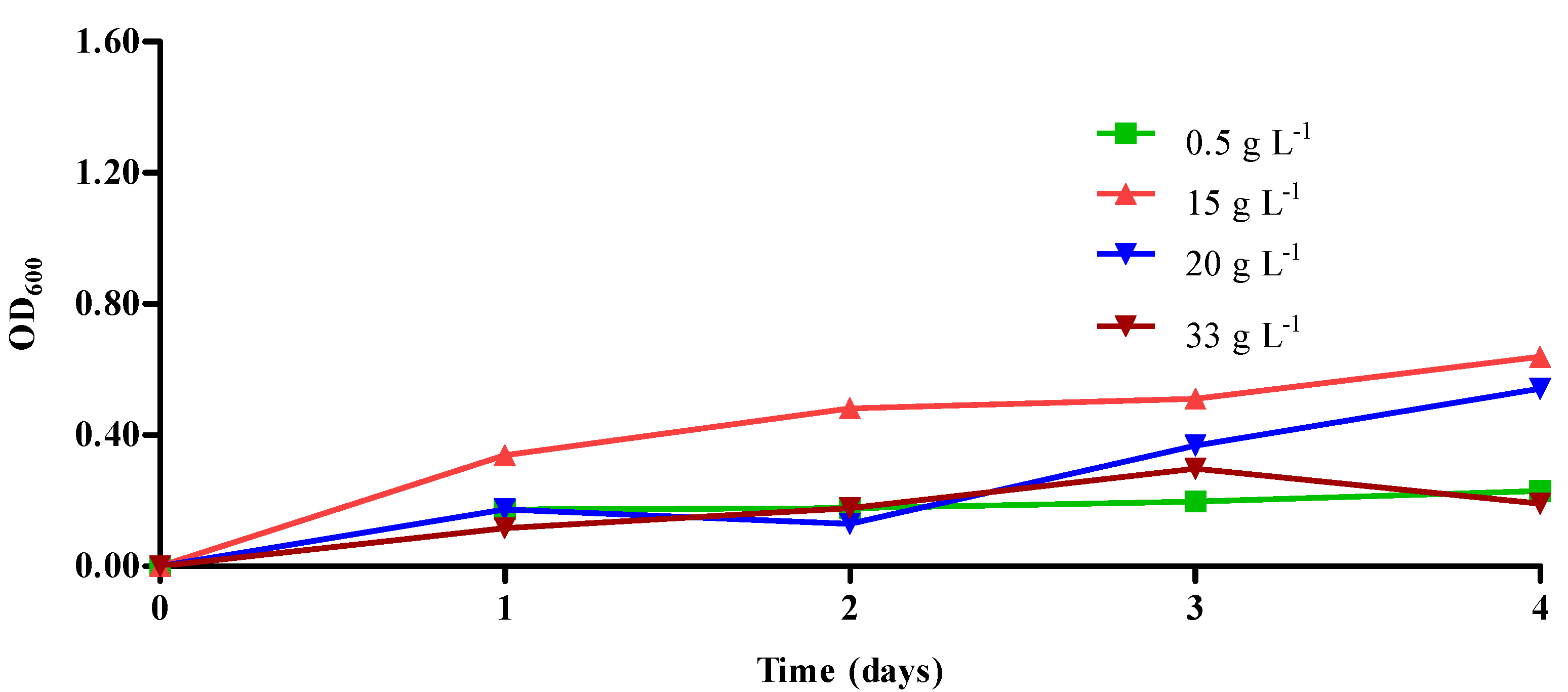
Figure 1. Growth of Citrobacter sp. D2 under different salinity conditions in the presence of 150 mg L−1 of phenanthrene.
Water 14 01350 g001
Figure 2. Biodegradation of phenanthrene (C0 = 150 mg L−1) over time by Citrobacter sp. D2 under different salinity conditions.
Water 14 01350 g002
Figure 3. Decrease of COD concentration (g L−1) over time in bilge water (high range COD) after cultivation with isolated bacterial strains.
Water 14 01350 g003
Figure 4. Decrease of COD concentration (g L−1) over time in bilge water (low range) after cultivation with isolated bacterial strains.
Water 14 01350 g004
Figure 5. Chromatographic analysis by GC-MS of: (a) real untreated BW and after 7 days treatment with (b) Citrobacter sp. D2, (c) Halomonas sp. Hal-CG, (d) Exiguobacterium sp. Ex-Ind2. X axis: Time (min); Y axis: Abundance.
Water 14 01350 g005
Table 1. Major organic compounds detected in real bilge water (BW) after GC-MS analysis.
NameMolecular FormulaRTProbability (%)
HeptaneC7H167.0581.8
Methylyclohexane (toluene)C7H147.6855.6
2,3,4-trimethyl-pentaneC8H188.4355.4
3-methyl-heptane (octane)C8H189.1659
OctaneC8H1810.1465.3
1-butoxy-2-propanolC7H16O215.2693.7
2-ethyl-1-hexanolC8H18O18.3671.6
Methyl isobutyl ketoneC6H12O18.9292.9
2-methyl-phenolC7H8O19.3159.0
AcetophenoneC8H8O19.7152.3
PentadecaneC15H3231.9852.5
HexadecaneC16H3434.4256.3
Hexadecanoic acid, methyl ester (palmitic acid)C17H34O241.4083.9
DocosaneC22H4646.5558.5
TricosaneC23H4848.2656.0
Table 2. Major organic compounds detected in untreated and treated bilge water by isolated bacterial strains after GC-MS analysis. % Probability according to NIST 5 Library is given in the parenthesis.
CompoundUntreated BWCitrobacter
sp. D2.
Exiguobacterium
sp. Ex-Ind2
Halomonas
sp. Hal-CG
Heptane32008407
(81.8%)
n.d.14717758
(80,7%)
6195579
(83.7%)
Hexane n.d.5182909n.d.n.d.
N-hexadecanoic acid 2092353
(83.9%)
n.d.n.d.n.d.
N-hexadecanoic, methyl ester n.d.37200692
(80%)
5698795
(81.0)
n.d.
N-hexadecanoic, ethyl estern.d.2945055
(86.4%)
14717758
(83.2%)
4921636
(87.4%)
Methyl isobutyl Ketone8198875
(92.9%)
n.d.n.d.n.d.
1-butoxy-2-propanol728060
(93.7%)
n.d.n.d.n.d.
Octadecanoic acid methyl estern.d.9251735
(82.7%)
4955474
(84.1%)
2722022
(80.5%)
Publisher’s Note: MDPI stays neutral with regard to jurisdictional claims in published maps and institutional affiliations.