Article

Effects of Temperature on the Growth Performance, Biochemical Indexes and Growth and Development-Related Genes Expression of Juvenile Hybrid Sturgeon (Acipenser baerii♀ × Acipenser schrenckii♂)

1
Faculty of Animal Science and Technology, Plateau Aquacultural College, Yunnan Agricultural University, Kunming 650201, China
2
Key Laboratory of Plateau Fishery Resources Protection and Sustainable Utilization of Yunnan Province, Yunnan Agricultural University, Kunming 650201, China
*
Authors to whom correspondence should be addressed.
Water 2022, 14(15), 2368; https://doi.org/10.3390/w14152368
Submission received: 19 June 2022 / Revised: 28 July 2022 / Accepted: 28 July 2022 / Published: 31 July 2022
(This article belongs to the Special Issue Effect of Aquatic Environment on Fish Ecology)

Abstract

:
In order to investigate the effects of temperature on the growth performance, biochemical indexes and genes expression in juvenile hybrid sturgeon (Acipenser baerii♀ × Acipenser schrenckii♂), five temperature treatment groups (18 °C, 21 °C, 24 °C, 27 °C, 30 °C) were set in this study. After 25 days of domestication, the specific growth rate (SGR) and relative weight gain rate of juvenile sturgeon increased at first and then decreased with the increase in temperature in the range of 18–30 °C and reached the highest value at 21 °C. A quadratic equation was used to fit the regression equation of SGR and temperature (T). The result showed that the temperature of the maximum SGR is 23.45 °C. Moreover, the expression level of the growth hormone (gh) and the content of blood glucose (GLU) increased significantly at 21 °C, while the expression level of the growth hormone receptor (ghr) in the liver decreased significantly. When the temperature exceeded 27 °C, the thyroid hormone levels were significantly affected, and the levels of total antioxidants (T-AOC) and reduced glutathione (GSH) were significantly higher than those in the control group. In addition, with the increase in temperature, the expression levels of hsp70 and hsp90 in the liver increased significantly, while the expression level of the luteinizing hormone (lh) in the brain decreased significantly. To summarize, the effect of temperature on the growth and development of juvenile sturgeon mainly occurs through the effects of glucose metabolism, thyroid hormone level, total antioxidant capacity and growth-related genes. Therefore, in a temperature range between 21 and 24 °C, juvenile sturgeon can obtain the maximum growth rate and survival rate.

1. Introduction

As an important environmental factor, temperature can directly affect the stability of chemical bonds, the chemical reaction rate, enzyme activity and biofilm permeability, which are very important for the survival of organisms. In particular, as a typical ectothermic animal, fish are sensitive and highly dependent on ambient temperature, and their body temperature fluctuates with the change in the water temperature. Therefore, temperature has become a very important environmental and ecological factor affecting the physiological process of fish.
Water temperature has a multifaceted effect on feeding, growth, development, metabolism, reproduction and immunity in fish [1]. High temperatures for prolonged periods augmented blood glucose (GLU), triglycerides (TG) and total protein (TP) levels in European seabass (Dicentrarchus labrax) [2]. When the ambient temperature is altered, it causes changes in the metabolic pathway and blood biochemical indexes of fish [3]. In addition, heat stress will form a large number of reactive oxygen species (ROS), resulting in oxidative stress in organisms, thus causing cell damage. The heat shock protein (HSP) plays an important role in oxidative stress. As a sensor of the redox reaction, it can activate superoxide dismutase (SOD), catalase (CAT) and peroxidase to detoxify and repair the damage [4]. Meanwhile, ROS significantly increased the expression of HSPs [5]. Therefore, temperature fluctuation can affect the antioxidant defense system of fish, resulting in physiological metabolism abnormalities in fish.
Temperature affects the thyroid function of fish, including thyroxine (T4), free thyroxine (FT4), triiodothyronine (T3) and free triiodothyronine (FT3), thus affecting the growth, development and survival of fish. An increase in temperature enhances the degradation rate and internal conversion rate of T4 and T3 of trout and European eel (Anguilla anguilla) [6]. Conversely, a too-low water temperature could affect the growth of Atlantic Cod (Gadus morhua) by inhibiting part of the function of the thyroid gland [6]. In addition, temperature also affects the gene expression of the growth hormone (GH)/insulin-like growth factor (IGF) system. During the embryonic development of rainbow trout (Oncorhynchus mykiss), ghr promoted growth through the effect of temperature on the plasma growth hormone (GH), and igf-II also had a temperature-mediated effect on growth [7,8]. Therefore, there is a close relationship between temperature and thyroid function, gene expression of the GH/IGF system and growth.
Fish sense temperature fluctuations naturally, collect signals and transmit them to the brain through the hypothalamus–pituitary–gonad (HPG) axis and secrete various hormones to regulate physiological effects. When the water temperature rises continuously, the maturation process of goldfish (Carassius auratus L.) oocytes is significantly accelerated, and ovulation occurs [9]. However, cold-water fish breeding in autumn and winter can accelerate ovarian maturation and advance spawning time with the decrease in water temperature, while ovarian maturation and spawning are delayed in higher water temperatures [10]. Therefore, the water temperature can significantly affect the gonadal development, maturation and ovulation of fish.
In addition, gonadal development is regulated not only by temperature but also by genes, especially the synthesis of the gonadotropic hormone (GtH) at different stages of gonadal development in fish. As the important component of GtH, the follicle-stimulating hormone (FSH) and luteinizing hormone (LH) regulate gonadal development and stimulate estrogen production, which are mediated by their receptors and thus regulate their expression [11]. During the vitellogenesis of O. mykiss, the levels of plasma FSH and LH increased significantly. It has been proven that the gene expression levels of fsh and lh in the pituitary are similar to those of plasma FSH and LH [12]. The embryo survival rate of Atlantic salmon (Salmo salar) and O. mykiss decreased after high-temperature treatment during vitellogenesis [13]. Similarly, the study of female pejerrey (Odontesthes bonariensis) showed that the levels of fshr and E2 in short-term temperature treatment (23 °C, 27 °C) were lower than those in the control group (19 °C) [14]. When S. salar is raised in high temperatures, the binding affinity of ers in the liver decreases, which may be affected by the high temperature in the process of signal transduction downstream [13]. The expression of vtg is stimulated by estrogen. Studies have shown that fish exposed to E2 can better respond to the effect of temperature on Vitellogenin (VTG) [15]. When O. mykiss is exposed to E2 at 14 °C and 22 °C, VTG is induced faster at 22 °C [16]. Therefore, temperature can have an effect on the gonadal development, vitellogenesis and ovulation of fish. However, there are relatively few reports on the effect of temperature on the early gonadal development of fish, especially sturgeon, which have a longer gonadal development cycle. It is very important to study the regulation of temperature on the gonadal development of sturgeon.
At present, sturgeon is one of the earliest vertebrates and it is also one of the most primitive groups of fish. There are approximately 2 families, 6 genera and 27 species of sturgeon in the world, most of which are endangered and belong to worldwide protected species. However, the flood of cultured sturgeon germplasm, the decline of growth performance, the backward scale of seedling production, culture methods and water environment and other problems restrict the healthy development of the sturgeon industry in Yunnan. Among them, the environmental factors affecting the growth and development of sturgeon mainly include temperature, salinity, light, dissolved oxygen, pH, food richness and so on. Therefore, combined with the rich cold-water resources and topographic conditions in Yunnan, this study carried out a more systematic study on the effects of different temperatures on the regulation of juvenile sturgeon growth and development, in order to enrich the biological data and summarize the optimum water temperature for growth of juvenile sturgeon. It has important reference significance for the production practice and disease prevention of juvenile sturgeon culture and promotes the sustainable development of cold-water fisheries in Yunnan.

2. Materials and Methods

2.1. Experimental Fish

Juvenile sturgeon were purchased from Huize Dianze Aquaculture Company of Yunnan. After being temporarily cultured in the Aquatic Laboratory of Yunnan Agricultural University for 7 d (10–12 °C), individuals with healthy, dynamic and uniform initial weight (8.63 ± 0.17 g) were selected for the temperature domestication experiment. The experimental animals used in this experiment are strictly in accordance with the requirements of the guidelines for the use of Experimental Animals of Yunnan Agricultural University and were approved by the Experimental Ethics Committee of Yunnan Agricultural University (202203026).

2.2. Experimental Design

Five domestication temperatures (18 °C, 21 °C, 24 °C, 27 °C and 30 °C) were set to domesticate juvenile sturgeon. A total of 225 juvenile sturgeons were selected, and three parallel groups of 15 juveniles were set in each treatment group. The experiment was carried out under the conditions of a natural photoperiod in the laboratory. The test temperature control system was an indoor circulating water type, each group of aquariums had 3 parallel water tanks and the water temperature of each tank was kept the same. Before reaching 27 °C, a rise of 1 °C/h was implemented, after which a rise of 0.5 °C/h was used. After the acclimation temperature rises to the set temperature, the domestication experiment was started and lasted 25 d. During the experiment, the water was changed twice a day, and the daily water exchange rate was 50%. The commercial diet (42% crude protein, 12% crude lipid, 6.0% crude fiber, supplied by Kunming Evergrande Feed Co. Ltd., Kunming, China) was fed twice a day at 10:00 and 19:00, and the bottom was cleaned 1 hour after feeding. Water quality parameters were as follows: pH (7–8.5), ammonia nitrogen less than 0.5 mg/L, nitrite less than 0.1 mg/L and dissolved oxygen content more than 6 mg/L. During the experiment, the daily behavior of experimental fish was observed and recorded, the weight of 15 juveniles in each parallel group was measured every 5 d and the average weight gain of each group was calculated.

2.3. Sample Collection

After fasting for 24 hours, 5 random fish in each tank were anesthetized with MS-222 (tricaine methane sulfonate, Sigma, St. Louis, MO, USA) and weighed (g). Blood was taken from the caudal artery with a disposable aseptic syringe and placed in a 1.5 mL centrifuge tube. After standing at 4 °C overnight, the supernatant was collected (centrifuged at 1500× g for 30 min) and stored at −80 °C for the determination of plasma biochemical indexes and hormones. Then, the fish were dissected, and liver and brain tissue were placed into a 2.0 mL RNase-free centrifuge tube and stored at −80 °C for the determination of gene expression.

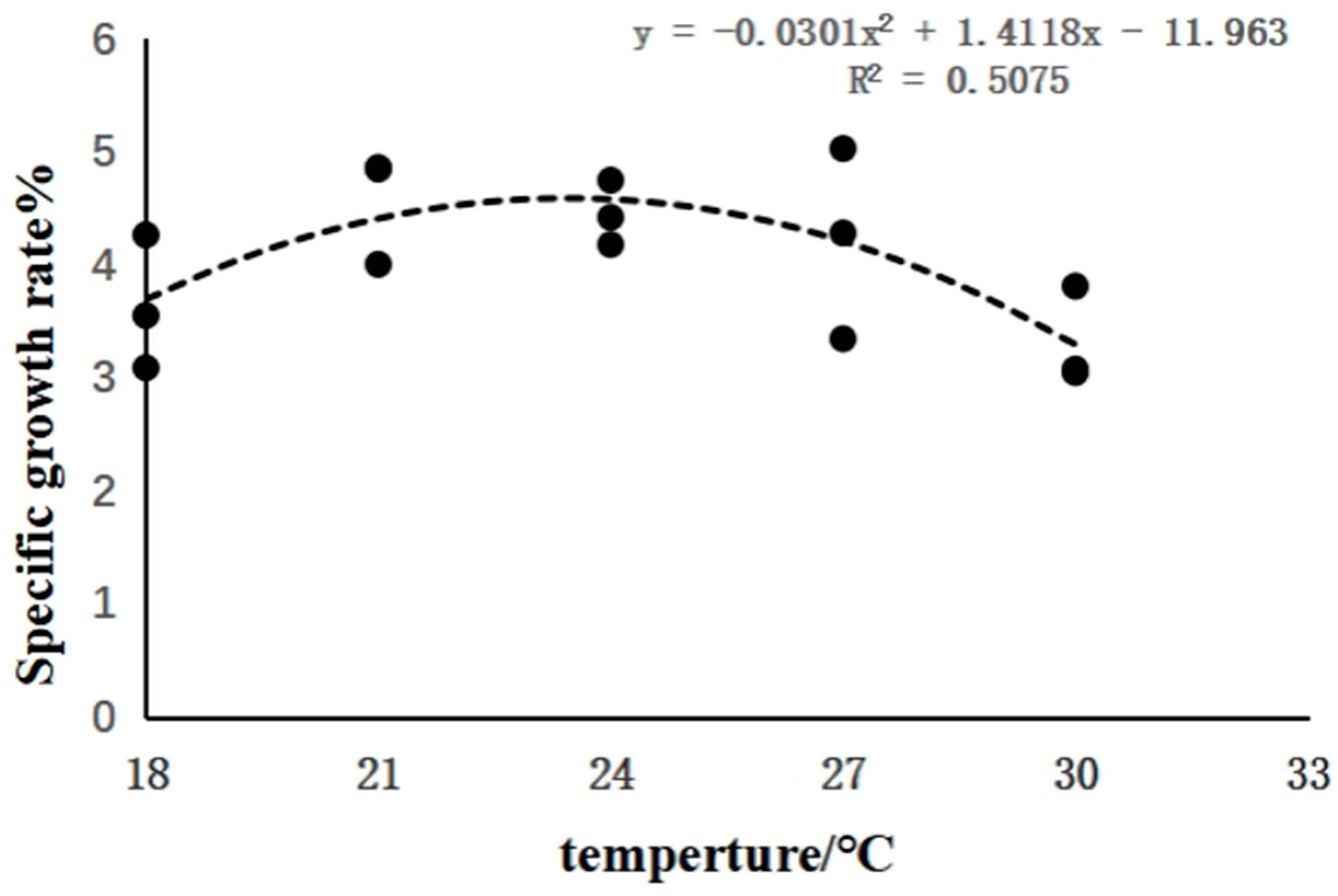
2.4. Determination of SGR and RWG

After 25 d of domestication, the final weight of the juvenile sturgeon was obtained by weighing. The specific growth rate (SGR) and relative weight gain rate (RWG) were calculated according to the following formula:
SGR = 100% × (lnW2 − lnW1)/(T2 − T1)
RWG = 100% × (W2 − W1)/W1
In the formula, W1 and W2 are the average body weight (g) of time T1 and T2.

2.5. Determination of Plasma Biochemical Indexes and Hormones

Total cholesterol (TC), reduced glutathione (GSH), TG, GLU, CAT, total antioxidant (T-AOC) and malondialdehyde (MDA) kits were purchased from Beijing Box Shenggong Technology Co., Ltd. (Beijing, China). The TP kit was purchased from Beijing Coolaber Science Technology Co., Ltd. (Beijing, China). Plasma Na+ and Cl kits were purchased from Nanjing Jiancheng Biochemical Corporation (Nanjing, China). T3 and FT3 kits were purchased from the Beijing Northern Institute of Biotechnology (Beijing, China). Fish cortisol kits were purchased from Shanghai Enzymatic Biotechnology (Shanghai, China). The measurement methods of these indicators are carried out according to the manufacturer’s instructions.

2.6. RNA Extraction and cDNA Synthesis

The total RNA of the liver and brain was extracted according to the total RNA Isolation Reagent Kit (Biosharp, China) instructions. The concentration and purity of total RNA were determined by an ultramicro spectrophotometer (NanoPhotometer 80 Touch, Germany). The A260 nm/A280 nm was between 1.8 and 2.0. One microgram of total RNA was reverse-transcribed into cDNA by the Goldenstar reverse RT6 cDNA Synthesis Kit reverse transcription kit (Qingke Biotechnology Co., Ltd., Kunming, China), which was used for the determination of gene expression.

2.7. Gene Expression

Real-time fluorescence quantification was carried out under CFX ConnectTM Optics Module (Bio-Rad, Hercules, CA, USA). The primer sequence is shown in Table 1. The reaction system is 20 μL: 2× T5 Fast qPCR mix (SYBP Green I, Beijing TsingKe Biotech Co., Ltd., Beijing, China) 10 μL, 1 μL of forward primer and reverse primer, respectively, cDNA template 2 μL, sterile water 6 μL. Each treatment group was repeated three times, and each gene had a negative control (excluding cDNA). The reaction conditions are as follows: Pre-denaturation at 95 °C for 3 min, then denaturation at 95 °C for 20 s, annealing at 53–57 °C for 20 s (Table 1), extension at 72 °C for 20 s, cycle for 40 s and, finally, 10 min extension at 72 °C. The 18sRNA was used as the internal control to calculate the relative expression of the target gene by the 2−△△Ct method [17].

2.8. Statistical Analysis

SAS 9.2 software (IBM Inc., Cary, NC, USA) was used to test the normality and homogeneity of variance of the data, and then the Tukey method of one-way analysis of variance (ANOVA) was used to evaluate the differences in values among the treatment groups. A value of p < 0.05 was considered statistically significant. All data were expressed as mean ± standard deviation (SD).

3. Results

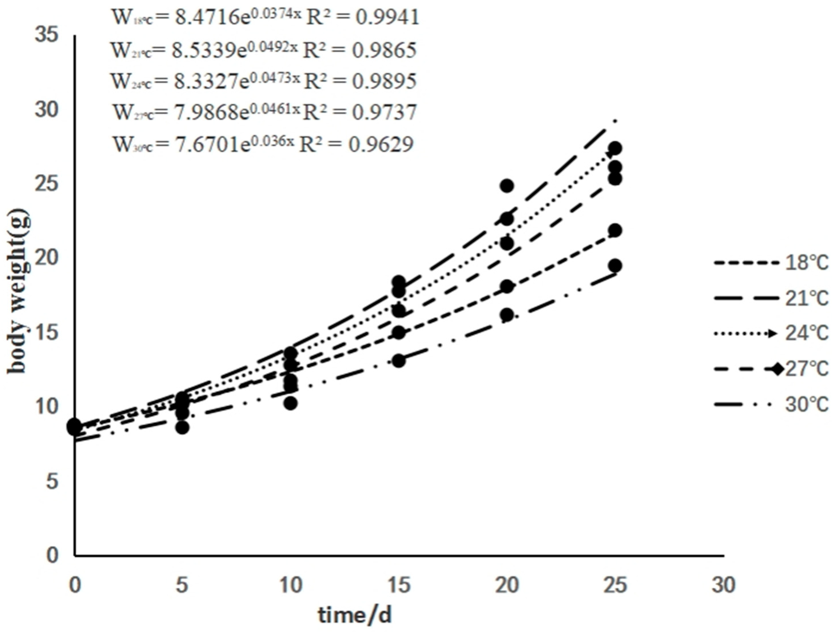
3.1. The Effects of Rearing Temperature on the Growth of Juvenile Sturgeon

As the rearing temperature increased, the final weight, SGR and RWG of juvenile sturgeon increased at first and then decreased. The highest values were detected in the 21 °C treatment group and were significantly higher than that in the 30 °C treatment group (Table 2). The weight growth curves of juvenile sturgeon are shown in Figure 1. During the experiment, the body weight of juvenile sturgeon increased exponentially in all treatment groups, and the growth rate of body weight from high to low was 21 °C, 24 °C, 27 °C, 18 °C and 30 °C. A quadratic equation is used to fit the regression curve of SGR and temperature (T) (Figure 2). The regression equation is SGR = −0.0301T2 + 1.4118T − 11.963 (R2 = 0.5075). Through the derivative calculation of the curve, it is found that the temperature of the maximum SGR is 23.45 °C.

3.2. The Effects of Rearing Temperature on Plasma Biochemical Indexes of Juvenile Sturgeon

With the increase in temperature, the GLU content of juvenile sturgeon was highest at 30 °C, which was significantly higher than that of other treatment groups (p < 0.05), except for the 21 °C treatment group.
The concentration of Cl in the plasma of juvenile sturgeon increased at first and then decreased with the increase in temperature, but there was no significant difference among different treatment groups (p > 0.05). Meanwhile, the concentration of Na+ and TC in the plasma of juvenile sturgeon showed irregular fluctuations, but there was no significant difference among different treatment groups (p > 0.05). The concentrations of plasma TP and TG in juvenile sturgeon decreased gradually, but there was also no significant difference among different treatment groups (p > 0.05) (Table 3).

3.3. The Effects of Rearing Temperature on Antioxidant Capacity of Juvenile Sturgeon

With the increase in temperature, the activity of plasma CAT and MDA content of juvenile sturgeon did not change significantly (p > 0.05) (Figure 3A,D), but the content of GSH in the plasma of juvenile sturgeon treated at 27 °C and 30 °C was significantly higher than that of other treatment groups (p < 0.05) (Figure 3B). The plasma T-AOC of juvenile sturgeon treated at 27 °C was the highest, which was significantly higher than that at 18 °C and 21 °C (p < 0.05), and then decreased at 30 °C (Figure 3C).

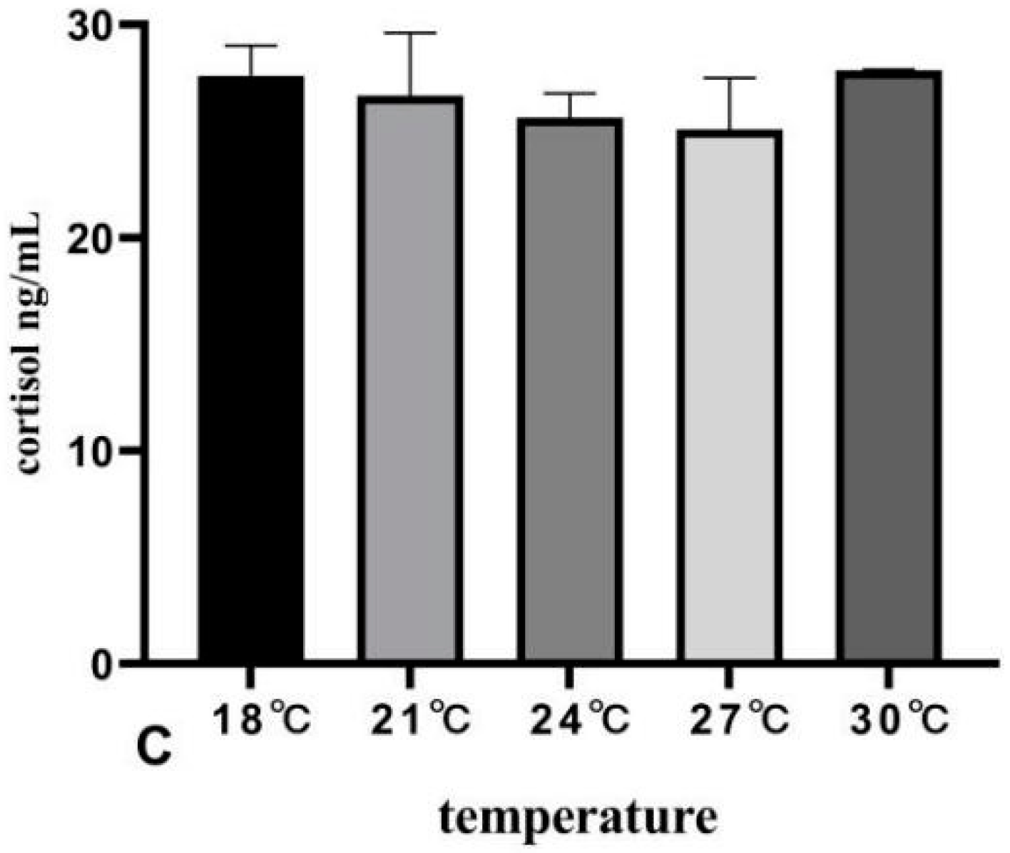
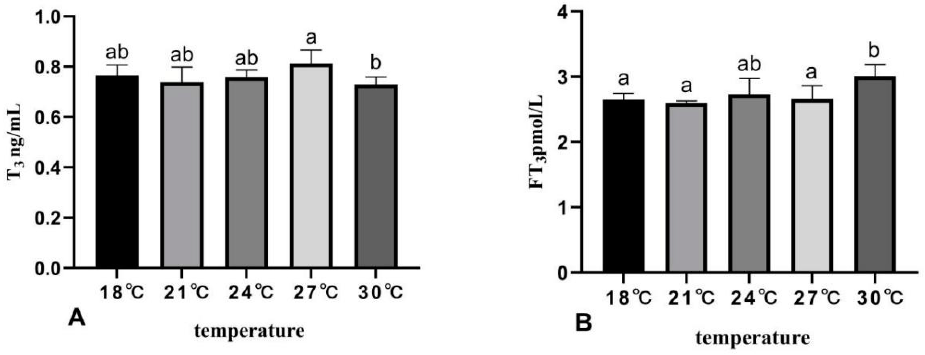
3.4. The Effects of Rearing Temperature on Hormone Levels in Juvenile Sturgeon

After rearing at different water temperatures, the plasma T3 level of juvenile sturgeon was the highest at 27 °C, which was significantly higher than that of 30 °C (p < 0.05) (Figure 4A). The plasma FT3 level increased with the increase in temperature, and the level in the 30 °C group was significantly higher than that in the 18 °C, 21 °C and 27 °C treatment groups (p < 0.05) (Figure 4B).
With the increase in temperature, the plasma cortisol level of juvenile sturgeon fluctuated and decreased, but there was no significant change among the treatment groups (p > 0.05) (Figure 4C).

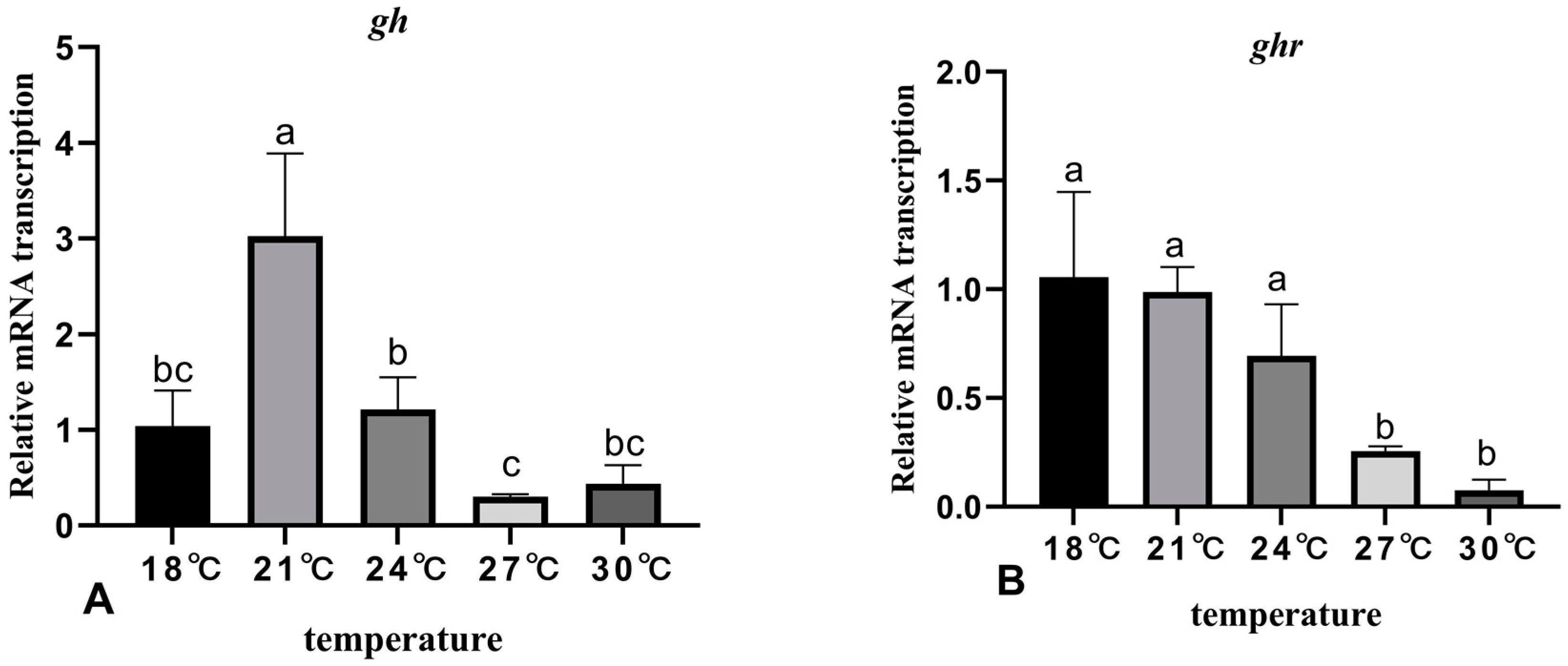
3.5. The Effects of Rearing Temperature on the Expression Levels of Growth-Related Genes in Juvenile Sturgeon

The effects of different temperature rearing on the expression levels of GH/IGF-related genes in juvenile sturgeon are shown in Figure 5. The expression level of gh in the brain of the 21 °C treatment group was significantly higher than that of other treatment groups, but there was no significant change among other treatment groups (Figure 5A). With the increase in temperature, the expression level of the ghr gene in the liver decreased, and the expression level in the 27 °C and 30 °C treatment groups was significantly lower than that in other treatment groups (Figure 5B). The expression level of the igf-I gene in the liver was the highest in the 21 °C treatment group, but there was no significant change among treatment groups (p > 0.05) (Figure 5C). With the increase in temperature, the expression level of the igf-II gene in the liver increased at first and then decreased, which was the highest in the 27 °C treatment group, but there was no significant change among all treatment groups (p > 0.05) (Figure 5D).

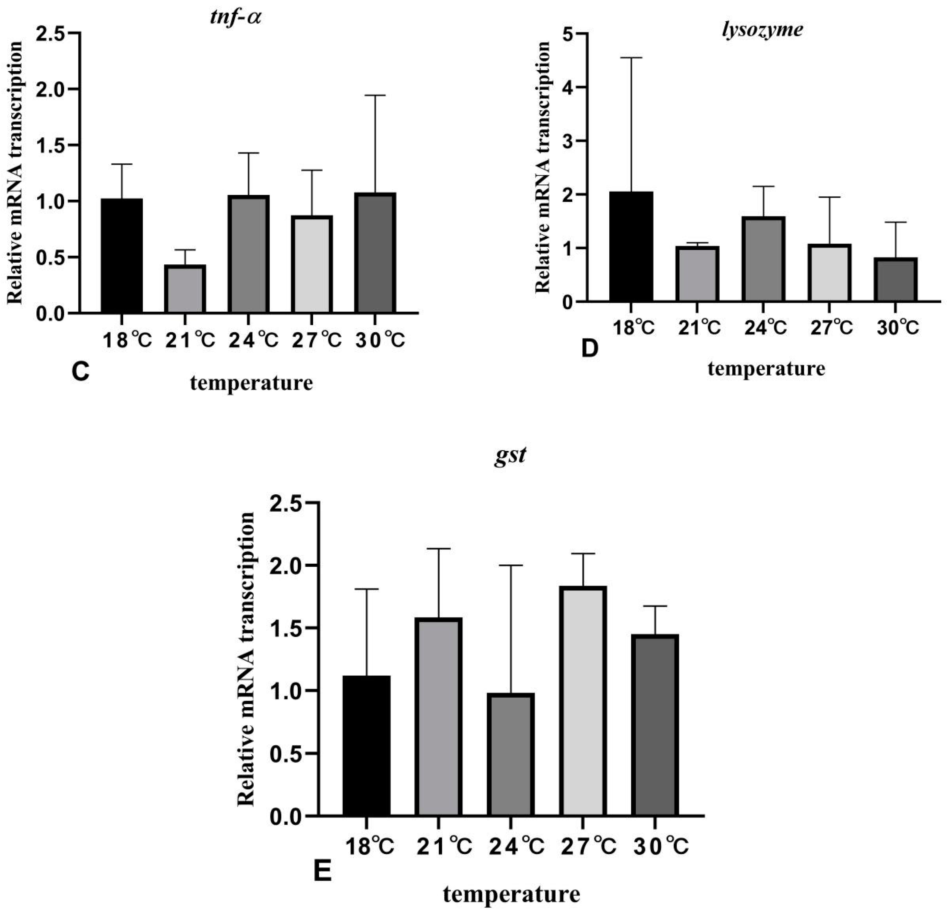
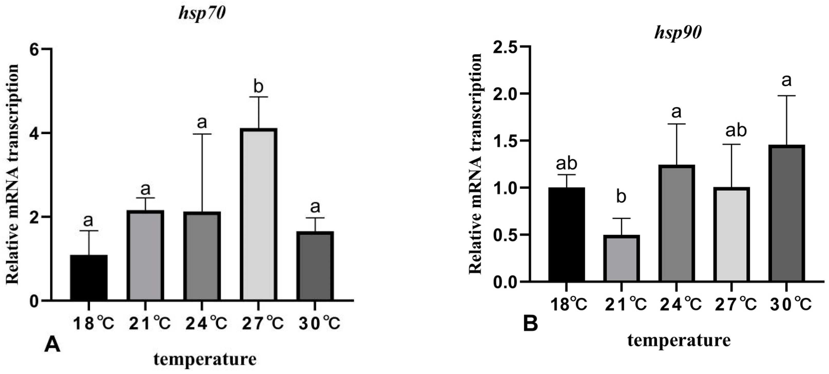
3.6. The Effects of Rearing Temperature on the Expression Levels of Stress- and Immunity-Related Genes in Juvenile Sturgeon

After rearing at different temperatures, the expression level of hsp70 in the liver of the 27 °C treatment group was significantly higher than that of other treatment groups, but there was no significant change among other treatment groups (Figure 6A). With the increase in temperature, the expression level of hsp90 in the liver of the 21 °C treatment group was the lowest, which was significantly different from that of the 24 °C and 30 °C groups (Figure 6B). The expression level of tnf-α in the liver of the 21 °C treatment group was the lowest, but there was no significant change among the treatment groups (Figure 6C). The expression level of lysozyme decreased with the increase in temperature, but there was no significant change among the treatment groups (Figure 6D). The expression level of gst in the liver of the 24 °C treatment group was the lowest, but there was also no significant change among the treatment groups (Figure 6E).

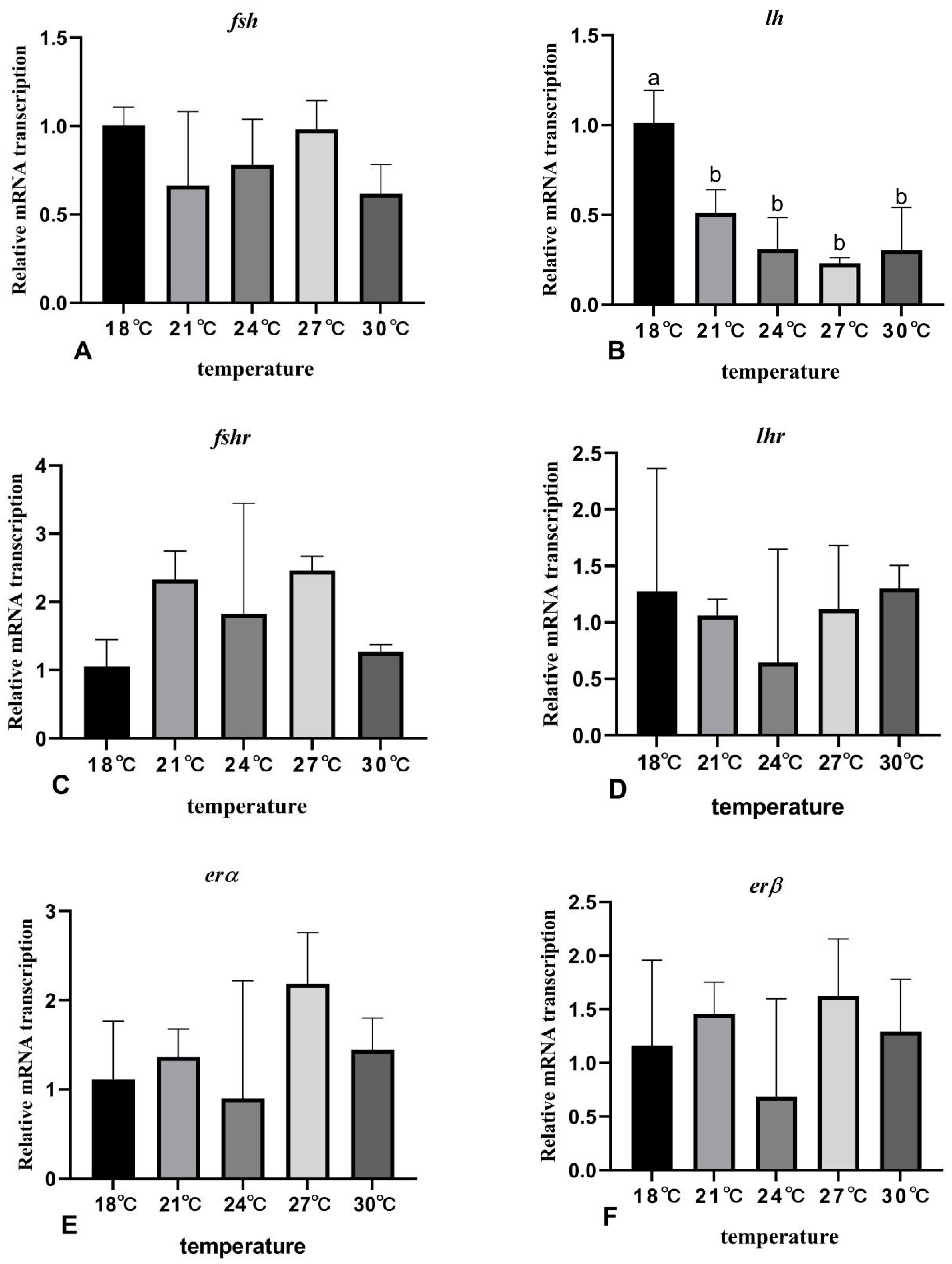
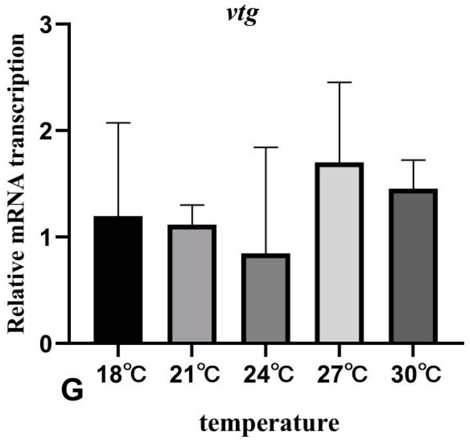
3.7. The Effects of Rearing Temperature on the Expression Levels of Gonad-Development-Related Genes in Juvenile Sturgeon

With the increase in temperature, the expression level of lh in the brain decreased, the expression level in the 18 °C treatment group was significantly higher than that in other treatment groups and the expression level of lhr in the liver decreased at first and then increased, but there was no significant change among all treatment groups. After rearing at different temperatures, there was no significant change in the expression levels of fsh in the brain and fshr, erα, erβ and vtg in the liver of juvenile sturgeon (Figure 7).

4. Discussion

As an environmental factor, temperature plays an important role in the metabolic reaction rate of fish and affects the growth and development of fish [25]. Within the optimum temperature range, it will promote the feeding, digestion and absorption of fish. When the critical value of the optimum temperature is exceeded, the feeding rate, SGR and RWG of fish decrease with the increase in temperature. In the study of S. salar [26], the optimum temperature for the growth of 70–150 g fry is 12.8 °C, but that for 150–300 g is 14 °C, indicating that the optimum growth temperature increases with the size of the fish. In the study of Tra Catfish (Pangasianodon hypophthalmus) [27], the optimum temperature for maximum SGR and RWG is 34 °C. However, it began to decline at 36°C. In this study, the growth rate of juvenile sturgeon changes with the increase in water temperature in the range of 18–30 °C, and the optimum temperature for maximum SGR and RWG of juvenile sturgeon is 21 °C. The growth efficiency decreases when the water temperature is higher than 24 °C. The regression curve of SGR and temperature (T) showed that the optimum culture temperature of juvenile sturgeon was 23.45 °C, and the growth performance was the best. Therefore, when the culture environment is maintained at approximately 23.45 °C, the juvenile sturgeon will show better growth performance.
Water temperature affects the biochemical indexes of metabolic activity, and directly or indirectly influences the growth, development and reproduction of fish [28]. The heterogenetic effect of glycogen in the liver is enhanced, resulting in an increase in GLU. In this study, the plasma GLU concentration of juvenile sturgeon at 21 °C and 30 °C is higher than that of other treatment groups, which may be due to the strong vitality and metabolism of juvenile sturgeon at 21 °C. However, a high concentration of GLU was also detected at 30 °C, which reflected the fact that 30 °C exceeded the optimum water temperature for the growth and development of juvenile sturgeon. Moreover, TC and TG are important metabolic substances and indicators of protein uptake and synthesis in fish. In this study, the concentrations of TC and TG in the plasma of juvenile sturgeon fluctuated, but there was no significant change among different treatment groups. Plasma protein is the raw material of tissue protein synthesis. In this study, the concentration of plasma TP did not change significantly with the increase in temperature. These results indicated that the temperature increase had no significant effect on the ability of protein synthesis in the liver of juvenile sturgeon. Fish maintain their osmotic pressure balance by regulating the ability of ion absorption and excretion to adapt to different temperature environments. In this study, the contents of plasma Na+ and Cl reached the highest levels at 30 °C and 24 °C, respectively, but there was no significant change. It was shown that when juvenile sturgeon were domesticated at a suitable temperature range, the metabolism increased and the cell membrane permeability changed with the increase in temperature, but it did not affect the osmotic regulation function of juvenile sturgeon.
Temperature changes lead to oxidative stress in fish, affecting the immune and metabolic function of fish. Therefore, it is necessary to enhance the ability of antioxidant defense. The antioxidant indexes, such as the concentration of MDA and T-AOC, CAT and SOD activity, are important parameters that reflect the potential antioxidant capacity of the organism, which can determine the degree of tissue peroxidation damage, and can also protect the organism from free radical damage [5]. Some studies have shown that the concentrations of MDA and CAT activity are lowest at the optimum temperature range of juvenile D. labrax [29]. However, when organisms were exposed to conditions outside the optimum temperature, the concentrations of MDA and CAT activity in fish were the highest. Similar to the results of D. labrax, the activities of CAT and MDA content of juvenile sturgeon were lowest when the optimum temperature was 21 °C to 24 °C, and the highest value was detected when the temperature was higher than the optimum temperature. In bald notothen (Pagothenia borchgrevinki), a species endemic to Antarctica, the increase in temperature leads to the enhancement of fish oxidation defense ability [30]. GSH is the most important antioxidant in organisms, which can help the immune system maintain normal function, and has the functions of antioxidation, integration and detoxification. As a ROS scavenger, GSH also plays an important role in long-term exposure to high temperatures [31]. After the Antarctic fish (Notothenia coriicep) were exposed to 0 °C, 2 °C and 4 °C for one day, the content of GSH increased with the increase in temperature [31]. In this study, the activity of T-AOC, CAT and MDA content fluctuated with the increase in temperature. Among them, the activity of T-AOC and GSH content at 27 °C and 30 °C was significantly higher than that of 18 °C. These results indicated that the antioxidant capacity of juvenile sturgeon increased with the increase in temperature, and the ability of scavenging free radicals reached its maximum at 27 °C and 30 °C.
Oxidative stress can be affected by environmental changes, which leads to the oxidation of some components in cells, such as DNA, proteins and lipids. Heat stress causes proteins in cells to be misfolded [4]. Hsps play an important role in the repair and clearance of damaged proteins, which can prevent proteins from misfolding under oxidative stress and act as antioxidants [32]. Studies have detected the up-regulation of hsps expression in C. auratus in summer [33]. The expression level of hsp70 in carp (Cyprinus carpio) was up-regulated after 1 hour of high-temperature exposure [34]. Heat stress can up-regulate the expression of hsp90 in the hepatopancreas of grass carp (Ctenopharyngodon idella) [35]. In this study, the expression level of hsp90 increased with the increase in temperature, which was significantly higher at 30 °C than that at 21 °C. The expression level of hsp70 at 27 °C was significantly higher than that in other treatment groups. Therefore, it was suggested that when the temperature exceeds 27 °C, the regulation of hsps expression in juvenile sturgeon is enhanced, the cell tolerance is increased after stress, and the scavenging and antioxidation of damaged proteins are enhanced, thus improving the stress resistance of fish [4,36]. Temperature affects the specific immune system of fish [37], and an increase in the domestication temperature (19–35 °C) would lead to a decrease in lysozyme activity of Mozambique tilapia (Oreochromis mossambicus) [38]. Similar to the results of this study, the lysozyme expression of juvenile sturgeon decreased with the increase in temperature. Moreover, the temperature increase in summer can up-regulate the expression of gst in the liver of O. niloticus [39]. The expression of gst in the brain and kidney of C. auratus increased after exposure to 23 °C [40]. Temperature also induces a stress response and up-regulates the expression of hsp70, which can promote the detoxification, anti-inflammation and protection of fish cells by affecting the expression level of tnf-α [41]. However, in this study, the expression levels of gst, lysozyme and tnf-α in juvenile sturgeon did not change significantly with the increase in temperature. Therefore, long-term temperature domestication would help fish tolerate and survive in a stressed environment.
In fish, the secretion of the thyroid hormone (THs) is regulated by the hypothalamus–pituitary–thyroid (HPT) axis. The thyrotropin (TSH) is released from the anterior pituitary and transported to the thyroid follicle through the blood to stimulate THs synthesis and be released into the blood [42]. THs is a key factor in regulating growth and has a wide range of metabolic physiological functions [43]. Studies have shown that the level of thyroid hormone changed seasonally. In the study of Channel catfish (Ictalurus punctatus), the levels of T4 and T3 changed significantly when the water temperature fluctuated from 7 °C to 32 °C [44]. With the increase in temperature in summer, the levels of T3 and T4 of O. mykiss increased too [11]. In this study, the plasma T3 level decreased significantly when the temperature exceeded 27 °C, and the FT3 level increased significantly at 30 °C. Moreover, THS, corticosteroids and growth hormones play an important role in regulating the development, growth, metabolism and osmotic regulation of fish [45]. Among them, the secretion of cortisol is regulated by the hypothalamic–pituitary–interrenal tissue (HPI) axis. When fish produce a stress response, it leads to a change in plasma cortisol content [46]. Studies have shown that there was no significant change in plasma cortisol levels of cutthroat trout (Salmo clarki) at 9 °C and 23 °C [47]. When Red Spotted Grouper (Epinephelus akaara) were domesticated at 15 °C, 20 °C and 25 °C for 7 days, there was also no change in cortisol levels in different treatment groups [48]. The plasma cortisol level of C. carpio increased after acute exposure to 4 °C, but there was no change in chronic exposure [49]. Similar to this study, there was no significant change in the plasma cortisol level of juvenile sturgeon after 25 d of temperature rearing, indicating that juvenile sturgeon gradually adapted to different temperature environments. However, the temperature change would affect the thyroid function and participate in the early growth and development of juvenile sturgeon.
The growth and development of fish are mainly regulated by the GH/IGF system, which promotes the growth and development of organisms by secreting related hormones and combining them with corresponding receptors [50]. GH is the most important hormone to promote the growth and development of fish, and its promoting effect on fish growth is realized directly or indirectly through IGFs. Temperature is an important factor affecting the GH/IGF system. With an increase in temperature within the suitable temperature range, the release of GH/IGF system-related hormones and gene expression are relatively elevated, and the growth rate of fish is accelerated. On the contrary, it can be inhibited beyond the suitable range, affecting the normal growth of fish, thus resulting in the retardation of growth in fish [7,51]. Some studies have shown that after domesticating at 30 °C, 33 °C and 36 °C for 30 d in rohu (Labeo rohita), the expression level of gh increased in the 33 °C group, while gh expression decreased significantly in the 36 °C group [52]. Similar to this study, the expression level of gh in the liver of juvenile sturgeon increased significantly at 21 °C, and then decreased significantly. The expression level of ghr decreased significantly with the increase in temperature. These results clarified that the expression of gh was promoted within the optimum water temperature, but high temperatures inhibited the binding of GH to its receptor. S. salar [53] were domesticated at 13 °C, 15 °C, 17 °C and 19 °C for 45 d, and O. mykiss [8] were domesticated at 22 °C to 25 °C; there was no significant change in liver igf-I expression levels. However, in the study of L. rohita and S. salar, the expression level of igf-II in the liver decreased with the increase in temperature [52,53]. In this study, there was no significant change in the expression of liver igf-I after juvenile sturgeon were domesticated at different temperatures for 25 d. When the water temperature exceeded 27 °C, the expression of igf-II in the liver decreased, and there was also no significant change between different treatment groups. Therefore, it was suggested that high temperatures affect the growth performance of juvenile sturgeon by inhibiting the expression of gh in the GH/IGF system. However, domestication within the optimum temperature range promoted the growth and development of juvenile sturgeon.
The process of gonadal development in fish is mainly regulated by the hypothalamus–pituitary–gonad (HPG) axis. Under the stimulation of environmental factors, the HPG axis secretes a gonadotropin-releasing hormone (GnRH) and gonadotropin (GtHs), namely FSH and LH, which act synergistically with steroids in gonadal tissues and combine with corresponding receptors to jointly regulate the reproductive process of fish. Long-term temperature stimulation has adverse effects on the reproduction of fish, because it affects the sensitivity of gonads to GtHs and disrupts the synthesis and release of GtHs. Temperature fluctuations also lead to alterations in signal transduction in the HPG axis, which reduces the release of steroids and ultimately hinders the development of gametes [54]. Some studies have shown that, compared with a yolk period at 14 °C, the plasma FSH level increased after the exposure of female S. salar to 22 °C, but the LH level did not change and the expression of fsh in the gonads was inhibited [13]. In coho salmon (Oncorhynchus kisutch), the expression of fshr was significantly lower in fish at 22 °C than at 14 °C [55]. The expression levels of fsh and lh subtypes in the grass puffer (Takifugu niphobles) decreased significantly after high- or low-temperature treatment [56]. Moreover, after exposure to 37 °C for 14 d in sheepshead minnow (Cyprinodon variegatus), the expression levels of fsh and lh in the gonads and the level of E2 in plasma decreased, and the spawning ability also decreased [57]. These results showed that unsuitable temperatures inhibited the regulation of gene expression on the HPG axis, resulting in a series of cascade reactions. In addition, ers regulates the expression of vtg and shows a positive correlation trend. For example, the changes in temperature decreased the ers binding affinity of tilapia (Oreochromis aureus) [58] and the decrease in binding affinity increases the level of E2 in circulation, which stimulates the liver to synthesize VTG. However, the temperature change had no effect on the expression of ers in O. mykiss and Atlantic halibut (Hippoglossus hippoglossus) [59,60]. As for sturgeon, with its long gonadal development cycle, the lack of secondary sexual characteristics and difficult sex identification, the regulatory mechanism of temperature on the expression of related genes on the HPG axis is not clear. Therefore, this study discussed the regulation of temperature rearing on the expression of genes related to the early gonadal development of juvenile sturgeon. These results showed that there was no significant change in the expression levels of fsh, fshr, lhr, erα, erβ and vtg in juvenile sturgeon with the increase in temperature. However, with the increase in temperature, the expression level of lh in the brain decreased significantly. Therefore, it was suggested that the temperature change is involved in the early gonadal development of juvenile sturgeon through the regulation of lh.

5. Conclusions

In conclusion, water temperature has significant effects on the growth and development, antioxidant capacity and immunity of juvenile sturgeon. Juvenile sturgeon can normal survival at a temperature range of 18–30 °C. In this study, the maximum SGR was obtained at 23.45 °C and the high expression of gh and GLU were also detected at approximately 21 °C, which indicated that the metabolic activity and growth rate of juvenile sturgeon were enhanced at 21–24 °C. However, with the increase in temperature, the expression level of ghr in the liver and lh in the brain decreased significantly. Moreover, temperature can affect the resistance of juvenile sturgeon to environmental changes by affecting the content of the plasma thyroid hormone, GSH and T-AOC activity, and the expression of hsp70 and hsp90 in the liver. Therefore, in order to promote the healthy culture of sturgeon and obtain sturgeon with fast growth, less disease, strong adaptability and high economic value, the best culture temperature recommended in this study is 21–24 °C.

Author Contributions

Conceptualization, H.C.; data curation, H.C.; funding acquisition, Q.H. and B.B.; methodology, H.C.; project administration, Q.H.; resources, B.B. and L.K.; software, H.R.; writing—original draft, H.C.; writing—review and editing, Q.H. and B.B. All authors have read and agreed to the published version of the manuscript.

Funding

This work was supported by the Zhuangping expert workstation of Yunnan Province (grant number 202005AF150042), the Scientific Research Foundation Project of Yunnan Provincial Department of Education (grant number 2022Y268), and the National Natural Science Foundation of China (grant number 32060825, 31602141).

Institutional Review Board Statement

The experimental animals used in this experiment are strictly in accordance with the requirements of the guidelines for the use of Experimental Animals of Yunnan Agricultural University and have been approved by the Experimental Ethics Committee of Yunnan Agricultural University (202203026).

Informed Consent Statement

Not applicable.

Data Availability Statement

Data are contained within the article.

Acknowledgments

The author would like to thank the students majoring in Aquaculture in Yunnan Agricultural University for their help in this study.

Conflicts of Interest

The authors declare no conflict of interest.

References

  1. ACW. Temperature in Relation to Fish. Nature 1887, 36, 213–214. [Google Scholar] [CrossRef] [Green Version]
  2. Mjia, B.; Mjs, B.; Mb, B.; Sz, B.; Ak, A. Extreme ambient temperature effects in European seabass, Dicentrarchus labrax: Growth performance and hemato-biochemical parameters. Aquaculture 2020, 522, 735093. [Google Scholar]
  3. Hahjahan, M.; Uddin, M.H.; Bain, V.; Haque, M.M. Increased water temperature altered hemato-biochemical parameters and structure of peripheral erythrocytes in striped catfish Pangasianodon hypophthalmus. Fish Physiol. Biochem. 2018, 44, 1309–1318. [Google Scholar] [CrossRef] [PubMed]
  4. Kalmar, B.; Greensmith, L. Induction of heat shock proteins for protection against oxidative stress. Adv. Drug Deliv. Rev. 2009, 61, 310–318. [Google Scholar] [CrossRef]
  5. Madeira, D.; Narciso, L.; Cabral, H.N.; Vinagre, C.; Diniz, M.S. Influence of temperature in thermal and oxidative stress responses in estuarine fish. Comp. Biochem. Physiol. Part A Mol. Integr. Physiol. 2013, 166, 237–243. [Google Scholar] [CrossRef]
  6. Cyr, D.G.; Idler, D.R.; Audet, C.; Mcleese, J.M.; Eales, J.G. Effects of long-term temperature acclimation on thyroid hormone deiodinase function, plasma thyroid hormone levels, growth, and reproductive status of male Atlantic cod, Gadus morhua. Gen. Comp. Endocrinol. 1998, 109, 24–36. [Google Scholar] [CrossRef]
  7. Gabillard, J.C. Differential expression of two GH receptor mRNAs following temperature change in rainbow trout (Oncorhynchus mykiss). J. Endocrinol. 2006, 190, 29–37. [Google Scholar] [CrossRef]
  8. Gabillard, J.C.; Rescan, P.Y.; Fauconneau, B.; Weil, C.; Bail, P. Effect of temperature on gene expression of the Gh/Igf system during embryonic development in rainbow trout (Oncorhynchus mykiss). J. Exp. Zool. Part A Comp. Exp. Biol. 2003, 298, 134–142. [Google Scholar] [CrossRef]
  9. Stacey, N.E. Hormones and reproductive behaviour in teleosts. Control. Processes Fish Physiol. 1983, 145, 117–129. [Google Scholar]
  10. Zanug, S.; Carrillo, M.; Ruiz, F. Delayed gametogenesis and spawning of sea bass (Dicentrarchus labrax) kept under different photoperiod and temperature regimes. Fish Physiol. Biochem. 1986, 2, 53–63. [Google Scholar]
  11. Chen, H.; Bi, B.; Kong, L.; Rong, H.; Su, Y.; Hu, Q. Seasonal changes in plasma hormones, sex-related genes transcription in brain, liver and ovary during gonadal development in female Rainbow Trout (Oncorhynchus mykiss). Fishes 2021, 6, 62. [Google Scholar] [CrossRef]
  12. Miranda, L.A.; Strüssmann, C.A.; Somoza, G.M. Effects of light and temperature conditions on the expression of GnRH and GtH genes and levels of plasma steroids in Odontesthes bonariensis females. Fish Physiol. Biochem. 2009, 35, 101–108. [Google Scholar] [CrossRef] [PubMed]
  13. Anderson, K.; Swanson, P.; Pankhurst, N.; King, H.; Elizur, A. Effect of thermal challenge on plasma gonadotropin levels and ovarian steroidogenesis in female maiden and repeat spawning Tasmanian Atlantic salmon (Salmo salar). Aquaculture 2012, 334, 205–212. [Google Scholar] [CrossRef] [Green Version]
  14. Soria, F.N.; Strüssmann, C.A.; Miranda, L.A. High water temperatures impair the reproductive ability of the pejerrey fish Odontesthes bonariensis: Effects on the hypophyseal-gonadal axis. Physiol. Biochem. Zool. 2008, 81, 898–905. [Google Scholar] [CrossRef] [PubMed]
  15. Brian, J.V.; Harris, C.A.; Runnalls, T.J.; Fantinati, A.; Pojana, G.; Marcomini, A.; Sumpter, J.P. Evidence of temperature-dependent effects on the estrogenic response of fish: Implications with regard to climate change. Sci. Total Environ. 2008, 397, 72–81. [Google Scholar] [CrossRef] [Green Version]
  16. Anderson, K.; King, H.; Pankhurst, N.; Ruff, N.; Pankhurst, P.; Elizur, A. Effect of elevated temperature on estrogenic induction of vitellogenesis and zonagenesis in juvenile Atlantic salmon (Salmo salar). Mar. Freshw. Behav. Phy. 2012, 45, 1–15. [Google Scholar] [CrossRef] [Green Version]
  17. Livak, K.J.; Schmittgen, T.D. Analysis of relative gene expression data using real-time quantitative PCR and the 2−∆∆CT method. Methods. 2001, 25, 402–408. [Google Scholar] [CrossRef]
  18. Din, S.Y.; Hurvitz, A.; Goldberg, D.; Jackson, K.; Levavi-Sivan, B.; Degani, G. Cloning of Russian sturgeon (Acipenser gueldenstaedtii) growth hormone and insulin-like growth factor I and their expression in male and female fish during the first period of growth. J. Endocrinol. Investig. 2008, 31, 201–210. [Google Scholar] [CrossRef]
  19. Hu, Y.; Xiao, K.; Yang, J.; Liu, X.; Wang, B.; Zeng, Q.; Du, H. Effects of feeding frequency on juvenile Chinese sturgeon Acipenser sinensis. Sci. Rep. 2020, 10, 17399. [Google Scholar] [CrossRef]
  20. Long, L.; Zhang, H.; Ni, Q.; Liu, H.; Wu, F.; Wang, X. Effects of stocking density on growth, stress, and immune responses of juvenile Chinese sturgeon (Acipenser sinensis) in a recirculating aquaculture system. Comp. Biochem. Physiol. Part C Toxicol. Pharmacol. 2019, 219, 25–34. [Google Scholar] [CrossRef]
  21. Fajkowska, M.; Głowacka, D.K.; Adamek-Urbańska, D.; Ostaszewska, T.; Krajnik, K.A.; Rzepkowska, M. Sex-related gene expression profiles in various tissues of juvenile Russian sturgeon (Acipenser gueldenstaedtii). Aquaculture 2019, 500, 532–539. [Google Scholar] [CrossRef]
  22. Wang, W.; Zhu, H.; Tian, Z.; Sun, A.; Dong, Y.; Dong, T.; Hu, H. Effects of 11-Ketotestosterone on development of the previtellogenic ovary in the sterlet, Acipenser ruthenus. Front. Endocrinol. 2020, 11, 115. [Google Scholar] [CrossRef] [PubMed]
  23. Atre, I.; Mizrahi, N.; Yebra-Pimentel, E.S.; Hausken, K.; Yom-Din, S.; Hurvitz, A.; Levavi-Sivan, B. Molecular characterization of two Russian sturgeon gonadotropin receptors: Cloning, expression analysis, and functional activity. Gen. Comp. Endocrinol. 2020, 298, 113557. [Google Scholar] [CrossRef]
  24. Jackson, K.; Hurvitz, A.; Din, S.Y.; Goldberg, D.; Pearlson, O.; Degani, G.; Levavi-Sivan, B. Anatomical, hormonal and histological descriptions of captive Russian sturgeon (Acipenser gueldenstaedtii) with intersex gonads. Gen. Comp Endocrinol. 2006, 148, 359–367. [Google Scholar] [CrossRef] [PubMed]
  25. Volkoff, H.; Rønnestad, I. Effects of temperature on feeding and digestive processes in fish. Temperature 2020, 7, 307–320. [Google Scholar] [CrossRef] [PubMed]
  26. Handeland, S.O.; Imsland, A.K.; Stefansson, S.O. The effect of temperature and fish size on growth, feed intake, food conversion efficiency and stomach evacuation rate of Atlantic salmon post-smolts. Aquaculture 2008, 283, 36–42. [Google Scholar] [CrossRef]
  27. Nguen, T.H.P. Effects of temperatures on growth performance, hematological parameters and plasma IGF-1 level of Tra catfish (Pangasianodon hypophthalmus). Hydrobiol. J. 2022, 58, 87–101. [Google Scholar] [CrossRef]
  28. Dahlhoff, E.P. Biochemical indicators of stress and metabolism: Applications for marine ecological studies. Annu. Rev. Physiol. 2004, 66, 183–207. [Google Scholar] [CrossRef] [Green Version]
  29. Vinagre, C.; Madeira, D.; Narciso, L.; Cabral, H.N.; Diniz, M. Effect of temperature on oxidative stress in fish: Lipid peroxidation and catalase activity in the muscle of juvenile seabass, Dicentrarchus labrax. Ecol. Indic. 2012, 23, 274–279. [Google Scholar] [CrossRef]
  30. Almroth, B.C.; Asker, N.; Wassmur, B.; Rosengren, M.; Jutfelt, F.; Gräns, A.; Sturve, J. Warmer water temperature results in oxidative damage in an Antarctic fish, the bald notothen. J. Exp. Mar. Biol. Ecol. 2015, 468, 130–137. [Google Scholar] [CrossRef]
  31. Klein, R.D.; Borges, V.D.; Rosa, C.E.; Colares, E.P.; Robaldo, R.B.; Martinez, P.E.; Bianchini, A. Effects of increasing temperature on antioxidant defense system and oxidative stress parameters in the Antarctic fish Notothenia coriiceps and Notothenia rossii. J. Therm. Biol. 2017, 68, 110–118. [Google Scholar] [CrossRef] [PubMed]
  32. Preville, X.; Salvemini, F.; Giraud, S.; Chaufour, S.; Paul, C.; Stepien, G.; Ursini, M.V.; Arrigo, A.P. Mammalian small stress proteins protect against oxidative stress through their ability to increase glucose-6-phosphate dehydrogenase activity and by maintaining optimal cellular detoxifying machinery. Exp. Cell Res. 1999, 247, 61–78. [Google Scholar] [CrossRef] [PubMed]
  33. Wang, J.; Wei, Y.; Li, X.; Cao, H.; Xu, M.; Dai, J. The identification of heat shock protein genes in goldfish (Carassius auratus) and their expression in a complex environment in Gaobeidian Lake, Beijing, China. Comp. Biochem. Physiol. Part C Toxicol. Pharmacol. 2007, 145, 350–362. [Google Scholar] [CrossRef] [PubMed]
  34. Hermesz, E.; Ábrahám, M.; Nemcsók, J. Tissue-specific expression of two metallothionein genes in common carp during cadmium exposure and temperature shock. Comp. Biochem. Physiol. Part C Toxicol. Pharmacol. 2001, 128, 457–465. [Google Scholar] [CrossRef]
  35. Wu, C.X.; Zhao, F.Y.; Zhang, Y.; Zhu, Y.J.; Ma, M.S.; Mao, H.L.; Hu, C.Y. Overexpression of Hsp90 from grass carp (Ctenopharyngodon idella) increases thermal protection against heat stress. Fish Shellfish Immunol. 2012, 33, 42–47. [Google Scholar] [CrossRef]
  36. Sharma, J.G.; Singh, S.P.; Chakrabarti, R. Effect of temperature on digestive physiology, immune-modulatory parameters, and expression level of Hsp and LDH genes in Catla catla (Hamilton, 1822). Aquaculture 2017, 479, 134–141. [Google Scholar] [CrossRef]
  37. Pérez-Casanova, J.C.; Rise, M.L.; Dixon, B.; Afonso, L.O.B.; Hall, J.R.; Johnson, S.C.; Gamperl, A.K. The immune and stress responses of Atlantic cod to long-term increases in water temperature. Fish Shellfish Immunol. 2008, 24, 600–609. [Google Scholar] [CrossRef]
  38. Ndong, D.; Chen, Y.Y.; Lin, Y.H.; Vaseeharan, B.; Chen, J.C. The immune response of Tilapia Oreochromis mossambicus and its susceptibility to Streptococcus iniae under stress in low and high temperatures. Fish Shellfish Immunol. 2007, 22, 686–694. [Google Scholar] [CrossRef]
  39. Hassan, A.M.; El Nahas, A.F.; Mahmoud, S.; Barakat, M.E.; Ammar, A.Y. Thermal stress of ambient temperature modulate expression of stress and immune-related genes and DNA fragmentation in Nile tilapia (Oreochromis niloticus (Linnaeus, 1758)). Appl. Ecol. Environ. Res. 2017, 15, 1343–1354. [Google Scholar] [CrossRef]
  40. Bagnyukova, T.V.; Lushchak, O.V.; Storey, K.B.; Lushchak, V.I. Oxidative stress and antioxidant defense responses by goldfish tissues to acute change of temperature from 3 to 23 °C. J. Therm. Biol. 2007, 32, 227–234. [Google Scholar] [CrossRef]
  41. Chowdhury, S.; Saikia, S.K. Oxidative stress in fish: A review. J. Sci. Res. 2020, 12, 145–160. [Google Scholar] [CrossRef] [Green Version]
  42. Blanton, M.L.; Specker, J.L. The Hypothalamic-Pituitary-Thyroid (HPT) axis in fish and its role in fish development and reproduction. Crit. Rev. Toxicol. 2007, 37, 97–115. [Google Scholar] [CrossRef] [PubMed]
  43. Rabah, S.A.; Gowan, I.L.; Pagnin, M.; Osman, N.; Richardson, S.J. Thyroid hormone distributor proteins during development in vertebrates. Front. Endocrinol. 2019, 10, 506. [Google Scholar] [CrossRef] [PubMed] [Green Version]
  44. Loter, T.C.; MacKenzie, D.S.; McLeese, J.; Eales, J. G Seasonal changes in channel catfish thyroid hormones reflect increased magnitude of daily thyroid hormone cycles. Aquaculture 2007, 262, 451–460. [Google Scholar] [CrossRef]
  45. Peyghan, R.; Enayati, A.; Sabzevarizadeh, M. Effect of salinity level on TSH and thyroid hormones of grass carp, Ctenophayngodon idella. Vet. Res. Forum 2013, 4, 175–178. [Google Scholar] [PubMed]
  46. Barton, B.A.; Iwama, G.K. Physiological changes in fish from stress in aquaculture with emphasis on the response and effects of corticosteroids. Ann. Rev. Fish. Dis. 1991, 1, 3–26. [Google Scholar] [CrossRef]
  47. Strange, R.J.; Schreck, C.B.; Golden, J.T. Corticoid stress responses to handling and temperature in salmonids. Trans. Am. Fish. Soc. 1977, 106, 213–218. [Google Scholar] [CrossRef]
  48. Cho, H.C.; Kim, J.E.; Kim, H.B.; Baek, H.J. Effects of water temperature change on the hematological responses and plasma cortisol levels in growing of red spotted grouper, Epinephelus akaara. Dev. Reprod. 2015, 19, 19–24. [Google Scholar] [CrossRef] [Green Version]
  49. Chen, G.R.; Sun, L.T.; Lee, Y.H.; Chang, C.F. Characteristics of blood in common carp, Cyprinus carpio, exposed to low temperatures. J. Appl. Aquac. 1995, 5, 21–31. [Google Scholar] [CrossRef]
  50. Gabillard, J.C.; Weil, C.; Rescan, P.Y.; Navarro, I.; Gutiérrez, J.; Le Bail, P.Y. Effects of environmental temperature on IGF1, IGF2, and IGF type I receptor expression in rainbow trout (Oncorhynchus mykiss). Gen. Comp. Endocrinol. 2003, 133, 233–242. [Google Scholar] [CrossRef]
  51. Deane, E.E.; Woo, N. Modulation of fish growth hormone levels by salinity, temperature, pollutants and aquaculture related stress: A review. Rev. Fish Biol. Fish. 2009, 19, 97–120. [Google Scholar] [CrossRef]
  52. Shahjahan, M.; Zahangir, M.M.; Islam, S.M.; Ashaf-Ud-Doulah, M.; Ando, H. Higher acclimation temperature affects growth of rohu (Labeo rohita) through suppression of GH and IGFs genes expression actuating stress response. J. Therm. Biol. 2021, 100, 103032. [Google Scholar] [CrossRef]
  53. Hevrøy, E.M.; Hunskår, C.; de Gelder, S.; Shimizu, M.; Waagbø, R.; Breck, O.; Hansen, T. GH–IGF system regulation of attenuated muscle growth and lipolysis in Atlantic salmon reared at elevated sea temperatures. J. Comp. Physiol. B 2013, 183, 243–259. [Google Scholar] [CrossRef] [PubMed]
  54. Maruska, K.P.; Fernald, R.D. Social regulation of gene expression in the hypothalamic-pituitary-gonadal axis. Physiology 2011, 26, 412–423. [Google Scholar] [CrossRef] [PubMed]
  55. Anderson, K.; Pankhurst, N.; King, H.; Elizur, A. Effect of thermal challenge on the expression of genes involved in ovarian steroidogenesis in Tasmanian Atlantic salmon (Salmo salar). Aquaculture 2017, 479, 474–478. [Google Scholar] [CrossRef] [Green Version]
  56. Shahjahan, M.D.; Kitahashi, T.; Ando, H. Temperature affects sexual maturation through the control of kisspeptin, kisspeptin receptor, GnRH and GTH subunit gene expression in the grass puffer during the spawning season. Gen. Comp. Endocrinol. 2017, 243, 138–145. [Google Scholar] [CrossRef]
  57. Bock, S.L.; Chow, M.I.; Forsgren, K.L.; Lema, S.C. Widespread alterations to hypothalamic-pituitary-gonadal (HPG) axis signaling underlie high temperature reproductive inhibition in the eurythermal sheepshead minnow (Cyprinodon variegatus). Mol. Cell. Endocrinol. 2021, 537, 111447. [Google Scholar] [CrossRef]
  58. Tan, N.S.; Frecer, V.; Lam, T.J.; Ding, J.L. Temperature dependence of estrogen binding: Importance of a subzone in the ligand binding domain of a novel piscine estrogen receptor. Biochim. Biophys. Acta (BBA)-Mol. Cell. Res. 1999, 1452, 103–120. [Google Scholar] [CrossRef] [Green Version]
  59. Pawlowski, S.; Islinger, M.; Völkl, A.; Braunbeck, T. Temperature-dependent vitellogenin-mRNA expression in primary cultures of rainbow trout (Oncorhynchus mykiss) hepatocytes at 14 °C and 18 °C. Toxicol. In Vitro 2000, 14, 531–540. [Google Scholar] [CrossRef]
  60. van Nes, S.; Andersen, Ø. Temperature effects on sex determination and ontogenetic gene expression of the aromatases cyp19a and cyp19b, and the estrogen receptors esr1 and esr2 in Atlantic halibut (Hippoglossus hippoglossus). Mol. Reprod. Deve. 2006, 73, 1481–1490. [Google Scholar] [CrossRef]
Figure 1. Weight growth curves of juvenile sturgeon under different temperatures.
Water 14 02368 g001
Figure 2. Correlation between water temperature and SGR of juvenile sturgeon.
Water 14 02368 g002
Figure 3. The effects of different temperature rearing on the levels of plasma antioxidant capacity in juvenile sturgeon. (A) CAT, (B) GSH, (C) T-AOC and (D) MDA. Data are expressed as mean ± SD (n = 3). Different letters denote statistically significant differences among the groups (Tukey method of the one-way ANOVA test, p < 0.05).
Water 14 02368 g003
Figure 4. The effects of different temperature rearing on plasma hormone levels of juvenile sturgeon. (A) T3, (B) FT3 and (C) cortisol. Data are expressed as mean ± SD (n = 3). Different letters denote statistically significant differences among the groups (Tukey method of the one-way ANOVA test, p < 0.05).
Water 14 02368 g004aWater 14 02368 g004b
Figure 5. The effects of different temperature rearing on the expression levels of growth-related genes in juvenile sturgeon. (A) gh, (B) ghr, (C) igf-I and (D) igf-II. Data are expressed as mean ± SD (n = 3). Different letters denote statistically significant differences among the groups (Tukey method of the one-way ANOVA test, p < 0.05).
Water 14 02368 g005aWater 14 02368 g005b
Figure 6. The effects of different temperature rearing on the expression levels of stress- and immunity-related genes in juvenile sturgeon. (A) hsp70, (B) hsp90, (C) tnf-ɑ, (D) lysozyme and (E) gst. Data are expressed as mean ± SD (n = 3). Different letters denote statistically significant differences among the groups (Tukey method of the one-way ANOVA test, p < 0.05).
Water 14 02368 g006aWater 14 02368 g006b
Figure 7. The effects of different temperature rearing on the expression levels of gonad-development-related genes in juvenile sturgeon. (A) fsh, (B) lh, (C) fshr, (D) lhr, (E) erɑ, (F) erβ and (G) vtg. Data are expressed as mean ± SD (n = 3). Different letters denote statistically significant differences among the groups (Tukey method of the one-way ANOVA test, p < 0.05).
Water 14 02368 g007aWater 14 02368 g007b
Table 1. Primers used in this experiment.
GenePrimer NameSequence (5′–3′)TMGenBank No. or Article Source
igf-Iigf-I-FGAACGAGTGCTGCTTCCAGAG57 °C[18]
igf-I-RAGGCTTTGGCTGGCTTAACA
igf-IIigf-II-FATCGCCCTCACAGTCTACAT57 °C[19]
igf-II-RGTGGCTTGCTGAAATAAAA
ghgh-FATGGCATCAGGTCTGCTTCT57 °C[19]
gh-RACGCTGCTCATCTGGAACATAG
ghrghr-FCATAGAAATCCAGGTTTACCCAACTC57 °C[19]
ghr-RCTGAACATCAAGGACGACGACTC
hsp70hsp70-FACAGCCATGTTGTATACTGAGTCC53 °C[20]
hsp70-RTGCACACCTTCTCCAGTTCTT
hsp90hsp90-FGCCAACCAATTTGATCAGAGC53 °C[20]
hsp90-RTGGACACTGTGACCTGGAAAG
gstgst-FTTGATAGGGCGGCTCTTGT57 °C[20]
gst-RCACCTGGATGTGTCGACTTGT
lysozymelysozyme-FGAGGGACCCAAATGGAATG57 °C[20]
lysozyme-RCCCACCCAGTTATTTTATGCT
tnf-αtnf-α-FTGTGTCTGTAGAGCACTCCGAT57 °C[20]
tnf-α-RCATGGCCAGCAAGTCGAT
erαerα-FCAGGCCAAGTATGGAAGGCA57 °C[21]
erα-RCACCGCACAGAACCTCATCT
erβerβ-FATTGCTGCTGGAGATGCTG57 °C[22]
erβ-RTTCTGGCTTTGAACAGGTGA
vtgvtg-FCAAGTCAGCTAACCCAGCCA57 °C[21]
vtg-RGCATGTTCAGGATCCCCCTC
lhrlhr-FTTCAATCCCTGCGAAGACAT57 °C[23]
lhr-RGCAGAACAAGCAGTACAGCAAA
fshrfshr-FGAGGCTGAGATTGCGAAGAAGGAG57 °C[24]
fshr-RCGCCGTCTGTGTCTGCTATGTAAG
fshfsh-FGTCTGTCAACACCACCTCCT57 °CEU523732.1
fsh-RCTTAGGGTGCCACAGTCAGT
lhlh-FTCCTCCTCCTCTTCTCTGCT57 °CEU523733.1
lh-RCACCGTCACAAAGCGAAGAT
18sRNA18SRNA-FCCATAAACGATGCCGACTGG57 °C[21]
18SRNA-RTGAGGTTCCCCGTGTTGAGT
Table 2. Effect of temperature on growth performance of juvenile sturgeon.
Parameter18 °C21 °C24 °C27 °C30 °C
Initial weight (g)8.74 ± 0.058.69 ± 0.198.55 ± 0.128.71 ± 0.228.47 ± 0.14
Final weight (g)21.83 ±3.16 ab27.35 ± 2.75 a26.07 ± 2.24 a25.31 ± 4.71 ab19.45 ± 1.93 b
Specific growth rate (%/d)3.64 ± 0.59 ab4.57 ± 0.49 a4.45 ± 0.29 a4.22 ± 0.84 ab3.31 ± 0.43 b
Relative weight gain rate (%)149.97 ± 37.44 ab215.07 ± 37.17 a204.67 ± 21.84 ab191.34 ± 60.35 ab129.80 ± 25.58 b
Note: Data are expressed as mean ± SD (n = 3). Different letters above the parameters represent significant differences (p < 0.05). There was no significant difference when they were the same (p > 0.05).
Table 3. The effects of rearing at different temperatures on plasma biochemical indices of juvenile sturgeon.
Parameter18 °C21 °C24 °C27 °C30 °C
Blood Glucose (µg/mL)598.83 ± 127.45 ab647.47 ± 63.64 bc550.37 ± 41.59 ab481.70 ± 20.24 a739.90 ± 36.13 c
Triglyceride (mg/dL)1392.49 ± 903.491126.59 ± 381.39924.86 ± 290.10793.64 ± 128.80877.46 ± 351.57
Total Cholesterol (µmol/dL)117.78 ± 36.11121.06 ± 40.44102.13 ± 10.96110.05 ± 0.89123.09 ± 1.75
Total protein (µg/mL)16.56 ± 1.6515.45 ± 0.6614.94 ± 3.1314.06 ± 0.3214.69 ± 0.28
Na+ (mmol/L)171.66 ± 20.19149.82 ± 9.78139.76 ± 29.19136.52 ± 17.72204.61 ± 78.04
Cl (mmol/L)107.01 ± 8.52113.72 ± 13.22123.33 ± 18.32115.46 ± 3.15121.36 ± 11.75
Note: Data are expressed as mean ± SD (n = 3). Different letters above the parameters represent significant differences (p < 0.05). There was no significant difference when they were the same (p > 0.05).
Publisher’s Note: MDPI stays neutral with regard to jurisdictional claims in published maps and institutional affiliations.

