Article

Interacting Effects of Polystyrene Microplastics and the Antidepressant Amitriptyline on Early Life Stages of Brown Trout (Salmo trutta f. fario)

1
Animal Physiological Ecology, Institute of Evolution and Ecology, University of Tübingen, Auf der Morgenstelle 5, D-72076 Tübingen, Germany
2
Chair of Water Quality Control, Technische Universität Berlin, Sekr. KF 4, Str. des 17. Juni 135, D-10623 Berlin, Germany
3
German Environment Agency (UBA), Section II 3.1 (National and International Development of Drinking Water Quality and Resource Protection), Schichauweg 58, D-12307 Berlin, Germany
4
Environmental Analytical Chemistry, Center for Applied Geoscience, University of Tübingen, Schnarrenbergstr.94-96, D-72076 Tübingen, Germany
5
Steinbeis Transfer Center for Ecotoxicology and Ecophysiology, Blumenstr. 13, D-72108 Rottenburg, Germany
*
Author to whom correspondence should be addressed.
Water 2020, 12(9), 2361; https://doi.org/10.3390/w12092361
Submission received: 21 July 2020 / Revised: 18 August 2020 / Accepted: 20 August 2020 / Published: 22 August 2020
(This article belongs to the Special Issue Microplastics in Aquatic Environments and Wastewater Treatment )

Abstract

:
Whether microplastics themselves or their interactions with chemicals influence the health and development of aquatic organisms has become a matter of scientific discussion. In aquatic environments, several groups of chemicals are abundant in parallel to microplastics. The tricyclic antidepressant amitriptyline is frequently prescribed, and residues of it are regularly found in surface waters. In the present study, the influence of irregularly shaped polystyrene microplastics (<50 µm), amitriptyline, and their mixture on early life-stages of brown trout were investigated. In a first experiment, the impacts of 100, 104, and 105 particles/L were studied from the fertilization of eggs until one month after yolk-sac consumption. In a second experiment, eggs were exposed in eyed ova stages to 105, 106 particles/L, to amitriptyline (pulse-spiked, average 48 ± 33 µg/L) or to two mixtures for two months. Microplastics alone did neither influence the development of fish nor the oxidative stress level or the acetylcholinesterase activity. Solely, a slight effect on the resting behavior of fry exposed to 106 particles/L was observed. Amitriptyline exposure exerted a significant effect on development, caused elevated acetylcholinesterase activity and inhibition of two carboxylesterases. Most obvious was the severely altered swimming and resting behavior. However, effects of amitriptyline were not modulated by microplastics.

1. Introduction

Microplastic particles (MP) are detected worldwide from densely populated and rural areas to remote regions [1,2,3,4]. The presence of MP has globally been reported for sediment, surface water and even for air samples [5,6,7,8]. Representing the recent state of knowledge in freshwater systems, MP concentrations range from 0.00012 particles/L up to 2867 particles/L (according to [9]). However, the potential risk for organisms and ecosystems caused by MP is still a matter of discussion. MP were shown to be ingested and egested by fish [10,11,12] and small particles (mostly nanoplastics) can even transfer into tissues [13,14,15,16]. Microplastic particles can injure organisms mechanically resulting in inflammation and other histopathological effects in contact epithelia [17,18,19], disturb the energy metabolism [19,20], and induce oxidative stress [13,18,19]. Early life stages of fish are considered as very sensitive for pollutants [21]. In this context, high concentrations of MP have been shown to reduce and delay hatching as well as negatively influence growth and heart rate of marine medaka (Oryzias melastigma) [11]. Moreover, Malafaia, et al. [22] reported that exposure of zebrafish (Danio rerio) to polyethylene (PE) MP reduced the hatching time and survival rates and led to morphological changes. In contrast, LeMoine, et al. [23] found no effects of PE MP on hatching, mortality, and growth rates of zebrafish. However, zebrafish exposed to MP exhibit transcriptomic changes as, for example, downregulation of genes involved in the neural development. Mazurais, et al. [12] observed that a diet that incorporated about 200 PE microbeads per day caused a slightly higher mortality rate in sea bass larvae (Dicentrarchus labrax). Apart from that MP had only limited effects on the development of sea bass larvae in the experiment [12].
The evaluation of the risk of MP for aquatic organisms in general is complex since different polymer types with manifold additives in various sizes and shapes are present in the environment [24]. With our study we therefore solely address a selected aspect of MP aquatic ecotoxicology.
The topic is even more complex since not only MP themselves, but also their interaction with chemicals have to be regarded. For example, polymerization solvents, residual monomers, plasticizers, or other additives can leak from the particles and affect MP-exposed organisms [25,26]. In addition, MP has the potential to ad- or absorb organic pollutants (reviewed by [27] and [28]). The sorption can modulate the toxicity of the pollutants in different ways: If the particles are ingested and excreted together with an adherent pollutant this would be without consequences for the organism. However, the bioavailability of otherwise free pollutants may be reduced due to sorption what can led to less negative effects in organisms [11,29,30]. On the other hand, pollutants ingested together with MP can desorb in the digestive track, for example due to different pH conditions. In such cases, MP act as a vector and adverse effects can be enhanced by the presence of MP [31,32,33]. Batel, et al. [10] showed that MP and associated benzo[a]pyrene can also be transported along an artificial food web. Nevertheless, the relevance of MP as vectors for organic pollutants in comparison to other exposure pathways in the environment remains a matter of discussion [34,35,36]. Since the concentrations of persistent organic pollutants in continental environments are expected to be higher than in marine ecosystems, sorption of hydrophobic organic pollutants to MP might be especially important for freshwater ecosystems [37].
One group of chemicals commonly found in aquatic environments are pharmaceuticals [38,39]. Residues of these or their metabolites enter surface waters mainly via wastewater treatment plants [39,40]. Non-selective monoamine reuptake inhibitors more known as tricyclic antidepressants are one of the oldest groups of pharmaceuticals to treat depression. Amitriptyline is the most prescribed drug of this group [41,42,43]. Beside depression, amitriptyline is also used for migraine prophylaxis and to treat chronic pain [43]. In comparison to other antidepressants, its mode of action is rather unspecific: In addition to the inhibition of the reuptake of the neurotransmitters serotonin and noradrenaline, amitriptyline acts as muscarinic acetylcholine receptor antagonist [44]. Furthermore, it has been shown to bind to histamine receptors [45] as well as to neurotrophic tyrosine kinase A/B receptors resulting in an upregulation of acetyl transferase and an influence on cell differentiation [46]. Amitriptyline is mainly metabolized in the liver by cytochrome P450 [47]. An important metabolite is nortriptyline which is also in use as an antidepressant itself [41,47,48]. Amitriptyline has been found in surface waters around the world [38,48,49]. The highest concentration of 71.0 ng/L was reported by Baker and Kasprzyk-Hordern [38] for a large river in the UK. Mean surface water concentrations are normally in the low nanogram per liter range up to 22 ng/L [49,50,51]. Togola and Budzinski [52] reported that in France residues of amitriptyline (1.4 ng/L) were even found in drinking water. Pharmaceuticals are designed to be bioactive at low concentrations, and it can be, therefore, not excluded that they also may affect non-target organisms at low environmental concentrations [39,40].
Demin, et al. [53] showed that amitriptyline increases the serotonin-triggered neurotransmission in the brain of adult zebrafish in a dose-dependent way, with significantly higher 5-hydroxyindoleacetic (5-HIAA)/serotonin ratios in fish exposed to 5 and 10 mg/L amitriptyline for a few hours. No effects on the noradrenaline level were shown in this study. In contrast, Meshalkina, et al. [54] found a significant reduction of the 5-HIAA/serotonin ratio and an increased dopamine and noradrenaline level in the brain of adult zebrafish exposed for two weeks to 10 and 50 µg/L amitriptyline. Moreover, the antidepressant was shown to alter the immune response in in vitro studies with primary macrophages of common carp (Cyprinus carpio) as well as in vivo studies with zebrafish [55,56] and to affect the oxidative stress level of both fish species [56,57,58]. Furthermore, amitriptyline was found to affect the swimming behavior of zebrafish, by reducing for example the swimming activity as well as the covered distance, and high concentrations of the antidepressant even led to side or vertical swimming behavior [53,54,59,60]. In addition, exposure of common carp and zebrafish to the antidepressant stimulated hatching and caused retarded development and malformations in common carp as well as reduced body length of zebrafish larvae [56,57]. In contrast, high amitriptyline concentrations were found to extend the time until hatch and to decrease the heart rate in zebrafish [60]. Most studies about the ecotoxicity of amitriptyline were performed with model species, but ecotoxicological studies with feral aquatic species are lacking. Brown trout are known to be a sensitive test organism and as important predators are of ecological relevance [61,62].
In the present study, effects of polystyrene (PS) MP at an environmental relevant concentration of 100 particles/L and higher concentrations of 104 and 105 particles/L on the development of brown trout were investigated. Fish were exposed for 182 days from freshly fertilized eggs until about one month after the fry completed their yolk sac consumption. In a second experiment, eggs were exposed from eyed ova stage until one week after yolk sac consumption. In addition to PS MP (105 and 106 particles/L) fish were also exposed to amitriptyline and co-exposed to the mixtures of PS MP and the antidepressant. The co-exposure allows to investigate a potential modulation of the effects of amitriptyline by PS MP. In both experiments, the impact of exposure on the development and biomarkers for oxidative stress (activity of superoxide dismutase (SOD) and the level of lipid peroxidation (LPO)) were analyzed. Since the chorion of zebrafish has been reported to act as an effective protective barrier against carbon nanotubes [63], we examined the structure of the chorion of brown trout in the first experiment by means of scanning electron microscopy. In the second experiment, also the behavior of larvae and endpoints for neurotoxicity (activity of acetylcholinesterase (AChE) and two carboxylesterases (CbE)) were investigated.

2. Materials and Methods

2.1. Test Organism

Eggs of brown trout (Salmo trutta f. fario) were obtained from a commercial fish breeder (Forellenzucht Lohmühle, D-72275 Alpirsbach-Ehlenbogen, Germany). According to the EC Council Directive, the breeding facility is listed as category 1, disease-free [64]. All experiments started directly after purchase of the eggs: In experiment 1, eggs (in total 360 eggs) were exposed on the same day of their fertilization, in experiment 2 (in total 540 eggs), exposure started 47 days post fertilization (dpf) in the eyed ova stage.

2.2. Test Substances

In both experiments, transparent PS pellets (Polystyrol 158 K, BASF, Ludwigshafen, Germany, density 1.05 g/mL) were cryo-milled (CryoMill, Retsch, Haan, Germany) according to the method of Eitzen, et al. [65]. The resulting irregularly shaped particles were suspended in ultra-pure water (without any surfactant), fractionated using a micro-sieve (polyamide monofilament) with nominal mesh-size of 50 µm and the permeate was used as stock suspension. The particle concentration in the stock suspensions were analyzed with a particle counter (SVSS, PAMAS, Rutesheim, Germany) by light extinction in a laser-diode sensor (type HCB-LD-50/50). Exemplary particle numbers with the analyzed size ranges are provided in Figure 1 and in Table S1 in the supplement. The stock suspensions were diluted with respective ratios to obtain the target particle concentrations for the exposure experiments.
Amitriptyline hydrochloride was purchased from Sigma Aldrich (CAS Number: 549–18–8; Lot: BCBV1175; molecular formula: C20H23N · HCl; purity ≥ 98%; molecular weight 313.86). Amitriptyline hydrochloride in the used concentration is water soluble without adding organic solvents. For the stock solutions, 8.5 mg/L amitriptyline hydrochloride were solved in bidestilled water. Bottles with stock solutions were covered in aluminum foil to protect them from light. All further given amitriptyline concentrations refer to pure amitriptyline not amitriptyline hydrochloride. The predicted logP octanol-water coefficient (pH 7.4) of amitriptyline is 4.92 [66].

2.3. Exposure and Sampling of Brown Trout

In both experiments, each treatment was tested in triplicates in a semi-static three-block design. Exposures took place in a thermostat-controlled chamber with a light/dark cycle of 10/14 h. Petri dishes and aquaria were shaded from direct light. Aquaria were aerated with glass pipettes connected via silicone tubes to compressed air. Test suspensions were prepared from defined PS MP stock suspensions (56,240 particles/mL). Vessels containing the respective stock suspension were rinsed four times to avoid loss of particles. After consumption of the yolk sacs, fish were fed daily approximately 3% of their body weight with commercial fish feed (0.5 mm, Biomar, Brande, Denmark). At the end of the experiments, brown trout were anesthetized and killed by an overdose of tricaine methanesulfonate ((MS-222), 1 g/L, buffered with NaHCO3). Death was ensured by severance of the spine. Length and weight of each fish were recorded. The level of LPO, the activity of SOD and the activity of AChE and CbE had to be analyzed in different tissues, due to the small size of the fish.

2.3.1. Experiment 1a: Exposure of Embryos and Sac-Fry Stages

The first part of experiment 1 was conducted according to the OECD guideline 212 for exposing fish embryos and sac-fry stages to dissolved chemicals [67]. Freshly fertilized eggs (fertilization and start of experiment 07 December 2016) were exposed to 0 particles/L (C1), 100 particles/L (MP1h), 104 particles/L (MP1tt) and 105 particles/L (MP1ht). This first part of the experiment 1 was performed in glass Petri dishes containing 200 mL of the respective test suspension. To achieve the final concentration, the stock suspension was diluted with aerated artificial water (294 mg/L CaCl2 × 2 H2O, 123.25 mg/L MgSO4 × 7 H2O, 64.75 mg/L NaHCO3, 5.75 mg/L KCl in pure water). In each Petri dish, 30 brown trout eggs were exposed to the test suspensions (90 eggs per treatment). Until the eyed ova stage, the eggs were kept in complete darkness. The temperature in the Petri dishes was 6.7 ± 0.2°C. To maintain good water quality, 25 to 50% of the test suspensions were renewed every second day (detailed information in the supplement Table S2). From day 138 dpf, filtered aerated tap water (iron filter, particle filter, activated charcoal filter) was used to prepare the test suspensions to habituate the growing larvae to the water used in the second part of the experiment. The first part of the experiment ended after 150 days when the fry had completely consumed their yolk sacs (07 December 2016–05 May 2017). Investigated parameters were time of development until eyed ova stage, time until hatch, heart rate 93 dpf, and mortality (excluding unfertilized eggs). After the first part of the experiment, ten larvae were sampled from each Petri dish. For determination of the LPO level, heads of the larvae were immediately frozen in liquid nitrogen and stored at −80 °C until further usage.

2.3.2. Experiment 1b: Exposure of Fry

The second part of experiment 1 lasted 33 days until 182 dpf (05 May 2017–06 June 2017). The remaining fry of experiment 1a were transferred into 12 L aquaria with 5 L of the corresponding test suspensions (in total C1: n = 57, MP1h: n = 57, MP1tt: n = 53 and MP1ht: n = 58). PS MP stock suspensions were diluted with filtered tap water. To ensure good water quality, half of the test suspension was renewed twice a week. Water parameters were checked 177 dpf and at the end of the experiment (average values: Temperature 6.38 ± 0.45 °C, pH 8.5 ± 0.1, oxygen concentration 12.71 ± 0.22 mg/L, oxygen saturation 107.33 ± 1.60%; conductivity 493.33 ± 7.30 μS/cm; see supplement Table S3). Samples for analysis of SOD activity (muscle/kidney) as well as for determination of the LPO level (head) were frozen in liquid nitrogen and stored at −80 °C.

2.3.3. Experiment 2

In experiment 2, embryos/larvae were exposed in total for 60 days from eyed ova stage (47 dpf) until one week after yolk sac consumption (29 December 2017–26/27. February 2018). Exposure groups included a control group (C2) and groups exposed to 105 particles/L (MP2ht), 106 particles/L (MP2mio), pulse-spiked amitriptyline (AMI2, nominal concentration 300 µg/L, average concentration calculated and given as follows), 105 particles/L + pulse-spiked amitriptyline (MIX2ht, nominal concentration 300 µg/L amitriptyline, average concentration calculated and given as follows), and 106 particles/L + pulse-spiked amitriptyline (MIX2mio, nominal concentration 300 µg/L amitriptyline, average concentration calculated and given as follows). Test media were prepared with filtered tap water. Exposure took place in 12 L aquaria filled with 5 L of the corresponding test media. Per aquarium, 30 individuals were exposed (3 × 30 per treatment group). 2.5 L of the test media were exchanged on average once a week (see Table S2 in the supplement). Water parameters were determined to control water quality at the start, after 55 days of exposure and at the end of the experiment. The average values were pH 8.3 ± 0.2, temperature 7.06 ± 0.20 °C, conductivity 430.83 ± 17.24 µS/cm, oxygen content 10.92 ± 0.10 mg/L, oxygen saturation 95.06 ± 0.79% (see supplement Table S4). Nitrite (NO2) values did not exceed 0.05 mg/L. The heart rate was counted 21 days after the start of the experiment. Samples for LPO (head), SOD (muscle/kidney), AChE and CbE (muscle) were frozen in liquid nitrogen and stored at −80 °C.

2.4. Chemical Analyses

At the start of the second experiment as well as prior and past a water exchange (17 January 2018) mixed samples of all three blocks of each treatment group (4 mL per aquarium 12 mL in total) were taken and frozen at −20 °C until further analysis. The water concentrations of amitriptyline were determined using LC-MS with a 1290 Infinity HPLC system (Agilent Technologies, Waldbronn, Germany) and a triple quadrupole mass spectrometer (6490 iFunnel Triple Quadrupole LC/MS, Agilent Technologies, Waldbronn, Germany) in ESI (+) mode. An Agilent Poroshell-120-EC-C18 (2.7 µm, 2.1 × 100 mm) column at a flow rate of 0.4 mL/min was used for separation, and column temperature was maintained at 40 °C. Eluent A and B were water (+0.1% formic acid) and acetonitrile (+0.1% formic acid), respectively. Gradient elution was used: 0–1 min 5% B, linear increase to 100% B within 7 min, hold for 7 min at 100% B. After switching back to the starting conditions, reconditioning time of 3 min was employed. Samples were kept in the autosampler at 10 °C, the injection volume was 10 µL. Samples of the control experiments were measured undiluted and samples of the experiments pulse-spiked with amitriptyline were measured after 50-times dilution. The detection limit of amitriptyline (mass transition m/z 278.2 → 117.1) for undiluted samples was 10 ng/L (10 µL injection volume). Further details on operating parameters of the triple quadrupole are provided in Tables S5 and S6 in the supplement.

2.5. Development Parameters

Mortality, malformations, eye pigmentation (only experiment 1), and hatch were checked daily. Coagulated eggs, dead fish and remains of chorions were removed. To determine the heart rate, five animals of each Petri dish/aquaria were transferred into a Petri dish with fresh test medium. The heart rate was counted under a stereo microscope for 20 s and water temperature was measured. Subsequently, fish were placed back into the corresponding Petri dish/aquaria.

2.6. Scanning Electron Microscopy

After hatching in experiment 1, chorions were immediately fixed in 2% glutardialdehyde in 0.1 M cacodylate buffer (pH 7.6) for several days. Specimens were rinsed three times with 0.1 M cacodylate buffer and subsequently incubated in 1% osmium tetroxide overnight. The next day, chorions were transferred in a graded series of ethanol for dehydration. Subsequently, samples were fixed to specimen holder stubs and sputter-coated with gold. Analyses were conducted using a scanning electron microscope EVO LS 10 (Zeiss, Jena, Germany).

2.7. Behavior

In the second experiment, resting behavior of fry was determined after 42 days of exposure. In each tank, the positions of all fish were recorded (resting on the side/resting in ventral position). Additionally, the swimming behavior under stressful conditions (bright illumination, no aeration) was quantified at the end of the experiment (27 February 2018). For this, five fish per replicate were transferred into small tanks (17 cm × 17 cm × 8.5 cm) filled with 0.5 L of the corresponding test medium. Four of these tanks were measured simultaneously. Tanks were surrounded with white polystyrene plates and indirectly illuminated with lamps (one lamp per tank, 2700 K, 1521 Lm per lamp) facing the top plate. After a habitation period of 2 min, swimming behavior of fish was recorded for 18 min with four cameras (Basler acA 1300–60 gm, 1.3 megapixels resolution, Basler AG, Ahrensburg, Germany, lens: 4.5–12.5 mm; 1:1.2; IR 1/2”) positioned 32 cm above the water surface of each tank. Fry were center-point tracked individually, and total distance moved, mean velocity over time, time of no movement and body contact were assessed using the EthoVision 12 XT software (Noldus Information Technology bv, Wageningen, The Netherlands). Whenever the system exhibited difficulties in automatic tracking, data were manually corrected for swaps between tracked individuals. After the video tracking fish were sampled as described above.

2.8. Level of Lipid Peroxides

The degree of LPO was quantified with the ferrous oxidation xylenol orange (FOX) assay. The assay was performed according to Hermes-Lima, et al. [68] and Monserrat, et al. [69], slightly modified for 96-well plates. In pre-tests, the dilution factor with methanol as well as sample volume and incubation time were adjusted for optimal output (see Table S7). Frozen heads of fry were homogenized with HPLC grade methanol. Samples were centrifuged (15,000 rcf, 5 min, 4 °C) and the supernatants were stored at −80 °C. For the final assay, the following compounds were added to each well: 50 µL of 0.75 mM FeSO4-solution, 50 µL of 75 mM sulfuric acid, and 50 µL of 0.3 mM xylenol orange solution. Subsequently, the corresponding sample volume was added. To be able to correct for potential Fe in the samples, additionally, a sample blank in which the FeSO4-solution was replaced by bidistilled water was performed. Bidistilled water was used to achieve a total volume of 200 µL in each well. Well plates were incubated at room temperature and the absorbance at 570 nm (ABS570) was measured in a photometer (Bio-Tek Instruments, Winooski, VT, USA). In a next step, 1 µL of 1 mM cumene hydroperoxide solution (CHP) was added into each well and the plates were incubated for another 30 min (at room temperature). Afterwards, the absorbance of the samples with CHP was measured at 570 nm. Data were related to the corresponding sample blanks. Each sample was analyzed in triplicates. CHP equivalents were calculated according to the following equation:
C H P e q u i v .   =   A B S 570   A B S 570   C H P   × volume   CHP   ( 1   µ L ) × total   volume   ( 200   µ L ) sample   volume × dilution   factor

2.9. Activity of Superoxide Dismutase

Samples containing muscle and kidney tissue were rinsed in phosphate buffered saline (PBS; pH 7.4) before they were frozen. Superoxide dismutase (Cu/Zn SOD, Mn SOD and Fe SOD) activity was determined with a superoxide dismutase assay kit (item no. 706002, Cayman Chemical Company, Ann Arbor, MI, USA). Samples were homogenized with 1:5 20 mM HEPES buffer (pH 7.2) and stored at −80 °C. Prior to the assay, samples were diluted 5:150 with TRIS buffer (50 mM TRIS-HCl, pH 8.0). In the assay formazan dye is formed as a product of the reduction of tetrazolium salt by superoxide radicals (generated by xanthine oxidase and hypoxanthine). SOD catalyzes the dismutation of the superoxide anion to hydrogen peroxide and molecular oxygen. After incubation for 30 min, the absorbance at 450 nm (Bio-Tek Instruments, Winooski, VT, USA) was measured and the SOD activity was calculated. All samples were analyzed in duplicates.

2.10. Neurotoxicity

Muscle tissue was homogenized in TRIS buffer (20 mM TRISbase, 20 mM NaCl, inhibitor mix, pH 7.3) in a ratio of 1:5 and centrifuged (5000 rcf, 10 min, 4 °C). Subsequently, 50% glycerol (1/4 of the volume of the supernatant) were added to the supernatant. Until final analysis, samples were stored at −20 °C. The Lowry method [70] modified by Markwell, et al. [71] was used to determine the total protein content in the samples. The activity of acetylcholinesterase (AChE) was photometrically analyzed at 405 nm (Bio-Tek Instruments, Winooski, VT, USA) according to the method of Ellman, et al. [72] and modified by Rault, et al. [73]. Additionally, the activity of two carboxylesterases (CbE) with 5 mM 4-nitrophenyl acetate (pnpa) and 5 mM 4-nitrophenyl valerate (pnpv) were determined according to Sanchez-Hernandez, et al. [74]. All samples were analyzed in triplicates. Reported are specific activities of the enzyme per mg of total protein content. One unit corresponds to one µmol substrate hydrolyzed per min.

2.11. Statistical Analysis

All analyses were performed with the software R 3.6.2. The α-level was set to 0.05. Developmental time until eyed ova stage, time until hatch and mortality were analyzed with mixed effects Cox models (package coxme) including the treatment as fixed effect and the Petri dish/tank as random effect to consider potential position effects and influences among the fish in the Petri dish/tank. Post-hoc comparisons with the control were performed with Dunnett’s test (experiment 1), and among all groups with Tukey-HSD test (experiment 2). If necessary, data were transformed to achieve normal distribution (see supplement Table S8). Length (experiment 1a) and CbE-pnpa (experiment 2) could not be transformed to normal distributed data. In these cases, a Kruskal-Wallis test was performed. Data for weight, AChE activity, CbE activity, SOD activity, LPO level, body contact, total distance, and length (experiment 1b and experiment 2) were analyzed with a linear mixed model (package lme4) including treatment as fixed effect and Petri dish/aquarium as random effect. Heart rate was similarly analyzed but with temperature during measurement as additional random effect. For mean velocity and no movement beside treatment, recording time was included in the model as fixed effect, and again the aquarium as random effect. Resting behavior was analyzed with a likelihood-ratio test followed by Fisher’s exact tests. The method of Benjamini and Hochberg [75] was used to correct for multiple testing. All p-values not mentioned in the manuscript are given in Table S9 in the supplement.

2.12. Animal Welfare

The animal welfare committee of the regional council of Tübingen, Germany has approved the experiments (authorization number ZO 2/16).

2.13. Credibility of Data

Details on the fulfillment of the criteria for reporting and evaluation of ecotoxicity of data (CRED) proposed by Moermond, et al. [76] are provided in the supplement.

3. Results

Both experiments were considered valid as the survival of fish in the control groups as well as the oxygen saturation were above 95%, and the difference in temperature between the aquaria in each test was smaller than 1.5 °C. Prevalence of malformations was negligible in both tests.

3.1. Experiment 1a: Exposure of Embryos and Sac-Fry Stages

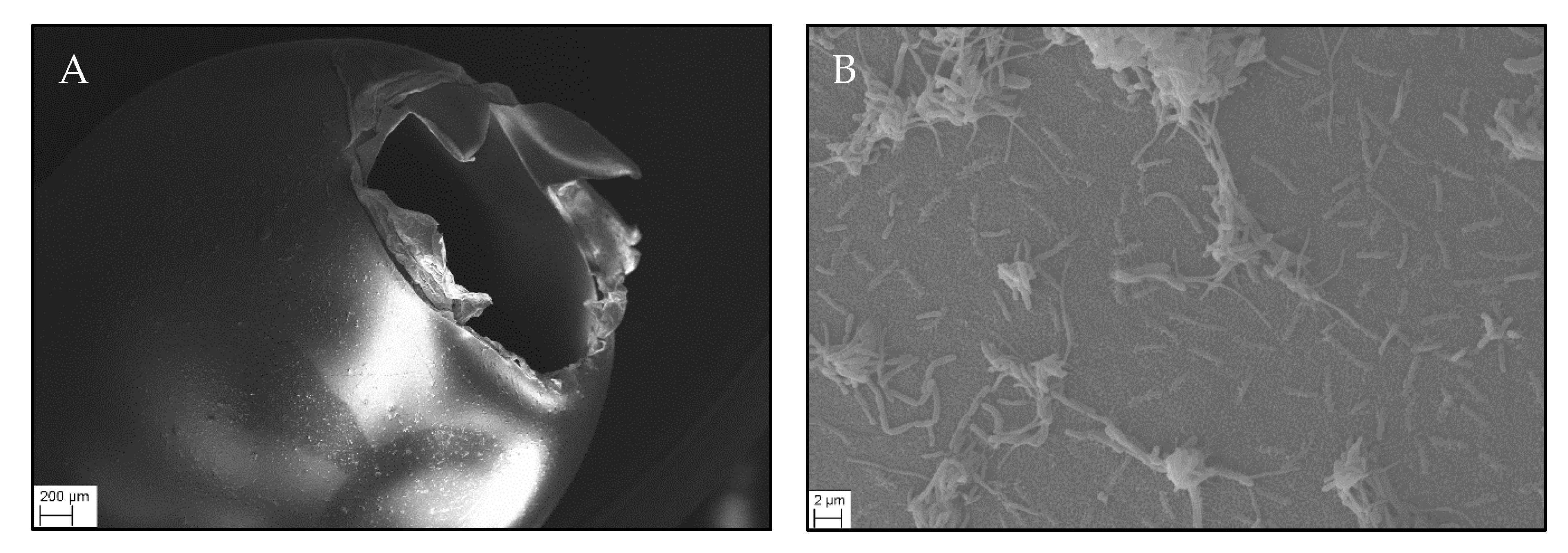
The fertilization rate of the eggs was 98%. Table 1 summarizes the results of the first part of experiment 1. Eye pigmentation started 37 dpf and was completed after 55 dpf. Over 99% of the larvae hatched between 68 and 84 dpf. No significant differences in time until eyed ova stage and hatch were found between the groups exposed to MP (MP1h, MP1tt, MP1ht) and the control group (eyed ova: d.f. = 3, n = 352, Χ2 = 16.401, p < 0.001, C1/MP1h p = 0.977, C1/MP1tt and C1/MP1ht p = 1; hatch d.f. = 3, n = 349, Χ2 = 171.66, p < 0.001, C1/MP1h p = 0.230, C1/MP1tt p = 0.999, C1/MP1ht p = 0.814). It became evident that the chorion of the eggs consists of several layers and exhibits no pores in the micrometer range (Figure 2). The heart rate of brown trout larvae was not affected by the exposure to PS MP (d.f. = 3/6.649, F = 3.971, p = 0.066).
The mortality did not differ between the treatment groups and was below 2% (d.f. = 3, n = 354, Χ2 = 0.0017, p = 1) in all groups. At 150 dpf, fish were 2.72 ± 0.18 cm long and weighted 0.15 ± 0.03 g on average. No significant differences were measured in length and body mass compared to the control group (length: n = 120, d.f. = 3, Χ2 = 4.857, p = 0.183; body mass: d.f. = 3/8, F = 0.928, p = 0.471). The level of LPO was alike in all treatment groups (d.f. = 3/8.072, F = 0.928, p = 0.471).

3.2. Experiment 1b: Exposure of Fry

No fish died during the second part of the experiment. The fish were 2.92 ± 0.21 cm long and weighted 0.19 ± 0.04 g on average. MP had neither a significant effect on biometric values nor on the oxidative stress level compared to the control (Table 2; length: d.f. = 3/7.819, F = 2.511, p = 0.134; body mass: d.f. = 3/220, F = 3.576, p = 0.015, C1/MP1h: p = 0.064, C1/MP1tt: p = 0.729, C1/MP1ht: p = 0.997; SOD: d.f. = 3/115, F = 0.341, p = 0.795; LPO: d.f. = 3/116, F = 2.904, p = 0.038, C1/MP1h: p = 0.355, C1/MP1tt: p = 0.286, C1/MP1ht: p = 0.286).

3.3. Experiment 2

At every time point sampled, the concentration of amitriptyline in C2, MP2ht, MP2mio was below the limit of detection (10 ng/L). The nominal concentration of amitriptyline in the groups AMI2, MIX2ht and MIX2mio was 300 µg/L. However, at the beginning of the experiment the real concentration was only between 76% and 56% of the nominal concentration (Table 3).
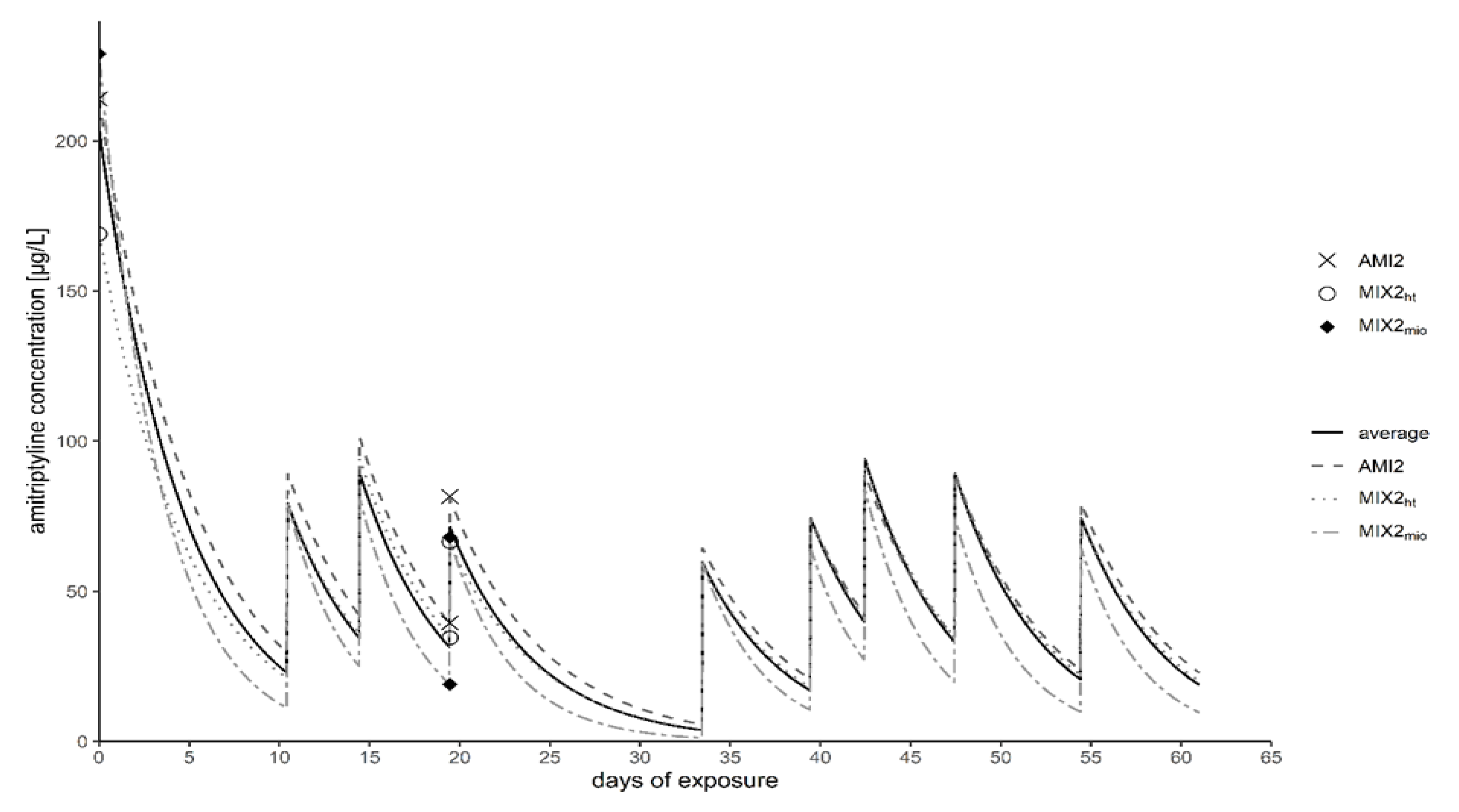
Prior to the water exchange, the measured concentration of amitriptyline was only between 6% and 13% of the nominal concentration in all three groups. After the exchange of half of the media with freshly prepared amitriptyline solutions, it was between 22% and 27% of the nominal concentration. Due to the strong depletion of amitriptyline during the experiment, we modeled the concentration fish were exposed to over time (Figure 3). Based on the measured concentrations, the removal of amitriptyline from the aqueous phase was assumed as pseudo-first order:
  y =   c 0 × e k × t e x p .
where c0 is the initial concentration and texp are the days of exposure. To adjust the model to the measured amitriptyline concentrations, for k values between 0.19 and 0.29 were presumed. According to this model, the average amitriptyline concentration during the experiment was 48 ± 33 µg/L.
Results of the second experiment are summarized in Table 4. No mortality occurred in the control group (C2) and it was below 5% in all treatment groups (Χ2 = 0.3102, d.f. = 5, p = 0.9974).
Fish hatched on average 11 days after the start of the experiment (Figure 4). While no differences were found between C2 and MP2ht as well as between C2 and MP2mio, all exposure groups with amitriptyline (AMI2, MIX2ht and MIX2mio) hatched significantly earlier than the control group and the two MP treatment groups (Χ2 = 164.6, d.f. = 5, p < 0.0001). Neither trout treated with MP nor those exposed to amitriptyline or the mixture of both showed an influence on their heart rate (d.f. = 5/7.5129, F = 0.2909, p = 0.9044).
MP had no effect on body mass and length of the fry while fish exposed to AMI2, MIX2ht and MIX2mio weighted significantly less and were significantly smaller than C2, MP2ht and MP2mio (Table 4; body mass: d.f. = 5/8.6811, F = 36.602, p < 0.0001; length: d.f. = 5/10.75, F = 65.262, p < 0.0001).
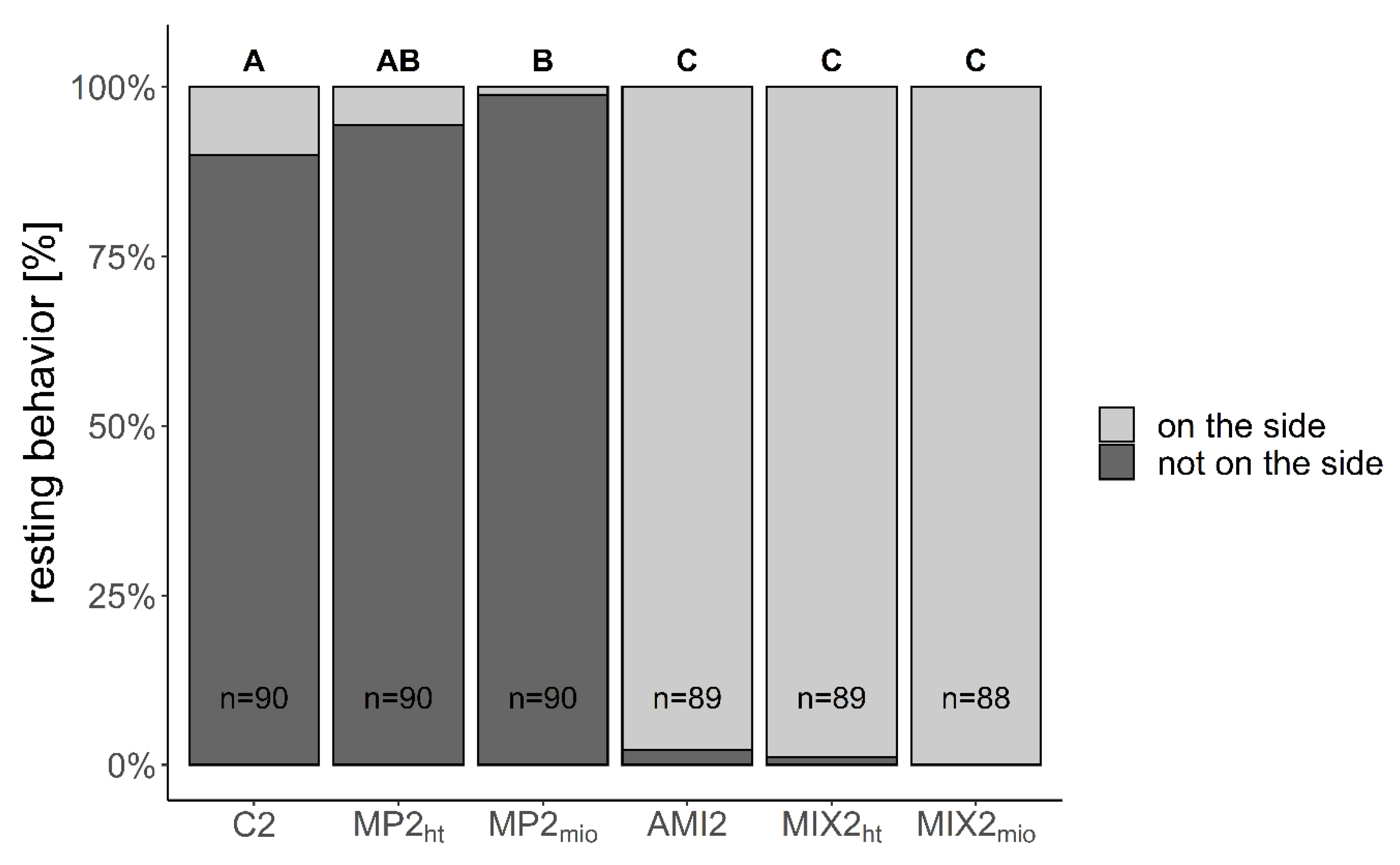
The resting behavior was influenced by the different treatments: Compared to C2, significantly less fish exposed to MP2mio were resting on their side, while nearly all fish exposed to AMI2, MIX2ht and MIX2mio showed this behavior (Figure 5; Χ2 = 604.081, d.f. = 5, p < 0.001). Furthermore, abnormal swimming behavior was observed as larvae exposed to AMI2, MIX2ht and MIX2mio showed looping behavior as well as side swimming.
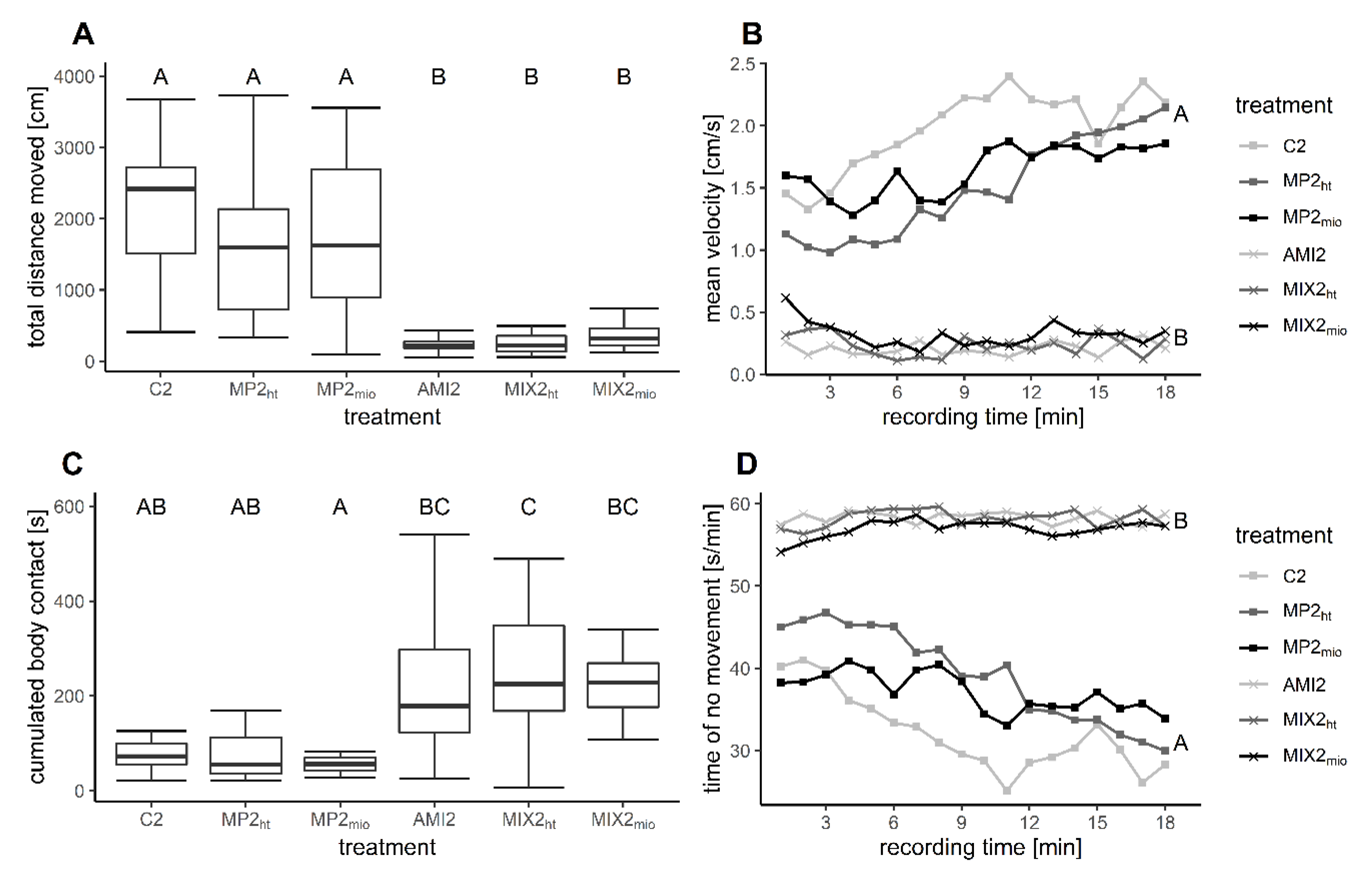
Video tracking revealed considerable differences in the behavior of fry (Figure 6, Table 4). For all investigated parameters, fish exposed to MP2ht and MP2mio did not differ from the control group. In contrast, fish exposed to AMI2, MIX2ht, and MIX2mio covered only 11–19% of the distance compared to the control (Figure 6A). In addition, in the three groups containing amitriptyline, fry swam significantly (84–90%) slower than the control fish (Figure 6B). In the control group and in MP2ht and MP2mio, the mean velocity increased over the recording time. This effect did not occur in the groups exposed to AMI2 or the mixtures. It is especially noticeable that the fry in the three groups containing amitriptyline had more body contact. Compared to the control group, the time fry had body contact to another fish was tripled in AMI2, MIX2ht, and MIX2mio (Figure 6C). Nevertheless, this behavior was highly variable among the different aquaria. A significant difference was only found between C2 and MIX2ht, MP2ht, and MIX2ht as well as MP2mio and MIX2mio. AMI2 and MIX2mio only showed a trend towards more body contact than the fry in the control group. Furthermore, fry exposed to AMI2, MIX2ht and MIX2mio spent significantly more time in inactivity than the fish in the control group and the two exposure groups containing solely MP (Figure 6D; distance: d.f. = 5/84, F = 21.25, p < 0.001; velocity: d.f. = 5/12, F = 35.290, p < 0.001, time: d.f. = 1/1601, F = 33.115 p < 0.001; no movement: d.f. = 5/12, F = 30.028, p < 0.001, time: d.f. = 1/2.0794 x 1021, F = 40.300, p < 0.001; body contact: d.f. = 5/12, F = 5.302, p = 0.008).
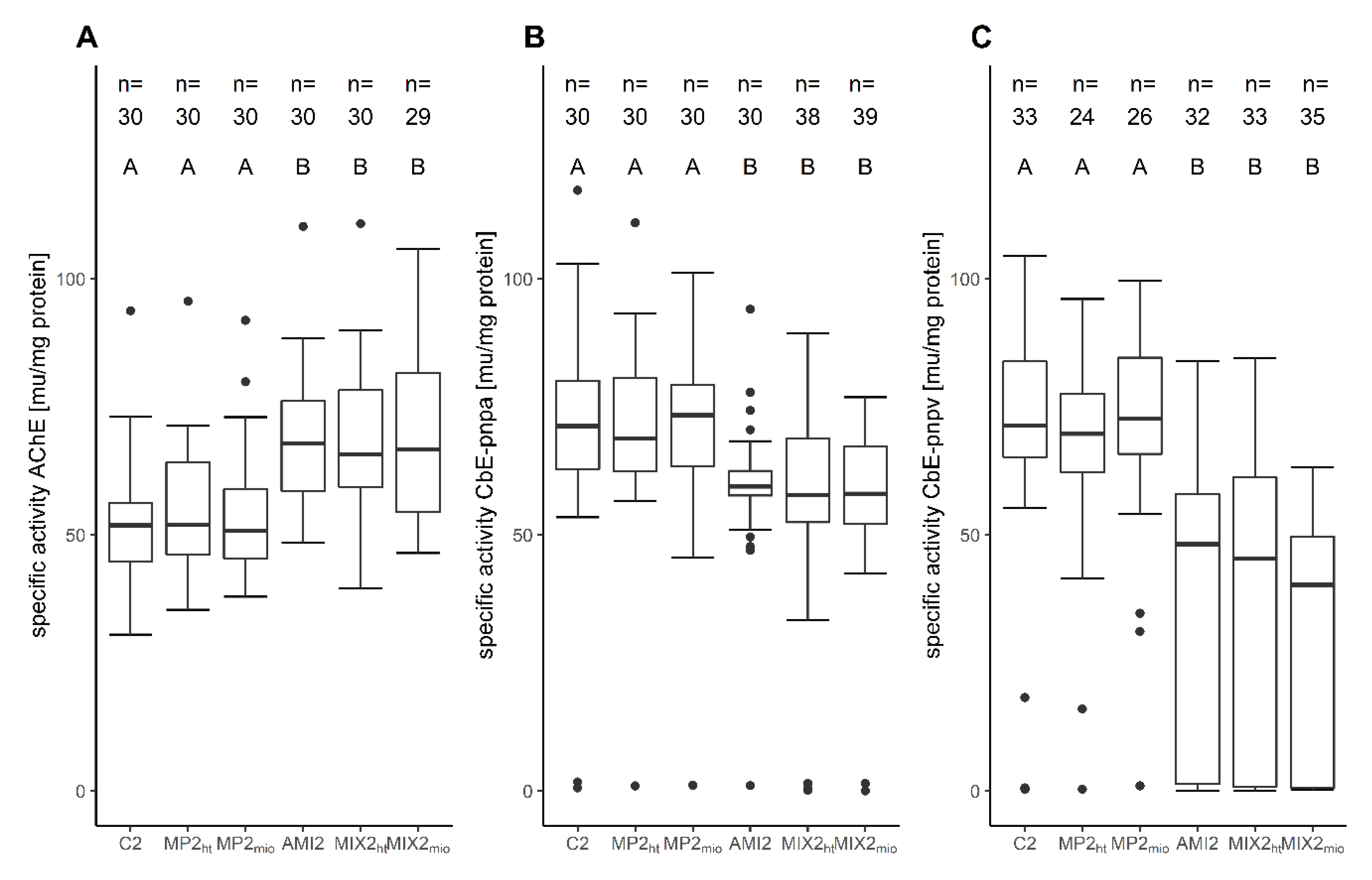
No differences occurred in the activity of SOD and the level of LPO between all exposure groups (SOD: d.f. = 5/173, F = 2.081, p = 0.070; LPO: d.f. = 5/12.287, F = 1.010, p = 0.452). The AChE activity was increased about 30% in all treatment groups with amitriptyline (Figure 7a). Contrarily, CbE-pnpa was reduced between 14% and 22% in the three exposure groups with the antidepressant. Outliers with very low activity of CbE-pnpa were found in all groups but occurred cumulatively in AMI2, MIX2ht, MIX2mio (Figure 7b). The activity of CbE-pnpv was even more reduced in the AMI2 and the mixture exposure groups (Figure 7c). Compared to the control, the activity of CbE-pnpv was 44 - 55% lower in AMI2, MIX2ht, MIX2mio (AChE: d.f. = 5/11.822, F = 6.081, p = 0.005; CbE-pnpa: d.f. = 5, Χ2 = 39.211, p < 0.001; CbE-pnpv: d.f. = 5/177, F = 22.135, p < 0.001).

4. Discussion

4.1. Amitriptyline Concentration

The measured amitriptyline concentrations were considerably lower than the nominal concentration of 300 µg/L. Due to the strong depletion of the amitriptyline concentration, fish were not constantly exposed to the same concentration. The concentration was considerably lower and fluctuated due to the water exchange design. Amitriptyline was found to be one of the most stable pharmaceuticals at pH 7 [77]. However, degradation of the antidepressant via photolysis occurs at low or high pH [77]. In our experiment, the pH was 8.3 ± 0.2 and thereby slightly alkaline. At pH 9, Baena-Nogueras, et al. [77] found a half-life of amitriptyline of 9.63 h (wavelength 300–800 nm, irradiance 500 W/m2). In our experiment stock solutions were covered in aluminum foil and aquaria were shaded from direct light which reduces the impact of photodegradation. Another factor that likely influenced the concentration of amitriptyline is a possible sorption of the chemical to the glass of the aquaria. Amitriptyline has a high adsorption capacity to kaolinite and Ca-montmorillonite [78,79]. One day prior to the start of the experiment, the tanks were filled with the corresponding amitriptyline solutions to saturate sorptive surfaces, and the test media were renewed before the start of the experiment. Nonetheless, it cannot be excluded that, still, sorption of amitriptyline to the glass occurred. A third process that has likely reduced the measured amitriptyline concentration is the uptake and metabolization of the antidepressant by the fish. Amitriptyline was found to bioconcentrate in brain tissue, gills, liver, blood plasma as well as in bile and muscle tissue of fish with bioconcentration factors between 4 up to 198 [80,81,82,83]. Furthermore, in gilt-head bream (Sparus aurata), amitriptyline was shown to be degraded to a broad range of metabolites including nortriptyline which is known to be also a bioactive antidepressant [83]. In our experiment, amitriptyline exposure led to severe effects in early life stages of brown trout which supports the assumption that fish had taken up the antidepressant. To counteract the decline of amitriptyline concentration, we performed as many water exchanges with freshly prepared amitriptyline solution and/or cryo-milled PS MP (whose amount was limited) as possible. However, the goal of our study, which was to show a possible modulation of amitriptyline-induced effects in early life stages of brown trout, was not influenced by the unexpectedly low amitriptyline concentrations, since even those induced strong reactions in the exposed fish.

4.2. Effects of MP

In the first experiment, brown trout eggs exposed to MP developed in a similar way as fish of the control group. This might be due to an impeded passage of the tested MP through the egg chorion which protects the first developmental stages of the fish embryo [84]. This explanation is highly probable since our own REM images showed the chorion of brown trout eggs to be free of micropores > 1 µm. Similarly, Li, et al. [11] describes that 10 µm PS MP accumulated at the outside of the chorion of marine medaka but failed to pass it. In a study with zebrafish, van Pomeren, et al. [15] found the uptake of nanoplastics via chorion and epidermis to be marginal. Furthermore, LeMoine, et al. [23] observed that MP could not pass the chorion of zebrafish and that the larvae started to ingest and accumulate MP (10–45 µm) not earlier than 5 d post hatch, i.e., as soon as they start to feed on exogenous food. Larvae of marine medaka ingested MP from two days post hatching [11]. In the second experiment we therefore decided to expose the fish in a later developmental stage. In addition to the different developmental stages at the start of the experiment, a slight difference in the temperature explains the faster development and higher heart rate of the fish in the second experiment. Killeen, et al. [85] investigated the influence of temperature on the development of brown trout in detail and described that the development is generally delayed at lower temperatures.
In both experiments of our study, PS MP had no effect on hatching, growth, mortality, or heart rate. LeMoine, et al. [23] and Chen, et al. [29] also observed that neither PE MP (10–45 µm, 480 particles/mL) nor PS MP (45 µm, 1 mg/L) had any effect on mortality, hatching or growth rate of zebrafish embryos. While PE MP (10–45 µm) incorporated into the diet of European sea bass likewise did not affect the growth of the fish, high dosages (105 particles/g diet) of PE MP had a slight but significant effect on the survival [12]. Furthermore, Malafaia, et al. [22] reported a trend to early hatching and lower survival of zebrafish larvae exposed to PE MP (38.26 ± 15.64 µm; up to 7.07 × 10³ particles/L). In contrast, Li, et al. [11] observed that PE MP (6534.0 ± 247.8 and 63,640.0 ± 723.5 particles/L) delayed the hatching and reduced the heart rate and growth of marine medaka. Overall, in most studies only minor effects on mortality and developmental parameters were observed in fish exposed to MP. A plausible reason for this might be that the chorion acts as protective barrier, and effects on this life stage are therefore more likely caused by chemicals leaking from the MP than by the particles themselves.
The exposure of brown trout to PS MP during the development apparently did not lead to oxidative stress as neither the activity of SOD nor the LPO level was influenced. Also, Chen, et al. [29] did not observe any influence of PS MP on the activity of catalase (CAT) and glutathione peroxidase (GPx) but they reported a significant decrease in the level of the reduced form of glutathione (GSH) in zebrafish larvae. In juvenile brown trout, also no effect of PS MP on oxidative stress was found [86]. Furthermore, in common gobies (Pomatoschistus microps) the LPO level was not affected by PE MP exposure in several studies [33,87,88,89]. Nevertheless, other studies showed that some MP may induce oxidative stress in fish [19,90,91]. Thus, it should be further investigated which parameters of MP as for example particle size, additives or the way of exposure can be responsible for MP-induced oxidative stress in fish.
PS MP did neither affect the AChE activity nor the activity of two CbEs of brown trout. Similar results were found for juvenile brown trout exposed to the same PS MP (104 particles/L) for 96 h [86]. Furthermore, PS MP (45 µm) did not cause alterations in the activity of AChE in zebrafish [29]. However, several studies showed that PE MP (1–5 µm) reduced the AChE activity in common gobies significantly [33,88,89]. Moreover, 30 days exposure to 200 µg/L fluorescent PE (70–88 μm) led to a significantly decreased AChE activity in the Amazonian discus fish (Symphysodon aequifasciatus) [20]. Considering the different polymer types and organisms investigated, the size of the administered particles seems to be important as the most neurotoxic effects are reported for small MP or even nanoplastics [13,29].
Exposure to 106 PS MP resulted in a minimal change in the resting behavior of trout larvae. However, it seems rather unlikely that this slight change is biologically relevant especially when considering the huge variation between the replicas. Furthermore, in the video tracking of brown trout fry, no differences in swimming and shoaling behavior were observed in response to MP exposure. Likewise, exposure of embryo and larvae of zebrafish to PE MP (10–45 µm) did not affect the covered distance during darkness [29]. Exposure of Krefft’s frillgobies (Bathygobius kreffti) to PE MP (38–45 mm) via diet did not affect their boldness or exploration behavior [92]. Moreover, Critchell and Hoogenboom [93] observed that the feeding and aggression behavior of juvenile planktivorous fish (Acanthochromis polyacanthus) was not affected by polyethylene terephthalate (PET) MP (<300 µm and 2 mm). In addition, the foraging activity and survival facing predators of post-larvae surgeonfish (Acanthurus triostegus) was not affected by PS MP (90 µm) exposure [94]. In contrast, juvenile black rockfish (Sebastes schlegelii) stayed more closely together when exposed to about 106 particles/L PS MP (15 µm) than the control group. Furthermore, black rockfish exposed to PS MP swam with reduced speed and showed both a diminished explorative behavior during search for food and an increased feeding time [95,96]. In European sea bass exposed to MP (0.69 mg/L; 1–5 µm) a significantly reduced swimming velocity and resistance time (0.26 and 0.69 mg/L) until being dragged away by water flow was observed [97]. Moreover, PE MP (1–5 µm and 420–500 µm) led, depending on the influence of the environmental condition during development, to a significant reduction of the predatory performance in early juvenile of common goby [89,98]. In two other studies investigating the effects of PE MP (1–5 µm) in common gobies, a non-significant reduction of the post exposure predatory performance was observed [87,88]. Likewise, the predatory performance of juvenile barramundi (Lates calcarifer) was not influenced by PS MP (97 µm) but the fish showed more curved swimming paths than control fish [99].
The factors that delimit effects of MP on the behavior of fish remain unclear. It does not seem that chemical properties of the polymer have a major importance as, for example, after exposure to PE MP and PS MP some studies found effects on the behavior of fish and some did not. It is possible that different size ranges or additives of the particles are responsible for the reported differences. Moreover, the discrepancies can be due to different sensitivity of the test species or the used study design.
In the present study, no biologically relevant effects of PS MP on the development and behavior of brown trout were observed.

4.3. Effects of Amitriptyline

In our experiment the survival of fish was not affected by the exposure to amitriptyline. However, the antidepressant led to a significantly reduced time until hatching. In common carp Sehonova, et al. [57] also observed a significant stimulation of hatching speed in fish exposed to 10, 100 and 500 µg/L amitriptyline. In addition, Yang, et al. [56] found that amitriptyline (1 ng up to 1 mg) reduced the time to hatch of zebrafish in a concentration-dependent manner. In contrast, higher concentrations of 3 mg/L amitriptyline led to an increased hatching time and a higher mortality in zebrafish [60]. This is, per se, not necessarily contradictory: In common carp, amitriptyline caused either developmental stimulation or retardation depending on the developmental stage and the concentration of the antidepressant [57]. Based on these data, amitriptyline seems to stimulate hatching of fish in general but may also delay hatching at high concentrations. In the present study, no influence was found on the heart rate of the larvae. In contrast, the considerably higher concentration of 3 mg/L led to a reduced heart rate in zebrafish larvae [60].
After two months, brown trout larvae exposed to amitriptyline were significantly smaller and weighted less than fish of the control group. Yang, et al. [56] reported that the body length of zebrafish was significantly reduced after exposure to concentrations as low as 100 ng/L amitriptyline for 120 h. Wu, et al. [100] found only a slight but insignificant reduction in body size of zebrafish and a modulation of the expression of genes encoding early growth response factors after exposure to 0.1 µg/L amitriptyline for 120 h. Thus, amitriptyline affects the growth of different fish species which can influence the survival of the larvae. Another reason for growth effects might be behavioral changes that led to less food consumption.
Exposure to amitriptyline did not cause an alteration of the activity of SOD or the level of LPO in early life stages of brown trout. Likewise, amitriptyline led to only moderate or minor effects on the oxidative stress level of zebrafish during development: The transcription of glutathione S-transferase (GST), GPx, and SOD genes in zebrafish embryos was not significantly altered in response to the antidepressant [58]. Nonetheless, a significant upregulation of CAT mRNA formation in fish exposed to 30 µg/L amitriptyline was observed after 144 h [58]. In common carp, exposure to 10 µg/L amitriptyline during development had no effect on the activity of CAT, GPx, GST and cytosolic SOD or the amount of protein carbonylation [57]. However, the antidepressant led to an increase in the level of LPO and in glutathione reductase activity [57]. Furthermore, Yang, et al. [56] have found a significantly positive influence of 100 ng/L amitriptyline to the antioxidant capacity of zebrafish as the activities of both SOD and CAT were enhanced and the formation of hydroxyl radicals and LPO was significantly suppressed. Nevertheless, in fish treated with higher amitriptyline concentrations the activity of SOD, CAT and peroxidase was found to be inhibited, and in zebrafish exposed to 1 mg/L amitriptyline the hydroxyl radical formation and the LPO level were significantly increased [56]. Brown trout might be less susceptible to oxidative stress caused by the tricyclic antidepressant than common carp and zebrafish whereas the rather low amitriptyline concentrations applied in our study do not allow any predictions about effects of higher concentrations that caused hydroxyl radical formation and an increase of LPO level in zebrafish.
Amitriptyline is a neuroactive compound and has been shown to induce neurotoxic effects in human cell lines as well as in non-target organisms. In a neuroblastoma cell line 100 µM amitriptyline caused a total loss of viability in neurons and a 30% loss of viability in astrocytes [101]. Sehonova, et al. [57] observed neuronal dystrophy in common carp exposed to amitriptyline (lowest observed effect concentration 10 µg/L). Moreover, mRNA expression of genes related to the development of eyes and the central nervous system (pax 6) were significantly downregulated in zebrafish exposed to 30 µg/L amitriptyline [60]. In the present study, the antidepressant increased the activity of AChE and significantly inhibited two other CbEs in early life stages of brown trout. The two CbE are involved in the detoxification of pollutants and are assumed to act in a protective way against AChE inhibiting pesticides [74]. An impact of amitriptyline on AChE activity is likely since amitriptyline was shown to act as a muscarinic acetylcholine receptor antagonist [44]. In human serum and erythrocyte ghosts, amitriptyline caused a decrease of the AChE activity [102]. However, to the best of our knowledge, our study was the first to investigate the effects of amitriptyline on the activities of AChE, CbE-pnpv, and CbE-pnpa in non-target organisms.
Considering its influence on neuronal processes and the general purpose of antidepressants to alter behavior, it is not surprising that amitriptyline caused also behavioral changes in non-target organisms. In our study fish showed altered resting and swimming behavior. Video tracking conditions in the used artificial system are rather stressful for the fish [103], therefore reduced velocity and covered distance can be interpreted as a result from an anxiolytic effect of amitriptyline. Even though it cannot be excluded that freezing behavior and, therefore, anxiogenic behavior increased the time of inactivity, it seems more probable that the effect was caused by the sedative effect of amitriptyline. Another possible explanation is that amitriptyline interfered with neuronal processes resulting in ataxic behavior, like looping or side swimming. Likewise, zebrafish exposed to 5 mg/L and 10 mg/L amitriptyline exhibited a significantly reduced maximal swimming velocity. Furthermore, 10 mg/L amitriptyline caused ataxic movement, like swimming vertically or on the side [53]. Moreover, exposure of adult zebrafish to 50 µg/L, 1 and 5 mg/L amitriptyline significantly reduced the time until fish entered the top in a novel tank test and increased the time they spent in the top region of the tank [53,54]. Meshalkina, et al. [54] also observed a reduction in the covered distance and mean velocity, and an increased meandering and angular velocity. In addition, decreased swimming activity was observed in zebrafish larvae treated with 100 µg/L amitriptyline in different exposure scenarios [59]. In another study, Sehonova, et al. [60] observed that 300 µg/L amitriptyline resulted in a significantly decreased swimming distance of zebrafish in the dark and, when exposed to 3000 µg/L amitriptyline, even in both dark and light conditions. Common carp exposed to 100 µg/L and 500 µg/L amitriptyline just floated apathetically and sank to the bottom of the aquarium [57].
Synoptically, our study revealed severe effects of amitriptyline on the behavior of brown trout at concentrations in the µg/L range (calculated average concentration of 48 µg/L amitriptyline). Even though also other antidepressants like citalopram [103,104], fluoxetine [105,106] or venlafaxine [106,107] exhibit an amitriptyline-analogous mode of action and have been shown to cause similar effects in non-target organisms our study was not designed to draw conclusions on the environmental risk of the antidepressant, since measured amitriptyline concentrations in surface waters are largely in the range of 22 ng/L [49,51,81]. To evaluate the ecotoxicological risk on non-target organisms, definitely further research on environmental relevant concentrations of amitriptyline as single substance and in combination with other antidepressants would be necessary. The focus of our study, however, was on the potential interactions of a psychoactive drug and MP.

4.4. Co-Exposure of MP and Amitriptyline

In our experiment brown trout were co-exposed to amitriptyline and PS MP. Like recently pointed out by Heinrich, et al. [108] environmental pollutants are present both in liquid (water) and solid (e.g., MP) compartments. Co-exposure allows to establish an equilibrium in the overall system including water, MP, and organisms. Co-exposure of brown trout to MP plus amitriptyline led to the same effects as the exposure to amitriptyline alone and the amitriptyline concentrations in the water phase of the treatment groups showed no considerable difference. While the mortality and heart rate as well as the oxidative stress level were not affected, larvae hatched significantly earlier, were smaller and weighted less than control fish. Furthermore, the swimming behavior was altered, and the activity of AChE was increased while the activities of the two other tested CbEs were inhibited. Therefore, we did not find any indication for PS MP to modulate the effects of amitriptyline on the development and behavior of brown trout. Several studies reported that MP can alleviate some negative effects caused by other pollutants by reducing their bioavailability [11,29,88]. On the other hand, MP was reported to increase negative effects of other contaminants [32,33,109]. In our experiments, measured amitriptyline concentrations were only slightly lower in those exposure groups which also included MP. Nevertheless, the data basis of the chemical analytic seems to be too limited to draw clear conclusions about the sorption of amitriptyline to MP. Only few studies found, like the present study, no interaction between MP and pollutants [86,90]. However, it cannot be excluded that this might have been resulting from a bias against the publication of negative results [27].

5. Conclusions

In our study PS MP (<50 µm up to 106 particles per liter) did not influence the development of brown trout. It is very likely that MP is not capable to pass the chorion so that the fish are protected until hatch. Therefore, it might be reasonable to focus on life stages that have hatched and started to feed on exogenous food. The only observed effect caused by MP in our experiment was a slight change in the resting behavior that occurred at the highest tested (but environmentally irrelevant) concentration. The biological relevance seems negligible considering the solely small change in behavior and the high variability among the replicas. The antidepressant amitriptyline affected development and behavior at considerably higher concentrations than reported in the environment. These effects were not modulated by the co-exposure of the antidepressant with PS MP. Overall, no harmful effects were caused by PS MP in the tested concentrations in brown trout and PS MP did not modulate the effects of amitriptyline on fish. Nevertheless, these results do not allow to deduce a general statement about the risk of MP considering the complexity caused by different polymers, size classes, and additives reported in the environment as well as different sensitivities of affected organisms.

Supplementary Materials

The following are available online at https://www.mdpi.com/2073-4441/12/9/2361/s1, Table S1: Size ranges (in µm) and counted particle numbers (per mL) of polystyrene particles in the stock suspension. Table S2: Volume of changed test medium in each Petri dish/tank during experiment 1 and experiment 2. Table S3: Average measured water parameters of the experiment 1b. Table S4: Average measured water parameters of the experiment 2. Table S5: Operating parameters of the triple quadrupole MS (Agilent 6490 QqQ) in positive mode. Table S6: Specific measurement parameters for amitriptyline with LC-QqQ in water samples. Intraday variations (RSD) is calculated with 1 µg/L standard (10 µL injection volume and 4 replicates (n)). Limit of quantification = LOQ. Table S7: Parameters for the determination of the lipid peroxide content in the different experiments. Table S8: Used data transformations for the statistical analysis. Table S9: Summary of all p-values of the single comparisons of the different endpoints. CRED reporting. Raw data experiment 1. Raw data experiment 2.

Author Contributions

Conceptualization: R.T., H.-R.K., and H.S.; methodology, H.S., S.K., S.T., A.S.R.; formal analysis, H.S.; investigation, H.S., J.K.Y.B., and S.T.; resources, H.-R.K., R.T., C.Z., and A.S.R.; data curation, H.S.; writing—original draft preparation, H.S. and S.T.; writing—review and editing, H.S., J.K.Y.B., S.K., A.S.R., S.T., C.Z., H.-R.K., and R.T.; visualization, H.S.; supervision, R.T., C.Z., and H.-R.K.; project administration, R.T.; funding acquisition, R.T. All authors have read and agreed to the published version of the manuscript.

Funding

This research was funded by the German Federal Ministry of Education and Research, Support Code: 02WRS1378. The experiments were conducted within the joint project MiWa (Microplastics in the water cycle—sampling, sample preparation, analytics, occurrence, removal, and assessment).

Acknowledgments

We wish to thank Martin Jekel for the initiation and coordination of the MiWa project. For the scanning electron microscope analysis, we thank Monika Meinert and Oliver Betz. Furthermore, the authors wish to thank Michael Ziegler, Stefanie Jacob, Katharina Peschke, Simon Schwarz, Carla Lorenz, Paul Thellmann, Andreas Dieterich, Sabrina Wilhelm as well as Katharina Reitter, Lea Schuster and Aron Meral for help, discussion, and technical assistance. We acknowledge support by Open Access Publishing Fund of University of Tübingen.

Conflicts of Interest

The authors declare no conflict of interest. The funders had no role in the design of the study; in the collection, analyses, or interpretation of data; in the writing of the manuscript, or in the decision to publish the results.

References

  1. Allen, S.; Allen, D.; Phoenix, V.R.; Le Roux, G.; Durántez Jiménez, P.; Simonneau, A.; Binet, S.; Galop, D. Atmospheric transport and deposition of microplastics in a remote mountain catchment. Nat. Geosci. 2019, 12, 339–344. [Google Scholar] [CrossRef]
  2. Peeken, I.; Primpke, S.; Beyer, B.; Gütermann, J.; Katlein, C.; Krumpen, T.; Bergmann, M.; Hehemann, L.; Gerdts, G. Arctic sea ice is an important temporal sink and means of transport for microplastic. Nat. Commun. 2018, 9, 1–12. [Google Scholar] [CrossRef] [Green Version]
  3. Piehl, S.; Leibner, A.; Löder, M.G.J.; Dris, R.; Bogner, C.; Laforsch, C. Identification and quantification of macro- and microplastics on an agricultural farmland. Sci. Rep. 2018, 8, 17950. [Google Scholar] [CrossRef] [Green Version]
  4. Wang, W.; Ndungu, A.W.; Li, Z.; Wang, J. Microplastics pollution in inland freshwaters of China: A case study in urban surface waters of Wuhan, China. Sci. Total Environ. 2017, 575, 1369–1374. [Google Scholar] [CrossRef]
  5. Claessens, M.; Meester, S.D.; Landuyt, L.V.; Clerck, K.D.; Janssen, C.R. Occurrence and distribution of microplastics in marine sediments along the Belgian coast. Mar. Pollut. Bull. 2011, 62, 2199–2204. [Google Scholar] [CrossRef]
  6. Dris, R.; Gasperi, J.; Mirande, C.; Mandin, C.; Guerrouache, M.; Langlois, V.; Tassin, B. A first overview of textile fibers, including microplastics, in indoor and outdoor environments. Environ. Pollut. 2017, 221, 453–458. [Google Scholar] [CrossRef] [Green Version]
  7. Leslie, H.A.; Brandsma, S.H.; Van Velzen, M.J.M.; Vethaak, A.D. Microplastics en route: Field measurements in the Dutch river delta and Amsterdam canals, wastewater treatment plants, North Sea sediments and biota. Environ. Int. 2017, 101, 133–142. [Google Scholar] [CrossRef]
  8. Vianello, A.; Jensen, R.L.; Liu, L.; Vollertsen, J. Simulating human exposure to indoor airborne microplastics using a Breathing Thermal Manikin. Sci. Rep. 2019, 9, 8670. [Google Scholar] [CrossRef]
  9. Li, C.; Busquets, R.; Campos, L.C. Assessment of microplastics in freshwater systems: A review. Sci. Total Environ. 2020, 707, 135578. [Google Scholar] [CrossRef]
  10. Batel, A.; Linti, F.; Scherer, M.; Erdinger, L.; Braunbeck, T. Transfer of benzo[a]pyrene from microplastics to Artemia nauplii and further to zebrafish via a trophic food web experiment: CYP1A induction and visual tracking of persistent organic pollutants. Environ. Toxicol. Chem. 2016, 35, 1656–1666. [Google Scholar] [CrossRef]
  11. Li, Y.; Wang, J.; Yang, G.; Lu, L.; Zheng, Y.; Zhang, Q.; Zhang, X.; Tian, H.; Wang, W.; Ru, S. Low level of polystyrene microplastics decreases early developmental toxicity of phenanthrene on marine medaka (Oryzias melastigma). J. Hazard. Mater. 2020, 385, 121586. [Google Scholar] [CrossRef] [PubMed]
  12. Mazurais, D.; Ernande, B.; Quazuguel, P.; Severe, A.; Huelvan, C.; Madec, L.; Mouchel, O.; Soudant, P.; Robbens, J.; Huvet, A.; et al. Evaluation of the impact of polyethylene microbeads ingestion in European sea bass (Dicentrarchus labrax) larvae. Mar. Environ. Res. 2015, 112, 78–85. [Google Scholar] [CrossRef] [PubMed] [Green Version]
  13. Ding, J.; Zhang, S.; Razanajatovo, R.M.; Zou, H.; Zhu, W. Accumulation, tissue distribution, and biochemical effects of polystyrene microplastics in the freshwater fish red tilapia (Oreochromis niloticus). Environ. Pollut. 2018, 238, 1–9. [Google Scholar] [CrossRef] [PubMed]
  14. Mattsson, K.; Johnson, E.V.; Malmendal, A.; Linse, S.; Hansson, L.-A.; Cedervall, T. Brain damage and behavioural disorders in fish induced by plastic nanoparticles delivered through the food chain. Sci. Rep. 2017, 7, 11452. [Google Scholar] [CrossRef] [PubMed]
  15. Van Pomeren, M.; Brun, N.R.; Peijnenburg, W.J.G.M.; Vijver, M.G. Exploring uptake and biodistribution of polystyrene (nano)particles in zebrafish embryos at different developmental stages. Aquat. Toxicol. 2017, 190, 40–45. [Google Scholar] [CrossRef] [PubMed]
  16. Triebskorn, R.; Braunbeck, T.; Grummt, T.; Hanslik, L.; Huppertsberg, S.; Jekel, M.; Knepper, T.P.; Krais, S.; Müller, Y.K.; Pittroff, M.; et al. Relevance of nano- and microplastics for freshwater ecosystems: A critical review. Trac. Trends Anal. Chem. 2019, 110, 375–392. [Google Scholar] [CrossRef]
  17. Karami, A.; Romano, N.; Galloway, T.; Hamzah, H. Virgin microplastics cause toxicity and modulate the impacts of phenanthrene on biomarker responses in African catfish (Clarias gariepinus). Environ. Res. 2016, 151, 58–70. [Google Scholar] [CrossRef] [PubMed]
  18. Lei, L.; Wu, S.; Lu, S.; Liu, M.; Song, Y.; Fu, Z.; Shi, H.; Raley-Susman, K.M.; He, D. Microplastic particles cause intestinal damage and other adverse effects in zebrafish Danio rerio and nematode Caenorhabditis elegans. Sci. Total Environ. 2018, 619–620, 1–8. [Google Scholar] [CrossRef] [PubMed]
  19. Lu, Y.; Zhang, Y.; Deng, Y.; Jiang, W.; Zhao, Y.; Geng, J.; Ding, L.; Ren, H. Uptake and accumulation of polystyrene microplastics in zebrafish (Danio rerio) and toxic effects in liver. Environ. Sci. Technol. 2016, 50, 4054–4060. [Google Scholar] [CrossRef] [PubMed]
  20. Wen, B.; Zhang, N.; Jin, S.-R.; Chen, Z.-Z.; Gao, J.-Z.; Liu, Y.; Liu, H.-P.; Xu, Z. Microplastics have a more profound impact than elevated temperatures on the predatory performance, digestion and energy metabolism of an Amazonian cichlid. Aquat. Toxicol. 2018, 195, 67–76. [Google Scholar] [CrossRef] [PubMed]
  21. Mohammed, A. Why are early life stages of aquatic organisms more sensitive to toxicants than adults? In New Insights into Toxicity and Drug Testing; Gowder, S.J.T., Ed.; IntechOpen: London, UK, 2013; pp. 49–62. [Google Scholar] [CrossRef] [Green Version]
  22. Malafaia, G.; De Souza, A.M.; Pereira, A.C.; Gonçalves, S.; Da Costa Araújo, A.P.; Ribeiro, R.X.; Rocha, T.L. Developmental toxicity in zebrafish exposed to polyethylene microplastics under static and semi-static aquatic systems. Sci. Total Environ. 2020, 700, 134867. [Google Scholar] [CrossRef] [PubMed]
  23. LeMoine, C.M.R.; Kelleher, B.M.; Lagarde, R.; Northam, C.; Elebute, O.O.; Cassone, B.J. Transcriptional effects of polyethylene microplastics ingestion in developing zebrafish (Danio rerio). Environ. Pollut 2018, 243, 591–600. [Google Scholar] [CrossRef] [PubMed]
  24. Wagner, M.; Scherer, C.; Alvarez-Muñoz, D.; Brennholt, N.; Bourrain, X.; Buchinger, S.; Fries, E.; Grosbois, C.; Klasmeier, J.; Marti, T.; et al. Microplastics in freshwater ecosystems: What we know and what we need to know. Environ. Sci. Eur. 2014, 26, 12. [Google Scholar] [CrossRef] [PubMed] [Green Version]
  25. Lithner, D.; Damberg, J.; Dave, G.; Larsson, Å. Leachates from plastic consumer products—Screening for toxicity with Daphnia magna. Chemosphere 2009, 74, 1195–1200. [Google Scholar] [CrossRef] [PubMed]
  26. Schiavo, S.; Oliviero, M.; Romano, V.; Dumontet, S.; Manzo, S.; Liu, G. Ecotoxicological assessment of virgin plastic pellet leachates in freshwater matrices. J. Environ. Account. Manag. 2018, 6, 345–353. [Google Scholar] [CrossRef]
  27. De Sá, L.C.; Oliveira, M.; Ribeiro, F.; Rocha, T.L.; Futter, M.N. Studies of the effects of microplastics on aquatic organisms: What do we know and where should we focus our efforts in the future? Sci. Total Environ. 2018, 645, 1029–1039. [Google Scholar] [CrossRef] [PubMed]
  28. Wang, F.; Wong, C.S.; Chen, D.; Lu, X.; Wang, F.; Zeng, E.Y. Interaction of toxic chemicals with microplastics: A critical review. Water Res. 2018, 139, 208–219. [Google Scholar] [CrossRef] [PubMed]
  29. Chen, Q.; Gundlach, M.; Yang, S.; Jiang, J.; Velki, M.; Yin, D.; Hollert, H. Quantitative investigation of the mechanisms of microplastics and nanoplastics toward zebrafish larvae locomotor activity. Sci. Total Environ. 2017, 584–585, 1022–1031. [Google Scholar] [CrossRef] [PubMed]
  30. Rehse, S.; Kloas, W.; Zarfl, C. Microplastics reduce short-term effects of environmental contaminants. Part I: Effects of bisphenol A on freshwater zooplankton are lower in presence of polyamide particles. Int. J. Environ. Res. Public Health 2018, 15, 280. [Google Scholar] [CrossRef] [PubMed] [Green Version]
  31. Guilhermino, L.; Vieira, L.R.; Ribeiro, D.; Tavares, A.S.; Cardoso, V.; Alves, A.; Almeida, J.M. Uptake and effects of the antimicrobial florfenicol, microplastics and their mixtures on freshwater exotic invasive bivalve Corbicula fluminea. Sci. Total Environ. 2018, 622–623, 1131–1142. [Google Scholar] [CrossRef] [PubMed]
  32. Nematdoost Haghi, B.; Banaee, M. Effects of micro-plastic particles on paraquat toxicity to common carp (Cyprinus carpio): Biochemical changes. Int. J. Environ. Sci. Technol. 2017, 14, 521–530. [Google Scholar] [CrossRef]
  33. Oliveira, M.; Ribeiro, A.; Hylland, K.; Guilhermino, L. Single and combined effects of microplastics and pyrene on juveniles (0+ group) of the common goby Pomatoschistus microps (Teleostei, Gobiidae). Ecol. Indic. 2013, 34, 641–647. [Google Scholar] [CrossRef]
  34. Bakir, A.; O’Connor, I.A.; Rowland, S.J.; Hendriks, A.J.; Thompson, R.C. Relative importance of microplastics as a pathway for the transfer of hydrophobic organic chemicals to marine life. Environ. Pollut. 2016, 219, 56–65. [Google Scholar] [CrossRef] [PubMed] [Green Version]
  35. Beckingham, B.; Ghosh, U. Differential bioavailability of polychlorinated biphenyls associated with environmental particles: Microplastic in comparison to wood, coal and biochar. Environ. Pollut. 2017, 220, 150–158. [Google Scholar] [CrossRef] [PubMed]
  36. Koelmans, A.A.; Bakir, A.; Burton, G.A.; Janssen, C.R. Microplastic as a vector for chemicals in the aquatic environment: Critical review and model-supported reinterpretation of empirical studies. Environ. Sci. Technol. 2016, 50, 3315–3326. [Google Scholar] [CrossRef] [PubMed]
  37. Dris, R.; Imhof, H.; Sanchez, W.; Gasperi, J.; Galgani, F.; Tassin, B.; Laforsch, C. Beyond the ocean: Contamination of freshwater ecosystems with (micro-) plastic particles. Environ. Chem. 2015, 12, 539–550. [Google Scholar] [CrossRef]
  38. Baker, D.R.; Kasprzyk-Hordern, B. Spatial and temporal occurrence of pharmaceuticals and illicit drugs in the aqueous environment and during wastewater treatment: New developments. Sci. Total Environ. 2013, 454–455, 442–456. [Google Scholar] [CrossRef] [PubMed]
  39. Fent, K.; Weston, A.A.; Caminada, D. Ecotoxicology of human pharmaceuticals. Aquat. Toxicol. 2006, 76, 122–159. [Google Scholar] [CrossRef] [PubMed]
  40. Sanderson, H.; Johnson, D.J.; Wilson, C.J.; Brain, R.A.; Solomon, K.R. Probabilistic hazard assessment of environmentally occurring pharmaceuticals toxicity to fish, daphnids and algae by ECOSAR screening. Toxicol. Lett. 2003, 144, 383–395. [Google Scholar] [CrossRef]
  41. Breyer-Pfaff, U. The Metabolic Fate of Amitriptyline, Nortriptyline and Amitriptylinoxide in Man. Drug Metab. Rev. 2004, 36, 723–746. [Google Scholar] [CrossRef] [PubMed]
  42. Chockalingam, R.; Gott, B.M.; Conway, C.R. Tricyclic antidepressants and monoamine oxidase Inhibitors: Are They Too Old for a New Look? In Antidepressants: From Biogenic Amines to Newe Mechanisms of Action; Macaluso, M., Preskorn, S.H., Eds.; Springer: Cham, Switzerland, 2019; Volume 250. [Google Scholar]
  43. Schwabe, U.; Paffrath, D.; Ludwig, W.-D.; Klauber, J. Arzneiverordnungs-Report 2019–Aktuelle Daten, Kosten, Trends und Kommentare; Springer: Berlin, Germany, 2019; p. 927. [Google Scholar]
  44. Snyder, S.H.; Yamamura, H.I. Antidepressants and the muscarinic acetylcholine receptor. Arch. Gen. Psychiatry 1977, 34, 236–239. [Google Scholar] [CrossRef] [PubMed]
  45. Nguyen, T.; Shapiro, D.A.; George, S.R.; Setola, V.; Lee, D.K.; Cheng, R.; Rauser, L.; Lee, S.P.; Lynch, K.R.; Roth, B.L.; et al. Discovery of a novel member of the histamine receptor family. Mol. Pharmacol. 2001, 59, 427. [Google Scholar] [CrossRef] [PubMed] [Green Version]
  46. Jang, S.-W.; Liu, X.; Chan, C.-B.; Weinshenker, D.; Hall, R.A.; Xiao, G.; Ye, K. Amitriptyline is a TrkA and TrkB receptor agonist that promotes TrkA/TrkB heterodimerization and has potent neurotrophic activity. Chem. Biol. 2009, 16, 644–656. [Google Scholar] [CrossRef] [PubMed] [Green Version]
  47. Rudorfer, M.V.; Potter, W.Z. Metabolism of tricyclic antidepressants. Cell. Mol. Neurobiol. 1999, 19, 373–409. [Google Scholar] [CrossRef] [PubMed]
  48. Ma, L.-D.; Li, J.; Li, J.-J.; Liu, M.; Yan, D.-Z.; Shi, W.-Y.; Xu, G. Occurrence and source analysis of selected antidepressants and their metabolites in municipal wastewater and receiving surface water. Environ. Sci. Process. Impacts 2018, 20, 1020–1029. [Google Scholar] [CrossRef] [PubMed]
  49. Ferrey, M.L.; Heiskary, S.; Grace, R.; Hamilton, M.C.; Lueck, A. Pharmaceuticals and other anthropogenic tracers in surface water: A randomized survey of 50 Minnesota lakes. Environ. Toxicol. Chem. 2015, 34, 2475–2488. [Google Scholar] [CrossRef] [PubMed]
  50. Lajeunesse, A.; Gagnon, C.; Sauvé, S. Determination of Basic Antidepressants and Their N-Desmethyl Metabolites in Raw Sewage and Wastewater Using Solid-Phase Extraction and Liquid Chromatography−Tandem Mass Spectrometry. Anal. Chem. 2008, 80, 5325–5333. [Google Scholar] [CrossRef] [PubMed]
  51. Thomas, K.V.; Da Silva, F.M.A.; Langford, K.H.; De Souza, A.D.L.; Nizzeto, L.; Waichman, A.V. Screening for selected human pharmaceuticals and cocaine in the urban streams of Manaus, Amazonas, Brazil. Jawra J. Am. Water Resour. Assoc. 2014, 50, 302–308. [Google Scholar] [CrossRef]
  52. Togola, A.; Budzinski, H. Multi-residue analysis of pharmaceutical compounds in aqueous samples. J. Chromatogr. A 2008, 1177, 150–158. [Google Scholar] [CrossRef] [PubMed]
  53. Demin, K.A.; Kolesnikova, T.O.; Khatsko, S.L.; Meshalkina, D.A.; Efimova, E.V.; Morzherin, Y.Y.; Kalueff, A.V. Acute effects of amitriptyline on adult zebrafish: Potential relevance to antidepressant drug screening and modeling human toxidromes. Neurotoxicol. Teratol. 2017, 62, 27–33. [Google Scholar] [CrossRef] [PubMed]
  54. Meshalkina, D.A.; Kysil, E.V.; Antonova, K.A.; Demin, K.A.; Kolesnikova, T.O.; Khatsko, S.L.; Gainetdinov, R.R.; Alekseeva, P.A.; Kalueff, A.V. The effects of chronic amitriptyline on zebrafish behavior and monoamine neurochemistry. Neurochem. Res. 2018, 43, 1191–1199. [Google Scholar] [CrossRef] [PubMed]
  55. Qiu, W.; Wu, M.; Liu, S.; Chen, B.; Pan, C.; Yang, M.; Wang, K.-J. Suppressive immunoregulatory effects of three antidepressants via inhibition of the nuclear factor-κB activation assessed using primary macrophages of carp (Cyprinus carpio). Toxicol. Appl. Pharmacol. 2017, 322, 1–8. [Google Scholar] [CrossRef] [PubMed]
  56. Yang, M.; Qiu, W.; Chen, J.; Zhan, J.; Pan, C.; Lei, X.; Wu, M. Growth inhibition and coordinated physiological regulation of zebrafish (Danio rerio) embryos upon sublethal exposure to antidepressant amitriptyline. Aquat. Toxicol. 2014, 151, 68–76. [Google Scholar] [CrossRef] [PubMed]
  57. Sehonova, P.; Plhalova, L.; Blahova, J.; Doubkova, V.; Marsalek, P.; Prokes, M.; Tichy, F.; Skladana, M.; Fiorino, E.; Mikula, P.; et al. Effects of selected tricyclic antidepressants on early-life stages of common carp (Cyprinus carpio). Chemosphere 2017, 185, 1072–1080. [Google Scholar] [CrossRef] [PubMed]
  58. Sehonova, P.; Zikova, A.; Blahova, J.; Svobodova, Z.; Chloupek, P.; Kloas, W. mRNA expression of antioxidant and biotransformation enzymes in zebrafish (Danio rerio) embryos after exposure to the tricyclic antidepressant amitriptyline. Chemosphere 2019, 217, 516–521. [Google Scholar] [CrossRef] [PubMed]
  59. Huang, I.J.; Sirotkin, H.I.; McElroy, A.E. Varying the exposure period and duration of neuroactive pharmaceuticals and their metabolites modulates effects on the visual motor response in zebrafish (Danio rerio) larvae. Neurotoxicol. Teratol. 2019, 72, 39–48. [Google Scholar] [CrossRef] [PubMed]
  60. Sehonova, P.; Hodkovicova, N.; Urbanova, M.; Örn, S.; Blahova, J.; Svobodova, Z.; Faldyna, M.; Chloupek, P.; Briedikova, K.; Carlsson, G. Effects of antidepressants with different modes of action on early life stages of fish and amphibians. Environ. Pollut. 2019, 254, 112999. [Google Scholar] [CrossRef] [PubMed]
  61. Schmidt-Posthaus, H.; Bernet, D.; Wahli, T.; Burkhardt-Holm, P. Morphological organ alterations and infectious diseases in brown trout Salmo trutta and rainbow trout Oncorhynchus mykiss exposed to polluted river water. Dis. Aquat. Org. 2001, 44, 161–170. [Google Scholar] [CrossRef] [PubMed] [Green Version]
  62. Klemetsen, A.; Amundsen, P.-A.; Dempson, J.B.; Jonsson, N.; Jonsson, B.; O’connell, M.; Mortensen, E. Atlantic salmon Salmo salar L., brown trout Salmo trutta L. and Arctic charr Salvelinus alpinus (L.): A review of aspects of their life histories. Ecol. Freshw. Fish. 2003, 12, 1–59. [Google Scholar] [CrossRef]
  63. Cheng, J.; Flahaut, E.; Cheng, S.H. Effect of carbon nanotubes on developing zebrafish (Danio rerio) embryos. Environ. Toxicol. Chem. 2007, 26, 708–716. [Google Scholar] [CrossRef] [PubMed]
  64. EU. Council Directive 2006/88/EC. Off. J. Eur. Union 2006, L 328, 14–56. [Google Scholar]
  65. Eitzen, L.; Paul, S.; Braun, U.; Altmann, K.; Jekel, M.; Ruhl, A.S. The challenge in preparing particle suspensions for aquatic microplastic research. Environ. Res. 2019, 168, 490–495. [Google Scholar] [CrossRef] [PubMed]
  66. Hansch, C.; Leo, A.; Hoekman, D. Exploring QSAR–Hydrophobic, Electronic, and Steric Constants; American Chemical Society: Washington, DC, USA, 1995; p. 167. [Google Scholar]
  67. OECD. Test. No. 212: Fish, Short-term Toxicity Test on Embryo and Sac-fry Stages; OECD Publishing: Paris, France, 1998. [Google Scholar] [CrossRef]
  68. Hermes-Lima, M.; Willmore, W.G.; Storey, K.B. Quantification of lipid peroxidation in tissue extracts based on Fe(III)xylenol orange complex formation. Free Radic. Biol. Med. 1995, 19, 271–280. [Google Scholar] [CrossRef]
  69. Monserrat, J.M.; Geracitano, L.A.; Pinho, G.L.L.; Vinagre, T.M.; Faleiros, M.; Alciati, J.C.; Bianchini, A. Determination of Lipid Peroxides in Invertebrates Tissues Using the Fe(III) Xylenol Orange Complex Formation. Arch. Environ. Contam. Toxicol. 2003, 45, 177–183. [Google Scholar] [CrossRef] [PubMed]
  70. Lowry, O.H.; Rosebrough, N.J.; Farr, A.L.; Randall, R.J. Protein measurement with the Folin phenol reagent. J. Biol. Chem. 1951, 193, 265–275. [Google Scholar] [PubMed]
  71. Markwell, M.A.K.; Haas, S.M.; Bieber, L.L.; Tolbert, N.E. A modification of the Lowry procedure to simplify protein determination in membrane and lipoprotein samples. Anal. Biochem. 1978, 87, 206–210. [Google Scholar] [CrossRef]
  72. Ellman, G.L.; Courtney, K.D.; Andres, V.; Featherstone, R.M. A new and rapid colorimetric determination of acetylcholinesterase activity. Biochem. Pharmacol. 1961, 7, 88–95. [Google Scholar] [CrossRef]
  73. Rault, M.; Collange, B.; Mazzia, C.; Capowiez, Y. Dynamics of acetylcholinesterase activity recovery in two earthworm species following exposure to ethyl-parathion. Soil Biol. Biochem. 2008, 40, 3086–3091. [Google Scholar] [CrossRef]
  74. Sanchez-Hernandez, J.C.; Mazzia, C.; Capowiez, Y.; Rault, M. Carboxylesterase activity in earthworm gut contents: Potential (eco)toxicological implications. Comp. Biochem. Physiol. Part C Toxicol. Pharmacol. 2009, 150, 503–511. [Google Scholar] [CrossRef] [PubMed]
  75. Benjamini, Y.; Hochberg, Y. Controlling the false discovery rate: A practical and powerful approach to multiple testing. J. R. Stat. Soc. Ser. B 1995, 57, 289–300. [Google Scholar] [CrossRef]
  76. Moermond, C.T.A.; Kase, R.; Korkaric, M.; Ågerstrand, M. CRED: Criteria for reporting and evaluating ecotoxicity data. Environ. Toxicol. Chem. 2016, 35, 1297–1309. [Google Scholar] [CrossRef] [PubMed] [Green Version]
  77. Baena-Nogueras, R.M.; González-Mazo, E.; Lara-Martín, P.A. Degradation kinetics of pharmaceuticals and personal care products in surface waters: Photolysis vs biodegradation. Sci. Total Environ. 2017, 590–591, 643–654. [Google Scholar] [CrossRef] [PubMed]
  78. Chang, P.-H.; Jiang, W.-T.; Li, Z.; Kuo, C.-Y.; Jean, J.-S.; Chen, W.-R.; Lv, G. Mechanism of amitriptyline adsorption on Ca-montmorillonite (SAz-2). J. Hazard. Mater. 2014, 277, 44–52. [Google Scholar] [CrossRef] [PubMed]
  79. Lv, G.; Stockwell, C.; Niles, J.; Minegar, S.; Li, Z.; Jiang, W.-T. Uptake and retention of amitriptyline by kaolinite. J. Colloid Interface Sci. 2013, 411, 198–203. [Google Scholar] [CrossRef] [PubMed]
  80. David, A.; Lange, A.; Tyler, C.R.; Hill, E.M. Concentrating mixtures of neuroactive pharmaceuticals and altered neurotransmitter levels in the brain of fish exposed to a wastewater effluent. Sci. Total Environ. 2018, 621, 782–790. [Google Scholar] [CrossRef] [PubMed]
  81. Lajeunesse, A.; Gagnon, C.; Gagné, F.; Louis, S.; Čejka, P.; Sauvé, S. Distribution of antidepressants and their metabolites in brook trout exposed to municipal wastewaters before and after ozone treatment—Evidence of biological effects. Chemosphere 2011, 83, 564–571. [Google Scholar] [CrossRef] [PubMed]
  82. Muir, D.; Simmons, D.; Wang, X.; Peart, T.; Villella, M.; Miller, J.; Sherry, J. Bioaccumulation of pharmaceuticals and personal care product chemicals in fish exposed to wastewater effluent in an urban wetland. Sci. Rep. 2017, 7, 16999. [Google Scholar] [CrossRef] [PubMed]
  83. Ziarrusta, H.; Mijangos, L.; Izagirre, U.; Plassmann, M.M.; Benskin, J.P.; Anakabe, E.; Olivares, M.; Zuloaga, O. Bioconcentration and biotransformation of amitriptyline in gilt-head bream. Environ. Sci. Technol. 2017, 51, 2464–2471. [Google Scholar] [CrossRef] [PubMed]
  84. Grierson, J.P.; Neville, A.C. Helicoidal architecture of fish eggshell. Tissue Cell 1981, 13, 819–830. [Google Scholar] [CrossRef]
  85. Killeen, J.; McLay, H.A.; Johnston, I.A. Development in Salmo trutta at different temperatures, with a quantitative scoring method for intraspecific comparisons. J. Fish Biol. 1999, 55, 382–404. [Google Scholar] [CrossRef]
  86. Schmieg, H.; Huppertsberg, S.; Knepper, T.P.; Krais, S.; Reitter, K.; Rezbach, F.; Ruhl, A.S.; Köhler, H.-R.; Triebskorn, R. Polystyrene microplastics do not affect juvenile brown trout (Salmo trutta f. fario) or modulate effects of the pesticide methiocarb. Environ. Sci. Eur. 2020, 32, 49. [Google Scholar] [CrossRef]
  87. Fonte, E.; Ferreira, P.; Guilhermino, L. Temperature rise and microplastics interact with the toxicity of the antibiotic cefalexin to juveniles of the common goby (Pomatoschistus microps): Post-exposure predatory behaviour, acetylcholinesterase activity and lipid peroxidation. Aquat. Toxicol. 2016, 180, 173–185. [Google Scholar] [CrossRef] [PubMed]
  88. Luís, L.G.; Ferreira, P.; Fonte, E.; Oliveira, M.; Guilhermino, L. Does the presence of microplastics influence the acute toxicity of chromium(VI) to early juveniles of the common goby (Pomatoschistus microps)? A study with juveniles from two wild estuarine populations. Aquat. Toxicol. 2015, 164, 163–174. [Google Scholar] [CrossRef] [PubMed]
  89. Miranda, T.; Vieira, L.R.; Guilhermino, L. Neurotoxicity, behavior, and lethal effects of cadmium, microplastics, and their mixtures on Pomatoschistus microps juveniles from two wild populations exposed under laboratory conditions―Implications to environmental and human risk assessment. Int. J. Environ. Res. Public Health 2019, 16, 2857. [Google Scholar] [CrossRef] [PubMed] [Green Version]
  90. Ferreira, P.; Fonte, E.; Soares, M.E.; Carvalho, F.; Guilhermino, L. Effects of multi-stressors on juveniles of the marine fish Pomatoschistus microps: Gold nanoparticles, microplastics and temperature. Aquat. Toxicol. 2016, 170, 89–103. [Google Scholar] [CrossRef] [PubMed]
  91. Qiao, R.; Sheng, C.; Lu, Y.; Zhang, Y.; Ren, H.; Lemos, B. Microplastics induce intestinal inflammation, oxidative stress, and disorders of metabolome and microbiome in zebrafish. Sci. Total Environ. 2019, 662, 246–253. [Google Scholar] [CrossRef] [PubMed]
  92. Tosetto, L.; Williamson, J.E.; Brown, C. Trophic transfer of microplastics does not affect fish personality. Anim. Behav. 2017, 123, 159–167. [Google Scholar] [CrossRef]
  93. Critchell, K.; Hoogenboom, M.O. Effects of microplastic exposure on the body condition and behaviour of planktivorous reef fish (Hoogen). PLoS ONE 2018, 13, e0193308. [Google Scholar] [CrossRef] [PubMed] [Green Version]
  94. Jacob, H.; Gilson, A.; Lanctôt, C.; Besson, M.; Metian, M.; Lecchini, D. No Effect of Polystyrene Microplastics on Foraging Activity and Survival in a Post-larvae Coral-Reef Fish, Acanthurus triostegus. Bull. Environ. Contam. Toxicol. 2019, 102, 457–461. [Google Scholar] [CrossRef] [PubMed]
  95. Yin, L.; Chen, B.; Xia, B.; Shi, X.; Qu, K. Polystyrene microplastics alter the behavior, energy reserve and nutritional composition of marine jacopever (Sebastes schlegelii). J. Hazard. Mater. 2018, 360, 97–105. [Google Scholar] [CrossRef] [PubMed]
  96. Yin, L.; Liu, H.; Cui, H.; Chen, B.; Li, L.; Wu, F. Impacts of polystyrene microplastics on the behavior and metabolism in a marine demersal teleost, black rockfish (Sebastes schlegelii). J. Hazard. Mater. 2019, 380, 120861. [Google Scholar] [CrossRef] [PubMed]
  97. Barboza, L.G.A.; Vieira, L.R.; Guilhermino, L. Single and combined effects of microplastics and mercury on juveniles of the European seabass (Dicentrarchus labrax): Changes in behavioural responses and reduction of swimming velocity and resistance time. Environ. Pollut. 2018, 236, 1014–1019. [Google Scholar] [CrossRef] [PubMed]
  98. De Sá, L.C.; Luís, L.G.; Guilhermino, L. Effects of microplastics on juveniles of the common goby (Pomatoschistus microps): Confusion with prey, reduction of the predatory performance and efficiency, and possible influence of developmental conditions. Environ. Pollut. 2015, 196, 359–362. [Google Scholar] [CrossRef] [PubMed]
  99. Guven, O.; Bach, L.; Munk, P.; Dinh, K.V.; Mariani, P.; Nielsen, T.G. Microplastic does not magnify the acute effect of PAH pyrene on predatory performance of a tropical fish (Lates calcarifer). Aquat. Toxicol. 2018, 198, 287–293. [Google Scholar] [CrossRef] [PubMed]
  100. Wu, M.; Liu, S.; Hu, L.; Qu, H.; Pan, C.; Lei, P.; Shen, Y.; Yang, M. Global transcriptomic analysis of zebrafish in response to embryonic exposure to three antidepressants, amitriptyline, fluoxetine and mianserin. Aquat. Toxicol. 2017, 192, 274–283. [Google Scholar] [CrossRef] [PubMed]
  101. Mannerström, M.; Toimela, T.; Ylikomi, T.; Tähti, H. The combined use of human neural and liver cell lines and mouse hepatocytes improves the predictability of the neurotoxicity of selected drugs. Toxicol. Lett. 2006, 165, 195–202. [Google Scholar] [CrossRef] [PubMed]
  102. Müller, T.C.; Rocha, J.B.T.; Morsch, V.M.; Neis, R.T.; Schetinger, M.R.C. Antidepressants inhibit human acetylcholinesterase and butyrylcholinesterase activity. Biochim. Biophys. Acta Mol. Basis Dis. 2002, 1587, 92–98. [Google Scholar] [CrossRef] [Green Version]
  103. Ziegler, M.; Knoll, S.; Köhler, H.R.; Tisler, S.; Huhn, C.; Zwiener, C.; Triebskorn, R. Impact of the antidepressant citalopram on the behaviour of two different life stages of brown trout. PeerJ 2020, 8, e8765. [Google Scholar] [CrossRef] [PubMed]
  104. Kellner, M.; Porseryd, T.; Porsch-Hällström, I.; Borg, B.; Roufidou, C.; Olsén, K.H. Developmental exposure to the SSRI citalopram causes long-lasting behavioural effects in the three-spined stickleback (Gasterosteus aculeatus). Ecotoxicology 2018, 27, 12–22. [Google Scholar] [CrossRef] [PubMed]
  105. Meijide, F.J.; Da Cuña, R.H.; Prieto, J.P.; Dorelle, L.S.; Babay, P.A.; Lo Nostro, F.L. Effects of waterborne exposure to the antidepressant fluoxetine on swimming, shoaling and anxiety behaviours of the mosquitofish Gambusia holbrooki. Ecotoxicol. Environ. Saf. 2018, 163, 646–655. [Google Scholar] [CrossRef] [PubMed]
  106. Painter, M.M.; Buerkley, M.A.; Julius, M.L.; Vajda, A.M.; Norris, D.O.; Barber, L.B.; Furlong, E.T.; Schultz, M.M.; Schoenfuss, H.L. Antidepressants at environmentally relevant concentrations affect predator avoidance behavior of larval fathead minnows (Pimephales promelas). Environ. Toxicol. Chem. 2009, 28, 2677–2684. [Google Scholar] [CrossRef] [PubMed] [Green Version]
  107. Maulvault, A.L.; Santos, L.H.M.L.M.; Paula, J.R.; Camacho, C.; Pissarra, V.; Fogaça, F.; Barbosa, V.; Alves, R.; Ferreira, P.P.; Barceló, D.; et al. Differential behavioural responses to venlafaxine exposure route, warming and acidification in juvenile fish (Argyrosomus regius). Sci. Total Environ. 2018, 634, 1136–1147. [Google Scholar] [CrossRef] [PubMed]
  108. Heinrich, P.; Hanslik, L.; Kämmer, N.; Braunbeck, T. The tox is in the detail: Technical fundamentals for designing, performing, and interpreting experiments on toxicity of microplastics and associated substances. Environ. Sci. Pollut. Res. 2020, 27, 22292–22318. [Google Scholar] [CrossRef] [PubMed]
  109. Lu, K.; Qiao, R.; An, H.; Zhang, Y. Influence of microplastics on the accumulation and chronic toxic effects of cadmium in zebrafish (Danio rerio). Chemosphere 2018, 202, 514–520. [Google Scholar] [CrossRef] [PubMed]
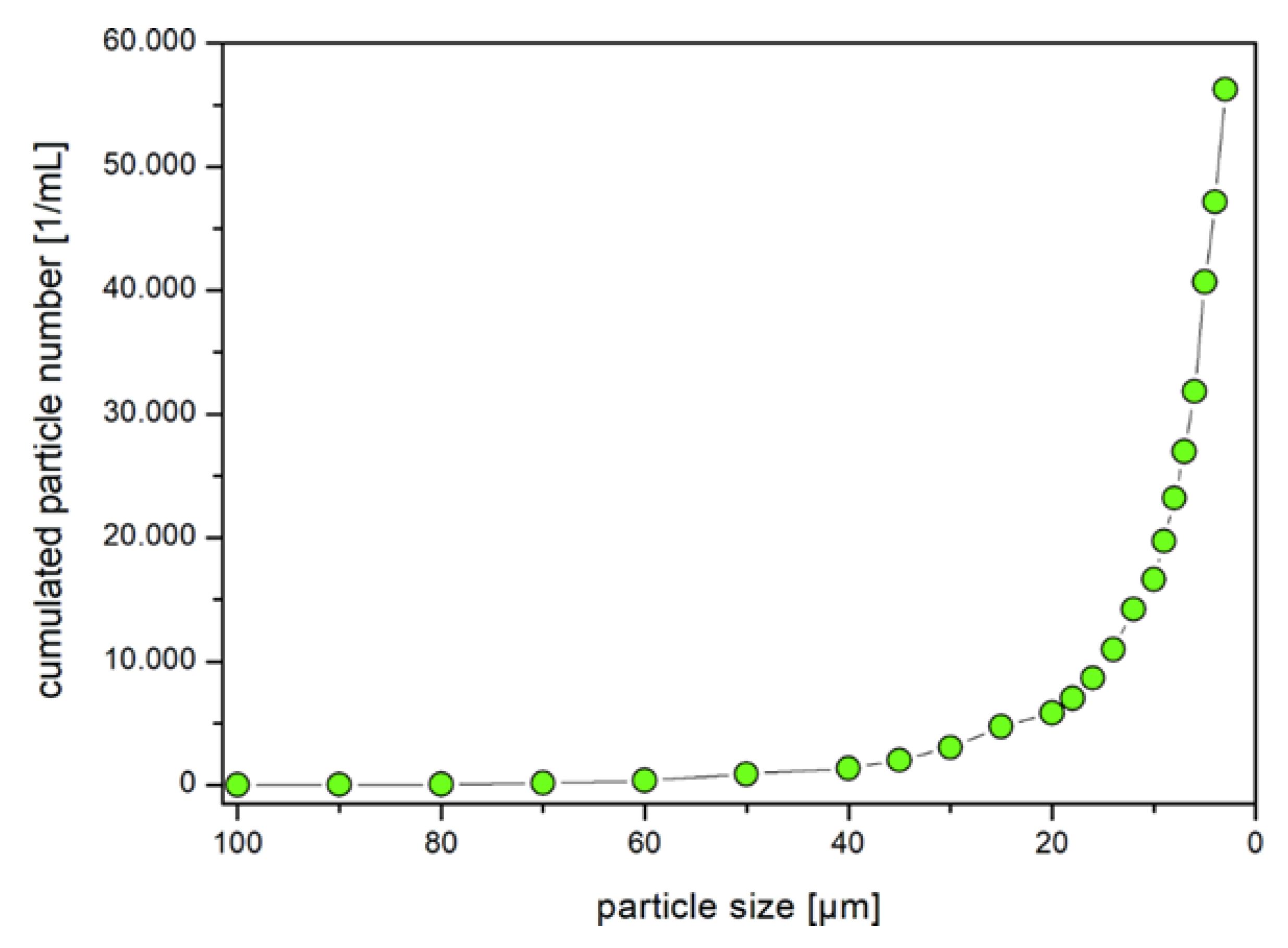
Figure 1. Size distribution of the used polystyrene microplastic particles (PS MP).
Water 12 02361 g001
Figure 2. SEM images of the chorion of a recently hatched brown trout. (A): Overview with distinguishable layers at the opening. (B): Detailed view of the chorion’s surface. No pores in µm range are present.
Water 12 02361 g002
Figure 3. Modelled amitriptyline concentration over the duration of the experiment. Measured concentrations of the three exposure groups (AMI2, MIX2ht, and MIX2mio) are depicted as points.
Water 12 02361 g003
Figure 4. Percent of brown trout hatched in the different treatment groups at the different days after exposure. All fish exposed to amitriptyline (AMI2, MIX2ht, MIX2mio) hatched significantly earlier than the control group and the groups containing solely MP (MP2ht and MP2mio).
Water 12 02361 g004
Figure 5. Resting behavior of brown trout fry after 42 days after exposure. Compared to the control significantly less fish exposed to MP2mio are resting on their side while significantly more fish exposed to amitriptyline (AMI2) or the mixture of amitriptyline and MP (MIX2ht and MIX2mio) are resting on their side (Χ2 = 604.0806, d.f. = 5, p < 0.0001, C2/MP2ht: p = 0.4048, C2/MP2mio: p = 0.01816, MP2ht /MP2mio: p = 0.2108, C2/AMI2, C2/MIX2ht, C2/MIX2mio, MP2ht /MIX2ht and MP2mio/MIX2mio p < 0.001, AMI2/ MIX2ht and MIX2ht/MIX2mio: p = 1, AMI2/ MIX2mio: p = 0.4972). Different letters indicate significant differences.
Water 12 02361 g005
Figure 6. Behavior of brown trout during video tracking (n = 15 in each group). (A): Total distance moved in 18 min. (B): Mean velocity. (C): Total time individuals had body contact. (D): Time of no movement over recording time. (A) and (C): The box plots display the median, the 25th and 75th percentiles as well as minimum and maximum values (whiskers); the dots indicate outliers. Different letters indicate significant differences.
Water 12 02361 g006
Figure 7. Enzyme activity in brown trout fry exposed to microplastic particles (MP), amitriptyline, or the mixture of both. (A): Specific activity of acetylcholinesterase (AChE). (B): Specific activity of carboxylesterases (CbE) with the substrate pnpa. (C): Specific activity of CbE with the substrate pnpv. The box plots display the median, the 25th and 75th percentiles as well as minimum and maximum values (whiskers); the dots indicate outliers. Different letters indicate significant differences.
Water 12 02361 g007
Table 1. Summary of data for the investigated endpoints in experiment 1a. All data are given as arithmetic means ± standard deviation.
Control
(C1)
100 Particles/L
(MP1h)
104 Particles/L
(MP1tt)
105 Particles/L
(MP1ht)
Mortality
(%)
1
±2
1
±2
1
±2
1
±2
Time until eyed ova stage
(dpf)
39
±1
39
±1
40
±3
40
±2
Time to hatch
(dpf)
75
±3
73
±3
73
±2
73
±3
Heart rate
(beats/min)
51
±2
56
±3
52
±2
53
±3
Length
(cm)
2.7
±0.2
2.7
±0.2
2.7
±0.2
2.8
±0.2
Body mass
(g)
0.15
±0.03
0.14
±0.02
0.14
±0.03
0.16
±0.02
Lipid peroxidation
(CHP-equiv.)
57.89
±10.20
60.12
±13.70
62.42
±12.17
61.23
±13.01
Table 2. Summary of data for the investigated endpoints in experiment 1b. All data are given as arithmetic means ± standard deviation.
Control
(C1)
100 Particles/L
(MP1h)
104 Particles/L
(MP1tt)
105 Particles/L
(MP1ht)
Mortality
(%)
0
±0
0
±0
0
±0
0
±0
Length
(cm)
3.0
±0.1
2.9
±0.2
3.0
±0.2
2.9
±0.2
Body mass
(g)
0.20
±0.02
0.17
±0.04
0.20
±0.04
0.19
±0.04
Lipid peroxidation
(CHP-equiv.)
58.28
±15.46
65.46
±22.14
52.30
±18.77
58.57
±15.51
SOD
(U/mL)
95.43
±28.47
105.60
±42.84
96.15
±31.87
98.60
±30.56
Table 3. Nominal and measured amitriptyline concentrations at the start of the experiment as well as prior and after a water exchange.
Nominal ConcentrationMeasured Concentration
Start of ExperimentPrior Water Exchange After Water Exchange
C20 µg/L<0.01 µg/L<0.01 µg/L<0.01 µg/L
MP2ht0 µg/L<0.01 µg/L<0.01 µg/L<0.01 µg/L
MP2mio0 µg/L<0.01 µg/L<0.01 µg/L<0.01 µg/L
AMI2300 µg/L214 µg/L40 µg/L82 µg/L
MIX2ht300 µg/L169 µg/L35 µg/L67 µg/L
MIX2mio300 µg/L229 µg/L19 µg/L68 µg/L
Table 4. Summary of data for the investigated endpoints in experiment 2. All data are given as arithmetic means ± standard deviation. p-values in comparison to the control group are given if significant differences occurred.
Control
(C2)
105 Particles/L
(MP2ht)
106 Particles/L
(MP2mio)
Amitriptyline
(AMI2)
Amitriptyline + 105 Particles/L
(MIX2ht)
Amitriptyline +106 Particles/L
(MIX2mio)
Mortality
(%)
0 ± 01 ± 10 ± 03 ± 42 ± 14 ± 3
Time to hatch
(days of exposure)
12 ± 212 ± 1
p = 0.786
11 ± 2
p = 0.580
10 ± 2
p< 0.001
10 ± 2
p< 0.001
10 ±2
p< 0.001
Heart rate
(beat /min)
56 ± 455 ± 256 ± 256 ± 154 ± 455 ± 2
Larvae resting on their side (%)10.0 ± 11.95.6 ± 4.2
p = 0.405
1.1 ± 1.6
p= 0.018
97.8 ± 3.1
p< 0.001
98.9 ± 1.6
p< 0.001
100.0 ± 0.0
p< 0.001
Total distance moved (cm)2135 ± 8621617 ± 1008
p = 0.385
1772 ± 1103
p = 0.756
224 ± 95
p< 0.001
253 ± 136
p< 0.001
345 ± 165
p< 0.001
Body contact (s)75 ± 3177 ± 49
p = 1
56 ± 17
p = 0.999
217 ± 137
p = 0.082
241 ± 137
p= 0.023
222 ± 63
p = 0.066
Mean velocity (cm/s)2.0 ± 0.81.5 ± 0.9
p = 0.129
1.6 ± 1.0
p = 0.505
0.2 ± 0.1
p< 0.001
0.2 ± 0.1
p< 0.001
0.3 ± 0.2
p< 0.001
No movement (s)579 ± 207706 ± 243
p = 0.628
668 ± 282
p = 0.902
1050 ± 20
p< 0.001
1049 ± 28
p< 0.001
1025 ± 36
p< 0.001
Length
(cm)
2.7 ± 0.12.6 ± 0.1
p = 0.603
2.7 ± 0.1
p = 1
2.4 ± 0.1
p< 0.001
2.4 ± 0.1
p< 0.001
2.4 ± 0.1
p< 0.001
Body mass
(g)
0.14 ± 0.030.14 ± 0.02
p = 1
0.15 ± 0.02
p = 0.963
0.11 ± 0.02
p< 0.001
0.11 ± 0.02
p< 0.001
0.12 ± 0.02
p< 0.001
Lipid peroxidation
(CHP-equiv.)
18.59 ± 2.6219.61 ± 4.0118.04 ± 2.4619.05 ± 2.4019.24 ± 3.7119.29 ± 2.53
SOD
(U/mL)
117.54 ± 28.89122.62 ± 28.11121.55 ± 24.58137.88 ± 33.58133.66 ± 29.16129.09 ± 31.48
AChE activity
(mu/mg protein)
52.46 ± 12.6654.48 ± 13.04
p = 0.997
53.69 ± 12.39
p = 0.999
68.29 ± 13.59
p= 0.008
68.53 ± 14.70
p= 0.008
68.87 ± 14.78
p= 0.008
CbE-pnpa activity
(mu/mg protein)
69.21 ± 22.8570.25 ± 17.96
p = 0.915
70.15 ± 17.54
p = 0.915
59.23 ± 14.10
p= 0.001
54.11 ± 23.21
p= 0.001
57.22 ± 15.89
p= 0.008
CbE-pnpv activity
(mu/mg protein)
70.28 ± 24.0466.15 ± 21.79
p = 0.859
70.43 ± 21.51
p = 1
39.12 ± 26.11
p< 0.001
38.27 ± 27.86
p< 0.001
31.33 ± 23.59
p< 0.001
Significant differences from the control group are highlighted in bold.