Review

Genetic Landscape of Familial Melanoma

by
Carmela Scarano
1,2,
Iolanda Veneruso
1,2,* and
Valeria D’Argenio
1,3
1
CEINGE-Biotecnologie Avanzate Franco Salvatore, 80145 Napoli, Italy
2
Department of Molecular Medicine and Medical Biotechnologies, Federico II University, 80131 Napoli, Italy
3
Department for the Promotion of Human Sciences and Quality of Life, San Raffaele Roma Open University, 00166 Rome, Italy
*
Author to whom correspondence should be addressed.
Genes 2025, 16(8), 857; https://doi.org/10.3390/genes16080857
Submission received: 2 July 2025 / Revised: 20 July 2025 / Accepted: 22 July 2025 / Published: 23 July 2025
(This article belongs to the Section Molecular Genetics and Genomics)

Abstract

About 10% of all forms of melanoma occur in a familial context and may be due to germline predisposing mutations transmitted as autosomal dominant traits within the affected families. CDKN2A is a highly penetrant gene associated to familial melanomas, being responsible of up to 40% of the cases. Other high, moderate, and low penetrance genes are being discovered, even if their own contribution to melanoma risk is still under debate. Indeed, next generation sequencing-based strategies enable large genomic regions to be analyzed, thus identifying novel candidate genes. These strategies, in diagnostic settings, may also improve the identification of the hereditary cases between all melanomas. The identification of the at-risk subjects gives an important opportunity for cancer surveillance in order to reduce the risk of onset and/or make early diagnosis. In addition, the identification of molecular biomarkers may drive the future development of specific targeted therapies, as already done for other inherited cancer syndromes. Here, we summarize the state of the art regarding the molecular basis of the hereditary susceptibility to develop melanoma.

1. Introduction

Melanoma is the most lethal skin cancer and its incidence has risen over the last decades, currently being the 17th most common cancer in both sexes. According to GLOBOCAN 2020 (https://gco.iarc.fr/, accessed on 20 July 2025) and the National Cancer Institute (https://seer.cancer.gov/statfacts/html/melan.html, accessed on 20 July 2025), the global incidence of melanoma is estimated at 21.9 cases per 100,000 persons per year, with the highest rates reported in Australia and New Zealand (up to 55–60/100,000). The incidence has continued to rise in recent decades, particularly among fair-skinned populations in Western countries. About 300,000 new cases have been diagnosed worldwide just in 2022 (https://www.wcrf.org/, accessed on 20 July 2025) and it has been estimated that in 2025 about 104,960 new melanomas will be diagnosed and about 8430 people will die of melanoma, only in the US (https://www.cancer.org/, accessed on 20 July 2025). Melanomas may develop through a multi-step process that start from benign, pre-cancerous lesions (nevus); however, most of them are developed de novo and it has also been reported that melanomas arising on a nevus sometimes do not share driver mutations with the preexisting nevus, suggesting that they may be two different and independent entities [1,2,3,4,5,6]. Even if most melanomas are sporadic and due principally to environmental risk factors, about 10% of all melanoma cases are familial and may be due to germline mutations in predisposing genes [7,8]. To date, several genes have been identified as risk factors for familial melanoma and, based on the probability of developing a form of melanoma if mutated, they are commonly classified into high, medium, and low penetrance genes [7,8]. As with other inherited cancers, recognizing individuals carrying an inherited predisposition to develop melanoma has important implications for identifying at-risk subjects within affected families and implementing preventive programs aimed at reducing cancer onset or enabling earlier diagnosis. Recent technological advances in the genomics field have made it possible to study multiple genomic loci, in several patients with a high resolution and at progressively reducing costs [9,10]. Accordingly, multiple genes panel testing is becoming commonly offered for the molecular diagnosis of an increasing number of human diseases, including hereditary cancers and also melanoma [11,12,13]. In this review, we will describe the different high, medium, and low penetrance genes that have been associated to date to a hereditary risk of melanoma, the diagnostic tests, and the recommendations currently available for the identification and management of the mutations’ carriers.

2. Methods

A comprehensive literature search was conducted using PubMed and the following keywords: “familial melanoma”, “familial melanoma predisposition”, “familial melanoma germline mutations”, “familial melanoma predisposing genes”, “CDKN2A germline variants”, “familial melanoma high penetrant genes”, “familial melanoma moderate penetrant genes”, “familial melanoma low penetrant genes”, “novel familial melanoma genes”, “familial melanoma susceptibility loci”, and “familial melanoma management”. As temporal windows, publications from 2000 to 2025 were searched; however, a manual search for the oldest references mentioned in the found articles was also carried out. Inclusion criteria required peer-reviewed original articles, meta-analyses, reviews, and guideline-based recommendations. Non-English reports were excluded. Papers in the search results reporting somatic mutations were not included since they are out of the topic of the present review. Clinvar and dbSNP databases have been used for variants assessment.

3. Defining Familial Melanoma Concept

Melanomas can develop in a familial context suggesting that a genetic susceptibility to melanoma development may be inherited within these affected families. Familial melanoma (7–15% of all melanomas) is defined as the occurrence of three or more melanoma cases within the same family branch, or at least two cases among first-degree relatives. Within these are defined as “hereditary melanomas”, those cases related to the presence of a germline pathogenic/likely pathogenic variant in a melanoma susceptibility gene (2% of all melanomas and about 20% of familial melanomas) [14].
Thus, in the presence of a personal history of multiple primary melanomas, and/or melanomas diagnosed in at least two first-degree relatives, and/or in families with at least three melanoma cases, genetic testing and counseling should be carried out to assess the presence of germline predisposing mutations in the proband and, consequently, in his/her family. It is important to underline that the criteria to include melanoma patients into genetic counseling may vary between countries taking into account the highest incidence in some geographic areas (i.e., USA and Australia) and also patient’s features, like the age at diagnosis [15,16,17,18,19].
The cyclin-dependent kinase inhibitor 2A (CDKN2A) gene is the most commonly mutated gene in familial melanoma; however, CDKN2A mutations explain up to 40% of hereditary cases [20,21]. Even if other susceptibility genes are being discovered (and other are probably yet to be discovered), it has also been suggested that a polygenic inheritance, featured by the co-occurrence of multiple low-risk variants and phenotype-modifier genes, may be involved. All the genes that have been associated to an increased risk of melanoma are reported in the next paragraphs according to their penetrance (Table 1).
Genes related to other hereditary cancer syndromes in which melanoma may be a “secondary” cancer, such as BRCA1/2, TP53, and PTEN, will be not covered by the present review; please refer to specific papers on these topics for more details [22,23,24,25].

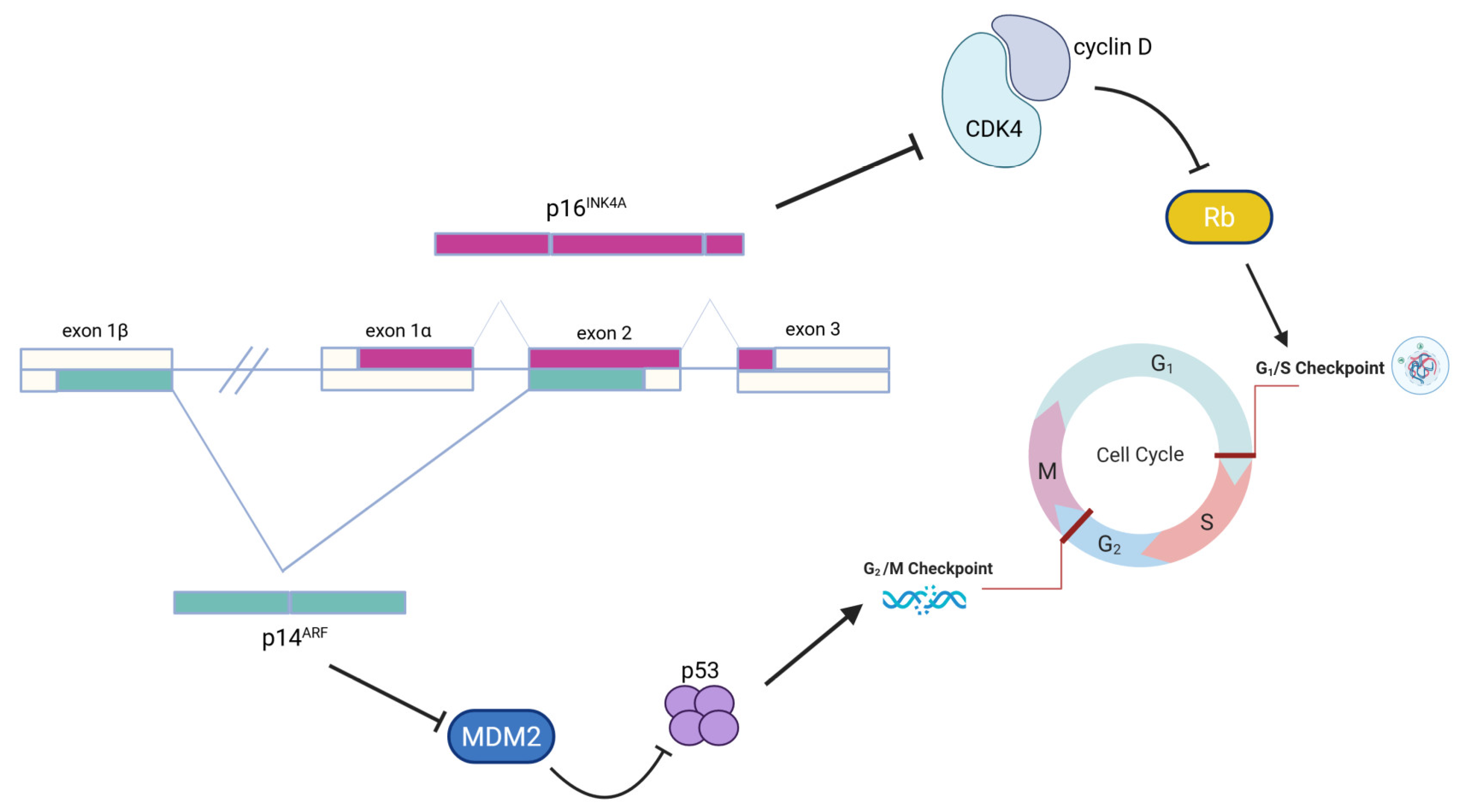
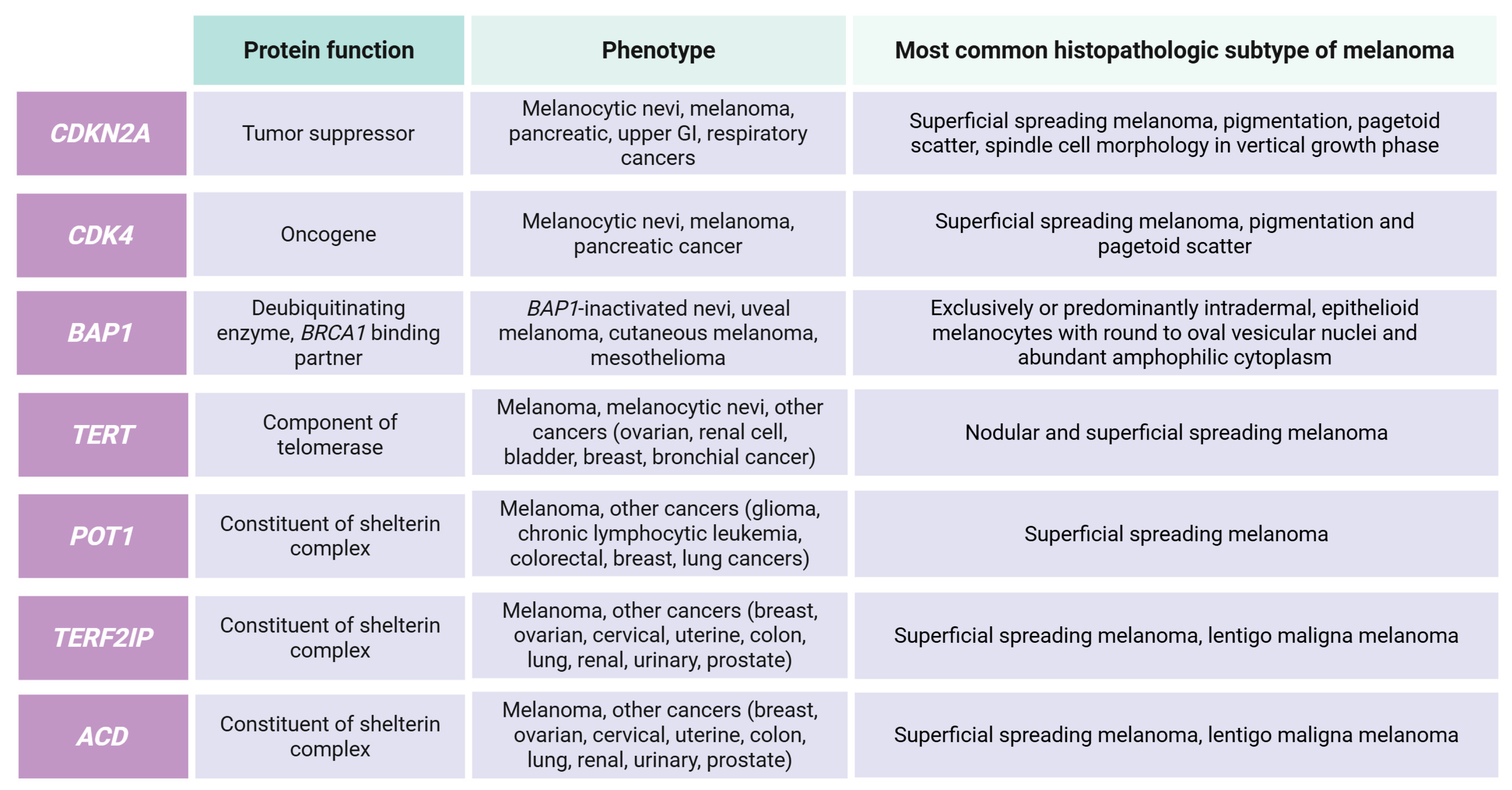
3.1. High-Risk Genes

As mentioned above, within the highly penetrant genes, the most commonly associated to hereditary susceptibility to melanoma is the CDKN2A gene [18]. Indeed, while the overall probability to find a germline CDKN2A mutation in people with melanoma is lower than 2%, this value raises up to 20–40% in familial melanomas [19]. The CDKN2A gene is located on chromosome 9p21.3 and, by using two different first exons, namely 1a and 1β, it encodes for two proteins: p16INK4A (156 amino acids) and p14ARF (132 amino acids), respectively [20]. Protein p16INK4A interacts with the Retinoblastoma protein and controls the G1 to S transition during the cell cycle. Protein p14ARF also plays an important role in the cell cycle regulation process: it induces cell cycle arrest in the G2 phase and the subsequent cell death (Figure 1). In malignant melanoma, usually, the two proteins are affected together [26].
To date, 14,786 sequence variations from dbSNP (https://www.ncbi.nlm.nih.gov/snp/, accessed on 20 July 2025) are known and, among them, 163 seem to be pathogenic/likely pathogenic for the ClinVar Database (https://www.ncbi.nlm.nih.gov/clinvar/, accessed on 20 July 2025).
Interestingly, there seems to be an association between the CDKN2A mutations’ frequency and the geographic area: different founder mutations have been described in Northern Europe, Iceland, UK, and European Mediterranean countries (France, Italy, and Spain) [27,28,29,30,31]. Nevertheless, also considering that penetrance varies depending on other environmental and genetic factors, CDKN2A mutations are usually related to early age of melanoma onset, multiple primary melanomas, and a high number of affected family members [23,32].
It has to be noticed that penetrance estimates for CDKN2A mutations vary widely, ranging from 30% to 70% by age 80, depending on geographic region and family selection criteria. In particular, the lifetime risk has been estimated at approximately 58% in Europe and up to 91% in Australia by age 80, according to data from the International Melanoma Genetics Consortium (https://genomel.org/, accessed on 20 July 2025). However, these evaluations may be also impaired by sample size and geographic region. As reported elsewhere [14], the Mediterranean population is often underrepresented in genetic melanoma studies, thus impairing unbiased risk estimations.
In addition to familial melanoma, CDKN2A germline mutations have been also associated with an elevated risk of pancreatic [33], lung, and other tobacco-related cancers [7,8,27,28,29,30,31]. These features characterize the FAMMM syndrome (OMIM 155601), which includes atypical nevi, multiple primary melanomas, and a family history of melanoma and/or pancreatic cancer [30]. Finally, CDKN2A mutated families have shown also an increased risk to develop other type of cancers, like breast cancer [28,29,34,35,36].
The CDK4 (Cyclin-Dependent Kinase 4) gene is located on the chromosome 12q14.1. The encoded protein is a Ser/Thr-kinase component of the cyclin D-CDK4 (DC) complex that is implicated in the phosphorylation and inhibition of retinoblastoma (RB) members, including RB1, and regulates the cell cycle during G1 to S phase transition [37]. Totally, 4485 CDK4 sequence variants have been reported to date, seven being classified as pathogenic/likely pathogenic and associated to cutaneous malignant melanoma onset. All these CDK4 variants are located in exon 2; in particular, two of them affect the codon 24 (p.Arg24His and p.Arg24Cys), which is involved in the binding of p16INK4A [14]. So far, CDK4 mutations have been reported in a limited number of families from different geographic areas (including Europe, Australia, and USA). Thus, due to their low frequency, CDK4 mutations’ contribution to familial melanoma risk still remain difficult to be accurately assessed.
BAP1 gene (BRCA1-Associated Protein 1) is located on chromosome 3p21.1. The protein function is to help in regulating many proteins involved in different cellular processes; indeed, the BAP1 protein controls cell growth, proliferation, and cell death mechanisms. In total, 248 out of 8420 BAP1 variants are known to be pathogenic/likely pathogenic (https://www.ncbi.nlm.nih.gov/clinvar/, accessed on 20 July 2025) [38]. These BAP1 gene mutations have been mainly associated to uveal melanoma and to an increasing number of cancers, including mesothelioma, meningioma, and renal cell carcinoma [39]. The different phenotypic expression seems to be related to different BAP1 gene mutations, showing variable penetrance for different tumor types [40]. However, since most of the families carrying a BAP1 germline mutation present at least one melanoma case, the high penetrance for familial melanoma seems to be confirmed [7]. To improve the understanding regarding the spectrum of BAP1-related cancers, a comprehensive study on the clinical features associated to all known BAP1 mutations has been carried out, highlighting the need for a curated registry in order to improve mutation carriers’ management [41]. However, the interpretation of BAP1 variants remains challenging, as most published studies are based on limited family cohorts or targeted sequencing in referral populations, often lacking proper control groups.
Recent studies have shown the importance in familial melanoma susceptibility, of genes that play a role in telomere maintenance, in the regulation of telomeres length, and in their protection [42,43]. Telomers are regions of repetitive nucleotide sequences at each end of a chromosome. Their principal function is to maintain the genomic stability and chromosomal integrity protecting chromosome ends from degradation, fusions, and atypical recombinations. Telomeres became smaller with age, smoking, and UV radiations. Consequently, these factors affecting telomeres maintenance processes are perfect candidates as risk factors for the onset of cancers [26,42,43]. Four genes belonging to this class have been implicated in familial melanoma susceptibility: (1) TERT (Telomerase Reverse Transcriptase); (2) POT1 (Protection of Telomeres 1); (3) ACD (ACD Shelterin Complex Subunit and Telomerase Recruitment Factor); and (4) TERF2IP (TERF2 Interacting Protein) [44,45].
The TERT gene is located on chromosome 5p15.33 and encodes for a catalytic subunit of telomerase involved in telomere length maintenance. A high penetrant mutation has been described so far in the TERT promoter region. This mutation has been found in a German family with four affected individuals, all of them carrying the same mutation [46]. A subsequent large population-based study identified this rare TERT promoter mutation in another melanoma family confirming its high penetrance, its association with early onset melanoma, and highlighting an increased risk of developing other cancers [47,48].
The POT1 gene is located on chromosome 7q31.33 and encodes a protein involved in telomeres protection. Mutations affecting this gene have been reported as highly penetrant for familial melanoma occurrence [49,50]. Roblez-Espinoza et al., analyzing several families from different countries, identified POT1 as a novel familial melanoma susceptibility gene; interestingly, they found that all POT1 mutations carriers were affected by melanoma, and most of them had early age of onset and multiple primary cancers, suggesting the high penetrance of this gene [49]. At the same time, Shi J et al., using whole exome sequencing, identified a POT1 founder mutation in apparently unrelated Italian families [50]. Subsequently, a novel POT1 mutation has been reported in a family with multiple cases of primary melanomas, as well as other kind of cancers [51], suggesting that a broader range of cancers may be associated to predisposing mutations in POT1. A large-population study was carried out in 694 Austrian patients to assess the role of POT1 germline mutations in predisposing familial melanoma [52]. The entire sequence analysis of POT1 in all the study subjects identified only eight variants in high-risk melanoma patients, and only one of these has been predicted as potentially pathogenic, suggesting that POT1 mutations are almost rare [52]. On the contrary, another study by Poltrony et al. carried out in 228 Spanish melanoma families suggests that POT1 mutations are involved in familial melanoma susceptibility in Spain [53]. Moreover, Wong et al. described a novel POT1 mutation supporting the need of including POT1 analysis in the assessment of familial melanoma inherited risk [54]. Finally, Abu Shtaya et al. recently described the identification of the POT1 p.(Ile78Thr) variant, previously classified with conflicting interpretations of pathogenicity, as a pathogenic variant with founder effect in the Ashkenazi Jews population [55].
ACD and TERF2IP genes are located on chromosome 16q22.1 and 16q23.1, respectively. These genes were identified as risk factors for familial melanoma by a large cohort study carried out on 510 melanoma families [56]. Seven ACD and three TERF2IP pathogenic/likely pathogenic variants were totally identified supporting the pivotal role of telomere dysregulation in melanoma susceptibility. Moreover, a recent study by Goldstein et al. highlighted the association between germline variants in telomere maintenance genes (POT1, TERF2IP, ACD, and TERT) and the spitzoid morphology in familial melanoma [57].
High-risk genes associated to hereditary melanoma are summarized in Figure 2.

3.2. Moderate-Risk Genes

The MC1R (Melanocortin 1 Receptor) gene is located on chromosome 16q24.3; the encoded protein, a transmembrane G protein coupled receptor, controls melanogenesis and in particular it plays an important role in normal skin pigmentation [58,59]. In this gene, 3107 variants are known and 42 are classified as pathogenic/likely pathogenic (https://www.ncbi.nlm.nih.gov/clinvar/, accessed on 20 July 2025). Interestingly, some MC1R polymorphisms may play a role in familial melanoma risk and it seems to be correlated to the number of inherited variants [7]. According to this finding, a multigene panel testing has been recently carried out on a large Dutch population, showing that MC1R risk variants had a strong association with the melanoma respect to the control group [60]. It has to be underlined that MC1R variants are common in the general population, and their modest effect size is difficult to isolate due to environmental confounders. Additionally, case-control studies often differ in their inclusion criteria and definitions, which complicates cross-study comparison.
The MITF gene (Melanocyte-Inducing Transcription Factor) is located on chromosome 3p13. MITF helps the development and functions of melanocytes. Melanocytes control the production of the pigment melanin, which contributes to hair, eye, and skin color [61]. In total, 90,634 variants were found spanning on the gene and 153 of them show a pathogenic/likely pathogenic interpretation (https://www.ncbi.nlm.nih.gov/clinvar/, accessed on 20 July 2025). To date, only one MITF variant, the p.Glu318Lys, has been associated to melanoma susceptibility [62]. The MITF p.Glu318Lys mutation not only increases the risk of melanoma, but has also been associated to high nevi count and fast-growing melanomas; interestingly, this mutation can be present also in p16INK4A mutations carriers [63,64]. Potjer et al. detected the MITF p.Glu318Lys mutation in 15 melanoma families suggesting a pivotal role of this mutation, at least in the Dutch population [60]. Mutation carriers should be encouraged to strict dermatologic surveillance, periodic self-examination, and renal cell carcinoma surveillance.
Finally, TERT variants not occurring in the promoter (see above) can behave as low-risk variants [17].

3.3. Low-Risk Genes

Recently, variants in other genes have shown an association with familial melanoma, but the risk conferred by these variants is low and still partly unclear [26,65].
The MGMT (O-6-Methylguanine-DNA Methyltransferase) gene is located on chromosome 10q26.3. The protein encoded is involved in the cellular defense against the biological effects of O6-methylguanine (O6-MeG) and O4-methylthymine (O4-MeT). More in detail, it repairs the methylated nucleobase in DNA by transferring the methyl group to a cysteine residue in the enzyme. This gene is considered a susceptibility gene for cutaneous melanoma [66] and a biomarker for glioblastoma and gliosarcoma. Moreover, MGMT gene promoter methylation was found in brain metastases of melanoma, lung, breast, and renal carcinoma [67].
Association studies (Genome-wide association studies—GWAS) found many SNPs in a different group of genes all involved in skin pigmentation, in particular, TPCN2, ASIP, KITLG, SLC24A5, SLC45A2, TYR, IRF4, OCA2, and TYRP1 genes, as well as the MC1R gene already discussed [68,69,70]. These results highlight the importance of pigmentation-related genes in the onset of melanoma and tumorigenesis through genetic–environment interactions. In particular, TPCN2 is an ion transport gene containing SNPs associated with hair pigmentation that have also been associated to increased melanoma risk, especially in males [71]. The ASIP gene inhibits MC1R shifting melanin color to a less protective isoform; this gene has also been associated to high-risk phenotype and melanoma susceptibility [72]. KITLG is involved in melanocytes development and maintenance; SNPs in this gene have been associated to altered skin pigmentation and increased risk of melanoma [69]. SLC24A5 transports calcium or potassium ions into the melanosome and plays a role in melanogenesis, while SLC45A2 is a sodium–hydrogen exchanger of melanosomes, regulating tyrosinase activity in human melanocyte; both genes have been proposed as risk factors for melanoma development [73]. TYR catalyzes the conversion of tyrosine to melanin; variants affecting this gene have been related to albinism and increased melanoma risk [64]. IRF4 is involved in gene expression regulation in response to cytokines and interferon: a SNP in this gene has been associated with increased risk of melanoma [74]. Finally, OCA2 and TYRP1 genes are both involved in oculocutaneous albinism: variants affecting these genes have been related to increased susceptibility to familial melanoma [75].

3.4. Candidate Genes

A GWAS study, reporting a meta-analysis combining data from 11 previous GWAS and an additional data set, identified 20 significant loci associated to familial melanoma: 13 already known, two previously-reported but not considered significant, and five novels (explaining an additional 2.3% of the familial relative risk) [76]. In particular, one locus includes putative melanocyte and keratinocyte enhancers and the CYP1B1 gene. CYP1B1 metabolizes hormones and has been associated to hormone-related cancers, such as prostate and breast cancers [77]. Interestingly, CYP1B1 is regulated by ARNT, a gene present in a melanoma-associated locus, suggesting a common pathogenetic pathway [78]. Another identified locus involves CDKAL1, a gene involved in gene expression regulation. The most significant SNP identified in this gene is localized in a predicted melanocyte enhancer that binds IRF4: this is intriguing since an IRF4 SNP (rs12203592) has been associated with nevus count, skin pigmentation, and tanning response [79,80]. Another region involves the AGR3 gene, a member of the protein disulfide isomerase family, already associated to breast cancer risk and poor survival in ovarian cancer [81,82]. Next, an association was found with a region including TMEM38B, ZNF462, and RAD23B genes, the latter being of particular interest due to its involvement in DNA repair. A locus spanning the OBFC1 gene and the SH3PXD2A promoter was highlighted and since this region is associated with telomere length; this suggests that OBFC1, as a member of telomeres maintenance complex, may be a candidate susceptibility gene for familial melanoma [76]. Another locus involves the CCND1 gene, in which somatic mutations has been already reported as a marker of poor survival in melanoma [76]. Finally, a locus including both OCA2 and HERC2 genes was also identified. These genes have been reported to be important determinants of eye color, thus suggesting that they may be potential candidates for susceptibility to familial melanoma [76].
Melanoma is an immunogenic tumor but the relationship between the immune system and the skin cancer is currently under investigation [83,84,85,86]. Indeed, another group of genes, including genes of the immune system, may be implicated in melanoma onset: IL-10, IL-1β, TNF-α (tumor necrosis factor alpha), HLA (human leukocyte antigen class II) genes, and IRF4 (interferon regulatory factor 4) genes [83,84].
Finally, novel genes are being proposed as risk factors for familial melanoma.
A rare non-synonymous variant has been identified in Golgi Membrane Protein 1 (GOLM1), rs149739829, in two patients from the same family [87]. The variant co-segregates with the phenotype in this family and was confirmed in independent cases, thus suggesting that GOLM1 may be a candidate susceptibility gene.
Similarly, an exome sequencing study performed in high-risk patients highlighted two TP53AIP1 truncating mutations in four patients, suggesting a role in melanoma predisposition [88].
In conclusion, moderate and low susceptibility genes have a weak impact on melanoma onset and families that carry these variants usually have only one or two melanoma cases. Nevertheless, it is possible that a combination of low to moderate inherited variants, also in combination with environmental factors, may contribute to the melanoma carcinogenesis.

3.5. Susceptibility Loci
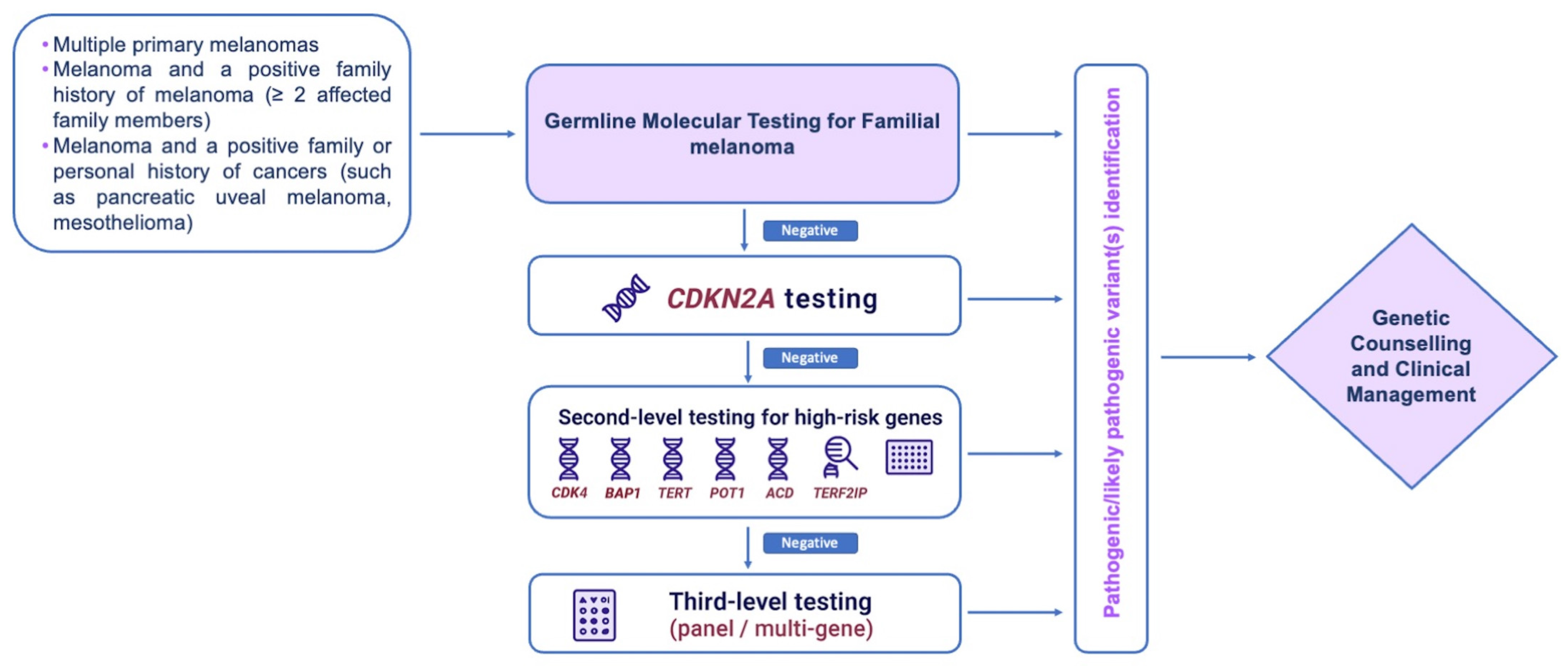
In addition to the well-established and emerging genes associated with an increased risk of familial melanoma, there are also some susceptibility loci that have been identified by GWASs that may harbor other susceptibility genes.
Gillanders et al., by analyzing 49 Australian families with familial melanoma and no mutations in CDKN2A and CDK4 genes, identified a novel melanoma susceptibility locus on Chr1p22, confirmed by the further analysis of other families from other countries [89]. A subsequent GWAS, carried out on 35 Swedish families with no CDKN2A mutations, detected a strong association with the region Chr17p12–p11 suggesting that this locus may contain genes involved in melanoma susceptibility, at least in Sweden [90]. A linkage analysis on 34 high-risk families highlighted an association with the region Chr9q21 [91]; interestingly, several genes are present in this region including GOLM1, recently described as a candidate gene [87]. Tuominen et al. investigated a putative susceptibility locus in the region Chr3q29 harboring 44 genes that may be novel melanoma-associated genes [92]. Finally, a GWAS carried out in Spanish CDKN2A-negative patients identified a novel familial melanoma-associated locus at Chr11 harboring four genes (DLG2, PRSS23, FZD4, and TMEM135) with the strongest linkage evidence [93].
A large meta-analysis led by Landi et al. reported 68 independent cutaneous melanoma-associated variants across 54 loci confirming the importance of key functional pathways and highlighting previously unknown cutaneous melanoma etiologic routes [94].
Future studies investigating these regions through next-generation sequencing may allow the identification of novel familial melanoma susceptibility genes.

4. Genetic Testing

CDKN2A molecular screening is recommended to assess the presence of a melanoma predisposing-syndrome [95]. Although CDKN2A and CDK4 are the most commonly tested genes to date, no unique standard guidelines on which genes to include in familial melanoma genetic testing are currently available. According to NCCN Guidelines (Version 2.2025, accessed on July 2025), genetic predisposition is defined by the presence of germline mutations or polymorphisms predisposing to melanoma (e.g., CDKN2A, CDK4, MC1R, BAP1 [especially for uveal melanoma], TERT, MITF, and PTEN) and other cancer predisposition genes with increased melanoma risk (e.g., CHEK2, BRCA1/2, BLM, and ATM).
In addition, the availability of next-generation sequencing (NGS)-based approaches, able to comprehensively analyze more candidate genes (from a few to the whole exome) in several patients simultaneously, has enhanced the diffusion of molecular analyses in a diagnostic context [96,97,98,99]. Indeed, this approach enables, with an acceptable cost and in a relatively short time, gaining multiple information regarding the presence of causative mutations, risk factors, drug response-related variants, and phenotype-modifiers variants, thus stratifying patients and identifying the at-risk subjects within their families. In addition, by analyzing a group of candidate genes in large cohorts, it is also possible to estimate the contribution of a specific gene to a disease of interest or discover novel associations. As a consequence, multi-gene panels analysis is becoming the best option for the molecular diagnosis of several inherited and genetically heterogeneous diseases, like hereditary cancers [11,12,100,101,102].
The main advantages of a molecular diagnosis for familial melanoma are the possibility to make patients aware about their cancer risk and contributing risk factors, and plan more frequent surveillance in order to make early diagnosis and modulate the melanoma risk in other family members [103].
Considering all the above, gene panels testing is becoming a common practice also in patients affected by melanoma in order to assess an inherited predisposition. However, considering the low frequency of melanoma-related germline mutations, their variable penetrance, and the absence of standardized guidelines for the clinical management of the mutations’ carriers, the use of these tests as a diagnostic tool is currently under debate. In addition, since the prevalence of predisposing mutations is different in different geographic areas, this makes it difficult to establish a common panel for all countries. Finally, expanding the number of genes included in testing inevitably increases the likelihood of identifying variants of uncertain significance (VUSs). These findings may lead to inconclusive results, complicate risk communication, and create uncertainty for both clinicians and patients. Indeed, the detection of VUSs represents a known limitation of broad genetic testing approaches. While such variants may be reclassified over time, their initial ambiguity poses challenges in clinical decision-making and genetic counseling. It is therefore essential to balance diagnostic yield with the risk of generating uncertain results.
Thus, it is important to underline that genetic counselling should always be offered to patients in the presence of a suspicion of a hereditary cancer syndrome, including familial melanoma [23,95]. Indeed, patients should be informed about the significance of the test, the probability of inconclusive results, and the possible personal and familial implications. Aiming to reduce uninformative tests, Leachman et al. proposed a diagnostic algorithm for the molecular analysis of hereditary susceptibility to melanoma based on the separation between “melanoma dominant” (the melanoma is the most frequent cancer in the family) and “melanoma subordinate” (the melanoma is secondary respect to other predominant cancers in the family) cases [23]. According to this classification, they suggested different gene panels to be tested based on the penetrance of the genes for the cancer found as predominant in the patient’s family.
Potjer et al. used a custom panel, including 30 genes, to analyze 488 melanoma cases from CDKN2A and CDK4-negative families [60]. They were able to identify 18 pathogenic/likely pathogenic variants (3 in BAP1 and 15 in MITF) obtaining a 4% increase of the diagnostic yield. Their results not only support the use of a multi-genes panel test in familial melanoma, but also suggests a pivotal role of BAP1 and MITF genes. More recently, Bruno et al. successfully used multi genes panel testing for familial melanoma evaluation and, by analyzing 1044 family members and 940 cutaneous melanoma index cases, found that CDKN2A pathogenic variants’ detection rate was 5.53% (52 cases), while adding the variants identified in the other genes, the panel diagnostic sensitivity increases up to 9.47% (89 cases) [104]. Based on all the reviewed studies and current NCCN guidelines, a possible diagnostic algorithm for familial melanoma is reported in Figure 3.

5. Genetic Counseling and Mutations Carriers’ Management

The burden of melanoma-risk genes is becoming even more clear with the emergence of other predisposing genes and the diffusion of extended genomic analyses that allow multiple genetic loci to be analyzed simultaneously.
Thus, pre-test genetic counseling should be mandatory to explain the significance of the test proposed, the possible results, and their significance for the proband and for the family. Similarly, the post-test genetic counseling has to be carried out to discuss the results and their repercussions in the familial context. Indeed, the combination of the appropriate genetic test in properly selected melanoma patients and the personalized recommendation for follow-up based on the test results has shown to both improve early detection and reduce mortality [23]. This means that the selection criteria to access genetic testing are crucial in order to identify those patients that are more likely to carry a germline predisposing mutation. However, established guidelines for melanoma-predisposing genes testing are not yet available.
About fifteen years ago, a pre-test probabilities estimation was proposed for the identification, within the melanoma patients, of proper candidates for the molecular analysis [15]. This study suggested using as criteria the number of primary melanomas in the proband, the number of melanomas in blood relatives, and the numbers of pancreatic cancers in the proband or blood relatives, normalizing these criteria for the geographic area. This is the so-called “rule of twos or threes” for melanoma molecular testing. This rule has been recently updated to include the other cancers that may co-occur in the families; interestingly, based on this a priori probability, authors suggest different gene panels to be assessed in different patients [23].
In addition, even if today genomic screening methods allow comprehensive genomic analyses to be carried out, it is important to underline that their results may be inconclusive. Indeed, a number of VUSs may be identified. Lastly, for more known pathogenetic mutations, established guidelines for mutations carriers’ clinical management and follow-up may be lacking. In an attempt to minimize this uncertainty, Leachman et al. suggest reporting only genes and variants with established clinical significance [23].
In the presence of positive family history or of an identified predisposing mutation, melanoma patients should receive specific recommendations for follow-up and therapy. First of all, patients should be advised on the importance of melanoma prevention and early detection: photoprotection has to be encouraged, as well as self-skin examination. In addition, the frequency of dermatological visit must be planned based on the patient’s history and skin features.
Dermoscopy, in particular, is an important instrument for the monitoring of these patients, also considering the variability of the phenotype [16,105]. Indeed, most studies suggested that an interval of 6 months between subsequent dermoscopies should be adequate [106,107], while Haenssle et al. suggested to reduce it to 3 months [108]; to date, there are still inconclusive data supporting that this reduction may ameliorate patients’ outcome. Other advanced techniques, like total body photography and sequential digital dermoscopy imaging, may be also useful for patients at extreme risk of melanoma [109].
A study carried out on 311 high-risk patients evaluated at 6-month intervals, by using total body photography and sequential digital dermoscopy imaging, showed an improvement in post-baseline melanoma detection [110]. In addition, it has been reported that the use of total body photography and sequential digital dermoscopy imaging allows diagnosis of thinner melanomas in respect of those identified by physical examination [111]. Also in this case, the frequency of follow-up is still under debate. A cost-analysis study has demonstrated that the cost for the surveillance of the high-risk subjects, even if high, are cheaper than the cost to treat a melanoma [112].
Considering that familial melanoma is associated often to an early age of onset, the dermatological surveillance should begin from adolescence.
Annual screening for other cancers should be also evaluated depending on family history and on the spectrum of cancer related to the mutated gene.
Finally, in the presence of a predisposing mutation within a melanoma family, the blood relatives of the proband should be admitted to genetic counseling in order to receive adequate information regarding their own risk, the most appropriate surveillance strategies and evaluate the possibility of undergoing genetic analysis to exclude or confirm the inheritance of the familial mutation. It is important to underline that, as for other cancers, inherited mutations confer a risk and do not allow a melanoma diagnosis to be made or evaluate if and when a melanoma may develop. Over time, some risk scores have been developed in an attempt to evaluate personal melanoma risk. In particular, Davies et al. developed a melanoma risk algorithm based on the evaluation of seven phenotypic features (hair color, skin type, family history, freckling, nevus count, number of large nevi, and history of sunburn) available as a web tool for self-assessment [113]. Subsequently, Cho et al. tested the use of a genetic risk score based on the use of 21 SNPs reported as significantly related to melanoma risk; however, this study highlighted that, despite the associated risk, genetic factors seem to have a limited impact on melanoma development with respect to other environmental factors [114]. To deeply investigate the contribution of genetic factors to melanoma risk, a study derived a polygenic risk score from the analysis of two independent cohorts: interestingly, they found that genetic factors evaluation may identify at-risk subjects in absence of typical phenotypic features [115]. Finally, Gu et al. reported a polygenic risk score combining 204 SNPs that, in combination with skin pigmentation features and the number of nevi, showed its potential in the identification of the high-risk subjects who could benefit from increased surveillance [116]. Further studies and prospective analyses on different populations are required to establish the use of these scores and their utility in familial melanoma management.
Currently, specific dermatological surveillance recommendations are available only for carriers of mutations in a few high-risk genes, primarily CDKN2A (see Table 2). These protocols typically include full-body skin examinations every 3–12 months, beginning in early adulthood. For the other familial melanoma susceptibility genes, evidence remains insufficient to support standardized surveillance guidelines [14].

6. Role of Germline Variants in Prognosis and Therapy

Germline-inherited DNA variants, in addition to increasing the risk for developing melanoma, may also play a role in patients’ prognosis and/or survival. The identification of germline variants able to affect melanoma survival enables an increase in the knowledge on the molecular bases of melanoma. In particular, specific key pathways may be identified, supporting the development of novel therapies for the treatment of melanoma cases with a poor prognosis. To this attempt, the GenoMel consortium (https://genomel.org/, accessed on 20 July 2025) was launched and is investigating the role of inherited DNA variants on melanoma survival.
In this context, a BioGenoMel collaborative study aimed at verifying the hypothesis that inherited MC1R variants may impact melanoma survival expectation [117]. By analyzing 10 melanoma cohorts from different countries, they suggested a survival benefit for melanoma patients carrying inherited specific MIC1R variants [118]. Subsequently, the association between MIC1R variants and survival rate has been further investigated by an international, large population-based project within the Genes, Environment, and Melanoma study [117]. By analyzing the MIC1R genotype in 2200 patients with a primary melanoma, this study suggested that melanoma survival may be improved in the carriers of two inherited variants in the MIC1R gene [118]. Finally, a study by Lira et al. investigated the association between MC1R variants, melanoma survival, and sex [119]. Interestingly, they found that inherited MC1R variants correlated with an improved overall survival rate only in women, suggesting that sex-dependent features may influence the role of specific genes involved in melanoma prognosis [119].
In addition to MIC1R, germline variants in other genes have been identified as predictive of melanoma patients’ outcome. In particular, the same population-based study carried out in the context of the Genes, Environment, and Melanoma study, mentioned above [118], aimed to evaluate the possible role on melanoma survival of polymorphisms located in the ASIP (Agouti signaling protein) gene. Indeed, ASIP protein is the antagonist of MC1R and GWASs have highlighted an association between ASIP polymorphisms and the risk of melanoma [68,120]. In this study, the ASIP risk haplotype TG was found to significatively affect melanoma survival with respect to the most common GG haplotype [118].
Davies at al. reported an association between a rare, inherited variant in the PARP1 (polyadenosine diphosphate-ribose polymerase 1) gene and melanoma survival. This study showed that the investigated PARP1 SNP was associated with increased survival of melanoma patients and that the reduced expression of PARP1, affecting both melanoma growth and angiogenesis, may positively impact on melanoma progression [121].
SNPs in the VDR (Vitamin D Receptor) gene have been reported to affect the survival rate of several cancers [122,123]. Thus, the Genes, Environment, and Melanoma study also aimed to assess their role in melanoma-affected patients [124]. By analyzing 3566 primary melanoma cases, this study found that VDR SNPs may affect melanoma survival even if tumor aggressiveness was not modified [124].
The IRF4 (Interferon regulatory factor 4) gene encodes for a transcription factor within the interferon regulatory family and is expressed in the melanocytic lesions [125]. An IRF4 SNP has been also associated to an increased risk of melanoma [80,126]. Poltrony et al. (2017) aimed to verify the role of this IRF4 SNP in melanoma patients’ outcome [127]. By analyzing two independent cohorts, this study found that the IRF4 SNP was associated with a worse melanoma outcome [127].
Finally, some studies have also investigated the different survival in familial melanoma patients according to their CDKN2A mutation status [128,129,130,131]. In particular, Florell et al. aimed to demonstrate different prognosis and survival rates in familial melanoma with respect to the sporadic cases [128]. They carried out a large population-based study that did not highlight any different biological behavior between the two studied groups. Instead, De Giorgi et al. reported different clinical and prognostic features in melanoma patients carrying a CDKN2A mutation with respect to the non-carriers, the latter showing the worst prognosis [129]. On the contrary, Helgadottir et al., comparing the survival in germline CDKN2A mutated and nonmutated melanoma cases, found the CDKN2A mutated cases had statistically significantly worse survival than melanoma cases with no CDKN2A mutations [130]. Finally, a subsequent study, confirming the previous report from De Giorgi et al. (2015), reported no difference on the overall survival between CDKN2A mutated and nonmutated melanoma cases, at least in the Italian population [131].
In addition to the above-mentioned role on melanoma prognosis and survival, germline-inherited variants may also be involved in patients’ response to specific therapies.
Indeed, as for other cancers, melanoma therapies include different approaches, the choice being based on several factors, including the patient’s health, tumor stage, and location. In addition to the classic protocols (surgical resection, chemotherapy, photodynamic therapy, and biochemotherapy), targeted therapy, and immunotherapy were recently approved for metastatic melanoma [132,133,134]. Targeted therapy refers to a group of drugs able to inhibit specific pathways by acting on specific somatic mutations, such as the MAPK pathway in patients that carry the V600E/K in BRAF gene [135].
Immunotherapy, instead, acts through the inhibition of different cellular checkpoints that directly activate the patient’s immune system [136]. More details can be found in specific papers focusing on these aspects [132,137,138,139,140,141,142,143,144,145,146].
All these therapeutic approaches are targeted to somatic melanoma-related hallmarks and little is known regarding their efficacy in melanoma patients carrying germline predisposing variants.
In this context, Helgadottir et al. evaluated the efficacy of immunotherapy agents in CDKN2A germline mutation carriers affected by metastatic melanoma. Interestingly, these patients showed a better outcome and a higher efficacy of immunotherapy with respect to nonmutated patients. This finding was probably due to high neoantigens production resulting in high sensitivity to immunotherapy, as a consequence of the high mutation load in the presence of a germline predisposition [147]. Similarly, Guida et al. evaluated the effects of MC1R variants on BRAF-targeted therapy. They found that the presence of MC1R variants significantly correlate with a worse outcome considered as both overall response rate and progression free survival [148].
Finally, a recent paper by Chat et al. showed that germline autoimmunity risk factors are able to influence the response to melanoma immune-checkpoint inhibition, suggesting that systematic testing of autoimmune risk loci could be useful to identify personalized biomarkers predictive of immunotherapy response [149].

7. Discussion and Conclusions

Autosomal dominant hereditary mutations have been identified as genetic predisposing factors for melanoma [26,150]. To date, up to 15% of melanoma cases are considered familial and may be related to germline predisposing mutations, CDKN2A being the most common and highly penetrant gene [151]. Nevertheless, single gene testing for the evaluation of familial melanoma risk is under debate. Indeed, considering that nonspecific therapies targeting the mutations are available (if not for somatic mutations), the clinical relevance of these tests is not clearly established. On the other hand, the possibility of using multi-gene panels at reasonable costs reduces the interest for single-gene testing.
While it is obvious that the greater the number of genes analyzed, the higher the risk for inconclusive results or difficult-to-interpret variants [152], it is also true that the more the genes panel analysis will be extended, the more our knowledge about the molecular landscape of hereditary susceptibility to melanoma will increase.
In addition, it is noteworthy that most of our knowledge about germline mutations predisposing to familial melanoma is focused on mutations occurring as a consequence of single nucleotide substitutions or small insertions/deletions, while little is known regarding the role of copy number variants (CNVs). As for other diseases, the frequency of CNVs and their role in melanoma progression may be underestimated [153]. Yang et al. identified in all the affected members of a melanoma family a duplicated region on chromosome 4, containing genes encoding CXC chemokines, and suggested their possible involvement in melanoma susceptibility [154]. Recently, Rocca et al. reported that germline CNVs in the gene encoding for the E2F1 transcription factors, that is known to play a role in cell cycle regulation and apoptosis, may be a predisposing factor to melanoma [155]. The use of specific bioinformatic software for NGS data, to be confirmed by an independent quantitative method (quantitative PCR, MLPA or a-CGH), has already shown its reliability and may further improve our comprehension regarding the molecular bases of familial melanoma. Moreover, it is becoming evident that genomic variants may not be the sole molecular mechanism underlying genetic predisposition to familial melanoma but epigenetic factors may also play a role. In particular, MGMT promoter methylation has received increasing attention. Hypermethylation of the MGMT gene may contribute to impaired DNA repair capacity, increased mutational burden, and a potentially altered response to alkylating agents. While not yet integrated into routine clinical management, MGMT methylation status may represent a promising biomarker deserving further investigation. This growing body of knowledge will provide valuable insights not only for a better identification of the molecular bases of familial melanomas, but also for at-risk subjects’ ascertainment, and for the development of novel targeted therapies, as already done for other hereditary cancer syndromes [156,157].
In conclusion, while CDKN2A remains the most established high-penetrance gene for familial melanoma, recent findings on other genes have broadened the genetic landscape and complexity of this condition. However, current knowledge on familial melanoma susceptibility is largely centered around a few high-penetrance genes, such as CDKN2A, CDK4, BAP1, and POT1, for which clinical management guidelines are available.
The growing use of multigene panels, including moderate-, low-risk, and emerging genes, has improved mutations detection but raises challenges, such as the careful interpretation of VUSs and awareness of the limitations associated with uncertain findings. As highlighted in this review, the inclusion of CNVs evaluation and the refinement of bioinformatic pipelines are crucial steps toward a more comprehensive assessment of genetic risk. Moreover, surveillance protocols are currently limited to a few genes and should be expanded as new evidence emerges. Improving genetic counseling, standardizing testing strategies, and integrating CNVs analysis will be crucial steps toward optimizing patient care and developing tailored prevention strategies. The diagnostic algorithm we propose (Figure 3) integrates current evidence and may serve as a guide for clinicians in tailoring genetic testing and surveillance strategies in individuals and families at increased risk of melanoma. Future research should focus on better characterizing moderate-penetrance genes, clarifying the role of CNVs, and refining genotype-based clinical recommendations.

Author Contributions

Conceptualization V.D.; methodology, C.S. and I.V.; data curation, C.S. and I.V.; writing—original draft preparation, C.S.; writing—review and editing, I.V. and V.D. All authors have read and agreed to the published version of the manuscript.

Funding

This research received no external funding.

Institutional Review Board Statement

Not applicable.

Informed Consent Statement

Not applicable.

Data Availability Statement

No new data were created or analyzed in this study.

Conflicts of Interest

The authors declare no conflicts of interest.

Abbreviations

The following abbreviations are used in this manuscript:
ACDACD Shelterin Complex Subunit and Telomerase Recruitment Factor
AGR3Anterior Gradient 3
ARNTAryl Hydrocarbon Receptor Nuclear Translocator
ASIPAgouti Signaling Protein
BAP1BRCA1-Associated Protein 1
BRAFB-Raf Proto-Oncogene, Serine/Threonine Kinase
CCND1Cyclin D1
CDK4Cyclin-Dependent Kinase 4
CDKAL1CDK5 Regulatory Subunit-Associated Protein 1-Like 1
CDKN2ACyclin-Dependent Kinase Inhibitor 2A
CNVCopy Number Variant
DCCyclin D–CDK4 Complex
FAMMMFamilial Atypical Multiple Mole Melanoma
GWASGenome-Wide Association Study
HERC2HECT and RLD Domain Containing E3 Ubiquitin Protein Ligase 2
ILInterleukin
IRF4Interferon Regulatory Factor 4
MC1RMelanocortin-1 Receptor
MGMTO6-Methylguanine-DNA Methyltransferase
MITFMelanocyte Inducing Transcription Factor
NGSNext Generation Sequencing
OCA2Oculocutaneous Albinism II
OBFC1Oligosaccharide-Binding Fold Containing 1
PARP1Poly (ADP-Ribose) Polymerase 1
POT1Protection of Telomeres 1
PTENPhosphatase and Tensin Homolog
RAD23BRAD23 Homolog B
SH3PXD2ASH3 and PX Domains 2A
SLC24A5Solute Carrier Family 24 Member 5
SLC45A2Solute Carrier Family 45 Member 2
SNPSingle Nucleotide Polymorphism
TERTTelomerase Reverse Transcriptase
TERF2IPTERF2 Interacting Protein
TPCN2Two-Pore Segment Channel 2
TP53Tumor Protein P53
TP53AIP1Tumor Protein P53 Regulated Apoptosis Inducing Protein 1
TYRTyrosinase
TYRP1Tyrosinase Related Protein 1
VDRVitamin D Receptor
VUSVariant of Uncertain Significance
ZNF462Zinc Finger Protein 462

References

  1. Thomas, N.E.; Groben, P. Invasive superficial spreading melanomas arising from clinically normal skin. J. Am. Acad. Dermatol. 2004, 51, 466–470. [Google Scholar] [CrossRef] [PubMed]
  2. Weatherhead, S.C.; Haniffa, M.; Lawrence, C.M. Melanomas Arising from Naevi and de Novo Melanomas—Does Origin Matter? Br. J. Dermatol. 2007, 156, 72–76. [Google Scholar] [CrossRef] [PubMed]
  3. Goldstein, A.M.; Tucker, M.A. Dysplastic Nevi and Melanoma. Cancer Epidemiol. Biomark. Prev. 2013, 22, 528–532. [Google Scholar] [CrossRef] [PubMed]
  4. Tschandl, P.; Berghoff, A.S.; Preusser, M.; Burgstaller-Muehlbacher, S.; Pehamberger, H.; Okamoto, I.; Kittler, H. NRAS and BRAF Mutations in Melanoma-Associated Nevi and Uninvolved Nevi. PLoS ONE 2013, 8, e0069639. [Google Scholar] [CrossRef] [PubMed]
  5. Shitara, D.; Tell-Martí, G.; Badenas, C.; Enokihara, M.M.S.S.; Alõs, L.; Larque, A.B.; Michalany, N.; Puig-Butille, J.A.; Carrera, C.; Malvehy, J.; et al. Mutational Status of Naevus-Associated Melanomas. Br. J. Dermatol. 2015, 173, 671–680. [Google Scholar] [CrossRef] [PubMed]
  6. Cymerman, R.M.; Shao, Y.; Wang, K.; Zhang, Y.; Murzaku, E.C.; Penn, L.A.; Osman, I.; Polsky, D. De Novo vs Nevus-Associated Melanomas: Differences in Associations with Prognostic Indicators and Survival. J. Natl. Cancer Inst. 2016, 108, djw121. [Google Scholar] [CrossRef] [PubMed]
  7. Read, J.; Wadt, K.A.W.; Hayward, N.K. Melanoma Genetics. J. Med. Genet. 2016, 53, 1–14. [Google Scholar] [CrossRef] [PubMed]
  8. Šerman, N.; Vranić, S.; Glibo, M.; Šerman, L.; Mokos, Z.B. Genetic Risk Factors in Melanoma Etiopathogenesis and the Role of Genetic Counseling: A Concise Review. Bosn. J. Basic Med. Sci. 2022, 22, 673–682. [Google Scholar] [PubMed]
  9. Precone, V.; Del Monaco, V.; Esposito, M.V.; De Palma, F.D.E.; Ruocco, A.; Salvatore, F.; D’Argenio, V. Cracking the Code of Human Diseases Using Next-Generation Sequencing: Applications, Challenges, and Perspectives. Biomed. Res. Int. 2015, 2015, 161648. [Google Scholar] [CrossRef] [PubMed]
  10. Scarano, C.; Veneruso, I.; De Simone, R.R.; Di Bonito, G.; Secondino, A.; D’Argenio, V. The Third-Generation Sequencing Challenge: Novel Insights for the Omic Sciences. Biomolecules 2024, 14, 568. [Google Scholar] [CrossRef] [PubMed]
  11. Nunziato, M.; Di Maggio, F.; Pensabene, M.; Esposito, M.V.; Starnone, F.; De Angelis, C.; Calabrese, A.; D’Aiuto, M.; Botti, G.; De Placido, S.; et al. Multi-gene panel testing increases germline predisposing mutations’ detection in a cohort of breast/ovarian cancer patients from Southern Italy. Front. Med. 2022, 9, 894358. [Google Scholar] [CrossRef] [PubMed]
  12. Secondino, A.; Starnone, F.; Veneruso, I.; Di Tella, M.A.; Conato, S.; De Angelis, C.; De Placido, S.; D’Argenio, V. Evaluation of a Four-Gene Panel for Hereditary Cancer Risk Assessment. Genes 2022, 13, 682. [Google Scholar] [CrossRef] [PubMed]
  13. Veneruso, I.; Ranieri, A.; Falcone, N.; Tripodi, L.; Scarano, C.; La Monica, I.; Pastore, L.; Lombardo, B.; D’Argenio, V. The Potential Usefulness of the Expanded Carrier Screening to Identify Hereditary Genetic Diseases: A Case Report from Real-World Data. Genes 2023, 14, 1651. [Google Scholar] [CrossRef] [PubMed]
  14. Fiasconaro, C.A.; Carbone, A.; Giordano, S.; Cavallo, F.; Fava, P.; Pasini, B.; Yakymiv, Y.; Marchisio, S.; Quaglino, P.; Ribero, S.; et al. Germline Non-CDKN2A Variants in Melanoma and Associated Hereditary Cancer Syndromes. Diseases 2025, 13, 180. [Google Scholar] [CrossRef] [PubMed]
  15. Leachman, S.A.; Carucci, J.; Kohlmann, W.; Banks, K.C.; Asgari, M.M.; Bergman, W.; Bianchi-Scarrà, G.; Brentnall, T.; Bressac-de Paillerets, B.; Bruno, W.; et al. Selection Criteria for Genetic Assessment of Patients with Familial Melanoma. J. Am. Acad. Dermatol. 2009, 61, 677.e1–677.e14. [Google Scholar] [CrossRef] [PubMed]
  16. Soura, E.; Eliades, P.J.; Shannon, K.; Stratigos, A.J.; Tsao, H. Hereditary Melanoma: Update on Syndromes and Management Emerging Melanoma Cancer Complexes and Genetic Counseling. J. Am. Acad. Dermatol. 2016, 74, 395–407. [Google Scholar] [CrossRef] [PubMed]
  17. Soura, E.; Eliades, P.J.; Shannon, K.; Stratigos, A.J.; Tsao, H. Hereditary Melanoma: Update on Syndromes and Management Genetics of Familial Atypical Multiple Mole Melanoma Syndrome. J. Am. Acad. Dermatol. 2016, 74, 411–420. [Google Scholar] [CrossRef] [PubMed]
  18. Ransohoff, K.J.; Jaju, P.D.; Tang, J.Y.; Carbone, M.; Leachman, S.; Sarin, K.Y. Familial Skin Cancer Syndromes: Increased Melanoma Risk. J. Am. Acad. Dermatol. 2016, 74, 423–434. [Google Scholar] [CrossRef] [PubMed]
  19. Jansen, M.R.; El Moumni, M.; van Leeuwen, B.L.; van den Akker, P.C.; Rácz, E. Identifying high-risk melanoma patients: The importance of acquiring a detailed family history. J. Eur. Acad. Dermatol. Venereol. 2025, 1–9. [Google Scholar] [CrossRef] [PubMed]
  20. Toussi, A.; Mans, N.; Welborn, J.; Kiuru, M. Germline Mutations Predisposing to Melanoma. J. Cutan. Pathol. 2020, 47, 606–616. [Google Scholar] [CrossRef] [PubMed]
  21. Harland, M.; Cust, A.E.; Badenas, C.; Chang, Y.M.; Holland, E.A.; Aguilera, P.; Aitken, J.F.; Armstrong, B.K.; Barrett, J.H.; Carrera, C.; et al. Prevalence and Predictors of Germline CDKN2A Mutations for Melanoma Cases from Australia, Spain and the United Kingdom. Hered. Cancer Clin. Pract. 2014, 12, 20. [Google Scholar] [CrossRef] [PubMed]
  22. Hajkova, N.; Hojny, J.; Nemejcova, K.; Dundr, P.; Ulrych, J.; Jirsova, K.; Glezgova, J.; Ticha, I. Germline Mutation in the TP53 Gene in Uveal Melanoma. Sci. Rep. 2018, 8, 7618. [Google Scholar] [CrossRef] [PubMed]
  23. Leachman, S.A.; Lucero, O.M.; Sampson, J.E.; Cassidy, P.; Bruno, W.; Queirolo, P.; Ghiorzo, P. Identification, genetic testing, and management of hereditary melanoma. Cancer Metastasis Rev. 2017, 36, 77–90. [Google Scholar] [CrossRef] [PubMed]
  24. Monnerat, C.; Chompret, A.; Kannengiesser, C.; Avril, M.F.; Janin, N.; Spatz, A.; Guinebretière, J.M.; Marian, C.; Barrois, M.; Boitier, F.; et al. BRCA1, BRCA2, TP53, and CDKN2A Germline Mutations in Patients with Breast Cancer and Cutaneous Melanoma. Fam. Cancer 2007, 6, 453–461. [Google Scholar] [CrossRef] [PubMed]
  25. National Cancer Institute. Melanoma of the Skin-Cancer Stat Facts. National Cancer Institute. 2018. Available online: https://seer.cancer.gov/statfacts/html/melan.html (accessed on 15 May 2025).
  26. Potrony, M.; Badenas, C.; Aguilera, P.; Puig-Butille, J.A.; Carrera, C.; Malvehy, J.; Puig, S. Update in Genetic Susceptibility in Melanoma. Ann. Transl. Med. 2015, 3, 210. [Google Scholar] [PubMed]
  27. Funchain, P.; Ni, Y.; Heald, B.; Bungo, B.; Arbesman, M.; Behera, T.R.; McCormick, S.; Song, J.M.; Kennedy, L.B.; Nielsen, S.M.; et al. Germline Cancer Susceptibility in Individuals with Melanoma. J. Am. Acad. Dermatol. 2024, 91, 265–272. [Google Scholar] [CrossRef] [PubMed]
  28. Potrony, M.; Puig-Butillé, J.A.; Aguilera, P.; Badenas, C.; Carrera, C.; Malvehy, J.; Puig, S. Increased Prevalence of Lung, Breast, and Pancreatic Cancers in Addition to Melanoma Risk in Families Bearing the Cyclin-Dependent Kinase Inhibitor 2A Mutation: Implications for Genetic Counseling. J. Am. Acad. Dermatol. 2014, 71, 888–895. [Google Scholar] [CrossRef] [PubMed]
  29. Helgadottir, H.; Höiom, V.; Jönsson, G.; Tuominen, R.; Ingvar, C.; Borg, Å.; Olsson, H.; Hansson, J. High Risk of Tobacco-Related Cancers in CDKN2A Mutation-Positive Melanoma Families. J. Med. Genet. 2014, 51, 545–552. [Google Scholar] [CrossRef] [PubMed]
  30. Rashid, S.; Gupta, S.; McCormick, S.R.; Tsao, H. New Insights into Melanoma Tumor Syndromes. JID Innov. Ski. Sci. Mol. Popul. Health 2022, 2, 100152. [Google Scholar] [CrossRef] [PubMed]
  31. Goldstein, A.M.; Chan, M.; Harland, M.; Gillanders, E.M.; Hayward, N.K.; Avril, M.F.; Azizi, E.; Bianchi-Scarra, G.; Bishop, D.T.; Bressac-De Paillerets, B.; et al. High-Risk Melanoma Susceptibility Genes and Pancreatic Cancer, Neural System Tumors, and Uveal Melanoma across GenoMEL. Cancer Res. 2006, 66, 9818–9828. [Google Scholar] [CrossRef] [PubMed]
  32. Pedace, L.; De Simone, P.; Castori, M.; Sperduti, I.; Silipo, V.; Eibenschutz, L.; De Bernardo, C.; Buccini, P.; Moscarella, E.; Panetta, C.; et al. Clinical Features Predicting Identification of CDKN2A Mutations in Italian Patients with Familial Cutaneous Melanoma. Cancer Epidemiol. 2011, 35, e116-20. [Google Scholar] [CrossRef] [PubMed]
  33. Goldstein, A.M.; Tucker, M.A. Genetic Epidemiology of Familial Melanoma. Dermatol. Clin. 1995, 3, 605–612. [Google Scholar] [CrossRef]
  34. Borg, Å.; Sandberg, T.; Nilsson, K.; Johannsson, O.; Klinker, M.; Måsbäck, A.; Westerdahl, J.; Olsson, H.; Ingvar, C. High Frequency of Multiple Melanomas and Breast and Pancreas Carcinomas in CDKN2A Mutation-Positive Melanoma Families. J. Natl. Cancer Inst. 2000, 92, 1260–1266. [Google Scholar] [CrossRef] [PubMed]
  35. Cremin, C.; Howard, S.; Le, L.; Karsan, A.; Schaeffer, D.F.; Renouf, D.; Schrader, K.A. CDKN2A Founder Mutation in Pancreatic Ductal Adenocarcinoma Patients without Cutaneous Features of Familial Atypical Multiple Mole Melanoma (FAMMM) Syndrome. Hered. Cancer Clin. Pract. 2018, 16, 7. [Google Scholar] [CrossRef] [PubMed]
  36. McWilliams, R.R.; Wieben, E.D.; Chaffee, K.G.; Antwi, S.O.; Raskin, L.; Olopade, O.I.; Li, D.; Highsmith, W.E.; Colon-Otero, G.; Khanna, L.G.; et al. CDKN2A Germline Rare Coding Variants and Risk of Pancreatic Cancer in Minority Populations. Cancer Epidemiol. Biomark. Prev. 2018, 27, 1364–1370. [Google Scholar] [CrossRef] [PubMed]
  37. Garutti, M.; Targato, G.; Buriolla, S.; Palmero, L.; Minisini, A.M.; Puglisi, F. CDK4/6 Inhibitors in Melanoma: A Comprehensive Review. Cells 2021, 10, 1334. [Google Scholar] [CrossRef] [PubMed]
  38. Louie, B.H.; Kurzrock, R. BAP1: Not Just a BRCA1-Associated Protein. Cancer Treat. Rev. 2020, 90, 102091. [Google Scholar] [CrossRef] [PubMed]
  39. Masoomian, B.; Shields, J.A.; Shields, C.L. Overview of BAP1 Cancer Predisposition Syndrome and the Relationship to Uveal Melanoma. J. Curr. Ophthalmol. 2018, 30, 102–109. [Google Scholar] [CrossRef] [PubMed]
  40. Wadt, K.A.; Aoude, L.G.; Johansson, P.; Solinas, A.; Pritchard, A.; Crainic, O.; Andersen, M.T.; Kiilgaard, J.F.; Heegaard, S.; Sunde, L.; et al. A Recurrent Germline BAP1 Mutation and Extension of the BAP1 Tumor Predisposition Spectrum to Include Basal Cell Carcinoma. Clin. Genet. 2015, 88, 267–272. [Google Scholar] [CrossRef] [PubMed]
  41. Walpole, S.; Pritchard, A.L.; Cebulla, C.M.; Pilarski, R.; Stautberg, M.; Davidorf, F.H.; de la Fouchardière, A.; Cabaret, O.; Golmard, L.; Stoppa-Lyonnet, D.; et al. Comprehensive Study of the Clinical Phenotype of Germline BAP1 Variant-Carrying Families Worldwide. J. Natl. Cancer Inst. 2018, 110, 1328–1341. [Google Scholar] [CrossRef] [PubMed]
  42. Rachakonda, S.; Kong, H.; Srinivas, N.; Garcia-Casado, Z.; Requena, C.; Fallah, M.; Heidenreich, B.; Planelles, D.; Traves, V.; Schadendorf, D.; et al. Telomere Length, Telomerase Reverse Transcriptase Promoter Mutations, and Melanoma Risk. Genes Chromosomes Cancer 2018, 57, 564–572. [Google Scholar] [CrossRef] [PubMed]
  43. Guo, Y.; Chen, Y.; Zhang, L.; Ma, L.; Jiang, K.; Yao, G.; Zhu, L. TERT Promoter Mutations and Telomerase in Melanoma. J. Oncol. 2022, 2022, 6300329. [Google Scholar] [CrossRef] [PubMed]
  44. Jones, M.; Bisht, K.; Savage, S.A.; Nandakumar, J.; Keegan, C.E.; Maillard, I. The Shelterin Complex and Hematopoiesis. J. Clin. Investig. 2016, 126, 1621–1629. [Google Scholar] [CrossRef] [PubMed]
  45. Savage, S.A. Beginning at the Ends: Telomeres and Human Disease. F1000Research 2018, 7, F1000. [Google Scholar] [CrossRef] [PubMed]
  46. Horn, S.; Figl, A.; Rachakonda, P.S.; Fischer, C.; Sucker, A.; Gast, A.; Kadel, S.; Moll, I.; Nagore, E.; Hemminki, K.; et al. TERT Promoter Mutations in Familial and Sporadic Melanoma. Science 2013, 339, 959–961. [Google Scholar] [CrossRef] [PubMed]
  47. Harland, M.; Petljak, M.; Robles-Espinoza, C.D.; Ding, Z.; Gruis, N.A.; van Doorn, R.; Pooley, K.A.; Dunning, A.M.; Aoude, L.G.; Wadt, K.A.W.; et al. Germline TERT Promoter Mutations Are Rare in Familial Melanoma. Fam. Cancer 2016, 15, 139–144. [Google Scholar] [CrossRef] [PubMed]
  48. Zaremba, A.; Meier, F.; Schlein, C.; Jansen, P.; Lodde, G.; Song, M.; Kretz, J.; Möller, I.; Stadtler, N.; Livingstone, E.; et al. Clinical and Pathological Characteristics of Familial Melanoma with Germline TERT Promoter Variants. Pigment. Cell Melanoma Res. 2022, 35, 573–586. [Google Scholar] [CrossRef] [PubMed]
  49. Robles-Espinoza, C.D.; Harland, M.; Ramsay, A.J.; Aoude, L.G.; Quesada, V.; Ding, Z.; Pooley, K.A.; Pritchard, A.L.; Tiffen, J.C.; Petljak, M.; et al. POT1 Loss-of-Function Variants Predispose to Familial Melanoma. Nat. Genet. 2014, 46, 478–481. [Google Scholar] [CrossRef] [PubMed]
  50. Shi, J.; Yang, X.R.; Ballew, B.; Rotunno, M.; Calista, D.; Fargnoli, M.C.; Ghiorzo, P.; Bressac-De Paillerets, B.; Nagore, E.; Avril, M.F.; et al. Rare Missense Variants in POT1 Predispose to Familial Cutaneous Malignant Melanoma. Nat. Genet. 2014, 46, 482–486. [Google Scholar] [CrossRef] [PubMed]
  51. Wilson, T.L.S.; Hattangady, N.; Lerario, A.M.; Williams, C.; Koeppe, E.; Quinonez, S.; Osborne, J.; Cha, K.B.; Else, T. A New POT1 Germline Mutation-Expanding the Spectrum of POT1-Associated Cancers. Fam. Cancer 2017, 16, 561–566. [Google Scholar] [CrossRef] [PubMed]
  52. Müller, C.; Krunic, M.; Wendt, J.; von Haeseler, A.; Okamoto, I. Germline Variants in the POT1-Gene in High-Risk Melanoma Patients in Austria. G3 Bethesda 2018, 8, 1475–1480. [Google Scholar] [CrossRef] [PubMed]
  53. Potrony, M.; Puig-Butille, J.A.; Ribera-Sola, M.; Iyer, V.; Robles-Espinoza, C.D.; Aguilera, P.; Carrera, C.; Malvehy, J.; Badenas, C.; Landi, M.T.; et al. POT1 germline mutations but not TERT promoter mutations are implicated in melanoma susceptibility in a large cohort of Spanish melanoma families. Br. J. Dermatol. 2019, 181, 105–113. [Google Scholar] [CrossRef] [PubMed]
  54. Wong, K.; Robles-Espinoza, C.D.; Rodriguez, D.; Rudat, S.S.; Puig, S.; Potrony, M.; Wong, C.C.; Hewinson, J.; Aguilera, P.; Puig-Butille, J.A.; et al. Association of the POT1 Germline Missense Variant p.I78T with Familial Melanoma. JAMA Dermatol. 2019, 155, 604–609. [Google Scholar] [CrossRef] [PubMed]
  55. Abu Shtaya, A.; Kedar, I.; Bazak, L.; Basel-Salmon, L.; Barhom, S.F.; Naftali, M.; Eskin-Schwartz, M.; Birk, O.S.; Polager-Modan, S.; Keidar, N.; et al. A POT1 Founder Variant Associated with Early Onset Recurrent Melanoma and Various Solid Malignancies. Genes 2024, 15, 355. [Google Scholar] [CrossRef] [PubMed]
  56. Aoude, L.G.; Pritchard, A.L.; Robles-Espinoza, C.D.; Wadt, K.; Harland, M.; Choi, J.; Gartside, M.; Quesada, V.; Johansson, P.; Palmer, J.M.; et al. Nonsense Mutations in the Shelterin Complex Genes ACD and TERF2IP in Familial Melanoma. J. Natl. Cancer Inst. 2014, 107, dju408. [Google Scholar] [CrossRef] [PubMed]
  57. Goldstein, A.M.; Qin, R.; Chu, E.Y.; Elder, D.E.; Massi, D.; Adams, D.J.; Harms, P.W.; Robles-Espinoza, C.D.; Newton-Bishop, J.A.; Bishop, D.T.; et al. Association of Germline Variants in Telomere Maintenance Genes (POT1, TERF2IP, ACD, and TERT) with Spitzoid Morphology in Familial Melanoma: A Multi-Center Case Series. JAAD Int. 2023, 11, 43–51. [Google Scholar] [CrossRef] [PubMed]
  58. Tagliabue, E.; Fargnoli, M.C.; Gandini, S.; Maisonneuve, P.; Liu, F.; Kayser, M.; Nijsten, T.; Han, J.; Kumar, R.; Gruis, N.A.; et al. MC1R Gene Variants and Non-Melanoma Skin Cancer: A Pooled-Analysis from the M-SKIP Project. Br. J. Cancer 2015, 113, 354–363. [Google Scholar] [CrossRef] [PubMed]
  59. Wendt, J.; Mueller, C.; Rauscher, S.; Fae, I.; Fischer, G.; Okamoto, I. Contributions by MC1R Variants to Melanoma Risk in Males and Females. JAMA Dermatol. 2018, 154, 789–795. [Google Scholar] [CrossRef] [PubMed]
  60. Potjer, T.P.; Bollen, S.; Grimbergen, A.J.E.M.; van Doorn, R.; Gruis, N.A.; van Asperen, C.J.; Hes, F.J.; van der Stoep, N. Multigene Panel Sequencing of Established and Candidate Melanoma Susceptibility Genes in a Large Cohort of Dutch Non-CDKN2A/CDK4 Melanoma Families. Int. J. Cancer 2019, 144, 2453–2464. [Google Scholar] [CrossRef] [PubMed]
  61. Hartman, M.L.; Czyz, M. MITF in Melanoma: Mechanisms behind Its Expression and Activity. Cell. Mol. Life Sci. 2015, 72, 1249–1260. [Google Scholar] [CrossRef] [PubMed]
  62. Ciccarese, G.; Dalmasso, B.; Bruno, W.; Queirolo, P.; Pastorino, L.; Andreotti, V.; Spagnolo, F.; Tanda, E.; Ponti, G.; Massone, C.; et al. Clinical, pathological and dermoscopic phenotype of MITF p.E318K carrier cutaneous melanoma patients. J. Transl. Med. 2020, 18, 78. [Google Scholar] [CrossRef] [PubMed]
  63. Yokoyama, S.; Woods, S.L.; Boyle, G.M.; Aoude, L.G.; MacGregor, S.; Zismann, V.; Gartside, M.; Cust, A.E.; Haq, R.; Harland, M.; et al. A Novel Recurrent Mutation in MITF Predisposes to Familial and Sporadic Melanoma. Nature 2011, 480, 99–103. [Google Scholar] [CrossRef] [PubMed]
  64. Potrony, M.; Puig-Butille, J.A.; Aguilera, P.; Badenas, C.; Tell-Marti, G.; Carrera, C.; Del Pozo, L.J.; Conejo-Mir, J.; Malvehy, J.; Puig, S. Prevalence of MITF p.E318K in Patients with Melanoma Independent of the Presence of CDKN2A Causative Mutations. JAMA Dermatol. 2016, 152, 405–412. [Google Scholar] [CrossRef] [PubMed]
  65. Goldstein, A.M.; Xiao, Y.; Sampson, J.; Zhu, B.; Rotunno, M.; Bennett, H.; Wen, Y.; Jones, K.; Vogt, A.; Burdette, L.; et al. Rare Germline Variants in Known Melanoma Susceptibility Genes in Familial Melanoma. Hum. Mol. Genet. 2017, 26, 4886–4895. [Google Scholar] [CrossRef] [PubMed]
  66. Appelqvist, F.; Yhr, M.; Erlandson, A.; Martinsson, T.; Enerbäck, C. Deletion of the MGMT Gene in Familial Melanoma. Genes Chromosomes Cancer 2014, 53, 703–711. [Google Scholar] [CrossRef] [PubMed]
  67. Qi, F.; Yin, Z.; Wang, G.; Zeng, S. Clinical and Prognostic Significance of O6-Methylguanine-DNA Methyltransferase Promoter Methylation in Patients with Melanoma: A Systematic Meta-Analysis. Ann. Dermatol. 2018, 30, 129–135. [Google Scholar] [CrossRef] [PubMed]
  68. Helsing, P.; Nymoen, D.A.; Rootwelt, H.; Vårdal, M.; Akslen, L.A.; Molven, A.; Andresen, P.A. MC1R, ASIP, TYR, and TYRP1 gene variants in a population-based series of multiple primary melanomas. Genes Chromosomes Cancer 2012, 51, 654–661. [Google Scholar] [CrossRef] [PubMed]
  69. Pho, L.N.; Leachman, S.A. Genetics of Pigmentation and Melanoma Predisposition. G. Ital. Dermatol. Venereol. 2010, 1, 37–45. [Google Scholar]
  70. Gibbs, D.C.; Small, B.M.; Autuori, I.; Leong, S.F.; Ali, E.; Kenney, J.; Luo, L.; Kanetsky, P.A.; Busam, K.J.; Cust, A.E.; et al. Association of Inherited Genetic Variants with Multiple Primary Melanoma. Cancer Epidemiol. Biomark. Prev. 2025, 34, 805–814. [Google Scholar] [CrossRef] [PubMed]
  71. Kocarnik, J.M.; Park, S.L.; Han, J.; Dumitrescu, L.; Cheng, I.; Wilkens, L.R.; Schumacher, F.R.; Kolonel, L.; Carlson, C.S.; Crawford, D.C.; et al. Pleiotropic and Sex-Specific Effects of Cancer GWAS SNPs on Melanoma Risk in the Population Architecture Using Genomics and Epidemiology (PAGE) Study. PLoS ONE 2015, 10, e.0120491. [Google Scholar] [CrossRef] [PubMed]
  72. Maccioni, L.; Rachakonda, P.S.; Scherer, D.; Bermejo, J.L.; Planelles, D.; Requena, C.; Hemminki, K.; Nagore, E.; Kumar, R. Variants at Chromosome 20 (ASIP Locus) and Melanoma Risk. Int. J. Cancer 2013, 132, 42–54. [Google Scholar] [CrossRef] [PubMed]
  73. Nan, H.; Kraft, P.; Hunter, D.J.; Han, J. Genetic Variants in Pigmentation Genes, Pigmentary Phenotypes, and Risk of Skin Cancer in Caucasians. Int. J. Cancer 2009, 125, 909–917. [Google Scholar] [CrossRef] [PubMed]
  74. Zhang, M.; Song, F.; Liang, L.; Nan, H.; Zhang, J.; Liu, H.; Wang, L.E.; Wei, Q.; Lee, J.E.; Amos, C.I.; et al. Genome-Wide Association Studies Identify Several New Loci Associated with Pigmentation Traits and Skin Cancer Risk in European Americans. Hum. Mol. Genet. 2013, 22, 2948–2959. [Google Scholar] [CrossRef] [PubMed]
  75. Nathan, V.; Johansson, P.A.; Palmer, J.M.; Howlie, M.; Hamilton, H.R.; Wadt, K.; Jönsson, G.; Brooks, K.M.; Pritchard, A.L.; Hayward, N.K. Germline Variants in Oculocutaneous Albinism Genes and Predisposition to Familial Cutaneous Melanoma. Pigment. Cell Melanoma Res. 2019, 32, 854–863. [Google Scholar] [CrossRef] [PubMed]
  76. Law, M.H.; Bishop, D.T.; Lee, J.E.; Brossard, M.; Martin, N.G.; Moses, E.K.; Song, F.; Barrett, J.H.; Kumar, R.; Easton, D.F.; et al. Genome-Wide Meta-Analysis Identifies Five New Susceptibility Loci for Cutaneous Malignant Melanoma. Nat. Genet. 2015, 47, 987–995. [Google Scholar] [CrossRef] [PubMed]
  77. Gajjar, K.; Martin-Hirsch, P.L.; Martin, F.L. CYP1B1 and Hormone-Induced Cancer. Cancer Lett. 2012, 324, 13–30. [Google Scholar] [CrossRef] [PubMed]
  78. MacGregor, S.; Montgomery, G.W.; Liu, J.Z.; Zhao, Z.Z.; Henders, A.K.; Stark, M.; Schmid, H.; Holland, E.A.; Duffy, D.L.; Zhang, M.; et al. Genome-Wide Association Study Identifies a New Melanoma Susceptibility Locus at 1q21.3. Nat. Genet. 2011, 43, 1114–1118. [Google Scholar] [CrossRef] [PubMed]
  79. Han, J.; Kraft, P.; Nan, H.; Guo, Q.; Chen, C.; Qureshi, A.; Hankinson, S.E.; Hu, F.B.; Duffy, D.L.; Zhen, Z.Z.; et al. A Genome-Wide Association Study Identifies Novel Alleles Associated with Hair Color and Skin Pigmentation. PLoS Genet. 2008, 4, e1000074. [Google Scholar] [CrossRef] [PubMed]
  80. Duffy, D.L.; Iles, M.M.; Glass, D.; Zhu, G.; Barrett, J.H.; Höiom, V.; Zhao, Z.Z.; Sturm, R.A.; Soranzo, N.; Hammond, C.; et al. IRF4 Variants Have Age-Specific Effects on Nevus Count and Predispose to Melanoma. Am. J. Hum. Genet. 2010, 87, 6–16. [Google Scholar] [CrossRef] [PubMed]
  81. Fletcher, G.C.; Patel, S.; Tyson, K.; Adam, P.J.; Schenker, M.; Loader, J.A.; Daviet, L.; Legrain, P.; Parekh, R.; Harris, A.L.; et al. HAG-2 and HAG-3, Human Homologues of Genes Involved in Differentiation, Are Associated with Oestrogen Receptor-Positive Breast Tumours and Interact with Metastasis Gene C4.4a and Dystroglycan. Br. J. Cancer 2003, 88, 579–585. [Google Scholar] [CrossRef] [PubMed]
  82. King, E.R.; Tung, C.S.; Tsang, Y.T.M.; Zu, Z.; Lok, G.T.M.; Deavers, M.T.; Malpica, A.; Wolf, J.K.; Lu, K.H.; Birrer, M.J.; et al. The Anterior Gradient Homolog 3 (AGR3) Gene Is Associated with Differentiation and Survival in Ovarian Cancer. Am. J. Surg. Pathol. 2011, 35, 904–912. [Google Scholar] [CrossRef] [PubMed]
  83. Mukherji, B. Immunology of Melanoma. Clin. Dermatol. 2013, 31, 156–165. [Google Scholar] [CrossRef] [PubMed]
  84. Passarelli, A.; Mannavola, F.; Stucci, L.S.; Tucci, M.; Silvestris, F. Immune System and Melanoma Biology: A Balance between Immunosurveillance and Immune Escape. Oncotarget 2017, 8, 106132–106142. [Google Scholar] [CrossRef] [PubMed]
  85. Marzagalli, M.; Ebelt, N.D.; Manuel, E.R. Unraveling the Crosstalk between Melanoma and Immune Cells in the Tumor Microenvironment. Semin. Cancer Biol. 2019, 59, 236–250. [Google Scholar] [CrossRef] [PubMed]
  86. Eddy, K.; Chen, S. Overcoming Immune Evasion in Melanoma. Int. J. Mol. Sci. 2020, 21, 8984. [Google Scholar] [CrossRef] [PubMed]
  87. Teerlink, C.C.; Huff, C.; Stevens, J.; Yu, Y.; Holmen, S.L.; Silvis, M.R.; Trombetti, K.; Zhao, H.; Grossman, D.; Farnham, J.M.; et al. A Nonsynonymous Variant in the GOLM1 Gene in Cutaneous Malignant Melanoma. J. Natl. Cancer Inst. 2018, 110, 1380–1385. [Google Scholar] [CrossRef] [PubMed]
  88. Benfodda, M.; Gazal, S.; Descamps, V.; Basset-Seguin, N.; Deschamps, L.; Thomas, L.; Lebbe, C.; Saiag, P.; Zanetti, R.; Sacchetto, L.; et al. Truncating Mutations of TP53AIP1 Gene Predispose to Cutaneous Melanoma. Genes Chromosomes Cancer 2018, 57, 294–303. [Google Scholar] [CrossRef] [PubMed]
  89. Gillanders, E.; Juo, S.H.H.; Holland, E.A.; Jones, M.P.; Nancarrow, D.; Freas-Lutz, D.; Sood, R.; Park, N.; Faruque, M.; Markey, C.; et al. Localization of a Novel Melanoma Susceptibility Locus to 1p22. Am. J. Hum. Genet. 2003, 73, 301–313. [Google Scholar] [CrossRef] [PubMed]
  90. Höiom, V.; Tuominen, R.; Hansson, J. Genome-Wide Linkage Analysis of Swedish Families to Identify Putative Susceptibility Loci for Cutaneous Malignant Melanoma. Genes Chromosomes Cancer 2011, 50, 1076–1084. [Google Scholar] [CrossRef] [PubMed]
  91. Cannon-Albright, L.A.; Teerlink, C.C.; Farnham, J.M.; Thomas, A.W.; Zone, J.J.; Leachman, S.A. Linkage Analysis of Extended High-Risk Pedigrees Replicates a Cutaneous Malignant Melanoma Predisposition Locus on Chromosome 9q21. J. Investig. Dermatol. 2013, 133, 128–134. [Google Scholar] [CrossRef] [PubMed]
  92. Tuominen, R.; Jönsson, G.; Enerbäck, C.; Appelqvist, F.; Olsson, H.; Ingvar, C.; Hansson, J.; Höiom, V. Investigation of a Putative Melanoma Susceptibility Locus at Chromosome 3q29. Cancer Genet. 2014, 207, 70–74. [Google Scholar] [CrossRef] [PubMed]
  93. Potrony, M.; Puig-Butille, J.A.; Farnham, J.M.; Giménez-Xavier, P.; Badenas, C.; Tell-Martí, G.; Aguilera, P.; Carrera, C.; Malvehy, J.; Teerlink, C.C.; et al. Genome-Wide Linkage Analysis in Spanish Melanoma-Prone Families Identifies a New Familial Melanoma Susceptibility Locus at 11q. Eur. J. Hum. Genet. 2018, 26, 1188–1193. [Google Scholar] [CrossRef] [PubMed]
  94. Landi, M.T.; Bishop, D.T.; MacGregor, S.; Machiela, M.J.; Stratigos, A.J.; Ghiorzo, P.; Brossard, M.; Calista, D.; Choi, J.; Fargnoli, M.C.; et al. Genome-Wide Association Meta-Analyses Combining Multiple Risk Phenotypes Provide Insights into the Genetic Architecture of Cutaneous Melanoma Susceptibility. Nat. Genet. 2020, 52, 494–504. [Google Scholar] [CrossRef] [PubMed]
  95. Rossi, M.; Pellegrini, C.; Cardelli, L.; Ciciarelli, V.; Nardo, L.D.; Fargnoli, M.C. Familial Melanoma: Diagnostic and Management Implications. Dermatol. Pract. Concept. 2019, 9, 10–16. [Google Scholar] [CrossRef] [PubMed]
  96. Cirino, A.L.; Lakdawala, N.K.; McDonough, B.; Conner, L.; Adler, D.; Weinfeld, M.; O’Gara, P.; Rehm, H.L.; MacHini, K.; Lebo, M.; et al. A Comparison of Whole Genome Sequencing to Multigene Panel Testing in Hypertrophic Cardiomyopathy Patients. Circ. Cardiovasc. Genet. 2017, 10, e.001768. [Google Scholar] [CrossRef] [PubMed]
  97. Kuo, F.C.; Mar, B.G.; Lindsley, R.C.; Lindeman, N.I. The Relative Utilities of Genome-Wide, Gene Panel, and Individual Gene Sequencing in Clinical Practice. Blood 2017, 130, 433–439. [Google Scholar] [CrossRef] [PubMed]
  98. Mazzaccara, C.; Lombardi, R.; Mirra, B.; Barretta, F.; Esposito, M.V.; Uomo, F.; Caiazza, M.; Monda, E.; Losi, M.A.; Limongelli, G.; et al. Next-Generation Sequencing Gene Panels in Inheritable Cardiomyopathies and Channelopathies: Prevalence of Pathogenic Variants and Variants of Unknown Significance in Uncommon Genes. Biomolecules 2022, 12, 1417. [Google Scholar] [CrossRef] [PubMed]
  99. Ranieri, A.; Veneruso, I.; La Monica, I.; Pascale, M.G.; Pastore, L.; D’argenio, V.; Lombardo, B. Combined ACGH and Exome Sequencing Analysis Improves Autism Spectrum Disorders Diagnosis: A Case Report. Medicina 2022, 58, 522. [Google Scholar] [CrossRef] [PubMed]
  100. Desmond, A.; Kurian, A.W.; Gabree, M.; Mills, M.A.; Anderson, M.J.; Kobayashi, Y.; Horick, N.; Yang, S.; Shannon, K.M.; Tung, N.; et al. Clinical Actionability of Multigene Panel Testing for Hereditary Breast and Ovarian Cancer Risk Assessment. JAMA Oncol. 2015, 1, 943–951. [Google Scholar] [CrossRef] [PubMed]
  101. Neben, C.L.; Zimmer, A.D.; Stedden, W.; van den Akker, J.; O’Connor, R.; Chan, R.C.; Chen, E.; Tan, Z.; Leon, A.; Ji, J.; et al. Multi-Gene Panel Testing of 23,179 Individuals for Hereditary Cancer Risk Identifies Pathogenic Variant Carriers Missed by Current Genetic Testing Guidelines. J. Mol. Diagn. 2019, 21, 646–657. [Google Scholar] [CrossRef] [PubMed]
  102. Veneruso, I.; Scarano, C.; D’Argenio, V. Hereditary Breast Cancer Susceptibility Genes. Biochim. Clin. 2023, 47, 328–339. [Google Scholar]
  103. Taber, J.M.; Aspinwall, L.G.; Stump, T.K.; Kohlmann, W.; Champine, M.; Leachman, S.A. Genetic Test Reporting Enhances Understanding of Risk Information and Acceptance of Prevention Recommendations Compared to Family History-Based Counseling Alone. J. Behav. Med. 2015, 38, 740–753. [Google Scholar] [CrossRef] [PubMed]
  104. Bruno, W.; Dalmasso, B.; Barile, M.; Andreotti, V.; Elefanti, L.; Colombino, M.; Vanni, I.; Allavena, E.; Barbero, F.; Passoni, E.; et al. Predictors of Germline Status for Hereditary Melanoma: 5 Years of Multi-Gene Panel Testing within the Italian Melanoma Intergroup. ESMO Open 2022, 7, 100525. [Google Scholar] [CrossRef] [PubMed]
  105. Jones, O.T.; Jurascheck, L.C.; Van Melle, M.A.; Hickman, S.; Burrows, N.P.; Hall, P.N.; Emery, J.; Walter, F.M. Dermoscopy for Melanoma Detection and Triage in Primary Care: A Systematic Review. BMJ Open 2019, 9, e027529. [Google Scholar] [CrossRef] [PubMed]
  106. Goldstein, A.M.; Struewing, J.P.; Chidambaram, A.; Fraser, M.C.; Tucker, M.A. Genotype-Phenotype Relationships in U.S. Melanoma-Prone Families with CDKN2A and CDK4 Mutations. J. Natl. Cancer Inst. 2000, 92, 1006–1010. [Google Scholar] [CrossRef] [PubMed]
  107. Kefford, R.F.; Newton Bishop, J.A.; Bergman, W.; Tucker, M.A. Counseling and DNA Testing for Individuals Perceived to Be Genetically Predisposed to Melanoma: A Consensus Statement of the Melanoma Genetics Consortium. J. Clin. Oncol. 1999, 17, 3245–3251. [Google Scholar] [CrossRef] [PubMed]
  108. Haenssle, H.A.; Korpas, B.; Hansen-Hagge, C.; Buhl, T.; Kaune, K.M.; Johnsen, S.; Rosenberger, A.; Schön, M.P.; Emmert, S. Selection of Patients for Long-Term Surveillance with Digital Dermoscopy by Assessment of Melanoma Risk Factors. Arch. Dermatol. 2010, 146, 257–264. [Google Scholar] [CrossRef] [PubMed]
  109. Salerni, G.; Carrera, C.; Lovatto, L.; Puig-Butille, J.A.; Badenas, C.; Plana, E.; Puig, S.; Malvehy, J. Benefits of Total Body Photography and Digital Dermatoscopy (“two-Step Method of Digital Follow-up”) in the Early Diagnosis of Melanoma in Patients at High Risk for Melanoma. J. Am. Acad. Dermatol. 2012, 67, e17–e27. [Google Scholar] [CrossRef] [PubMed]
  110. Moloney, F.J.; Guitera, P.; Coates, E.; Haass, N.K.; Ho, K.; Khoury, R.; O’Connell, R.L.; Raudonikis, L.; Schmid, H.; Mann, G.J.; et al. Detection of Primary Melanoma in Individuals at Extreme High Risk: A Prospective 5-Year Follow-up Study. JAMA Dermatol. 2014, 150, 819–827. [Google Scholar] [CrossRef] [PubMed]
  111. Rademaker, M.; Oakley, A. Digital Monitoring by Whole Body Photography and Sequential Digital Dermoscopy Detects Thinner Melanomas. J. Prim. Health Care 2010, 4, 268–272. [Google Scholar] [CrossRef]
  112. Watts, C.G.; Cust, A.E.; Menzies, S.W.; Coates, E.; Mann, G.J.; Morton, R.L. Specialized Surveillance for Individuals at High Risk for Melanoma: A Cost Analysis of a High-Risk Clinic. JAMA Dermatol. 2015, 151, 178–186. [Google Scholar] [CrossRef] [PubMed]
  113. Davies, J.R.; Chang, Y.M.; Bishop, D.T.; Armstrong, B.K.; Bataille, V.; Bergman, W.; Berwick, M.; Bracci, P.M.; Elwood, J.M.; Ernstoff, M.S.; et al. Development and Validation of a Melanoma Risk Score Based on Pooled Data from 16 Case-Control Studies. Cancer Epidemiol. Biomark. Prev. 2015, 24, 817–824. [Google Scholar] [CrossRef] [PubMed]
  114. Cho, H.G.; Ransohoff, K.J.; Yang, L.; Hedlin, H.; Assimes, T.; Han, J.; Stefanick, M.; Tang, J.Y.; Sarin, K.Y. Melanoma Risk Prediction Using a Multilocus Genetic Risk Score in the Women’s Health Initiative Cohort. J. Am. Acad. Dermatol. 2018, 79, 36–41.e10. [Google Scholar] [CrossRef] [PubMed]
  115. Cust, A.E.; Drummond, M.; Kanetsky, P.A.; Mann, G.J.; Schmid, H.; Hopper, J.L.; Aitken, J.F.; Armstrong, B.K.; Giles, G.G.; Holland, E.; et al. Assessing the Incremental Contribution of Common Genomic Variants to Melanoma Risk Prediction in Two Population-Based Studies. J. Investig. Dermatol. 2018, 138, 2617–2624. [Google Scholar] [CrossRef] [PubMed]
  116. Gu, F.; Chen, T.H.; Pfeiffer, R.M.; Fargnoli, M.C.; Calista, D.; Ghiorzo, P.; Peris, K.; Puig, S.; Menin, C.; De Nicolo, A.; et al. Combining Common Genetic Variants and Non-Genetic Risk Factors to Predict Risk of Cutaneous Melanoma. Hum. Mol. Genet. 2018, 27, 4145–4156. [Google Scholar] [CrossRef] [PubMed]
  117. Davies, J.R.; Randerson-Moor, J.; Kukalizch, K.; Harland, M.; Kumar, R.; Madhusudan, S.; Nagore, E.; Hansson, J.; Höiom, V.; Ghiorzo, P.; et al. Inherited Variants in the MC1R Gene and Survival from Cutaneous Melanoma: A BioGenoMEL Study. Pigment. Cell Melanoma Res. 2012, 25, 384–394. [Google Scholar] [CrossRef] [PubMed]
  118. Taylor, N.J.; Reiner, A.S.; Begg, C.B.; Cust, A.E.; Busam, K.J.; Anton-Culver, H.; Dwyer, T.; From, L.; Gallagher, R.P.; Gruber, S.B.; et al. Inherited Variation at MC1R and ASIP and Association with Melanoma-Specific Survival. Int. J. Cancer 2015, 136, 2659–2667. [Google Scholar] [CrossRef] [PubMed]
  119. Lira, F.E.; Podlipnik, S.; Potrony, M.; Tell-Martí, G.; Calbet-Llopart, N.; Barreiro, A.; Carrera, C.; Malvehy, J.; Puig, S. Inherited MC1R Variants in Patients with Melanoma Are Associated with Better Survival in Women. Br. J. Dermatol. 2020, 182, 138–146. [Google Scholar] [CrossRef] [PubMed]
  120. Landi, M.T.; Kanetsky, P.A.; Tsang, S.; Gold, B.; Munroe, D.; Rebbeck, T.; Swoyer, J.; Ter-Minassian, M.; Hedayati, M.; Grossman, L.; et al. MC1R, ASIP, and DNA Repair in Sporadic and Familial Melanoma in a Mediterranean Population. J. Natl. Cancer Inst. 2005, 97, 998–1007. [Google Scholar] [CrossRef] [PubMed]
  121. Davies, J.R.; Jewell, R.; Affleck, P.; Anic, G.M.; Randerson-Moor, J.; Ozola, A.; Egan, K.M.; Elliott, F.; García-Casado, Z.; Hansson, J.; et al. Inherited Variation in the PARP1 Gene and Survival from Melanoma. Int. J. Cancer 2014, 135, 1625–1633. [Google Scholar] [CrossRef] [PubMed]
  122. Bilinski, K.; Boyages, J. Association between 25-Hydroxyvitamin D Concentration and Breast Cancer Risk in an Australian Population: An Observational Case-Control Study. Breast Cancer Res. Treat. 2013, 137, 599–607. [Google Scholar] [CrossRef] [PubMed]
  123. Ordonez-Mena, J.M.; Schottker, B.; Haug, U.; Muller, H.; Kohrle, J.; Schomburg, L.; Holleczek, B.; Hermann, B.H. Serum 25-Hydroxyvitamin d and Cancer Risk in Older Adults: Results from a Large German Prospective Cohort Study. Cancer Epidemiol. Biomark. Prev. 2013, 22, 905–916. [Google Scholar] [CrossRef] [PubMed]
  124. Orlow, I.; Reiner, A.S.; Thomas, N.E.; Roy, P.; Kanetsky, P.A.; Luo, L.; Paine, S.; Armstrong, B.K.; Kricker, A.; Marrett, L.D.; et al. Vitamin D Receptor Polymorphisms and Survival in Patients with Cutaneous Melanoma: A Population-Based Study. Carcinogenesis 2016, 37, 30–38. [Google Scholar] [CrossRef] [PubMed]
  125. Sundram, U.; Harvell, J.D.; Rouse, R.V.; Natkunam, Y. Expression of the B-Cell Proliferation Marker MUM1 by Melanocytic Lesions and Comparison with S100, Gp100 (HMB45), and MelanA. Mod. Pathol. 2003, 16, 802–810. [Google Scholar] [CrossRef] [PubMed]
  126. Peña-Chilet, M.; Blanquer-Maceiras, M.; Ibarrola-Villava, M.; Martinez-Cadenas, C.; Martin-Gonzalez, M.; Gomez-Fernandez, C.; Mayor, M.; Aviles, J.A.; Lluch, A.; Ribas, G. Genetic Variants in PARP1 (Rs3219090) and IRF4 (Rs12203592) Genes Associated with Melanoma Susceptibility in a Spanish Population. BMC Cancer 2013, 13, 160. [Google Scholar] [CrossRef] [PubMed]
  127. Potrony, M.; Rebollo-Morell, A.; Giménez-Xavier, P.; Zimmer, L.; Puig-Butille, J.A.; Tell-Marti, G.; Sucker, A.; Badenas, C.; Carrera, C.; Malvehy, J.; et al. IRF4 Rs12203592 Functional Variant and Melanoma Survival. Int. J. Cancer 2017, 140, 1845–1849. [Google Scholar] [CrossRef] [PubMed]
  128. Florell, S.R.; Boucher, K.M.; Garibotti, G.; Astle, J.; Kerber, R.; Mineau, G.; Wiggins, C.; Noyes, R.D.; Tsodikov, A.; Cannon-Albright, L.A.; et al. Population-Based Analysis of Prognostic Factors and Survival in Familial Melanoma. J. Clin. Oncol. 2005, 23, 7168–7177. [Google Scholar] [CrossRef] [PubMed]
  129. De Giorgi, V.; Savarese, I.; D’Errico, A.; Gori, A.; Papi, F.; Colombino, M.; Sini, M.C.; Grazzini, M.; Stanganelli, I.; Rossari, S.; et al. Epidemiological Features and Prognostic Parameters of Multiple Primary Melanomas in CDKN2A-Mutations Patients. Pigment. Cell Melanoma Res. 2015, 28, 747–751. [Google Scholar] [CrossRef] [PubMed]
  130. Helgadottir, H.; Hoiom, V.; Tuominen, R.; Nielsen, K.; Jonsson, G.; Olsson, H.; Hansson, J. Germline CDKN2A Mutation Status and Survival in Familial Melanoma Cases. J. Natl. Cancer Inst. 2016, 108, 11. [Google Scholar] [CrossRef] [PubMed]
  131. Dalmasso, B.; Pastorino, L.; Ciccarese, G.; Andreotti, V.; Grillo, F.; Mastracci, L.; Spagnolo, F.; Ballestrero, A.; Queirolo, P.; Bruno, W.; et al. CDKN2A Germline Mutations Are Not Associated with Poor Survival in an Italian Cohort of Melanoma Patients. J. Am. Acad. Dermatol. 2019, 80, 1263–1271. [Google Scholar] [CrossRef] [PubMed]
  132. Domingues, B.; Lopes, J.M.; Soares, P.; Pópulo, H. Melanoma Treatment in Review. Immunotargets Ther. 2018, 7, 35–49. [Google Scholar] [CrossRef] [PubMed]
  133. Guo, W.; Wang, H.; Li, C. Signal Pathways of Melanoma and Targeted Therapy. Signal Transduct. Target. Ther. 2021, 6, 424. [Google Scholar] [CrossRef] [PubMed]
  134. Kreuger, I.Z.M.; Slieker, R.C.; van Groningen, T.; van Doorn, R. Therapeutic Strategies for Targeting CDKN2A Loss in Melanoma. J. Investig. Dermatol. 2023, 143, 18–25.e1. [Google Scholar] [CrossRef] [PubMed]
  135. Ferrucci, P.F.; Lens, M.; Cocorocchio, E. Combined BRAF-Targeted Therapy with Immunotherapy in BRAF-Mutated Advanced Melanoma Patients. Curr. Oncol. Rep. 2021, 23, 138. [Google Scholar] [CrossRef] [PubMed]
  136. LoRusso, P.M.; Schalper, K.; Sosman, J. Targeted Therapy and Immunotherapy: Emerging Biomarkers in Metastatic Melanoma. Pigment. Cell Melanoma Res. 2020, 33, 390–402. [Google Scholar] [CrossRef] [PubMed]
  137. Castet, F.; Garcia-Mulero, S.; Sanz-Pamplona, R.; Cuellar, A.; Casanovas, O.; Caminal, J.M.; Piulats, J.M. Uveal Melanoma, Angiogenesis and Immunotherapy, Is There Any Hope? Cancers 2019, 11, 834. [Google Scholar] [CrossRef] [PubMed]
  138. Chae, Y.K.; Arya, A.; Iams, W.; Cruz, M.R.; Chandra, S.; Choi, J.; Giles, F. Current Landscape and Future of Dual Anti-CTLA4 and PD-1/PD-L1 Blockade Immunotherapy in Cancer; Lessons Learned from Clinical Trials with Melanoma and Non-Small Cell Lung Cancer (NSCLC). J. Immunother. Cancer 2018, 6, 39. [Google Scholar] [CrossRef] [PubMed]
  139. Cheng, L.; Lopez-Beltran, A.; Massari, F.; Maclennan, G.T.; Montironi, R. Molecular Testing for BRAF Mutations to Inform Melanoma Treatment Decisions: A Move toward Precision Medicine. Mod. Pathol. 2018, 31, 24–38. [Google Scholar] [CrossRef] [PubMed]
  140. Kollmann, K.; Briand, C.; Bellutti, F.; Schicher, N.; Blunder, S.; Zojer, M.; Hoeller, C. The Interplay of CDK4 and CDK6 in Melanoma. Oncotarget 2019, 10, 1346–1359. [Google Scholar] [CrossRef] [PubMed]
  141. Lugowska, I.; Teterycz, P.; Rutkowski, P. Immunotherapy of Melanoma. Contemp. Oncol. 2018, 22, 61–67. [Google Scholar] [CrossRef] [PubMed]
  142. Ma, X.; Wu, Y.; Zhang, T.; Song, H.; Jv, H.; Guo, W.; Ren, G. The Clinical Significance of C-Kit Mutations in Metastatic Oral Mucosal Melanoma in China. Oncotarget 2017, 8, 82661–82673. [Google Scholar] [CrossRef] [PubMed]
  143. Rowshanravan, B.; Halliday, N.; Sansom, D.M. CTLA-4: A Moving Target in Immunotherapy. Blood 2018, 131, 58–67. [Google Scholar] [CrossRef] [PubMed]
  144. Tran, K.A.; Cheng, M.Y.; Mitra, A.; Ogawa, H.; Shi, V.Y.; Olney, L.P.; Kloxin, A.M.; Maverakis, E. MEK Inhibitors and Their Potential in the Treatment of Advanced Melanoma: The Advantages of Combination Therapy. Drug Des. Devel Ther. 2015, 10, 43–52. [Google Scholar] [PubMed]
  145. Liu, H.; Gou, X.; Tan, Y.; Fan, Q.; Chen, J. Immunotherapy and Delivery Systems for Melanoma. Hum. Vaccin. Immunother. 2024, 20, 2394252. [Google Scholar] [CrossRef] [PubMed]
  146. Amarillo, D.; Flaherty, K.T.; Sullivan, R.J. Targeted Therapy Innovations for Melanoma. Hematol. Oncol. Clin. North Am. 2024, 38, 973–995. [Google Scholar] [CrossRef] [PubMed]
  147. Helgadottir, H.; Olsson, H.; Tucker, M.A.; Yang, X.R.; Höiom, V.; Goldstein, A.M. Phenocopies in Melanoma-Prone Families with Germ-Line CDKN2A Mutations. Genet. Med. 2018, 20, 1087–1090. [Google Scholar] [CrossRef] [PubMed]
  148. Guida, S.; Ciardo, S.; De Pace, B.; De Carvalho, N.; Peccerillo, F.; Manfredini, M.; Farnetani, F.; Chester, J.; Kaleci, S.; Manganelli, M.; et al. The Influence of MC1R on Dermal Morphological Features of Photo-Exposed Skin in Women Revealed by Reflectance Confocal Microscopy and Optical Coherence Tomography. Exp. Dermatol. 2019, 28, 1321–1327. [Google Scholar] [CrossRef] [PubMed]
  149. Chat, V.; Ferguson, R.; Simpson, D.; Kazlow, E.; Lax, R.; Moran, U.; Pavlick, A.; Frederick, D.; Boland, G.; Sullivan, R.; et al. Autoimmune Genetic Risk Variants as Germline Biomarkers of Response to Melanoma Immune-Checkpoint Inhibition. Cancer Immunol. Immunother. 2019, 68, 897–905. [Google Scholar] [CrossRef] [PubMed]
  150. Meyle, K.D.; Guldberg, P. Genetic Risk Factors for Melanoma. Hum. Genet. 2009, 126, 499–510. [Google Scholar] [CrossRef] [PubMed]
  151. Marzuka-Alcalá, A.; Gabree, M.J.; Tsao, H. Melanoma Susceptibility Genes and Risk Assessment. Methods Mol. Biol. 2014, 1102, 381–393. [Google Scholar] [PubMed]
  152. D’Argenio, V. The High-Throughput Analyses Era: Are We Ready for the Data Struggle? High Throughput 2018, 7, 8. [Google Scholar] [CrossRef] [PubMed]
  153. Shi, J.; Zhou, W.; Zhu, B.; Hyland, P.L.; Bennett, H.; Xiao, Y.; Zhang, X.; Burke, L.S.; Song, L.; Hsu, C.H.; et al. Rare Germline Copy Number Variations and Disease Susceptibility in Familial Melanoma. J. Investig. Dermatol. 2016, 136, 2436–2443. [Google Scholar] [CrossRef] [PubMed]
  154. Yang, X.R.; Brown, K.; Landi, M.T.; Ghiorzo, P.; Badenas, C.; Xu, M.; Hayward, N.K.; Calista, D.; Landi, G.; Bruno, W.; et al. Duplication of CXC Chemokine Genes on Chromosome 4q13 in a Melanoma-Prone Family. Pigment. Cell Melanoma Res. 2012, 25, 243–247. [Google Scholar] [CrossRef] [PubMed]
  155. Rocca, M.S.; Benna, C.; Mocellin, S.; Rossi, C.R.; Msaki, A.; Di Nisio, A.; Opocher, G.; Foresta, C. E2F1 Germline Copy Number Variations and Melanoma Susceptibility. J. Transl. Med. 2019, 17, 181. [Google Scholar] [CrossRef] [PubMed]
  156. Iyevleva, A.G.; Imyanitov, E.N. Cytotoxic and Targeted Therapy for Hereditary Cancers. Hered. Cancer Clin. Pract. 2016, 14, 17. [Google Scholar] [CrossRef] [PubMed]
  157. Lima, Z.S.; Ghadamzadeh, M.; Arashloo, F.T.; Amjad, G.; Ebadi, M.R.; Younesi, L. Recent Advances of Therapeutic Targets Based on the Molecular Signature in Breast Cancer: Genetic Mutations and Implications for Current Treatment Paradigms. J. Hematol. Oncol. 2019, 12, 38. [Google Scholar] [CrossRef] [PubMed]
Figure 1. CDKN2A gene-encoded proteins and their role in cells cycle regulation.
Genes 16 00857 g001
Figure 2. High-risk melanoma susceptibility genes and associated clinical–pathologic features. This figure summarizes key genes implicated in hereditary melanoma predisposition, detailing their protein function, associated phenotypes, and the most common histopathologic subtypes of melanoma.
Genes 16 00857 g002
Figure 3. Diagnostic algorithm for familial melanoma. In the presence of clinical suspicion for familial melanoma, based on a patient’s personal and family history, molecular testing to verify the presence of a germline predisposing variant should be considered. Given the frequency and penetrance of specific gene mutations—and the availability of clinical guidelines for mutation carriers—a stepwise approach is recommended. This includes first-level testing of CDKN2A, followed by the analysis of the other high-risk genes. In patients with strong clinical indications but negative results at earlier steps, broader testing, such as enlarged multigene panels or whole-exome sequencing, may be appropriate. This flowchart aims to balance comprehensive testing with the need to avoid unnecessary analyses and reduce the risk of uncertain results.
Genes 16 00857 g003
Table 1. Genes related to hereditary melanoma reported according to their chromosomal location and penetrance.
GeneNameChr * LocalizationPenetrance
CDKN2ACyclin-Dependent Kinase Inhibitor 2A9p21.3High
CDK4Cyclin-Dependent Kinase 412q14.1High
BAP1BRCA1-Associated Protein 13p21.1High
TERTTelomerase Reverse Transcriptase5p15.33High
POT1Protection of Telomeres 17q31.33High
ACDACD Shelterin Complex Subunit and Telomerase Recruitment Factor16q22.1High
TERF2IPTERF2-Interacting Protein16q23.1High
MC1RMelanocortin 1 Receptor16q24.3Moderate
MITFMelanocyte-Inducing Transcription Factor3p13Moderate
MGMTO-6-Methylguanine-DNA Methyltransferase10q26.3Low
TPCN2Two Pore Segment Channel 211q13.3Low
ASIPAgouti Signaling Protein20q11.22Low
KITLGKIT Ligand12q21.32Low
SLC24A5Solute Carrier Family 24 Member 515q21.1Low
SLC45A2Solute Carrier Family 45 Member 25p13.2Low
TYRTyrosinase11q14.3Low
IRF4Interferon Regulatory Factor 46p25.3Low
OCA2OCA2 Melanosomal Transmembrane Protein15q12-q13.1Low
TYRP1Tyrosinase-Related Protein 19p23Low
CYP1B1Cytochrome P450 family 1 subfamily B member 12p22.2Low
ARNTAryl Hydrocarbon Receptor Nuclear Translocator1q21.3Low
CDKAL1CDKAL1 threonylcarbamoyladenosine tRNA methylthiotransferase6p22.3Low
AGR3Anterior Gradient 3, protein disulfide isomerase family member7p21.1Low
TMEM38BTransmembrane protein 38B9q31.2Low
ZNF462Zinc Finger protein 4629q31.2Low
RAD23BRAD23 nucleotide excision repair protein B9q31.2Low
OBFC1 (STN1)STN1 subunit of CST complex10q24.33Low
SH3PXD2ASH3 and PX domains 2A10q24.33Low
CCND1Cyclin D111q13.3Low
HERC2HECT and RLD domain containing E3 ubiquitin protein ligase 215q13.1Low
GOLM1Golgi membrane protein 19q21.33Low
TP53AIP1Tumor Protein p53 regulated Apoptosis Inducing Protein 111q24.3Low
IL-10Interleukin 101q32.1Low
IL-1βInterleukin 1 Beta2q14.1Low
TNF-αTumor Necrosis Factor6p21.33Low
* Chr, chromosome.
Table 2. Clinical recommendation for dermatological surveillance in individuals carrying a pathogenic/likely pathogenic variant.
GeneCancer Risk Management
CDKN2A
-
Comprehensive skin examination by a dermatologist with total body photography and dermoscopy every 6 months.
-
Pancreatic cancer screening from age 40 with annual contrast-enhanced MRI/magnetic resonance cholangiopancreatography (MRCP) and/or endoscopic ultrasound (EUS).
-
Multidisciplinary surveillance may include annual full-body and brain MRI depending on individuals’ presentation and/or family history.
CDK4
-
Dermatological examination with dermoscopy every 3, 6, or 12 months depending on individual risk.
-
No established guidelines for extra-cutaneous cancer surveillance.
BAP1
-
Full skin examination with dermoscopy every 6 months starting at age 18.
-
Annual ophthalmologic examination starting at age 16.
-
Chest ultrasound (US) or MRI, abdomen, and urinary tract US every 2 years from age 30.
POT1
-
Annual dermatological examination with dermoscopy.
-
Annual physical examination and complete blood count.
-
Neck US every 2 years and full-body MRI annually.
Disclaimer/Publisher’s Note: The statements, opinions and data contained in all publications are solely those of the individual author(s) and contributor(s) and not of MDPI and/or the editor(s). MDPI and/or the editor(s) disclaim responsibility for any injury to people or property resulting from any ideas, methods, instructions or products referred to in the content.

