Article

Transcriptomic Differences Related to Neck Pain in Patients with Oropharyngeal Squamous Cell Carcinoma

by
Monica A. Wagner
1,*,
Charles Djordjevic
1 and
Marci L. Nilsen
2
1
Frances Payne Bolton School of Nursing, Case Western Reserve University, Cleveland, OH 44106, USA
2
Department of Otolaryngology, University of Pittsburgh, Pittsburgh, PA 15260, USA
*
Author to whom correspondence should be addressed.
Genes 2025, 16(11), 1277; https://doi.org/10.3390/genes16111277
Submission received: 10 September 2025 / Revised: 9 October 2025 / Accepted: 13 October 2025 / Published: 28 October 2025
(This article belongs to the Section Human Genomics and Genetic Diseases)

Abstract

Background/Objectives: Neck-specific pain and disability are common and burdensome for survivors of oropharyngeal squamous cell carcinoma (OPSCC), yet the biological mechanisms underlying these symptoms remain poorly understood. While patient-reported outcomes (PROs) offer valuable insight into pain and function, their limited integration with molecular data restricts the development of targeted interventions. The purpose of this study was to explore transcriptomic differences associated with neck pain and disability in OPSCC survivors. Methods: Bulk RNA sequencing was performed on blood samples collected from OPSCC survivors either pre-radiation or more than one year post treatment. DESeq2 was used to determine differentially expressed genes between survivors reporting no versus any neck-related pain, as measured by the validated Neck Disability Index. Ingenuity Pathway Analysis was used to explore interaction among the genes. Results: We identified 24 significantly differentially expressed genes (adjusted p < 0.05) linked to hematopoietic, immune, and neuronal functions. Pathway analysis of the top 50 differentially expressed genes revealed overlap in interferon signaling, iron homeostasis, and blood cell development, suggesting molecular connectivity in hematologic and immunologic disease, cellular movement, and connective tissue disorders. Conclusions: These findings suggest the existence of molecular phenotypes associated with patient-reported neck pain and disability in OPSCC survivors and highlight the importance of integrating PROs with molecular profiling to better understand survivorship burden.

1. Introduction

Head and neck cancer (HNC) represents a growing public health concern, accounting for 3–4% of all cancers in the United States (US) and affecting nearly 70,000 individuals annually [1,2]. Despite advancements in treatment and prevention, the overall incidence of HNC continues to rise, with a predicted 30% annual increase by 2030 [3,4]. Oropharyngeal squamous cell carcinoma (OPSCC) is a distinct and increasingly prevalent HNC subtype, primarily driven by human papillomavirus (HPV) infection [5]. HPV-positive OPSCC now accounts for over 70% of all OPSCC cases and is expected to represent more than half of all HNC diagnoses in the US by 2030 [6]. As the number of HNC survivors increases, evaluating quality of life (QOL) and functional outcomes has become a critical component of survivorship care.
Neck pain and disability are highly prevalent among HNC survivors and can persist for years after treatment [7,8,9]. Studies show that approximately 55% of HNC survivors experience some degree of neck disability [7]. Additionally, persistent pain, including that in the neck area, is reported in nearly 31% of survivors, even months or years after completing treatment [10]. This pain and disability are often linked to treatment-related factors such as surgery, radiation, and neck dissection and can significantly impact long-term QOL [11].
Patient-reported outcomes (PROs) offer direct, patient-centered insights into symptoms such as pain, fatigue, and anxiety, and their impact on daily functioning and QOL, factors that cannot be measured through clinical observation [12,13]. Subjective PRO measures correlate with objective indicators of dysfunction and, due to their low cost and ease of implementation, are valuable tools in clinical practice [8]. PROs facilitate early detection and intervention, support personalized care, and contribute to more informed clinical decision-making, and more efficient resource allocation [14]. Assessing the functional limitations associated with neck pain and disability in the OPSCC community is essential for clinical decision-making, treatment planning, and outcome evaluation [15].
Despite growing recognition of the importance of survivorship outcomes in OPSCC, significant gaps remain in our understanding of the biological and clinical underpinning of neck pain and disability in this population. The molecular mechanisms driving these symptoms are poorly characterized, limiting the development of targeted interventions [16,17,18]. Current research rarely integrates clinical symptomatology, PROs, and molecular data, an approach that could yield more comprehensive insights into disease burden and survivorship trajectories. Additionally, pain features specific to the head and neck region are often under-characterized, despite their relevance to functional impairment and QOL in cancer survivors [19,20]. Addressing these gaps has the potential to transform clinical care by enabling earlier identification of high-risk patients, guiding personalized symptom management strategies, and improving long-term functional outcomes [21,22].
Understanding the biological basis of symptom burden in OPSCC is critical to improving survivorship outcomes. While PROs offer valuable insights into pain, fatigue, and function impairment, they are rarely integrated with molecular data, limiting their utility in guiding targeted interventions. Transcriptomic profiling provides a dynamic snapshot of gene expression that reflects real-time cellular responses to disease and treatment, offering a powerful tool to explore the mechanisms underlying symptom biology. Linking PROs to transcriptomic data enables the identification of molecular pain signatures and symptoms associated pathways, which may serve as novel therapeutic targets. This integrative approach has the potential to advance personalized care by stratifying patients based on biological risk, tailoring symptom management strategies, and ultimately improving long-term functional outcomes.
The purpose of this study was to investigate transcriptomic differences associated with patient-reported neck pain and disability in OPSCC survivors via self-reported responses to the Neck Disability Index (NDI). By integrating gene expression data with patient-reported symptom profiles, we sought to characterize molecular mechanisms underlying symptom development and identify potential therapeutic targets. This work lays the foundation for biologically informed, personalized interventions that improve symptom management and long-term QOL for OPSCC survivors.

2. Materials and Methods

2.1. Sample and Setting

The current study capitalizes on clinical, sociodemographic, and gene expression (transcriptome) data collected from participants seen at the UPMC Head & Neck Cancer Survivorship Clinic. The sample consisted of 51 participants diagnosed with OPSCC, an anatomical subsite of head and neck cancer. Participants were able to speak and read English, had completed at least 8 years of education, and were scheduled to or had already received radiation. Participants were characterized according to clinician-assigned grades of radiation-induced fibrosis using the National Cancer Institute Common Terminology Criteria for Adverse Events (CTCAE). Those assessed prior to radiation were classified as Grade 0 (no fibrosis). Participants with Grade 1 fibrosis (asymptomatic or mild symptoms; no intervention indicated) were grouped as ‘mild’, while those scored with Grades 2 (moderate symptoms; minimal, local or noninvasive intervention indicated) or 3 (severe or medically significant symptoms, intense intervention indicated) were grouped as ‘moderate/severe’. All stages of cancer were included, based on the American Joint Commission on Cancer (AJCC) TNM classification system [23]. Informed consent was obtained from all participants. This study was approved by the University of Pittsburgh Institutional Review Board.

2.2. Sociodemographic and Clinical Characteristics

Sociodemographic and clinical characteristics were obtained via medical record review. Sociodemographic factors included age (years), sex (female, male), race (White, Other), tobacco use (yes, no), and alcohol intake (yes, no). Clinical data included tumor stage (T1/T2, T3/T4), radiation dose (Gray), time since last radiation (months), whether they received adjuvant chemotherapy, and National Area Deprivation Index (ADI). ADI is a measure of neighborhood disadvantage that is determined by entering the complete home address into the Neighborhood Atlas database [24,25]. National ADI ranges from 1 to 100, with higher values indicating greater neighborhood disadvantage.

2.3. Evaluation of Neck Pain

Neck pain and disability were assessed via the NDI [26]. The NDI is the most commonly used self-report tool for neck pain and has been shown to have both internal consistency and a high degree of test/retest reliability [27,28,29]. NDI scores are determined based on degree of disability (none: 0 to 4, mild to moderate: 5 to 24, and severe to complete: 25 to 50) [30]. The 10-question NDI assesses: pain intensity, personal care, lifting, reading, headaches, concentration, work, driving, sleeping, and recreation. Items are scored on a scale from 0 (no disability) to 5 (complete disability). For this study, NDI was dichotomized into two groups: none (0 to 4) or any (5 to 50) disability. This dichotomization has been used in previous studies [13,31]. We analyzed gene expression differences between individuals with any versus no neck disability to identify associated genes.
Participants also self-reported how much general pain they were experiencing from 0 (no pain) to 10 (worst pain imagined) at time of assessment. NCCN guidelines [32] classify pain into the following categories: no pain (0), mild pain (1 to 3), moderate/severe pain (4 to 10). Pain was also dichotomized into no pain (0) or any pain (1 to 10) for this study. We controlled for the presence or absence of pain in our gene expression analysis.

2.4. Peripheral Blood Collection and RNA Extraction

All samples were collected using standardized protocols. RNA was extracted from intracellular peripheral blood cells preserved in PAXgene Blood RNA Tubes (PreAnaltiX, Qiagen/BD, okHombrechtikon, Switzerland) and stored at −70 °C until processing. RNA was extracted using the PAXgene Blood RNA Kit (Qiagen), following the manufacturer’s protocol. RNA concentration and purity were assessed using the Agilent 2100 Bioanalyzer (Agilent Technologies, Santa Clara, CA, USA). Samples with an RNA integrity number (RIN) ≥ 7.0 were selected for sequencing. Batch effects were controlled during sample preprocessing.

2.5. Library Preparation and Sequencing

RNA libraries were prepared by the University of Pittsburgh Genomics Core using the Illumina (Santa Clara, CA, USA) TruSeq Stranded mRNA Library Prep Kit, which includes poly-A selection. The poly-A selections step further enriched for mature mRNA transcripts from cellular sources. Free circulating RNA (e.g., cell-free RNA or exosomal RNA) was not targeted or captured in this workflow. Libraries were quantified using Qubit fluorometry (Thermo Fisher Scientific, Waltham, MA, USA) and pooled for sequencing on an Illumina NovaSeq 6000 Platform, generating paired-end 150 bp reads with a target depth of ~30–40 million reads per sample.

2.6. Bioinformatic Processing and Quality Control

Raw sequencing data were processed using CLC Genomics Workbench v24 (Qiagen, Santa Clarita, CA, USA) for initial quality control, trimming, and alignment to the GRCh38/hg38 human reference genome. Gene level count data were exported and analyzed in R (v4.1.2) using DESeq2 (v1.34.0, Bioconductor, Boston, MA, USA). The design formula used for DESeq2 was ‘~Fibrosis Level + Pain + Neck Disability’. In this analysis, DEseq2 controlled for Fibrosis Level and Pain while testing the effect of Neck Disability. We included relevant covariates in the design formula to account for potential batch effects. Genes with fewer than 10 total counts across all samples were filtered prior to normalization to reduce noise and improve statistical power. Wald test p-values were adjusted using the Benjamini–Hochberg method to control the false discovery rate (FDR). Genes with an adjusted p-value (FDR) ≤ 0.05 were considered statistically significant. A heatmap of the top 50 differentially expressed genes using the unadjusted p-values (p-value ≤ 0.05) was also generated to explore relative differences and help identify trends in differential expression that many warrant further investigation.

2.7. Pathway Enrichment Analysis

To identify biological processes associated with neck pain and disability in OPSCC, we performed pathway enrichment analysis using Ingenuity Pathway Analysis (IPA, Qiagen, Germantown, MD, USA) on the set of differentially expressed genes. Differentially expressed genes identified using a p-value threshold of ≤0.10 and a normalized fold change of ±1.5 were used for pathway analysis in IPA. This filtered gene set also served as the background reference gene set for statistical enrichment. Pathway enrichment significance was determined using the right-tailed Fisher’s exact test to determine overrepresentation (i.e., pathways with more molecules from the reference set than expected). The Benjamini–Hochberg (B-H) FDR correction was applied to control for false positives [33].

2.8. Statistical Analysis

Statistical comparisons across pre-radiation, mild and moderate groups were conducted using IBM SPSS Statistics Version 29 (IBM Corp, Armonk, NY, USA). For the descriptive analysis, mean and standard deviation were calculated for continuous variables; frequency counts and percentage were calculated for categorical variables. The Pearson correlation coefficient (r) was calculated between the NDI values and patient-reported pain scores. A negligible pain score was defined as an absolute value of r between 0 and 0.29, a low correlation between 0.3 and 0.49, a moderate correlation between 0.5 and 0.69, and a strong correlation between 0.7 and 1.0. We used basic statistical tests to compare fibrosis cohorts across the variables in Table 1. Continuous variables were assessed for normality using the Shapiro–Wilk test. Depending on distribution, either one-way ANOVA (for normally distributed data) or Kruskal–Wallis test (for non-parametric data) were applied. Categorical variables were analyzed using Chi-square tests or Fisher’s exact tests when appropriate. A p-value threshold of <0.05 was used to determine statistical significance.

3. Results

3.1. Participant Characteristics

Sociodemographic, clinical, and transcriptome data were available for 51 participants divided into clinical cohorts of no fibrosis (n = 13, 25.5%), mild fibrosis (n = 20, 39.2%), and moderate fibrosis (n = 18, 35.3%; Table 1). Patient characteristics reveal that cohorts had no statistical differences in age, race, sex, tobacco use, alcohol use, tumor stage, or national ADI. There were statistical differences in marital status (p = 0.034), neck disability (p = 0.003), and self-reported pain (p < 0.001).
There was no statistical difference between the mild and moderate/severe fibrosis cohorts in radiation dose or elapsed time (in months) since last radiation treatment. There was also no difference between the two cohorts in number of participants who also received chemotherapy. There was a strong correlation (r = 0.779) between reported pain score and NDI scores.

3.2. Differential Gene Expression

Differential expression analysis comparing patients with no neck disability (NDI = none; n = 39) and those with any level of disability (NDI = mild/moderate/severe; n = 12) identified 24 significantly differentially expressed genes (adjusted p < 0.05; Table 2). A volcano plot (Figure 1) illustrates the distribution of gene expression changes highlighting genes with both statistical significance (adjusted p < 0.05) and fold change (|log2FC| > 0.5).
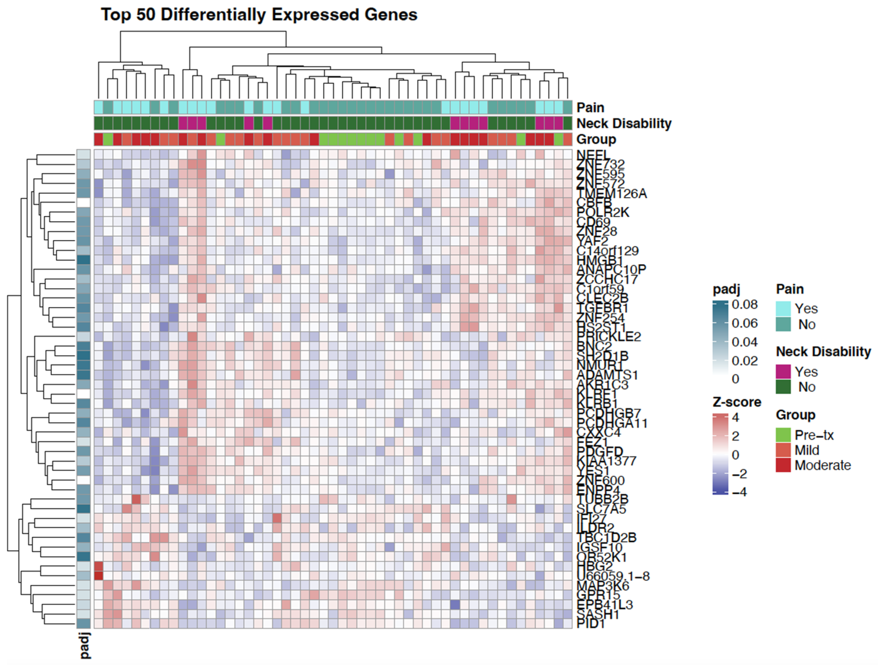
To further explore expression patterns, a heatmap of the top 50 differentially expressed genes was generated (Figure 2). This visualization revealed notable clustering of samples related to neck disability, with several genes showing consistent up- or down-regulation in participants reporting any neck disability.

3.3. Pathway and Network Analysis

IPA was used to explore biological pathways associated with differential gene expression between participants with and without any neck disability. The top canonical pathways include: Interferon alpha/beta signaling (B-H p-value = 2.31 × 10⁻¹¹; 17/76 genes, 22.4% overlap), Iron homeostasis signaling pathway (B-H p-value = 5.65 × 10⁻⁷; 17/145 genes, 11.7% overlap), Erythrocytes take up oxygen and release carbon dioxide (B-H p-value = 1.16 × 10⁻⁵; 5/7 genes, 71.4% overlap), Erythrocytes take up carbon dioxide and release oxygen (B-H p-value =1.80 × 10⁻⁴; 5/11 genes, 45.5% overlap), and Transcriptional Regulation of Granulopoiesis (B-H p-value = 5.83 × 10⁻⁴; 8/51 genes, 15.7% overlap).
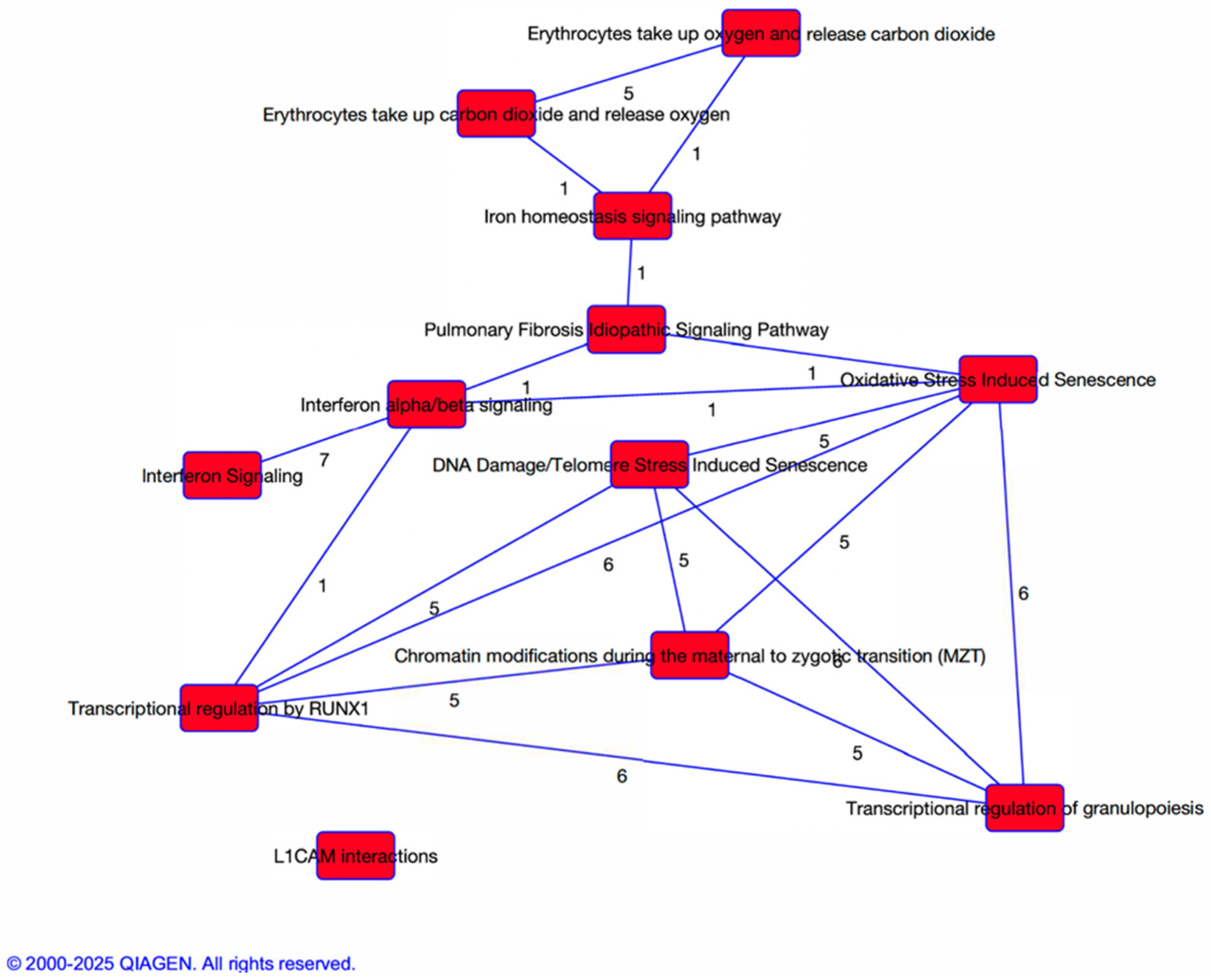
Evaluation of the interaction between the pathways revealed functional overlap among pathways related to interferon signaling, iron homeostasis, and blood cell development suggesting strong molecular connectivity in functions such as hematological and immunological disease, cellular movement, and connective tissue disorders (Figure 3).

4. Discussion

This study provides novel insights into the molecular and clinical landscape of neck disability in OPSCC survivors, with a particular focus on neck pain measured via NDI. Despite comparable sociodemographic and clinical characteristics across clinical cohorts, patients reporting any level of neck disability exhibited notable gene expression profiles compared to those who reported no disability. Differential gene expression analysis revealed 24 significantly dysregulated genes with clustering patterns that aligned with self-reported neck disability status. Pathway enrichment and network analyses further implicated interferon signaling, iron homeostasis, and erythrocyte gas exchange patterns, highlighting potential biological mechanisms underlying post-radiation tissue remodeling and neuromuscular dysfunction. Findings of this exploratory study suggest that self-reported neck pain and disability may reflect a broader biological phenotype beyond localized fibrotic changes captured by traditional clinical grading. Clinically, these insights may inform the development of biomarker-driven strategies for early identification, risk stratification, and targeted interventions to mitigate symptom burden associated with neck disability in OPSCC survivors.
The composition of the cohorts was comparable in age, race, and tobacco/alcohol use, significant differences emerged in marital status, neck disability, and self-reported pain. Although the differences in neck disability and self-reported pain were expected, as these are signs and symptoms of worsening degree of radiation-induced fibrosis, the differences in marital status were interesting as they were not directly related to the cancer or cancer treatment. In our study, we found that as fibrosis grade increased, so did the number of people who were not married. It has previously been shown that not only are married patients less likely to report neck disability, but might also experience a survival benefit as they have been shown to have less metastatic disease and are more likely to receive adequate treatment [13,34,35]. These findings suggest that social support and physical function may be important correlates of post-treatment disability in OPSCC survivors and warrants further research. The significant association between NDI and pain highlights the ability of the NDI to capture perceived pain in our cohort of patients. It is important to note that the differences observed occurred in the absence of significant variation in tumor stage or HPV status, reinforcing the hypothesis that neck disability may be driven by biological and psychosocial factors beyond initial disease severity.
Differential gene expression analysis revealed 24 significantly dysregulated genes associated with neck pain and disability in OPSCC survivors, spanning key biological domains including immune regulation, hematopoiesis, neuronal function, and transcriptional control. Notably, genes such as IFI27, GPR15, and ILDR2 were upregulated, implicating heightened interferon signaling and immune activation in patients with neck disability [36,37,38]. Concurrently, downregulation of neuronal genes like NEFL, FEZ1, and PRICKLE2 suggest potential disruptions in neuromuscular integrity and sensory processing [39,40,41,42]. The upregulation of HBG2 and MAP3K6, both involved in hematopoiesis and iron metabolism, may reflect compensatory vascular or oxygenation responses to tissue stress [43,44]. These transcriptomic patterns align with pathway enrichment results, reinforcing the hypothesis that neck disability reflects a multifaceted biological phenotype involving immune, vascular, and neuromuscular dysfunction rather than isolated fibrotic changes.
In addition to the previously highlighted immune and neuronal genes, the heatmap analysis revealed differential expression of transcriptional regulators and cellular function genes that may contribute to the biological complexity of neck pain and disability. Downregulation of ZNF732 and POLR2K suggest altered transcriptional control mechanisms, potentially affecting cellular stress responses and repair pathways [45,46]. Genes such as AKR1C3 and PID1, involved in tissue distributions and metabolic regulation, were also dysregulated, pointing to possible disruptions in local energy metabolism and tissue remodeling [47,48,49]. The upregulation of EPB41L3, a gene linked to cytoskeletal structure and neuronal stability, may reflect compensatory responses to neuromuscular strain [50]. Together, these findings expand the biological scope of neck pain and disability beyond immune and pain signaling, implicating transcriptional, metabolic, and structural pathways in the chronic sequelae experienced by HNC survivors.
Pathway enrichment analysis further contextualized the transcriptomic findings, revealing significant involvement of interferon alpha/beta signaling, iron homeostasis, and erythrocyte gas exchange, pathways that may underlie the persistent symptoms reported by OPSCC survivors with neck disability. The identification of oxidative stress-induced senescence and DNA damage/telomere stress pathways suggests that chronic cellular stress and impaired regenerative capacity may contribute to neuromuscular dysfunction [51]. Additionally, transcriptional regulation pathways such as RUNX1-mediated granulopoiesis and chromatin modification during the maternal-to-zygotic transition point to broader epigenetic and hematopoietic shifts [52,53]. These findings support a model in which neck pain and disability reflect not only localized tissue damage but also systemic biological responses involving immune activation, vascular remodeling, and cellular aging processes.
Notably, several genes identified in the transcriptomic analysis appear to be involved in multiple enriched pathways, suggesting they may serve as key regulatory nodes in the biological response to radiation and neck disability. For example, IFI27 and GPR15 are implicated in both interferon signaling and immune-related senescence pathways, highlighting their potential role in sustaining chronic inflammation and immune dysregulation [38,54]. Similarly, genes like HBG2 and MAP3K6, which contribute to iron homeostasis and erythrocyte gas exchange, also intersect with pathways related to oxidative stress and hematopoiesis [55,56]. The recurrence of these genes across functionally related biological processes suggest that neck disability may arise from a convergence of immune, vascular, and metabolic dysfunctions rather than isolated pathway activation. This integrative view supports the development of multitargeted therapeutic strategies and reinforces the value of transcriptomic profiling in uncovering complex, overlapping mechanisms of survivorship burden.
It has previously been shown that although most changes in pain perception are likely due to alterations in nerve impulse transmission pathways, whole blood transcriptomic profiles can differentiate vulnerability to chronic low back pain [57]. In this study, the authors found significant enrichment for known pain genes, supporting the feasibility of detecting pain-related molecular signatures in blood, even though the mechanisms may be indirect or reflect systemic correlates of pain processing. In our study, the lack of enrichment for pain genes may reflect differences in pain type. Our cohort experienced radiation-induced musculoskeletal pain, which often involves fibrosis, vascular remodeling, and localized inflammation, whereas chronic low back pain may stem from mechanical dysfunction or systemic inflammatory conditions, and its molecular signature in blood may reflect different pathways. Another difference might be that our analysis was guided by subjective self-reported neck disability, which reflects the impact of pain on daily functioning, whereas the Dorsey et al. study incorporated both subjective and objective assessments. Importantly, our study did not exclude participants taking opioids, whereas Dorsey et al. did. This difference may influence the peripheral transcriptomic profile, as opioid use can modulate immune and inflammatory gene expression.
Findings from this study provide evidence that neck pain and disability in OPSCC survivors are possibly linked to molecular profiles marked by immune activation, neuronal disruption, and vascular-metabolic dysregulation. Transcriptomic profiling combined with pathway enrichment analysis revealed not only individual gene dysregulation but also recurring molecular signatures across key biological pathways—including interferon signaling, iron homeostasis, and erythrocyte gas exchange—suggesting a coordinated biological response to radiation-induced tissue stress that may underlie chronic pain and functional impairment. These molecular distinctions underscore the importance of integrative approaches that pair patient-reported outcomes (PROs) with transcriptomic data to better characterize post-treatment sequelae and uncover hidden dimensions of survivorship burden. Future research should explore whether these gene expression patterns can serve as predictive biomarkers and whether they are modifiable through targeted therapies or rehabilitation strategies. Moreover, deeper understanding of the interplay between interferon signaling, iron metabolism, and erythrocyte function may open new therapeutic avenues to mitigate neuromuscular complications and enhance long-term survivorship care.

4.1. Implications for Clinical Practice

This study identified candidate genes and pathways that may serve as therapeutic targets and biomarkers for symptom burden and rehabilitation response in OPSCC. Genes linked to neuroinflammation, and neuromuscular function could inform risk stratification and early intervention for patients at higher risk of persistent neck pain and disability. Integrating transcriptomic data with PROs supports a personalized approach to survivorship care, enabling tailored physical therapy, pharmacologic treatment, and supportive services. These findings align with precision health goals and emphasize the role of symptom biology in improving QOL for OPSCC survivors. If replicated, these results could inform precision care strategies by combining clinical, demographic, behavioral, and gene expression data to characterize individual symptom experiences and guide self-management. Patients with gene dysregulation linked to severe pain risk may benefit from targeted education and early intervention options to prevent or reduce symptom burden.

4.2. Limitations and Future Directions

In this exploratory analysis, the small sample size limited our ability to break up the categories of neck disability and pain into mild, moderate, and severe, instead we used the bivariate categories of any pain or any neck disability versus no pain or no neck disability, limiting our ability to determine differential gene expression related to pain severity. Also, as this was a cross-sectional study, we did not have the ability to look at intra-patient pain duration. We do recognize that there is overlap between NDI grouping and treatment timing. However, the primary objective of this exploratory study was to explore transcriptomic correlates of symptom burden, not to model treatment effects per se. While treatment timing is inherently linked to symptom development, our analysis focused on identifying molecular features associated with dysfunction regardless of treatment stage. Disentangling treatment effects from symptom-related molecular changes is challenging in cross-sectional designs. In future studies, we plan to pursue longitudinal sampling to better isolate temporal effects. Moreover, we acknowledge that the dichotomization of continuous variables such as NDI and pain score could lead to information loss and reduced statistical power. However, we chose this this approach based on the exploratory nature of the study and considering the modest sample size. The threshold that we used for dichotomization are grounded in established clinical cutoffs that reflect meaningful symptom burden. We also recognize that it is possible that our results may also be influenced by residual RNA from erythroid or reticulocyte populations; however, in the context of gene expression, we do not see a uniform expression level of these genes (e.g., HBG2) but rather an enrichment in specific subgroups, suggesting a potential biological relevance rather than a universal artifact. Additionally, our sample consisted mainly of White males over the age of 60, limiting the ability to generalize the results. Future studies should validate these findings in larger, longitudinal cohorts and explore functional roles of identified genes through in vitro or in vivo models. Multi-omics approaches, including epigenomics and proteomics, may further elucidate the complex biology of neck pain and disability in OPSCC. Ultimately, translating these insights into clinical tools could improve symptom prediction, monitoring, and intervention.

Author Contributions

Conceptualization, M.A.W. and M.L.N.; methodology, M.A.W.; formal analysis, M.A.W.; resources, C.D. and M.L.N.; writing—original draft preparation, M.A.W.; writing—review and editing, M.A.W., C.D. and M.L.N.; visualization, M.A.W.; supervision, M.A.W. and M.L.N.; funding acquisition, M.A.W. and M.L.N. All authors have read and agreed to the published version of the manuscript.

Funding

This research received no external funding.

Institutional Review Board Statement

The study was conducted according to the guidelines of the Declaration of Helsinki and approved by the Institutional Review Board of the University of Pittsburgh (Protocol #20100024, date of approval 11 December 2020).

Informed Consent Statement

All study participants provided informed consent before initiating study procedures.

Data Availability Statement

De-identified data can be made available upon request for research purposes to qualified individuals within the scientific community.

Conflicts of Interest

The authors declare no conflicts of interest.

References

  1. Siegel, R.L.; Giaquinto, A.N.; Jemal, A. Cancer statistics, 2024. CA A Cancer J. Clin. 2024, 74, 12–49. [Google Scholar] [CrossRef] [PubMed]
  2. Ellington, T.D.; Henley, S.J.; Senkomago, V.; O’Neil, M.E.; Wilson, R.J.; Singh, S.; Thomas, C.C.; Wu, M.; Richardson, L.C. Trends in Incidence of Cancers of the Oral Cavity and Pharynx—United States 2007–2016. MMWR Morb. Mortal. Wkly. Rep. 2020, 69, 433–438. [Google Scholar] [CrossRef]
  3. Sung, H.; Ferlay, J.; Siegel, R.L.; Laversanne, M.; Soerjomataram, I.; Jemal, A.; Bray, F. Global Cancer Statistics 2020: GLOBOCAN Estimates of Incidence and Mortality Worldwide for 36 Cancers in 185 Countries. CA A Cancer J. Clin. 2021, 71, 209–249. [Google Scholar] [CrossRef]
  4. Johnson, D.E.; Burtness, B.; Leemans, C.R.; Lui, V.W.Y.; Bauman, J.E.; Grandis, J.R. Head and neck squamous cell carcinoma. Nat. Rev. Dis. Primers 2020, 6, 92. [Google Scholar] [CrossRef]
  5. Fonsêca, T.C.; Jural, L.A.; Marañón-Vásquez, G.A.; Magno, M.B.; Roza, A.L.O.C.; Ferreira, D.M.T.P.; Maia, L.C.; Romañach, M.J.; Agostini, M.; Abrahão, A.C. Global prevalence of human papillomavirus-related oral and oropharyngeal squamous cell carcinomas: A systematic review and meta-analysis. Clin. Oral. Investig. 2023, 28, 62. [Google Scholar] [CrossRef]
  6. Kim, S.I.; Lee, J.W.; Eun, Y.-G.; Lee, Y.C. A SEER-based analysis of trends in HPV-associated oropharyngeal squamous cell carcinoma. Infect. Agents Cancer 2024, 19, 29. [Google Scholar] [CrossRef]
  7. Harris, A.; Branstetter, B.; Li, J.; Piva, S.R.; Johnson, J.T.; Nilsen, M.L. Evaluation of Neck Disability Using Computed-Tomography in Head and Neck Cancer Survivors. Front. Pain. Res. 2022, 3, 910247. [Google Scholar] [CrossRef]
  8. Ghiam, M.K.; Mannion, K.; Dietrich, M.S.; Stevens, K.L.; Gilbert, J.; Murphy, B.A. Assessment of musculoskeletal impairment in head and neck cancer patients. Support. Care Cancer 2017, 25, 2085–2092. [Google Scholar] [CrossRef] [PubMed]
  9. Gane, E.M.; McPhail, S.M.; Hatton, A.L.; Panizza, B.J.; O’Leary, S.P. The relationship between physical impairments, quality of life and disability of the neck and upper limb in patients following neck dissection. J. Cancer Surviv. 2018, 12, 619–631. [Google Scholar] [CrossRef] [PubMed]
  10. Fernández-Gualda, M.Á.; Postigo-Martin, P.; Fernandez-Gonzalez, M.; Martin-Martin, L.; Vargas-Arrabal, P.; Lozano-Lozano, M.; Fernández-Lao, C. Prevalence and characteristics of persistent pain among head and neck cancer survivors: A systematic review and meta-analysis. Pain. Med. 2025, 26, 712–721. [Google Scholar] [CrossRef]
  11. Shuman, A.G.; Terrell, J.E.; Light, E.; Wolf, G.T.; Bradford, C.R.; Chepeha, D.; Jiang, Y.; McLean, S.; Ghanem, T.A.; Duffy, S.A. Predictors of Pain Among Patients with Head and Neck Cancer. Arch. Otolaryngol.–Head Neck Surg. 2012, 138, 1147. [Google Scholar] [CrossRef] [PubMed]
  12. Tschiesner, U. Changing the Perspective: Current Trends in the Assessment of Functional Outcome in Patients with Head and Neck Cancer. Curr. Oncol. Rep. 2011, 13, 126–131. [Google Scholar] [CrossRef] [PubMed]
  13. Nilsen, M.L.; Lyu, L.; Belsky, M.A.; Mady, L.J.; Zandberg, D.P.; Clump, D.A., 2nd; Skinner, H.D.; Peddada, S.D.; George, S.; Johnson, J.T. Impact of Neck Disability on Health-Related Quality of Life among Head and Neck Cancer Survivors. Otolaryngol. Head Neck Surg. 2020, 162, 64–72. [Google Scholar] [CrossRef]
  14. Bonsel, J.M.; Itiola, A.J.; Huberts, A.S.; Bonsel, G.J.; Penton, H. The use of patient-reported outcome measures to improve patient-related outcomes—A systematic review. Health Qual. Life Outcomes 2024, 22, 101. [Google Scholar] [CrossRef]
  15. Zeqiri, E.; Qorri, E.; Todri, J.; Lena, O. Establishing the Neck Disability Index as a Valid Tool for Assessing Persistent Neck Pain in the Albanian Population. Medicina 2025, 61, 955. [Google Scholar] [CrossRef]
  16. Martinelli, C.; Ercoli, A.; Parisi, S.; Iatì, G.; Pergolizzi, S.; Alfano, L.; Pentimalli, F.; De Laurentiis, M.; Giordano, A.; Cortellino, S. Molecular Mechanisms and Clinical Divergences in HPV-Positive Cervical vs. Oropharyngeal Cancers: A Critical Narrative Review. BMC Med. 2025, 23, 405. [Google Scholar] [CrossRef] [PubMed]
  17. Wang, H.; Song, X.; Shen, H.; Liu, W.; Wang, Y.; Zhang, M.; Yang, T.; Mou, Y.; Ren, C.; Song, X. Cancer neuroscience in head and neck: Interactions, modulation, and therapeutic strategies. Mol. Cancer 2025, 24, 101. [Google Scholar] [CrossRef]
  18. Wang, W.-L.; Hao, Y.-H.; Pang, X.; Tang, Y.-L. Cancer pain: Molecular mechanisms and management. Mol. Biomed. 2025, 6, 45. [Google Scholar] [CrossRef]
  19. Anarte-Lazo, E.; Bernal-Utrera, C. A Scoping Review of Clinical Features and Mechanisms of Orofacial Pain and Headache in Patients with Head and Neck Cancer. J. Clin. Med. 2025, 14, 5722. [Google Scholar] [CrossRef]
  20. Ye, Y.; Cardoso, D.M.; Kayahara, G.M.; Bernabé, D.G. A pilot study to improve pain phenotyping in head and neck cancer patients. Front. Pain Res. 2023, 4, 1146667. [Google Scholar] [CrossRef]
  21. Mackey, S.; Aghaeepour, N.; Gaudilliere, B.; Kao, M.C.; Kaptan, M.; Lannon, E.; Pfyffer, D.; Weber, K. Innovations in acute and chronic pain biomarkers: Enhancing diagnosis and personalized therapy. Reg. Anesth. Pain. Med. 2025, 50, 110–120. [Google Scholar] [CrossRef]
  22. Zhi, W.I.; Gentile, D.; Diller, M.; Kinney, A.; Bao, T.; Master, V.; Wang, X.S. Patient-Reported Outcomes of Pain and Related Symptoms in Integrative Oncology Practice and Clinical Research: Evidence and Recommendations. Oncology 2021, 35, 35–41. [Google Scholar] [CrossRef]
  23. American Joint Committee on Cancer. AJCC Cancer Staging Manual, 8th ed.; Springer: New York, NY, USA, 2017. [Google Scholar]
  24. Powell, W.R.; Sheehy, A.M.; Kind, A.J. The area deprivation index is the most scientifically validated social exposome tool available for policies advancing health equity. Health Aff. Forefr. 2023. [Google Scholar] [CrossRef]
  25. Kind, A.J.H.; Buckingham, W.R. Making Neighborhood-Disadvantage Metrics Accessible—The Neighborhood Atlas. N. Engl. J. Med. 2018, 378, 2456–2458. [Google Scholar] [CrossRef]
  26. Vernon, H. The Neck Disability Index: State-of-the-art, 1991–2008. J. Manip. Physiol. Ther. 2008, 31, 491–502. [Google Scholar] [CrossRef]
  27. Cleland, J.A.; Fritz, J.M.; Whitman, J.M.; Palmer, J.A. The Reliability and Construct Validity of the Neck Disability Index and Patient Specific Functional Scale in Patients with Cervical Radiculopathy. Spine 2006, 31, 598–602. [Google Scholar] [CrossRef]
  28. MacDermid, J.C.; Walton, D.M.; Avery, S.; Blanchard, A.; Etruw, E.; McAlpine, C.; Goldsmith, C.H. Measurement properties of the neck disability index: A systematic review. J. Orthop. Sports Phys. Ther. 2009, 39, 400–417. [Google Scholar] [CrossRef]
  29. Cleland, J.A.; Childs, J.D.; Whitman, J.M. Psychometric properties of the Neck Disability Index and Numeric Pain Rating Scale in patients with mechanical neck pain. Arch. Phys. Med. Rehabil. 2008, 89, 69–74. [Google Scholar] [CrossRef] [PubMed]
  30. Vernon, H. Assessment of self-rated disability, impairment, and sincerity of effort in whiplash-associated disorder. J. Musculoskelet. Pain. 2000, 8, 155–167. [Google Scholar] [CrossRef]
  31. Magaña, L.C.; Murati, S.; Riffitts, M.; Harrison, C.; Harris, A.; Sowa, G.; Johnson, J.T.; Bell, K.; Nilsen, M. Subjective and Objective Measures in Assessing Neck Disability and Pain in Head and Neck Cancer. Laryngoscope 2021, 131, 2015–2022. [Google Scholar] [CrossRef] [PubMed]
  32. Network, N.C.C. Adult Cancer Pain (Version 2.2024). Available online: https://www.nccn.org/professionals/physician_gls/pdf/pain.pdf (accessed on 12 September 2024).
  33. Benjamini, Y.; Hochberg, Y. Controlling the false discovery rate: A practical and powerful approach to multiple testing. J. R. Stat. Soc. Ser. B (Methodol.) 1995, 57, 289–300. [Google Scholar] [CrossRef]
  34. Inverso, G.; Mahal, B.A.; Aizer, A.A.; Donoff, R.B.; Chau, N.G.; Haddad, R.I. Marital status and head and neck cancer outcomes. Cancer 2015, 121, 1273–1278. [Google Scholar] [CrossRef] [PubMed]
  35. Simpson, M.C.; Challapalli, S.D.; Cass, L.M.; Zahirsha, Z.S.; Adjei Boakye, E.; Massa, S.T.; Osazuwa-Peters, N. Impact of gender on the association between marital status and head and neck cancer outcomes. Oral. Oncol. 2019, 89, 48–55. [Google Scholar] [CrossRef] [PubMed]
  36. Hecht, I.; Toporik, A.; Podojil, J.R.; Vaknin, I.; Cojocaru, G.; Oren, A.; Aizman, E.; Liang, S.C.; Leung, L.; Dicken, Y.; et al. ILDR2 Is a Novel B7-like Protein That Negatively Regulates T Cell Responses. J. Immunol. 2018, 200, 2025–2037. [Google Scholar] [CrossRef]
  37. Huang, C.H.; Huang, Y.C.; Xu, J.K.; Chen, S.Y.; Tseng, L.C.; Huang, J.L.; Lin, C.S. ATM Inhibition-Induced ISG15/IFI27/OASL Is Correlated with Immunotherapy Response and Inflamed Immunophenotype. Cells 2023, 12, 1288. [Google Scholar] [CrossRef]
  38. Okamoto, Y.; Shikano, S. Emerging roles of a chemoattractant receptor GPR15 and ligands in pathophysiology. Front. Immunol. 2023, 14, 1179456. [Google Scholar] [CrossRef]
  39. Bayat, A.; Iqbal, S.; Borredy, K.; Amiel, J.; Zweier, C.; Barcia, G.; Kraus, C.; Weyhreter, H.; Bassuk, A.G.; Chopra, M.; et al. PRICKLE2 revisited-further evidence implicating PRICKLE2 in neurodevelopmental disorders. Eur. J. Hum. Genet. 2021, 29, 1235–1244. [Google Scholar] [CrossRef]
  40. Gehr, N.L.; Mortensen, C.; Stage, T.B.; Pedersen, M.R.V.; Rafaelsen, S.R.; Madsen, J.S.; Olsen, D.A.; Timm, S.; Jensen, L.H.; Hansen, T.F.; et al. Neurofilament light chain as a marker for neuronal damage: Integrating in vitro studies and clinical findings in patients with oxaliplatin-induced neuropathy. Cancer Chemother. Pharmacol. 2025, 95, 53. [Google Scholar] [CrossRef]
  41. Gunaseelan, S.; Wang, Z.; Tong, V.K.J.; Ming, S.W.S.; Razar, R.; Srimasorn, S.; Ong, W.Y.; Lim, K.L.; Chua, J.J.E. Loss of FEZ1, a gene deleted in Jacobsen syndrome, causes locomotion defects and early mortality by impairing motor neuron development. Hum. Mol. Genet. 2021, 30, 5–20. [Google Scholar] [CrossRef]
  42. Shi, G.; Zhou, X.; Wang, X.; Zhang, X.; Zhang, P.; Feng, S. Signatures of altered DNA methylation gene expression after central and peripheral nerve injury. J. Cell Physiol. 2020, 235, 5171–5181. [Google Scholar] [CrossRef] [PubMed]
  43. Hadanny, A.; Forer, R.; Volodarsky, D.; Daniel-Kotovsky, M.; Catalogna, M.; Zemel, Y.; Bechor, Y.; Efrati, S. Hyperbaric oxygen therapy induces transcriptome changes in elderly: A prospective trial. Aging 2021, 13, 24511–24523. [Google Scholar] [CrossRef] [PubMed]
  44. Liu, X.; Zhang, P.; Ji, K.; Zhang, J.; Yang, S.; Du, B.; Hu, S.; Fan, R. Cyclin-dependent kinase 5 regulates MAPK/ERK signaling in the skin of mice. Acta Histochem. 2018, 120, 15–21. [Google Scholar] [CrossRef] [PubMed]
  45. Cui, J.; Wang, L.; Zhong, W.; Chen, Z.; Chen, J.; Yang, H.; Liu, G. Development and Validation of Epigenetic Signature Predict Survival for Patients with Laryngeal Squamous Cell Carcinoma. DNA Cell Biol. 2021, 40, 247–264. [Google Scholar] [CrossRef]
  46. Ranaweera, S.S.; Zuniga-Hertz, J.P.; Chitteti, R.; Chernov, A.V.; Patel, H.H. Protective effect of energized structured water on bioenergetic function and oxidative stress in H9c2 cells. EXPLORE 2025, 21, 103232. [Google Scholar] [CrossRef] [PubMed]
  47. Deng, H.; Tang, J.; Xu, Y.; Wu, L.; Zhang, J.; Chen, H.; Wang, Z.; Yang, R.; Gao, W.; He, Z. Mitochondrial Damage-induced Ferroptosis: The Molecular Mechanism by Which Psoralen Inhibits the Proliferation and Invasion of Non-small-cell Lung Cancer Cells. Vivo 2025, 39, 2681–2702. [Google Scholar] [CrossRef]
  48. Shi, C.M.; Xu, G.F.; Yang, L.; Fu, Z.Y.; Chen, L.; Fu, H.L.; Shen, Y.H.; Zhu, L.; Ji, C.B.; Guo, X.R. Overexpression of TFAM protects 3T3-L1 adipocytes from NYGGF4 (PID1) overexpression-induced insulin resistance and mitochondrial dysfunction. Cell Biochem. Biophys. 2013, 66, 489–497. [Google Scholar] [CrossRef]
  49. Zhang, H.; Qian, Y.; Zhang, Y.; Zhou, X.; Shen, S.; Li, J.; Sun, Z.; Wang, W. Multi-omics analysis deciphers intercellular communication regulating oxidative stress to promote oral squamous cell carcinoma progression. NPJ Precis. Oncol. 2024, 8, 272. [Google Scholar] [CrossRef]
  50. Cifuentes-Diaz, C.; Chareyre, F.; Garcia, M.; Devaux, J.; Carnaud, M.; Levasseur, G.; Niwa-Kawakita, M.; Harroch, S.; Girault, J.A.; Giovannini, M.; et al. Protein 4.1B contributes to the organization of peripheral myelinated axons. PLoS ONE 2011, 6, e25043. [Google Scholar] [CrossRef]
  51. Sahin, E.; Depinho, R.A. Linking functional decline of telomeres, mitochondria and stem cells during ageing. Nature 2010, 464, 520–528. [Google Scholar] [CrossRef]
  52. de Bruijn, M.; Dzierzak, E. Runx transcription factors in the development and function of the definitive hematopoietic system. Blood 2017, 129, 2061–2069. [Google Scholar] [CrossRef]
  53. Eckersley-Maslin, M.A.; Alda-Catalinas, C.; Reik, W. Dynamics of the epigenetic landscape during the maternal-to-zygotic transition. Nat. Rev. Mol. Cell Biol. 2018, 19, 436–450. [Google Scholar] [CrossRef] [PubMed]
  54. Villamayor, L.; López-García, D.; Rivero, V.; Martínez-Sobrido, L.; Nogales, A.; DeDiego, M.L. The IFN-stimulated gene IFI27 counteracts innate immune responses after viral infections by interfering with RIG-I signaling. Front. Microbiol. 2023, 14, 1176177. [Google Scholar] [CrossRef] [PubMed]
  55. Kautz, L.; Nemeth, E. Molecular liaisons between erythropoiesis and iron metabolism. Blood 2014, 124, 479–482. [Google Scholar] [CrossRef]
  56. Morgan, P.K.; Murphy, A.J. Make hematopoiesis great again: Countering oxidative stress! Blood 2025, 145, 1102–1104. [Google Scholar] [CrossRef]
  57. Dorsey, S.G.; Renn, C.L.; Griffioen, M.; Lassiter, C.B.; Zhu, S.; Huot-Creasy, H.; McCracken, C.; Mahurkar, A.; Shetty, A.C.; Jackson-Cook, C.K.; et al. Whole blood transcriptomic profiles can differentiate vulnerability to chronic low back pain. PLoS ONE 2019, 14, e0216539. [Google Scholar] [CrossRef] [PubMed]
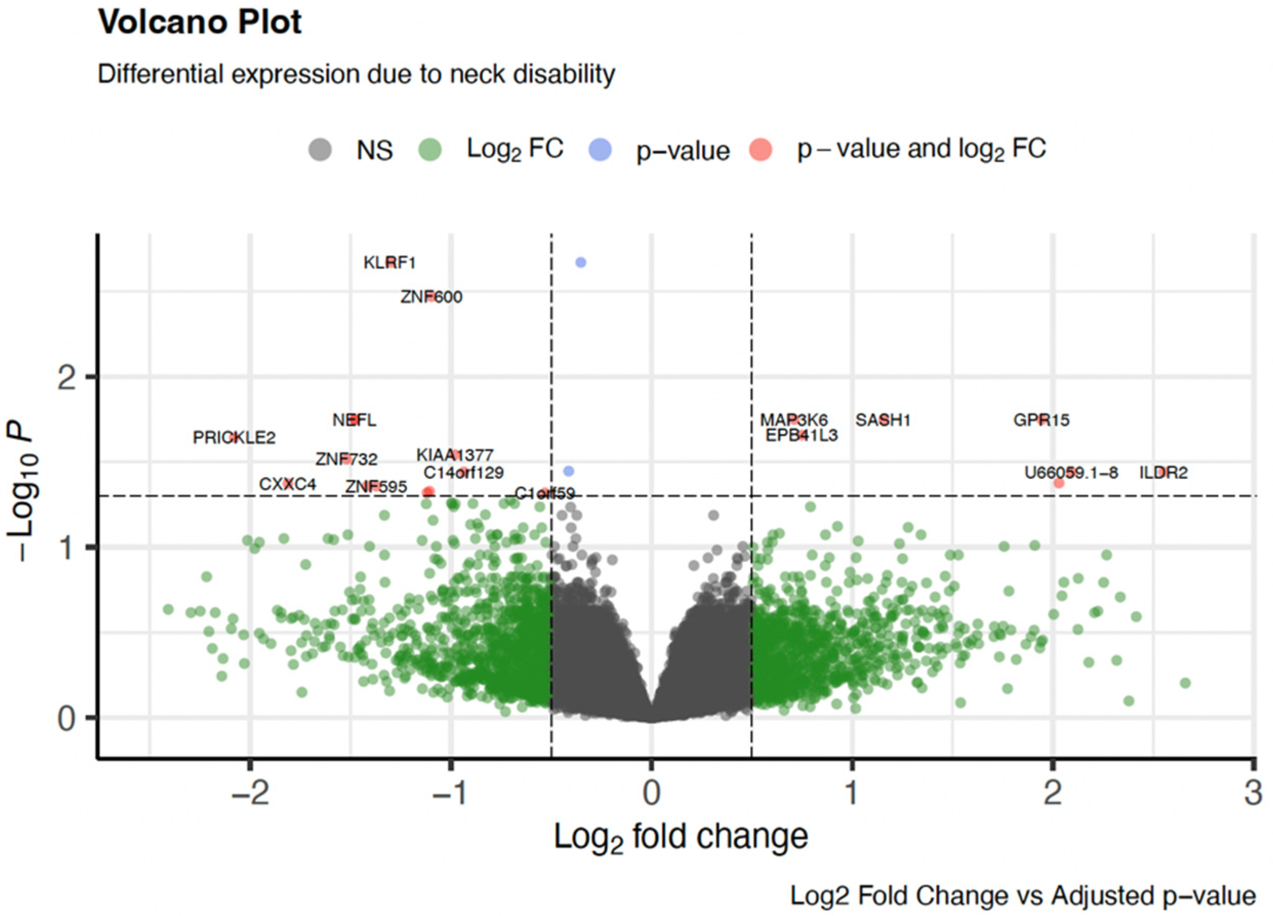
Figure 1. A volcano plot illustrating differential gene expression associated with neck disability. Each point represents a gene, plotted by it log2 fold change (x-axis) and −log10 adjusted p-value (y-axis). Genes with both statistically significant expression changes and substantial fold changes are highlighted in red, while those with either significant p-values (blue) or fold changes (green) alone are also marked. Non-significant genes are shown in grey. Notable genes such as KLRF1, ZNF600, PRICKLE2, and NEFL are labeled for emphasis. This visualization enables rapid identification of genes most affected by neck disability, supporting downstream pathway and functional enrichment analysis.
Genes 16 01277 g001
Figure 2. Heatmap showing the expression profiles of the top 50 DEGs (p ≤ 0.05) across all samples. Rows represent individual genes, and columns represent samples grouped by clinical annotations: Fibrosis Level (No Fibrosis, Mild Fibrosis, Moderate/Severe Fibrosis), Pain Category (None, Mild/Moderate-Severe), and NDI category (None, Mild/Moderate-Severe). Gene expression values were z-score-normalized, with red indicating higher expression and blue indicating lower expression. Hierarchical clustering was applied to both genes and samples using Euclidean distance and complete linkage. The adjusted p-value is represented on the left for visualization but the genes in this heatmap were chosen based on p-value. This visualization highlights differentiated expression patterns corresponding to clinical phenotypes in HNC patients and provides a broader visual overview of transcriptomic variation across the sample.
Genes 16 01277 g002
Figure 3. IPA identified top canonical pathway enriched among genes differentially expressed in relation to neck disability. The numbers adjacent to each pathway indicate the number of genes shared between the pathways, reflecting molecular overlap and potential crosstalk among biological processes. This figure highlights key mechanisms such as cellular stress responses, immune signaling, erythrocyte function, and transcriptional regulation, suggesting interconnected molecular pathways underlying neck disability.
Genes 16 01277 g003
Table 1. Participant characteristics.
CharacteristicNo Fibrosis
(n = 13)
Mild Fibrosis
(n = 20)
Moderate/
Severe Fibrosis
(n = 18)
p-Value
Age (years, mean ± SD)63.69 ± 7.0468.80 ± 7.3666.83 ± 10.980.271
Race: White (count, %)12 (92.3%)17 (85.0%)17 (94.4%)0.593
Sex: Male (count, %)12 (92.3)18 (90.0)12 (66.7)0.094
   Female (count, %)1 (7.7)2 (10.0)6 (33.3)
Marital Status: Married (count, %)11 (84.6)15 (75.0)8 (44.4)0.034 *
        Other a (count, %)2 (15.4)5 (25.0)10 (55.6)
Tobacco Use (ever): Yes (count, %)7 (53.8)10 (50.0)8 (44.4)0.870
          No (count, %)6 (46.2)10 (50.0)10 (55.6)
Alcohol Use (ever): Yes (count, %)11 (84.6)18 (90.0)12 (66.7)0.176
          No (count, %)2 (15.4)2 (10.0)6 (33.3)
Tumor Stage: T1/T2 (count, %)8 (61.5)18 (90.0)12 (66.7)0.119
        T3/T4 (count, %)5 (38.5)2 (10.0)6 (33.3)
HPV Status: Yes (count, %)12 (92.3)16 (80.0)12 (66.7)0.225
      No (count, %)1 (7.6)4 (20.0)6 (33.3)
Neck Disability: None (count, %)12 (92.3)19 (95.0)8 (44.4)0.003 *
        Any (count, %)1 (7.7)1 (5.0)10 (55.6)
Self-reported Pain: None (count, %)12 (92.3)13 (65.0%)4 (22.2)<0.001 *
         Any (count, %)1 (7.7)7 (35%)14 (77.8)
Area Deprivation (mean ± SD)59.38 ± 29.3759.05 ± 23.1264.22 ± 18.590.884
* Statistically significant difference; a divorced/living with partner/widowed/single.
Table 2. Significant genes.
Gene NameGene SymbolAdjusted p-Valuelog2 Fold ChangeGene Function
Killer Cell Lectin Like Receptor F1KLRF10.002−1.30Immune Function
Core−Binding Factor Subunit BetaCBFB0.002−0.35Hematopoiesis
Zinc Finger Protein 600ZNF6000.003−1.09Transcriptional Regulation
Mitogen−Activated Protein 3 Kinase Kinase Kinase 6MAP3K60.0170.71Hematopoiesis
G Protein−Coupled Receptor 15GPR150.0171.94Immune Function
SAM And SH3 Domain Containing 1SASH10.0171.16Immune Function
Neurofilament Light ChainNEFL0.017−1.48Neuronal Function
Hemoglobin Subunit Gamma 2HBG20.0174.21Hematopoiesis
Fasciculation And Elongation Protein Zeta 1FEZ10.017−1.48Neuronal Function
Interferon Alpha Inducible Protein 27IFI270.0183.01Immune Function
Erythrocyte Membrane Protein Band 4.1 Like 3EPB41L30.0220.75Neuronal Function
Prickle Planar Cell Polarity Protein 2PRICKLE20.022−2.08Neuronal Function
Centrosomal Protein 126CEP1260.028−0.98Cellular Function
Zinc Finger Protein 732ZNF7320.030−1.52Transcriptional Regulation
Zinc Finger CCHC−Type Containing 17ZCCHC170.035−0.41Transcriptional Regulation
Immunoglobulin Like Domain Containing Receptor 2ILDR20.0362.55Immune Function
GSK3B Interacting ProteinGSKIP0.036−0.94Neuronal Function (neurite outgrowth)
Immunoglobulin Superfamily Member 10IGSF100.0412.03Neuronal Function
CXXC Finger Protein 4CXXC40.042−1.81Hematopoiesis (Fe) Wnt signaling
Zinc Finger Protein 595ZNF5950.043−1.37Transcriptional Regulation
Protocadherin Gamma Subfamily B, 7PCDHGB70.043−1.42Neuronal Function
Aldo−Keto Reductase Family 1 Member C3AKR1C30.047−1.11Cellular Function (tissue distribution)
HEN Methyltransferase 1HENMT10.047−0.53Transcriptional Regulation
RNA polymerase II, I and III Subunit KPOLR2K0.047−1.12Transcriptional Regulation
Disclaimer/Publisher’s Note: The statements, opinions and data contained in all publications are solely those of the individual author(s) and contributor(s) and not of MDPI and/or the editor(s). MDPI and/or the editor(s) disclaim responsibility for any injury to people or property resulting from any ideas, methods, instructions or products referred to in the content.