Article

Molecular Characterization and Expression Patterns of Sox3 and Sox30 Genes and Response to Exogenous Hormones in the Chinese Soft-Shelled Turtle (Pelodiscus sinensis)

1
College of Marine Science and Fisheries, Jiangsu Ocean University, Lianyungang 222005, China
2
Key Laboratory of Tropical & Subtropical Fishery Resource Application & Cultivation of Ministry of Agriculture and Rural Affairs, Pearl River Fisheries Research Institute, Chinese Academy of Fishery Sciences, Guangzhou 510380, China
3
Science and Technology Research Center of China Customs, Beijing 100026, China
4
School of Marine and Fisheries, Guangdong Eco-Engineering Polytechnic, Guangzhou 510520, China
*
Authors to whom correspondence should be addressed.
These authors contributed equally to this work.
Genes 2025, 16(11), 1249; https://doi.org/10.3390/genes16111249
Submission received: 25 September 2025 / Revised: 18 October 2025 / Accepted: 21 October 2025 / Published: 22 October 2025
(This article belongs to the Section Animal Genetics and Genomics)

Abstract

Background/Objectives: The Sox transcription factor family is critical for gonadal development and sex differentiation in animals, yet its roles in chelonians, particularly in the Chinese soft-shelled turtle (Pelodiscus sinensis), have rarely been investigated. Methods: This study cloned and analyzed the cDNA sequences of Sox3 and Sox30 genes from P. sinensis, examining their amino acid sequences and structural properties. Real-time quantitative PCR (RT-qPCR) was used to assess the expression of these two genes in different adult tissues and at various stages of embryonic gonadal development. Additionally, the effects of exogenous hormones (17β-estradiol, E2 and 17α-Methyltestosterone, MT) on the expression of Sox3 and Sox30 were also investigated. Results: The results indicated that Sox3 showed significantly elevated expression in female gonads, kidney, brain, liver, lung, spleen, and muscle relative to male counterparts, displaying a female-biased expression pattern. In contrast, Sox30 showed a male-biased pattern, with higher expression in male gonads, spleen, muscle, brain, and liver than in females, showing expression. Both genes were expressed at low levels. Exogenous hormone treatments revealed that MT significantly downregulated Sox3 expression in female embryos, whereas E2 significantly enhanced Sox3 expression in male embryos. Furthermore, MT treatment significantly upregulated Sox30 expression in female embryos, and E2 treatment also significantly increased Sox30 expression in male embryos. Conclusions: These findings suggest that Sox3 and Sox30 play crucial roles in the gonadal development of P. sinensis, with Sox3 potentially involved in ovarian development and Sox30 in testicular maturation. Both genes are regulated by exogenous hormones, highlighting their importance in sex differentiation and gonadal development. This study provides valuable theoretical insights for further exploration of the molecular mechanisms of sex regulation in reptiles.

1. Introduction

Members of the Sox gene family are transcription factors defined by a conserved high-mobility group (HMG) box that mediates DNA binding, and they are involved in processes such as early embryonic development, gonadal differentiation, and nervous system development [1]. As major members of this family, the Sox3 and Sox30 genes were first identified in humans (Homo sapiens) and mice (Mus musculus) [2]. Previous studies have shown that Sox3 mRNA plays a role in neural crest development and adult gonadal differentiation in M. musculus. Furthermore, Sox3 expression levels have been reported to be higher in the ovaries than in the testes of Japanese eel (Anguilla japonica) [3], Japanese flounder (Paralichthys olivaceus) [4], and Nile tilapia (Oreochromis niloticus) [5]. Knockout of the Sox3 gene in M. musculus has been shown to impair germline stem cell differentiation during development [6]. Similarly, Hong et al. [7] demonstrated that Sox3 knockout in zebrafish (Danio rerio) led to delayed follicular development and reduced fecundity in females. Sox30 is the sole member of the Sox H subfamily. In M. musculus, Sox30 expression exhibits remarkable tissue specificity, being abundantly expressed only in the adult male testis, predominantly in spermatocytes and spermatogonia. Its expression increases significantly at postnatal day 21, the period when spermatocytes first appear, suggesting that Sox30 may be involved in spermatid differentiation during the post-meiotic haploid stage [8]. Wei et al. [9] reported that the Sox30 gene is specifically expressed in the testes of O. niloticus. The Sox30 gene is primarily involved in male germ cell maturation, and studies have shown that its knockout its inhibits sperm formation while having no effect on oocyte formation or meiosis [10,11]. Fei Han et al. [12] reported that loss of the Sox30 can cause non-obstructive azoospermia (NOA), whereas re-expression of Sox30 rescues spermatogenesis and restores fertility, consistent with observations in Sox30-deficient mice. In the transcriptomic analysis of gonadal differentiation in the Chinese soft-shelled turtle (Pelodiscus sinensis), Sox3 and Sox30 were found to be highly expressed in the ovary and testis, respectively [13]. However, their potential functions in the gonadal differentiation and development of P. sinensis remain unclear.
P. sinensis, belonging to the class Reptilia, order Testudines, family Trionychidae, and genus Pelodiscus, is a high-value, is high-value “famous, special, and high-quality” aquaculture species in China. It is widely favored for its medicinal and edible value [14]. P. sinensis exhibits pronounced sexual dimorphism, with males growing faster than females under the same conditions. Consequently, there is demand for all-male breeding in the aquaculture industry. Identifying and studying genes related to gonadal differentiation and development may provide molecular targets for sex-controlled breeding in aquatic animals. In addition, previous studies have demonstrated that sex hormones play an important role in gonadal differentiation and development. 17β-Estradiol (E2) is a natural estrogen [15]. 17α-Methyltestosterone (MT) has been synthesized as an androgen agonist [16]. Both have been widely used in studies related to gonadal differentiation and development. Pieau et al. [17] reported that injecting of E2 into European pond turtles (Emys orbicularis) at 25 °C induces male-to-female sex reversal. El-Greisy et al. reported that treatment with MT induced masculinization of gonadal development in female juveniles of the red-spotted grouper (Epinephelus akaara) [18] and O. niloticus [19]. However, whether E2 and MT regulate the expression of Sox3 and Sox30 in P. sinensis, thereby participating in gonadal differentiation and development, remains unclear. Given the aforementioned background, this study cloned the cDNA sequences of Sox3 and Sox30 genes, analyzed their sequence characteristics, examined their expression patterns across different adult tissues and in gonads at various embryonic developmental stages, and evaluated their expression responses to exogenous hormone treatments (E2 and MT). These results will provide fundamental data for further elucidating the physiological roles of Sox3 and Sox30 in gonadal differentiation and development of P. sinensis.

2. Materials and Methods

2.1. Collection of Experimental Materials

P. sinensis used in this study were purchased from Caixing Industrial Co., Ltd. (Huizhou, China). Three healthy, three-year-old P. sinensis were selected, anesthetized, and euthanized by exsanguination. Heart, liver, brain, kidney, spleen, muscle, ovary, and testis tissues were collected from each individual, placed into RNase-free cryogenic tubes, rapidly frozen in liquid nitrogen, and stored at −80 °C until use. Three biological replicates were obtained for each tissue type. Additionally, 1000 fertilized P. sinensis eggs were collected, and gonadal tissues were sampled at developmental stages 14, 16, 18, 20, 22, 24, and 26. Embryonic staging was performed according to morphological characteristics described in a previous study [20], and other tissues from the embryos were collected for sex identification. At each stage, 120 embryos were collected, and based on the results of sex identification (Section 2.2), gonads from 20 male or female embryos were pooled into one tube, with three biological replicates per stage. Fertilized eggs were incubated using a medium-free method [21] under controlled conditions at 31 ± 1 °C and humidity of 75–85%.

2.2. DNA Extraction and Embryonic Sex Identification

DNA was extracted from P. sinensis embryonic samples following the instructions provided with the kit (ONREW, Shanghai, China). The extracted DNA was collected and stored at −20 °C until use. PCR amplification was performed using the P. sinensis species-specific primer pair PB1 [22] (Table 1). The 20 μL reaction system consisted of 7 μL of double-distilled water (ddH2O), 1 μL of PB1-F, 1 μL of PB1-R, 1 μL of genomic DNA template (50 ng/μL), and 10 μL of 2× Accurate Taq Master Mix. The amplification program was as follows: initial denaturation at 94 °C for 30 s; 35 cycles of denaturation at 98 °C for 10 s, annealing at 60 °C for 30 s, and extension at 72 °C for 60 s; and a final extension at 72 °C for 120 s. The resulting products were analyzed via 1% agarose gel electrophoresis to determine the genetic sex of the embryos.

2.3. Total RNA Extraction and First-Strand cDNA Synthesis

Total RNA was isolated from various tissues of P. sinensis using Trizol reagent (Takara, Beijing, China) according to the manufacturer’s protocol. RNA concentration and quality were assessed using the NanoQ™ nucleic acid analyzer (Thermo Scientific, Madison, WI, USA), and 1% agarose gel electrophoresis. Genomic DNA was removed using the HiScript® III RT SuperMix for qPCR (+gDNA wiper) kit (Vazyme, Nanjing, China). First-strand cDNA was synthesized following the manufacturer’s instructions, and the resulting cDNA was stored at −20 °C.

2.4. Cloning of Sox3 and Sox30 Genes in P. sinensis

Based on the P. sinensis Sox3 gene sequence (GenBank accession no. XM_006115453.3) and Sox30 gene sequence (GenBank accession no. XM_006138296.4) available in the NCBI database, specific primer pairs (Sox3-F/Sox3-R and Sox30-F/Sox30-R; Table 1) were designed. The open reading frame (ORF) regions of Sox3 and Sox30 were amplified using cDNA from the ovaries and testes of three-year-old P. sinensis as templates. PCR products were analyzed on 1% agarose gels, and target bands were excised and purified using a Gel Extraction Kit (Omega Bio-tek, Norcross, GA, USA). Purified fragments were ligated into the pMD19-T vector (TaKaRa) and transformed into Escherichia coli DH5α competent cells (TaKaRa). Positive clones were verified by colony PCR and submitted to Guangzhou Tianyi Huiyuan Gene Technology Co., Ltd. (Guangzhou, China) for sequencing.

2.5. Bioinformatic Analysis of Sox3 and Sox30 Genes

The open reading frames (ORFs) were predicted using the ExPASy online tool (https://web.expasy.org/translate/ (accessed on 30 October 2024)) [23]. The molecular weight (kDa), isoelectric point (pI), instability index, total fat content, and grand average of hydropathicity (GRAVY) of the Sox3 and Sox30 proteins were calculated using ProtParam software [24]. The hydrophobicity, structural domains, and phosphorylation sites of the Sox3 and Sox30 proteins were predicted using ProtScale, SMART, and NetPhos-3.1 [25], respectively. The secondary structures of Sox3 and Sox30 proteins were analyzed using SOPMA [26], and their tertiary structures were constructed via homology modeling with SWISS-MODEL [27]. Amino acid sequence translation and homology comparison were perform using DNAMAN 8 software [28] (Table 2). Phylogenetic analysis of the Sox3 and Sox30 protein sequences was carried out in MEGA 11 [29] using the neighbor-joining method with 1000 bootstrap replicates.

2.6. Tissue Expression Analysis

Specific quantitative primers for Sox3 (Sox3-qF/Sox3-qR) and Sox30 (Sox30-qF/Sox30-qR) were designed using Primer 5 software, with Ef1α [30] selected as the reference gene (Table 1). Real-time quantitative PCR (RT-qPCR) was performed using cDNA from different P. sinensis tissues as templates on an Applied Biosystems StepOnePlus Real-Time PCR System (Applied Biosystems, Singapore). Each 20 μL reaction mixture contained 10 μL of 2× iTaq Universal SYBR Green Supermix (Bio-Rad, Hercules, CA, USA), 7 μL of double-distilled water (ddH2O), 1 μL of cDNA template, and 1 μL each of forward and reverse primers. The cycling conditions were as follows: initial denaturation at 95 °C for 5 min; 40 cycles of 95 °C for 10 s, 60 °C for 20 s, and 72 °C for 20 s. Melting curve analysis was subsequently performed at 95 °C for 15 s, 60 °C for 60 s, and 95 °C for 15 s.

2.7. Exogenous Hormone Treatment of P. sinensis Embryos

A total of 2600 embryos, confirmed to be fertilized at 12 h post-oviposition, were randomly divided into three groups: MT treatment group, E2 treatment group, and control group (treated with 95% ethanol). E2 and MT were each dissolved in 95% ethanol at a concentration of 20 μg/μL [31]. Prior to gonadal differentiation (embryonic stages 12–14) [32], 5 μL of the dissolved E2 solution or MT was applied to the eggshell, with the treatment repeated for two consecutive days. The control group received the same volume of solvent (95% ethanol). Gonads were then randomly collected at embryonic stages 14, 16, 18, 20, 22, 24, and 26. The methods for fertilized egg incubation, gonad collection, and sex identification are detailed in Section 2.1 and Section 2.2.

2.8. Data Analysis

The relative expression levels of the target gene were calculated using the 2−ΔΔCt method. Each experiment was performed with at least two independent replicates to ensure the reliability of the results. Statistical analysis was conducted using SPSS 22.0 software, and one-way analysis of variance (ANOVA) was applied to analyze differential expression in the following three comparisons: between males and females in the same tissue, between male and female embryos at the same stage, and between the treatment group and control group at the same stage. The criteria for determining significant differences were as follows: p < 0.05 indicated a significant difference, and p < 0.01 indicated an extremely significant difference. All data were presented as mean ± standard error of the mean (SEM).

3. Results

3.1. Cloning of Sox3 and Sox30 Genes in P. sinensis and Analysis of Their Deduced Amino Acid Sequences

I cloned the coding regions (CDS) of Sox3 and Sox30 genes, and aligned their sequences with the corresponding reference sequences in the NCBI database, respectively. The results showed that the cloned CDS sequence of Sox3 gene had 100% sequence identity with the database sequence, and the cloned CDS sequence of Sox30 gene also had 100% sequence identity with the database sequence (Figures S1 and S2).
The cloned Sox3 gene cDNA sequence is 1062 bp in length, with a coding sequence (CDS) of 933 bp that encodes 310 amino acids (Figure 1A). The molecular formula of the P. sinensis Sox3 protein is C1437H2266N424O437S20. Its theoretical molecular weight is 33,115.58, theoretical isoelectric point (pI) is 9.66, and instability index (II) is 69.35, indicating that this protein is unstable. The aliphatic index of the P. sinensis Sox3 protein is 57.10, with a grand average of hydrophilicity (GRAVY) of −0.597 (Figure 1B). The entire peptide chain of P. sinensis Sox3 protein exhibits hydrophilic properties, so it is classified as a hydrophilic protein. The P. sinensis Sox3 protein contains an HMG domain, which is a DNA-binding domain (Figure 1C). The Sox3 protein of P. sinensis highly conserved and possesses 2 serine phosphorylation sites, 9 threonine phosphorylation sites, and 4 tyrosine phosphorylation sites (Figure 1D). Secondary structure analysis of the protein using the Protean program in DNAStar (Figure 1E) revealed that the secondary structure of the P. sinensis Sox3 protein comprises 18.39% α-helix, 2.26% extended chain, and 79.35% disordered coil. The tertiary of the Sox3 protein was predicted using SWISS-MODEL (Figure 2F). The results showed that the protein is mainly composed of α-helices, disordered coils, and extended chains, which is basically consistent with the secondary structure prediction.
Similarly, the full-length cDNA sequence of the P. sinensis Sox30 gene was 2340 bp, with a CDS of 1659 bp that encodes 552 amino acids (Figure 2A). The molecular formula of the P. sinensis Sox30 protein is C2702H4149N741O811S16, with a theoretical molecular weight of 60,503.11 Da, a pI of 6.52, and an instability index of 60.31, which also indicates that the protein is unstable. The aliphatic index of the P. sinensis Sox30 protein was 63.61, and its grand average of hydrophilicity was −0.564 (Figure 2B), classifying it as a hydrophilic protein. Domain analysis identified a conserved HMG domain in this protein (Figure 2C). Phosphorylation site prediction of the P. sinensis Sox30 protein indicated 6 serine, 17 threonine, and 6 tyrosine sites (Figure 2D). Secondary structure prediction revealed that the P. sinensis Sox30 protein is composed of 11.78% α-helices, 1.09% extended chains, and 87.14% random coils (Figure 2E). The tertiary structure of the P. sinensis Sox30 protein was predicted using SWISS-MODEL (Figure 2F). The results showed that the protein is mainly composed of α-helices, disordered coils, and extended chains, which is basically consistent with the secondary structure prediction.

3.2. Homology Alignment and Phylogenetic Tree Construction of the Sox3 and Sox30 Genes in P. sinensis

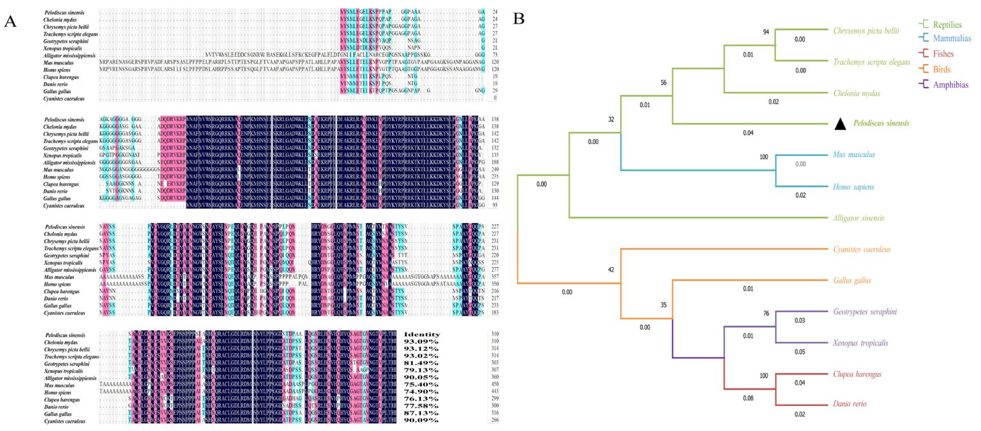
Sequence homology analysis revealed that the Sox3 protein of P. sinensis exhibited 93.09, 93.12, and 93.02% identity with green sea turtle (C. mydas), western painted turtle (C. picta bellii), and red-eared slider (T. scripta elegans), respectively. Sequence identities with chicken (G. gallus), Eurasian blue tit (C. caeruleus), western clawed frog (X. tropicalis), Gaboon caecilian (G. seraphini), and American alligator (A. mississippiensis) were 87.13, 90.09, 79.13, 81.49, and 90.05%, respectively. More distant similarities were observed with Atlantic herring (C. harengus, 76.13%), D. rerio (77.58%), M. musculus (75.40%), and H. sapiens (74.90%) (Figure 3A). The maximum likelihood (ML) phylogenetic tree indicated that Sox3 from P. sinensis clustered most closely with other turtle species, followed by amphibians, mammals, and birds, while showing a more distant relationship with teleost fishes (Figure 3B).
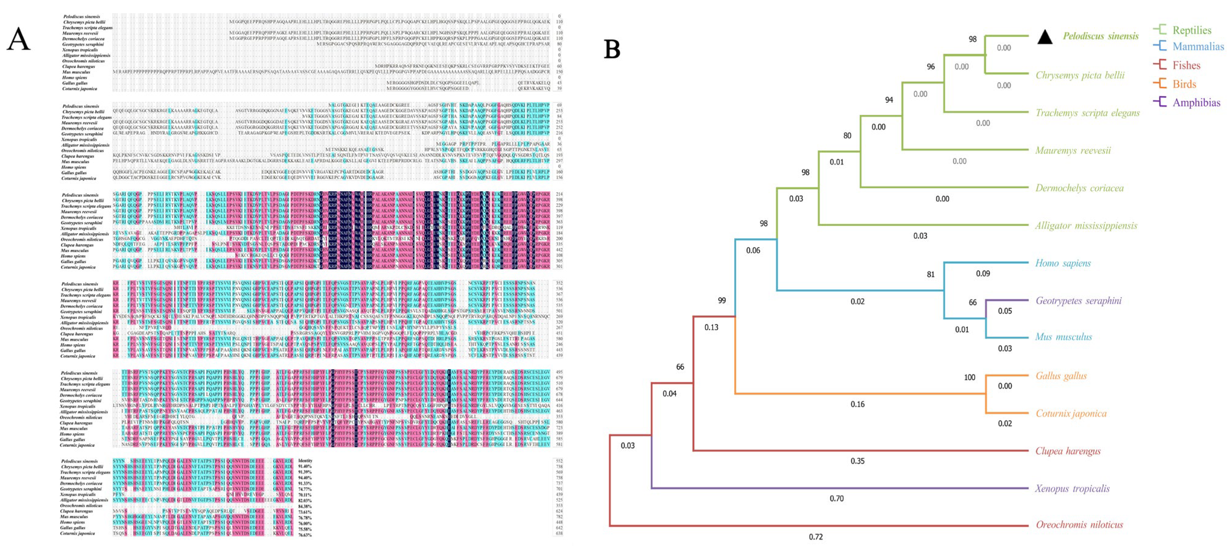
Homology alignment revealed that the Sox30 sequence of P. sinensis exhibited 91.40, 91.39, 94.40, and 91.33% homology with the western C. picta bellii, T. scripta elegans, Chinese pond turtle (M. reevesii), and leatherback sea turtle (D. coriacea), respectively. Sequence identities with the A. mississippiensis, Japanese quail (C. japonica), and G. gallus were 82.03, 75.63, and 75.58%, respectively. More distant similarities were observed with M. musculus (76.78%), H. sapiens (76.00%), C. picta bellii (70.11%), G. seraphini (74.77%), C. harengus (73.61%), and O. niloticus (84.38%) (Figure 4A). The ML phylogenetic tree indicated that Sox30 from P. sinensis showed the highest similarity to other turtle species, followed by mammals, birds, amphibians, and fishes, suggesting that P. sinensis shares closer evolutionary relationships with reptiles and occupies a distinct phylogenetic position within the group (Figure 4B).

3.3. Expression of Sox3 and Sox30 Genes in Embryonic Gonads and Adult Tissues of P. sinensis

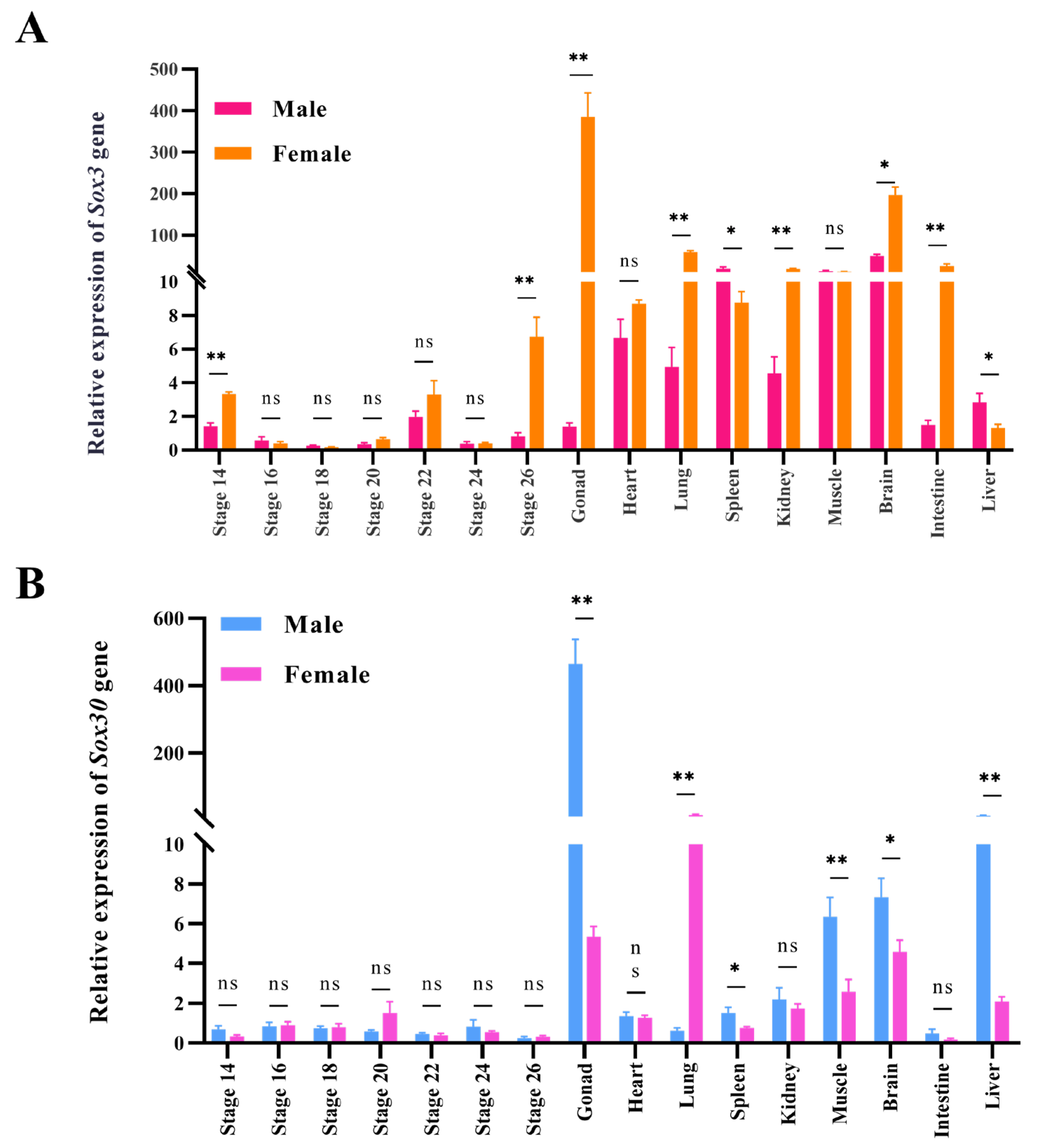
RT-qPCR was performed to examine the mRNA expression levels of Sox3 and Sox30 in the gonads at different embryonic stages and in various adult tissues of P. sinensis. The results showed that Sox3 in P. sinensis exhibited a female-biased expression pattern, with significantly higher expression in the female gonads, kidney, brain, liver, lung, spleen, and muscle compared with those in males (p < 0.05). In embryonic gonads of both sexes, the expression level of Sox3 was relatively low across all stages, and significant sex-dependent differences were only observed at stages 14 and 22 (p < 0.01) (Figure 5A). In contrast, Sox30 displayed a male-biased expression pattern, being expressed at significantly higher levels in male gonads, spleen, muscle, brain, and liver compared with females (p < 0.05). However, in embryonic gonads of P. sinensis, the expression of Sox30 remained relatively low across all stages, with no significant differences detected between the sexes (Figure 5B).

3.4. Effects of E2 and MT Treatments on the Expression of Sox3 and Sox30 Genes in Gonads of P. sinensis Embryonic Gonads at Different Developmental Stages

Compared to the control group, MT treatment led to an overall reduction in Sox3 expression in female embryonic gonads of P. sinensis, with significant differences observed at stages 14, 20, and 26 (p < 0.05) (Figure 6A). In contrast, E2 treatment increased Sox3 expression in male embryonic gonads of P. sinensis, peaking at stage 20 (p < 0.01) (Figure 6B). Additionally, MT treatment in female embryos of P. sinensis significantly upregulated Sox30 expression compared with the NC group (Figure 6C). Similarly, E2 treatment in male embryos of P. sinensis significantly elevated Sox30 expression compared with the NC group (Figure 6D).

4. Discussion

Gonadal differentiation and development is a crucial process in the sex determination of Testudines, resulting from the interaction between sex-related genes and endogenous hormones; however, the underlying molecular mechanisms remain poorly characterized [33]. In the present study, we cloned the cDNA sequences of Sox3 and Sox30 from P. sinensis, with lengths of 1062 bp and 2340 bp, respectively. The CDS were 933 bp and 1659 bp in length, encoding 310 and 552 amino acids, respectively. Systematic analyses of the two genes and their encoded proteins revealed that both Sox3 and Sox30 proteins are hydrophilic, with instability indices greater than 40, which is consistent with the flexible structural characteristics typical of transcription factors. The secondary structure prediction indicated that both Sox3 and Sox30 predominantly consist of random coils (79.35 and 87.14%, respectively), with limited α-helices and very low proportions of β-sheets, suggesting that their overall conformation is flexible, which facilitates binding with DNA and other regulatory factors [10,34]. Notably, Sox30 contained more putative phosphorylation sites than Sox3, implying that it may be subject to more complex signaling regulation. Comparative amino acid sequence analysis and maximum likelihood (ML) phylogenetic tree construction further revealed that both genes are most closely related to those of turtles, while being most distantly related to those of fish. In summary, these findings suggest that P. sinensis Sox3 and Sox30 share common physicochemical properties and secondary structural features but also exhibit distinct differences, which may be associated with their functional divergence in the reproduction and development of this species.
RT-qPCR indicated that Sox3 expression in the brain and gonads of adult P. sinensis was significantly higher than in tissues such as the heart, liver, spleen, and intestine (p < 0.05). Previous studies have shown that Sox3 is expressed in undifferentiated spermatogonia and in the developing hypothalamic–pituitary axis of mammals [35], and is widely involved in neural development and neural stem cell differentiation across vertebrates [36,37]. Notably, the expression of Sox3 in the ovary was significantly higher than in the testis (p < 0.01), exhibiting clear sexual dimorphism. This is consistent with the expression pattern of Sox3 in various fish species, including D. rerio [7] and Malabar red snapper (Lutjanus malabaricus) [38]. During embryonic development of P. sinensis, Sox3 expression was significantly higher in female gonads than in male gonads at stage 14 (early gonadal differentiation stage [32] (p < 0.01), suggesting that Sox3 may play a role in female gonadal development. Studies have also shown that in the giant grouper E. coioides [39] and A. japonica [3], the Sox3 gene plays a more significant role in ovarian development and oogenesis than in spermatogenesis. RNA-seq analysis of gonadal tissues from male and female P. sinensis embryos also revealed that Sox3 was upregulated in the ovaries, whereas Sox8 and Sox9 were elevated in the testes, suggesting a coordinated role of these genes in gonadal development [40]. Further functional studies using knockout or overexpression approaches will be required to clarify the regulatory mechanisms of Sox3 in gonadal differentiation and development of P. sinensis.
In contrast, the expression pattern of Sox30 is markedly different from that of Sox3. In adult P. sinensis, Sox30 is highly expressed in the testis (p < 0.05). The expression pattern is highly consistent with those observed in animals such as L. malabaricus [38], O. niloticus [9], and M. musculus [41]. The relatively high expression level of Sox30 in the brain may be associated with neural development, which is consistent with findings in D. rerio, where in situ hybridization localized Sox30 expression to the midbrain and hindbrain [42]. During the critical stages of embryonic development in P. sinensis, the expression level of Sox30 is relatively low in both female and male embryos. Previous studies have shown that in carp (Cyprinus carpio), Sox30 is localized in spermatocytes and sperm [10]; in M. musculus, it is primarily expressed during spermatogenesis after meiosis [43]. Similarly, Tang et al. [44] showed that Sox30 is localized in spermatocytes, round spermatids, epithelial cells of the efferent duct, sperm, and mature sperm cells in the testes of O. niloticus but was not detected in the ovaries. Collectively, these findings suggest that Sox30 exhibits male-specific expression in P. sinensis and likely functions in testis maturation and spermatogenesis, rather than in the early events of sex determination.
In this experiment, after treatment with E2 and MT hormones, the expression of Sox3 and Sox30 genes in the male and female P. sinensis embryos underwent rapid changes during the critical stages of sex determination and differentiation (stages 14–26). Previous studies have demonstrated that exogenous hormones can interact with sex-related genes to influence sex divergence and gonadal development in lower vertebrates, including reptiles [45,46]. In MT treatment female embryos, Sox3 expression exhibited a downward trend, which may have resulted from the disruption of the endogenous hormonal balance. Specifically, MT stimulation increases androgen levels, antagonizing endogenous estrogen and thereby exerting negative feedback regulation on Sox3. Similarly, Deng et al. [47] reported that Foxl2 expression was significantly downregulated in MT-treated catfish (Clarias fuscus). Previous studies have indicated that stages 15–20 represent the critical window for sex differentiation in P. sinensis [13,48]. In contrast, E2 treatment significantly upregulated Sox3 expression in male embryos, a pattern comparable to the E2-induced response of Foxl2 in embryos. This suggests that Sox3 is estrogen-dependent and positively regulated by estrogen receptors, likely playing a role in gonadal development of P. sinensis [31].
After MT hormone treatment in female P. sinensis embryos, the expression of Sox30 gene showed an upward trend, which may be closely associated with the shift in gonadal development from ovary to testis, suggesting its important role in sex reversal and masculinization in P. sinensis. Previous studies have shown. that in some protandrous fish species, a significant downregulation of Cyp19a1 expression was observed, leading to a reduction in estrogen synthesis. The disrupting of the positive feedback loop that maintains feminization removes the inhibition on the male-related gene regulatory network. Allowing gonads to develop in the male direction [49]. Studies have shown that Sox30 is highly expressed in the testes of various vertebrates and plays a role in spermatogenesis [50]. Similarly, in common C. carpio, Sox30 expression was found to be elevated during spermatogenesis and sperm formation [10]. In E2-treated male embryos, Sox30 expression was also upregulated compared with the NC group. This altered expression may reflect a genetically triggered compensatory mechanism or a resilience response, in which males enhance the expression of testis-associated genes to counteract feminization. Supporting this hypothesis, studies in O. niloticus have demonstrated that Dmrt1 positively regulates Sox30 expression by binding to putative cis-regulatory elements in its promoter region [51]. Similarly, transcriptomic analysis in the South American teleost (Astyanax altiparanae) revealed that male-biased genes such as Dmrt1, Sox9, and amh exhibited comparable regulatory mechanisms [52].

5. Conclusions

In this study, the full-length cDNAs of the Sox3 and Sox30 genes were successfully cloned from P. sinensis. Their sequence lengths were 1062 bp and 2340 bp, encoding 310 and 552 amino acids, respectively. Both genes exhibited a close phylogenetic relationship with those from other reptilian species. The Sox3 gene showed a female-biased expression pattern, while the Sox30 gene displayed a male-biased expression pattern. Both genes were expressed at relatively low levels during embryonic gonadal development. Exogenous hormone treatments further revealed that MT suppressed Sox3 but promoted Sox30 expression in female embryos, while E2 enhanced the expression of both genes in male embryos. Taken together, these findings suggest that Sox3 may be involved in ovarian development, while Sox30 plays a role in testis development and maturation, potentially contributing spermatogenesis. Both genes are subject to regulation by exogenous hormones. Overall, this study provides novel insights and a theoretical foundation for future research on gonadal differentiation, development, and sex regulation in turtles and other related reptilian species.

Supplementary Materials

The following supporting information can be downloaded at: https://www.mdpi.com/article/10.3390/genes16111249/s1, Figure S1: Alignment of the cloned Sox3 sequence from P. sinensis with the NCBI reference sequence (XM_006115453); Figure S2: Alignment of the cloned Sox30 sequence from P. sinensis with the NCBI reference sequence (XM_006138296).

Author Contributions

Conceptualization, X.L. (Xiaofang Lai) and W.L.; methodology, J.Z., C.C., L.J. and W.L.; validation, K.X. and Y.L.; formal analysis, K.X., Y.L., T.R. and W.L.; investigation, K.X. and Y.L.; resources, T.R., X.H., X.L. (Xiaoli Liu) and C.W.; data curation, K.X. and Z.W.; writing—original draft, K.X. and Y.L.; writing—review and editing, K.X. and T.R.; visualization, K.X.; supervision, T.R., Z.W., J.Z., C.C., L.J., X.H., X.L. (Xiaoli Liu), C.W., H.C. and X.Z.; project administration, Z.W., X.L. (Xiaofang Lai) and W.L.; funding acquisition, X.Z. and W.L. All authors have read and agreed to the published version of the manuscript.

Funding

This research was funded by the National Key R&D Program of China (2022YFD2401800), the National Natural Science Foundation of China (32202941), the Selection Breeding of the high-quality fast-growing Chinese Soft-Shelled Turtle and Studies on Efficient and Healthy Farming Technology (2024-SPY-00-014), the Central Public-interest Scientific Institution Basal Research Fund, CAFS (2023TD38), and the China-ASEAN Mari-time Cooperation Fund (CAMC-2018F).

Institutional Review Board Statement

The animal study protocol was approved by the Institutional Review Board of the Pearl River Fisheries Research Institute, Chinese Academy of Fishery Sciences (approval number: LAEC-PRFRI-2024-05-13; approval date: 13 May 2024).

Informed Consent Statement

Not applicable.

Data Availability Statement

The data presented in this study are available on request from the author. Email: xkl954334197@163.com.

Conflicts of Interest

The authors declare no conflicts of interest.

References

  1. Foster, J.W.; Dominguez-Steglich, M.A.; Guioli, S.; Kwok, C.; Weller, P.A.; Stevanović, M.; Weissenbach, J.; Mansour, S.; Young, I.D.; Goodfellow, P.N.; et al. Campomelic Dysplasia and Autosomal Sex Reversal Caused by Mutations in an SRY-Related Gene. Nature 1994, 372, 525–530. [Google Scholar] [CrossRef] [PubMed]
  2. Osaki, E. Identification of a Novel Sry-Related Gene and Its Germ Cell-Specific Expression. Nucleic Acids Res. 1999, 27, 2503–2510. [Google Scholar] [CrossRef] [PubMed]
  3. Jeng, S.R.; Wu, G.C.; Yueh, W.S.; Kuo, S.F.; Dufour, S.; Chang, C.F. Gonadal Development and Expression of Sex-Specific Genes during Sex Differentiation in the Japanese Eel. Gen. Comp. Endocrinol. 2018, 257, 74–85. [Google Scholar] [CrossRef]
  4. Gao, J.; Li, P.; Zhang, W.; Wang, Z.; Wang, X.; Zhang, Q. Molecular Cloning, Promoter Analysis and Expression Profiles of the Sox3 Gene in Japanese Flounder, Paralichthys olivaceus. Int. J. Mol. Sci. 2015, 16, 27931–27944. [Google Scholar] [CrossRef]
  5. Wei, L.; Yang, C.; Tao, W.; Wang, D. Genome-Wide Identification and Transcriptome-Based Expression Profiling of the Sox Gene Family in the Nile Tilapia (Oreochromis niloticus). Int. J. Mol. Sci. 2016, 17, 270. [Google Scholar] [CrossRef]
  6. Raverot, G.; Weiss, J.; Park, S.Y.; Hurley, L.; Jameson, J.L. Sox3 Expression in Undifferentiated Spermatogonia is Required for the Progression of Spermatogenesis. Dev. Biol. 2005, 283, 215–225. [Google Scholar] [CrossRef]
  7. Hong, Q.; Li, C.; Ying, R.; Lin, H.; Li, J.; Zhao, Y.; Cheng, H.; Zhou, R. Loss-of-Function of Sox3 Causes Follicle Development Retardation and Reduces Fecundity in Zebrafish. Protein Cell 2018, 10, 347–364. [Google Scholar] [CrossRef]
  8. Bai, S.; Fu, K.; Yin, H.; Cui, Y.; Yue, Q.; Li, W.; Cheng, L.; Tan, H.; Liu, X.; Guo, Y.; et al. Sox30 Initiates Transcription of Haploid Genes during Late Meiosis and Spermiogenesis in Mouse Testes. Development 2018, 145, dev164855. [Google Scholar] [CrossRef]
  9. Wei, L.; Tang, Y.; Zeng, X.; Li, Y.; Zhang, S.; Deng, L.; Wang, L.; Wang, D. The Transcription Factor Sox30 is Involved in Nile Tilapia Spermatogenesis. J. Genet. Genom. 2022, 49, 666–676. [Google Scholar] [CrossRef]
  10. Anitha, A.; Senthilkumaran, B. Role of Sox30 in Regulating Testicular Steroidogenesis of Common Carp. J. Steroid Biochem. Mol. Biol. 2020, 204, 105769. [Google Scholar] [CrossRef]
  11. Li, S.F.; Li, Y.P. Roles of SOX Transcription Factors on Fate Decision of Germ Cells. Chin. J. Cell Biol. 2019, 41, 961–966. (In Chinese) [Google Scholar]
  12. Han, F.; Jiang, X.; Li, Z.; Zhuang, X.; Zhang, X.; Ouyang, W.; Liu, W.; Mao, C.; Chen, Q.; Huang, C.; et al. Epigenetic Inactivation of SOX30 is Associated with Male Infertility and Offers a Therapy Target for Non-Obstructive Azoospermia. Mol. Ther. Nucleic Acids 2020, 19, 72–83. [Google Scholar] [CrossRef]
  13. Lei, L.; Chen, C.; Zhu, J.; Wang, Y.; Liu, X.; Liu, H.; Geng, L.; Su, J.; Li, W.; Zhu, X. Transcriptome Analysis Reveals Key Genes and Pathways Related to Sex Differentiation in the Chinese Soft-Shelled Turtle (Pelodiscus sinensis). Comp. Biochem. Physiol. D 2022, 42, 100986. [Google Scholar] [CrossRef]
  14. Zhu, J.; Wang, Y.; Lei, L.; Chen, C.; Ji, L.; Li, J.; Wu, C.; Yu, W.; Luo, L.; Chen, W.; et al. Comparative Genomic Survey and Functional Analysis of DKKL1 during Spermatogenesis in the Chinese Soft-Shelled Turtle (Pelodiscus sinensis). Int. J. Biol. Macromol. 2024, 254, 127696. [Google Scholar] [CrossRef] [PubMed]
  15. Jiang, Y.; Luo, H.; Hou, M.; Chen, J.; Tao, B.; Zhu, Z.; Song, Y.; Hu, W. Aromatase Inhibitor Induces Sex Reversal in the Protogynous Hermaphroditic Rice Field Eel (Monopterus albus). Aquaculture 2022, 551, 737960. [Google Scholar] [CrossRef]
  16. Liu, S.; Chen, Y.; Li, T.; Qiao, L.; Yang, Q.; Rong, W.; Liu, Q.; Wang, W.; Song, J.; Wang, X.; et al. Effects of 17α-Methyltestosterone on the Transcriptome and Sex Hormones in the Brain of Gobiocypris rarus. Int. J. Mol. Sci. 2023, 24, 3571. [Google Scholar] [CrossRef] [PubMed]
  17. Pieau, C.; Girondot, M.; Richard-Mercier, N.; Desvages, G.; Dorizzi, M.; Zaborski, P. Temperature Sensitivity of Sexual Differentiation of Gonads in the European Pond Turtle: Hormonal Involvement. J. Exp. Zool. 1994, 270, 86–94. [Google Scholar] [CrossRef]
  18. El-Greisy, Z.A.; El-Gamal, A.E. Monosex Production of Tilapia, Oreochromis niloticus Using Different Doses of 17α-Methyltestosterone with Respect to the Degree of Sex Stability After One Year of Treatment. Egypt. J. Aquat. Res. 2012, 38, 59–66. [Google Scholar] [CrossRef]
  19. Lee, C.H.; Hur, S.W.; Na, O.S.; Baek, H.J.; Noh, C.H.; Han, S.H.; Lee, Y.D. Induction of Primary Male in Juvenile Red Spotted Grouper Epinephelus akaara by Immersion of 17α-Methyltestosterone. Dev. Reprod. 2014, 18, 127–131. [Google Scholar] [CrossRef]
  20. Tokita, M.; Kuratani, S. Normal Embryonic Stages of the Chinese Softshelled Turtle Pelodiscus sinensis (Trionychidae). Zool. Sci. 2001, 18, 705–715. [Google Scholar] [CrossRef]
  21. Zhu, J.; Lei, L.; Chen, C.; Li, J.; Wang, Y.; Wu, C.; Wang, Y.; Hong, X.; Liu, X.; Yu, L.; et al. Development and Evaluation of a Medium-Free Incubation Method for Hatching Chinese Soft-Shelled Turtle (Pelodiscus sinensis) Eggs. Aquac. Rep. 2023, 31, 101643. [Google Scholar] [CrossRef]
  22. Zhu, J.; Wang, Y.; Chen, C.; Ji, L.; Hong, X.; Liu, X.; Chen, H.; Wei, C.; Zhu, X.; Li, W. Identification of Sex-Specific Markers and Candidate Genes Using WGS Sequencing Reveals a ZW-Type Sex-Determination System in the Chinese Soft-Shell Turtle (Pelodiscus sinensis). Int. J. Mol. Sci. 2024, 25, 819. [Google Scholar] [CrossRef] [PubMed]
  23. Artimo, P.; Jonnalagedda, M.; Arnold, K.; Baratin, D.; Csardi, G.; De Castro, E.; Duvaud, S.; Flegel, V.; Fortier, A.; Gasteiger, E.; et al. ExPASy: SIB Bioinformatics Resource Portal. Nucleic Acids Res. 2012, 40, W597–W603. [Google Scholar] [CrossRef] [PubMed]
  24. Schneider, M.; Tognolli, M.; Bairoch, A. The Swiss-Prot Protein Knowledgebase and ExPASy: Providing the Plant Community with High Quality Proteomic Data and Tools. Plant Physiol. Biochem. 2004, 42, 1013–1021. [Google Scholar] [CrossRef]
  25. Blom, N.; Sicheritz-Pontén, T.; Gupta, R.; Gammeltoft, S.; Brunak, S. Prediction of Post-translational Glycosylation and Phosphorylation of Proteins from the Amino Acid Sequence. Proteomics 2004, 4, 1633–1649. [Google Scholar] [CrossRef]
  26. Geourjon, C.; Deléage, G. SOPMA: Significant Improvements in Protein Secondary Structure Prediction by Consensus Prediction from Multiple Alignments. Bioinformatics 1995, 11, 681–684. [Google Scholar] [CrossRef]
  27. Waterhouse, A.; Bertoni, M.; Bienert, S.; Studer, G.; Tauriello, G.; Gumienny, R.; Heer, F.T.; de Beer, T.A.P.; Rempfer, C.; Bordoli, L.; et al. SWISS-MODEL: Homology Modelling of Protein Structures and Complexes. Nucleic Acids Res. 2018, 46, W296–W303. [Google Scholar] [CrossRef]
  28. Zhang, Y.; Zhang, Y.; Xu, L.; Wang, H.; Shao, F.; Yu, J.; Gilbert, E.; Gu, Z. Molecular Cloning, Tissue Expression and Polymorphism Analysis of the Caveolin-3 Gene in Ducks. Br. Poult. Sci. 2020, 62, 17–24. [Google Scholar] [CrossRef]
  29. Tamura, K.; Stecher, G.; Kumar, S. MEGA11: Molecular Evolutionary Genetics Analysis Version 11. Mol. Biol. Evol. 2021, 38, 3022–3027. [Google Scholar] [CrossRef]
  30. Zhu, J.; Chen, C.; Liu, X.; Wang, Y.; Lei, L.; Hong, X.; Yu, L.; Xu, H.; Li, W.; Zhu, X. Cloning and expression analysis of histone H2A variant in oocytes of Chinese soft-shelledturtle (Pelodiscus sinensis). Acta Hydrobiol. Sin 2021, 45, 1207–1213. (In Chinese) [Google Scholar] [CrossRef]
  31. Gao, L.L. Molecularcloning, Expression of Foxl2 and the Influence of Temperature and Exogenoushormone on Expression of Foxl2 in the Pelodiscus sinensis. Master’s Thesis, Southwest University, Chongqing, China, 26 May 2016. (In Chinese). [Google Scholar]
  32. Jin, L.; Sun, W.; Bao, H.; Liang, X.; Li, P.; Shi, S.; Wang, Z.; Qian, G.; Ge, C. The Forkhead Factor Foxl2 Participates in the Ovarian Differentiation of Chinese Soft-Shelled Turtle Pelodiscus sinensis. Dev. Biol. 2022, 492, 101–110. [Google Scholar] [CrossRef] [PubMed]
  33. Thépot, D. Sex Chromosomes and Master Sex-Determining Genes in Turtles and Other Reptiles. Genes 2021, 12, 1822. [Google Scholar] [CrossRef] [PubMed]
  34. Feng, Y.; Zhong, Z.; Xu, Y.; Zhang, Z.; Ao, L.; Yang, Z.; Wang, Y.; Jiang, Y. Characterization of the Transcription Factor Sox3 Regulating the Gonadal Development of Pearlscale Angelfish (Centropyge vrolikii). Fish Physiol. Biochem. 2022, 48, 1193–1207. [Google Scholar] [CrossRef] [PubMed]
  35. McAninch, D.; Mäkelä, J.-A.; La, H.M.; Hughes, J.N.; Lovell-Badge, R.; Hobbs, R.M.; Thomas, P.Q. SOX3 Promotes Generation of Committed Spermatogonia in Postnatal Mouse Testes. Sci. Rep. 2020, 10, 6751. [Google Scholar] [CrossRef]
  36. Hong, Q. Molecular Mechanismof Oocyte Development Regulated by Sox3 in Zebrafish. Master’s Thesis, Wuhan University, Wuhan, China, 10 April 2018. (In Chinese). [Google Scholar]
  37. Gui, L.; Xiong, X.X.; Chen, X.R.; Liu, Y.Y.; Tan, G.H. Transcriptional regulatory role of the Sox gene family in adult neurogenesis. Prog. Physiol. Sci. 2020, 51, 289–293. (In Chinese) [Google Scholar]
  38. Liang, B.; Jerry, D.R.; Shen, X.; Koh, J.; Terence, C.; Nayfa, M.G.; Nguyen, V.; Loo, G.; Vij, S.; Domingos, J.A. Transcriptomic Analysis of Gonads in Malabar Red Snapper (Lutjanus malabaricus) Reveals Genes Associated with Gonad Development. Aquaculture 2024, 592, 741258. [Google Scholar] [CrossRef]
  39. Yao, B.; Zhou, L.; Wang, Y.; Xia, W.; Gui, J. Differential Expression and Dynamic Changes of SOX3 during Gametogenesis and Sex Reversal in Protogynous Hermaphroditic Fish. J. Exp. Zool. Part A Ecol. Genet. Physiol. 2007, 307, 207–219. [Google Scholar] [CrossRef]
  40. Zhang, J.; Yu, P.; Liu, T.; Qiao, D.; Hu, Q.; Su, S. Identification and Functional Analysis of SOX Transcription Factors in the Genome of the Chinese Soft-Shell Turtle (Pelodiscus sinensis). Comp. Biochem. Phys. B 2020, 242, 110407. [Google Scholar] [CrossRef]
  41. Han, F.; Yin, L.; Jiang, X.; Zhang, X.; Zhang, N.; Yang, J.; Ouyang, W.; Hao, X.; Liu, W.; Huang, Y.; et al. Identification of SRY-Box 30 (Sox30) as an Age-Related Essential Gatekeeper for Male Germ-Cell Meiosis and Differentiation. Aging Cell 2021, 20, e13343. [Google Scholar] [CrossRef]
  42. De Martino, S.P.; Errington, F.; Ashworth, A.; Jowett, T.; Austin, C.A. Sox30: A Novel Zebrafish Sox Gene Expressed in a Restricted Manner at the Midbrain-Hindbrain Boundary during Neurogenesis. Dev. Genes Evol. 1999, 209, 357–362. [Google Scholar] [CrossRef]
  43. Feng, C.-W.A.; Spiller, C.; Merriner, D.J.; O’Bryan, M.K.; Bowles, J.; Koopman, P. SOX30 is Required for Male Fertility in Mice. Sci. Rep. 2017, 7, 17619. [Google Scholar] [CrossRef]
  44. Tang, Y.H. Study on the Functionand Regulatory Mechanism of Transcription Factor Sox30 in Spermatogenesis of Nile tilapia. Master’s Thesis, Southwest University, Chongqing, China, 24 April 2020. (In Chinese). [Google Scholar]
  45. Shioda, K.; Odajima, J.; Kobayashi, M.; Kobayashi, M.; Cordazzo, B.; Isselbacher, K.J.; Shioda, T. Transcriptomic and Epigenetic Preservation of Genetic Sex Identity in Estrogen-Feminized Male Chicken Embryonic Gonads. Mol. Endocrinol. 2020, 162, bqaa208. [Google Scholar] [CrossRef]
  46. Matsumoto, Y.; Crews, D. Molecular Mechanisms of Temperature-Dependent Sex Determination in the Context of Ecological Developmental Biology. Mol. Cell. Endocrinol. 2012, 354, 103–110. [Google Scholar] [CrossRef]
  47. Deng, S.; Zhu, C.; Sun, J.; Wang, W.; Wu, T.; Chen, H.; Shi, S.; Li, G. Foxl2 of the Hong Kong Catfish (Clarias fuscus): cDNA Cloning, Tissue Distribution and Changes in Gene Expression Towards Methyltestosterone, Estradiol and Letrozole Exposure of the Fries During Gonadal Differentiation. Genes Genom. 2015, 37, 669–677. [Google Scholar] [CrossRef]
  48. Zhang, H.; Gao, Y.; Wang, G.; Xin, Q.; Tian, X.; Wu, L.; Shi, X.; Ma, W.; Liu, H.; Jiang, H.; et al. Molecular Cloning and Characterization of Sirt1 and Its Role in the Follicle of Juvenile Chinese Soft-shelled Turtle (Pelodiscus sinensis). Gene 2023, 860, 147211. [Google Scholar] [CrossRef] [PubMed]
  49. Ortega-Recalde, O.; Goikoetxea, A.; Hore, T.A.; Todd, E.V.; Gemmell, N.J. The Genetics and Epigenetics of Sex Change in Fish. Annu. Rev. Anim. Biosci. 2020, 8, 47–69. [Google Scholar] [CrossRef] [PubMed]
  50. Tang, Y.; Li, X.; Xiao, H.; Li, M.; Li, Y.; Wang, D.; Wei, L. Transcription of the Sox30 Gene is Positively Regulated by Dmrt1 in Nile Tilapia. Int. J. Mol. Sci. 2019, 20, 5487. [Google Scholar] [CrossRef] [PubMed]
  51. Tenugu, S.; Pranoty, A.; Mamta, S.K.; Senthilkumaran, B. Development and Organisation of Gonadal Steroidogenesis in Bony Fishes—A Review. Aquac. Fish. 2021, 6, 223–246. [Google Scholar] [CrossRef]
  52. Martinez-Bengochea, A.; Doretto, L.; Rosa, I.F.; Oliveira, M.A.; Silva, C.; Silva, D.M.Z.A.; Santos, G.R.; Santos, J.S.F.; Avelar, M.M.; Silva, L.V.; et al. Effects of 17β-Estradiol on Early Gonadal Development and Expression of Genes Implicated in Sexual Differentiation of a South American Teleost, Astyanax altiparanae. Comp. Biochem. Phys. B 2020, 248–249, 110467. [Google Scholar] [CrossRef]
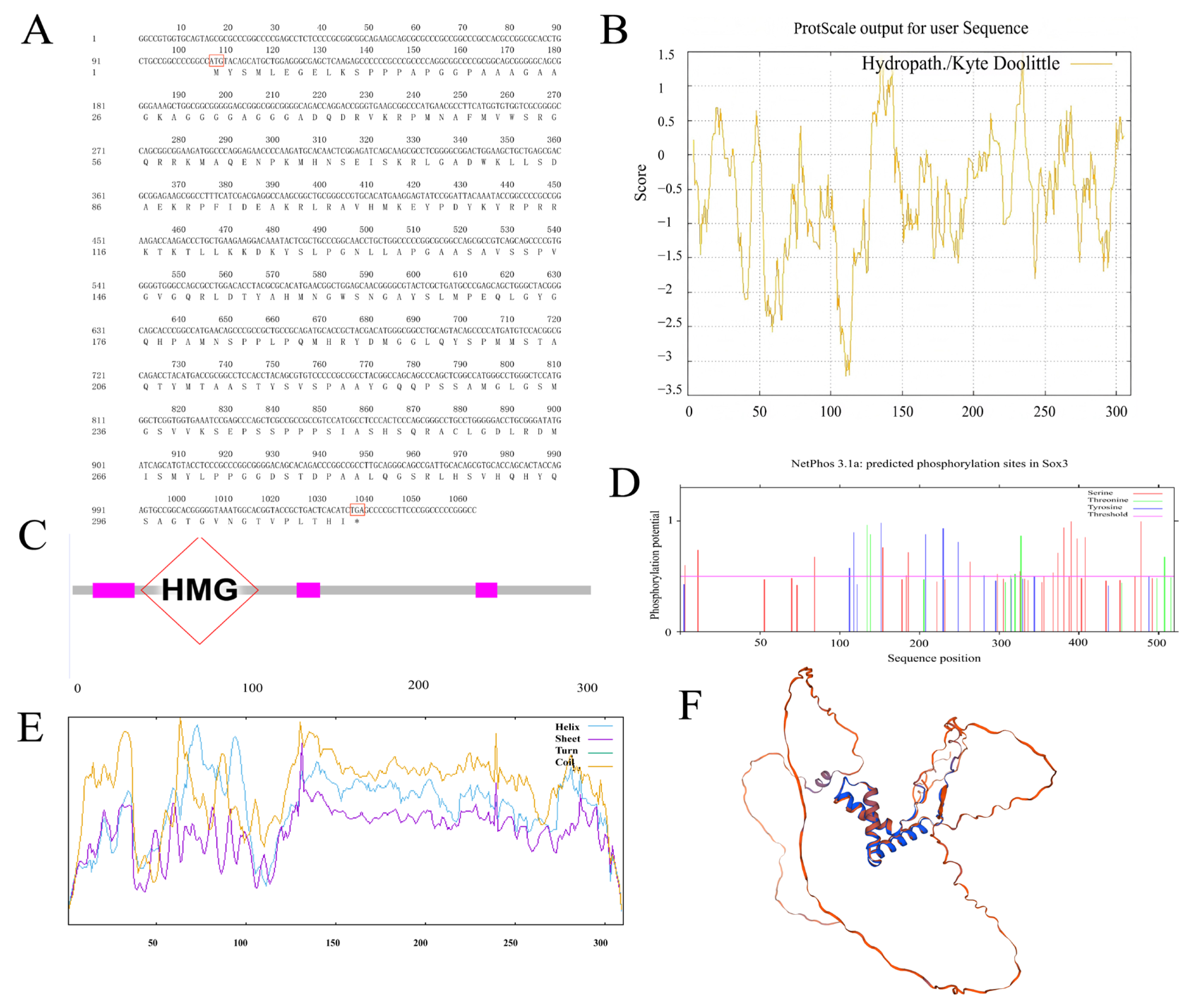
Figure 1. (A) Nucleotide sequence and deduced amino acid sequence of the Sox3 gene in P. sinensis. (B) Hydrophilicity/hydrophobicity profile of Sox3. (C) Sox3 protein domain. (D) Predicted phosphorylation sites of Sox3. (E) Predicted secondary structure of Sox3. (F) Predicted tertiary structure of Sox3. Notation: (A) ATG denotes the initiation codon, while TGA denotes the termination codon. (C) The gray bar represents the protein backbone; magenta boxes denote conserved motifs. The red diamond highlights the HMG box domain—the canonical DNA-binding domain of SOX family proteins—which recognizes specific DNA sequences and mediates transcriptional regulation. (D) Serine (Ser), Threonine (Thr), and Tyrosine (Tyr) indicate putative phosphorylation sites. (E) Blue (Helix): α-helix, Purple (Sheet): β-sheet, Green (Turn): β-turn, Orange (Coil): Random coil.
Genes 16 01249 g001
Figure 2. (A) Nucleotide sequence and deduced amino acid sequence of the Sox30 gene in P. sinensis. (B) Hydrophilicity/hydrophobicity profile of Sox30. (C) Sox30 protein domain. (D) Predicted phosphorylation sites of Sox30. (E) Predicted secondary structure of Sox30. (F) Predicted tertiary structure of Sox30. Notation: (A) ATG denotes the initiation codon, while TAA denotes the termination codon. (C) The gray bar represents the protein backbone; magenta boxes denote conserved motifs. The red diamond highlights the HMG box domain—the canonical DNA-binding domain of SOX family proteins—which recognizes specific DNA sequences and mediates transcriptional regulation. (D) Serine (Ser), Threonine (Thr), and Tyrosine (Tyr) indicate putative phosphorylation sites. (E) Blue (Helix): α-helix, Purple (Sheet): β-sheet, Green (Turn): β-turn, Orange (Coil): Random coil.
Genes 16 01249 g002
Figure 3. Multiple sequence alignment and phylogenetic tree analysis of Sox3 amino acid sequences in P. sinensis and representative species. (A) Alignment of Sox3 amino acid sequences. (B) phylogenetic tree of Sox3 amino acid sequences. Notation: (B). The black triangle indicates P. sinensis, the target species of this study, which is marked as a key node in this phylogenetic tree.
Genes 16 01249 g003
Figure 4. Multiple sequence alignment and phylogenetic tree analysis of Sox30 amino acid sequences in P. sinensis and representative species. (A) Alignment of Sox30 amino acid sequences. (B) phylogenetic tree of Sox30 amino acid sequences. Notation: (B). The black triangle indicates P. sinensis, the target species of this study, which is marked as a key node in this phylogenetic tree.
Genes 16 01249 g004
Figure 5. (A) Expression of Sox3 gene in gonads of male and female embryos and adult tissues of P. sinensis. (B) Expression of Sox30 gene in gonads of male and female embryos and adult tissues of P. sinensis. Notation: ** (p < 0.01), * (p < 0.05), ns indicates no significant difference. All data are presented as mean ± standard error (n = 3).
Genes 16 01249 g005
Figure 6. (A) Expression of Sox3 in female P. sinensis embryos after MT treatment. (B) Expression of Sox3 in male P. sinensis embryos after E2 treatment. (C) Expression of Sox30 in female P. sinensis embryos after MT treatment. (D) Expression of Sox30 in male P. sinensis embryos after E2 treatment. Notation: ** (p < 0.01), * (p < 0.05), ns indicates no significant difference. All data are presented as mean ± standard error (n = 3).
Genes 16 01249 g006
Table 1. Primers used for cDNA cloning and expression analysis of P. sinensis.
NameSequences (5′–3′)Purpose
PB1-FGGATCTCATTTGTGAGCCTACATGTSex identification
PB1-RCCCACAGCTTGCTTTCCWTGTTTAG
Sox3-FTTGGCCGTGGTGCAGTAGCcDNA cloning
Sox3-RATGAGTGTAGAGGTGGAATGGAAA
Sox30-FGAGGAGAGCCAGAAAGAGGAGCcDNA cloning
Sox30-RTATTAGTGGGAGTGGGGGTGACAGAA
Sox3-qFGCAGTACAGCCCCATGATGTReal-time fluorescence quantification
Sox3-qRGATCATATCCCGCAGGTCC
Sox30-qFCGAATGCCTGGGCTTTTAReal-time fluorescence quantification
Sox30-qRGATGGGGTTGCCGTGAAA
Ef1αFACTCGTCCAACTGACAAGCCTCReference genes
Ef1αRCACGGCGAACATCTTTCACAG
Table 2. Test Comparison Species Sequence Numbers.
GeneSpeciesAccession
Sox3Chelonia mydasXP_037764741.1
Sox3Chrysemys picta belliiXP_005294674.2
Sox3Trachemys scripta elegansXP_034636385.1
Sox3Geotrypetes seraphiniXP_033802893.1
Sox3Xenopus tropicalisNP_001007502.1
Sox3Alligator mississippiensisXP_006031121.1
Sox3M. musculusNP_033263.2
Sox3H. sapiensNP_005625.2
Sox3Clupea harengusXP_012695760.1
Sox3D. rerioNP_001001811.2
Sox3Gallus gallusNP_989526.1
Sox3Cyanistes caeruleusXP_023782651.1
Sox30C. picta belliiXP_005300115.3
Sox30T. scripta elegansXP_034635714.1
Sox30Mauremys reevesiiXP_039340705.1
Sox30Dermochelys coriaceaXP_038267805.1
Sox30G. seraphiniXP_033783385.1
Sox30X. tropicalisXP_031753986.1
Sox30A. mississippiensisXP_019331538.2
Sox30O. niloticusXP_003447014.1
Sox30Clupea harengusXP_042564328.1
Sox30M. musculusNP_775560.1
Sox30G. gallusXP_414564.1
Sox30Coturnix japonicaXP_015731686.1
Disclaimer/Publisher’s Note: The statements, opinions and data contained in all publications are solely those of the individual author(s) and contributor(s) and not of MDPI and/or the editor(s). MDPI and/or the editor(s) disclaim responsibility for any injury to people or property resulting from any ideas, methods, instructions or products referred to in the content.