Article

HOXA7 Expression Is an Independent Prognostic Biomarker in Esophageal Squamous Cell Carcinoma

by
Jennifer Vieira Gomes
1,†,
Pedro Nicolau-Neto
2,†,
Júlia Nascimento de Almeida
1,
Lilian Brewer Lisboa
1,
Paulo Thiago de Souza-Santos
3,
Luis Felipe Ribeiro-Pinto
1,2,
Sheila Coelho Soares-Lima
4 and
Tatiana de Almeida Simão
1,*
1
Laboratório de Toxicologia e Biologia Molecular, Departamento de Bioquímica, Universidade do Estado do Rio de Janeiro (UERJ), Rio de Janeiro 20550-013, RJ, Brazil
2
Programa de Carcinogênese Molecular, Instituto Nacional de Câncer (INCA), Rio de Janeiro 20230-130, RJ, Brazil
3
Beneficência Portuguesa de São Paulo, São Paulo 01323-001, SP, Brazil
4
Programa de Pesquisa Clínica e Desenvolvimento Tecnológico, Instituto Nacional de Câncer (INCA), Rio de Janeiro 20230-130, RJ, Brazil
*
Author to whom correspondence should be addressed.
These authors contributed equally to this study.
Genes 2024, 15(11), 1430; https://doi.org/10.3390/genes15111430
Submission received: 3 October 2024 / Revised: 29 October 2024 / Accepted: 31 October 2024 / Published: 1 November 2024
(This article belongs to the Section Human Genomics and Genetic Diseases)

Abstract

:
Background/Objectives: Homeobox (HOX) genes encode conserved transcription factors essential for tissue and organ development and cellular differentiation. In humans, these genes are organized into four clusters: HOXA, HOXB, HOXC, and HOXD. While HOX genes have been extensively studied in cancer biology, their roles in esophageal squamous cell carcinoma (ESCC) remain poorly understood. Given the increasing incidence and high mortality rate of ESCC, exploring the molecular drivers of this tumor is urgent. Methods: Therefore, this study investigated the mutational landscape and expression profiles of HOX genes in ESCC and their differentially expressed targets using ESCC data from The Cancer Genome Atlas (TCGA) and two independent transcriptome datasets. Results: We found that copy number alterations and single nucleotide variations were rare, while seven HOX genes (HOXA2, HOXA7, HOXB13, HOXC9, HOXC10, HOXC13, and HOXD10) were significantly differentially expressed in ESCC compared to paired non-malignant mucosa. Further analysis identified 776 potential HOX target genes differentially expressed in ESCC, many of which are involved in critical cancer pathways such as PI3K-AKT, cell cycle regulation, and epithelial–mesenchymal transition (EMT). The HOXA7 overexpression was associated with poor overall survival rates in ESCC. This finding opens new possibilities for targeted therapies, offering hope for improved patient outcomes. Conclusions: Thus, this study underscored the pivotal role of HOX gene dysregulation in ESCC and classified HOXA7 as a potential prognostic biomarker in this tumor.

1. Introduction

Homeobox (HOX) genes are a group of major transcription factors, distinguished by a highly conserved 61-amino-acid helix-turn-helix DNA binding homeodomain, preserved throughout animal evolution. In humans, there are 39 HOX genes organized into four clusters located on different chromosomes: HOXA (7p15), HOXB (17q21), HOXC (12q13), and HOXD (2q31) [1]. HOX proteins can function as monomers or homodimers to regulate the transcription of downstream targets. Sometimes, they form heterodimers or heterotrimers with members of the TALE (three amino acid loop extension) family of cofactors, depending on the specific cell type [2]. The coordinated expression of HOX genes is essential for defining the embryonic anterior–posterior body axis and determining temporospatial limb development, tissue and organ formation, and cellular differentiation [3,4].
While the role of HOX genes in cancer is becoming more evident, it is still not fully understood. In certain cancers, specific HOX genes that generally suppress tumors are silenced, whereas, in other contexts, HOX genes are overexpressed and contribute to oncogenic processes [2]. HOXB13 is a classic example of this deregulation, displaying a dual role depending on tissue type. For instance, while HOXB13 is essential for proper prostate development and differentiation [5], its overexpression in breast cancer is linked to greater invasiveness by downregulating the estrogen receptor α and upregulating interleukin-6 expression [6]. HOX gene deregulation has been reported in various cancers, including leukemia, breast, ovarian, prostate, gastric, and esophageal cancers [7,8,9,10,11,12,13,14]. Previous studies have highlighted the role of HOXA in cellular processes, particularly within the context of innate immune cell activation [12,14]. The roles of HOXB, HOXC, and HOXD in esophageal squamous cell carcinoma (ESCC) are not yet fully elucidated. Research suggests that specific genes within these clusters contribute to tumor progression, like HOXB13, HOXC10, and HOXD13 [14]. Esophageal cancer (EC) is the seventh most common cancer type and the sixth leading cause of cancer-related deaths worldwide among men. Alarmingly, the incidence of EC is projected to increase 1.8-fold in both sexes by 2050 [15]. There are two primary histological subtypes of EC: esophageal adenocarcinoma and ESCC, with ESCC accounting for 85% of all EC cases. Over the past two decades, treatment advancements have included a combination of neoadjuvant chemotherapy, radiotherapy, and surgery [16]. However, despite these efforts, the survival rates for EC remain poor. The underlying molecular mechanisms driving ESCC initiation, promotion, and progression are still not fully understood, contributing to the bleak patient outcomes. Therefore, research to identify biomarkers for improved ESCC diagnosis, prognosis, and treatment is critical to changing this scenario [17].
This study investigated the mutational landscape and gene expression profiles of HOX genes in ESCC. Through comparative analysis between tumor tissues and their paired nonmalignant mucosa, we identified HOXA7 overexpression as a critical prognostic biomarker associated with poor outcomes in ESCC patients.

2. Materials and Methods

2.1. Analysis of HOX Gene Expression Using Publicly Available Esophageal

Two transcriptome datasets of tumor and matched non-malignant surrounding mucosa (NMSM) were utilized to investigate the HOX gene expression profile. These included our previously published transcriptome analysis dataset (GSE75241) [18] and the dataset by Li and colleagues (GSE53625) [19]. HOX gene expression in ESCC was quantified by calculating gene expression values in array units.
Gene expression analyses were conducted in the R environment, with differential expression assessed using the Limma package from the Bioconductor project. Differentially expressed genes (DEGs) were identified based on an adjusted p-value < 0.05 and a fold-change threshold of |1.5| [20,21].

2.2. Evaluation of Somatic Alterations in HOX Genes in the TCGA Data

The frequency of copy number alterations (CNAs) and single nucleotide variants (SNVs) in HOX genes in ESCC was assessed using The Cancer Genome Atlas (TCGA) dataset by applying The Genomic Identification of Significant Targets in Cancer (GISTIC) 2.0 protocol to whole exome sequencing [22].

2.3. Snap-Frozen Human Tissue Samples

Forty-one ESCC patients treated between 2012 and 2015 at the Instituto Nacional de Câncer (INCA, Rio de Janeiro, Brazil) were enrolled in this study. Snap-frozen tumor and matched non-malignant surrounding mucosa (NMSM) samples were collected by endoscopy before chemotherapy or radiotherapy. These samples were stored at the INCA Tumor Bank (Banco Nacional de Tumores e DNA, BNT) until RNA extraction. The INCA Ethics Committee approved the study (Protocol 116/11). Patient characteristics are summarized in Table 1.

2.4. Evaluation of Gene Expression by Quantitative PCR (qPCR)

Total RNA was extracted from snap-frozen biopsies using the RNeasy mini kit (Qiagen) following the manufacturer’s protocol. Complementary DNA (cDNA) was synthesized using SuperScript II (Invitrogen, CA, USA) and random primers (Invitrogen, California, USA), according to the manufacturer’s instructions.
HOX expression was assessed by qPCR using 41 paired ESCC and NMSM samples. qPCR was performed with the Quantifast SYBR Green PCR kit (Qiagen), Valencia, CA, USA) in a Rotor-Gene 6000 thermal cycler (Qiagen). Gene expression quantification was conducted as previously described [23]. Specific oligonucleotide sequences are listed in Supplementary Table S1.
A schematic diagram (Figure 1) was created to highlight the primary methodological information addressed in this study and facilitate understanding of the methods and sample sets used.

2.5. Identification of HOX Targets and Gene-Set Enrichment Analyses

The Transcriptome Factor Target Gene database identified the putative transcriptional targets of selected HOX genes [24]. Next, we evaluated whether these targets were among the 1368 DEGs identified in ESCC from our previously published study (GSE75241) [18]. The differentially expressed HOX targets were then subjected to gene-set enrichment analysis using the WEB-based GEne SeT AnaLysis Toolkit 2024 (WebGestalt) [25], with pathways evaluated using the Kyoto Encyclopedia of Genes and Genomes (KEGG) and Hallmark_50 databases. Pathways were considered enriched if the adjusted p-value was <0.05.

2.6. Statistical Analyses

The Kolmogorov–Smirnov test was used to assess the normality of continuous data, followed by paired t-tests, Wilcoxon matched-pairs signed-rank tests, unpaired t-tests, or Mann–Whitney U tests, as appropriate. These analyses were performed using GraphPad Prism 8 software. The Kaplan–Meier method and log-rank test were applied to estimate the impact of individual variables on overall survival (OS). Variables with a p-value < 0.05 were selected for multivariate analysis. Cox regression analysis was performed using the stepwise backward method [26]. Survival analyses were conducted in the R environment using the survival package [27]. Gene expression cut-offs were determined using the best-performing threshold [28], with at least 20% of samples in one of the comparison groups. For Brazilian patients, samples were classified as high and low HOXA7 expression with an expression cut-off of 0.00256 relative to GAPDH. For the GSE53625 dataset, the HOXA7 expression cut-off was 13.0277 array units.

3. Results

3.1. Mutational Profile of HOX Genes

The frequency of somatic genetic alterations in HOX genes in ESCC was evaluated in the TCGA dataset. Among ninety-five ESCC samples, six (6.3%) exhibited amplification, and one showed deletion in the HOXA locus 7p15.2. One sample showed amplification in the HOXB locus 17q21.3, and one showed amplification of HOXD locus 2q31. In addition, missense mutations occurred at a low frequency in ESCC samples (≤1%) (Supplementary Figure S1).

3.2. HOX Genes’ Expression Profile

The expression profile of HOX genes in ESCC was assessed using our previously published transcriptome dataset (GSE75241), comparing paired ESCC and NMSM samples. We identified seven HOX genes with differential expression in ESCC, six of which were upregulated (HOXA7, HOXB13, HOXC9, HOXC10, HOXC13, and HOXD10) and one downregulated (HOXA2) (Table 2). The differential expression of these seven HOX genes was further validated using the independent dataset GSE53625 (Table 2).

3.3. Targets of ESCC-Overexpressed HOX Genes Are Associated with Enriched Signaling Pathways and Cellular Processes

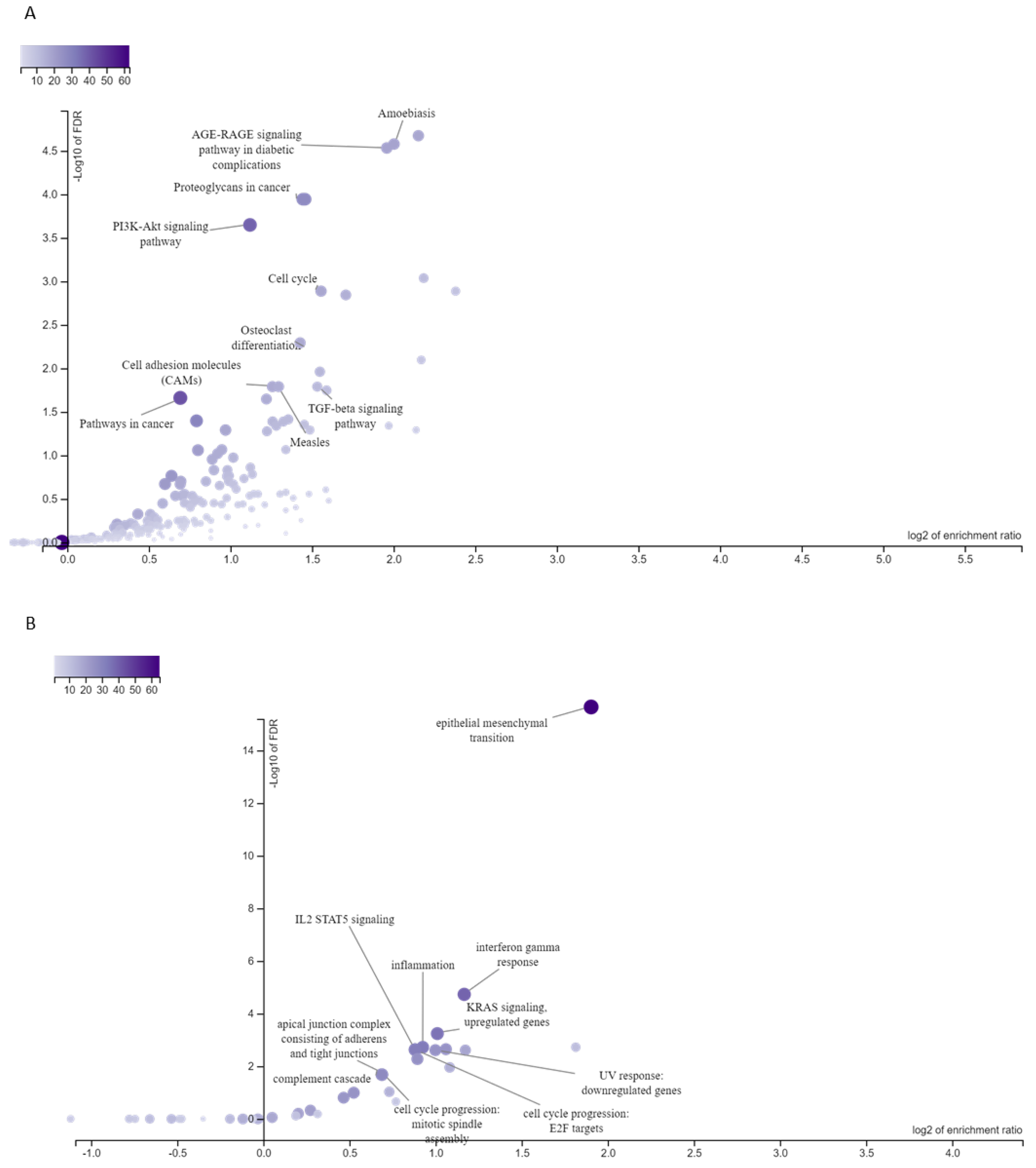
Initially, we identified potential transcriptional targets of HOXA2, HOXA7, HOXB13, HOXC9, HOXC10, HOXC13, and HOXD10 using the Transcription Factor Target Gene database. We then integrated these data with our previously published ESCC transcriptome dataset (GSE75241) to identify which HOX gene targets were differentially expressed (DEG) in ESCC. Of 1368 DEGs identified in ESCC, 776 were potential HOX gene targets (Supplementary Table S2). A thorough gene-set enrichment analysis of these 776 putative HOX targets using the KEGG database revealed significant enrichment in pathways related to PI3K-AKT signaling, cell cycle regulation, cell adhesion molecules, and cancer-associated signaling pathways (Figure 2A). A detailed analysis using the Cellular Hallmarks database highlighted the enrichment of key cancer hallmarks, including inflammation (inflammatory response) and cellular plasticity (epithelial–mesenchymal transition) (Figure 2B). We applied the same methodology to analyze HOXA7 targets and found that 223 of the 776 putative HOX targets were identified as targets of this gene (Supplementary Table S3). The KEGG pathway enrichment analysis of these putative targets is strongly associated with cell adhesion molecule pathways. In contrast, the Cellular Hallmarks database analysis highlighted significant enrichment in epithelial–mesenchymal transition, reflecting critical aspects of cellular plasticity and tumor progression.

3.4. Association Analyses Between ESCC-Overexpressed HOX Genes and Clinicopathological Features

Association analyses between the expression of HOXA2, HOXA7, HOXB13, HOXC9, HOXC10, HOXC13, and HOXD10 and clinicopathological features were conducted with the GSE53625 dataset. HOXB13 was overexpressed in older patients (>60 years) relative to younger patients. The expression of HOXD10 was higher in tumors of males relative to female patients and in tumors of patients with a history of alcohol drinking. Furthermore, ESCC from patients with tobacco smoking habits and with poorly tumor grades showed HOXC9 overexpression relative to never-smokers and patients with other tumor grades, respectively (Table 3).
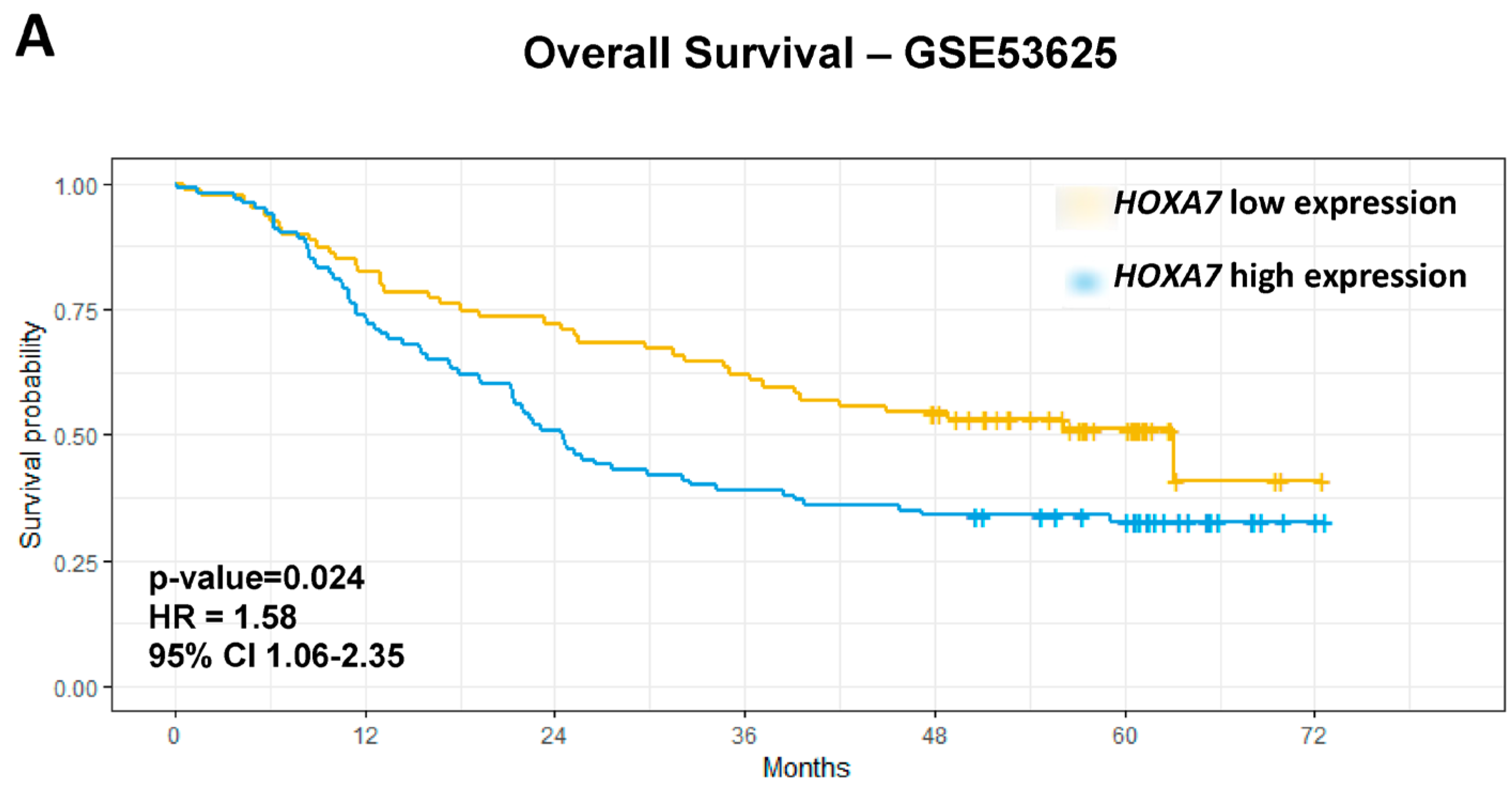
Regarding overall survival, univariate analyses showed that age > 60 years (p = 0.028), late-stage (III/IV) (p = 0.0002), high-grade tumors (G2/G3) (p = 0.048), HOXA2 low expression (p = 0.032), HOXA7 overexpression (p = 0.013), HOXB13 overexpression (p = 0.03), and HOXD10 overexpression (p = 0.018) were associated with worse prognosis in GSE53625 dataset. The final multivariate model further confirmed late-stage tumors (p = 0.0002; HR = 2.97; 95% CI 1.40–3.12) and HOXA7 overexpression (p = 0.024; HR = 1.58; 95% CI 1.06–2.35) as independent biomarkers of OS in ESCC (Table 4; Figure 3A).
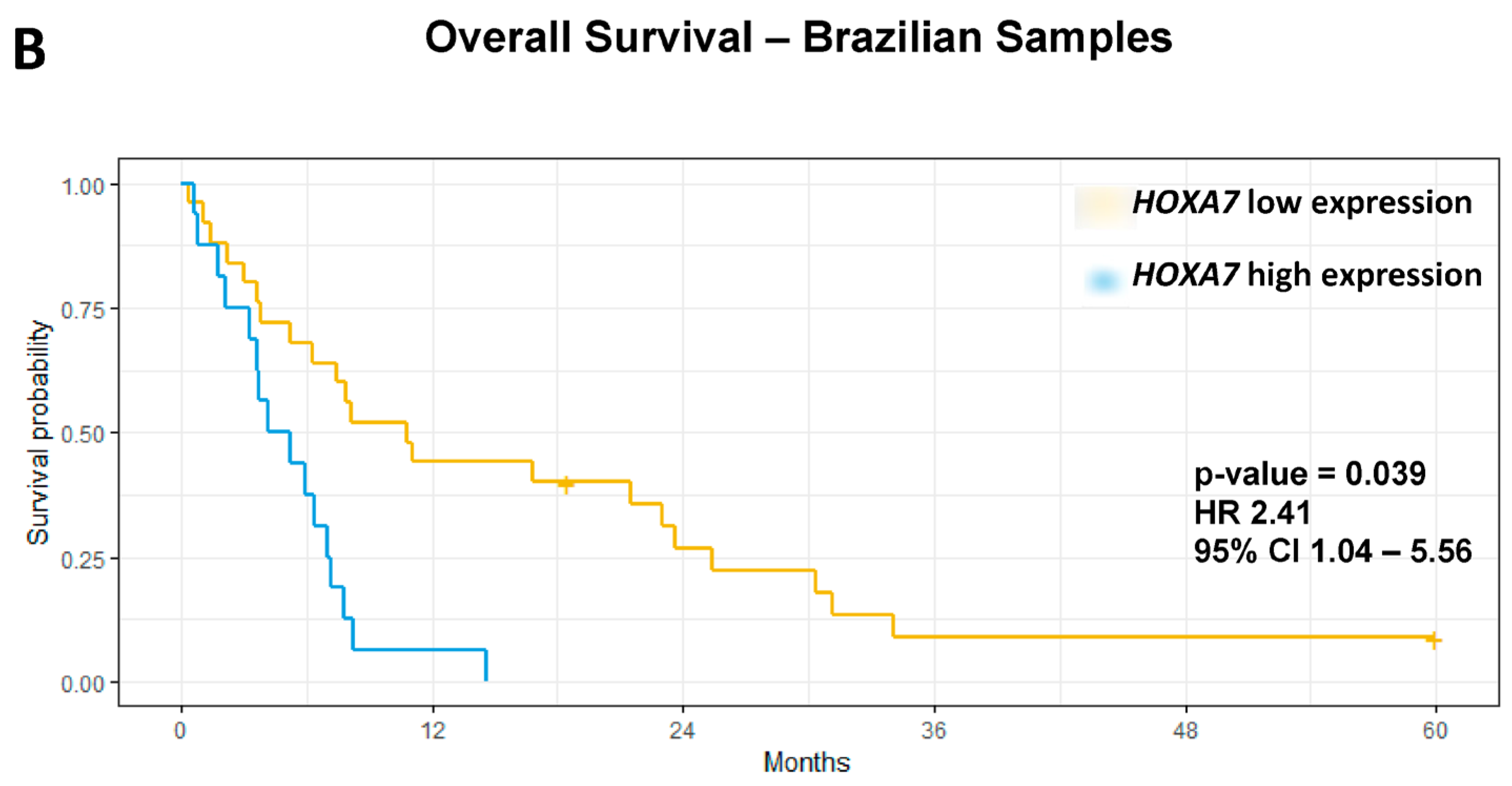
We evaluated the expression profile of HOXA7 in an independent set of ESCC Brazilian patients, observing HOXA7 overexpression in ESCC compared to paired NMSM (p = 0.016) (Supplementary Figure S3). Furthermore, we performed OS univariate analysis, and HOXA7 overexpression (p-value = 0.0019, HR = 3.29, 95%CI 1.54–6.99) and late-stage tumors (p-value = 0.012, HR = 3.02, 95% CI 1.27–7.19) were associated with worse OS rates. Multivariate analysis confirmed that HOXA7 overexpression is an independent biomarker of worse prognosis in this dataset (p-value = 0.039, HR 2.41, 95% CI 1.04–5.56) (Figure 3B; Table 4).

4. Discussion

In the present study, we investigated the mutational and expression profiles of HOX genes in esophageal squamous cell carcinoma (ESCC). We identified the signaling pathways associated with the putative targets of HOX genes overexpressed in ESCC. Our findings demonstrate that HOXA7 overexpression is an independent biomarker for poor prognosis in this malignancy.
The low frequency of somatic genetic alterations in HOX genes observed in ESCC (0–8%) is consistent with other cancers characterized by similar risk factors, such as alcohol consumption and tobacco smoking. For example, 4% of cases of lung squamous carcinoma exhibit copy number alterations or missense mutations in HOX genes [29,30]. In comparison, 6% of head and neck tumors show somatic alterations at the HOXD locus [30]. These data suggest that somatic mutations are not the primary mechanism underlying the dysregulation of HOX genes in tumorigenesis, underscoring the importance of regulatory mechanisms governing HOX gene expression in cancer.
We analyzed two transcriptomic datasets, comparing paired ESCC and NMSM, and identified seven HOX genes differentially expressed in ESCC. Furthermore, we validated the HOXA7 expression profile using an independent sample set. Previous studies have attempted to evaluate the expression profiles of HOX genes in ESCC using the methodologies available then. Chen and collaborators [11] assessed the expression of HOX genes in paired ESCC and NMSM samples from Chinese patients using non-quantitative RT-PCR without adjustment for multiple comparisons. They reported a higher number of deregulated HOX genes in ESCC, with nine genes exclusively expressed in tumors (HOXA10, HOXA13, HOXB7, HOXC4, HOXC8, HOXD9, HOXD10, and HOXD13) and three overexpressed in ESCC relative to NMSM (HOXA7, HOXA9, and HOXC6). Similarly, using quantitative RT-PCR, Takahashi and collaborators [13] observed that 24 of 39 HOX genes were overexpressed in ESCC compared to paired NMSM from Japanese patients. Again, they did not use adjustment methods for multiple comparisons. Therefore, the available studies confirm that a subset of HOX genes is overexpressed in ESCC, and the technique and statistical criteria applied could explain the discrepancies between our data and other studies.
In addition to the dysregulation of HOX genes, we identified putative HOX transcriptional targets using in silico analyses and assessed their differential expression in ESCC. Gene-set enrichment analyses highlighted various signaling pathways previously associated with the natural history of ESCC. Notably, in the PI3K-AKT signaling pathway, molecular alterations in PIK3CA and PIK3R3 have been linked to poor prognosis [17,18]. Furthermore, epithelial–mesenchymal transition is recognized as a relevant event for ESCC carcinogenesis, and the aberrant activation of the WNT signaling pathway is also associated with ESCC prognosis [31,32]. This study also identified the enrichment of signaling pathways related to IL-6-JAK, inflammatory responses, and apoptosis. We previously demonstrated that IL-6 mRNA and protein expression are upregulated in ESCC compared to NMSM, with IL-6 overexpression playing a crucial role in ESCC carcinogenesis by promoting anti-apoptotic signaling via BCL3 overexpression [33].
The dysregulation of HOXA7 in ESCC is associated with a poorer prognosis, echoing findings from mixed-lineage leukemia, where HOXA7 overexpression correlates with lower survival rates [34]. Additionally, HOXA7 has been reported to be overexpressed in colorectal cancer [35], which is also linked to poor prognoses. Similar to our results, which show higher HOXA7 expression in ESCC mucosa compared to NMSM samples, oral tumors exhibit elevated HOXA7 levels compared to the dysplastic oral mucosa [36]. This altered expression profile suggests that HOXA7 could be a central player in the carcinogenesis of the upper aerodigestive tract.
HOXA7 promotes hepatocellular carcinoma through cyclin E1/CDK2, inducing proliferation, migration, colony formation, and tumorigenesis in vivo [37]. In oral squamous cell carcinoma, HOXA7 overexpression is linked to aggressive markers such as tumor size, high-grade tumors, vascular and perineural invasion, and lymph node and distant metastases [38]. In ESCC, it has been shown that HOXA7 induces tumor-associated macrophage infiltration and M2 polarization by promoting CCL2 secretion. Additionally, macrophage secretion of EGF induced by CCL2 promotes ESCC tumor growth [12]. HOXA7 can also induce EGFR expression in ESCC cells [39] and in granuloma cells [39].
The dysregulation of HOX genes is a multifactorial process mediated by temporospatial deregulation, gene dominance, and epigenetic perturbations [2]. Epigenetic control, mainly through DNA methylation mediated by DNA methyltransferases/ten-eleven translocations (DNMTs/TETs) regulation of DNA methylation, as well as histone methylation and long non-coding RNA [40], emerges as a crucial mechanism governing HOX genes. In breast cancer, TET1 has been shown to impair HOXA7 function by modulating HOXA promoters, thereby promoting breast tumor growth and metastasis [41]. Recent studies have illuminated the significant role of HOX-related non-coding RNAs in modulating chromatin dynamics and gene expression, with implications for the progression of oral squamous cell carcinoma. The authors showed that miR196a was upregulated in oral squamous cell carcinoma samples from patients undergoing surgery and radiotherapy, suggesting its interaction with HOXA7 and a potential tumorigenic role [36].
This study has limitations, such as the lack of assessment of HOXA7 protein levels. Nevertheless, we applied stringent statistical criteria and validated the association between HOXA7 expression and the prognosis of ESCC patients in independent sample sets.

5. Conclusions

In conclusion, our findings underscore the rarity of somatic alterations in HOX genes in ESCC and shed light on the pivotal role of altered gene expression in driving their dysregulation. Furthermore, the HOXA7 overexpression emerges as an independent biomarker of poor prognosis in ESCC, providing valuable insights into the disease.

Supplementary Materials

The following supporting information can be downloaded at: https://www.mdpi.com/article/10.3390/genes15111430/s1, Figure S1: HOX genes somatic alteration in ESCC. Oncoprint showed the somatic alterations of the HOX genes in esophageal squamous cell carcinoma (ESCC) samples from TCGA. Each row represents one HOX gene, and each column represents one tumor sample; Figure S2: Enrichment analyses of differentially expressed HOXA7 gene targets were conducted to identify significant pathways. Using potential targets of differentially expressed HOXA7 genes in esophageal squamous cell carcinoma (ESCC), this analysis highlighted key signaling pathways in the KEGG (A) and Hallmarks (B) databases. The shades of blue represent the gene count observed within each signaling pathway, with darker blues indicating a higher number of observed genes in each pathway. Figure S3: Validation of HOXA7 gene expression profile in ESCC. HOXA7 expression was evaluated by qPCR in esophageal squamous cell carcinoma (ESCC) and paired non-malignant surrounding mucosa (NMSM) samples from the Brazilian population, and significantly higher expression was observed in ESCC samples than in paired NMSM samples (p = 0.0168). Min: Minimum; Max: Maximum. Supplementary Table S1: Oligonucleotide sequences. Supplementary Table S2: Potential transcriptional targets of HOXA2, HOXA7, HOXB13, HOXC9, HOXC10, HOXC13 and HOXD10. Supplementary Table S3: Potential transcriptional targets of HOXA7.

Author Contributions

Conceptualization: T.d.A.S., P.N.-N., S.C.S.-L. and L.F.R.-P.; Methodology: P.N.-N., J.V.G., T.d.A.S., J.N.d.A. and L.B.L.; Validation and formal analysis: T.d.A.S., P.N.-N., S.C.S.-L., J.V.G., J.N.d.A., L.B.L. and P.T.d.S.-S.; Supervision: T.d.A.S., S.C.S.-L. and L.F.R.-P.; Writing original draft preparation: T.d.A.S., P.N.-N., S.C.S.-L., J.V.G. and L.F.R.-P.; Funding acquisition, Project administration and Resources: T.d.A.S. All authors have read and agreed to the published version of the manuscript.

Funding

This research was funded by Fundação Carlos Chagas Filho de Amparo à Pesquisa do Estado do Rio de Janeiro), grant number E26/210.526/2019 and The APC was funded by MDPI.

Institutional Review Board Statement

The study was conducted in accordance with the Declaration of Helsinki and approved by the Ethics Committee of Instituto Nacional de Câncer (no. 116/11-CAAE-0086.0.007.000-11 on 5 December 2011).

Informed Consent Statement

Informed consent was obtained from all subjects involved in the study.

Data Availability Statement

Data are contained within the article or Supplementary Material.

Acknowledgments

We thank Fundação Carlos Chagas Filho de Amparo à Pesquisa do Estado do Rio de Janeiro (FAPERJ-E26/210.526/2019) for financially supporting this study and the team at INCA’s Banco Nacional de Tumores e DNA (BNT) for helping acquire and extract tumor samples.

Conflicts of Interest

The authors state that they do not have any conflicts of interest.

References

  1. Lewis, E.B. Clusters of master control genes regulate the development of higher organisms. JAMA 1992, 267, 1524–1531. [Google Scholar] [CrossRef] [PubMed]
  2. Shah, N.; Sukumar, S. The HOX genes and their roles in oncogenesis. Nat. Rev. Cancer 2010, 10, 361–371. [Google Scholar] [CrossRef] [PubMed]
  3. Krumlauf, R. HOX genes in vertebrate development. Cell 1994, 78, 191–201. [Google Scholar] [CrossRef] [PubMed]
  4. Abate-Shen, C. Deregulated homeobox gene expression in cancer: Cause or consequence? Nat. Rev. Cancer 2002, 2, 777–785. [Google Scholar] [CrossRef] [PubMed]
  5. Jung, C.; Kim, R.S.; Zhang Lee, S.J.; Jeng, M.H. HOXB13 induces growth suppression of prostate cancer cells as a hormone-activated androgen receptor signaling repressor. Cancer Res. 2004, 64, 9185–9191. [Google Scholar] [CrossRef] [PubMed]
  6. Wang, Z.; Dahiya, S.; Provencher, H.; Muir, B.; Carney, E.; Coser, K.; Shioda, T.; Ma, X.J.; Sgroi, D.C. The prognostic biomarkers HOXB13, IL17BR, and CHDH are regulated by estrogen in breast cancer. Clin. Cancer Res. 2007, 13, 6327–6334. [Google Scholar] [CrossRef]
  7. Sun, Y.; Zhou, B.; Mao, F.; Xu, J.; Miao, H.; Zou, Z.; Jang, Y.; Cai, S.; Witkin, M.; Koche, R.; et al. HOXA9 reprograms the enhancer landscape to promote leukemogenesis. Cancer Cell 2018, 34, 643–658. [Google Scholar] [CrossRef]
  8. Bhatlekar, S.; Fields, J.Z.; Boman, B.M. HOX genes and their role in the development of human cancers. J. Mol. Med. 2014, 92, 811–823. [Google Scholar] [CrossRef]
  9. Ewing, C.M.; Ray, A.M.; Lange, E.M.; Zuhlke, K.A.; Robbins, C.M.; Tembe, W.D.; Wiley, K.E.; Isaacs, S.D.; Johng, D.; Wang, Y.; et al. Germline mutations in HOXB13 and prostate-cancer risk. N. Engl. J. Med. 2012, 366, 141–149. [Google Scholar] [CrossRef]
  10. Luo, Z.; Rhie, S.K.; Farnham, P.J. The enigmatic HOX genes: Can we crack their code? Cancers 2019, 11, 323. [Google Scholar] [CrossRef]
  11. Chen, K.N.; Gu, Z.D.; Ke, Y.; Li, J.Y.; Shi, X.T.; Xu, G.W. Expression of 11 HOX genes is deregulated in esophageal squamous cell carcinoma. Clin. Cancer Res. 2005, 11, 1044–1049. [Google Scholar] [CrossRef] [PubMed]
  12. Feng, A.; He, L.; Jiang, J.; Chu, Y.; Zhang, Z.; Fang, K.; Wang, Z.; Li, Z.; Sun, M.; Zhao, Z.; et al. Homeobox A7 promotes esophageal squamous cell carcinoma progression through C-C motif chemokine ligand 2-mediated tumor-associated macrophage recruitment. Cancer Sci. 2023, 114, 3270–3286. [Google Scholar] [CrossRef] [PubMed]
  13. Takahashi, O.; Hamada, J.I.; Abe, M.; Hata, S.; Asano, T.; Takahashi, Y.; Tada, M.; Miyamoto, M.; Kondo, S.; Moriuchi, T. Dysregulated expression of HOX and ParaHOX genes in human esophageal squamous cell carcinoma. Oncol. Rep. 2007, 17, 753–760. [Google Scholar] [CrossRef] [PubMed]
  14. Zhao, J.; Jia, X.; Li, Q.; Zhang, H.; Wang, J.; Huang, S.; Hu, Z.; Li, C. Genomic and transcriptional characterization of early esophageal squamous cell carcinoma. BMC Med. Genom. 2023, 16, 67. [Google Scholar] [CrossRef]
  15. Ferlay, J.; Ervik, M.; Lam, F.; Laversanne, M.; Colombet, M.; Mery, L.; Piñeros, M.; Znaor, A.; Soerjomataram, I.; Bray, F. Global Cancer Observatory: Cancer Today. Lyon, France: International Agency for Research on Cancer. 2024. Available online: https://gco.iarc.who.int/today (accessed on 30 August 2024).
  16. van Hagen, P.; Hulshof, M.C.C.M.; van Lanschot, J.J.B.; Steyerberg, E.W.; van Berge Henegouwen, M.I.; Wijnhoven, B.P.L.; Richel, D.J.; Nieuwenhuijzen, G.A.P.; Hospers, G.A.P.; Bonenkamp, J.J.; et al. Preoperative chemoradiotherapy for esophageal or junctional cancer. N. Engl. J. Med. 2012, 366, 2074–2084. [Google Scholar] [CrossRef]
  17. The Cancer Genome Atlas Research Network. Integrated genomic characterization of oesophageal carcinoma. Nature 2017, 541, 169–175. [Google Scholar] [CrossRef]
  18. Nicolau-Neto, P.; Da Costa, N.M.; de Souza Santos, P.T.; Gonzaga, I.M.; Ferreira, M.A.; Guaraldi, S.; Moreira, M.A.; Seuánez, H.N.; Brewer, L.; Bergmann, A.; et al. Esophageal squamous cell carcinoma transcriptome reveals the effect of FOXM1 on patient outcome through novel PIK3R3 mediated activation of PI3K signaling pathway. Oncotarget 2018, 9, 16634–16647. [Google Scholar] [CrossRef]
  19. Li, J.; Chen, Z.; Tian, L.; Zhou, C.; He, M.Y.; Gao, Y.; Wang, S.; Zhou, F.; Shi, S.; Feng, X.; et al. LncRNA profile study reveals a three-lncRNA signature associated with the survival of patients with oesophageal squamous cell carcinoma. Gut 2014, 63, 1700–1710. [Google Scholar] [CrossRef]
  20. Sean, D.; Meltzer, P.S. GEOquery: A bridge between the Gene Expression Omnibus (GEO) and BioConductor. Bioinformatics 2007, 23, 1846–1847. [Google Scholar]
  21. Wettenhall, J.M.; Smyth, G.K. limmaGUI: A graphical user interface for linear modeling of microarray data. Bioinformatics 2004, 20, 3705–3706. [Google Scholar] [CrossRef]
  22. Mermel, C.H.; Schumacher, S.E.; Hill, B.; Meyerson, M.L.; Beroukhim, R.; Getz, G. GISTIC2.0 facilitates sensitive and confident localization of the targets of focal somatic copy-number alteration in human cancers. Genome Biol. 2011, 12, R41. [Google Scholar] [CrossRef] [PubMed]
  23. Livak, K.J.; Schmittgen, T.D. Analysis of relative gene expression data using real-time quantitative PCR and the 2−ΔΔCT method. Methods 2001, 25, 402–408. [Google Scholar] [CrossRef] [PubMed]
  24. Plaisier, C.L.; O’Brien, S.; Bernard, B.; Reynolds, S.; Simon, Z.; Toledo, C.M.; Ding, Y.; Reiss, D.J.; Paddison, P.J.; Baliga, N.S. Causal mechanistic regulatory network for glioblastoma deciphered using systems genetics network analysis. Cell Syst. 2016, 3, 172–186. [Google Scholar] [CrossRef] [PubMed]
  25. John MYuxing, L.; Zhiao, S.; Qian, Z.; Alexander, R.P.; Bing, Z. WebGestalt 2024: Faster gene set analysis and new support for metabolomics and multi-omics. Nucleic Acids Res. 2024, gkae456. [Google Scholar]
  26. Bradburn, M.J.; Clark, T.G.; Love, S.B.; Altman, D.G. Survival analysis part III: Multivariate data analysis—Choosing a model and assessing its adequacy and fit. Br. J. Cancer 2003, 89, 605–611. [Google Scholar] [CrossRef]
  27. Therneau, T.; Lumley, T. R Package, Version 2.38; Package Survival: A Package for Survival Analysis in R, 2015; Available online: https://cran.r-project.org/web/packages/survival/index.html (accessed on 1 October 2024).
  28. Lánczky, A.; Győrffy, B. Web-based survival analysis tool tailored for medical research (KMplot): Development and implementation. J. Med. Internet Res. 2021, 23, e27633. [Google Scholar] [CrossRef]
  29. Hammerman, P.S.; Lawrence, M.S.; Voet, D.; Jing, R.; Cibulskis, K.; Sivachenko, A.; Stojanov, P.; McKenna, A.; Lander, E.S.; Gabriel, S.; et al. Comprehensive genomic characterization of squamous cell lung cancers. Nature 2012, 489, 519–525. [Google Scholar]
  30. Hoadley, K.A.; Yau, C.; Wolf, D.M.; Cherniack, A.D.; Tamborero, D.; Ng, S.; Leiserson, M.D.; Niu, B.; McLellan, M.D.; Uzunangelov, V.; et al. Multiplatform analysis of 12 cancer types reveals molecular classification within and across tissues of origin. Cell 2014, 158, 929–944. [Google Scholar] [CrossRef]
  31. Oguma, J.; Ozawa, S.; Kazuno, A.; Nitta, M.; Ninomiya, Y.; Kajiwara, H. Wnt3a expression is associated with poor prognosis of esophageal squamous cell carcinoma. Oncol. Lett. 2018, 15, 3100–3108. [Google Scholar] [CrossRef]
  32. de Souza-Santos, P.T.; Lima, S.C.S.; Nicolau-Neto, P.; Boroni, M.; Costa, N.M.; Brewer, L.; Menezes, A.N.; Furtado, C.; Moreira, M.A.M.; Seuanez, H.N.; et al. Mutations, differential gene expression, and chimeric transcripts in esophageal squamous cell carcinoma show high heterogeneity. Transl. Oncol. 2018, 11, 1283–1291. [Google Scholar] [CrossRef]
  33. Soares-Lima, S.C.; Gonzaga, I.M.; Camuzi, D.; Nicolau-Neto, P.; Silva, R.V.D.; Guaraldi, S.; Ferreira, M.A.; Hernandez-Vargas, H.; Herceg, Z.; Ribeiro Pinto, L.F. IL6 and BCL3 expression are potential biomarkers in esophageal squamous cell carcinoma. Front. Oncol. 2021, 11, 610574. [Google Scholar] [CrossRef] [PubMed]
  34. Golub, T.R.; Slonim, D.K.; Tamayo, P.; Huard, C.; Gaasenbeek, M.; Mesirov, J.P.; Coller, H.; Loh, M.L.; Downing, J.R.; Caligiuri, M.A.; et al. Molecular classification of cancer: Class discovery and class prediction by gene expression monitoring. Science 1999, 286, 531–537. [Google Scholar] [CrossRef] [PubMed]
  35. Dang, Y.; Yu, J.; Zhao, S.; Cao, X.; Wang, Q. HOXA7 promotes the metastasis of KRAS mutant colorectal cancer by regulating myeloid-derived suppressor cells. Cancer Cell Int. 2022, 22, 89. [Google Scholar] [CrossRef] [PubMed]
  36. Padam, K.S.R.; Morgan, R.; Hunter, K.; Chakrabarty, S.; Kumar, N.A.N.; Radhakrishnan, R. Identification of HOX signatures contributing to oral cancer phenotype. Sci. Rep. 2022, 12, 2457. [Google Scholar] [CrossRef]
  37. Li, Y.; Yang, X.H.; Fang, S.J.; Qin, C.F.; Sun, R.L.; Liu, Z.Y.; Jiang, B.Y.; Wu, X.; Li, G. HOXA7 stimulates human hepatocellular carcinoma proliferation through cyclin E1/CDK2. Oncol. Rep. 2015, 33, 990–996. [Google Scholar] [CrossRef]
  38. Duan, X.; Chen, H.; Ma, H.; Song, Y. The expression and significance of the HOXA7 gene in oral squamous cell carcinoma. J. Oral Sci. 2017, 59, 329–335. [Google Scholar] [CrossRef]
  39. Zhang, Y.; Huang, Q.; Cheng, J.C.; Nishi, Y.; Yanase, T.; Huang, H.F.; Leung, P.C. Homeobox A7 increases cell proliferation by up-regulation of epidermal growth factor receptor expression in human granulosa cells. Reprod. Biol. Endocrinol. 2010, 8, 61. [Google Scholar] [CrossRef]
  40. Hu, X.; Wang, Y.; Zhang, X.; Li, C.; Zhang, X.; Yang, D.; Liu, Y.; Li, L. DNA methylation of HOX genes and its clinical implications in cancer. Exp. Mol. Pathol. 2023, 134, 104916. [Google Scholar] [CrossRef]
  41. Sun, M.; Song, C.X.; Huang, H.; Frankenberger, C.A.; Sankarasharma, D.; Gomes, S.; Chen, P.; Chen, J.; Chada, K.K.; He, C.; et al. HMGA2/TET1/HOXA9 signaling pathway regulates breast cancer growth and metastasis. Proc. Natl. Acad. Sci. USA 2013, 110, 9920–9925. [Google Scholar] [CrossRef]
Figure 1. Summary schematic of the principal methodologies and sample datasets used. ESCC: esophageal squamous cell carcinoma; matched non-malignant surrounding mucosa; CNA; copy number alteration; SNV: single nucleotide variant: GISTIC: The Genomic Identification of Significant Targets in Cancer (created in BioRender).
Genes 15 01430 g001
Figure 2. Enrichment analyses of differentially expressed HOX gene targets. Enrichment analyses using the potential targets of differentially expressed HOX genes in esophageal squamous cell carcinoma (ESCC) highlighted significant signaling pathways in KEGG (A) and Hallmarks (B) databases. Shades of blue indicate the number of genes observed in each signaling pathway. The darker blue shows more observed genes in the signaling pathway.
Genes 15 01430 g002
Figure 3. HOXA7 overexpression is an independent biomarker of worse prognosis in ESCC. Patients with esophageal squamous cell carcinoma (ESCC) expressing high HOXA7 gene levels showed worse survival rates than patients with ESCC expressing low HOXA7 levels in the GSE53625 dataset ((A) p = 0.024; HR = 1.58; 95% CI 1.06–2.35) and the Brazilian Samples ((B) p-value = 0.039, HR 2.41, 95% CI 1.04–5.56). Yellow lines—samples with low HOXA7 expression; Blue lines—samples with high HOXA7 expression.
Genes 15 01430 g003aGenes 15 01430 g003b
Table 1. ESCC patient’s features.
FeatureVariableBrazilian ESCC Patients
Age—Median (range) 59 (39–77)
GenderFemale8 (19.51%)
Male33 (80.49%)
Tobacco smokingNo4 (9.76%)
Yes35 (85.36%)
NA2 (4.88%)
Alcohol drinkingNo5 (12.20%)
Yes35 (85.36%)
NA1 (2.44%)
Esophagel tumor subsiteUpper5 (12.20%)
Middle11 (26.83%)
Lower3 (7.3%)
More than one subsite22 (53.66%)
Tumor gradeG234 (82.93%)
G37 (17.07%)
StageI-II12 (29.27%)
III-IV25 (60.97%)
NA4 (9.76%)
Lymph node metastasisNo8 (19.51%)
Yes17 (41.46%)
NA16 (39.03%)
Distant metastasisNo11 (26.83%)
Yes17 (41.46%)
NA13 (31.71%)
NA: not available.
Table 2. ESCC genes differentially expression in two datasets.
Gene SymbolGSE75241GSE53625
Probe IDFold Change (ESCC/NMSM)p ValueProbe IDFold Change (ESCC/NMSM)p Value
HOXA23042756−1.67<0.001CB_015587−1.58<0.001
HOXA730428811.500.002CB_0157372.13<0.001
HOXB1337615382.12<0.001CB_0152182.87<0.001
HOXC934163441.57<0.001CB_0157383.43<0.001
HOXC1034162902.32<0.001CB_01903710.56<0.001
HOXC1334162561.66<0.001CB_01903815.45<0.001
HOXD1025168344.17<0.001CB_0111324.44<0.001
Table 3. Association analyses between E SCC-overexpressed HOX genes and clinicopathological features.
FrequencyHOXA2HOXA7HOXB13HOXC9HOXC10HOXC13HOXD10
FeatureVariablen%Medianp ValueMedianp ValueMedianp ValueMedianp ValueMedianp ValueMedianp ValueMedianp Value
Age<608949.70%10.540.1112.940.225.760.02610.700.169.590.859.140.3811.890.92
≥609050.30%10.8312.876.8310.919.649.1811.88
SexMale14681.60%10.780.2412.950.126.530.7810.830.5010.140.889.130.6711.970.009
Female3318.40%10.6112.736.1510.79.959.2411.50
Alcohol DrinkingNo7340.80%10.770.6512.740.096.240.7310.650.099.940.668.980.0611.800.01
Yes10659.20%10.7313.046.3210.9110.229.4312.11
Tobacco SmokingNo6536.30%10.730.812.820.335.840.4110.530.0059.990.929.050.2711.880.53
Yes11463.70%10.7612.976.8210.9710.169.2712.00
Esophageal Tumor SubsiteUpper2011.20%10.960.8113.340.096.870.6211.230.369.880.708.880.311.760.48
Middle9754.20%10.6212.986.6910.9610.009.0912.06
Lower6234.60%10.6212.666.8710.5510.399.4311.81
Tumor DifferentiationWell3217.90%10.590.6813.110.745.660.050110.560.0067 *9.890.479.480.9211.80.75
Moderate9854.70%10.6112.896.3210.729.949.1512.03
Poorly4927.40%10.7913.037.3611.2710.729.0812.05
Lymph node metastasisNo8346.40%10.680.5512.830.746.870.3110.800.8910.000.729.060.3011.950.95
Yes9653.60%10.6212.955.7310.8210.259.4011.97
T (TNM)T1/T23921.70%10.540.312.830.826.350.6511.010.1610.340.659.400.3911.910.84
T3/T414078.80%10.6612.946.2710.7510.009.1011.88
Tumor StageEarly (I/II)8748.60%10.570.0812.820.556.870.0910.770.619.630.959.080.3711.880.95
Late (III)9251.40%10.6813.015.6510.849.619.2712.05
Legend: * well vs. poorly p = 0.008; moderate vs. poorly p = 0.029.
Table 4. Overall survival analysis.
FeatureVariableGSE53625Brazilian Samples
Univariate Survival AnalysisMultivariate AnalysisUnivariate Survival AnalysisMultivariate Analysis
95% CI95% CI95% CI95% CI
HRLowHighp ValueHRLowHighp ValueHRLowHighp ValueHRLowHighp Value
Age≥60 y vs. <60 y1.541.0472.260.028 0.980.511.890.97
GenderMale vs. Female0.780.491.250.32.971.43.120.00020.640.271.510.31
StageI vs. II vs. III2.151.443.20.00015 3.021.277.190.0122.360.935.960.06
GradeG3/G2 vs. G11.3511.820.048 0.570.281.170.12
HOXA2High vs. Low1.581.0412.4080.0316
HOXA7High vs. Low1.651.112.450.0131.581.062.350.0243.291.546.990.00192.411.045.560.039
HOXB13High vs. Low1.91.063.40.0302
HOXC9High vs. Low1.520.962.420.073
HOXC10High vs. Low0.280.141.040.061
HOXC13High vs. Low1.280.871.880.13
HOXD10High vs. Low1.30.871.90.018
Bold numbers refer to the significant p-value.
Disclaimer/Publisher’s Note: The statements, opinions and data contained in all publications are solely those of the individual author(s) and contributor(s) and not of MDPI and/or the editor(s). MDPI and/or the editor(s) disclaim responsibility for any injury to people or property resulting from any ideas, methods, instructions or products referred to in the content.