Article

RNAi Knockdown of EHMT2 in Maternal Expression of Prader–Willi Syndrome Genes

by
Violeta Zaric
1,
Hye Ri Kang
2,
Volodymyr Rybalchenko
1,
Jeffrey M. Zigman
1,2,3,4,
Steven J. Gray
2,3 and
Ryan K. Butler
1,2,3,*
1
Department of Psychiatry, UT Southwestern Medical Center, Dallas, TX 75390, USA
2
Department of Pediatrics, UT Southwestern Medical Center, Dallas, TX 75390, USA
3
O’Donnell Brain Institute, UT Southwestern Medical Center, Dallas, TX 75390, USA
4
Center for Hypothalamic Research, Department of Internal Medicine, UT Southwestern Medical Center, Dallas, TX 75390, USA
*
Author to whom correspondence should be addressed.
Genes 2024, 15(11), 1366; https://doi.org/10.3390/genes15111366
Submission received: 11 September 2024 / Revised: 13 October 2024 / Accepted: 18 October 2024 / Published: 24 October 2024
(This article belongs to the Section Human Genomics and Genetic Diseases)

Abstract

:
Background/objectives: Euchromatic histone lysine methyltransferase 2 (EHMT2, also known as G9a) is a mammalian histone methyltransferase that catalyzes the dimethylation of histone 3 lysine 9 (H3K9). On human chromosome 15, the parental-specific expression of Prader–Willi Syndrome (PWS)-related genes, such as SNRPN and SNORD116, are regulated through the genetic imprinting of the PWS imprinting center (PWS-IC). On the paternal allele, PWS genes are expressed whereas the epigenetic maternal silencing of PWS genes is controlled by the EHMT2-mediated methylation of H3K9 in PWS-IC. Here, we measured the effects of RNA interference of EHMT2 on the maternal expression of genes deficient in PWS in mouse model and patient iPSC-derived cells. Methods: We used small interfering RNA (siRNA) oligonucleotides and lentiviral short harpin RNA (shRNA) to reduce Ehtm2/EHMT2 expression in mouse Snord116 deletion primary neurons, PWS patient-derived induced pluripotent stem cell (iPSC) line and PWS iPSC-derived neurons. We then measured the expression of transcript or protein (if relevant) of PWS genes normally silenced on the maternal allele. Results: With an approximate reduction of 90% in EHMT2 mRNA and more than 80% of the EHMT2 protein, we demonstrated close to a 2-fold increase in the expression of maternal transcripts for SNRPN and SNORD116 in PWS iPSCs treated with siEHMT2 compared to PWS iPSC siControl. A similar increase in SNORD116 and SNRPN RNA expression was observed in PWS iPSC-derived neurons treated with shEHMT2. Conclusions: RNAi reduction in EHMT2 activates maternally silenced PWS genes. Further studies are needed to determine whether the increase is therapeutically relevant. This study confirms the role of EHMT2 in the epigenetic regulation of PWS genes.

1. Introduction

Prader–Willi Syndrome (PWS, OMIM176270) is an imprinted neurodevelopmental disorder. Major manifestations include early childhood obesity, hyperphagia, hypotonia with poor suck and poor weight gain in infancy, mild-to-moderate intellectual disability, hypogonadism, growth hormone insufficiency, and, frequently, psychiatric disturbance, including abnormal restricted repetitive behavior (e.g., skin picking, obsession, compulsion, sameness behavior, etc.). PWS is caused by the loss or loss-of-function of genes normally expressed on the paternal allele located in the chromosomal region 15q11-q13. Paternal expression is regulated by the PWS imprinting center (PWS-IC), whereas genes in this same region are epigenetically repressed due to the imprinting mechanism on the maternal allele. In most cases (70%), the large deletion (LD) of paternal 15q11-q13 region, comprised of several expressed protein-coding genes (MAGEL2, SNURF, and SNRPN, among others), a cluster of C/D box small nucleolar RNA noncoding genes (including SNORD116 and SNORD115), and several long noncoding transcripts (including IPW and the antisense transcript to UBE3A), leads to PWS pathogenesis [1,2,3]. Approximately 25% cases are due to the uniparental disomy (UPD) of maternal chromosome 15 [4,5], and ~1% of cases are due to aberrant DNA methylation throughout the imprinted domain on chromosome 15 [6]. Patients with the small deletion (SD) of 108 kb encompassing SNORD116 cluster and IPW genes appear to have most of the PWS-related clinical phenotypes, which shows the importance of SNORD116 in PWS pathogenesis [7,8,9,10]. Other rare PWS cases presenting the PWS symptoms of overeating and obesity have emerged and have been described with small, atypical deletions overlapping PWS-IC or a partial deletion of SNURF-SNRPN, or even a microdeletion of 78 kb that includes SNURF-SNRPN exons 2 and 3 [11,12,13]. However, although, in these last cases, patients do not express SNORD116, these patients do not manifest all PWS classical symptoms, as encountered in PWS patients with LD. Presently, there is no cure for PWS, and existing treatments address individual symptoms, necessitating a complex and burdensome regimen. There is a need for genetic therapies for PWS that can address the root cause of the disease and comprehensively mitigate the multitude of symptoms through a single treatment.
New methods of epigenetics-based intervention for the treatment of PWS targeting DNA methylation at the PWS-IC [14,15] or histone methylation at the SNORD116 locus have recently emerged [16]. In general, DNA methylation and histone lysine methylation are described as important epigenetic tools in maintaining gene silencing in numerous cellular processes, including embryonic development, genomic imprinting, X-chromosome inactivation, and the preservation of chromosome stability [17,18]. Particularly, the dimethylation of lysine 9 of histone H3 (H3K9me2) by euchromatic histone lysine N-methyltransferase-2 (EHMT2, also known as G9a) is associated with transcriptional repression and regulating genomic imprinting in PWS [19]. Indeed, not only is the PWS-IC on the maternal chromosome 15 found to be a methylated CG-rich region but H3K9me2 also acts as a repressor of maternal PWS gene expression in the 15q11-q13 chromosomal region. EHMT2 is essential in mouse embryonic development. Deletion and loss-of-function mutations result in lethality and severe growth defects by embryonic day 9.5. However, a neuron-specific deficiency in Ehmt2 did not reveal obvious neuronal developmental or architectural defects. Furthermore, a postnatal conditional genetic reduction in Ehtm2 does not affect skeletal muscle development [20] and reduces pain- and anxiety-like behaviors in mice [21,22,23]. Kim et al. recently demonstrated that the small molecules UNC0638 and UNC0642 can selectively reduce H3K9me2 [24]. The authors showed the activation of maternal PWS imprinted genes, including the cluster SNORD116, in fibroblasts from PWS patients and in a PWS mouse model without changing the methylation rate at the PWS-IC. This supports the idea that the activation of maternal genes is independent of PWS-IC DNA methylation. We are interested in pursuing epigenetics-based therapy by exploring the feasibility of the long-lasting gene downregulation of EHMT2 using short hairpin RNA (shRNA) expressed from a vector, modeled in cultured cells using either plasmids or lentivirus. shRNA, expressed as double-stranded RNA stems (19–29 bases) and joined by a short hairpin loop sequence, is processed by Dicer and incorporated into the RNA-induced silencing complex to become a small interfering RNA (siRNA), resulting in the targeting and degradation of cognate mRNAs [25,26,27]. Compared to siRNA, the benefit of shRNA is that it can be incorporated into permanent expression vectors and offer a long-standing effect on cellular function in therapeutic applications while maintaining a high level of specificity in gene silencing. Moreover, shRNA has also been widely used in research for over a decade, and there are currently five RNA interference-based drugs that have been approved by the FDA [28]. Our objective here is to test the hypothesis that a vector-derived anti-EHMT2 shRNA strategy is effective in elevating the maternal expression of PWS genes in vitro.

2. Materials and Methods

2.1. Animal Procedures

All animal studies were conducted in accordance with IACUC and under UT Southwestern Medical Center protocol 102619. All studies utilized C57Bl/6N Snord116p−/m+ mice (B6.Cg-Snord116tm1.1Uta/J, stock 008149) developed by the Francke lab [29], which were obtained from The Jackson Laboratory and have since been backcrossed onto a C57Bl/6N background over > 10 generations. Snord116p−/m+ study mice (which carry a paternally inherited chromosome 7 lacking the Snord116 gene cluster) and WT littermates were generated by crossing male C57Bl/6N Snord116p−/m+ mice with female C57BL/6N mice. Housing conditions included a 12 h light–dark cycle and ad libitum water access.

2.2. Genotyping

Genotyping was performed using genomic DNA extracted from tail snips and three primers to detect the Snord116del and WT Snord116 alleles: Snord116-M634 (5′-TGGATCTCTCCTTGCTTGTTTTCTC-3′), Snord116-M635 (5′-AATCCCCAACCTACTTCAAACAGTC-3′), and Snord116-M636 (5′-TTTACGGTACATGACAGCACTCAAG-3′). The following PCR protocol with an annealing temperature at +64 °C generated a WT band of 435 bp and a mutant band of 337 bp [30].

2.3. Cell Culture

The HEK293 cells were cultured in high-glucose (4.5 g/L) Dulbecco’s Modified Eagle Medium (DMEM) (Thermo-Fisher Scientific, Waltham, MA, USA) with added L-glutamine, penicillin–streptomycin antibiotic, and fetal bovine serum (FBS). The cells were passaged when they reached 80–90% confluency.

2.3.1. iPSC Lines

The iPSC lines used for this study originated from University of Connecticut Stem Cell Core (UConn Health, Farmington, CT, USA) and are shown in Table 1. Upon arrival, the iPSC lines were adapted to a feeder-free culture following the manufacturer’s instructions (StemCell Technologies, Cambridge, MA, USA) and passaged for an additional 2 passages for the removal of the feeder cells. The feeder-free iPSCs were cultured on Matrigel-coated plates with mTeSR™ Plus medium (StemCell Technologies, Cambridge, MA, USA) and passaged every 3–4 days at a ratio of 1:6–1:10 using Versene (Invitrogen, Waltham, MA, USA). The cells were cultured in a humidified incubator at 37 °C in a 5% CO2 atmosphere.

2.3.2. iPSC-Derived Neuronal Induction

iPSCs were induced to become neural progenitor cells (NPCs) according to the monolayer and dual SMAD inhibition protocol [32] using STEMdiff™ SMADi Neural Induction Kit (StemCell Technologies, Cambridge, MA, USA). iPSCs were detached using Accutase to obtain 2 × 10⁶ single cells per well in a 6-well plate pre-coated with Matrigel. Neural induction media were changed daily for 8 days before each passage. To induce the differentiation of NPCs into neural precursor cells (NPreCs), NPCs were detached with Accutase after three passages and plated in 6-well plates coated with 15 µg/mL PLO (Sigma-Aldrich, St. Louis, MO, USA, P3655) and 10 µg/mL Laminin (Sigma-Aldrich, St. Louis, MO, USA, L2020) at a plating density of 105,000 cells per cm2. STEMdiff Forebrain Neuron Differentiation medium (STEMCELL Technologies, 08600) was changed daily up to 7 days until confluency reached 80–90%. To generate iPSC-derived neurons, NPreCs were detached with Accutase and plated at 126,000 cells per cm2 into STEMdiff Forebrain Neuron Maturation medium (STEMCELL Technologies, 08605). Further maturation into forebrain neurons was conducted by changing the corresponding media every other day until the experiment was carried out.

2.3.3. Mouse Primary Neurons

Mouse primary neurons were isolated from the early postnatal (P0–P2) mouse hippocampus and cortex. The isolation method followed was that described by Beaudoin et al. [33] with minor modification. Briefly, cells from the mouse hippocampus and cortex were dissociated with a solution of trypsin, and the dissociated tissue was added on top of a 4% BSA cushion, followed by centrifugation at 300× g for 7 min at room temperature, as described by Moutin et al. [34]. The cell pellet was resuspended in a plating medium, and the cells were plated at 0.5 × 10⁶ cells per well in 12-well precoated plates with 0.5 mg/mL Poly L-Lysine (Sciencell Research, Carlsbad, CA, USA). The plating medium was removed 4 h after plating and replaced with maintenance media. The cells were cultured until DIV4, with half of the maintenance medium changed every other day prior to transfection.

2.4. siRNA Design

Several siRNA duplexes (21 nucleotides for sense and antisense) targeting the mouse Ehmt2 mRNA and human EHMT2 mRNA were designed using The Genetic Perturbation Platform from Broad Institute. Using the algorithm from the siRNA Sequence Probability-of-Off-Targeting Reduction online tool https://sispotr.icts.uiowa.edu/ (accessed on 1 March 2021), siRNA candidates were checked for Off-Targets in human and mouse RNA sequences, and the best matches were selected with the lowest Probability-of-Off-Targeting-Sites (POTS) values [35].

2.5. Plasmid Construct

Five siRNA sequences (Table 2; 21 nucleotides for sense and antisense) with silencing activities against human EHMT2 and mouse Ehmt2 coding regions were selected for constructing shRNA silencing plasmids, named U6-shRNA (hEHMT2/mEhmt2-11) U6-shRNA (hEHMT2/mEhmt2-12), U6-shRNA (hEHMT2/mEhmt2-13), U6-shRNA (hEHMT2/mEhmt2-14), and U6-shRNA (hEHMT2/mEhmt2-15). A negative control without silencing activity against human EHMT2 and mouse Ehmt2 coding regions was prepared and named U6-shRNA (Scramble). Each shRNA-expressing construct is driven by a Pol III U6 promoter. Each plasmid contains an additional cassette for EGFP controlled by the ubiquitous synthetic promoter (JeT) and placed upstream of the U6 promoter.

2.6. Cell Transfection with siRNA

PWS UPD iPSCs were transfected with 120 pmol of anti-EHMT2 siRNAs per well of a 6-well plate, using Lipofectamine RNAiMAX, as described [16]. Cells were transfected a second time 48 h following the first transfection and were harvested 72 h after the initial transfection.

2.7. Cell Transfection with Plasmid

HEK293 cells were seeded at 175,000 cells per well in 24-well plates and left to grow for 24 h before the transfection. HEK293 cells were transfected (Thermo-Fisher Scientific, Waltham, MA, USA, 161093) with different shRNA plasmid constructs at a total of 500 ng plasmid/well using PEI MAX (Polysciences, Warrington, PA, USA, 24765-1) at a ratio PEI:DNA of 4:1. The PEI:DNA complexes were prepared by mixing the PEI into a diluted solution of DNA with OptiMEM, and the mixture was left to incubate for 10 min before being added to the cells. The medium was replaced 24 h post transfection. Then, 72 h after transfection, cell lysates for RNA were harvested.
The iPSCs were transfected using the reverse transfection method following the manufacturer instructions. The TransIT-LT1 (Mirus Bio, Madison, WI, USA) was used to reverse-transfect 0.25 × 10⁶ iPSCs with the shRNA plasmid constructs. Reverse transfections were performed in 24-well plates using 1.5 µL of TransIT-LT1 to deliver 0.5 µg of DNA (3:1, reagent: DNA) in mTeSR™ Plus with 10 µM rock inhibitor for 4 h, after which the media was replaced with fresh mTeSR™ Plus with 10 µM rock inhibitor. The cells were incubated for 72 h until being harvested.

2.8. Transduction of iPSC-Derived Cells with Lentivirus

Non-integrating lentiviral vectors (NILVs) were designed by the VectorBuilder (Chicago, IL, USA) and are U6-based shRNA knockdown vectors containing the EGFP sequence driven by JeT promoter. The NILV-U6-shRNA candidates NILV-U6shRNA (hEHMT2/mEhmt2-11) and NILV-U6shRNA (hEHMT2/mEhmt2-14) targeting conserved mouse and human EHMT2 sequences and a control NILV-U6shRNA-Scramble were prepared.
NPreC derived from iPSC UPD and iPSC SD was detached with Accutase, and 240,000 cells per well were plated on 24-well plates coated with PLO and laminin. Mature neurons were generated for 15 days by feeding every other day with STEMdiff Forebrain Neuron Maturation medium (STEMCELL Technologies, 08605). NILVs were added to the wells at a range of viral concentrations [represented as multiplicity of infection (MOI)]. The media was replaced after 24 h of transduction, and the cells were cultured until harvest at the indicated time points.

2.9. Quantitative Real-Time Reverse Transcription (qRT)-PCR

Total RNA extraction from HEK293 cells, iPSCs, and mouse primary neurons was carried out using the RNeasy mini Kit (QIAGEN, Germantown, MA, USA). Briefly, the cells were lysed by adding 350 µL buffer RLT containing mercaptoethanol per well and stored at −80 °C until further extraction was carried out. The samples were then processed according to the manufacturer’s instructions. The RNA concentration was measured by NanoDrop One (Thermo-Fisher Scientific, Waltham, MA, USA). cDNA was prepared using the iScript cDNA Synthesis Kit (BioRad, Hercules, CA, USA) with 1ug of RNA input. Total RNA extraction from iPSC-derived neuron cells was carried out using the miRNeasy Micro kit (QIAGEN, Hilden, Germany).
The qRT primers used for expression analyses are given in Table 3. The EIF4A2 gene was used as the internal control for RNA expression. Expression was quantified by the ΔΔCt method using target-specific and control primers. Data are presented as the relative quantity of targets, normalized with respect to internal control, and relative to the calibrator control sample.

2.10. Western Blot Analysis

Total protein from UPD iPSCs was extracted by lysing the cells in cell lysis buffer (RIPA buffer: 25 mM Tris-HCl–pH 7.6, 150 mM NaCl, 1% NP-40, 1% sodium deoxycholate, 0.1% sodium dodecyl sulfate) containing 1X Protease Inhibitor Cocktail (Cell Signaling Technologies, Danvers, MA, USA). The cell lysate was cleared after centrifugation (12,000× g) for 10 min at +4 °C. Protein concentrations were determined by using the Pierce bicinchoninic acid protein assay kit (Thermo-Fisher Scientific, Waltham, MA, USA). Equal amounts of total protein (15–25 μg) from cells were subjected to SDS-PAGE. Proteins were then transferred to a polyvinylidene difluoride (PVDF) membrane using the Trans-Blot Turbo Mini 0.2 µm PVDF Transfer Packs (BioRad, Hercules, CA, USA). Membranes were blocked for 1 h at room temperature (RT) in EveryBlot Blocking Buffer (BioRad, Hercules, CA, USA). The membranes were then probed with primary antibody anti-EHMT2 antibody (#3306, Cell Signaling) at dilution 1:500 in EveryBlot Blocking Buffer and incubated overnight at +4 °C. The membranes were then washed with Tris-buffered saline with 0.1% TWEEN 20 (TBS-T) three times and incubated for 1h at RT with goat anti-rabbit StarBright blue 700 secondary antibody (BioRad, Hercules, CA, USA) and hFAB™ Rhodamine Anti-GAPDH antibody (BioRad, Hercules, CA, USA, Cat#: 12004167), and diluted at 1:2500 and at 1:2000 in EveryBlot Blocking Buffer, respectively. The membranes were washed with TBS-T three times, and the immunoreactive bands were visualized by the ChemiDoc™ MP System and analyzed using ImageJ software version 1.54j (National Institute of Health, Bethesda, MD, USA).

Statistics

GraphPad Prism 10.2.3 (Boston, MA, USA) was used for statistical analysis. T-test comparisons were used with two pairs of conditions. Unless otherwise indicated in the Figure legends, we analyzed three biological replicates for each data point in all graphs, and the level of significance was as follows: *, p < 0.05; **, p < 0.01; ***, p < 0.001; ****, p < 0.0001; ns, p > 0.05.

3. Results

3.1. siRNA Knockdown of EHMT2 in PWS-UPD iPSCs

Several siRNA oligonucleotides were designed to target human EHMT2 gene expression alone and conserved human and mouse sequences of EHMT2/Ehmt2. Universal Control (UNC) siRNA was used as a negative control. The expression levels of human EHMT2 RNA were reduced by at least 90% by six of the siRNA candidates (#1, 2, 5, 7, 9, and 10), as listed in Figure 1a. siRNA candidate #11, targeting both human and mouse sequences for EHMT2/Ehmt2, showed about 75% reduced mRNA levels of human EHMT2.
We confirmed that EHMT2 protein levels in PWS-UPD iPSCs were reduced post treatment with siRNA targeting EHMT2/Ehmt2 compared to UNC siRNA-treated cells. The blots showed an almost complete ablation of protein levels for EHMT2 with the siRNA targeting human or conserved human and mouse sequences of EHMT2/Ehmt2 (Figure 1c).
Transfection with siRNA targeting the human sequence for EHMT2 resulted in a less than 2-fold increase in SNORD116 and SNRPN expression levels (Figure 1d,e) compared to the control.

3.2. siRNA Knockdown of Ehmt2 in Primary Mouse Neurons

Distinct siRNA oligonucleotides were developed to target the mouse Ehmt2 sequence only and conserved sequences between the human and mouse EHMT2/Ehmt2 sequences. They are tested on mouse primary neurons from Snord116p−/m+ mice. Universal Negative Control (UNC) siRNA was used as a negative control. The siRNAs targeting the mouse Ehmt2 sequence reduced the expression levels of mouse Ehmt2 RNA by nearly 70% at 48 h post transfection (Figure 2a). The siRNA targeting both the human and mouse sequence for EHMT2/Ehmt2 showed an efficiency in inhibiting the expression levels of mouse Ehmt2 RNA similar to that of the siRNA targeting the mouse sequence only.
Probes to detect transcripts for Snord116 (Snord116 HG) were used. All siRNA candidates targeting mouse Ehmt2 including the siRNA for both the mouse and human sequence showed no significant increase in Snord116 HG RNA expression levels (Figure 2b) in the primary neurons of mouse Snord116p−/m+.

3.3. shRNA Knockdown of EHMT2 in HEK293 Cells

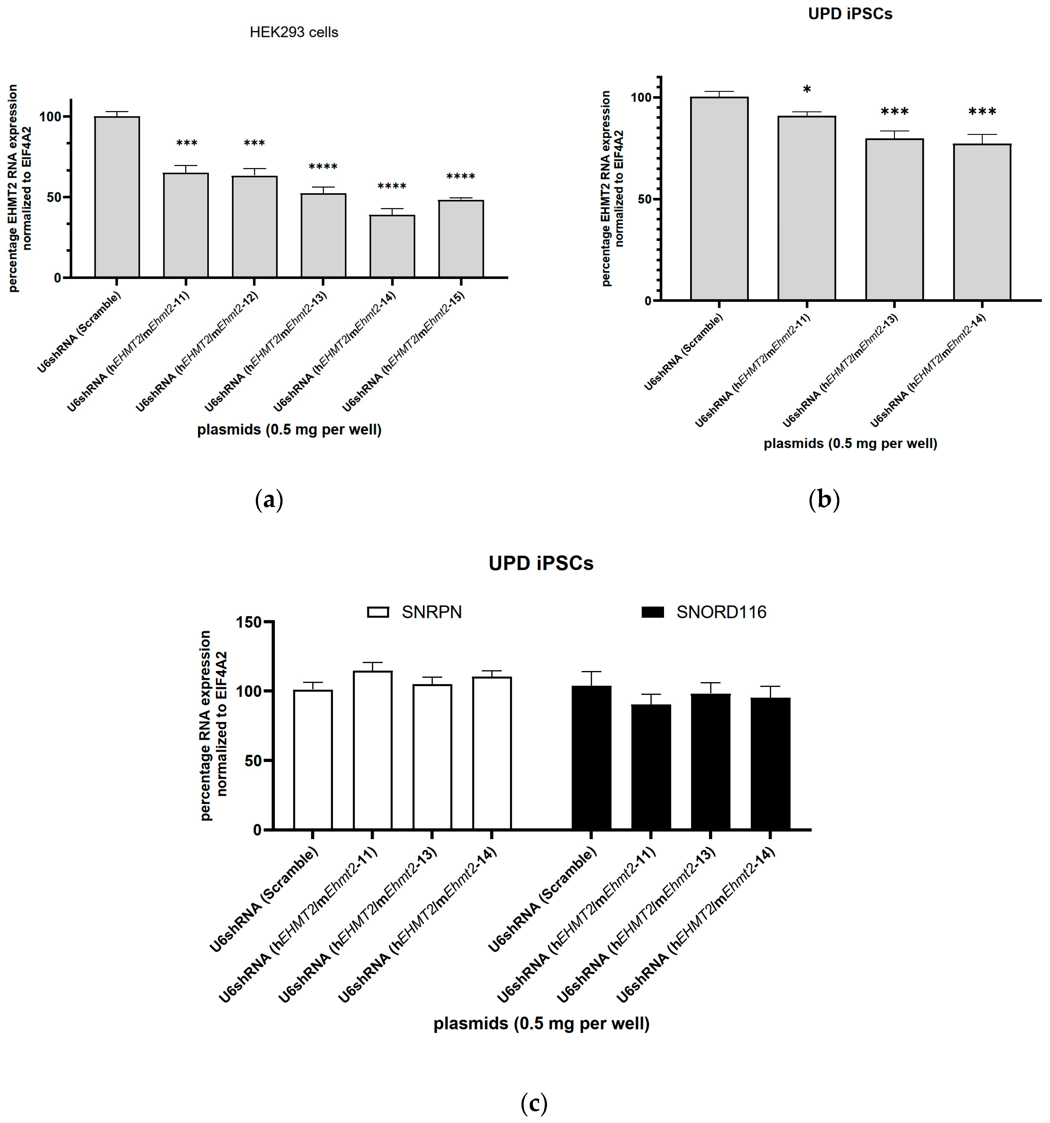
We designed four DNA plasmids with shRNA sequences targeting both human and mouse EHMT2 (named U6-shRNA(hEHMT2/mEhmt2-12), U6-shRNA(hEHMT2/mEhmt2-13), U6-shRNA(hEHMT2/mEhmt2-14), and U6-shRNA(hEHMT2/mEhmt2-15). To ensure the functionality of our plasmid constructs, we transfected into HEK293 cells the plasmid constructs encoding for shRNA targeting conserved mouse and human EHMT2 sequences and driven under the U6 promoter. The plasmid construct encoding for the shRNA sequence named scramble is used as a control. All the shRNA candidates significantly reduced the RNA levels of EHMT2 in HEK293 cells 72 h post transfection from 35% up to 60% (Figure 3a).

3.4. shRNA Knockdown of EHMT2 in PWS-iPSCs

The same shRNA plasmid constructs—shRNA(hEHMT2/mEhmt2-13) and U6-shRNA(hEHMT2/mEhmt2-14)—that were tested in HEK 293 cells were tested in PWS UPD iPSCs (Figure 3b,c). An additional plasmid construct containing sihEHMT2/mEhmt2-11 was added for comparison. The RNA levels for SNORD116 and SNRPN were measured 72 h post transfection. No statistically significant change in SNORD116 and SNRPN expression levels were measured compared to the control with shRNA Scramble (Figure 3c), despite a ~23% reduction in EHMT2 transcript levels with the U6-shRNA(hEHMT2/mEhmt2-14) plasmid construct (Figure 3b).

3.5. shRNA Knockdown of EHMT2 in PWS-iPSC-Derived Neurons 7 Days and 14 Days Post Transduction with Lentiviral Constructs

Non-integrative lentiviral constructs were designed encoding for the shRNA(hEHMT2/mEhmt2-11) and shRNA(hEHMT2/mEhmt2-14) targeting mouse Ehmt2 and human EHMT2 and were applied to mature iPSC-derived neurons from two different PWS patient cell lines (UPD and SD).
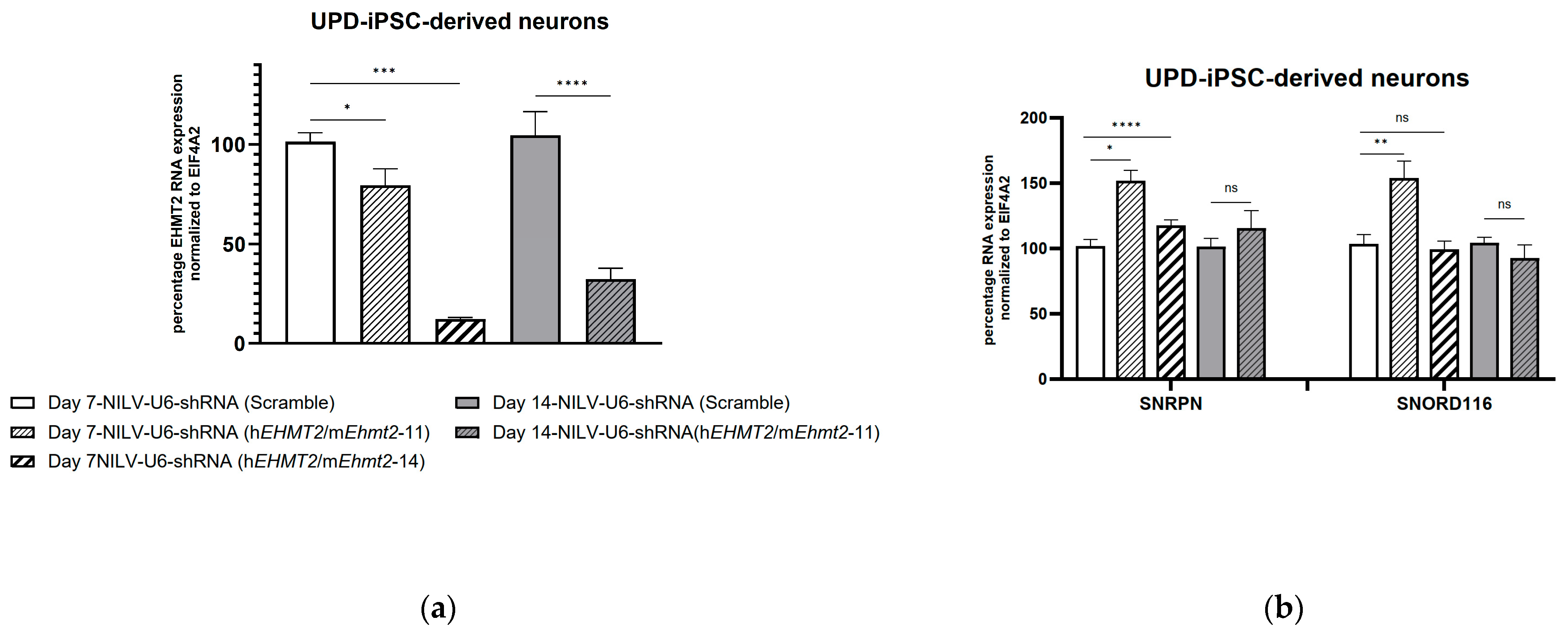
Seven days post transduction with NILV constructs, EHMT2 RNA levels were significantly downregulated in UPD-PWS iPSC neurons from 14% to 88% with NILV-U6- shRNA(hEHMT2/mEhmt2-11) and NILV-U6- shRNA(hEHMT2/mEhmt2-14), respectively (Figure 4a).
The levels of SNRPN and SNORD116 significantly increased by about 50% in the PWS UPD iPSC-derived neurons seven days post transduction with NILV-U6 shRNA(hEHMT2/mEhmt2-11) compared to the control (Figure 4b). A slight increase in RNA levels for SNRPN (about 17%) was observed in PWS UPD iPSC-derived neurons treated with NILV-U6 shRNA(hEHMT2/mEhmt2-14), whereas no increase in SNORD116 RNA expression level was measured (Figure 4b).
Fourteen days post transduction with NILV-U6, shRNA(hEHMT2/mEhmt2-14) induced toxicity in UPD-iPSC-derived neurons, and no data were collected for this time point. However, we were able to collect samples 14 days post transduction from UPD iPSC-derived neurons transduced with NILV-U6 shRNA(hEHMT2/mEhmt2-11). The results showed about 70% downregulation of EHMT2 RNA levels (Figure 4b), with no concomitant change in RNA levels for SNRPN and SNORD116 (Figure 4b).
In PWS SD-iPSC-derived neurons, EHMT2 RNA levels were significantly reduced seven days post transduction from 50% to 96% with NILV-U6 shRNA(hEHMT2/mEhmt2-11) and NILV-U6 shRNA(hEHMT2/mEhmt2-14), respectively (Figure 5a). To our surprise, we observed a significant increase in SNRPN and SNORD116 RNA levels from 37% to 47%, respectively (Figure 5b), compared to the control with NILV-U6 shRNA(hEHMT2/mEhmt2-11) in PWS SD-iPSC-derived neurons seven days post transduction. However, no significant increase in RNA levels for SNRPN and SNORD116 was measured with NILV-U6 shRNA(hEHMT2/mEhmt2-14), despite the highest decrease in EHMT2 RNA expression levels being observed.
The expression levels of MAGEL2 were significantly reduced by ~50% with NILV-U6 shRNA(hEHMT2/mEhmt2-14), whereas no significant changes were observed in MAGEL2 RNA levels in SD-iPSC-derived neurons seven days post transduction with NILV-U6 shRNA(hEHMT2/mEhmt2-11) (Figure 5b).

3.6. shRNA Knockdown of EHMT2 in PWS 2-9-iPSC-Derived Neurons 21 Days Post Transduction with Lentiviral Constructs

Twenty-one days post transduction with shRNA lentiviral constructs, EHMT2 RNA levels were significantly downregulated by about 10% to 60% with NILV-U6 shRNA(hEHMT2/mEhmt2-11) and NILV-U6 shRNA (hEHMT2/mEhmt2-14), respectively (Figure 5a). The levels of expression for SNRPN significantly increased by 20% with NILV-U6 shRNA(hEHMT2/mEhmt2-11) compared to the control, whereas no changes were observed with NILV-U6 shRNA (hEHMT2/mEhmt2-14) (Figure 5b). No significant differences in SNORD116 RNA levels were observed with NILV-U6 shRNA(hEHMT2/mEhmt2-11) compared to the control whereas about a 30% significant decrease in SNORD116 RNA levels was observed with NILV-U6 shRNA (hEHMT2/mEhmt2-14) compared to the control (Figure 5b). Twenty-one days post transduction with shRNA lentiviral constructs, the expression levels for MAGEL2 were significantly reduced to 20% and to 70% with NILV-U6 shRNA (hEHMT2/mEhmt2-11) and NILV-U6 shRNA (hEHMT2/mEhmt2-14), respectively (Figure 5b).

4. Discussion

We measured PWS gene transcripts such as Snord116/SNORD116, SNRPN, and MAGEL2 in PWS patient-derived iPSCs, PWS patient iPSC-derived neurons, and mouse primary neurons to assess maternal activation with anti-EHMT2 RNAi and therapeutic feasibility. We demonstrated that several siRNA oligonucleotides targeting Ehtm2/EHMT2 significantly decreased the transcript and protein expression levels of Ehmt2/EHMT2 in both mouse Snord116 deletion primary cortical neurons and human PWS iPSC lines. Several plasmid constructs encoding for shRNA, which targets EHMT2, also efficiently inhibited the expression levels of the EHMT2 protein in HEK293 cells. The lentiviral delivery of anti-EHMT2 shRNA produced a sustained reduction in Ehmt2/EHMT2 in paternally deleted Snord116 mouse primary neurons and PWS patient-derived iPSCs (up to 90% downregulation of ehmt2/EHMT2 transcript levels). In Snord116-deletion mice, we observed no increase in Snord116 transcript levels with Ehmt2 RNA interference. In UPD patient iPSC-derived neurons, time-dependent increases in SNRPN and SNORD116 transcript levels were observed; in SD patient iPSC-derived neurons, a statistically significant increase in SNRPN and a time-dependent increase in SNORD116 were observed; and MAGEL2 exhibited a sustained decrease in transcript levels. Taken together, our results show that inhibiting the expression of EHMT2 could partially activate the maternal expression of SNRPN and SNORD116 in PWS iPSCs.
Several studies have described the manipulation of the epigenetic regulation of PWS genes. Zinc finger protein ZNF274 inhibition induced histone modification in PWS patient iPSC-derived neurons and induced about a 20-fold increase in SNORD116 transcription levels relative to controls [36]. However, the increase was still approximately 1000-fold lower compared to PWS iPSC from healthy patients. Kim et al. showed that by inhibiting EHMT2 activity with small inhibitors such as UNC0638, activation of maternal expression PWS genes SNORD116 and SNRPN in fibroblasts from PWS patients (5–6 Mb deletion of the paternal copy of the 15q11-q13 region) approached healthy level controls [24]. Our approach was similar in targeting Ehmt2/EHMT2 but with a translationally relevant sustainable reduction.
In primary neurons from mice with Snord116 paternal deletion, the expression of downstream noncoding exons of Snurf/Snrpn are specifically expressed in neuronal cells [37]. We did not observe an induction of maternal Snord116 host gene expression levels in mouse primary neuronal cells from Snord116-deletion mice despite an siRNA-induced 70% inhibition of Ehmt2 mRNA expression. Kim et al. [24] have, however, identified small inhibitors of Ehmt2 capable of activating the maternal expression of Snrpn-EGFP in mouse embryonic fibroblasts carrying the Snrpn-EGFP fusion gene on the maternal chromosome. This might be explained by the fact that post-transcriptional modification resulting from reduced levels of Ehmt2 was not seen in our experimental procedure and cell type due to the limited exposure time of the anti-Ehmt2 siRNA.
To study a longer exposure in reducing EHMT2 in human neurons and its effects, we utilized an anti-EHMT2 shRNA as a lentiviral construct. We found a maternal activation of PWS maternal alleles for SNRPN and SNORD116 but no changes in MAGEL2 mRNA levels after one week of treatment with NILV EHMT2 in PWS UPD iPSC-derived neurons. The regulation of MAGEL2 expression is described as being regulated by the PWS-IC according to a mouse study investigating a paternally inherited deletion of the chromosome 7 PWS imprinting center [38]. However, the maternal expression of Magel2 was not described in the neurons of PWS mice treated with small inhibitors of EHMT2 [24]. Langouet et al. [36] described the maternal activation of MAGEL2 from PWS UPD patients from specifically knocking down ZNF274 [36,39]. Based on our work, the shRNA knockdown of EHMT2 is not enough to activate the maternal expression of MAGEL2 in UPD patient iPSC-derived neurons. A stable knockdown of zinc finger ZNF274 for 10 weeks in UPD iPSC-derived neurons restored SNORD116 and MAGEL2 to healthy levels [36,40]. Therefore, the data suggest that ZNF274, not EHMT2, is a regulatory element for MAGEL2.
The RNAi of EHMT2 resulted in the activation of maternally silenced SNRPN and SNORD116 transcripts in specific lines of PWS patient iPSC-derived neurons. We were unsuccessful in maintaining PWS UPD mature neurons beyond one week of treatment with anti-EHMT2 shRNA lentiviral exposure. We were able to maintain mature PWS SD neurons in culture for 3 weeks of treatment following anti-EHMT2 shRNA lentiviral exposure. Although the maternal activation of SNRPN was observed after 3 weeks of culture, a similar effect was not observed for SNORD116. This could be explained by the fact that the inhibitory effect of anti-EHMT2 shRNA via lentivirus was less pronounced 3 weeks later and that other mechanisms such as ZNF274 act independently of the PWS-IC in maintaining SNORD116 silencing on the maternal allele [16,36]. Paradoxically, 3 weeks after the incubation of anti-EHMT2 lentivirus in PWS SD mature neurons, we found that MAGEL2 transcript levels decreased in PWS SD iPSC-derived neurons. One potential explanation may be a reliance on mechanisms independent of SNORD116 regulation.
As mentioned above, EHMT2 does not act alone in regulating H3K9 methylation in neurons. Other methyltransferases including SETDB1 and SUV39H1 are shown to modulate the dimethylation of H3K9 [41,42,43]. Moreover, a paralog of EHMT2 called EHMT1 is also important for similar biological phenomena ascribed to EHMT2, such as modulating H3K9 methylation [44,45]. A recent publication described the role of both EHMT2 and EHMT1 knockdown as necessary to efficiently reduce H3K9me2 and exacerbate TNFα-induced lipolysis in mature adipocytes [46]. We recommend that future studies explore this hypothesis by knocking down several methyltransferases, such as SETDB1 and EHMT1.
Currently, the levels of PWS gene expression needed to exert a therapeutic effect on PWS patients are unknown. Unpublished data from our laboratory and others demonstrated approximately a 5000-fold increase in PWS genes in healthy versus PWS patient iPSC-derived neurons. Furthermore, exogenous alterations in the epigenetic regulation of genes in post-mitotic cells are slow compared to similar interrogations in pre-mitotic cells. As such, care must be taken in representing the data as evidence of a translationally relevant therapy. An additional consideration in the transition to human therapy with RNAi is the probability of off-target effects. As described, the siRNAs/shRNAs developed herein were designed with computer algorithms to estimate the probability of off-target binding. However, a detained transcriptomic analysis would be a requisite for safety testing. Further considerations include the burden of treatment through costs, the frequency of treatment, and the translational relevance of the route of administration.
Despite these considerations, recent clinical trials of epigenetic, “stop-the-stop” therapies with antisense oligonucleotides for the treatment of Angelman Syndrome (NCT04259281, NCT05127226), a neurodevelopmental disorder with genetic underpinnings on the same loci as PWS genes, can be considered as a positive advancement towards a similar therapy for PWS. While we would argue that the data presented here argue against singular RNAi targeting of EHMT2 as therapeutically relevant for PWS, our data confirm a partial role of EHMT2 in the maternal regulation of PWS genes. Furthermore, a combinatorial approach targeting multiple epigenetic regulators through translationally relevant approaches, while reducing the burden on the patient to a single dose, remains a viable consideration for future therapeutic studies.

5. Conclusions

Our study showed that reducing EHMT2 expression with either siRNA or shRNA resulted in modest increases in some PWS patient cell lines. However, these increases do not approach healthy control levels, which limits the potential therapeutic effect on PWS of such an approach.

6. Patents

A provisional patent has been filed for the anti-EHMT2 siRNAs/shRNAs and their use (with RKB, HRK, and SJG as co-inventors).

Author Contributions

Conceptualization, R.K.B. and S.J.G.; methodology, V.Z., H.R.K., V.R. and J.M.Z.; formal analysis, V.Z.; investigation, V.Z.; resources, J.M.Z., S.J.G. and R.K.B.; data curation, V.Z., H.R.K. and V.R.; writing—original draft preparation, V.Z.; writing—review and editing, R.K.B., S.J.G. and J.M.Z.; supervision, R.K.B.; project administration, R.K.B.; funding acquisition, R.K.B. All authors have read and agreed to the published version of the manuscript.

Funding

This research was funded by a Sponsored Research Agreement by Taysha Gene Therapies, Inc. and a grant from the Foundation for Prader–Willi Research.

Institutional Review Board Statement

The animal study protocol was approved by the Institutional Animal Care and Use Committee of UT Southwestern Medical Center (2018-102619-G approved on 6 May 2024).

Informed Consent Statement

Not applicable.

Data Availability Statement

The data presented in this study are available on request from the corresponding author due to an ongoing patent application.

Acknowledgments

We acknowledge technical support from Aymun Rahim and Samantha DeVries.

Conflicts of Interest

The authors have received past royalty income related to the license of intellectual properties to Taysha Gene Therapies. SJG has received consulting income from Taysha Gene Therapies unrelated to this study. The funders had no role in the design of the study; in the collection, analyses, or interpretation of data; in the writing of the manuscript; or in the decision to publish the results.

References

  1. Butler, M.G.; Miller, J.L.; Forster, J.L. Prader-Willi Syndrome—Clinical Genetics, Diagnosis and Treatment Approaches: An Update. Curr. Pediatr. Rev. 2019, 15, 207–244. [Google Scholar] [CrossRef] [PubMed]
  2. Cassidy, S.B.; Schwartz, S.; Miller, J.L.; Driscoll, D.J. Prader-Willi syndrome. Genet. Med. 2012, 14, 10–26. [Google Scholar] [CrossRef] [PubMed]
  3. Godler, D.E.; Butler, M.G. Special Issue: Genetics of Prader-Willi Syndrome. Genes 2021, 12, 1429. [Google Scholar] [CrossRef] [PubMed]
  4. Butler, M.G.; Hartin, S.N.; Hossain, W.A.; Manzardo, A.M.; Kimonis, V.; Dykens, E.; Gold, J.A.; Kim, S.J.; Weisensel, N.; Tamura, R.; et al. Molecular genetic classification in Prader-Willi syndrome: A multisite cohort study. J. Med. Genet. 2019, 56, 149–153. [Google Scholar] [CrossRef]
  5. Cassidy, S.B.; Dykens, E.; Williams, C.A. Prader-Willi and Angelman syndromes: Sister imprinted disorders. Am. J. Med. Genet. 2000, 97, 136–146. [Google Scholar] [CrossRef]
  6. Buiting, K.; Dittrich, B.; Robinson, W.P.; Guitart, M.; Abeliovich, D.; Lerer, I.; Horsthemke, B. Detection of aberrant DNA methylation in unique Prader-Willi syndrome patients and its diagnostic implications. Hum. Mol. Genet. 1994, 3, 893–895. [Google Scholar] [CrossRef]
  7. de Smith, A.J.; Purmann, C.; Walters, R.G.; Ellis, R.J.; Holder, S.E.; Van Haelst, M.M.; Brady, A.F.; Fairbrother, U.L.; Dattani, M.; Keogh, J.M.; et al. A deletion of the HBII-85 class of small nucleolar RNAs (snoRNAs) is associated with hyperphagia, obesity and hypogonadism. Hum. Mol. Genet. 2009, 18, 3257–3265. [Google Scholar] [CrossRef]
  8. Sahoo, T.; del Gaudio, D.; German, J.R.; Shinawi, M.; Peters, S.U.; Person, R.E.; Garnica, A.; Cheung, S.W.; Beaudet, A.L. Prader-Willi phenotype caused by paternal deficiency for the HBII-85 C/D box small nucleolar RNA cluster. Nat. Genet. 2008, 40, 719–721. [Google Scholar] [CrossRef]
  9. Schulze, A.; Hansen, C.; Skakkebaek, N.E.; Brondum-Nielsen, K.; Ledbeter, D.H.; Tommerup, N. Exclusion of SNRPN as a major determinant of Prader-Willi syndrome by a translocation breakpoint. Nat. Genet. 1996, 12, 452–454. [Google Scholar] [CrossRef]
  10. Duker, A.L.; Ballif, B.C.; Bawle, E.V.; Person, R.E.; Mahadevan, S.; Alliman, S.; Thompson, R.; Traylor, R.; Bejjani, B.A.; Shaffer, L.G.; et al. Paternally inherited microdeletion at 15q11.2 confirms a significant role for the SNORD116 C/D box snoRNA cluster in Prader-Willi syndrome. Eur. J. Hum. Genet. 2010, 18, 1196–1201. [Google Scholar] [CrossRef]
  11. Grootjen, L.N.; Juriaans, A.F.; Kerkhof, G.F.; Hokken-Koelega, A.C.S. Atypical 15q11.2-q13 Deletions and the Prader-Willi Phenotype. J. Clin. Med. 2022, 11, 4636. [Google Scholar] [CrossRef] [PubMed]
  12. Cao, Y.; AlHumaidi, S.S.; Faqeih, E.A.; Pitel, B.A.; Lundquist, P.; Aypar, U. A novel deletion of SNURF/SNRPN exon 1 in a patient with Prader-Willi-like phenotype. Eur. J. Med. Genet. 2017, 60, 416–420. [Google Scholar] [CrossRef] [PubMed]
  13. Crenshaw, M.M.; Graw, S.L.; Slavov, D.; Boyle, T.A.; Pique, D.G.; Taylor, M.; Baker, P., II. An Atypical 15q11.2 Microdeletion Not Involving SNORD116 Resulting in Prader-Willi Syndrome. Case Rep. Genet. 2023, 2023, 4225092. [Google Scholar] [CrossRef]
  14. Saitoh, S.; Wada, T. Parent-of-origin specific histone acetylation and reactivation of a key imprinted gene locus in Prader-Willi syndrome. Am. J. Hum. Genet. 2000, 66, 1958–1962. [Google Scholar] [CrossRef]
  15. Fulmer-Smentek, S.B.; Francke, U. Association of acetylated histones with paternally expressed genes in the Prader-Willi deletion region. Hum. Mol. Genet. 2001, 10, 645–652. [Google Scholar] [CrossRef]
  16. Cruvinel, E.; Budinetz, T.; Germain, N.; Chamberlain, S.; Lalande, M.; Martins-Taylor, K. Reactivation of maternal SNORD116 cluster via SETDB1 knockdown in Prader-Willi syndrome iPSCs. Hum. Mol. Genet. 2014, 23, 4674–4685. [Google Scholar] [CrossRef] [PubMed]
  17. Recillas-Targa, F. DNA methylation, chromatin boundaries, and mechanisms of genomic imprinting. Arch. Med. Res. 2002, 33, 428–438. [Google Scholar] [CrossRef]
  18. Kacem, S.; Feil, R. Chromatin mechanisms in genomic imprinting. Mamm. Genome 2009, 20, 544–556. [Google Scholar] [CrossRef] [PubMed]
  19. Jenuwein, T. The epigenetic magic of histone lysine methylation. FEBS J. 2006, 273, 3121–3135. [Google Scholar] [CrossRef]
  20. Zhang, R.H.; Judson, R.N.; Liu, D.Y.; Kast, J.; Rossi, F.M. The lysine methyltransferase Ehmt2/G9a is dispensable for skeletal muscle development and regeneration. Skelet. Muscle 2016, 6, 22. [Google Scholar] [CrossRef]
  21. Liang, L.; Gu, X.; Zhao, J.Y.; Wu, S.; Miao, X.; Xiao, J.; Mo, K.; Zhang, J.; Lutz, B.M.; Bekker, A.; et al. G9a participates in nerve injury-induced Kcna2 downregulation in primary sensory neurons. Sci. Rep. 2016, 6, 37704. [Google Scholar] [CrossRef] [PubMed]
  22. Pan, Z.; Du, S.; Wang, K.; Guo, X.; Mao, Q.; Feng, X.; Huang, L.; Wu, S.; Hou, B.; Chang, Y.J.; et al. Downregulation of a Dorsal Root Ganglion-Specifically Enriched Long Noncoding RNA is Required for Neuropathic Pain by Negatively Regulating RALY-Triggered Ehmt2 Expression. Adv. Sci. 2021, 8, e2004515. [Google Scholar] [CrossRef] [PubMed]
  23. Wang, D.Y.; Kosowan, J.; Samsom, J.; Leung, L.; Zhang, K.L.; Li, Y.X.; Xiong, Y.; Jin, J.; Petronis, A.; Oh, G.; et al. Inhibition of the G9a/GLP histone methyltransferase complex modulates anxiety-related behavior in mice. Acta Pharmacol. Sin. 2018, 39, 866–874. [Google Scholar] [CrossRef] [PubMed]
  24. Kim, Y.; Lee, H.M.; Xiong, Y.; Sciaky, N.; Hulbert, S.W.; Cao, X.; Everitt, J.I.; Jin, J.; Roth, B.L.; Jiang, Y.H. Targeting the histone methyltransferase G9a activates imprinted genes and improves survival of a mouse model of Prader-Willi syndrome. Nat. Med. 2017, 23, 213–222. [Google Scholar] [CrossRef]
  25. Bernstein, E.; Caudy, A.A.; Hammond, S.M.; Hannon, G.J. Role for a bidentate ribonuclease in the initiation step of RNA interference. Nature 2001, 409, 363–366. [Google Scholar] [CrossRef]
  26. Rand, T.A.; Ginalski, K.; Grishin, N.V.; Wang, X. Biochemical identification of Argonaute 2 as the sole protein required for RNA-induced silencing complex activity. Proc. Natl. Acad. Sci. USA 2004, 101, 14385–14389. [Google Scholar] [CrossRef] [PubMed]
  27. Wilson, R.C.; Doudna, J.A. Molecular mechanisms of RNA interference. Annu. Rev. Biophys. 2013, 42, 217–239. [Google Scholar] [CrossRef]
  28. Corydon, I.J.; Fabian-Jessing, B.K.; Jakobsen, T.S.; Jorgensen, A.C.; Jensen, E.G.; Askou, A.L.; Aagaard, L.; Corydon, T.J. 25 years of maturation: A systematic review of RNAi in the clinic. Mol. Ther. Nucleic Acids 2023, 33, 469–482. [Google Scholar] [CrossRef]
  29. Ding, F.; Prints, Y.; Dhar, M.S.; Johnson, D.K.; Garnacho-Montero, C.; Nicholls, R.D.; Francke, U. Lack of Pwcr1/MBII-85 snoRNA is critical for neonatal lethality in Prader-Willi syndrome mouse models. Mamm. Genome 2005, 16, 424–431. [Google Scholar] [CrossRef]
  30. Osborne-Lawrence, S.; Lawrence, C.; Metzger, N.P.; Klavon, J.; Baig, H.R.; Richard, C.; Varshney, S.; Gupta, D.; Singh, O.; Ogden, S.B.; et al. Effects of thermoneutrality on food intake, body weight, and body composition in a Prader-Willi syndrome mouse model. Obesity 2023, 31, 1644–1654. [Google Scholar] [CrossRef]
  31. Martins-Taylor, K.; Hsiao, J.S.; Chen, P.F.; Glatt-Deeley, H.; De Smith, A.J.; Blakemore, A.I.; Lalande, M.; Chamberlain, S.J. Imprinted expression of UBE3A in non-neuronal cells from a Prader-Willi syndrome patient with an atypical deletion. Hum. Mol. Genet. 2014, 23, 2364–2373. [Google Scholar] [CrossRef] [PubMed]
  32. Chambers, S.M.; Fasano, C.A.; Papapetrou, E.P.; Tomishima, M.; Sadelain, M.; Studer, L. Highly efficient neural conversion of human ES and iPS cells by dual inhibition of SMAD signaling. Nat. Biotechnol. 2009, 27, 275–280. [Google Scholar] [CrossRef] [PubMed]
  33. Beaudoin, G.M., III; Lee, S.H.; Singh, D.; Yuan, Y.; Ng, Y.G.; Reichardt, L.F.; Arikkath, J. Culturing pyramidal neurons from the early postnatal mouse hippocampus and cortex. Nat. Protoc. 2012, 7, 1741–1754. [Google Scholar] [CrossRef] [PubMed]
  34. Moutin, E.; Hemonnot, A.L.; Seube, V.; Linck, N.; Rassendren, F.; Perroy, J.; Compan, V. Procedures for Culturing and Genetically Manipulating Murine Hippocampal Postnatal Neurons. Front. Synaptic Neurosci. 2020, 12, 19. [Google Scholar] [CrossRef]
  35. Boudreau, R.L.; Spengler, R.M.; Hylock, R.H.; Kusenda, B.J.; Davis, H.A.; Eichmann, D.A.; Davidson, B.L. siSPOTR: A tool for designing highly specific and potent siRNAs for human and mouse. Nucleic Acids Res. 2013, 41, e9. [Google Scholar] [CrossRef]
  36. Langouet, M.; Gorka, D.; Orniacki, C.; Dupont-Thibert, C.M.; Chung, M.S.; Glatt-Deeley, H.R.; Germain, N.; Crandall, L.J.; Cotney, J.L.; Stoddard, C.E.; et al. Specific ZNF274 binding interference at SNORD116 activates the maternal transcripts in Prader-Willi syndrome neurons. Hum. Mol. Genet. 2020, 29, 3285–3295. [Google Scholar] [CrossRef]
  37. Cavaille, J.; Buiting, K.; Kiefmann, M.; Lalande, M.; Brannan, C.I.; Horsthemke, B.; Bachellerie, J.P.; Brosius, J.; Huttenhofer, A. Identification of brain-specific and imprinted small nucleolar RNA genes exhibiting an unusual genomic organization. Proc. Natl. Acad. Sci. USA 2000, 97, 14311–14316. [Google Scholar] [CrossRef]
  38. Yang, T.; Adamson, T.E.; Resnick, J.L.; Leff, S.; Wevrick, R.; Francke, U.; Jenkins, N.A.; Copeland, N.G.; Brannan, C.I. A mouse model for Prader-Willi syndrome imprinting-centre mutations. Nat. Genet. 1998, 19, 25–31. [Google Scholar] [CrossRef]
  39. Langouet, M.; Glatt-Deeley, H.R.; Chung, M.S.; Dupont-Thibert, C.M.; Mathieux, E.; Banda, E.C.; Stoddard, C.E.; Crandall, L.; Lalande, M. Zinc finger protein 274 regulates imprinted expression of transcripts in Prader-Willi syndrome neurons. Hum. Mol. Genet. 2018, 27, 505–515. [Google Scholar] [CrossRef]
  40. Wilson, C.; Giono, L.E.; Rozes-Salvador, V.; Fiszbein, A.; Kornblihtt, A.R.; Caceres, A. The Histone Methyltransferase G9a Controls Axon Growth by Targeting the RhoA Signaling Pathway. Cell Rep. 2020, 31, 107639. [Google Scholar] [CrossRef]
  41. Barral, A.; Pozo, G.; Ducrot, L.; Papadopoulos, G.L.; Sauzet, S.; Oldfield, A.J.; Cavalli, G.; Dejardin, J. SETDB1/NSD-dependent H3K9me3/H3K36me3 dual heterochromatin maintains gene expression profiles by bookmarking poised enhancers. Mol. Cell 2022, 82, 816–832.e12. [Google Scholar] [CrossRef] [PubMed]
  42. Tam, P.L.F.; Cheung, M.F.; Chan, L.Y.; Leung, D. Cell-type differential targeting of SETDB1 prevents aberrant CTCF binding, chromatin looping, and cis-regulatory interactions. Nat. Commun. 2024, 15, 15. [Google Scholar] [CrossRef] [PubMed]
  43. Weirich, S.; Khella, M.S.; Jeltsch, A. Structure, Activity and Function of the Suv39h1 and Suv39h2 Protein Lysine Methyltransferases. Life 2021, 11, 703. [Google Scholar] [CrossRef] [PubMed]
  44. Collins, R.E.; Northrop, J.P.; Horton, J.R.; Lee, D.Y.; Zhang, X.; Stallcup, M.R.; Cheng, X. The ankyrin repeats of G9a and GLP histone methyltransferases are mono- and dimethyllysine binding modules. Nat. Struct. Mol. Biol. 2008, 15, 245–250. [Google Scholar] [CrossRef]
  45. Tachibana, M.; Sugimoto, K.; Fukushima, T.; Shinkai, Y. Set domain-containing protein, G9a, is a novel lysine-preferring mammalian histone methyltransferase with hyperactivity and specific selectivity to lysines 9 and 27 of histone H3. J. Biol. Chem. 2001, 276, 25309–25317. [Google Scholar] [CrossRef]
  46. Able, A.A.; Richard, A.J.; Stephens, J.M. TNFalpha Effects on Adipocytes Are Influenced by the Presence of Lysine Methyltransferases, G9a (EHMT2) and GLP (EHMT1). Biology 2023, 12, 674. [Google Scholar] [CrossRef]
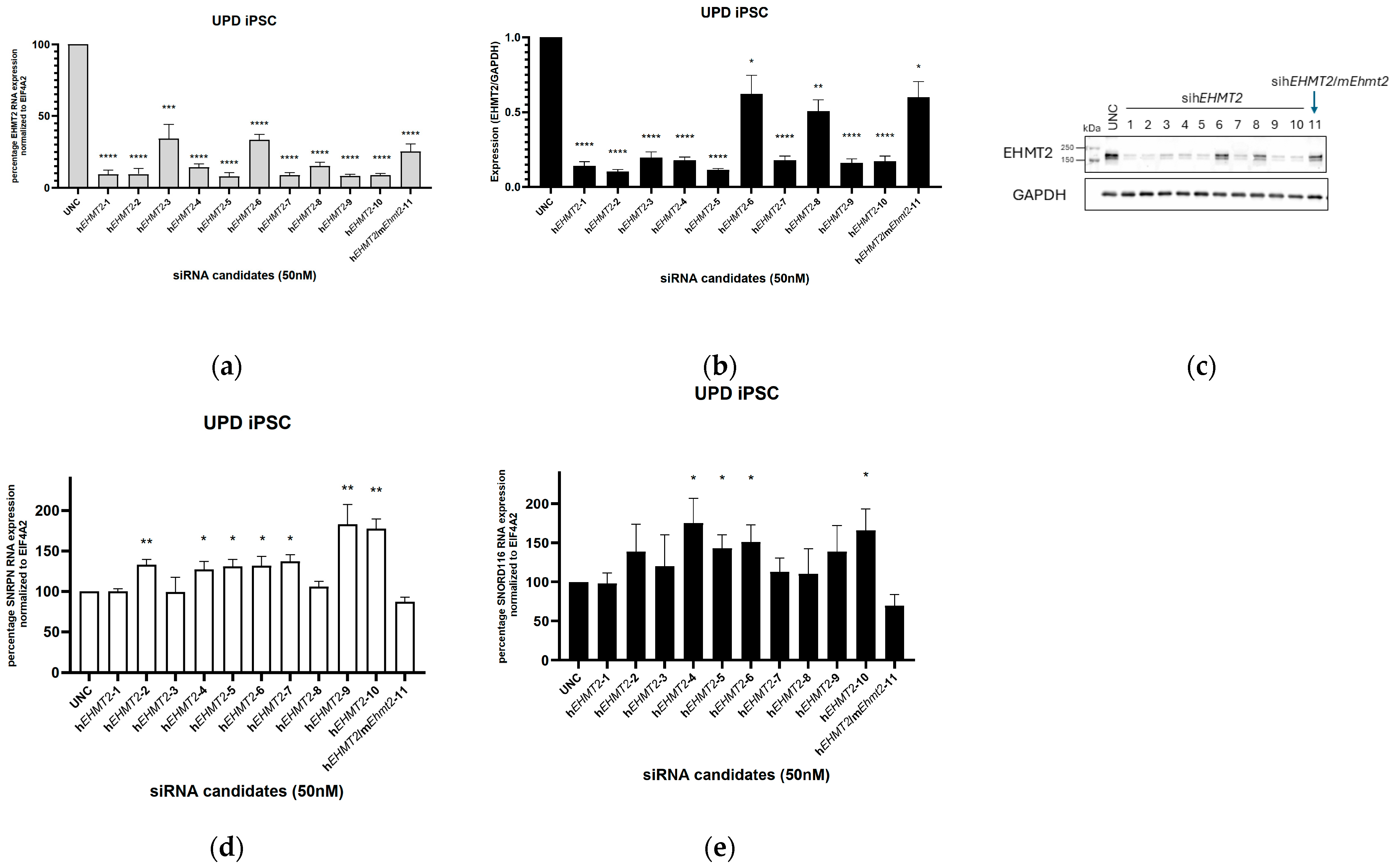
Figure 1. Efficacy of EHMT2 transcript reduction with specific siRNAs in human UPD patient-derived iPSCs. (a). Gene expression of EHMT2 in PWS UPD iPSCs following transfection with control siRNAs (UNC) or specific siRNAs targeting EHMT2. A significant reduction in EHMT2 was observed with all the siRNA candidates relative to the control; (b) quantification of the EHMT2 protein in UPD iPSCs treated with siRNA targeting EHMT2 and compared to treated cells with the control siRNA UNC; (c) representative image of EHMT2 protein from UPD iPSCs treated with siRNA candidates analyzed by Western blotting; (d) gene expression of SNRPN and (e) gene expression of SNORD116 in PWS UPD iPSCs following transfection with control siRNAs (UNC) or specific siRNAs targeting EHMT2. EIF4A2 was used as a control. Student’s t test: *, p < 0.05; **, p < 0.01; ***, p < 0.001; ****, p < 0.0001; data represent means ± S.E.M (N = 3–4).
Genes 15 01366 g001
Figure 2. Efficacy of Ehmt2 transcript reduction with specific siRNAs in mouse primary neurons. (a) Gene expression of Ehmt2 in primary neurons from Snord116p−/m+ mice following transfection with control siRNAs (UNC) or specific siRNAs targeting Ehmt2. A significant reduction in Ehmt2 was observed with all the siRNA candidates relative to the control; (b) gene expression of Snord116HG following transfection with control siRNAs (UNC) or specific siRNAs targeting Ehmt2. Eif4a2 was used as a control. Student’s t test: ****, p < 0.0001; data represent means ± S.E.M from four biological replicates.
Genes 15 01366 g002
Figure 3. Expression levels of EHMT2, SNRPN, and SNORD116 after transfection with U6 anti-EHMT2 shRNA DNA plasmids: (a) HEK293 cells were transfected with a plasmid construct control (shRNA Scramble) and shRNA specifically targeting EHMT2. A significant reduction in EHMT2 expression was observed with all the shRNA plasmid candidates relative to the control; (b) PWS UPD iPSC was transfected with the plasmid control and shRNA targeting EHMT2. A moderate but significant reduction in EHMT2 expression was observed 72 h post transfection; (c) SNRPN and SNORD116 expression levels in PWS UPD iPSC remained at the same level as for the control after transfection with U6shRNA plasmids targeting EHTM2. Student’s t test: *, p < 0.05; ***, p < 0.001; ****, p < 0.0001; data represent the means ± S.E.M from three biological replicates.
Genes 15 01366 g003
Figure 4. Expression levels of EHMT2, SNRPN and SNORD116 in UPD iPSC-derived neurons transduced with lentivirus containing anti-EHMT2 shRNA or control (Scramble): (a) NILV-U6shRNA(hEHMT2/mEhmt2-14) significantly reduced EHMT2 RNA levels as opposed to a mild reduction with NILV-U6shRNA (hEHMT2/mEhmt2-11) 7 days post transduction; EHMT2 expression is further reduced 14 days post transduction with NILV-U6shRNA (hEHMT2/mEhmt2-11). (b) NILV-U6shRNA (hEHMT2/mEhmt2-11) significantly increased SNRPN and SNORD116 transcript levels 7 days post transduction, as opposed to no change 14 days post transduction. There is a significant increase in SNRPN transcripts levels without any change in SNORD116 transcripts levels 7 days post transduction with NILV-U6shRNA (hEHMT2/mEhmt2-14). Student’s t test: *, p < 0.05; **, p < 0.01; ***, p < 0.001; ****, p < 0.0001; ns, p > 0.05; data represent the means ± S.E.M of four biological replicates.
Genes 15 01366 g004
Figure 5. Expression levels of EHMT2, MAGEL2, SNRPN, and SNORD116 in SD-iPSC-derived neurons transduced with lentivirus with shRNA targeting EHMT2 and control (Scramble): (a) NILV-U6shRNA(hEHMT2/mEhmt2-14) and NILV-U6shRNA (hEHMT2/mEhmt2-11) significantly reduced EHMT2 RNA levels 7 days post transduction as opposed to a milder reduction 21 days post transduction; (b) NILV-U6shRNA (hEHMT2/mEhmt2-11) significantly increased SNRPN transcript levels 7 days and 21 days post transduction as opposed to no change in SNRPN transcript levels 7 days and 21 days post transduction with NILV-U6shRNA(hEHMT2/mEhmt2-14). A significant increase in SNORD116 transcripts levels is shown at day 7 post transduction with NILV-U6shRNA (hEHMT2/mEhmt2-11). Student’s t test; *, p < 0.05; **, p < 0.01; ****, p < 0.0001; ns, p > 0.05; data represent the means ± S.E.M from four biological replicates.
Genes 15 01366 g005
Table 1. Prader-Willi Syndrome iPSC lines.
iPSC LinesDescriptionReferenceAbbreviation
PWS UPD 1–2PWS uniparental disomy[31]UPD iPSC
PWS 2–9PWS small deletion [31]SD iPSC
Table 2. List of shRNA sequences used in this study. hsa, Homo sapiens; mmu, Mus musculus.
shRNA NameSequence (5′-3′)—SenseSequence (5′-3′)—AntisenseSpecies Target EHMT2
hEHMT2/mEhmt2-11TAAATGTTGGGTTTGGTAATATATTACCAAACCCAACATTTAhsa and mmu
hEHMT2/mEhmt2-12GGCGCAAGGCCAAGAAGAAATATTTCTTCTTGGCCTTGCGCChsa and mmu
hEHMT2/mEhmt2-13GCGCAAGGCCAAGAAGAAATGCATTTCTTCTTGGCCTTGCGChsa and mmu
hEHMT2/mEhmt2-14TGATGTGAGAGAGGATGATTCGAATCATCCTCTCTCACATCAhsa and mmu
hEHMT2/mEhmt2-15GTGAGAGAGGATGATTCTTACGTAAGAATCATCCTCTCTCAChsa and mmu
Table 3. hsa, Homo sapiens; mmu, Mus musculus; R, reverse; F, forward.
Primer NameSequenceSpeciesSybR or with Probe
EIF4A2 FCAACGTGCATTGTGCTTCTThsaSybR
EIF4A2 RACGACTAACGTCGCTTTGCThsaSybR
SNORD116 FGGATCGATGATGAGTCCCChsaSybR
SNORD116 RTCCGATGAGAACGACGGTAThsaSybR
SNRPN FTTGCTGCGACTGCCAGTATThsaSybR
SNRPN RGCCCATGGGTGGTCTCATAChsaSybR
MAGEL2 FATCTGGAAGCCCAAGAGGAChsaSybR
MAGEL2 RACCTGGATAGGGCTTTGGAChsaSybR
EHMT2 FATGCAGTGGACAAACAGCAGhsaSybR
EHMT2 RACCGTCCTCCTCCTTGCTAThsaSybR
Snord116 HG FTGTTGCTGACTTGCCCTAGmmuprobe
Snord116 HG RGTTCGATGGAGACTCAGTTGGmmuprobe
mSnord116 (probe)AAACATGCAGAGGAAATGGCCCCmmuprobe
Eif4Mm01730183_gH/4448484mmuprobe
Ehmt2 FTGCGTACTCTGTGGATGAGCmmuSybR
Ehmt2 RTCTGACTGATTGCCCGACTCmmuSybR
Disclaimer/Publisher’s Note: The statements, opinions and data contained in all publications are solely those of the individual author(s) and contributor(s) and not of MDPI and/or the editor(s). MDPI and/or the editor(s) disclaim responsibility for any injury to people or property resulting from any ideas, methods, instructions or products referred to in the content.