Article

Escherichia albertii as a Potential Enteropathogen in the Light of Epidemiological and Genomic Studies

by
Katarzyna Leszczyńska
1,*,
Izabela Święcicka
2,
Tamara Daniluk
1,
Dariusz Lebensztejn
3,
Sylwia Chmielewska-Deptuła
1,
Dorota Leszczyńska
1,
Jan Gawor
4 and
Małgorzata Kliber
1
1
Department of Medical Microbiology and Nanobiomedical Enginnering, Medical University of Bialystok, ul. Mickiewicza 2C, 15-222 Bialystok, Poland
2
Department of Microbiology and Biotechnology, University of Bialystok, ul. Ciołkowskiego 1J, 15-245 Białystok, Poland
3
Department of Pediatrics, Gastroenterology, Hepatology, Nutrition, Allergology and Pulmonology, Medical University of Bialystok, ul. Waszyngtona 17, 15-274 Bialystok, Poland
4
DNA Sequecing and Synthesis Facility, Institute of Biochemistry and Biophysics, Polish Academy of Sciences, ul. Pawińskiego 5A, 02-106 Warszawa, Poland
*
Author to whom correspondence should be addressed.
Genes 2023, 14(7), 1384; https://doi.org/10.3390/genes14071384
Submission received: 25 April 2023 / Revised: 9 June 2023 / Accepted: 23 June 2023 / Published: 30 June 2023
(This article belongs to the Section Microbial Genetics and Genomics)

Abstract

:
Escherichia albertii is a new enteropathogen of humans and animals. The aim of the study was to assess the prevalence and pathogenicity of E. albertii strains isolated in northeastern Poland using epidemiological and genomic studies. In 2015–2018, a total of 1154 fecal samples from children and adults, 497 bird droppings, 212 food samples, 92 water samples, and 500 lactose-negative E. coli strains were tested. A total of 42 E. albertii strains were isolated. The PCR method was suitable for their rapid identification. In total, 33.3% of E. albertii isolates were resistant to one antibiotic, and 16.7% to two. Isolates were sensitive to cefepime, imipenem, levofloxacin, gentamicin, trimethoprim/sulfamethoxazole, and did not produce ESBL β-lactamases. High genetic variability of E. albertii has been demonstrated. In the PFGE method, 90.5% of the strains had distinct pulsotypes. In MLST typing, 85.7% of strains were assigned distinct sequence types (STs), of which 64% were novel ST types. Cytolethal distending toxin (CDT) and Paa toxin genes were found in 100% of E. albertii isolates. Genes encoding toxins, IbeA, CdtB type 2, Tsh and Shiga (Stx2f), were found in 26.2%, 9.7%, 1.7%, and 0.4% of E. albertii isolates, respectively. The chromosome size of the tested strains ranged from 4,573,338 to 5,141,010 bp (average 4,784,003 bp), and at least one plasmid was present in all strains. The study contributes to a more accurate assessment of the genetic diversity of E. albertii and the potential threat it poses to public health.

1. Introduction

Escherichia albertii (E. albertii) is the second—after Escherichia coli (E. coli)—pathogenic species in the Escherichia genus, and is regarded as an emerging human and animal enteropathogen [1]. E. albertii, similarly to enteropathogenic E. coli (EPEC) and enterohemorrhagic E. coli (EHEC) strains, possesses a specific intestinal cell invasion mechanism known as A/E lesions (attaching and effacing lesions), characterized by intimate attachment of bacteria to the residual apical enterocyte membrane and localized destruction of brush border microvilli [2,3]. To date, only a limited number of publications are available about this bacterium due to late recognition of E. albertii as a new species (in 2003) and lack of standard identification methods [4]. Although this bacterium was isolated in the early 1990s and recognized as the cause of diarrhea in five Bangladeshi children, originally it was misidentified as Hafnia alvei [5]. However, further analyses of the isolates reveled substantial phenotypic and genomic differences in comparison to the reference H. alvei strains, and their relatedness to Escherichia and Shigella, particularly Shigella boydii B13. Moreover, the presence of the pathogenicity island LEE (locus of enterocyte effacement) carrying the eae gene that encodes intimin, a major virulence factor of EPEC/EHEC strains, was also noted in E. albertii. Further research gave rise to the creation of a new species in the Escherichia genus [6]. Nevertheless, E. albertii seems to be similar to pathogenic E. coli strains, not only in terms of virulence but also reservoirs and ways of transmission, where animals—particularly birds and contaminated food—possibly play an essential role [7,8]. Cytolethal distending toxin (CDT) and Shiga toxin (Stx) are the main types of toxins that have been reported in E. albertii strains. CDTs are genotoxins that induce DNA double-strand breaks in both proliferating and nonproliferating cells and are produced by several Gram-negative bacteria, such as E. coli and Shigella spp. [9]. CDT induces nuclear distension in infected cells and kills target cells. CDT consists of three subunits (CdtA, CdtB, and CdtC), with CdtB corresponding to the active subunit, while CdtA and CdtC form a heterodimeric subunit required for binding and intracellular delivery of CdtB to the target cells [10,11,12,13]. Some E. albertii strains possess the stx genes encoding Shiga toxins (Stxs). Stx inhibits protein synthesis in eukaryotic cells, and its production was originally identified in Shigella dysenteriae type 1 and later in Shiga toxin-producing E. coli strains [14]. Stxs are classified into two types, Stx1 (subtyped as 1a, 1c, and 1d) and Stx2 (subtyped as 2a, 2b, 2c, 2d, 2e, 2f, and 2g) [15]. All stx-positive E. albertii strains reported so far contain the stx2f gene [16,17,18], with the single exception of an stx2a-positive isolate in Norway [19]. Potential virulence factors also include Paa (porcine attaching and effacing-associated) toxin, IbeA toxin (brain microvascular endothelial cell invasion toxin), and Tsh toxin (temperature-sensitive hemagglutinin) responsible for the pathogenicity of avian pathogenic E. coli (APEC) [20]. The paa gene, which encodes the porcine attaching-effacing associated protein, is highly conserved in E. albertii strains [21]. The ibe genes are associated with invasion of brain endothelial cells. ibeA is described as unique to E. coli K1 strains [22]. In contrast, the epidemiology, transmission, prevalence and incidence of E. albertii infections remain unexplored [23]. The aim of the research was to determine the prevalence and spread of E. albertii and the resulting potential threat to public health for the Polish population. In addition, we aimed to determine the biochemical features and/or biomarkers useful in the rapid identification of E. albertii, as well as the assessment of drug susceptibility and the study of the pathogenic potential of E. albertii strains from northeastern Poland. We aimed to investigate the relationship between the origin of E. albertii strains and their virulence, i.e., the occurrence of individual pathotypes and/or commensal strains, as in the case of E. coli, and to determine whether the E. albertii pangenome is open, i.e., genome in which the number of gene families increases continuously as new genomes are added for analysis, similarly to the E. coli pangenome.

2. Materials and Methods

2.1. Collection of Samples and Bacterial Cultures

In 2015–2018, a total of 1154 fecal samples from children and adults, 212 food samples as well as 92 water samples were collected. The latter were gathered from various locations within the city of Bialystok’s water distribution system. In addition, 497 bird droppings, collected from otherwise healthy wild birds, i.e., without visible disease symptoms, in the area of (i) the Biebrza National Park, (ii) the Czeremcha commune, (iii) the Siemianowka Lagoon, and (iv) the city of Bialystok, were analyzed for the presence of E. albertii. The study material consisted of 580 stool samples collected from children and adults with acute and chronic diarrhea (as outpatients and/or hospital patients; the children were treated at the Department of Pediatrics, Gastroenterology, Hepatology, Nutrition, Allergology and Pulmonology of the Medical University of Bialystok) and 574 stool samples from children and adults without diarrhea (control group). The criterion for exclusion from the study was antibiotic therapy conducted 1 month before stool sample collection and lack of consent to participate in the study. The study was approved by the Bioethics Committee of the Medical University of Bialystok. In addition, we conducted a retrospective re-identification of 500 lactose-negative E. coli strains (a trait of E. albertii) from the collection of bacterial strains at the Department of Microbiology of the Medical University of Bialystok, isolated from the feces of patients (children and adults) hospitalized at the University Pediatric Hospital in Bialystok, the Municipal Hospital in Bialystok, and the Psychiatric Hospital in Choroszcz.
Human fecal samples (20–80 g) were collected in sterile containers, while sterile swabs with a transport medium were used to collect bird droppings. In the next step, all stool samples were plated on MacConkey, Hektoen, CHROMagar E. coli selective agar media (bioMerieux, France), blood agar, BHI broth (Oxoid, United Kingdom), and incubated at 37 °C for 48 h. In addition, fecal samples from human diarrhea were tested for common bacterial enteropathogens using culture methods on additional selective media: (i) SS and SF agar broth (bioMerieux, France) for Salmonella spp. and Shigella spp., (ii) selective Campylobacter agar (bioMérieux, France), and (iii) selective Yersinia agar (Merck, Germany). Commercial enzyme immunoassays or immunochromatography were used to identify Clostridioides difficile, norovirus and rotavirus/adenovirus infections. The purpose of these additional tests was to determine, in the case of E. albertii positive samples, whether this was the only etiological factor of the diarrhea.
Water samples (100 mL) were taken into sterile glass bottles and analyzed in accordance with the PN-EN ISO 9308-1:2004 procedure, based on the enumeration of E. coli and coliform bacteria in water by membrane filtration method and culture on selective Chromogenic Coliform Agar (CCA). Food samples were analyzed in accordance with PN-ISO 4832: 2007, i.e., the enumeration of E. coli and coliform bacteria using colony-count technique.

2.2. PCR Detection of E. albertii, Enteropathogenic E. coli (EPEC), Enterohemorrhagic E. coli (EHEC) and Avian Pathogenic E. coli (APEC) Strains

Single bacterial colonies isolated on agar media specific for enteric rods, i.e., MacConkey, Hektoen, CHROMagar E. coli selective agar, were subcultured on LB agar plates (Biomaxima, Lublin, Poland) and subjected to DNA isolation using Genomic Mini (A&A Biotechnology, Poland) followed by PCR screening for E. albertii, EPEC, EHEC and APEC strains. Firstly, in order to detect E. albertii, EPEC, and EHEC, primers for the eae gene were applied. Secondly, E. albertii specific primers, which recognize unique fragments of lysP and mdh genes, were used to differentiate this species from EPEC/EHEC strains [6]; thus, eae(+)lysP(+)mdh(+) strains were identified as E. albertii. On the other hand, primers for genes encoding Shiga toxins (stx) and bfpA gene were used to recognize (i) EHEC-stx(+), (ii) typical tEPEC-stx(−)bfpA(+), and (iii) atypical aEPEC-stx(−)bfpA(−), respectively. APEC strains were detected by multiplex PCR with primers for APEC-specific virulence genes, i.e., astA, irp2, iss, papC, iucD and tsh [24]. Reference strains E. albertii DSM 17582, EPEC ATCC 43887, DSM-8695 and E. coli O157:H7 NCTC 12900 served as positive controls. The amplification process was performed on the Mastercycler gradient (Eppendorf). All PCRs were performed in duplicate. Amplicons were visualized by electrophoresis on a 2% agarose gel stained with ethidium bromide, documented by GelDoc 2000 system (Bio-Rad), and identified based on the size comparison with amplicons from the reference strains and DNA markers.

2.3. Phenotypic Features of E. albertii

Biochemical Profiles

Biochemical features were determined based on API 50 CHE, API 20E and ID32E (bioMérieux, Marcy-l’Étoile, France) (also in order to determine the most common species identified by these commercial tests, which do not include E. albertii) using ATB Expression (bioMérieux). The tests were performed according to the manufacturer’s recommendations.

2.4. Antimicrobial Susceptibility Patterns

The susceptibility of each isolate was tested with disc diffusion against a panel of 10 antibiotic agents used in E. coli infections in human: ampicillin 10 µg/disc, amoxicillin-clavulanic acid 20–10 µg/disc, cefuroxime 30 µg/disc, cefepime 30 µg/disc, imipenem 10 µg/disc, chloramphenicol 30 µg/disc, ciprofloxacin 5 µg/disc, levofloxacin 5 µg/disc, gentamicin 10 µg/disc and trimethoprim/sulfamethoxazole 1.25–23.75 µg/disc (all antibiotics from Oxoid Ltd., Cambridge, UK) (Table 1). Susceptibility testing was conducted using clinical breakpoints on Mueller–Hinton agar according to European Committee on Antimicrobial Susceptibility Testing (EUCAST) recommendations, including the E. coli ATCC 25922 as a reference strain [25]. When resistance of the strains was detected, the MIC values were determined using E-tests (bioMérieux). The production of E. albertii extended-spectrum β-lactamases (ESBLs) was determined using the double-disk synergy test (DDST) [26]. Specifically, this was performed with cefotaxime (30 μg) and ceftazidime (30 μg) disks placed at a distance of 20 mm (center to center) from the amoxicillin-clavulanic acid disk (20/10 μg). A cefepime (30 μg) disk was placed in the same culture medium in order to improve the detection of ESBL during the simultaneous stable hyperproduction of an AmpC β-lactamase. The test result was considered positive when an enhancement of the inhibition zone around at least one of the antibiotic disks (cefotaxime, ceftazidime, or cefepime) toward the clavulanic acid disk was observed. The control strains Klebsiella pneumoniae ATCC 700603 (ESBL positive) and E. coli ATCC 25922 (ESBL negative) were used for quality control.
  • Determination of Genetic Relatedness

2.5. Serotyping

All E. albertii and E. coli eae-positive or APEC isolates were serotyped using standard E. coli O antisera (Biomed, Kraków, Poland).

2.6. Restriction Fragment Length Polymorphism (RFLP)

All E. albertii isolates were typed by pulse field gel electrophoresis (PFGE) with XbaI endonuclease according to the PulseNet protocol (http://www.cdc.gov/pulsenet/pdf/ecoli-shigella-salmonella-pfge-protocol-508c.pdf) using CHEF Mapper (Bio-Rad, Cressier, Switzerland). PFGE gels after staining with ethidium bromide were captured in the GelDoc 2000 system (Bio-Rad, Hercules, CA, USA) and analyzed with BioNumerics software ver. 7 (Applied Maths). PFGE patterns were compared using the Dice coefficient and unweighted pair group method using arithmetic averages (UPGMA) clustering, with a 1% band position tolerance window and 1% optimization. Isolates were defined as having a clonal relationship if they possessed 85% similarity to the PFGE patterns. Further analyses (except for plasmid profiling) were performed only for nonclonal strains.

2.7. Plasmid Profiles

Plasmid profiling assays are necessary from the perspective of virulence determination, strain typing and assembly of whole genome sequences. Therefore, the number and the size of E. albertii plasmids < 100 kb were determined using a commercial plasmid isolation kit (e.g., Qiagen Plasmid Plus Midi Kit; Qiagen) and regular gel electrophoresis. On the other hand, larger plasmids (>100 kb) were detected by means of the PFGE method after plasmid linearization by nuclease S1 [27].

2.8. Multilocus Sequence Typing (MLST)

MLST was performed for nonclonal E. albertii isolates based on seven housekeeping genes (adk, fumC, gyrB, icd, mdh, purA, recA) according to the protocol available at http://mlst.warwick.ac.uk/mlst/dbs/Ecoli/ (accessed on 27 April 2023) (Table 2).

2.9. Virulence Gene Sequencing

The genes common for E. albertii strains, e.g., eae, cdtA-B, ibeA, tsh, vat, stx, and aida, were sequenced using Expand long-template PCR kit (Roche, Switzerland) and ABI3500 genetic analyzer (Applied Biosystems). In addition, the expression level of major E. albertii virulence genes, notably associated with adhesion process and localized either on the pathogenicity island LEE, e.g., eac, espA, ler, or outside of it, e.g., aida, paa, were assessed using quantitative reverse transcriptase real-time PCR (qRT-PCR) and PikoReal termocycler (Termo Scientific); rpoA (RNA polymerase), rrsG (16S ribosomal RNA), and arcA (aerobic respiration control) served as reference genes.

2.10. Whole Genome Sequencing of E. Albertii Using Next-Generation Sequencing (NGS)

In total, genomes of 25 nonclonal E. albertii isolates were sequenced using short- and long-read sequencing with MiSeq (Illumina, San Diego, CA, USA) and MinION (Oxford Nanopore Technologies, Oxford, UK) instruments, respectively. In detail, the total genomic DNA of bacteria was extracted using the CTAB/lysozyme method [28], and for the short-read sequencing, Paired-End TruSeq-like libraries were constructed using KAPA Library preparation kit (KAPA/Roche, Basel, Switzerland) according to the manufacturer’s instructions, followed by sequencing in paired-end mode with MiSeq Reagent Kit v3 (600-cycle). MinION sequencing was performed using SQK-LSK108 chemistry and R9.4/R9.5 ONT flowcells. Raw nanopore data were basecalled using Albacore v2.0.2 (Oxford Nanopore Technologies, Oxford, UK). The obtained short and long reads were filtered by quality using FastX toolkit ver. 0.0.14 (http://hannonlab.cshl.edu/fastx_toolkit/) and Porechop ver. 0.2.4 (https://github.com/rrwick/Porechop/), respectively. Finally, de novo assembly of the genomes was performed using Canu ver. 1.6 [29], and the Canu assembled contigs were further polished using Illumina sequencing reads and Oxford Nanopore ont-assembly-polish pipeline (https://github.com/nanoporetech/ont-assembly-polish/; accessed on 21 April 2021). The remaining gaps in the genome assemblies were closed by the PCR amplification of DNA fragments, followed by Sanger sequencing with an ABI3730xl Genetic Analyzer (Life Technologies, Carlsbad, CA, USA) using BigDye Terminator Mix v. 3.1 chemistry (Life Technologies). All of the sequence errors and missassemblies were further corrected using Seqman software ver. 7 (DNAStar, Madison, WI, USA) to obtain complete nucleotide sequences of bacterial genomes. The complete sequences of chromosomes and plasmids have been deposited in the GenBank database (BioProject ID: PRJNA931640).

3. Data Analysis

3.1. Bioinformatic Analysis and Study of E. albertii Genomes

3.1.1. Genome Annotation

Genome annotations (genes, tRNA, rRNA operons, mobile genetic elements, etc.) were performed using Prokka [30] and were manually corrected based on BLAST comparison with sequences available in the GenBank, and additional bioinformatic tools and databases, such as: IS Finder (insertion sequences identification; http://www-is.biotoul.fr/; accessed on 12 January 2021), ARDB (Antibiotic Resistance Genes Database; http: ardb.cbcb.umd.edu/; accessed on 12 January 2021) and VFDB (Virulence Factors of Bacterial Pathogens; http://www.mgc.ac.cn/VFs/; accessed on 12 January 2021), and ABRicate ver. 0.8 (https://github.com/tseemann/abricate).

3.1.2. Pangenome Estimation

Roary software [31] was used to estimate the size of E. albertii pangenome and identify the core genes.

3.2. Statistical Analysis

The results from biochemical tests, PFGE, MLST, antimicrobial susceptibility patterns, and other binary or categorical variables were analyzed using statistical methods, e.g., cluster analysis with BioNumerics ver. 7 (Applied Maths), Statistica (StatSoft, Tulsa, OK, USA) and/or Stata (StataCorp, College Station, TX, USA) software.

4. Results

From the tested materials—(i) 497 samples of bird droppings, (ii) 1154 fecal samples from children and adults, (iii) 212 food samples, (iv) 92 samples of water from natural reservoirs and tap water—a total of 42 strains of E. albertii were isolated. E. albertii could not be isolated from the collected human stool samples, indicating that its distribution is mainly restricted to birds and the environment.
In the present study, diagnostic PCR proved useful for rapid identification of the E. albertii strains, since different biochemical profiles were found in 38.1% of E. albertii isolates, which influenced their identification (Figure 1). Briefly, the studied strains of E. albertii were identified mainly as Escherichia coli type 1 or 2 and Hafnia alvei, and occasionally as Escherichia fergusonii, Edwardsiella trada, Yersinia ruckei, Burkholderia cepacia, Vibrio parahaemolyticus, and Morganella morganii. Unlike many E. coli strains, E. albertii strains failed to ferment a number of sugars, including lactose, dulcitol, L-rhamnose, and melibiose. The ability to ferment D-sorbitol, a trait almost exclusively associated with E. coli among Escherichia species, clearly helps separate this group (D-sorbitol negative) from the type species of the genus.
They can be biochemically distinguished from other Escherichia species by a number of tests, including the inability to utilize malonate and the lack of acid production from D-xylose, D-arabitol, melibiose, and cellobiose. We also found that the weak to moderate L-prolinoaminopeptidase activity noted in E. albertii strains may be a useful auxiliary test to separate this group from H. alvei. Because the species E. albertii was recently discovered, commercial systems do not currently include the species in their database or do not take into account the differences in their biochemical profiles. The results of testing 42 strains of E. albertii on various commercial systems suggest that a majority (80%) of the obtained biochemical profiles are matched with other species included in their databases with an unacceptable probability (around 60%) or are classified as unidentified. When identifying bacteria, the possible presence of E. albertii may be indicated by a result identifying the isolate as H. alvei, which is both L-rhamnose negative and D-xylose negative. Such results should trigger a more in-depth analysis of the test strain with additional testing. However, 45% of E. albertii isolates generated acceptable (92%) to perfect (97 to 99%) identifications as H. alvei or E. coli. Therefore, E. albertii strains may not be identified in routine diagnostics.
When assessing drug susceptibility, we showed that, in total, 33.3% of the 42 tested E. albertii isolates were resistant to one antibiotic, and 16.7% of isolates showed resistance to two antibiotics. Most often, E. albertii was resistant to ampicillin (45.0%), followed by amoxicillin with clavulanic acid (9.0%), and very uncommonly to chloramphenicol (2.0%), ciprofloxacin and cefuroxime (5.0% each). However, none of the isolates were resistant to cefepime, imipenem, levofloxacin, gentamicin, or trimethoprim/sulfamethoxazole (Table 1). The tested E. albertii isolates did not produce extended-spectrum β-lactamases.
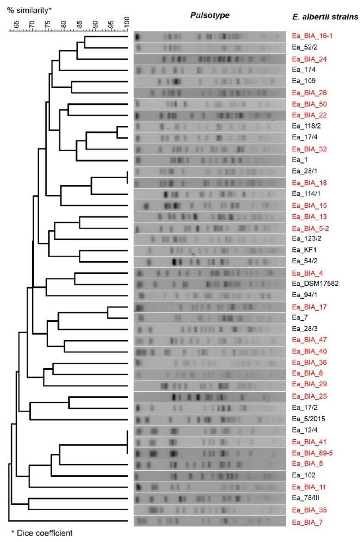
Of the 42 isolated strains of E. albertii, we were able to obtain complete sequences of the genomes (chromosomes and plasmids) of 25 by sequencing using the new generation NGS. The characteristics of the sequenced strains are presented in Table 2. Similarly, genetic typing showed high variability in isolates. In the PFGE method, 90.5% of the strains had distinct pulsotypes (Figure 2). In contrast, in MLST typing, 85.7% of the strains were assigned distinct sequence types (STs), of which 64% were new ST types (Table 2).
In the case of typing virulence factors, the genes encoding cytolethal distending toxin (CDT) and porcine attaching and effacing-associated (Paa) toxins were demonstrated in 100% of the tested E. albertii isolates. On the other hand, the presence of genes encoding IbeA toxin (brain microvascular endothelial cell invasion toxin), CdtB type 2 toxin, Tsh toxin (temperature-sensitive hemagglutinin), and Shiga toxin (Stx2f) was found in 26.2%, 9.7%, 1.7% and 0.4% of E. albertii isolates, respectively. A comparison of the virulence profiles of the sequenced E. albertii strains based on bioinformatic database analysis virulence genes (E. coli_vf n = 2710 and vfdb n = 3688) is shown in Figure 3.
However, among the resistance genes, only those encoding β-lactamases were detected: blaEC-5 (4.8% of the strains), blaEC-8 (54.8% of the strains), and blaTEM-116 (7.1% of strains).
The chromosome size of the tested strains ranged from 4,573,338 to 5,141,010 bp (average 4,784,003 bp). In each strain, the presence of at least one plasmid (range 1–8, average 3.8) was found in the range 1589–177,310 bp (average 58,791.6 bp). Among the plasmids, 20 different types were detected replicons, which were dominated by IncF, IncI, IncX, IncY (46.9%), and Col (37.5%) replicons. The genome characteristics are presented in Table 3.
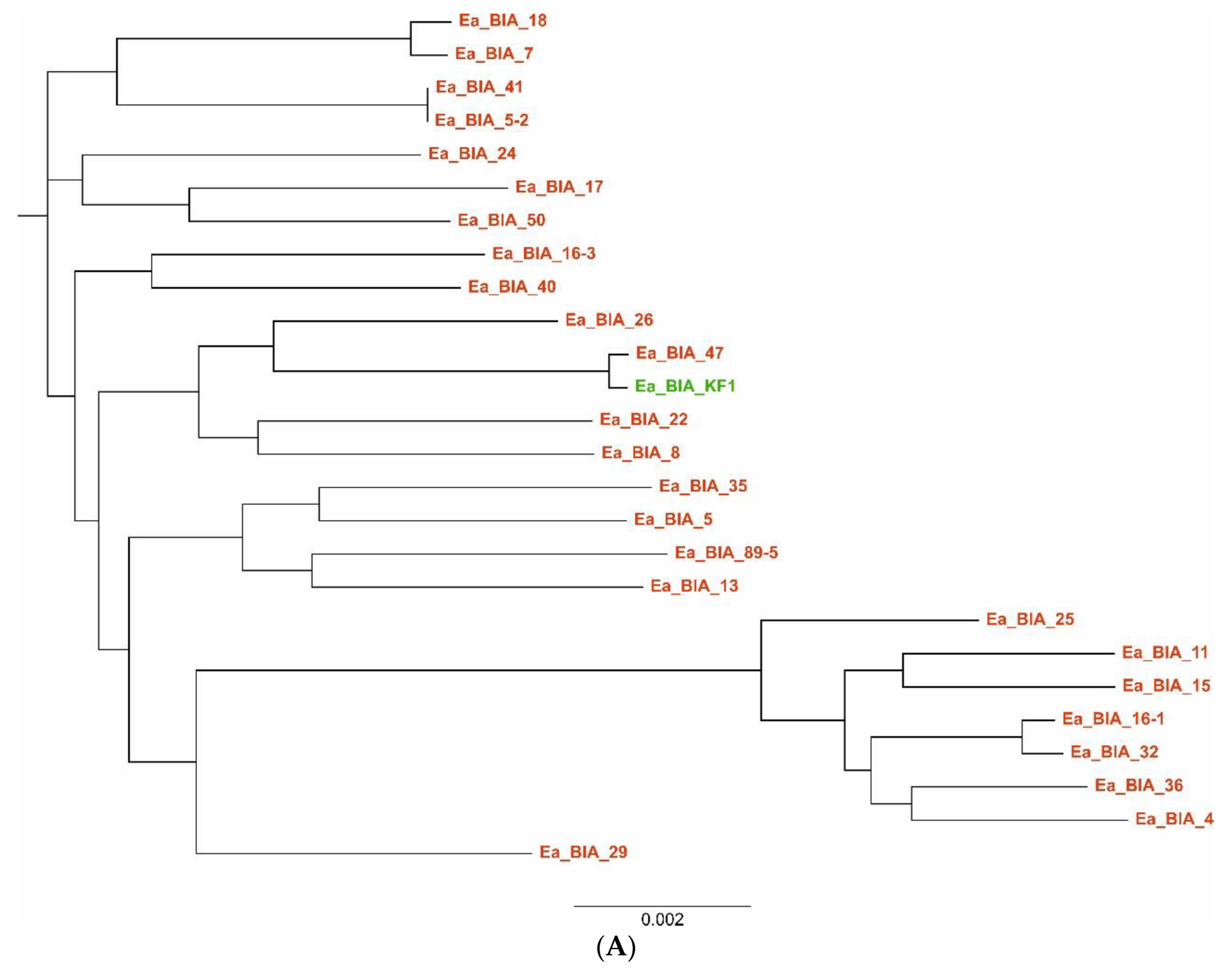
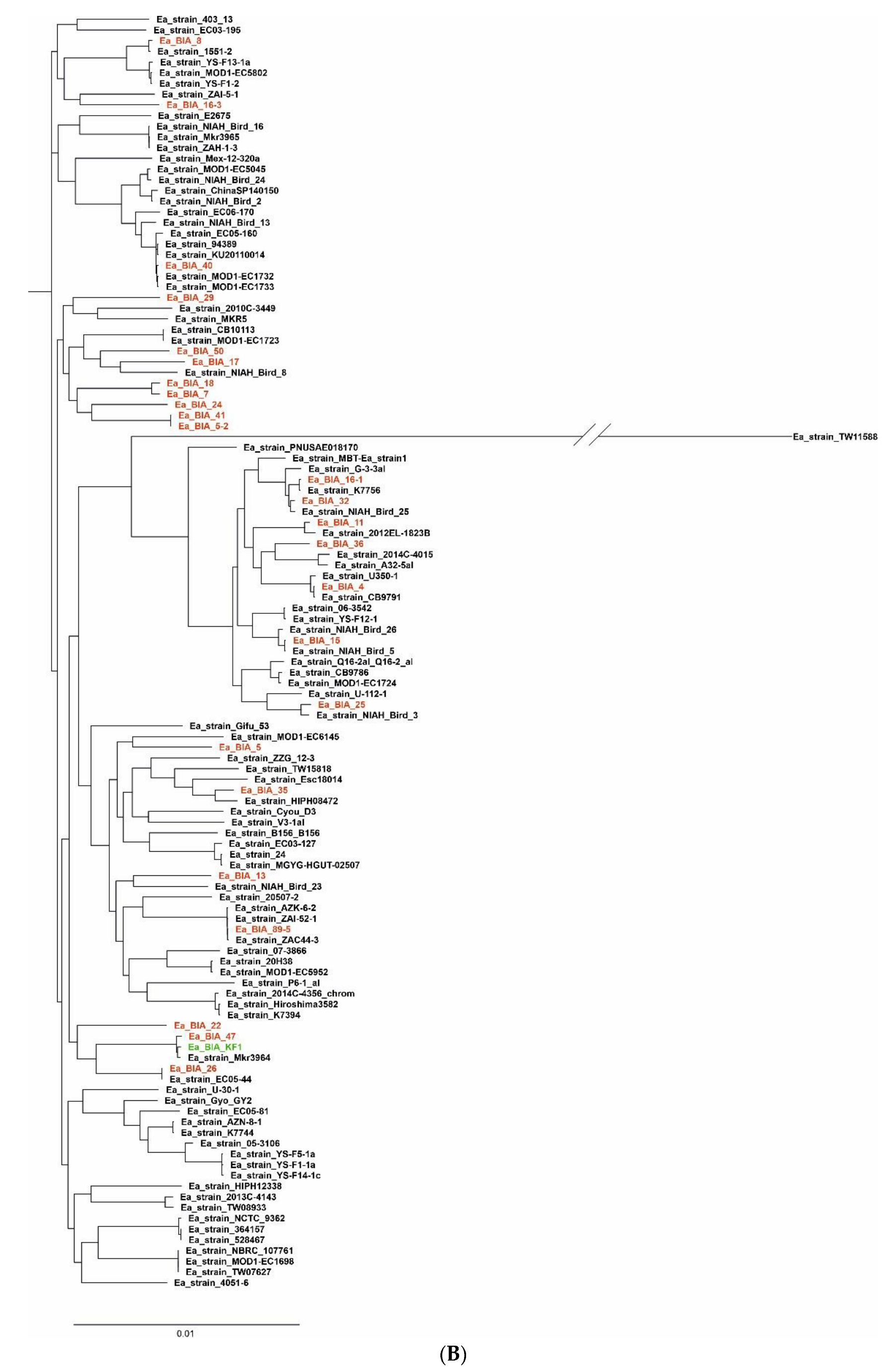
The results of the pangenome analysis of the studied E. albertii isolates, taking into account the earlier isolate of E. albertii z of our center (DOI: 10.1128/genomeA.00004-14) and the 94 strains of E. albertii available in GenBank database (119 strains in total), are presented in Table 4 and Figure 4. Their phylogenetic relationship is shown in Figure 5.

5. Discussion

According to WHO estimates, diarrheal diseases are responsible for two billion deaths worldwide each year. In addition, infectious diarrhea is the leading cause of death among children under age 5 in developing countries [32], and in developed countries it is mainly the problem of morbidity and socio-economic costs [33]. Infections caused by enteropathogens can also result in serious complications, such as neurological or autoimmune diseases (Guillain–Barré syndrome). Unfortunately, a number of factors (demographic changes, centralization of food production, changes in eating habits, globalization, etc.), combined with the rapid adaptation of bacteria to new conditions, result in the formation of new enteropathogens [34].
E. albertii is considered a novel enteropathogen for humans and animals. Reports of the isolation of E. albertii from human and animals, the environment and food samples come from different geographical areas of the world, confirming its globalization. The clinical significance of E. albertii is not yet fully understood, partially because it is difficult to distinguish it from other Enterobacteriaceae using routine identification protocols. E. albertii was first isolated from infants in Bangladesh, and in subsequent years its involvement in infections in children and adults was confirmed [4,5,35]. In 2010, we also isolated E. albertii from the diarrheal feces of a child, but to date this has been a single case [23].
People infected with E. albertii usually show symptoms related to gastroenteritis, including watery diarrhea, dehydration, abdominal distension, vomiting, and in some cases fever. In most cases, these infections are self-limiting, with patients often recovering with little to no treatment [5,33,35]. However, a small subset of E. albertii isolates carry the stx2a allele and can cause bloody diarrhea [19]. Incubation periods of E. albertii infections that present as diarrhea are relatively short (average 12–24 h), while mortality rates are unknown. However, morbidity rates appear to be relatively high, i.e., >50% of the exposed population [36,37].
Other researchers, Afshin et al. [38] and Zaki et al. [39], also described cases of urinary tract infections with E. albertii as the etiological agent. In addition, a single case of E. albertii bacteremia was reported in an elderly woman with multiple comorbidities, possibly due to its ability to move from the intestinal lumen to other extraintestinal sites [35,40]. It may seem that these are relatively rare cases of E. albertii involvement in human infections; however, this may be due to misidentification.
The problem of increasing antimicrobial resistance among many bacteria should also be taken into account. E. albertii strains resistant to important antibiotics, including ampicillin, gentamicin, ciprofloxacin, norfloxacin, trimethoprim/sulfamethoxazole, rifampicin, meropenem and imipenem, were found [21,41,42,43]. In addition, multidrug-resistant strains of E. albertii [42] that showed resistance or coded for resistance to antibiotics from at least eleven classes [44,45] have been described. Combined with the virulence potential of this pathogen, these strains pose a serious threat to public health.
The data obtained from the conducted studies suggest that E. albertii may occur in animals or humans as a commensal or pathogen. E. albertii has been recognized as the etiological agent of diseases in many bird species [7]. In 1994, numerous dead finches were recorded in Scotland [46,47]. The necropsy showed that the causative agent was E. coli belonging to the O86:K61 serotype [46,47], which was previously associated with diarrhea in calves, pigs and horses and with cellulitis in broiler chickens [47]. However, a later study that re-examined these isolates with MLST and a larger panel of phenotypic and biochemical tests showed that it was in fact the E. albertii species [1,7]. In Australia, studies of vertebrate gut bacteria have also shown that birds (chickens and magpies) were the main reservoir of E. albertii [1,48,49]. Our research conducted in northeastern Poland also confirmed the relationship between the occurrence of E. albertii and migratory birds and the water bodies at which they stop. We isolated our strains mainly from the feces of cranes, starlings and geese, and from the water near their habitats. Pigeons were carriers of E. albertii in the urban environment. Our research confirmed the results of other researchers [50,51,52,53] that E. albertii is present in the wild bird population, which may be the potential source of this pathogen for humans, other animals, and the environment.

6. Conclusions

The isolates tested in our center do not currently constitute an important reservoir of resistance genes. We also found them to be highly sensitive to the most commonly used antibacterial drugs. However, in terms of virulence genes, the pathogenic potential of the studied E. albertii strains may range from watery diarrhea typical of enteropathogenic E. coli strains to bloody diarrhea typical of EHEC strains, due to the presence of genes encoding Shiga toxin. An issue requiring further research is the potential of E. albertii strains containing the IbeA toxin (characteristic of NMEC strains; E. coli neonatal meningitides) to cause infections of the central nervous system. In addition, their pathogenicity to animals—pigs—is also noteworthy due to the presence of the Paa toxin typical of porcine enteropathogenic Escherichia coli (PEPEC) strains. Further understanding of the pathobiology of E. albertii, as well as the mechanisms of colonization, survival, and dissemination within and between hosts, is still limited and requires further investigation. This will only be possible if simple and effective diagnostic tools are developed to enable its effective isolation and identification.
The study provides several important data regarding the epidemiology of E. albertii; for instance, we noted a high genetic similarity between our human isolate—Ea_BIA_KF1 [22] and strains isolated from the environment (Ea_BIA_47) and wild birds (Ea_BIA_22) as well as those described in other countries, e.g., Japan [36] (Figure 5). As a result, it helps for a better estimation of a risk that E. albertii may pose for public health. In the conducted studies, we confirmed the high genetic diversity of E. albertii strains, as is the case with E. coli strains, and the open nature of the pangenome of this bacterium, in which plasmids are the main mobile genetic factors responsible for its plasticity. They may play an important role in the horizontal exchange of virulence genes with E. coli strains pathogenic to animals and humans.

Supplementary Materials

The following supporting information can be downloaded at: https://www.mdpi.com/article/10.3390/genes14071384/s1, Table S1 (Biochemical profiles of E. albertii strains estimated based on API20E, ID32E and API50CHE tests).

Author Contributions

All authors had full access to all of the data in the study and take responsibility for their integrity and the accuracy of the analysis. K.L. was responsible for coordination of all tasks and the logistic planning of the project. K.L. was responsible for the study concept, design, data acquisition, underlying data verification, statistical analysis and manuscript drafting. I.Ś. was responsible for genetic research and participation in bioinformatic elaboration of genomic data. T.D. she was responsible for bacterial culture, identification and determination of bacterial susceptibility, statistical analysis and manuscript drafting. D.L. (Dariusz Lebensztejn) was responsible for the selection of patients’ clinical data and coordination of the process of collection of samples. J.G. was responsible for the sequencing experiments. All other authors (D.L. (Dorota Leszczyńska), M.K., S.C.-D.) were directly responsible for execution of sample collection (food, water from birds) and acquisition of data and were involved in the final manuscript preparation. All authors have read and agreed to the published version of the manuscript.

Funding

The work was created as a result of the research project No. 2015/17/B/NZ6/03470 titled “Escherichia albertii as a potential enteropathogen in the light of epidemiological and genomic studies” funded by the National Science Center.

Institutional Review Board Statement

The study was conducted in accordance with the Declaration of Helsinki. The research provided for in the project was approved by the Bioethics Committee of the Medical University of Bialystok (No. R-I-002/480/2014; 27 November 2014). The application submitted to the Bioethical Committee of the Medical University of Bialystok, apart from the description of the research and the samples of materials used in them, also contained a template for patients entitled “Consent to perform microbiological tests” with information on the nature and purpose of the tests.

Informed Consent Statement

Informed consent was obtained from all subjects involved in the study. Written informed consent has been obtained from the patient(s) to publish this paper.

Data Availability Statement

The first complete genome sequence of Escherichia albertii strain KF1, a new potential human enteric pathogen was published in Genom. Announcement 2014, 2, e00004–e00014. https://doi.org/10.1128/genomeA.00004-14.

Acknowledgments

The authors would like to thank Krzysztof Fiedoruk, DSc for his cooperation and scientific support.

Conflicts of Interest

The authors declare no conflict of interest.

References

  1. Gordon, D.M. Reservoirs of Infection: The Epidemiological Characteristics of an Emerging Pathogen, Escherichia albertii; Research School of Biology. Australian National University: Canberra, Australia. Proceedings 2011, 61–73. Available online: https://www.aavac.com.au/files/2011-08.pdf (accessed on 27 April 2023).
  2. Nimri, L.F. Escherichia albertii, a newly emerging enteric pathogen with poorly defined properties. Diagn. Microbiol. Infect. Dis. 2013, 77, 91–95. [Google Scholar] [CrossRef] [PubMed]
  3. Donnenberg, M.S.; Tzipori, S.; McKee, M.L.; O’Brien, A.D.; Alroy, I.J.; Kapert, J.B. The role of the eae gene of enterohemorrhagic Escherichia coli in intimate attachment in vitro and in a porcine model. J. Clin. Investig. 1993, 92, 1418–1424. [Google Scholar] [CrossRef] [PubMed] [Green Version]
  4. Asoshima, N.; Matsuda, M.; Shigemura, K.; Honda, M.; Yoshida, H.; Hiwaki, H.; Ogata, K.T. Identification of Escherichia albertii as a Causative Agent of a Food-Borne Outbreak Occurred in 2003. JPN J. Infect. Dis. 2014, 67, 139–140. [Google Scholar] [CrossRef] [PubMed] [Green Version]
  5. Albert, M.J.; Alam, K.; Islam, M.; Montanaro, J.; Rahaman, A.S.; Haider, K.; Hossain, M.A.; Kibriya, A.K.; Tzipori, S. Hafnia alvei, a probable cause of diarrhea in humans. Infect. Immun. 1991, 59, 1507–1513. [Google Scholar] [CrossRef] [Green Version]
  6. Huys, G.; Cnockaert, M.; Janda, J.M.; Swings, J. Escherichia albertii sp. nov., a diarrhoeagenic species isolated from stool specimens of Bangladeshi children. Int. J. Syst. Evol. Microbiol. 2003, 53, 807–810. [Google Scholar] [CrossRef] [Green Version]
  7. Oaks, J.L.; Besser, T.E.; Walk, S.T.; Gordon, D.M.; Beckmen, K.B.; Burek, K.A.; Haldorson, G.J.; Bradway, D.S.; Ouellette, L. Escherichia albertii in wild and domestic birds. Emerg. Infect. Dis. 2010, 16, 638–646. [Google Scholar] [CrossRef]
  8. Blanco, M.; Schumacher, S.; Tasara, T.; Zweifel, C.; Blanco, J.E.; Dahbi, G.; Blanco, J.; Stephan, R. Serotypes, intimin variants and other virulence factors of eae positive Escherichia coli strains isolated from healthy cattle in Switzerland. Identification of a new intimin variant gene (eae-eta2). BMC Microbiol. 2005, 5, 23. [Google Scholar] [CrossRef] [PubMed] [Green Version]
  9. Okuda, J.; Kurazono, H.; Takeda, Y. Distribution of the cytolethal distending toxin A gene (cdtA) among species of Shigella and Vibrio, and cloning and sequencing of the cdt gene from Shigella dysenteriae. Microb. Pathog. 1995, 18, 167–172. [Google Scholar] [CrossRef]
  10. Jinadasa, R.N.; Bloom, S.E.; Weiss, R.S.; Duhamel, G.E. Cytolethal distending toxin: A conserved bacterial genotoxin that blocks cell cycle progression, leading to apoptosis of a broad range of mammalian cell lineages. Microbiology 2011, 157, 1851–1875. [Google Scholar] [CrossRef] [Green Version]
  11. Grasso, F.; Frisan, T. 2015. Bacterial genotoxins: Merging the DNA damage response into infection biology. Biomolecules 2015, 5, 1762–1782. [Google Scholar] [CrossRef] [Green Version]
  12. Taieb, F.; Petit, C.; Nougayrède, J.P.; Oswald, E. The enterobacterial genotoxins: Cytolethal distending toxin and colibactin. Ecosal. Plus 2016, 7. [Google Scholar] [CrossRef] [PubMed]
  13. Fedor, Y.; Vignard, J.; Nicolau-Travers, M.L.; Boutet-Robinet, E.; Watrin, C.; Salles, B.; Mirey, G. From single-strand breaks to double-strand breaks during S-phase: A new mode of action of the Escherichia coli cytolethal distending toxin. Cell Microbiol. 2013, 15, 1–15. [Google Scholar] [CrossRef] [PubMed]
  14. O’Brien, A.D.; LaVeck, G.D. 1983. Purification and characterization of a Shigella dysenteriae 1-like toxin produced by Escherichia coli. Infect. Immun. 1983, 40, 675–683. [Google Scholar] [CrossRef] [PubMed] [Green Version]
  15. Scheutz, F.; Teel, L.D.; Beutin, L.; Piérard, D.; Buvens, G.; Karch, H.; Mellmann, A.; Caprioli, A.; Tozzoli, R.; Morabito, S.; et al. Multicenter evaluation of a sequence-based protocol for subtyping Shiga toxins and standardizing Stx nomenclature. J. Clin. Microbiol. 2012, 50, 2951–2963. [Google Scholar] [CrossRef] [Green Version]
  16. Ooka, T.; Seto, K.; Kawano, K.; Kobayashi, H.; Etoh, Y.; Ichihara, S.; Kaneko, A.; Isobe, J.; Yamaguchi, K.; Horikawa, K.; et al. Clinical significance of Escherichia albertii. Emerg Infect. Dis. 2012, 18, 488–492. [Google Scholar] [CrossRef] [Green Version]
  17. Murakami, K.; Etoh, Y.; Tanaka, E.; Ichihara, S.; Horikawa, K.; Kawano, K.; Ooka, T.; Kawamura, Y.; Ito, K. Shiga toxin 2f-producing Escherichia albertii from a symptomatic human. JPN J. Infect. Dis. 2014, 67, 204–20857. [Google Scholar] [CrossRef] [Green Version]
  18. Hinenoya, A.; Yasuda, N.; Hibino, T.; Shima, A.; Nagita, A.; Tsukamoto, T.; Shinji Yamasaki, S. Isolation and characterization of an Escherichia albertii strain producing three different toxins from a child with diarrhea. JPN J. Infect. Dis. 2017, 70, 252–257. [Google Scholar] [CrossRef] [Green Version]
  19. Brandal, L.T.; Tunsjø, H.S.; Ranheim, T.E.; Løbersli, I.; Lange, H.; Astrid Louise Wester, A.L. Shiga toxin 2a in Escherichia albertii. J. Clin. Microbiol. 2015, 53, 1454–1455. [Google Scholar] [CrossRef] [Green Version]
  20. Lynne, A.M.; Kariyawasam, S.; Wannemuehler, Y.; Johnson, T.J.; Johnson, S.J.; Sinha, A.S.; Lynne, D.K.; Moon, H.W.; Jordan, D.M.; Logue, C.M.; et al. Recombinant Iss as a potential vaccine for avian colibacillosis. Avian Dis. 2012, 56, 192–199. [Google Scholar] [CrossRef] [Green Version]
  21. Lima, M.P.; Yamamoto, D.; Santos, A.C.D.M.; Ooka, T.; Hernandes, R.T.; Vieira, M.A.; Gomes, T.A. Phenotypic characterization and virulence-related properties of Escherichia albertii strains isolated from children with diarrhea in Brazil. Pathog. Dis. 2019, 77, ftz014. [Google Scholar] [CrossRef]
  22. Fedoruk, K.; Daniluk, T.; Święcicka, I.; Murawska, E.; Sciepuk, M.; Leszczynska, K. First complete genome sequence of Escherichia albertii strain KF1, a new potential human enteric pathogen. Genom. Announc. 2014, 2, e00004–e00014. [Google Scholar] [CrossRef] [Green Version]
  23. Joensen, K.G.; Tetzschner, A.M.M.; Iguchi, A.; Aarestrup, F.M.; Scheutzb, F. Rapid and easy in silico serotyping of Escherichia coli isolates by use of whole-genome sequencing data. J. Clin. Microbiol. 2015, 53, 2410–2426. [Google Scholar] [CrossRef] [Green Version]
  24. Janben, T.; Schwarz, C.; Preikschat, P.; Voss, M.; Philipp, H.C.; Wieler, L.H. Virulence-associated genes in avian pathogenic Escherichia coli (APEC) isolated from internal organs of poultry having died from colibacillosis. Int. J. Med. Microbiol. 2001, 291, 371–378. [Google Scholar] [CrossRef]
  25. Kahlmeter, G.; Brown, D.F.; Goldstein, F.W.; MacGowan, A.P.; Mouton, J.W.; Odenholt, I.; Rodloff, A.; Soussy, C.J.; Steinbakk, M.; Soriano, F.; et al. European Committee on Antimicrobial Susceptibility Testing (EUCAST) Technical Notes on antimicrobial susceptibility testing. Clin. Microbiol. Infect. 2006, 12, 501–503. [Google Scholar] [CrossRef] [Green Version]
  26. Yang, J.L.; Wang, J.T.; Lauderdale, T.L.; Chang, S.C. Prevalence of extended-spectrum β-lactamases in Enterobacter cloacae in Taiwan and comparison of 3 phenotypic confirmatory methods for detecting extended-spectrum β-lactamase production. J. Microbiol. Immunol. Infect. 2009, 42, 310–316. [Google Scholar]
  27. Barton, B.M.; Harding, G.P.; Zuccarelli, A.J. A general method for detecting and sizing large plasmids. Anal. Biochem. 1995, 226, 235–240. [Google Scholar] [CrossRef]
  28. Stirling, D. DNA Extraction from Fungi, Yeast, and Bacteria. In PCR Protocols; Bartlett, J.M.S., Stirling, D., Eds.; Humana Press: Totowa, NJ, USA, 2003; pp. 53–54. [Google Scholar]
  29. Koren, S.; Walenz, B.P.; Berlin, K.; Miller, J.R.; Bergman, N.H.; Phillippy, A.M. Canu: Scalable and accurate long-read assembly via adaptive k-mer weighting and repeat separation. Genom. Res. 2017, 27, 722–736. [Google Scholar] [CrossRef] [Green Version]
  30. Seemann, T. Prokka: Rapid prokaryotic genome annotation. Bioinformatics 2014, 30, 2068–2069. [Google Scholar] [CrossRef] [Green Version]
  31. Page, A.J.; Cummins, C.A.; Hunt, M.; Reuter, S.; Holden, M.T.; Fookes, M.; Falush, D.; Keane, J.A.; Parkhill. Roary: Rapid large-scale prokaryote pan genome analysis. Bioinformatics 2015, 31, 3691–3693. [Google Scholar] [CrossRef] [Green Version]
  32. Bryce, J.; Boschi-Pinto, C.; Shibuya, K.; Black, R.E. WHO Child Health Epidemiology Reference Group, 2005. WHO estimates of the causes of death in children. Lancet 2005, 365, 1147–1152. [Google Scholar] [CrossRef]
  33. Davidson, G.; Barnes, G.; Bass, D.; Cohen, M.; Fasano, A.; Fontaine, O.; Guandalini, S. Infectious diarrhea in children: Working Group Report of the First World Congress of Pediatric Gastroenterology, Hepatology, and Nutrition. J. Pediatr. Gastroenterol. Nutr. 2002, 35 (Suppl. 2), S143–S150. [Google Scholar] [CrossRef] [PubMed]
  34. Braden, C.R.; Tauxe, R.V. Emerging trends in foodborne diseases. Infect. Dis. Clin. North. Am. 2013, 27, 517–533. [Google Scholar] [CrossRef] [PubMed]
  35. Inglis, T.J.; Merritt, A.J.; Bzdyl, N.; Urosevic, M.N. First bacteraemic human infection with Escherichia albertii. New Microbes New Infect. 2015, 8, 171–173. [Google Scholar] [CrossRef] [PubMed] [Green Version]
  36. Ooka, T.; Tokuoka, E.; Furukawa, M. Human gastroenteritis outbreak associated with Escherichia albertii, Japan. Emerg. Infect. Dis. 2013, 19, 144–146. [Google Scholar] [CrossRef]
  37. Masuda, K.; Ooka, T.; Akita, H.; Hiratsuka, T.; Takao, S.; Fukada, M.; Inoue, K.; Honda, M.; Toda, J.; Sugitani, W.; et al. Epidemiological Aspects of Escherichia albertii Outbreaks in Japan and Genetic Characteristics of the Causative Pathogen. Foodborne Pathog. Dis. 2020, 17, 144–150. [Google Scholar] [CrossRef]
  38. Afshin, N.; Bahareh, R.; Azadeh, F. Detection of Escherichia albertii in urinary and gastrointestinal infections in Kermanshah, Iran Research square. Int. J. Enteric Pathog. 2020, 9, 37–42. [Google Scholar]
  39. Zaki, M.E.S.; Eid, A.E.; El-Kazzaz, S.S.; El-Sabbagh, A.M. Molecular Study of Escherichia albertii in Pediatric Urinary Tract Infections. Open Microbiol. J. 2021, 15, 139–144. [Google Scholar] [CrossRef]
  40. Gomes, T.A.T.; Ooka, T.; Hernandes, R.T.; Yamamoto, D.; Hayashi, T. Escherichia albertii Pathogenesis. EcoSal. Plus 2020, 9, 1–18. [Google Scholar] [CrossRef]
  41. Stock, I.; Rahman, M.; Sherwood, K.J.; Wiedemann, B. Natural antimicrobial susceptibility patterns and biochemical identification of Escherichia albertii and Hafnia alvei strains. Diagn. Microbiol. Infect. Dis. 2005, 51, 151–163. [Google Scholar] [CrossRef]
  42. Hinenoya, A.; Li, X.; Zeng, X.; Sahin, O.; Moxley, R.A.; Logue, C.M.; Gillespie, B.; Yamasaki, S.; Lin, J. Isolation and characterization of Escherichia albertii in poultry at the pre-harvest level. Zoonoses Public Health 2021, 68, 213–225. [Google Scholar] [CrossRef]
  43. Konno, T.; Yatsuyangi, J.; Takahashi, S.; Kumagai, Y.; Wada, E.; Chiba, M.; Saito, S. Isolation and identification of Escherichia albertii from a patient in an outbreak of gastroenteritidis. JPN J. Infect. Dis. 2012, 65, 203–207. [Google Scholar] [CrossRef] [PubMed] [Green Version]
  44. Li, Q.; Wang, H.; Xu, Y.; Bai, X.; Wang, J.; Zhang, Z.; Liu, X.; Miao, Y.; Zhang, L.; Li, X.; et al. Multidrug-resistant Escherichia albertii: Co-occurrence of β-lactamase and MCR-1 encoding genes. Front. Microbiol. 2018, 9, 258. [Google Scholar] [CrossRef] [PubMed] [Green Version]
  45. Luo, L.; Wang, H.; Payne, M.J.; Liang, C.; Bai, L.; Zheng, H.; Zhang, Z.; Zhang, L.; Zhang, X.; Yan, G.; et al. Comparative genomics of Chinese and international isolates of Escherichia albertii: Population structure and evolution of virulence and antimicrobial resistance. Microb. Genom. 2021, 7, 000710. [Google Scholar] [CrossRef]
  46. Pennycott, T.W.; Ross, H.M.; McLaren, I.M.; Park, A.; Hopkins, G.F.; Foster, G. Causes of death of wild birds of the family Fringillidae in Britain. Vet. Rec. 1998, 143, 155–158. [Google Scholar] [CrossRef]
  47. Foster, G.; Ross, H.M.; Pennycott, T.W.; Hopkins, G.F.; McLaren, I.M. Isolation of Escherichia coli O86, K61 producing cyto-lethal distending toxin from wild birds of the finch family. Lett. Appl. Microbiol. 1998, 26, 395–398. [Google Scholar] [CrossRef]
  48. La Ragione, R.M.; McLaren, I.M.; Foster, G.; Cooley, W.A.; Woodward, M.J. Phenotypic and genotypic characterization of avian Escherichia coli O86, K61 isolates possessing a γ-like intimin. Appl. Environ. Microbiol. 2002, 68, 4932–4942. [Google Scholar] [CrossRef] [Green Version]
  49. Gordon, D.M.; Cowling, A. The distribution and genetic structure of Escherichia coli in Australian vertebrates: Host and geographic effects. Microbiology 2003, 149, 3575–3586. [Google Scholar] [CrossRef] [Green Version]
  50. Maeda, E.; Murakami, K.; Sera, N.; Ito, K.; Fujimoto, S. Detection of Escherichia albertii from chicken meat and giblets. J. Vet. Med. Sci. 2015, 77, 871–873. [Google Scholar] [CrossRef] [Green Version]
  51. Barmettler, K.; Biggel, M.; Treier, A.; Muchaamba, F.; Vogler, B.R.; Stephan, R. Occurrence and Characteristics of Escherichia albertii in Wild Birds and Poultry Flocks in Switzerland. Microorganisms 2022, 10, 2265. [Google Scholar] [CrossRef] [PubMed]
  52. Asoshima, N.; Matsuda, M.; Shigemura, K.; Honda, M.; Yoshida, H.; Oda, T.; Hiwaki, H. Isolation of Escherichia albertii from raw chicken liver in Fukuoka City, Japan. Jpn J. Infect. Dis. 2015, 68, 248–250. [Google Scholar] [CrossRef] [Green Version]
  53. Wang, H.; Li, Q.; Bai, X.; Xu, Y.; Zhao, A.; Sun, H.; Deng, J.; Xiao, B.; Liu, X.; Sun, S.; et al. Prevalence of eae-positive, lactose non-fermenting Escherichia albertii from retail raw meat in China. Epidemiol. Infect. 2016, 144, 45–52. [Google Scholar] [CrossRef] [PubMed] [Green Version]
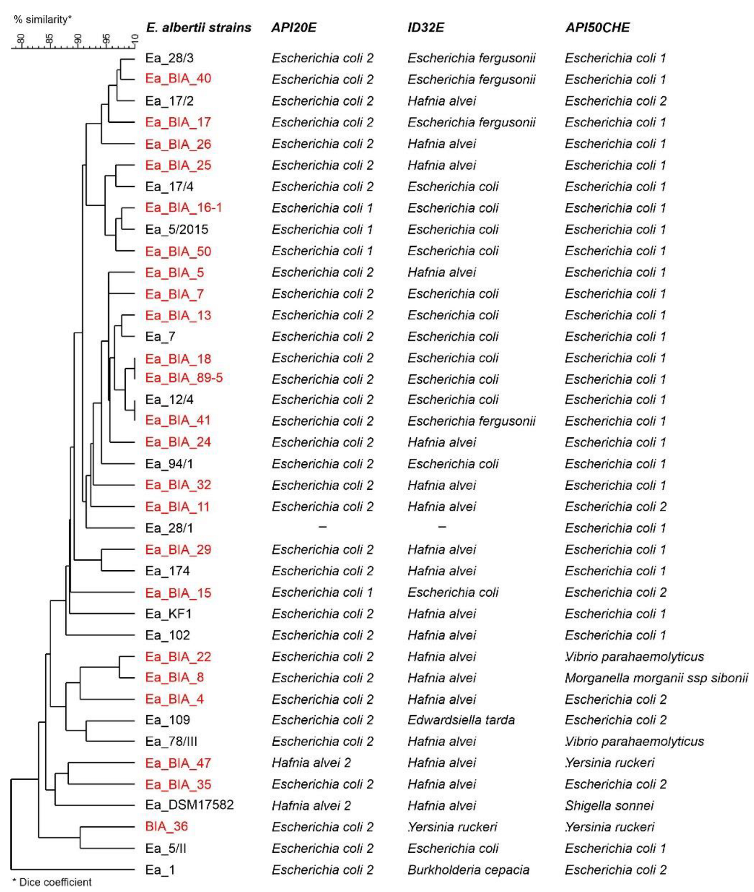
Figure 1. Similarity of E. albertii strains based on comparison of biochemical profiles determined based on API 50 CHE, API 20E and ID32E tests (bioMérieux). Dendrogram based on the average connection method (UPGMA) and the Dice similarity measure. E. albertii strains selected for WGS are highlighted in red color; Ea—E. albertii. Supplementary Table S1 (Biochemical profiles of E. albertii strains estimated based on API20E, ID32E and API50CHE tests).
Genes 14 01384 g001
Figure 2. Genetic profiles (pulsotypes) of E. albertii (Ea) strains. Dendrogram based on the average connection method (UPGMA) and the Dice similarity measure. E. albertii strains selected for WGS are highlighted in red color; Ea—E. albertii.
Genes 14 01384 g002
Figure 3. Similarity of sequenced strains of E. albertii (Ea) based on the presence of virulence genes (E. coli_vf n = 2710 and vfdb n = 3688 gene databases). Dendrogram based on the average connection method (UPGMA) and the Dice similarity measure. Analysis made in program ABRicate ver. 0.8 (https://github.com/tseemann/abricate).
Genes 14 01384 g003
Figure 4. Graph showing the frequency of occurrence of genes included in the pangenome of E. albertii depending on the number of genomes. Analysis based on 26 (A) and 119 genomes (B) performed in Roary ver. 3.13.0.
Genes 14 01384 g004
Figure 5. Phylogenetic similarity of E. albertii strains based on the analysis of genes included in the pangenome of 26 (A) and 119 strains (B). Twenty-five strains sequenced in the OPUS-9 project are marked in red, an earlier E. albertii isolate from our center (DOI: 10.1128/genomeA.00004-14) in green, strains from the GenBank database in black. Analysis made in Roary and FastTree programs. Ea—E. albertii.
Genes 14 01384 g005aGenes 14 01384 g005b
Table 1. Drug susceptibility profiles of E. albertii strains isolated in northeastern Poland.
E. albertii StrainsAntibiotics
AMAMCCXMCEFIPCHCIPLEVGNSXT
Ea_28/3; Ea_109; Ea_102; Ea_54/2; Ea_29/2; Ea_BIA_8; Ea_BIA_29; Ea_BIA_40; Ea_BIA_22; Ea_BIA_18; Ea_BIA_16-3; Ea_BIA_11; Ea_BIA_16-1;RSISSSSSSS
Ea_174; Ea_28/1; Ea_78/III; Ea_7; Ea_15/2; Ea_17/4; Ea_5/2015; Ea_17/2; Ea_1; Ea_69/2; Ea_BIA_29; Ea_BIA_36; Ea_BIA_47; Ea_BIA_5; Ea_BIA_50; Ea_BIA_5-2; Ea_BIA_7; Ea_BIA_13; Ea_BIA_15; Ea_BIA_17; Ea_BIA_24;SSISSSSSSS
Ea_12/4; Ea_94/2; Ea_BIA_89-5; Ea_BIA_26;RRISSSSSSS
Ea_BIA_24RSISSRSSSS
Ea_BIA_4RSRSSSSSSS
Ea_BIA_35SSRSSSRSSS
Ea_BIA_40SSISSSRSSS
Abbreviations: AM, Ampicillin; AMC, Amoxicillin/Clavulanic acid; CXM, Cefuroxime; CEF, Cefepime; CH, Chloramphenicol; CIP, Ciprofloxacin; IP, Imipenem; GN, Gentamicin; LEV, Levofloxacin SXT, Trimethoprim/Sulfamethoxazole. I, susceptible—increased exposure; S, susceptible—standard dosing regimen; R, resistant strain. E. albertii strains selected for WGS are highlighted in red color; Ea—E. albertii.
Table 2. Characterization of sequenced E. albertii strains.
E. albertii StrainsOriginSerotypeType/Sequential Profile a
STadkfumCgyrBicdmdhpurArecA
Ea_BIA_11Bird (Sturnus vulgaris)H33:O1191160822829385207116119106
Ea_BIA_13Bird (Grus grus)H52:O28nearest ST: 6059163395134386378444~373 b
Ea_BIA_15Bird (Passer domesticus)H52:O128473622817085332116149106
Ea_BIA_16-1Bird (Sturnus vulgaris)H52:O128596713217085207116206106
Ea_BIA_16-3Bird (Sturnus vulgaris)H5:-nearest ST: 1162222878885~468 b378149~103 b
Ea_BIA_17Bird (Grus grus)H52:-nearest ST: 10710133~399 b85189142141176
Ea_BIA_18Bird (Grus grus)H52:O115nearest ST: 1846133298235255~190 b66176
Ea_BIA_22Bird (Grus grus)H52:-nearest ST: 2683 c567~394 b1341787255113226
Ea_BIA_24Bird (Grus grus)H52:O69nearest ST: 6059 c228~788 b~85 b330109~69 b176
Ea_BIA_25Bird (Grus grus)H52:O1nearest ST: 1338~132 b17085207154149135
Ea_BIA_26Bird (Grus grus)H33:O182605853978813448325514975
Ea_BIA_29Bird (unidentified species)H5:-nearest ST: 119711321587134~872 b2566676
Ea_BIA_32Bird (Delichon urbicum)H5:O8nearest ST: 5399 c~379 b~170 b85207116206106
Ea_BIA_35Bird (Anser spp.)H52:O1521070916312838521896866225
Ea_BIA_36Bird (unidentified species)H33:O11511195228164585332116119106
Ea_BIA_4Bird (unidentified species)H33:O1604953717085332116206106
Ea_BIA_40Bird (Anser spp.)H52:-376213239413414411066102
Ea_BIA_41Bird (Sturnus vulgaris)H52:-nearest ST: 12964132298134~189 b885206~226 b
Ea_BIA_47Backwaters (Jancewicze)H33:-73656951118548310914975
Ea_BIA_5Bird (unidentified species)H52:-86911633938518968246125
Ea_BIA_50Bird (Grus grus)H52:O9nearest ST: 10710 c~393 b104~85 b~545 b111141~176 b
Ea_BIA_5-2Bird (Columba livia)H33:O9nearest ST: 12964132298134~189 b1238206885
Ea_BIA_7Bird (Columba livia)H52:O115184616329823525519066176
Ea_BIA_8Bird (Columba livia)H52:O13126811341648516925469103
Ea_BIA_89-5Bird (Grus grus)H52:O326802933958533068248226
a according to the MLST profile for E. coli; analysis was performed with MLST 2.0 server (https://cge.food.dtu.dk/services/MLST/; accessed on 21 April 2021). b alleles with less than 100% identity. c more than one nearest ST matched.
Table 3. Genome characterization of sequenced E. albertii strains.
E. albertii StrainsChromosome SizeNumber of GenesNumber of PlasmidsPlasmid Size
Ea_BIA_114,779,775449535952–110,945
Ea_BIA_134,819,152450371589–128,726
Ea_BIA_154,772,054458145140–130,064
Ea_BIA_16-14,573,338424832733–99,447
Ea_BIA_16-34,838,644454825055–82,983
Ea_BIA_174,854,152472831590–132,593
Ea_BIA_184,871,752458752760–144,030
Ea_BIA_224,884,955471881590–152,064
Ea_BIA_244,841,726451371589–136,544
Ea_BIA_254,810,099448041589–100,741
Ea_BIA_264,654,873427944703–114,713
Ea_BIA_294,687,526436224593–116,006
Ea_BIA_324,822,417457053758–125,136
Ea_BIA_354,725,775442242571–88,900
Ea_BIA_364,790,9014588440,185–103,275
Ea_BIA_44,776,2534548436,239–97,069
Ea_BIA_404,826,552455032719–95,160
Ea_BIA_414,677,2184327213,675–76,945
Ea_BIA_474,595,293428822720–40,979
Ea_BIA_54,782,897433043909–143,188
Ea_BIA_504,722,615436541627–165,614
Ea_BIA_5-24,681,1664356176,945
Ea_BIA_74,960,105470652283–177,310
Ea_BIA_84,709,8364505347,425–108,071
Ea_BIA_89-55,141,0104990175,610
Table 4. E. albertii pangenome estimated on the basis of genomic analysis of 26 tested strains (A) and an additional 94 from GenBank (B). The analysis was made in Roary ver. 3.13.0.
(A)
Type GenesNo. Strains (N = 26 *)No. Genes
Core genes(99% ≤ strains ≤ 100%)2966
Soft core genes(95% ≤ strains < 99%)252
Shell genes(15% ≤ strains < 95%)3062
Cloud genes(0% ≤ strains < 15%)7888
Total(0% ≤ strains ≤ 100%)14,168
* included previous E. albertii isolate from our center was used (DOI: 10.1128/genomeA.00004-14).
(B)
Type GenesNo. Strains (N = 119)No. Genes
Core genes(99% ≤ strains ≤ 100%)2088
Soft core genes(95% ≤ strains < 99%)511
Shell genes(15% ≤ strains < 95%)3052
Cloud genes(0% ≤ strains < 15%)19,506
Total(0% ≤ strains ≤ 100%)25,157
Disclaimer/Publisher’s Note: The statements, opinions and data contained in all publications are solely those of the individual author(s) and contributor(s) and not of MDPI and/or the editor(s). MDPI and/or the editor(s) disclaim responsibility for any injury to people or property resulting from any ideas, methods, instructions or products referred to in the content.