Review

Redox-Based Strategies against Infections by Eukaryotic Pathogens

Institut de Chimie des Substances Naturelles, CNRS UPR 2301, Université Paris-Saclay, 91198 Gif-sur-Yvette, France
*
Authors to whom correspondence should be addressed.
Genes 2023, 14(4), 778; https://doi.org/10.3390/genes14040778
Submission received: 27 February 2023 / Revised: 13 March 2023 / Accepted: 21 March 2023 / Published: 23 March 2023
(This article belongs to the Section Microbial Genetics and Genomics)

Abstract

:
Redox homeostasis is an equilibrium between reducing and oxidizing reactions within cells. It is an essential, dynamic process, which allows proper cellular reactions and regulates biological responses. Unbalanced redox homeostasis is the hallmark of many diseases, including cancer or inflammatory responses, and can eventually lead to cell death. Specifically, disrupting redox balance, essentially by increasing pro-oxidative molecules and favouring hyperoxidation, is a smart strategy to eliminate cells and has been used for cancer treatment, for example. Selectivity between cancer and normal cells thus appears crucial to avoid toxicity as much as possible. Redox-based approaches are also employed in the case of infectious diseases to tackle the pathogens specifically, with limited impacts on host cells. In this review, we focus on recent advances in redox-based strategies to fight eukaryotic pathogens, especially fungi and eukaryotic parasites. We report molecules recently described for causing or being associated with compromising redox homeostasis in pathogens and discuss therapeutic possibilities.

1. Introduction, Scope, and Aims of This Review

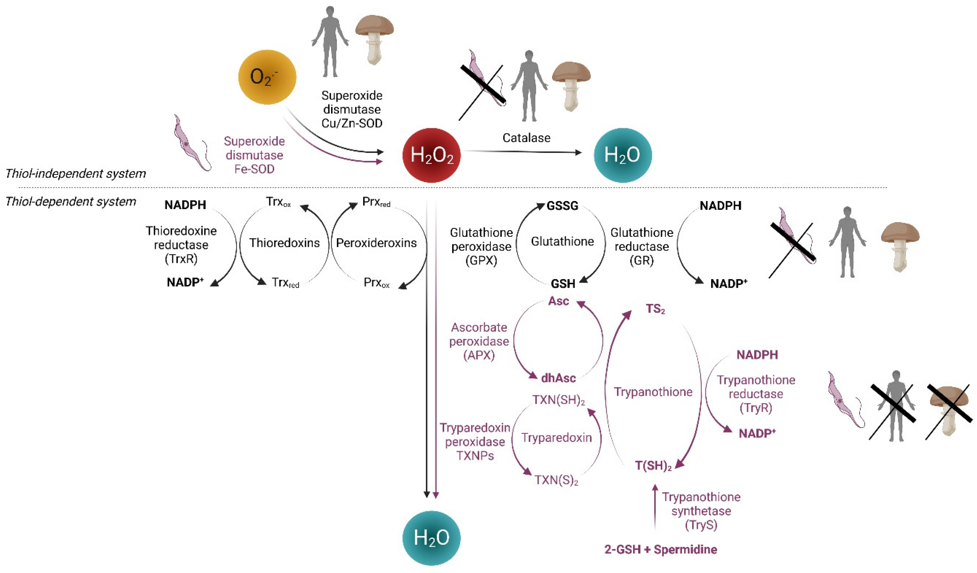
1.1. Reactive Oxygen Species (ROS) and Redox System in Human Cells

Oxygen-derived reactive chemical species or ROS are chemically reactive non-radical molecules involved in diverse biological functions. Under physiological conditions, ROS are important signaling molecules, and a balance between the generation and elimination of ROS ensures the proper function of ROS and redox-sensitive signaling proteins [1,2]. For example, a transient and rapid ROS burst is necessary for ROS-mediated signaling while excessive and prolonged ROS accumulation leads to oxidative stress that may disrupt the function of key transcription factors and signal-transduction pathways responsible for the development of various pathologies, including cardiovascular, neurodegenerative, inflammatory diseases, and cancer.
Cellular oxidation-reduction (redox) systems are broadly defined networks that consist of ROS producers, antioxidant enzymes, and redox effectors as well as ROS and several small redox molecules (NADPH, glutathione, etc.) (Figure 1) [3]. Superoxide (O2•-) is the primary ROS produced from a variety of sources, including mitochondrial respiratory chain, NADPH oxidases, and a number of other biochemical reactions. Typically, molecular oxygen (O2) may react with electron generating superoxide that can be rapidly converted to hydrogen peroxide (H2O2) by superoxide dismutases (SOD). H2O2, the most important non-radical molecule, can be converted to water by catalases, peroxiredoxins (Prx), or glutathione peroxidases (GPX). In the presence of transition metals, H2O2 can be converted to hydroxyl radicals (HO•), which are highly reactive and can cause damage to all cellular components. Thiol-dependent antioxidants constitute the thioredoxin-based system composed of thioredoxin reductases (TrxR), thioredoxin (Trx), and Prx, and the glutathione-based system with several glutathione-utilizing enzymes, such as GPX, glutathione reductase (GR), and glutaredoxin. NADPH, which can be supplied by the pentose phosphate pathway, is the ultimate electron donor for both thioredoxin and glutathione systems. Finally, the downstream effectors of redox systems, which may be directly or indirectly redox-regulated, include transcription factors, such as nuclear factor erythroid 2-related factor 2 (NRF2), hypoxia-inducible factor 1-alpha (HIF1α), and nuclear factor NF-kappa-B.
Excessive oxidative stress leads to cell death. Thus, based on the difference in redox systems between pathogens and host (human), it is conceivable that pharmacological intervention through the generation of ROS and/or targeting redox systems could selectively kill or inhibit the pathogens with limited damage to host cells. Indeed, ROS generation and redox-based anticancer strategy are fields of increasing interest as cancer cells are generally in a persistent pro-oxidative status compared to their normal counterparts and are more vulnerable to further oxidative insults via pharmacological interventions [4,5]. In this review, we will first briefly describe our current knowledge on the distinct redox systems in several important pathogenic fungi and protozoan parasites; highlighting the features of the redox systems unique to these pathogens (Figure 1), and then, we present recent attempts at targeting redox systems directly or indirectly as a strategy against these disease-causing pathogens (Table 1). Indeed, both pathogenic fungi and parasitic protozoa remain severe threats to human health across the globe. Pathogenic fungi present a global threat for immunocompromised patients as well as for agriculture and global food security. Regarding parasitic protozoa, current chemotherapies may show several drawbacks, including usual lower efficiency in the chronic stage of these diseases, significant side effects, long treatment duration, and the existence of naturally or drug-induced resistant strains. In both cases, the adeptness of the pathogen and the emergence of resistant strains have further complicated the situation. Therefore, there is a pressing need to discover and develop alternative antifungal and anti-parasite drugs to combat such threats.

1.2. Redox Metabolism of Pathogenic Fungi

Fungi share quite similar redox metabolism with human cells, making them hard to target using a redox-based therapeutic strategy. Fungi can infect multiple sites on the human body and cause both superficial and life-threatening infections. The most common human fungal infections come from several major fungal species, such as Candida, Cryptococcus neoformans, and Aspergillus fumigatus [6] (see also: WHO fungal priority pathogens list to guide research, development and public health action. World Health Organization. 2022. ISBN 978-92-4-006025-8). The host’s innate immune cells, including macrophages or neutrophils, are the first line of defense against fungal infections by phagocytizing and destroying the fungal cells via ROS generation. Like human cells, these pathogens possess the enzymatic (catalase, SOD, and peroxidase) and non-enzymatic (GSH) mechanisms to resist oxidative stress and ensure survival within the host. A robust ROS response capacity clearly contributes to fungal pathogenicity.
Fungal pathogens display differing degrees of sensitivity to the ROS and nitrogen species (RNS), yet they share high similarity in oxidative stress response mechanisms that protect them against these chemical insults [7,8]. Typically, fungal pathogens have conserved regulators central to such a stress response, (e.g., Hog1, Cap1, and Yap1), but there is divergence with respect to their upstream stress sensing mechanisms and some downstream transcriptional regulators. Increasing their sensitivity to host-generated ROS and RNS, sensitizing fungal pathogens to pharmacologically induced ROS and RNS or reducing their virulence and pathogenicity via redox manipulation are goals achievable by redox-based anti-fungi strategy.

1.3. Redox Metabolism in Protozoan Parasites

The major devastating protozoa-caused parasitic diseases are South American trypanosomiasis (Chagas disease), African trypanosomiasis (African sleeping sickness), malaria, and amoebiasis. Unicellular parasites of the trypanosomatidae family, responsible for Chagas diseases and sleeping sickness (Trypanosoma), respectively, as well as leishmaniasis (Leishmania), possess a redox system distinct from that of human cells [9]. These parasites heavily rely on the trypanothione, a thiol molecule that delivers electrons for the trypanothione-dependent pathways (Figure 1). Trypanothione contains two molecules of GSH joined by a spermidine linker, a reaction catalyzed by trypanothione synthetase [10]. It constitutes a central element in a peculiar trypanosomatid redox system, which includes trypanothione, a NADPH-dependent trypanothione-disulfide reductase, and a Trx-like protein called tryparedoxin, with a terminal electron acceptor, such as tryparedoxin-dependent peroxidase. Trypanothione reductase maintains trypanothione in the reduced state T(SH)2 using NADPH. Trypanothione directly reduces tryparedoxin, dehydroascorbate, and glutathione disulphide that, together with tryparedoxin peroxidase, ascorbate peroxidase, and GSH peroxidase-like enzymes, are responsible of the reductive detoxification of peroxides (H2O2, ROOH, and NOOH). The tryparedoxin plays an important role in catalyzing not only electron transfer from T(SH)2 to different molecular targets, such as peroxidases, but also ribonucleotide reductase and protein disulfides. Importantly, trypanosomatid cells lack catalase, GPX, and GR; therefore, their redox homeostasis is maintained mainly by biosynthesis, regeneration, and utilization of trypanothione, which represents a potential target for the development of new drugs to treat these diseases [9,11]. Indeed, many proteins of the parasite-specific trypanothione metabolism have shown to be essential for the survival of the parasites.
Plasmodium falciparum, the cause of malaria in humans, is by far the most representative and important member of the Apicomplexa. The life cycle of P. falciparum involves an intracellular stage during which the parasites multiply and generate high levels of ROS inside the human erythrocyte. To protect its own survival and proliferation, P. falciparum relies on a well-equipped antioxidant system, including Trx- and glutathione-dependent proteins as well as SOD. In addition, P. falciparum possesses an exclusive redox-active protein called plasmoredoxin that is a member of the Trx superfamily. In particular, the Plasmodium species being deficient in catalase and glutathione peroxidase, the defense mechanism against peroxides relies on a set of five Prxs, differentially localized in the cytosol, mitochondria, apicoplast, and nucleus. Therefore, ROS induction and targeting proteins involved in antioxidant defense in malaria parasites have potential for the development of antimalarial drugs [12]. Another characteristic in most species of the phylum Apicomplexa, including P. falciparum, is the presence of apicoplast, a plastid-like organelle. The apicoplast hosts the ferredoxin redox system, including a ferredoxin and its NADPH-dependent reductase, which supplies electrons to various metabolic pathways in this organelle. The ferredoxin redox system is essential for the parasite and with no human counterpart, making the apicoplast also an attractive target for novel and much-needed antimalarial drugs [13].
Entamoeba histolytica, human pathogen for amoebiasis, is a typical representative of unicellular parasites Amoebizoa with essentially anaerobic metabolism. During tissue invasion, E. histolytica is exposed to elevated concentrations of exogenous ROS. A functional thioredoxin redox system consisting of Trx, the low molecular weight TrxR (L-TrxR) variant, and Prx play a key role in the maintenance of redox balance [14]. In particular, E. histolytica completely lacks GSH, and L-cysteine represents the major intracellular low-molecular-mass thiol. In addition, to contend with H2O2, two specific oxidoreductases are involved and act in concert, the pyruvate-ferredoxin oxidoreductase and the NADPH-dependent rubredoxin reductase. Interestingly, metronidazole, the drug of choice currently used for invasive amoebiasis, is activated via action of redox mechanisms inside the parasite.

2. Targeting Redox Specificities in Parasites

An ideal target is a protein that is essential for the proliferation of the parasites but absent in their hosts or at least showing significant structural differences with the host protein. However, finding specific inhibitors of those enzymes is not an easy task. Reactive oxygen species (ROS), including hydrogen peroxide, superoxide radical, and hydroxyl radical, are by-products of oxygen metabolism in all cells. Parasitic infection leads to increased oxidative stress in the hosts. For instance, ROS are generated within the red blood cells infected by Plasmodium as a result of haemoglobin degradation in the food vacuole of the pathogen. Moreover, ROS arise from the production of oxygen radicals and nitric oxide by the immune system of the host in response to the microbial infection. Therefore, the parasites require efficient defense mechanisms to protect themselves against oxidative damage, such as specific ROS-detoxifying enzymes, in the particular absence of catalase. Several evidence points to the modulation of the redox system of parasites as a therapeutic strategy that is more rational than the manipulation of the redox metabolism of their vertebrate hosts. As several of those enzymes are not present in the hosts or differ strongly from their functional counterparts (see Figure 1), they represent potential molecular targets for novel drug development. In this section, we will present several parasitic proteins that are considered as promising drug targets and chemical compounds inhibiting those proteins. Interestingly, several inhibitors that have been designed for diverse human disorders also disrupt the redox metabolism of protozoan and helminth parasites, an effect that opens new avenues for drug repurposing for the treatment of infectious diseases.

2.1. Targeting Fe-SOD

Superoxide dismutases (SODs) are key components of antioxidant defence systems of Trypanosoma cruzi (responsible for Chagas disease), as they convert superoxide anions into H2O2 and oxygen. The overexpression of such enzymes in the parasite confers a protection against host-derived pro-oxidants. Fe-SOD is absent in mammals [15] and, therefore, can be promising molecular targets for drug development against the kinetoplastid. In an early study, phthalazine and polyamine macrocycles derivates, for instance, are effective against T. cruzi in vitro and in vivo in the acute and in the chronic phase of the infection and showed selective inhibitory effects on the Fe-SOD enzymes of the parasite in comparison with human CuZn-SOD [16].
In the parasite Leishmania infantum (causing leishmaniasis), attempts to remove the FeSOD-A allele proved unsuccessful, highly suggesting that this gene is essential [17]. Further, downregulating its expression caused the parasite to be more susceptible to oxidative stress and decreased its ability to maintain infection in macrophages, confirming the rationale for searching for FeSOD-A inhibitors to fight infections not only by L. infantum but also by T. cruzi [11].
Not many recent advances were identified in developing such inhibitors. Six compounds were identified based on tetradentate polyamines, with anti-Leishmania activity in vitro against Leishmania promastigotes, three of them proving to be specific of Fe-SOD as opposed to Cu- or Zn-SOD [18]. Additionally, several Mannich base-type compounds were first designed and tested in vitro and in vivo proving interesting capacity to bind Fe-SOD and good antichagasic activity in a murine model, both in the chronic and acute phases [19,20], making these compounds worthy of being considered for further clinical testing.

2.2. Targeting Trypanothione

Catalase is unexpectedly absent in some members of the Kinetoplastida and Apicomplexa, two groups including parasitic protists. Trypanosomatids and Plasmodium are also lacking glutathione peroxidases that are also capable of rapidly metabolising high levels of H2O2. Instead, kinetoplastids have developed a unique antioxidant defence system against hydroperoxides based on trypanothione as stated in the introduction section (see Figure 1). APXs are class I heme-containing enzymes that play an important role in the antioxidant defence Leishmania sp. and T. cruzi. TryR, TXNPs, and APXs being absent in the human host, those enzymes can thus be considered as rational drug targets in the treatment of kinetoplastid infections (for review see [11]). The ancient arsenical drug melarsoprol, although highly toxic, is critical for the treatment of sleeping sickness caused by T. brucei, especially when the parasite enters the central nervous system. Within the cells, the compound is converted to melarsen oxide, which irreversibly binds sulfhydryl groups in T(SH)2. The complex forms the stable adduct MelT, a TryR inhibitor, ultimately disrupting energy production in the parasite. However, melarsoprol treatment being associated to high host toxicity and naturally occurring drug resistance, new drugs are urgently needed [21].
Interestingly, a kinetic model was set up to predict the reduction flux involving trypanothione and its reductase TryR [22]. Perturbation introduced in the TryR reduction rate was the main determinant for perturbing the system, leading also to a decrease in trypanothione synthesis rate. Consequently, iron–sulfur metabolism proved also disturbed, likely due to free radicals’ production. This model suggests that developing highly potent and specific inhibitors of the TryR enzyme should be of interest in treating leishmaniasis and an infection caused by Trypanosoma.
Several compounds were picked up in ZINC, a free database of commercially available compounds for virtual screening on the basis of their docking scores on a tridimensional model of Leishmania Mexicana trypanothione reductase (LmTR), and evaluated against recombinant LmTR [23]. Compound ZINC12151998, an N-(6-quinolinemethyl)-3-pyrazole carboxamide, showed higher leishmanicidal activity than reference treatments, such as glucantime, and is less cytotoxic than amphotericin B [24,25]. Additionally, new amino naphtoquinone derivatives, derived from compounds with activity on T. cruzi, proved potent on the epimastigote and trypomastigote forms of T. cruzi strains, and docking analysis also showed a good interaction profile with the enzyme for one of them [26]. Finally, novel symmetrical triazole analogues were designed to specifically bind the unexplored hydrophobic region at the homodimeric interface in the L. infantum trypanothione disulfude reductase (LiTR), proving to be non-competitive, slow-binding inhibitors of LiTR, by dramatically disrupting LiTR dimerization. Remarkably, leishmanicidal activity was enhanced, and both extracellular and intracellular parasites in cell cultures were killed [27].
All those compounds are now in the course of development and provide a good basis for the development of new anti-Leishmania drugs, even more potent and less cytotoxic.

3. Targeting the Thioredoxin Reductase TrxR

As described in the introduction section, fungal and human redox systems share a relatively general common organization, as opposed to parasites. In most fungi and human cells, redox systems rely on the coordinate activity of catalase, glutathione, and thioredoxin reduction chains (see Figure 1). As opposed to fungi and human cells, the parasite Plasmodium lacks catalase and glutathione peroxidase, so that the remaining thioredoxin system consisting of the thioredoxin reductase (TrxR) and its substrate thioredoxin (Trx) is essential for maintaining redox homeostasis and antioxidant defense in the parasite. TrxR of P. falciparum shows structural differences with TrxR identified in other Apicomplexan parasites and in humans. Indeed, an extended insertion loop sets PfTrxR apart from its human counterpart. The loop of the parasite consists of nineteen residues while hTrxR contains only five, leading to differences in substrate binding and reduction [28]. PfTrx1 in the cytosol and PfTrx2 in the mitochondria are the substrates of PfTrxRs and are necessary electron donors in the thioredoxin systems. Knockout of TrxR has a lethal effect on P. falciparum demonstrating the importance of the thioredoxin systems for the survival of the parasite [29]. This is not surprising since TrxR acts as a central redox regulator controlling critical cellular functions, such as the reduction in Prx and of the ribonucleotide reductase, as well as the repair of oxidized proteins via the reduction in methionine sulfoxide.
Several years ago, TrxR was identified as a target of metronidazole and other 5-nitroimidazoles in E. histolytica [30], Trichomonas vaginalis [31], and Giardia lamblia [32]. In anaerobic/microaerophilic organisms, 5-nitroimidazoles are toxic, as a strong reductive environment is needed to reduce the nitro group of the compound. This further causes the cytotoxicity of the drug, as 5-nitroimidazoles can form covalent adducts with cysteines in TrxR and inhibit its reductase activity [30,31]. However, similarly to auranofin, 5-nitroimidazoles have a pleiotropic mechanism, i.e., they damage DNA and bind to non-protein thiols, such as cysteine disrupting the redox status of the parasite.
In the paragraphs below, we review several compounds quite recently either identified, repurposed, or revisited as inhibitors of TrxR both in parasites and fungi.
Auranofin (AUF) is an anti-inflammatory FDA-approved compound used to treat rheumatoid arthritis. It has been known for a long time to exhibit antibacterial activity [33,34,35] but recent repurposing of AUF for new indications also included broad antifungal activity against a variety of medically important species [36]. C. neoformans was effectively inhibited with a minimum inhibitory concentration (MIC) of 0.5 µg/mL to 2 µg/mL [37,38,39,40]. Related, an NIH-sponsored phase I trial to characterize the pharmacokinetics (PK) and safety of auranofin in healthy volunteers was performed, indicating that plasma concentrations of AUF high above the MIC values for auranofin towards E. histolytica and G. intestinalis were reached [41].
AUF was shown to reduce Candida albicans biofilm formation [39]. One target of AUF was proposed to be thioredoxin reductase (TrxR) in E. histolytica [42] and in bacteria [43], but others suggested TrxR was not the primary target [44,45]. In an attempt to identify AUF targets using chemogenomic profiling, the C. albicans strains haploinsufficient for Mia40 (mia40) or for Erv1 (erv1) proved highly sensitive to the drug. Together with Erv1, Mia40 controls a disulphide relay system leading to protein being imported in the mitochondrial intermembrane space. These data suggest that Mia40/Erv1 is a new target of AUF, as resistance to AUF is conferred by Erv1 overexpression in yeast, which strongly supports this finding [39]. AUF also showed toxic effects on a broad range of parasites, i.e., P. falciparum [46], L. infantum [47], E. histolytica [47], and others of public health significance. It was suggested that the monovalent gold released from the drug could bind to the redox-active dithiol group of TrxR, therefore disrupting the electron transport to the thioredoxin. Previous studies using trypanothione reductase and glutathione-thioredoxin reductase incubated with AUF showed that co-crystals were formed where gold is bound to reactive cysteines in both proteins [48], where the glutathione-thioredoxin reductase is a unique fusion of a glutaredoxin domain with a thioredoxin reductase domain in the parasite Schistosoma mansoni. Further molecular dynamics studies showed that bound AUF to catalytic cysteines actually remains stable in the binding pocket of thiol-reductases in L. infantum and P. falciparum [49]. However, a tenfold overexpression of TrxR in G. lamblia did not confer resistance to AUF [50]. In addition, the drug indiscriminately reacts with cysteine as cysteine supplementation decreased G. lamblia susceptibility to AUF [51]. Isolation and characterisation of AUF-resistant Toxoplasma gondii strains did not highlight resistance mutations in the thioredoxin reductase gene [52]. Altogether, this suggests that TrxR is not the molecular target of the drug or at least not the only target in parasites. Interestingly, the resistant parasites all carry several mutations in genes encoding redox-relevant proteins, such as superoxide dismutase (TgSOD2) and ribonucleotide reductase. The authors suggested either a high threshold in AUF resistance development or that the compound interferes with many cellular processes and therefore more than one target needs to be altered to confer resistance to the drug. Taking into consideration recent results obtained in fungi, it is a possibility that Erv1/Mia40 might also be targeted by AUF in parasites, but this has not yet been assessed to date.
Artemisinin is a well-known antimalarial molecule discovered by Pr Y. Tu in the early 70s [53]. Artemisinins, together with fexinidazole and benzonidazole used against T. brucei and T. cruzi, respectively, and metronidazole used to treat infections caused by intestinal protozoans, are drugs that are activated by chemical reduction inside the pathogens but not in the hosts, thus killing selectively the parasites (for review see [54]). The endoperoxide bridge of artemisinin is cleaved by a single-electron transfer. The potential electron donor is the ferrous heme, the end-product of haemoglobin degradation occurring in the digestive vacuole of Plasmodium. This is supported by some evidence, including the fact that blood parasites that do not digest haemoglobin, as T. brucei are less susceptible to artemisinin than P. falciparum [55]. The drawback of artemisinin is the very short half-life in the human body, rendering the drug inadequate for single-dose treatment. Novel antimalarial peroxides known as ozonides, which include arterolane and artefenomel, can overcome the short half time of artemisinin and the production issue [56,57]. Both compounds are synthetic, contrary to artemisinin, which is extracted from the plant Artemisia annua. The cleavage of artemisinin compounds (ARTs) generates alkylating carbon-centered radicals that react with heme, causing an increase in ROS production, leading to the parasite death. Chemoproteomics analyses showed that peroxide antimalarials disproportionately alkylate proteins involved in redox homeostasis and that impaired redox processes are involved in the mode of action of these antimalarials [58]. Artemisinin resistance is rapidly spreading. The antioxidant system, especially Fe-SOD and TrxR, is enhanced in the resistant strains, thus highlighting that artemisinin resistance is associated to the parasite ability to manage oxidative stress. The lower depletion of GSH in the artemisinin-resistant strain under artemisinin exposure demonstrates the importance of the GSH system in this resistance [59]. Interestingly, the inhibitors of TrxR are effective against wild-type and drug-resistant parasites. The use of molecules that specifically inhibit GSH synthesis in combination with artemisinin could also be a viable approach in winning the war against malaria due to the antimalarial resistance.
Importantly, artemisinin shows also antifungal activity against several species of Aspergilli (flavus and niger) and against C. albicans and C. Cryptococcus [60,61,62]. Several studies indicated that the mitochondrial NADH deshydrogenases are direct targets of artemisinin in yeast, leading to a potential disruption of membranes and a further ROS generation in yeast [63] and in A. fumigatus [64]. The same effect appeared to occur also in malaria parasites, Plasmodium sp. [65]. Transcriptomic and proteomic data analysis of A. fumigatus indicated that an oxidative phosphorylation pathway, as well as an ergosterol synthesis pathway, is consequently misregulated upon exposure to artemisinin [64], suggesting again that oxidative stress is produced during treatment by artemisinin. Interestingly, a stabilizing mutation in the ferredoxin, known to be involved in the oxidative stress response, confers resistance to parasites treated with artemisinin, in accordance with a protective role of ferredoxin against artemisinin [66].
Plasmodione (3-[4-(trifluoromethyl)benzyl]-menadione) is a new antimalarial drug. As a redox-active compound, it impairs the redox balance of parasites, mainly through the generation of benzhydrol and benzoyl metabolites leading to cell death [67,68]. Plasmodione and metabolites are actually substrates of mitochondrial respiratory chain flavoprotein NADH-dehydrogenases, leading to ROS production. Consistently, cells with a decreased antioxidant capacity show an increased sensitivity to plasmodione [69].

4. Targeting the Fungal TCA Cycle to Perturb the Redox Balance

In parasites, such as P. falciparum, most enzymes of the tricarboxylic acid (TCA) cycle are dispensable in asexual blood stages, except fumarate hydratase (FH) and malate–quinine oxidoreductase (MQO) [70]. However, asexual parasites can survive without a functional TCA cycle. For this reason, targeting the TCA cycle in parasites does not appear a rational approach at the asexual stage, even though TCA is now regarded as a target to prevent the parasite transmission after gametogenesis [71].
Opposite, several molecules targeting TCA in fungi have been identified. Ethanolic extract of Humulus lupulus containing isoxanthohumol exhibited moderate antifungal activity against five tested phytopathogenic fungi in vitro but proved highly efficient in vivo against Botrytis cinerea, a broad range plant pathogen. Interestingly, the antifungal mechanism was connected to the carbohydrate metabolic process: The TCA cycle as well as respiration were inhibited upon exposure to isoxanthohumol, consequently preventing ATP production [72]. Consequently, membrane lipid peroxidation occurred together with an increased expression of redox enzymes, including SOD, catalase, APX, and GPX, and of H2O2 concentration. These data thus point to a redox-regulated process, but the precise molecular target(s) of isoxanthohumol within the TCA process was not described yet.
Methylaervine is a natural antifungal agent with redox balance disrupting capacity. It exhibits effective activity against Fusarium solani (half maximal effective concentration EC50 = 10.56 µM) [73]. Exposure to methylaervine significantly induced lipid peroxidation, as malondialdehyde, the main product of polyunsaturated fatty acid peroxidation, was found to be increased. In addition, activities of two major antioxidant enzymes, catalase and SOD, were also increased. Interestingly, metabolomics analysis revealed that two major pathways are impacted upon exposure to methylaervine: The TCA cycle and steroid biosynthesis pathway. The activity of malate dehydrogenase and of succinate dehydrogenase were changed, suggesting the targeting of those two enzymes by methylarvine, accordingly with docking simulation studies [73].

5. Targeting Fungi through ROS Production

Phenazines are dibenzo-annulated pyrazines, which are found in nature and are of bacterial origin. Pseudomonas aeruginosa produces several phenazines, including the recently described 5-methyl-phenazine-1-carboxylic acid (5MPCA), exhibiting antifungal activity towards pathogenic fungi, such as Candida albicans [74]. This compound was found to generate ROS in vivo, which preceded 5MPCA-induced fungal death, suggesting that ROS production contributes to killing.
Interestingly, generating oxidative stress has been found to exacerbate the antifungal activity of conventional drugs, namely amphotericin B and itraconazole (ITZ), two drugs targeting ergosterol in fungal membranes [75]. The same strategy proved also true as Aspergillus fumigatus cells could be chemosensitized to the antifungal drug bithionol, a potent inhibitor of soluble adenylyl cyclase, by redox-active natural compounds or structural derivatives, such as thymol, and derivatives 4-isopropyl-3-methylphenol or 3,5-dimethoxybenzaldehyde [76].
Numerous nanoparticles (NPs) are being commonly used as potent antifungal agents for food preservation and also to fight fungal infections in humans [77]. Antifungal properties can occur through several mechanisms, including membrane damage, interactions with DNA, ion release, damage to hyphae and spore, ROS generation, and impact on mitochondria [78]. In the frame of this review, we will focus on this last one. Many studies indicated that intracellular ROS production occurs in the presence of metallic NPs and that this production is influenced by the type and specific surface of nanoparticles [79]. NPs can generate ROS due to transition metals on their surface through Fenton or Haber–Weiss reactions [80]. During these reactions, hydrogen peroxide is reduced in the presence of transition metals (Fe2+, Cu+) to form a highly active and toxic hydroxyl radical, underlying the significant role of NPs in ROS-mediated cell damage and cell death [81]. Interestingly, NPs are not prone to induce resistance, as opposed to chemical drugs, but opposite, ROS generation is not so much cell-specific and may lack selectivity needed otherwise to combat fungal infections.
Interestingly, new kinds of NPs, non-toxic chitosan-tripolyphosphate-chloroquine (CS-TPP CQ) nanoparticles, were developed that are biocompatible and biodegradable. They can both modulate the pro- and anti-inflammatory responses and trigger the redox-mediated parasite killing [82].
Table 1. List of molecules presented throughout this review.
Name of the MoleculeTargetMode of ActionReferences
Amphotericin BFungal membraneErgosterol binding[75]
ArtemisininNADH dehydrogenasesOxidative stress generation[63,65,66]
AuranofinThioredoxin reductases; Mia40/Erv1 import relayAnti-oxidative stress response pathway[37,38,39,40]
BithionolAdenylyl cyclasecAMP synthesis inhibition[76]
5-methyl-phenazine-1-carboxylic acid (5MPCA) ROS production[74]
5-nitroimidazolesCysteines in TrxRTrxR activity inhibition [30,31]
IsoxanthohumolTCA cycle and respirationATP production decreased, ROS production increased[72]
ItraconazoleFungal membraneErgosterol binding[75]
Mannich base-type compoundsFe-SODFe-SOD inhibition[19,20]
MelarsoprolPyruvate kinase enzyme
Metallic nanoparticles ROS production[78,79,80,81]
MethylaervineTCA cycle, steroid biosynthesisRedox balance disruptuin[73]
N-(6-quinolinemethyl)-3-pyrazole carboxamideLmTRLmTR inhibition[23,26]
PlasmodioneNADH dehydrogenasesOxidative stress generation[68,69]
Tetradentate polyaminesFe-SODFe-SOD inhibition[18]
Triazole analoguesLiTRSlow-binding inhibitors of LiTryR[27]

6. Conclusions

Effective treatments against eukaryotic pathogens are still lacking, mainly due to different obstacles, including the eukaryotic nature of both pathogens and hosts. Indeed, the close physiological proximity between host and pathogenic cells makes it difficult to find opportunistic therapeutic windows, as any treatment should be as harmless as possible for the host cells while killing efficiently the pathogens. Related, the identification of pathogen-specific targets is difficult. Some molecules have proven efficient by targeting specific proteins of the pathogens, but resistances logically appear rapidly.
Redox homeostasis unbalance/modulation proved useful to amplify drugs’ effects, such as in cancer treatment, for example [83]. This is the rational underlying so called redox-based approaches, using pro-oxidant compounds exhibiting synergistic effects when administered together with conventional treatments. As considered in this review, many newly identified compounds with antifungal or anti-parasitic properties also possess pro-oxidant capacity. This feature suggests that increasing oxidative stress using pro-oxidative compounds, administered together with antifungal or anti-parasitic treatments, might be of therapeutic interest. This has already been assessed using a single molecule capable of both generating massive oxidative stress and enhancing chloroquine action with effects on both sensitive and resistant parasites [82]. Combining two molecules in a therapeutic perspective would thus appear appealing, making it easier to fine-tune redox modulation and avoid possibly diminished or altered immune response in host cells due to hardly controlled redox perturbation.

Author Contributions

Writing—original draft preparation, C.V., M.-E.H. and L.V.; review and editing, C.V., M.-P.G.-C., O.G., M.L., M.-E.H. and L.V. All authors have read and agreed to the published version of the manuscript.

Funding

This research received no external funding.

Institutional Review Board Statement

Not applicable.

Informed Consent Statement

Not applicable.

Data Availability Statement

Not applicable.

Acknowledgments

This work was supported by regular funding from the Centre National de la Recherche Scientifique (CNRS). Cindy Vallières was supported by Marie Skłodowska-Curie Actions MSCA-IF-2020 (FungiFeS/ID 101030584). Laurence Vernis is supported by the Institut National de la Santé et de la Recherche Médicale (INSERM).

Conflicts of Interest

The authors declare no conflict of interest.

References

  1. Sies, H.; Jones, D.P. Reactive oxygen species (ROS) as pleiotropic physiological signalling agents. Nat. Rev. Mol. Cell. Biol. 2020, 21, 363–383. [Google Scholar] [CrossRef]
  2. Lennicke, C.; Cocheme, H.M. Redox metabolism: ROS as specific molecular regulators of cell signaling and function. Mol. Cell. 2021, 81, 3691–3707. [Google Scholar] [CrossRef]
  3. Tretter, V.; Hochreiter, B.; Zach, M.L.; Krenn, K.; Klein, K.U. Understanding Cellular Redox Homeostasis: A Challenge for Precision Medicine. Int. J. Mol. Sci. 2021, 23, 106. [Google Scholar] [CrossRef]
  4. Gorrini, C.; Harris, I.S.; Mak, T.W. Modulation of oxidative stress as an anticancer strategy. Nat. Rev. Drug. Discov. 2013, 12, 931–947. [Google Scholar] [CrossRef]
  5. Wang, Y.; Qi, H.; Liu, Y.; Duan, C.; Liu, X.; Xia, T.; Chen, D.; Piao, H.L.; Liu, H.X. The double-edged roles of ROS in cancer prevention and therapy. Theranostics 2021, 11, 4839–4857. [Google Scholar] [CrossRef]
  6. Sun, S.; Hoy, M.J.; Heitman, J. Fungal pathogens. Curr. Biol. 2020, 30, R1163–R1169. [Google Scholar] [CrossRef]
  7. Brown, A.J.; Haynes, K.; Quinn, J. Nitrosative and oxidative stress responses in fungal pathogenicity. Curr. Opin. Microbiol. 2009, 12, 384–391. [Google Scholar] [CrossRef] [Green Version]
  8. Yaakoub, H.; Mina, S.; Calenda, A.; Bouchara, J.P.; Papon, N. Oxidative stress response pathways in fungi. Cell. Mol. Life Sci. 2022, 79, 333. [Google Scholar] [CrossRef]
  9. Ali, V.; Behera, S.; Nawaz, A.; Equbal, A.; Pandey, K. Unique thiol metabolism in trypanosomatids: Redox homeostasis and drug resistance. Adv. Parasitol. 2022, 117, 75–155. [Google Scholar] [CrossRef]
  10. Fairlamb, A.H.; Blackburn, P.; Ulrich, P.; Chait, B.T.; Cerami, A. Trypanothione: A Novel Bis(glutathionyl)spermidine Cofactor for Glutathione Reductase in Trypanosomatids. Science 1985, 227, 1485–1487. [Google Scholar] [CrossRef]
  11. Santi, A.M.M.; Murta, S.M.F. Antioxidant defence system as a rational target for Chagas disease and Leishmaniasis chemotherapy. Mem. Do Inst. Oswaldo Cruz 2022, 117, e210401. [Google Scholar] [CrossRef] [PubMed]
  12. Egwu, C.O.; Augereau, J.M.; Reybier, K.; Benoit-Vical, F. Reactive Oxygen Species as the Brainbox in Malaria Treatment. Antioxidants 2021, 10, 1872. [Google Scholar] [CrossRef] [PubMed]
  13. Fichera, M.E.; Roos, D.S. A plastid organelle as a drug target in apicomplexan parasites. Nature 1997, 390, 407–409. [Google Scholar] [CrossRef]
  14. Jeelani, G.; Nozaki, T. Entamoeba thiol-based redox metabolism: A potential target for drug development. Mol. Biochem. Parasitol. 2016, 206, 39–45. [Google Scholar] [CrossRef] [PubMed]
  15. Radheshyam, M.; Madhulika, N. Superoxide Dismutase: A Key Enzyme for the Survival of Intracellular Pathogens in Host. In Reactive Oxygen Species; Rizwan, A., Ed.; IntechOpen: Rijeka, 2021; p. Ch. 3. [Google Scholar]
  16. Sánchez-Moreno, M.; Gómez-Contreras, F.; Navarro, P.; Marín, C.; Olmo, F.; Yunta, M.J.; Sanz, A.M.; Rosales, M.J.; Cano, C.; Campayo, L. Phthalazine derivatives containing imidazole rings behave as Fe-SOD inhibitors and show remarkable anti-T. cruzi activity in immunodeficient-mouse mode of infection. J. Med. Chem. 2012, 55, 9900–9913. [Google Scholar] [CrossRef]
  17. Santi, A.M.M.; Silva, P.A.; Santos, I.F.M.; Murta, S.M.F. Downregulation of FeSOD-A expression in Leishmania infantum alters trivalent antimony and miltefosine susceptibility. Parasit. Vectors 2021, 14, 366. [Google Scholar] [CrossRef]
  18. Urbanová, K.; Ramírez-Macías, I.; Martín-Escolano, R.; Rosales, M.J.; Cussó, O.; Serrano, J.; Company, A.; Sánchez-Moreno, M.; Costas, M.; Ribas, X.; et al. Effective Tetradentate Compound Complexes against Leishmania spp. that Act on Critical Enzymatic Pathways of These Parasites. Molecules 2018, 24, 134. [Google Scholar] [CrossRef] [Green Version]
  19. Martín-Escolano, R.; Moreno-Viguri, E.; Santivañez-Veliz, M.; Martin-Montes, A.; Medina-Carmona, E.; Paucar, R.; Marín, C.; Azqueta, A.; Cirauqui, N.; Pey, A.L.; et al. Second Generation of Mannich Base-Type Derivatives with in Vivo Activity against Trypanosoma cruzi. J. Med. Chem. 2018, 61, 5643–5663. [Google Scholar] [CrossRef]
  20. Paucar, R.; Martín-Escolano, R.; Moreno-Viguri, E.; Azqueta, A.; Cirauqui, N.; Marín, C.; Sánchez-Moreno, M.; Pérez-Silanes, S. Rational modification of Mannich base-type derivatives as novel antichagasic compounds: Synthesis, in vitro and in vivo evaluation. Bioorg. Med. Chem. 2019, 27, 3902–3917. [Google Scholar] [CrossRef]
  21. Fairlamb, A.H.; Horn, D. Melarsoprol Resistance in African Trypanosomiasis. Trends Parasitol. 2018, 34, 481–492. [Google Scholar] [CrossRef] [Green Version]
  22. Kumar, A.; Chauhan, N.; Singh, S. Understanding the Cross-Talk of Redox Metabolism and Fe-S Cluster Biogenesis in Leishmania Through Systems Biology Approach. Front. Cell. Infect. Microbiol. 2019, 9, 15. [Google Scholar] [CrossRef] [PubMed] [Green Version]
  23. Matadamas-Martínez, F.; Hernández-Campos, A.; Téllez-Valencia, A.; Vázquez-Raygoza, A.; Comparán-Alarcón, S.; Yépez-Mulia, L.; Castillo, R. Leishmania mexicana Trypanothione Reductase Inhibitors: Computational and Biological Studies. Molecules 2019, 24, 3216. [Google Scholar] [CrossRef] [PubMed] [Green Version]
  24. Ellis, D. Amphotericin B: Spectrum and resistance. J. Antimicrob. Chemother. 2002, 49, 7–10. [Google Scholar] [CrossRef] [PubMed] [Green Version]
  25. den Boer, M.; Davidson, R.N. Treatment options for visceral leishmaniasis. Expert. Rev. Anti-Infect. Ther. 2006, 4, 187–197. [Google Scholar] [CrossRef]
  26. Espinosa-Bustos, C.; Ortiz Pérez, M.; Gonzalez-Gonzalez, A.; Zarate, A.M.; Rivera, G.; Belmont-Díaz, J.A.; Saavedra, E.; Cuellar, M.A.; Vázquez, K.; Salas, C.O. New Amino Naphthoquinone Derivatives as Anti-Trypanosoma cruzi Agents Targeting Trypanothione Reductase. Pharmaceutics 2022, 14, 1121. [Google Scholar] [CrossRef]
  27. Revuelto, A.; de Lucio, H.; García-Soriano, J.C.; Sánchez-Murcia, P.A.; Gago, F.; Jiménez-Ruiz, A.; Camarasa, M.J.; Velázquez, S. Efficient Dimerization Disruption of Leishmania infantum Trypanothione Reductase by Triazole-phenyl-thiazoles. J. Med. Chem. 2021, 64, 6137–6160. [Google Scholar] [CrossRef]
  28. Fritz-Wolf, K.; Jortzik, E.; Stumpf, M.; Preuss, J.; Iozef, R.; Rahlfs, S.; Becker, K. Crystal structure of the Plasmodium falciparum thioredoxin reductase-thioredoxin complex. J. Mol. Biol. 2013, 425, 3446–3460. [Google Scholar] [CrossRef]
  29. Krnajski, Z.; Gilberger, T.W.; Walter, R.D.; Cowman, A.F.; Müller, S. Thioredoxin reductase is essential for the survival of Plasmodium falciparum erythrocytic stages. J. Biol. Chem. 2002, 277, 25970–25975. [Google Scholar] [CrossRef] [Green Version]
  30. Leitsch, D.; Kolarich, D.; Wilson, I.B.H.; Altmann, F.; Duchêne, M. Nitroimidazole Action in Entamoeba histolytica: A Central Role for Thioredoxin Reductase. PLoS Biol. 2007, 5, e211. [Google Scholar] [CrossRef]
  31. Leitsch, D.; Kolarich, D.; Binder, M.; Stadlmann, J.; Altmann, F.; Duchêne, M. Trichomonas vaginalis: Metronidazole and other nitroimidazole drugs are reduced by the flavin enzyme thioredoxin reductase and disrupt the cellular redox system. Implications for nitroimidazole toxicity and resistance. Mol. Microbiol. 2009, 72, 518–536. [Google Scholar] [CrossRef]
  32. Leitsch, D.; Schlosser, S.; Burgess, A.; Duchêne, M. Nitroimidazole drugs vary in their mode of action in the human parasite Giardia lamblia. Int. J. Parasitol. Drugs Drug. Resist. 2012, 2, 166–170. [Google Scholar] [CrossRef] [PubMed] [Green Version]
  33. Rhodes, M.D.; Sadler, P.J.; Scawen, M.D.; Silver, S. Effects of gold(I) antiarthritic drugs and related compounds on Pseudomonas putida. J. Inorg. Biochem. 1992, 46, 129–142. [Google Scholar] [CrossRef] [PubMed]
  34. Jackson-Rosario, S.; Cowart, D.; Myers, A.; Tarrien, R.; Levine, R.L.; Scott, R.A.; Self, W.T. Auranofin disrupts selenium metabolism in Clostridium difficile by forming a stable Au-Se adduct. J. Biol. Inorg. Chem. JBIC A Publ. Soc. Biol. Inorg. Chem. 2009, 14, 507–519. [Google Scholar] [CrossRef] [Green Version]
  35. Jackson-Rosario, S.; Self, W.T. Inhibition of selenium metabolism in the oral pathogen Treponema denticola. J. Bacteriol. 2009, 191, 4035–4040. [Google Scholar] [CrossRef] [PubMed] [Green Version]
  36. Wiederhold, N.P.; Patterson, T.F.; Srinivasan, A.; Chaturvedi, A.K.; Fothergill, A.W.; Wormley, F.L.; Ramasubramanian, A.K.; Lopez-Ribot, J.L. Repurposing auranofin as an antifungal: In vitro activity against a variety of medically important fungi. Virulence 2017, 8, 138–142. [Google Scholar] [CrossRef] [Green Version]
  37. Fuchs, B.B.; RajaMuthiah, R.; Souza, A.C.; Eatemadpour, S.; Rossoni, R.D.; Santos, D.A.; Junqueira, J.C.; Rice, L.B.; Mylonakis, E. Inhibition of bacterial and fungal pathogens by the orphaned drug auranofin. Future Med. Chem. 2016, 8, 117–132. [Google Scholar] [CrossRef] [Green Version]
  38. Roder, C.; Thomson, M.J. Auranofin: Repurposing an old drug for a golden new age. Drugs RD 2015, 15, 13–20. [Google Scholar] [CrossRef] [Green Version]
  39. Thangamani, S.; Maland, M.; Mohammad, H.; Pascuzzi, P.E.; Avramova, L.; Koehler, C.M.; Hazbun, T.R.; Seleem, M.N. Repurposing Approach Identifies Auranofin with Broad Spectrum Antifungal Activity That Targets Mia40-Erv1 Pathway. Front. Cell. Infect. Microbiol. 2017, 7, 4. [Google Scholar] [CrossRef] [Green Version]
  40. Rossi, S.A.; de Oliveira, H.C.; Agreda-Mellon, D.; Lucio, J.; Mendes-Giannini, M.J.S.; García-Cambero, J.P.; Zaragoza, O. Identification of Off-Patent Drugs That Show Synergism with Amphotericin B or That Present Antifungal Action against Cryptococcus neoformans and Candida spp. Antimicrob. Agents Chemother. 2020, 64, e01921-19. [Google Scholar] [CrossRef]
  41. Capparelli, E.V.; Bricker-Ford, R.; Rogers, M.J.; McKerrow, J.H.; Reed, S.L. Phase I Clinical Trial Results of Auranofin, a Novel Antiparasitic Agent. Antimicrob. Agents Chemother. 2017, 61, e01947-16. [Google Scholar] [CrossRef] [Green Version]
  42. Debnath, A.; Parsonage, D.; Andrade, R.M.; He, C.; Cobo, E.R.; Hirata, K.; Chen, S.; Garcia-Rivera, G.; Orozco, E.; Martinez, M.B.; et al. A high-throughput drug screen for Entamoeba histolytica identifies a new lead and target. Nat. Med. 2012, 18, 956–960. [Google Scholar] [CrossRef] [PubMed]
  43. Harbut, M.B.; Vilcheze, C.; Luo, X.; Hensler, M.E.; Guo, H.; Yang, B.; Chatterjee, A.K.; Nizet, V.; Jacobs, W.R., Jr.; Schultz, P.G.; et al. Auranofin exerts broad-spectrum bactericidal activities by targeting thiol-redox homeostasis. Proc. Natl. Acad. Sci. USA 2015, 112, 4453–4458. [Google Scholar] [CrossRef] [PubMed] [Green Version]
  44. Parsonage, D.; Sheng, F.; Hirata, K.; Debnath, A.; McKerrow, J.H.; Reed, S.L.; Abagyan, R.; Poole, L.B.; Podust, L.M. X-ray structures of thioredoxin and thioredoxin reductase from Entamoeba histolytica and prevailing hypothesis of the mechanism of Auranofin action. J. Struct. Biol. 2016, 194, 180–190. [Google Scholar] [CrossRef] [PubMed] [Green Version]
  45. Thangamani, S.; Mohammad, H.; Abushahba, M.F.; Sobreira, T.J.; Hedrick, V.E.; Paul, L.N.; Seleem, M.N. Antibacterial activity and mechanism of action of auranofin against multi-drug resistant bacterial pathogens. Sci. Rep. 2016, 6, 22571. [Google Scholar] [CrossRef] [PubMed] [Green Version]
  46. Sannella, A.R.; Casini, A.; Gabbiani, C.; Messori, L.; Bilia, A.R.; Vincieri, F.F.; Majori, G.; Severini, C. New uses for old drugs. Auranofin, a clinically established antiarthritic metallodrug, exhibits potent antimalarial effects in vitro: Mechanistic and pharmacological implications. FEBS Lett. 2008, 582, 844–847. [Google Scholar] [CrossRef]
  47. Ilari, A.; Baiocco, P.; Messori, L.; Fiorillo, A.; Boffi, A.; Gramiccia, M.; Di Muccio, T.; Colotti, G. A gold-containing drug against parasitic polyamine metabolism: The X-ray structure of trypanothione reductase from Leishmania infantum in complex with auranofin reveals a dual mechanism of enzyme inhibition. Amino Acids 2012, 42, 803–811. [Google Scholar] [CrossRef] [Green Version]
  48. Angelucci, F.; Sayed, A.A.; Williams, D.L.; Boumis, G.; Brunori, M.; Dimastrogiovanni, D.; Miele, A.E.; Pauly, F.; Bellelli, A. Inhibition of Schistosoma mansoni thioredoxin-glutathione reductase by auranofin: Structural and kinetic aspects. J. Biol. Chem. 2009, 284, 28977–28985. [Google Scholar] [CrossRef] [Green Version]
  49. Abhishek, S.; Sivadas, S.; Satish, M.; Deeksha, W.; Rajakumara, E. Dynamic Basis for Auranofin Drug Recognition by Thiol-Reductases of Human Pathogens and Intermediate Coordinated Adduct Formation with Catalytic Cysteine Residues. ACS Omega 2019, 4, 9593–9602. [Google Scholar] [CrossRef]
  50. Leitsch, D.; Müller, J.; Müller, N. Evaluation of Giardia lamblia thioredoxin reductase as drug activating enzyme and as drug target. Int. J. Parasitol. Drugs Drug. Resist. 2016, 6, 148–153. [Google Scholar] [CrossRef] [Green Version]
  51. Leitsch, D. Drug susceptibility testing in microaerophilic parasites: Cysteine strongly affects the effectivities of metronidazole and auranofin, a novel and promising antimicrobial. Int. J. Parasitol. Drugs Drug. Resist. 2017, 7, 321–327. [Google Scholar] [CrossRef]
  52. Ma, C.I.; Tirtorahardjo, J.A.; Jan, S.; Schweizer, S.S.; Rosario, S.A.C.; Du, Y.; Zhang, J.J.; Morrissette, N.S.; Andrade, R.M. Auranofin Resistance in Toxoplasma gondii Decreases the Accumulation of Reactive Oxygen Species but Does Not Target Parasite Thioredoxin Reductase. Front. Cell. Infect. Microbiol. 2021, 11, 618994. [Google Scholar] [CrossRef] [PubMed]
  53. Miller, L.H.; Su, X. Artemisinin: Discovery from the Chinese herbal garden. Cell 2011, 146, 855–858. [Google Scholar] [CrossRef] [PubMed] [Green Version]
  54. Wittlin, S.; Mäser, P. From Magic Bullet to Magic Bomb: Reductive Bioactivation of Antiparasitic Agents. ACS Infect. Dis. 2021, 7, 2777–2786. [Google Scholar] [CrossRef] [PubMed]
  55. Kaiser, M.; Wittlin, S.; Nehrbass-Stuedli, A.; Dong, Y.; Wang, X.; Hemphill, A.; Matile, H.; Brun, R.; Vennerstrom, J.L. Peroxide bond-dependent antiplasmodial specificity of artemisinin and OZ277 (RBx11160). Antimicrob. Agents Chemother. 2007, 51, 2991–2993. [Google Scholar] [CrossRef] [PubMed] [Green Version]
  56. Charman, S.A.; Arbe-Barnes, S.; Bathurst, I.C.; Brun, R.; Campbell, M.; Charman, W.N.; Chiu, F.C.; Chollet, J.; Craft, J.C.; Creek, D.J.; et al. Synthetic ozonide drug candidate OZ439 offers new hope for a single-dose cure of uncomplicated malaria. Proc. Natl. Acad. Sci. USA 2011, 108, 4400–4405. [Google Scholar] [CrossRef] [PubMed] [Green Version]
  57. Kim, H.S.; Hammill, J.T.; Guy, R.K. Seeking the Elusive Long-Acting Ozonide: Discovery of Artefenomel (OZ439). J. Med. Chem. 2017, 60, 2651–2653. [Google Scholar] [CrossRef]
  58. Siddiqui, G.; Giannangelo, C.; De Paoli, A.; Schuh, A.K.; Heimsch, K.C.; Anderson, D.; Brown, T.G.; MacRaild, C.A.; Wu, J.; Wang, X.; et al. Peroxide Antimalarial Drugs Target Redox Homeostasis in Plasmodium falciparum Infected Red Blood Cells. ACS Infect. Dis. 2022, 8, 210–226. [Google Scholar] [CrossRef]
  59. Egwu, C.O.; Pério, P.; Augereau, J.M.; Tsamesidis, I.; Benoit-Vical, F.; Reybier, K. Resistance to artemisinin in falciparum malaria parasites: A redox-mediated phenomenon. Free Radic. Biol. Med. 2022, 179, 317–327. [Google Scholar] [CrossRef]
  60. Tan, R.X.; Lu, H.; Wolfender, J.L.; Yu, T.T.; Zheng, W.F.; Yang, L.; Gafner, S.; Hostettmann, K. Mono- and sesquiterpenes and antifungal constituents from Artemisia species. Planta Med. 1999, 65, 64–67. [Google Scholar] [CrossRef]
  61. Galal, A.M.; Ross, S.A.; Jacob, M.; ElSohly, M.A. Antifungal activity of artemisinin derivatives. J. Nat. Prod. 2005, 68, 1274–1276. [Google Scholar] [CrossRef]
  62. Dhingra, V.; Pakki, S.R.; Narasu, M.L. Antimicrobial activity of artemisinin and its precursors. Curr. Sci. 2000, 78, 709–713. [Google Scholar]
  63. Li, W.; Mo, W.; Shen, D.; Sun, L.; Wang, J.; Lu, S.; Gitschier, J.M.; Zhou, B. Yeast model uncovers dual roles of mitochondria in action of artemisinin. PLoS Genet. 2005, 1, e36. [Google Scholar] [CrossRef] [PubMed] [Green Version]
  64. Gautam, P.; Upadhyay, S.K.; Hassan, W.; Madan, T.; Sirdeshmukh, R.; Sundaram, C.S.; Gade, W.N.; Basir, S.F.; Singh, Y.; Sarma, P.U. Transcriptomic and proteomic profile of Aspergillus fumigatus on exposure to artemisinin. Mycopathologia 2011, 172, 331–346. [Google Scholar] [CrossRef] [PubMed]
  65. Wang, J.; Huang, L.; Li, J.; Fan, Q.; Long, Y.; Li, Y.; Zhou, B. Artemisinin directly targets malarial mitochondria through its specific mitochondrial activation. PLoS ONE 2010, 5, e9582. [Google Scholar] [CrossRef] [Green Version]
  66. Kimata-Ariga, Y.; Morihisa, R. Effect of Artemisinin on the Redox System of NADPH/FNR/Ferredoxin from Malaria Parasites. Antioxidants 2022, 11, 273. [Google Scholar] [CrossRef]
  67. Cichocki, B.A.; Donzel, M.; Heimsch, K.C.; Lesanavičius, M.; Feng, L.; Montagut, E.J.; Becker, K.; Aliverti, A.; Elhabiri, M.; Čėnas, N.; et al. Plasmodium falciparum Ferredoxin-NADP(+) Reductase-Catalyzed Redox Cycling of Plasmodione Generates Both Predicted Key Drug Metabolites: Implication for Antimalarial Drug Development. ACS Infect. Dis. 2021, 7, 1996–2012. [Google Scholar] [CrossRef]
  68. Mounkoro, P.; Michel, T.; Golinelli-Cohen, M.P.; Blandin, S.; Davioud-Charvet, E.; Meunier, B. A role for the succinate dehydrogenase in the mode of action of the redox-active antimalarial drug, plasmodione. Free Radic. Biol. Med. 2021, 162, 533–541. [Google Scholar] [CrossRef]
  69. Mounkoro, P.; Michel, T.; Blandin, S.; Golinelli-Cohen, M.P.; Davioud-Charvet, E.; Meunier, B. Investigating the mode of action of the redox-active antimalarial drug plasmodione using the yeast model. Free Radic. Biol. Med. 2019, 141, 269–278. [Google Scholar] [CrossRef]
  70. Ke, H.; Lewis, I.A.; Morrisey, J.M.; McLean, K.J.; Ganesan, S.M.; Painter, H.J.; Mather, M.W.; Jacobs-Lorena, M.; Llinas, M.; Vaidya, A.B. Genetic investigation of tricarboxylic acid metabolism during the Plasmodium falciparum life cycle. Cell. Rep. 2015, 11, 164–174. [Google Scholar] [CrossRef] [Green Version]
  71. Laleve, A.; Vallieres, C.; Golinelli-Cohen, M.P.; Bouton, C.; Song, Z.; Pawlik, G.; Tindall, S.M.; Avery, S.V.; Clain, J.; Meunier, B. The antimalarial drug primaquine targets Fe-S cluster proteins and yeast respiratory growth. Redox Biol. 2016, 7, 21–29. [Google Scholar] [CrossRef]
  72. Yan, Y.F.; Wu, T.L.; Du, S.S.; Wu, Z.R.; Hu, Y.M.; Zhang, Z.J.; Zhao, W.B.; Yang, C.J.; Liu, Y.Q. The Antifungal Mechanism of Isoxanthohumol from Humulus lupulus Linn. Int. J. Mol. Sci. 2021, 22, 853. [Google Scholar] [CrossRef] [PubMed]
  73. Dan, W.; Gao, J.; Li, L.; Xu, Y.; Wang, J.; Dai, J. Cellular and non-target metabolomics approaches to understand the antifungal activity of methylaervine against Fusarium solani. Bioorg. Med. Chem. Lett. 2021, 43, 128068. [Google Scholar] [CrossRef] [PubMed]
  74. Morales, D.K.; Jacobs, N.J.; Rajamani, S.; Krishnamurthy, M.; Cubillos-Ruiz, J.R.; Hogan, D.A. Antifungal mechanisms by which a novel Pseudomonas aeruginosa phenazine toxin kills Candida albicans in biofilms. Mol. Microbiol. 2010, 78, 1379–1392. [Google Scholar] [CrossRef] [PubMed] [Green Version]
  75. Kim, J.H.; Chan, K.L.; Faria, N.C.; Martins Mde, L.; Campbell, B.C. Targeting the oxidative stress response system of fungi with redox-potent chemosensitizing agents. Front. Microbiol. 2012, 3, 88. [Google Scholar] [CrossRef] [Green Version]
  76. Kim, J.H.; Chan, K.L.; Cheng, L.W.; Tell, L.A.; Byrne, B.A.; Clothier, K.; Land, K.M. High Efficiency Drug Repurposing Design for New Antifungal Agents. Methods Protoc. 2019, 2, 31. [Google Scholar] [CrossRef] [Green Version]
  77. Pathakoti, K.; Manubolu, M.; Hwang, H.M. Nanostructures: Current uses and future applications in food science. J. Food Drug. Anal. 2017, 25, 245–253. [Google Scholar] [CrossRef] [Green Version]
  78. Slavin, Y.N.; Bach, H. Mechanisms of Antifungal Properties of Metal Nanoparticles. Nanomaterials 2022, 12, 4470. [Google Scholar] [CrossRef]
  79. Najibi Ilkhechi, N.; Mozammel, M.; Yari Khosroushahi, A. Antifungal effects of ZnO, TiO(2) and ZnO-TiO(2) nanostructures on Aspergillus flavus. Pestic. Biochem. Physiol. 2021, 176, 104869. [Google Scholar] [CrossRef]
  80. Abdal Dayem, A.; Hossain, M.K.; Lee, S.B.; Kim, K.; Saha, S.K.; Yang, G.M.; Choi, H.Y.; Cho, S.G. The Role of Reactive Oxygen Species (ROS) in the Biological Activities of Metallic Nanoparticles. Int. J. Mol. Sci. 2017, 18, 120. [Google Scholar] [CrossRef] [Green Version]
  81. Čapek, J.; Roušar, T. Detection of Oxidative Stress Induced by Nanomaterials in Cells-The Roles of Reactive Oxygen Species and Glutathione. Molecules 2021, 26, 4710. [Google Scholar] [CrossRef]
  82. Das, S.; Tripathy, S.; Pramanik, P.; Saha, B.; Roy, S. A novel nano-anti-malarial induces redox damage and elicits cytokine response to the parasite. Cytokine 2021, 144, 155555. [Google Scholar] [CrossRef] [PubMed]
  83. Chaiswing, L.; St Clair, W.H.; St Clair, D.K. Redox Paradox: A Novel Approach to Therapeutics-Resistant Cancer. Antioxid. Redox Signal. 2018, 29, 1237–1272. [Google Scholar] [CrossRef] [PubMed]
Figure 1. General scheme of redox systems in human, fungi, and parasites. Thiol-independent systems involve superoxide dismutases and catalases. Thiol-dependent systems are shown in black for human and fungi, mainly involving peroxiredoxins (Prxs) and thioredoxins (Trxs), on the one hand, and glutathione peroxidases (GPXs), on the other hand. Protozoa specificities are pictured in pink: two glutathione (GSH) molecules and spermidine are conjugated and catalysed by the trypanothione synthase. Trypanothione reductase (TryR) uses NADPH to maintain the trypanothione in its reduced form T(SH)2. In the cytosol, T(SH)2 reduces tryparedoxin TXN(S)2, which is will be used by the tryparedoxin peroxidases (TXNPs) to detoxify hydroperoxides (ROOH), and to deliver electrons to ribonucleotide reductase RNR, whose reduced form is required to produce dNTPs for DNA synthesis. T(SH)2 is also used for the conversion of dehydroascorbate to ascorbate by the ascorbate peroxidases (APXs), a reaction which is coupled to the reductive detoxification of H2O2.
Genes 14 00778 g001
Disclaimer/Publisher’s Note: The statements, opinions and data contained in all publications are solely those of the individual author(s) and contributor(s) and not of MDPI and/or the editor(s). MDPI and/or the editor(s) disclaim responsibility for any injury to people or property resulting from any ideas, methods, instructions or products referred to in the content.

