Article

Causal Association between Iritis or Uveitis and Glaucoma: A Two-Sample Mendelian Randomisation Study

1
Veterans Medical Research Institute, Veterans Health Service Medical Center, Seoul 05368, Republic of Korea
2
Department of Applied Statistics, Chung-Ang University, Seoul 06974, Republic of Korea
*
Author to whom correspondence should be addressed.
These authors contributed equally to this work.
Genes 2023, 14(3), 642; https://doi.org/10.3390/genes14030642
Submission received: 26 January 2023 / Revised: 22 February 2023 / Accepted: 2 March 2023 / Published: 3 March 2023
(This article belongs to the Section Human Genomics and Genetic Diseases)

Abstract

:
Recent studies have suggested an association between iritis or uveitis and glaucoma. This study investigated the causal relationship between glaucoma and iritis and uveitis as exposures in a multi-ethnic population. Single-nucleotide polymorphisms associated with exposures to iritis and uveitis from the genome-wide association study (GWAS) data of Biobank Japan (BBJ) and the meta-analysis data from BBJ and UK Biobank (UKB) were used as instrumental variables (IVs). The GWAS dataset for glaucoma was extracted from the meta-analysis data (n = 240,302) of Genetic Epidemiology Research in Adult Health and Aging and UKB. The casual estimates were assessed with a two-sample Mendelian randomisation (MR) test using the inverse-variance-weighted (IVW) method, weighted median method, MR–Egger method, and MR-Pleiotropy Residual Sum and Outlier test. The IVW method revealed a significant causal association between iritis and glaucoma using IVs (p < 5.0 ×   10 8 ) from the East Asian population (n = 2) (odds ratio [OR] = 1.01, p = 0.017), a significant association between iritis exposures (p < 5.0 × 10 8 ) in the multi-ethnic population (n = 11) (OR = 1.04, p = 0.001), and a significant causal association between uveitis exposures (n = 10 with p < 5.0 ×   10 8 ) and glaucoma in the multi-ethnic population (OR = 1.04, p = 0.001). Iritis and uveitis had causal effects on glaucoma risk based on IVs from the multi-ethnic population. These findings imply that the current classifications of uveitic glaucoma and open-angle glaucoma overlap, indicating the need for further investigating these complex relationships.

1. Introduction

Uveitis is an inflammatory condition that affects the middle layer of the eye wall [1,2]. Anterior uveitis, also known as iritis, is the most prevalent form of uveitis. A consequence of uveitis is glaucoma, a leading cause of irreversible vision loss characterized by the degeneration of the retinal ganglion cells and their axons [3]. The classification of glaucoma is based on anatomical and known-causal factors, with open-angle glaucoma (OAG) being the most common form according to the anatomical mechanism [4]. Uveitic glaucoma (UG) is a complex range of disorders characterized by the coexistence of iritis and glaucomatous optic neuropathy, which encompasses several diverse clinical entities with varying prognoses [5,6,7]. Nevertheless, not all patients with uveitis develop glaucoma, which can be caused by uveitis itself or occur as a side effect of treating inflammation. Previous studies have reported that the prevalence of glaucoma in uveitis ranges from 11–20% after 5 years [2,8,9,10].
Numerous studies have shown that multiple factors are involved in the pathophysiology of glaucoma; however, its pathogenesis is not fully understood [2,11]. Alterations in the intraocular pressure (IOP) are correlated with the development of glaucoma [11]. IOP elevation in UG occurs due to the obstruction of aqueous outflow due to changes in the trabecular meshwork tissue architecture or open-angle mechanisms [12] and angle closure mechanisms, such as pupillary block. Despite the complexity of the categorization and mechanism of UG, it is possible to discern whether the mechanism is open- or closed-angle based on the structure. Early stages of UG may present with open-angle and shared features of OAG. As uveitis and glaucoma are complex diseases with genetic predisposition and predisposing environmental factors [5,6,7,13,14,15,16,17,18,19], further research using genetic data is required to gain additional information regarding the association between uveitis and OAG.
Recent investigations on iritis and uveitis have indicated that uveitis is characterised by a strong relationship with the human leucocyte antigens (HLA) gene. Moreover, acute anterior uveitis (AAU), Behcet’s disease (BD), and Vogt–Koyanagi–Harada disease were found to be highly correlated with HLA-B27, HLA-B51, and HLA-DR4/DQA1, respectively [5,13]. The most recent genetic research data and information regarding single-nucleotide polymorphisms (SNPs) for glaucoma in a multi-ethnic population were obtained from a meta-analysis of the Genetic Epidemiology Research in Adult Health and Aging (GERA) cohort and UK Biobank (UKB) [20]. The disease pathophysiology and epidemiology can be studied by applying bioinformatics and statistical techniques to these data, and our study group recently evaluated a genetic risk score model based on the genome-wide association study results of OAG [21] and various conditions [22,23,24]. The causal effect of iritis and uveitis on glaucoma can be analysed by calculating the ratio of iritis-related SNPs and uveitis-related SNPs in the genetic data of patients with glaucoma. Mendelian randomisation (MR) analysis is a methodology used in genetic epidemiology that employs genetic variants linked with putative risk factors (exposures) as instrumental variables (IVs) to analyse the causal impact of these factors on clinical outcomes [25,26]. These techniques have been used in numerous studies that investigated the impact of risk factors, such as type 2 diabetes and the effect of coffee on glaucoma onset [27,28]. Thus, it may be possible to confirm whether uveitis and iritis are causal factors of glaucoma using MR analysis based on the calculated risk ratio or odds ratio (OR). In addition, it is anticipated that the selection of exposure factors from a large cohort of Biobank Japan (BBJ) and UKB data will lead to more significant results, which would provide a basis for recognizing iritis and uveitis as causal factors of glaucoma based on genetic data and aid in understanding UG pathology, as the pre-existing classification of glaucoma is a combination of anatomy and aetiology. For this purpose, we performed a two-sample MR test using summary data from BBJ for an East Asian population, a meta-analysis of BBJ and UKB for a multi-ethnic population [29], and glaucoma genetic data from a meta-analysis of GERA and UKB [20].

2. Materials and Methods

2.1. Study Design Overview

The institutional review board of the Veterans Health Service Medical Center approved this study protocol (IRB No. 2022-12-007) and waived the need for informed consent due to its retrospective nature. The study was conducted in compliance with the Declaration of Helsinki.

2.2. Data Source

A schematic plot of the design of this analytical study is shown in Figure 1. To explore the causal effect of exposure to iritis and uveitis on the risk of glaucoma, we selected datasets for SNP-related iritis and SNP-related uveitis as the exposures (Table 1) from the summary statistics of the genome-wide association study (GWAS) for (1) the East Asian population from BBJ (n = 175,653 for iritis, n = 174,725 for uveitis), and (2) the meta-analysis of the multi-ethnic population (n = 656,395 for iritis, n = 655,467 for uveitis) from BBJ and UKB [29]. The OAG summary statistics of the GWAS data were adopted from the meta-analysis (n = 240,302; 12,315 cases and 227,987 control) of GERA and UKB [30]. The OAG dataset was used in this study as most of the genetic data related to glaucoma have been investigated for OAG. Since most early stage UG cases are OAG, it was assumed that UG and OAG are classified on the basis of aetiology and angle shape, respectively [2]. Moreover, since this study was genome-based, this assumption is reasonable as the angle structure of elderly individuals with the angle-closure form is open-angle at birth, with the exception of congenital glaucoma, which presents with abnormal angle structure congenitally. The datasets for the summary statistics are described in detail in Table 1 and Figure 1.

2.3. Selection of the Genetic Instrumental Variables

SNPs associated with iritis and uveitis at the genome-wide significance threshold (p < 5.0 ×   10 8 ) were used as the IVs. However, SNPs with p < 5.0 ×   10 4 were chosen as the IVs when there were no noteworthy thresholds, as the number of participants was limited in comparison to the rarity of the disease. SNPs were clumped using linkage disequilibrium (LD) with R2 < 0.001 within 10,000 kb to ensure the independence of the IVs. The 1000 Genomes phase III dataset (East Asian) was used as the reference panel for computing LD for the clumping procedure. F-values were used to evaluate the strengths of the genetic IVs. The F-value was determined using the formula F = R2(n − 2)/(1 − R2), where n is the sample size and R2 is the proportion of variances of exposure by the genetic variances [31]. F-values larger than 10 are regarded as no evidence of weak instrument bias [32].

2.4. Mendelian Randomisation

The MR analysis was performed under the following assumption: (1) IVs should have a significant relationship with the exposure; (2) IVs should not be linked to confounders of the exposure–outcome association; and (3) IVs should affect the outcome solely through exposure. We used the inverse-variance-weighted (IVW) method as our primary analysis method [32,33,34]. A fixed-effects model with three or fewer IVs was applied; a multiplicative random-effects model was used otherwise. Weighted median [35] and MR–Egger (with or without adjustment using the Simulation Extrapolation [SIMEX] method) regression [36,37] were considered the sensitivity analyses. The IVW approach has maximum efficiency when all genetic variations satisfy the three IV assumptions [38]. The estimate of IVW might be biased if one or more of the variants are invalid [35]. However, the weighted median technique generates consistent estimations of causality even when up to 50% of the IVs are invalid [35]. The MR–Egger method provides estimates of suitable causal effects even in the presence of pleiotropic effects by considering a non-zero intercept, which represents the average horizontal pleiotropic effects [36]. Bias can be corrected using MR–Egger regression with SIMEX when the assumption of no measurement error is violated (I2 value < 90%) [37]. The Cochran’s Q statistic and Rücker’s Q′ statistic tests were used to assess the heterogeneity of the IVW and MR–Egger methods, respectively [33,39]. Pleiotropy in the genetic variant may be observed if the Cochran’s Q statistic and Rücker’s Q′ statistic tests have p-values less than 0.05. Directional horizontal pleiotropy was evaluated using the MR Polyhedral sum of residuals and outliers (MR-PRESSO) global test, and the presence of pleiotropic outliers was evaluated using the MR-PRESSO outlier test [40]. p-values of less than 0.05 for the MR-PRESSO global test and outlier test indicated possible pleiotropy in the genetic variations and the presence of pleiotropic outliers, respectively. All analyses were performed using the TwoSampleMR and simex packages in R version 3.6.3 (R Core Team, Vienna, Austria).

3. Results

3.1. Genetic Instrumental Variables

Two and eleven IVs at the significance level (p < 5.0 ×   10 8 ) were identified for iritis in the East Asian and multi-ethnic populations, respectively. Sixty and ten IVs at the significance level (p < 5.0 ×   10 4 ) were identified for uveitis in the East Asian and multi-ethnic populations, respectively (Table 2). The mean F statistics for iritis (41,588 for the East Asian population and 7704 for the multi-ethnic population) and uveitis (6433 for the East Asian population and 8396 for the multi-ethnic population) that were used for MR were greater than 10, demonstrating that there was a low chance of weak instrument bias (Table 2). Detailed information regarding the IVs utilized in this study is provided in Supplementary Table S1.

3.2. Heterogeneity and Horizontal Pleiotropy of Instrumental Variables

The IVW approach was employed as the IVs for iritis and uveitis were not heterogeneous in the Cochran’s Q test (all p > 0.05) (Table 2). In addition, the Rücker’s Q test from the MR–Egger test revealed no heterogeneity between IVs, and the MR–Egger regression intercepts showed that there was no horizontal pleiotropic effect before (all p > 0.05) and after SIMEX adjustment (all p > 0.05). These results indicated that there was no pleiotropic effect (Table 2). In addition, the MR-PRESSO global and outlier tests did not demonstrate horizontal pleiotropy (all p > 0.05; Table 2).

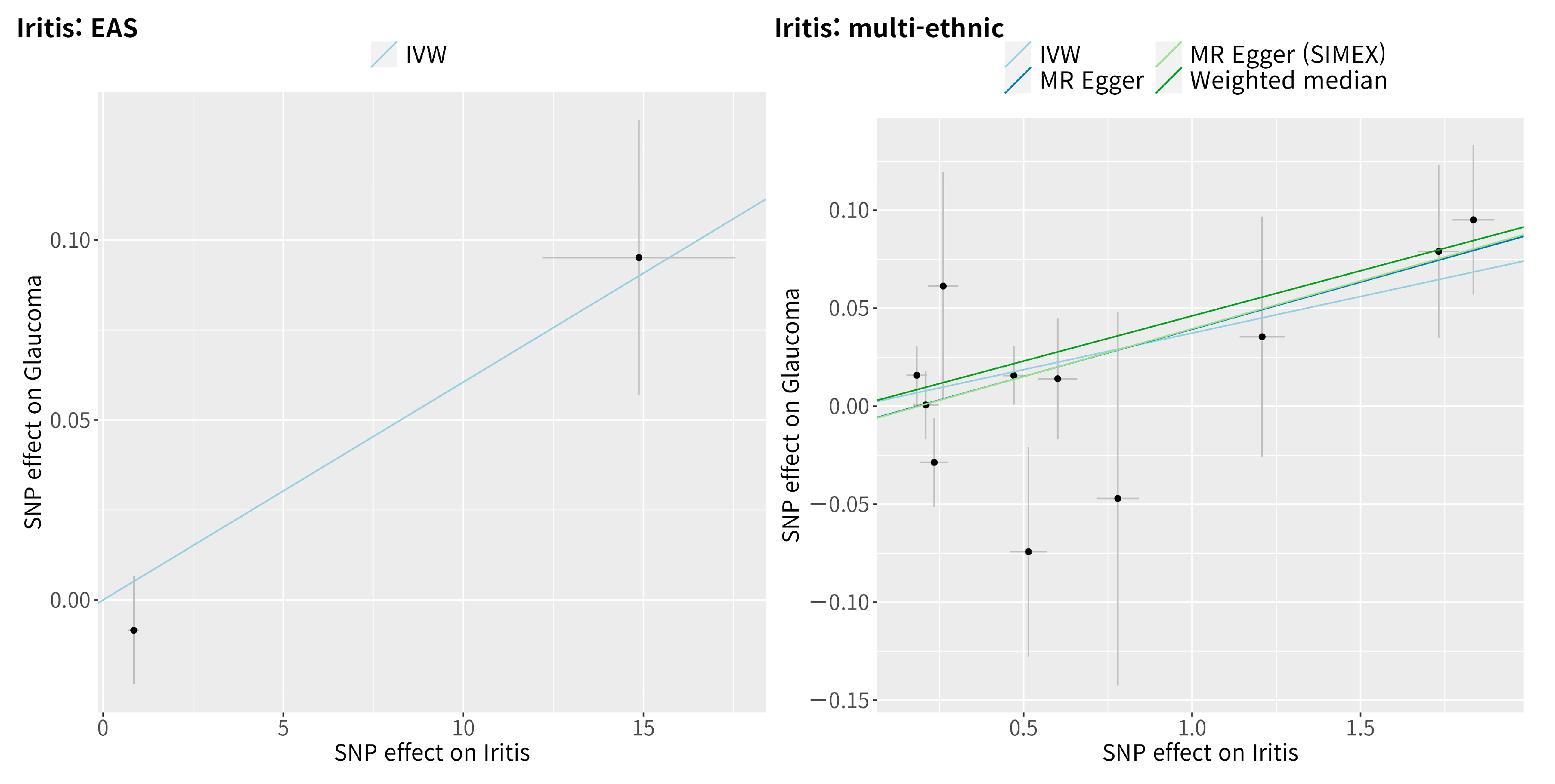
3.3. Mendelian Randomisation for the Casual Association between Iritis and Glaucoma

The IVW method showed a significant causal association between iritis and glaucoma in the East Asian population (MR OR = 1.01, 95% confidence interval (CI): 1.00–1.01, p = 0.017, Figure 2). Similarly, the IVW method also showed a significant causal association between iritis and glaucoma in the multi-ethnic population (IVW MR OR = 1.04, 95% CI: 1.01–1.06, p = 0.001, weighted median MR OR = 1.05, 95% CI: 1.02–1.08, p = 0.003, MR–Egger MR OR = 1.05, 95% CI: 1.01–1.09, p = 0.028, and MR–Egger [SIMEX] MR OR = 1.05, 95% CI: 1.02–1.09, p = 0.020). Scatter plots show the genetic association with iritis against the genetic association with glaucoma for each SNP (Figure 3).

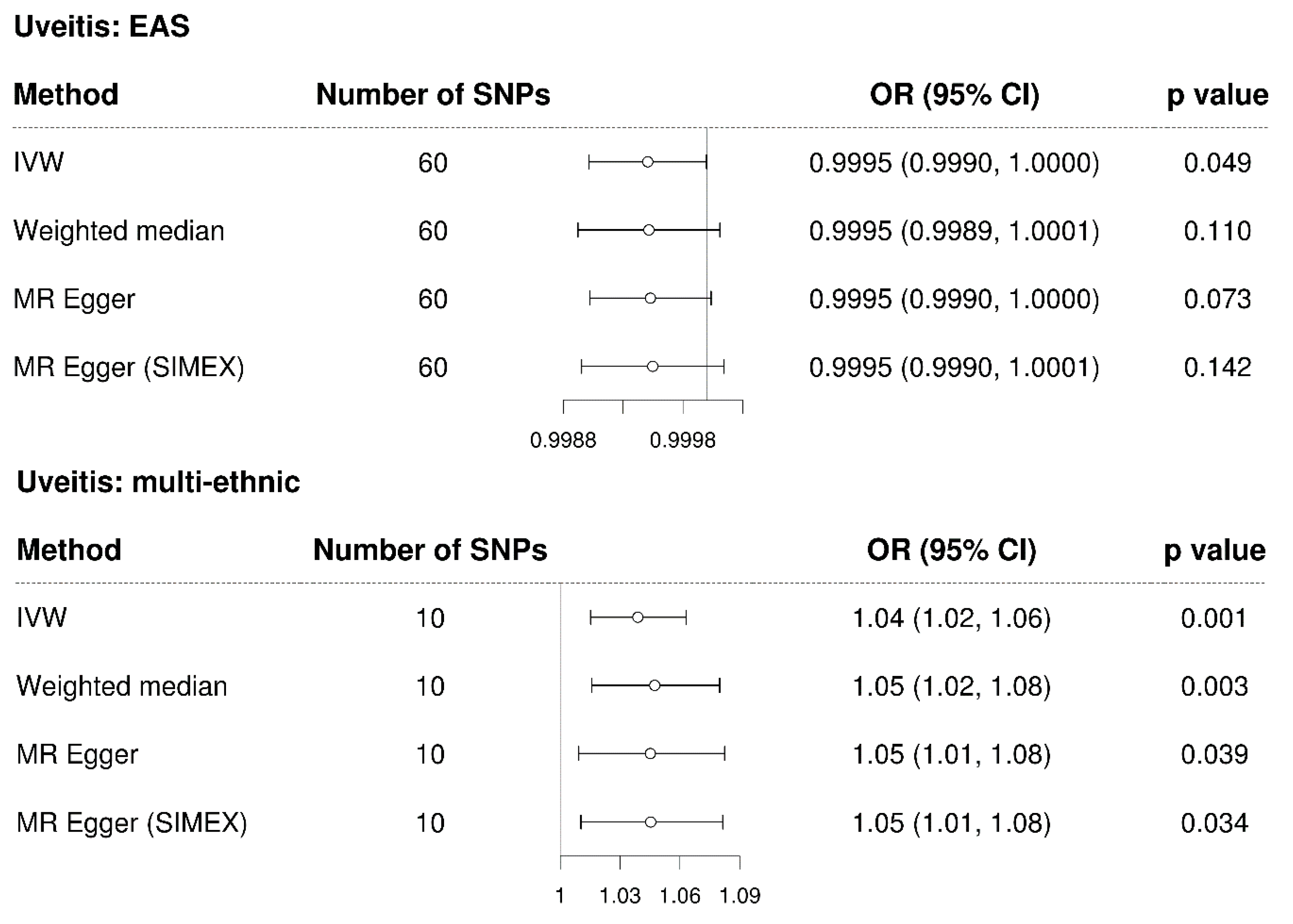
3.4. Mendelian Randomisation for the Casual Association between Uveitis and Glaucoma

The IVW method showed a non-significant causal association between uveitis and glaucoma in the East Asian population (MR OR = 0.9995, 95% CI: 0.9990–1.0000, p = 0.49, Figure 4). Since significant SNPs were not available (p < 5.0 ×   10 8 ), a less significant SNP (p < 5.0 ×   10 4 ) for uveitis in the East Asian population was chosen for analysis; therefore, the dependability of the analysis was low, as anticipated. In contrast, the IVW method showed a significant causal association between uveitis and glaucoma in the multi-ethnic population (IVW MR OR = 1.04, 95% CI: 1.02–1.06, p = 0.001, weighted median MR OR = 1.05, 95% CI: 1.02–1.08, p = 0.003, MR–Egger MR OR = 1.05, 95% CI: 1.01–1.08, p = 0.039, and MR–Egger [SIMEX] MR OR = 1.05, 95% CI: 1.01–1.08, p = 0.034). Scatter plots show the genetic association with uveitis against the genetic association with glaucoma for each SNP (Figure 5); a negative slope was observed for the East Asian population, whereas a significant positive slope was observed for the multi-ethnic population.

4. Discussion

Our study demonstrates a causal association between iritis and glaucoma in an East Asian population and a multi-ethnic population. In addition, uveitis showed a causal association with glaucoma in the multi-ethnic population, although power constraints impeded the detection of this in the East Asian population.
Research on the epidemiology of uveitis have shown results that are highly variable, primarily due to substantial methodological discrepancies. A recent systematic study on the frequency of uveitis showed prevalence numbers ranging from 9 to 730 occurrences per 100,000 and an overall incidence of 50.45 instances per 100,000, suggesting that geographical location is a significant driver of heterogeneity [41]. According to estimates, uveitis affects at least 2 million people worldwide and is a leading cause of blindness [42]. The inflammatory process that occurs within uveitis triggers the development of complications such as cataracts, macular edema, glaucoma, and retinal detachment [43]. According to one study, posterior synechiae (19.0%) was the most prevalent consequence of uveitis, followed by ocular hypertension (14.0%), macular edema (7.5%), and glaucoma (6.6%) [43]. Causal experimental studies on the induction of glaucoma by such severe uveitis have rarely been conducted. In this respect, this study is expected to be meaningful. Causal interference plays a major role in genetic epidemiology and clinical investigation and understanding the aetiology is implicit for identifying disease prevention and treatment opportunities. Existing epidemiological studies have limitations in the case of rare diseases; therefore, a prospective cohort study would be ideal. The MR is advantageous in this respect. Several MR studies have been conducted on ocular disease as a powerful approach to identify causal interference using human genetic data [44]. MR analysis has been used to investigate the impact of education levels, vitamin D, or medication on myopia; and lipid levels, refractive errors, or c-reactive protein levels on age-related macular degeneration [44]. In addition, lipid levels, central corneal thickness, type 2 diabetes, and refractive errors have been evaluated as risk factors for glaucoma in MR analyses [45,46,47]. From this perspective, the impact of iritis or uveitis exposure on the development of glaucoma can be investigated through a MR research approach.
The association between uveitis and glaucoma was first reported in 1813 as arthritic iritis followed glaucoma [2]. UG is one of the most common intraocular complications, and glaucoma after uveitis has an incidence of 11% after 5 years [10]. According to a Turkish epidemiologic study on 4604 eyes with glaucoma [48], UG accounted for 4.1% of all cases, and 92.4% of UG cases were OAG. In addition, glaucoma developed in 6.6% of patients with uveitis 1 year later [43]. While there have been numerous studies on the relationship between glaucoma and uveitis, the present study was performed as there was no data to support a causal relationship based on genetic determinants. Since uveitis is a rare disease and highly likely to be related to the HLA region, it is expected that fine-mapping analysis or next-generation sequencing will yield more desirable data. From this perspective, the exposure of SNPs associated with iritis and uveitis was confirmed to be involved in the occurrence of glaucoma, and a clear conclusion could be reached. SNPs, such as rs146683910, had a high positive beta value (beta = 14.87, Table S1), which was related to the HLA-B locus associated with ankylosing spondylitis [49] (https://pheweb.org/UKB-TOPMed/pheno/715.2). An empirical study reported that HLA-B may be associated with OAG [50,51]. The causative analysis of uveitis and glaucoma also included immune-related genes in additional to HLA. The major histocompatability complex class I polypeptide-related sequence A (MICA) gene-related rs115681000 (beta = 1.732, located MICA;LINC01149) and rs146683910 (beta = 1.835, located HLA-B;MICA-AS1) also had high beta values (Table S1). The LINC01149 variation modifies the expression of MICA, making it easier for it to function as a key gene in the susceptibility to uveitis development. Nevertheless, despite the fact that the MR analysis used SNPs were primarily concentrated on chromosome 6, significant results were still seen despite the analysis’s usage of a relatively small number of SNPs due to the LD with R2 < 0.001 within 10,000 kb to ensure the independence of the IVs.
AAU is the most common type of uveitis, which is related to synechiae formation. Glaucoma, which is frequently associated with AS, psoriatic arthritis, and inflammatory bowel disease [52,53], had characteristics of autoimmune and inflammatory illnesses. In addition, a recent study using whole-exome sequencing identified rare variants and genes associated with IOP and glaucoma, including BOD1L1, ACAD10, and HLA-B, demonstrating the power of including and aggregating rare variants [54]. HLA-associated regions, such as HLA-DRB1/DQA1, HLA-C, and HLA-DOA/HLA-DPA1, were adopted while evaluating the SNPs utilized in our analysis (Table S1). These loci showed significant association with uveitis, such as AAU, AS, and BD [5,13,49,52,53]. Moreover, significant SNPs were identified on comparing East Asian genetic data with the meta-analysis data, despite the ethnic differences. Nevertheless, our study demonstrated the value of using rare variants to enhance our understanding of the biological mechanisms regulating IOP and uncovered potential therapeutic targets for glaucoma [54], which we believe is consistent with our findings. Moreover, in our study, an association with glaucoma was found without intraocular pressure phenotype analysis, which further supports the findings. It is anticipated that future research will separate the description of disease into causal linkages rather than currently recognized categories.
The main strength of this study is the utilization of a relatively large Asian and European cohort dataset to reveal the causal association between iritis or uveitis and glaucoma. However, there are a few limitations of this study. First, we were unable to explain the presence of numerous confounding factors using summary statistics based on two-sample MR since we did not have access to individual-level data. Second, since iritis and uveitis are very infrequent disorders associated with immune-related (such as HLA) SNPs, they were not ideal study topics for MR analysis. However, the significant causality that emerged despite these limitations suggests that there is a very strong association. Third, there are test procedures to validate the MR hypotheses, but they do not provide complete validation. Therefore, as violation of MR assumptions can lead to invalid conclusions, the results should be interpreted with caution.

5. Conclusions

We provide strong genetic evidence that supports a causal relationship between iritis and glaucoma in East Asian and multi-ethnic populations. Additionally, uveitis was found to have a causal effect on glaucoma in the multi-ethnic population, although there was limited causal evidence of this in the East Asian population owing to the lower power of IVs for that assumption. These causal correlations between iritis or uveitis and glaucoma imply that the current classifications of UG and OAG overlap, highlighting the need for additional research and caution in interpreting these complex interactions.

Supplementary Materials

The following supporting information can be downloaded at: https://www.mdpi.com/article/10.3390/genes14030642/s1, Supplementary Table S1: List of single-nucleotide polymorphisms used in this MR study as instrumental variables.

Author Contributions

Conceptualization, J.H.S. and Y.L.; methodology, J.H.S. and Y.L.; software, Y.L.; validation, J.H.S. and Y.L.; formal analysis, J.H.S. and Y.L.; investigation, Y.L.; resources, J.H.S.; data curation, J.H.S. and Y.L.; writing—original draft preparation, J.H.S. and Y.L.; writing—review and editing, J.H.S. and Y.L.; visualization, Y.L.; supervision, J.H.S.; project administration, J.H.S.; funding acquisition, J.H.S. All authors have read and agreed to the published version of the manuscript.

Funding

This study was supported by the Veterans Health Service Medical Center Research Grant (No. VHSMC22045) and National Research Foundation of Korea (NRF) grant, funded by the Korean government (Ministry of Science and ICT) (No. 2022R1C1C1002929).

Institutional Review Board Statement

The study was conducted in accordance with the Declaration of Helsinki, and approved by the Institutional Review Board of the Veterans Health Service Medical Center (IRB No. 2022-12-007; 16 December 2022).

Informed Consent Statement

Informed consent was not required for this study because anonymized and de-identified data were used for the analyses. Patient consent was waived due to the retrospective nature of the study.

Data Availability Statement

The datasets for the genome-wide association study (GWAS) summary statistics can be found in the GWAS Catalog (https://www.ebi.ac.uk/gwas/summary-statistics, accessed on 19 July 2022).

Acknowledgments

Biobank Japan (BBJ https://pheweb.jp/, accessed on 19 July 2022) [29]. Genetic Epidemiology Research in Adult Health and Aging (GERA) cohort and UK biobank (UKB) [20] from GWAS catalogue (https://www.ebi.ac.uk/gwas/summary-statistics, accessed on 19 July 2022).

Conflicts of Interest

The authors declare no conflict of interest. The funders had no role in the design of the study; in the collection, analyses, or interpretation of data; in the writing of the manuscript; or in the decision to publish the results.

References

  1. Tsirouki, T.; Dastiridou, A.; Symeonidis, C.; Tounakaki, O.; Brazitikou, I.; Kalogeropoulos, C.; Androudi, S. A Focus on the Epidemiology of Uveitis. Ocul. Immunol. Inflamm. 2018, 26, 2–16. [Google Scholar] [CrossRef] [PubMed]
  2. Siddique, S.S.; Suelves, A.M.; Baheti, U.; Foster, C.S. Glaucoma and uveitis. Surv. Ophthalmol. 2013, 58, 1–10. [Google Scholar] [CrossRef] [PubMed]
  3. Quigley, H.A. Glaucoma. Lancet 2011, 377, 1367–1377. [Google Scholar] [CrossRef] [PubMed]
  4. Tham, Y.C.; Li, X.; Wong, T.Y.; Quigley, H.A.; Aung, T.; Cheng, C.Y. Global prevalence of glaucoma and projections of glaucoma burden through 2040: A systematic review and meta-analysis. Ophthalmology 2014, 121, 2081–2090. [Google Scholar] [CrossRef]
  5. Hou, S.; Li, N.; Liao, X.; Kijlstra, A.; Yang, P. Uveitis genetics. Exp. Eye Res. 2020, 190, 107853. [Google Scholar] [CrossRef] [PubMed]
  6. Takeuchi, M.; Mizuki, N.; Ohno, S. Pathogenesis of Non-Infectious Uveitis Elucidated by Recent Genetic Findings. Front. Immunol. 2021, 12, 640473. [Google Scholar] [CrossRef]
  7. Su, G.; Zhong, Z.; Zhou, Q.; Du, L.; Ye, Z.; Li, F.; Zhuang, W.; Wang, C.; Liang, L.; Ji, Y.; et al. Identification of Novel Risk Loci for Behcet’s Disease-Related Uveitis in a Chinese Population in a Genome-Wide Association Study. Arthritis. Rheumatol. 2022, 74, 671–681. [Google Scholar] [CrossRef] [PubMed]
  8. Merayo-Lloves, J.; Power, W.J.; Rodriguez, A.; Pedroza-Seres, M.; Foster, C.S. Secondary glaucoma in patients with uveitis. Ophthalmologica 1999, 213, 300–304. [Google Scholar] [CrossRef]
  9. Takahashi, T.; Ohtani, S.; Miyata, K.; Miyata, N.; Shirato, S.; Mochizuki, M. A clinical evaluation of uveitis-associated secondary glaucoma. Jpn. J. Ophthalmol. 2002, 46, 556–562. [Google Scholar] [CrossRef]
  10. Herbert, H.M.; Viswanathan, A.; Jackson, H.; Lightman, S.L. Risk factors for elevated intraocular pressure in uveitis. J. Glaucoma 2004, 13, 96–99. [Google Scholar] [CrossRef]
  11. Weinreb, R.N.; Aung, T.; Medeiros, F.A. The pathophysiology and treatment of glaucoma: A review. JAMA 2014, 311, 1901–1911. [Google Scholar] [CrossRef] [PubMed] [Green Version]
  12. Alvarado, J.A.; Murphy, C.G. Outflow obstruction in pigmentary and primary open angle glaucoma. Arch. Ophthalmol. 1992, 110, 1769–1778. [Google Scholar] [CrossRef]
  13. Huang, X.F.; Brown, M.A. Progress in the genetics of uveitis. Genes. Immun. 2022, 23, 57–65. [Google Scholar] [CrossRef] [PubMed]
  14. Wiggs, J.L.; Pasquale, L.R. Genetics of glaucoma. Hum. Mol. Genet. 2017, 26, R21–R27. [Google Scholar] [CrossRef] [PubMed] [Green Version]
  15. Gharahkhani, P.; Jorgenson, E.; Hysi, P.; Khawaja, A.P.; Pendergrass, S.; Han, X.; Ong, J.S.; Hewitt, A.W.; Segre, A.V.; Rouhana, J.M.; et al. Genome-wide meta-analysis identifies 127 open-angle glaucoma loci with consistent effect across ancestries. Nat. Commun. 2021, 12, 1258. [Google Scholar] [CrossRef] [PubMed]
  16. Youngblood, H.; Hauser, M.A.; Liu, Y. Update on the genetics of primary open-angle glaucoma. Exp. Eye Res. 2019, 188, 107795. [Google Scholar] [CrossRef]
  17. Sakurada, Y.; Mabuchi, F. Advances in glaucoma genetics. Prog. Brain Res. 2015, 220, 107–126. [Google Scholar] [CrossRef]
  18. Shin, J.H.; Lee, J.W.; Lim, S.H.; Yoon, B.W.; Lee, Y.; Seo, J.H. The microbiomes of the eyelid and buccal area of patients with uveitic glaucoma. BMC Ophthalmol. 2022, 22, 170. [Google Scholar] [CrossRef]
  19. Lee, J.W.; Lim, S.H.; Shin, J.H.; Lee, Y.; Seo, J.H. Differences in the eyelid and buccal microbiome between open-angle glaucoma and uveitic glaucoma. Acta Ophthalmol. 2022, 100, e770–e778. [Google Scholar] [CrossRef]
  20. Choquet, H.; Paylakhi, S.; Kneeland, S.C.; Thai, K.K.; Hoffmann, T.J.; Yin, J.; Kvale, M.N.; Banda, Y.; Tolman, N.G.; Williams, P.A.; et al. A multiethnic genome-wide association study of primary open-angle glaucoma identifies novel risk loci. Nat. Commun. 2018, 9, 2278. [Google Scholar] [CrossRef] [Green Version]
  21. Shin, H.T.; Yoon, B.W.; Seo, J.H. Analysis of risk allele frequencies of single nucleotide polymorphisms related to open-angle glaucoma in different ethnic groups. BMC Med. Genomics 2021, 14, 80. [Google Scholar] [CrossRef] [PubMed]
  22. Yoon, B.W.; Shin, H.T.; Seo, J.H. Risk Allele Frequency Analysis and Risk Prediction of Single-Nucleotide Polymorphisms for Prostate Cancer. Genes 2022, 13, 2039. [Google Scholar] [CrossRef]
  23. Yoon, B.W.; Shin, H.T.; Seo, J. Risk Allele Frequency Analysis of Single-Nucleotide Polymorphisms for Vitamin D Concentrations in Different Ethnic Group. Genes 2021, 12, 1530. [Google Scholar] [CrossRef]
  24. Shin, H.T.; Yoon, B.W.; Seo, J.H. Comparison of risk allele frequencies of single nucleotide polymorphisms associated with age-related macular degeneration in different ethnic groups. BMC Ophthalmol. 2021, 21, 97. [Google Scholar] [CrossRef]
  25. Burgess, S.; Thompson, S.G. Multivariable Mendelian randomization: The use of pleiotropic genetic variants to estimate causal effects. Am. J. Epidemiol. 2015, 181, 251–260. [Google Scholar] [CrossRef] [Green Version]
  26. Burgess, S.; Thompson, S.G. Interpreting findings from Mendelian randomization using the MR-Egger method. Eur. J. Epidemiol. 2017, 32, 377–389. [Google Scholar] [CrossRef] [PubMed] [Green Version]
  27. Hanyuda, A.; Goto, A.; Nakatochi, M.; Sutoh, Y.; Narita, A.; Nakano, S.; Katagiri, R.; Wakai, K.; Takashima, N.; Koyama, T.; et al. Association Between Glycemic Traits and Primary Open-Angle Glaucoma: A Mendelian Randomization Study in the Japanese Population. Am. J. Ophthalmol. 2022, 245, 193–201. [Google Scholar] [CrossRef] [PubMed]
  28. Kim, J.; Aschard, H.; Kang, J.H.; Lentjes, M.A.H.; Do, R.; Wiggs, J.L.; Khawaja, A.P.; Pasquale, L.R.; Modifiable Risk Factors for Glaucoma Collaboration. Intraocular Pressure, Glaucoma, and Dietary Caffeine Consumption: A Gene-Diet Interaction Study from the UK Biobank. Ophthalmology 2021, 128, 866–876. [Google Scholar] [CrossRef]
  29. Sakaue, S.; Kanai, M.; Tanigawa, Y.; Karjalainen, J.; Kurki, M.; Koshiba, S.; Narita, A.; Konuma, T.; Yamamoto, K.; Akiyama, M.; et al. A cross-population atlas of genetic associations for 220 human phenotypes. Nat. Genet. 2021, 53, 1415–1424. [Google Scholar] [CrossRef]
  30. Loh, M.; Zhang, W.; Ng, H.K.; Schmid, K.; Lamri, A.; Tong, L.; Ahmad, M.; Lee, J.J.; Ng, M.C.Y.; Petty, L.E.; et al. Identification of genetic effects underlying type 2 diabetes in South Asian and European populations. Commun. Biol. 2022, 5, 329. [Google Scholar] [CrossRef] [PubMed]
  31. Burgess, S.; Thompson, S.G.; Collaboration, C.C.G. Avoiding bias from weak instruments in Mendelian randomization studies. Int. J. Epidemiol. 2011, 40, 755–764. [Google Scholar] [CrossRef] [Green Version]
  32. Burgess, S.; Butterworth, A.; Thompson, S.G. Mendelian randomization analysis with multiple genetic variants using summarized data. Genet. Epidemiol. 2013, 37, 658–665. [Google Scholar] [CrossRef] [PubMed] [Green Version]
  33. Bowden, J.; Del Greco, M.F.; Minelli, C.; Davey Smith, G.; Sheehan, N.; Thompson, J. A framework for the investigation of pleiotropy in two-sample summary data Mendelian randomization. Stat. Med. 2017, 36, 1783–1802. [Google Scholar] [CrossRef] [PubMed] [Green Version]
  34. Lee, Y.; Kim, Y.A.; Seo, J.H. Causal Association of Obesity and Dyslipidemia with Type 2 Diabetes: A Two-Sample Mendelian Randomization Study. Genes 2022, 13, 2407. [Google Scholar] [CrossRef]
  35. Bowden, J.; Davey Smith, G.; Haycock, P.C.; Burgess, S. Consistent Estimation in Mendelian Randomization with Some Invalid Instruments Using a Weighted Median Estimator. Genet. Epidemiol. 2016, 40, 304–314. [Google Scholar] [CrossRef] [PubMed] [Green Version]
  36. Bowden, J.; Davey Smith, G.; Burgess, S. Mendelian randomization with invalid instruments: Effect estimation and bias detection through Egger regression. Int. J. Epidemiol. 2015, 44, 512–525. [Google Scholar] [CrossRef] [Green Version]
  37. Bowden, J.; Del Greco, M.F.; Minelli, C.; Davey Smith, G.; Sheehan, N.A.; Thompson, J.R. Assessing the suitability of summary data for two-sample Mendelian randomization analyses using MR-Egger regression: The role of the I2 statistic. Int. J. Epidemiol. 2016, 45, 1961–1974. [Google Scholar] [CrossRef] [PubMed] [Green Version]
  38. Burgess, S.; Davey Smith, G.; Davies, N.M.; Dudbridge, F.; Gill, D.; Glymour, M.M.; Hartwig, F.P.; Holmes, M.V.; Minelli, C.; Relton, C.L.; et al. Guidelines for performing Mendelian randomization investigations. Wellcome Open Res. 2019, 4, 186. [Google Scholar] [CrossRef] [Green Version]
  39. Greco, M.F.; Minelli, C.; Sheehan, N.A.; Thompson, J.R. Detecting pleiotropy in Mendelian randomisation studies with summary data and a continuous outcome. Stat. Med. 2015, 34, 2926–2940. [Google Scholar] [CrossRef] [Green Version]
  40. Verbanck, M.; Chen, C.Y.; Neale, B.; Do, R. Publisher Correction: Detection of widespread horizontal pleiotropy in causal relationships inferred from Mendelian randomization between complex traits and diseases. Nat. Genet. 2018, 50, 1196. [Google Scholar] [CrossRef]
  41. Garcia-Aparicio, A.; Garcia de Yebenes, M.J.; Oton, T.; Munoz-Fernandez, S. Prevalence and Incidence of Uveitis: A Systematic Review and Meta-analysis. Ophthalmic. Epidemiol. 2021, 28, 461–468. [Google Scholar] [CrossRef] [PubMed]
  42. Kanda, T.; Shibata, M.; Taguchi, M.; Ishikawa, S.; Harimoto, K.; Takeuchi, M. Prevalence and aetiology of ocular hypertension in acute and chronic uveitis. Br. J. Ophthalmol. 2014, 98, 932–936. [Google Scholar] [CrossRef] [PubMed]
  43. Garcia-Aparicio, A.; Alonso Martin, L.; Quiros Zamorano, R.; Lopez Lancho, R.; Del Olmo Perez, L.; Sanchez Fernandez, S.; Garcia de Yebenes, M.J.; Jimenez Escribano, R.; Gonzalez Del Valle, F.; Munoz-Fernandez, S. Complications of uveitis in a Spanish population, UveCAM study. Arch. Soc. Esp. Oftalmol. (Engl. Ed.) 2022, 97, 244–250. [Google Scholar] [CrossRef] [PubMed]
  44. Li, J.; Li, C.; Huang, Y.; Guan, P.; Huang, D.; Yu, H.; Yang, X.; Liu, L. Mendelian randomization analyses in ocular disease: A powerful approach to causal inference with human genetic data. J. Transl. Med. 2022, 20, 621. [Google Scholar] [CrossRef]
  45. Xu, M.; Li, S.; Zhu, J.; Luo, D.; Song, W.; Zhou, M. Plasma lipid levels and risk of primary open angle glaucoma: A genetic study using Mendelian randomization. BMC Ophthalmol. 2020, 20, 390. [Google Scholar] [CrossRef]
  46. Choquet, H.; Melles, R.B.; Yin, J.; Hoffmann, T.J.; Thai, K.K.; Kvale, M.N.; Banda, Y.; Hardcastle, A.J.; Tuft, S.J.; Glymour, M.M.; et al. A multiethnic genome-wide analysis of 44,039 individuals identifies 41 new loci associated with central corneal thickness. Commun. Biol. 2020, 3, 301. [Google Scholar] [CrossRef]
  47. Chong, R.S.; Li, H.; Cheong, A.J.; Fan, Q.; Koh, V.; Raghavan, L.; Nongpiur, M.E.; Cheng, C.Y. Mendelian Randomization Implicates Bidirectional Association Between Myopia and Primary Open Angle Glaucoma or Intraocular Pressure. Ophthalmology 2022. ahead of print. [Google Scholar] [CrossRef]
  48. Tekeli, O.; Elgin, U.; Takmaz, T.; Eksioglu, U.; Bas, Z.; Yarangumeli, A.; Karakurt, A.; Evren Kemer, O.; Mumcuoglu, T.; Aktas, Z.; et al. Characteristics of uveitic glaucoma in Turkish patients. Eur. J. Ophthalmol. 2021, 31, 1836–1843. [Google Scholar] [CrossRef]
  49. Chen, B.; Li, J.; He, C.; Li, D.; Tong, W.; Zou, Y.; Xu, W. Role of HLA-B27 in the pathogenesis of ankylosing spondylitis (Review). Mol. Med. Rep. 2017, 15, 1943–1951. [Google Scholar] [CrossRef] [Green Version]
  50. Mayr, W.R.; Grabner, G. HLA antigens-risk factors for primary open-angle glaucoma? Albrecht Von Graefes Arch Klin Exp Ophthalmol. 1978, 207, 291–297. [Google Scholar] [CrossRef]
  51. Wang, R.F.; Chu, R.Y.; Guo, B.K. HLA-A, B antigens and the relation between high myopia and high myopia with glaucoma. Chin. Med. J. 1986, 99, 775–778. [Google Scholar] [PubMed]
  52. Monnet, D.; Breban, M.; Hudry, C.; Dougados, M.; Brezin, A.P. Ophthalmic findings and frequency of extraocular manifestations in patients with HLA-B27 uveitis: A study of 175 cases. Ophthalmology 2004, 111, 802–809. [Google Scholar] [CrossRef] [PubMed]
  53. Wang, Y.; Huang, X.F.; Yang, M.M.; Cai, W.J.; Zheng, M.Q.; Mao, G.; Pang, C.P.; Jin, Z.B. CFI-rs7356506 is a genetic protective factor for acute anterior uveitis in Chinese patients. Br. J. Ophthalmol. 2014, 98, 1592–1596. [Google Scholar] [CrossRef] [PubMed]
  54. Gao, X.R.; Chiariglione, M.; Arch, A.J. Whole-exome sequencing study identifies rare variants and genes associated with intraocular pressure and glaucoma. Nat. Commun. 2022, 13, 7376. [Google Scholar] [CrossRef]
Figure 1. Schematic of the study design. SNP: single-nucleotide polymorphism.
Genes 14 00642 g001
Figure 2. Forest plot of causal associations between iritis and glaucoma. EAS: East Asians; SNP: single-nucleotide polymorphism; OR: odds ratio; CI: confidence interval; IVW: inverse-variance weight; MR: Mendelian randomisation; SIMEX: simulation extrapolation.
Genes 14 00642 g002
Figure 3. Scatter plots of Mendelian randomisation tests assessing the effect of the presence of iritis on glaucoma. The IVW, MR–Egger, MR–Egger (SIMEX), and weighted median estimate are indicated by the light blue, dark blue, light green, and dark green regression lines, respectively. SNP: single-nucleotide polymorphism; IVW: inverse-variance weight; MR: Mendelian randomisation; SIMEX: simulation extrapolation.
Genes 14 00642 g003
Figure 4. Forest plot of the causal associations between uveitis and glaucoma. EAS: East Asians; SNP: single-nucleotide polymorphism; OR: odds ratio; CI: confidence interval; IVW: inverse-variance weight; MR: Mendelian randomisation; SIMEX: simulation extrapolation.
Genes 14 00642 g004
Figure 5. Scatter plots of the Mendelian randomisation tests assessing the effect of the presence of uveitis on glaucoma. The IVW, MR–Egger, MR–Egger (SIMEX), and weighted median estimate are indicated by the light blue, dark blue, light green, and dark green regression lines, respectively. SNP: single-nucleotide polymorphism; IVW: inverse-variance weight; MR: Mendelian randomisation; SIMEX: simulation extrapolation.
Genes 14 00642 g005
Table 1. Summary statistics of the data sources.
TraitsData SourceNo. of ParticipantsPopulationNo. of VariantsReference
IritisBBJ Project175,653East Asian13,429,677[29]
BBJ Project + UKB656,395East Asian + European25,844,196[29]
UveitisBBJ Project174,725East Asian13,429,518[29]
BBJ Project + UKB655,467East Asian + European25,844,095[29]
Glaucoma(Open-angle glaucoma)GERA cohort + UKB240,302 (12,315 cases + 227,987 control)Multiethnic:
214,102 European
5103 African unspecified
3571 Other mixed ancestry
1847 African American or Afro-Caribbean
5189 Hispanic or Latin American
5370 East Asian
5120 South Asian
7,760,820[20]
BBJ, BioBank Japan; GERA, Genetic Epidemiology Research in Adult Health and Aging; UKB, UK Biobank.
Table 2. Heterogeneity and horizontal pleiotropy of instrumental variables.
Exposure HeterogeneityHorizontal Pleiotropy
Cochran’s Q Test from IVWRucker’s Q’ Test from MR-EggerMR-PRESSO Global TestMR–EggerMR–Egger (SIMEX)
NFI2 (%)p-Valuep-Valuep-ValueIntercept, β (SE)p-ValueIntercept, β (SE)p-Value
Iritis24158896.330.359
Iritis *11770499.110.5870.5580.659−0.009 (0.011)0.435−0.009 (0.010)0.389
Uveitis60643385.640.1480.1310.521−0.001 (0.003)0.688−0.001 (0.003)0.682
Uveitis *10839699.050.5580.4800.644−0.005 (0.01)0.644−0.005 (0.010)0.629
N, number of instrument variables; F, mean F statistic; IVW, inverse-variance weight; MR, Mendelian randomization; PRESSO, polyhedral sum of residuals and outliers; SIMEX, simulation extrapolation; β, beta coefficient; SE, standard error. *: indicates exposure using the SNPs selected from the meta-analysis from BioBank Japan Project and UK Biobank.
Disclaimer/Publisher’s Note: The statements, opinions and data contained in all publications are solely those of the individual author(s) and contributor(s) and not of MDPI and/or the editor(s). MDPI and/or the editor(s) disclaim responsibility for any injury to people or property resulting from any ideas, methods, instructions or products referred to in the content.

