Communication

The Significance of Mylohyoid Muscle Release (MMR) in the Vertical and Horizontal Ridge Augmentation Surgeries; Clinical and Human Cadaver Analyses of the Techniques

1
Ashman Department of Periodontics and Implant Dentistry, NYU College of Dentistry, New York University, 345 E. 24th Street, New York, NY 10010, USA
2
Department of Periodontics, School of Dentistry, Oregon Health Science University (OHSU), 2730 SW Moody Ave, Portland, OR 97201, USA
3
School of Dental Medicine, University of Nevada Las Vegas (UNLV), 1001 Shadow Ln., Las Vegas, NV 89106, USA
4
Department of Periodontics and Endodontics, School of Dental Medicine (SDM), Stony Brook University, 106 Rockland Hall, Stony Brook, NY 11794, USA
5
Department of Clinical Sciences, School of Dental Medicine, University of Nevada Las Vegas (UNLV), 1001 Shadow Ln., Las Vegas, NV 89106, USA
*
Author to whom correspondence should be addressed.
Genes 2023, 14(3), 595; https://doi.org/10.3390/genes14030595
Submission received: 3 December 2022 / Revised: 20 February 2023 / Accepted: 25 February 2023 / Published: 26 February 2023

Abstract

:
(1) Background: Ridge augmentations either horizontal (HRA) or vertical (VRA) in the posterior mandible are very challenging regenerative procedures. To attain and retain tension-free primary closure, buccal periosteal and mylohyoid muscle releases should be performed. The purpose of the present study was to review, analyze and discuss the three different techniques for the mylohyoid muscle release (MMR) in VRA and HRA surgeries on a clinical and human cadaver level. (2) Presentation of the techniques: Three different techniques are described in the literature regarding the lingual flap management: (i) the finger sweep technique (FST), (ii) the release of the mylohyoid muscle attachment on the lingual flap (MMALF), and (iii) the mylohyoid preservation technique (MPT) in three key anatomical zones. All three techniques, even though they use a different approach, can achieve similar amount of horizontal and vertical mylohyoid muscle release although MPT showed statistically significant higher flap advancement. The human cadaver analyses revealed that all three techniques are considered safe since they do not approximate vital anatomical structures. (3) Conclusions: All three techniques are considered safe, but they are not free of limitations or complications; therefore, they should be performed only by highly experienced and trained clinicians. MPT achieved statistically significant higher flap advancement.

1. Introduction

Severe cases of periodontal disease, congenital missing teeth, developmental defects, tooth extractions without ridge preservations, odontogenic cysts, tumors, and trauma are some of the etiological findings that can lead to Seibert class I, II, or III defects and require ridge augmentation surgeries [1,2,3,4]. Ridge augmentation can be achieved with the use of distraction osteogenesis, bone blocks, or guided bone regeneration (GBR). GBR has become more predictable approach, with less morbidity for the patients over the years, due to advancement of the surgical techniques and the barrier membranes, and the use of autogenous grafts, allografts, and xenografts [5,6,7,8,9,10,11,12].
Ridge augmentations in the posterior mandible, either HRA or VRA, are very challenging regenerative procedures. The consensus report of the 15th European workshop of Periodontology on bone regeneration and other studies reported that these techniques should be performed by highly experienced and trained clinicians due to complications such as wound dehiscence, membrane exposure, graft exposure, and post-operative infection [12,13,14,15]. Machtei’s findings, presented in a systematic review in 2001, agree with the consensus report since his results showed that membrane exposure yielded a 6-fold greater negative effect on GBR outcomes when compared to guided tissue regeneration (GTR) outcomes [16].
Wound stabilization, adequate blood supply, protection of the underlying blood clot, space maintenance, prevention of the migration of undesired cells from the overlying soft tissue, and tension-free primary closure are principles that should be followed to obtain a successful surgical outcome [13,15,17,18,19,20,21,22,23,24]. Flap tension of 0.01–0.1 N at the time of suturing may result in dehiscence in 10% of the cases, per Burkhardt and Lang in 2010. However, tension more than 0.1 N showed wound dehiscence in 40% of the cases [25].
In the posterior mandibular area, to attain and retain tension-free primary closure, buccal periosteal (after isolating the mental nerve) (Figure 1 and Figure 2a,b) and mylohyoid muscle releases should be performed.
The mylohyoid muscle or the diaphragma oris muscle is a flat and triangular muscle. It is located superior to the anterior belly of the digastric muscle and forms the floor of the mouth. It is one of the suprahyoid muscles, and it is derived from the first pharyngeal arch. It inserts into the body of hyoid bone and elevates the tongue and the hyoid bone. It is innervated by the inferior alveolar nerve. It has oblique line and runs more superior at the area of the first molar (which is very close to the attachment of the mandible) and then runs deeper at the area of first premolar and anterior teeth (Figure 3a,b) [26].
The posterior portion of the mylohyoid muscle derives from the lingual tuberosity below the retromolar pad [26]. The position of the lingual nerve is shown in the Figure 4 in relation to the design of the lingual flap and the MMR.
The mylohyoid muscle separates the sublingual from the submandibular space. These spaces are united in case of odontogenic infections penetrating the muscle [14,26]. A very good understanding of the head and neck anatomy is obligatory to avoid severe complications during VRA and HRA surgeries [14] (Figure 5a,b).
Three different techniques have been described in the literature regarding the lingual flap management using the MMR (Table 1): (i) Finger sweep technique (FST), (ii) the release of the mylohyoid muscle attachment on the lingual flap (MMALF), and (iii) the mylohyoid preservation technique (MPT) in three key anatomical zones [13,15,22,27].
The purpose of the present study was to review/analyze/discuss the three techniques for the release of the mylohyoid muscle in VRA and HRA surgeries in partial edentulous and atrophic posterior mandibular areas, in a clinical setting and in a human cadaver model.

2. Presentation of the Techniques

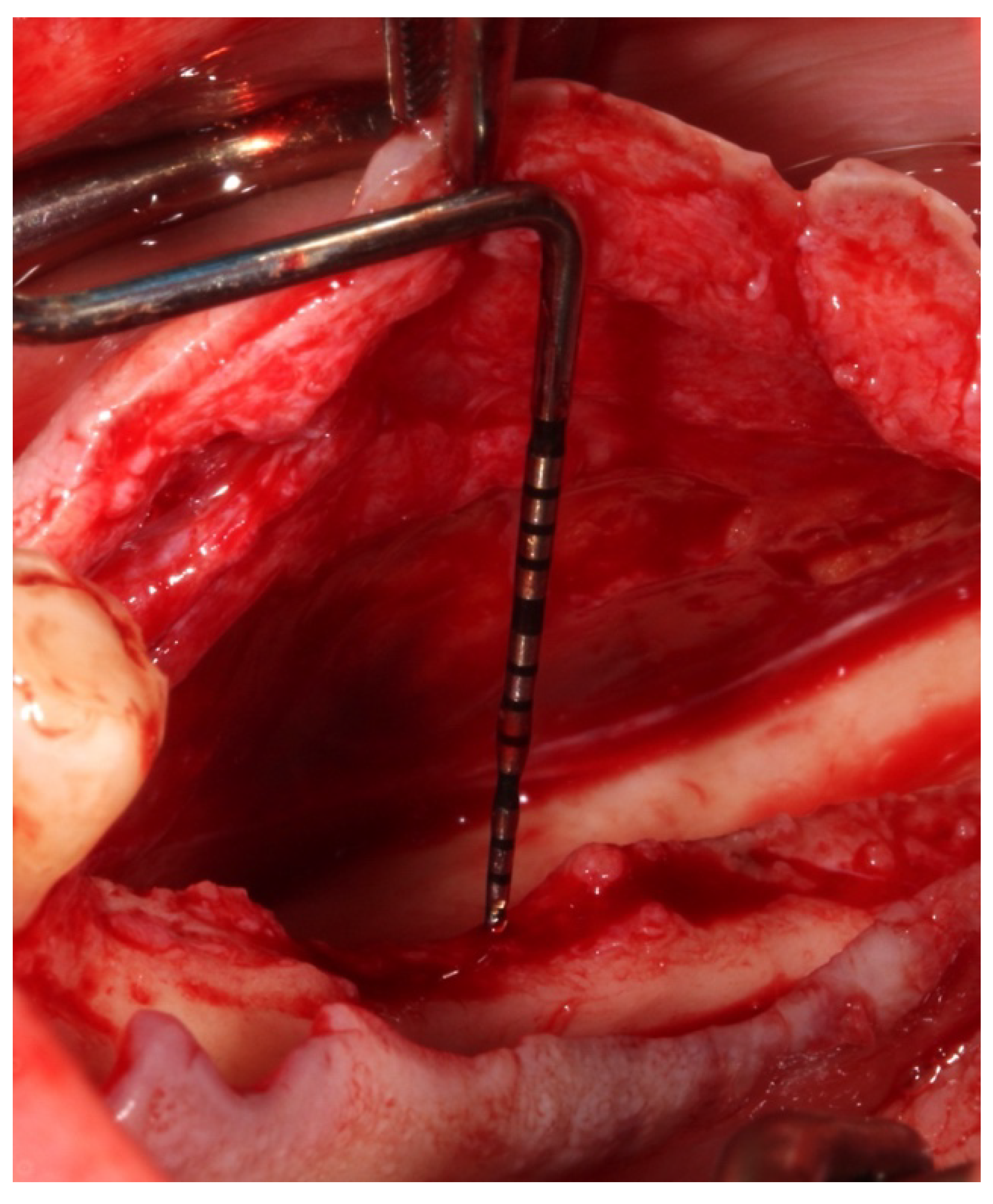
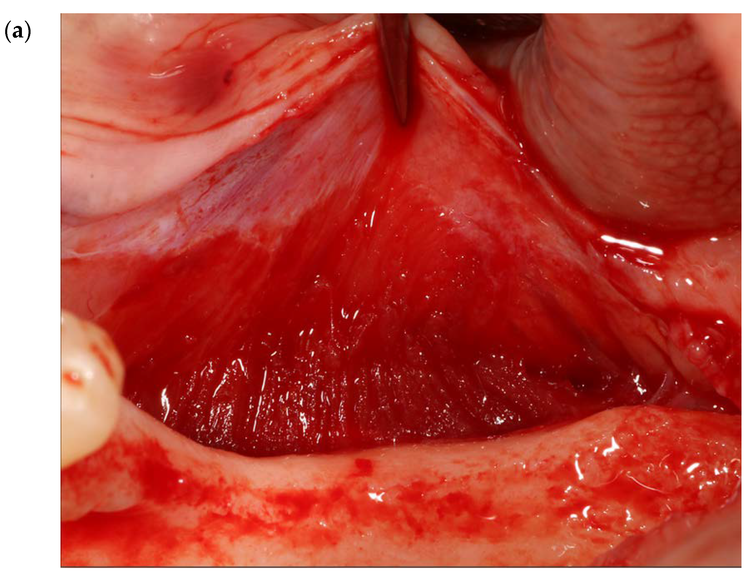
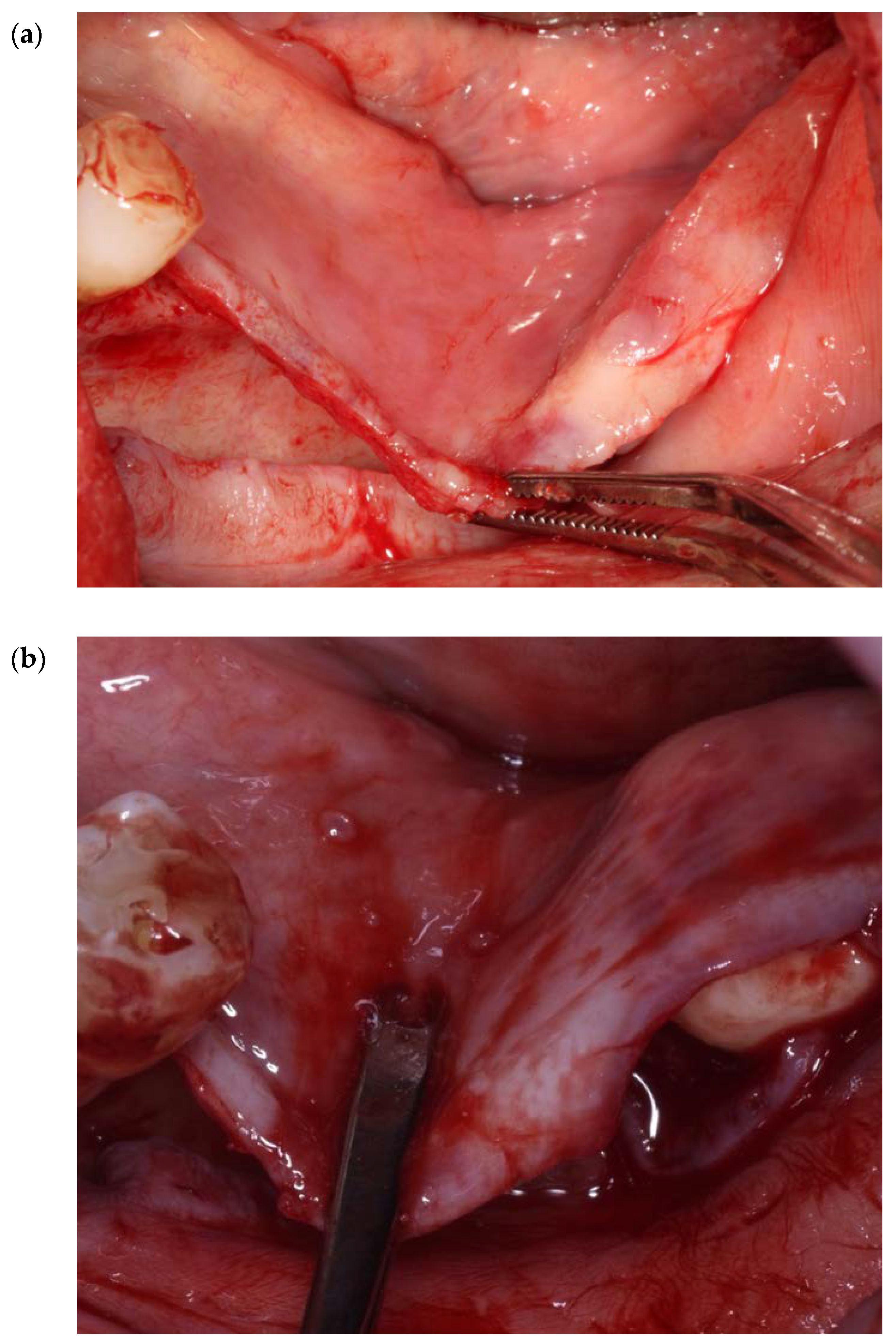
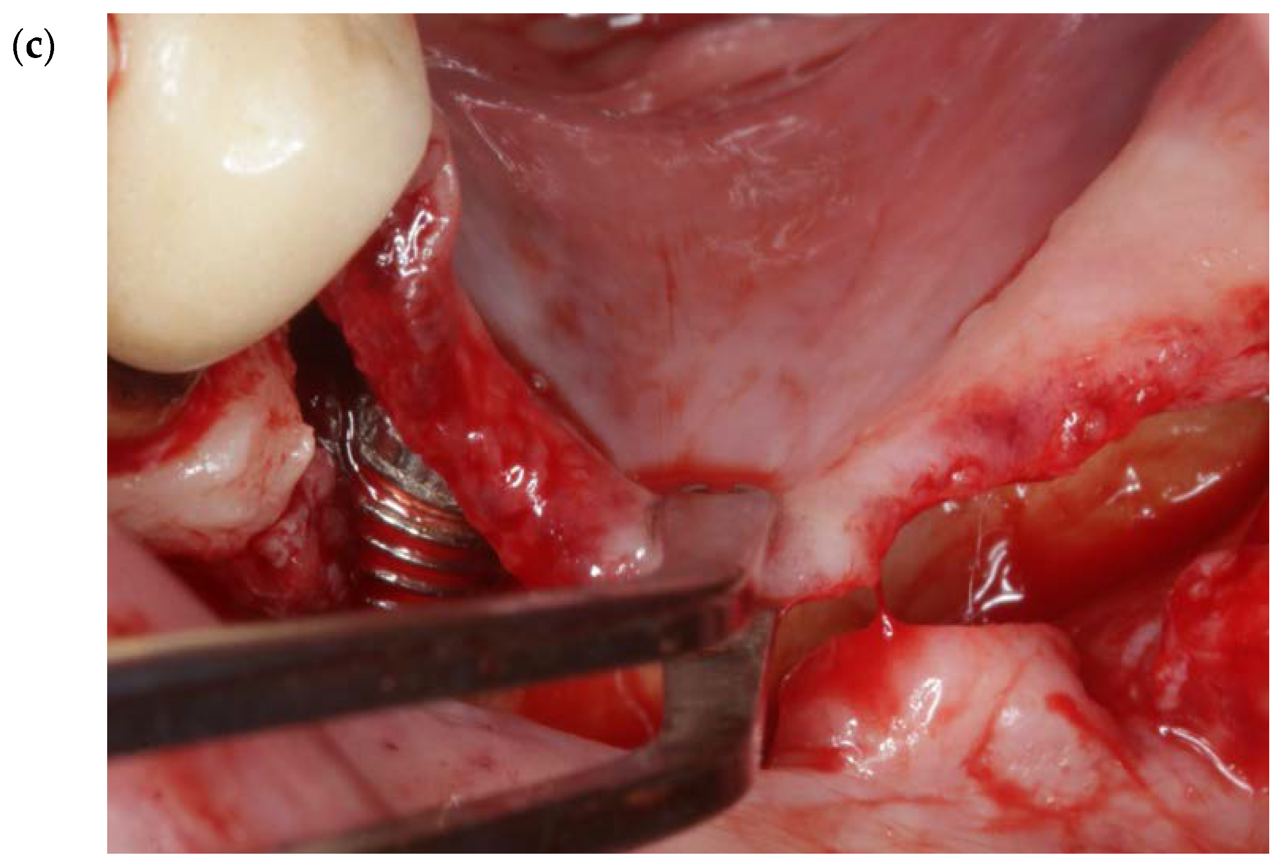
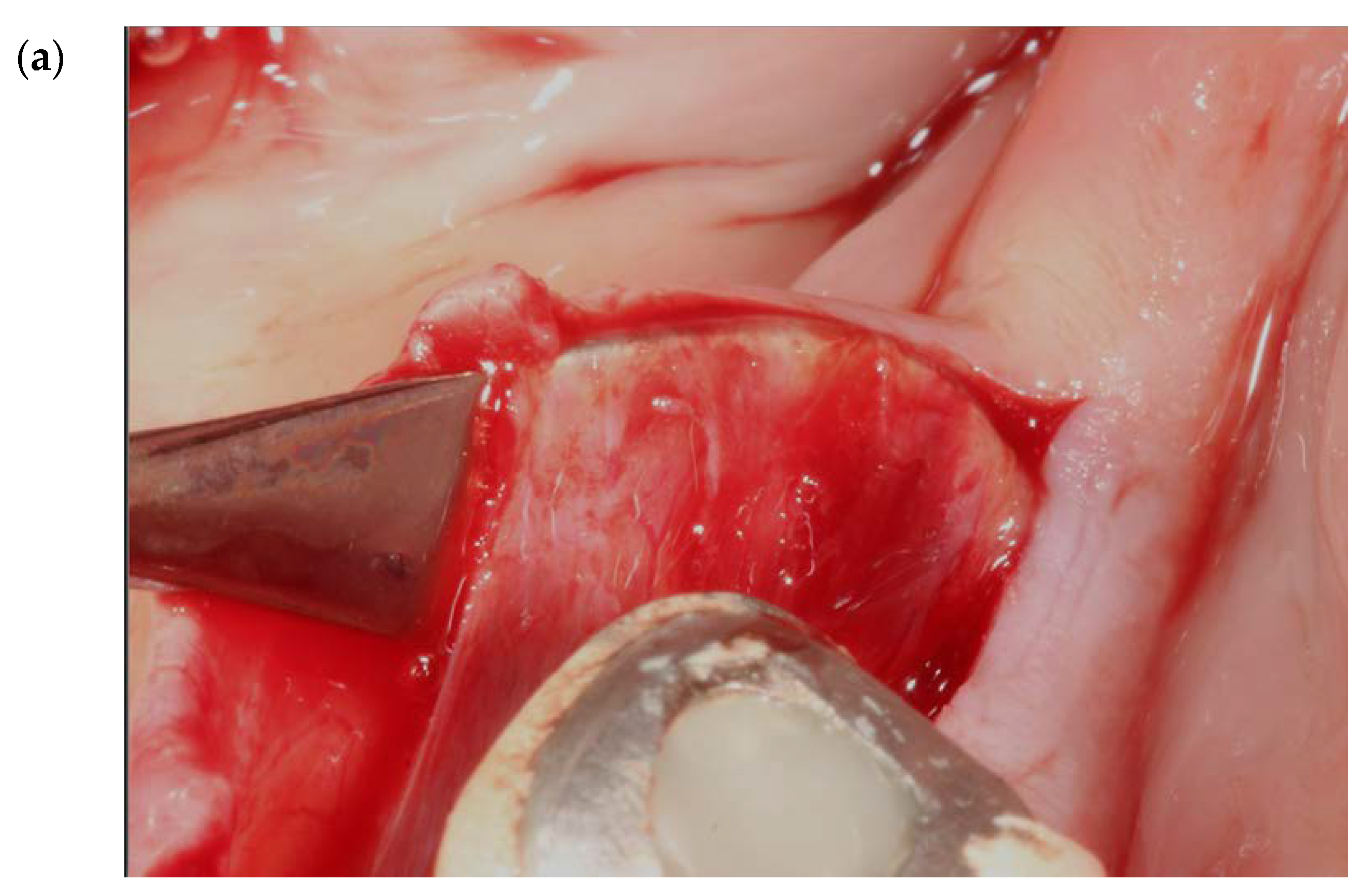
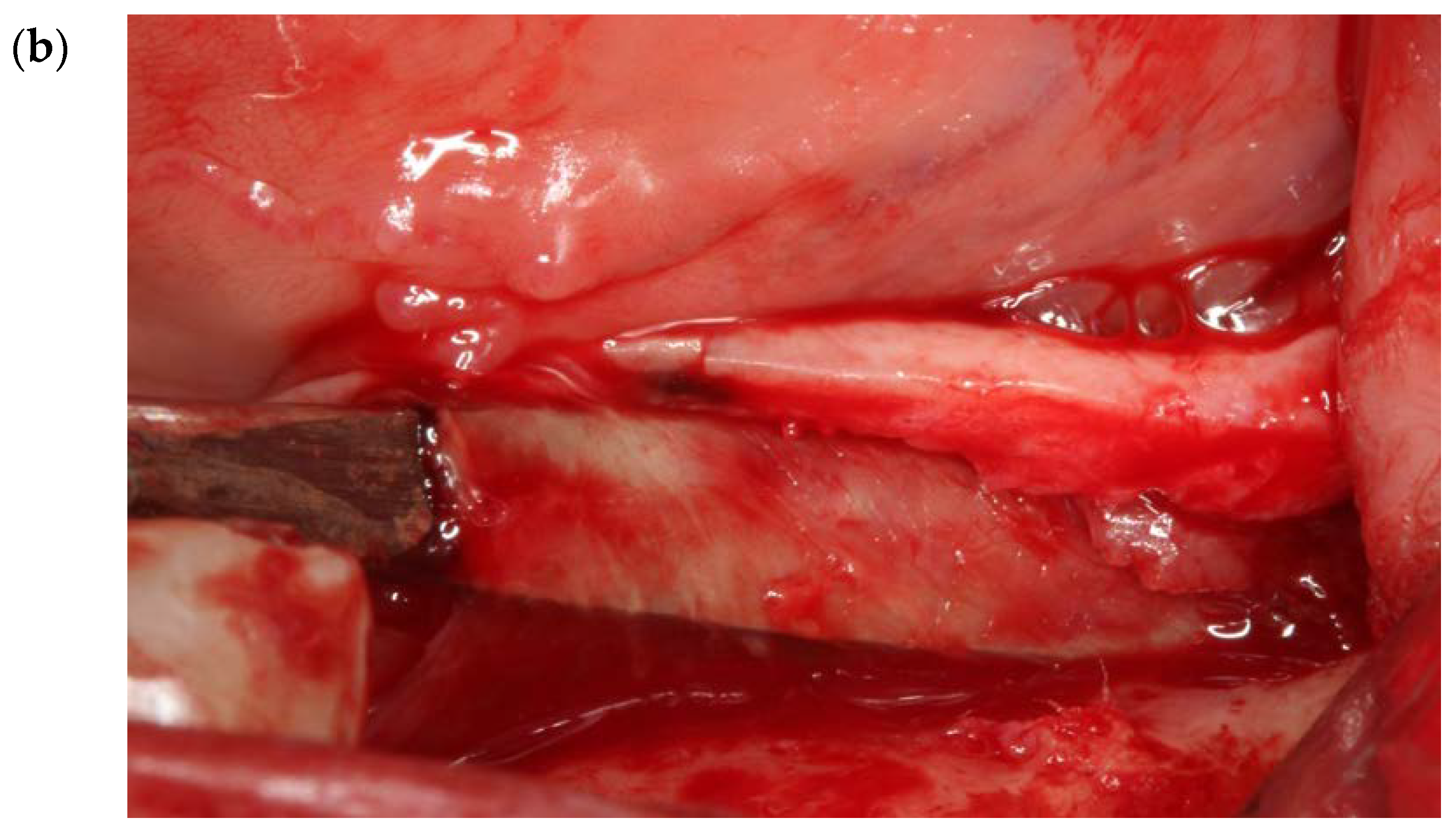
2.1. Finger Sweep Technique (FST) [19,22,27]

The flap design includes a full-thickness reflection of the buccal and lingual flaps, starting from a hockey stick releasing incision at the area of lateral incisors to the retromolar pad and ending in a 1 cm releasing incision to the ramus. FST is a blunt dissection/separation of the superficial from the deep mylohyoid muscle fibers. This finger dissection (using the index finger) includes stripping of the superficial fibers of the mylohyoid muscle in both an anterior and a posterior direction. No sharp dissection is needed. With the FST, the lingual flap release could reach up to 32 mm from the crest in a vertical direction (Figure 6a–c) and 6–10 mm in a horizontal direction towards the buccal aspect (Figure 7a–c and Figure 8).

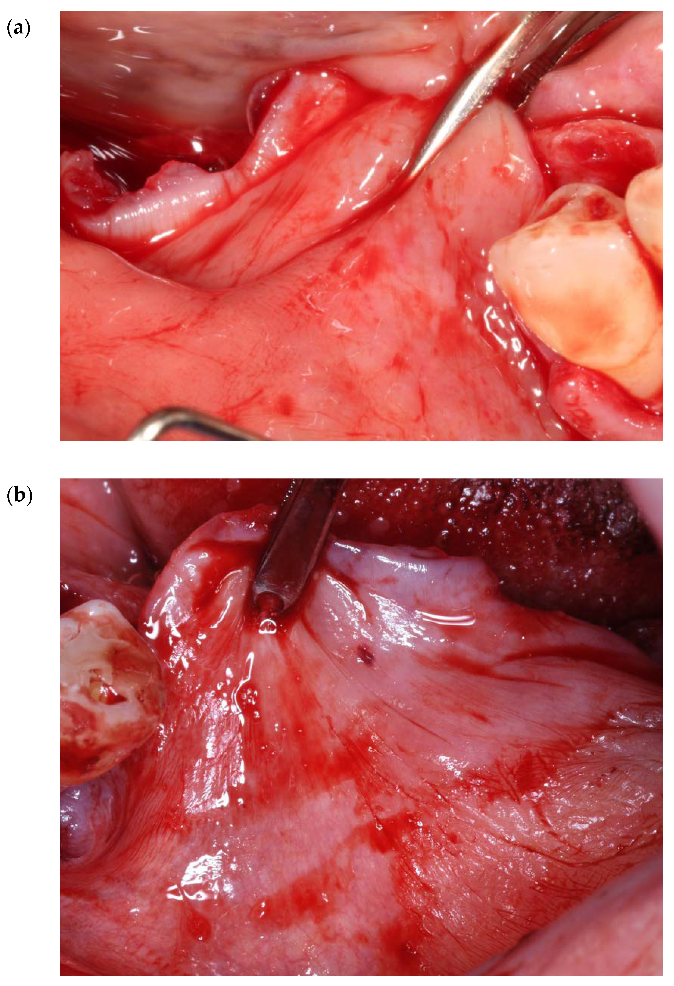
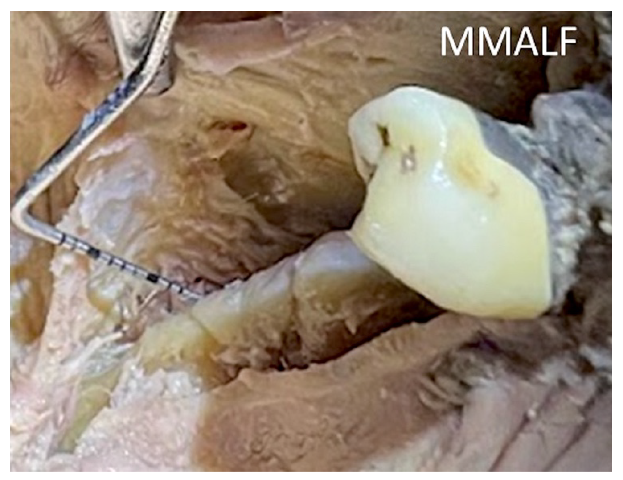
2.2. Release of the Mylohyoid Muscle Attachment on the Lingual Flap (MMALF) [15,28]

The flap design is similar with the FST (please see the description above). The mylohyoid insertion in the lingual flap is a connective tissue band (1–2 cm width) continuing from the epimysium of the mylohyoid muscle, around the area of the first molar (Figure 9).
A blunt instrument (e.g., Prichard elevator) is inserted under this connective tissue band, and the insertion of the muscle in the lingual flap is detached with a gentle traction in a coronal direction (Figure 10a,b and Figure 11).
This technique allows for a partial detachment of the mylohyoid muscle. Like the FST, the lingual flap release could reach more than 30 mm from the crest in a vertical direction and 6–10 mm in a horizontal direction towards the buccal aspect.

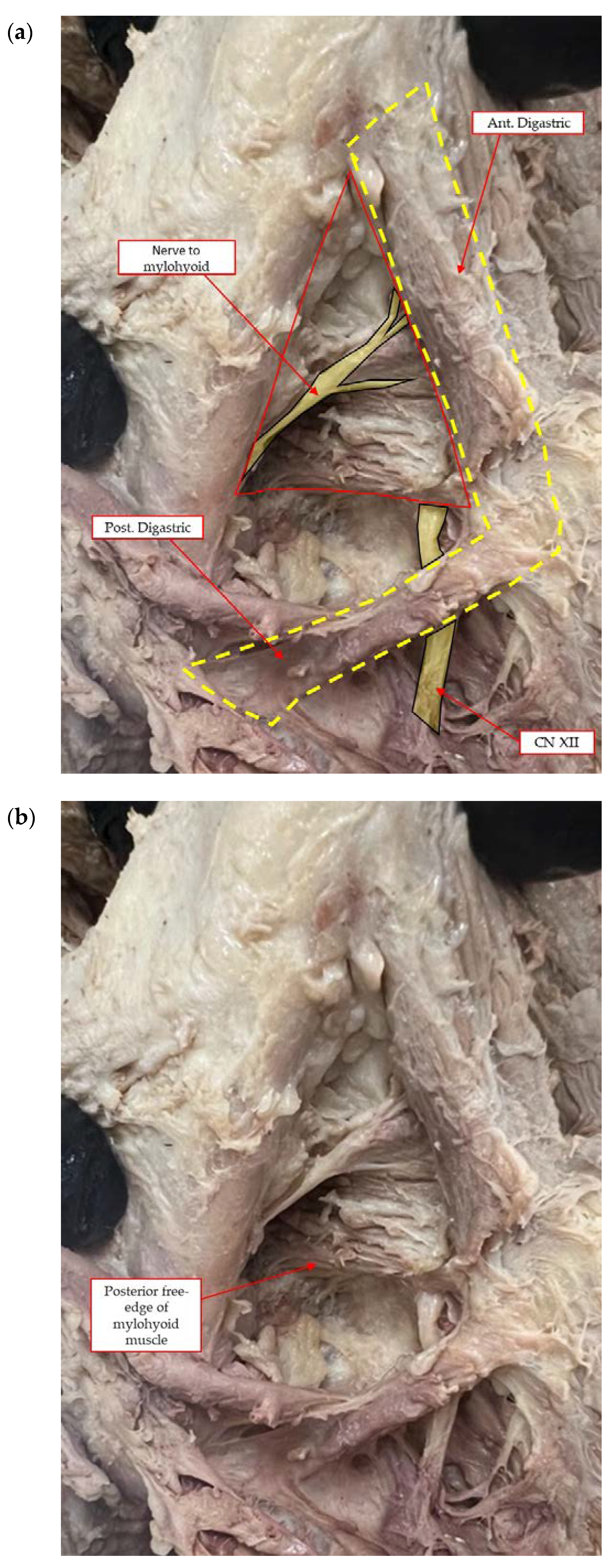
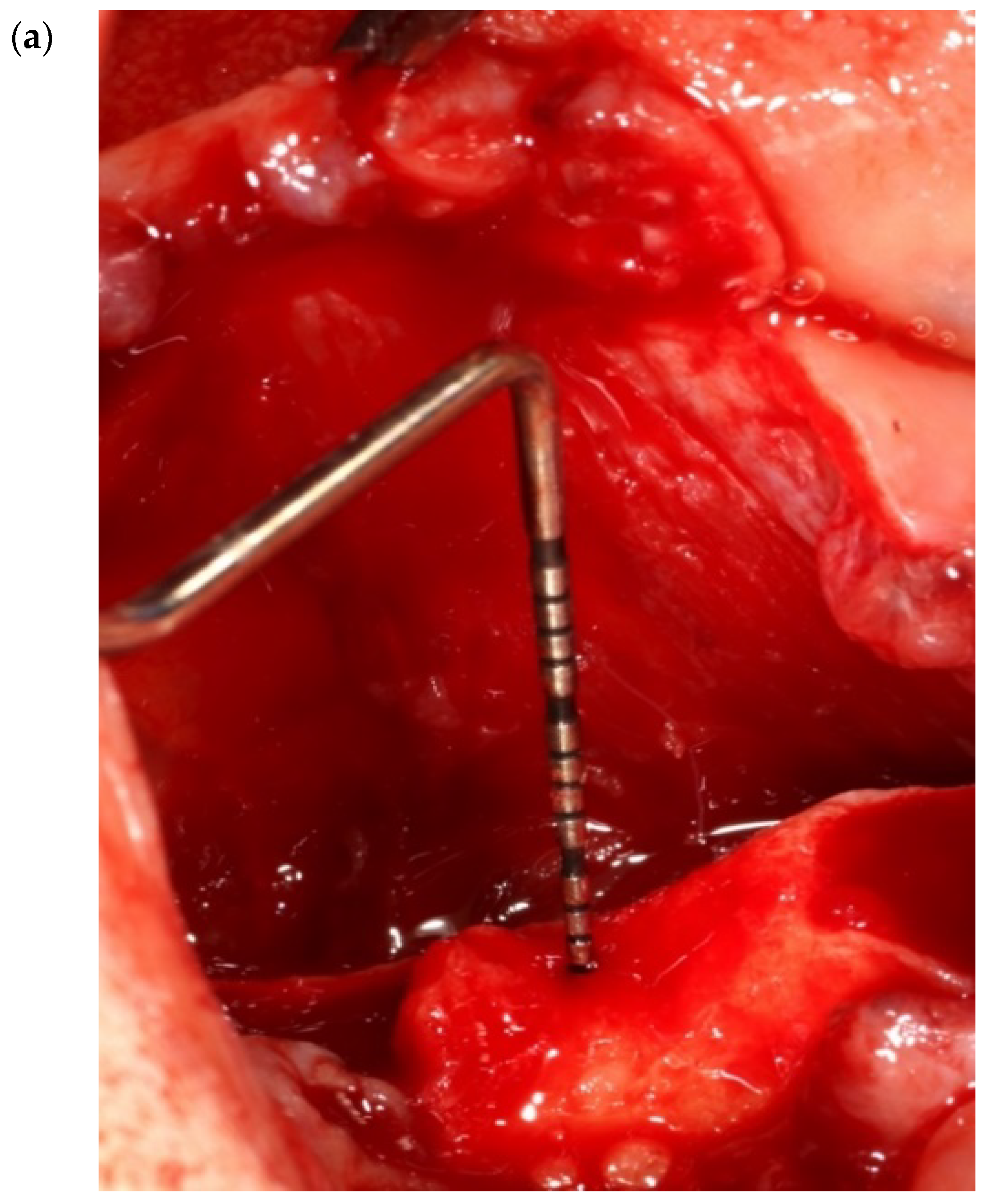
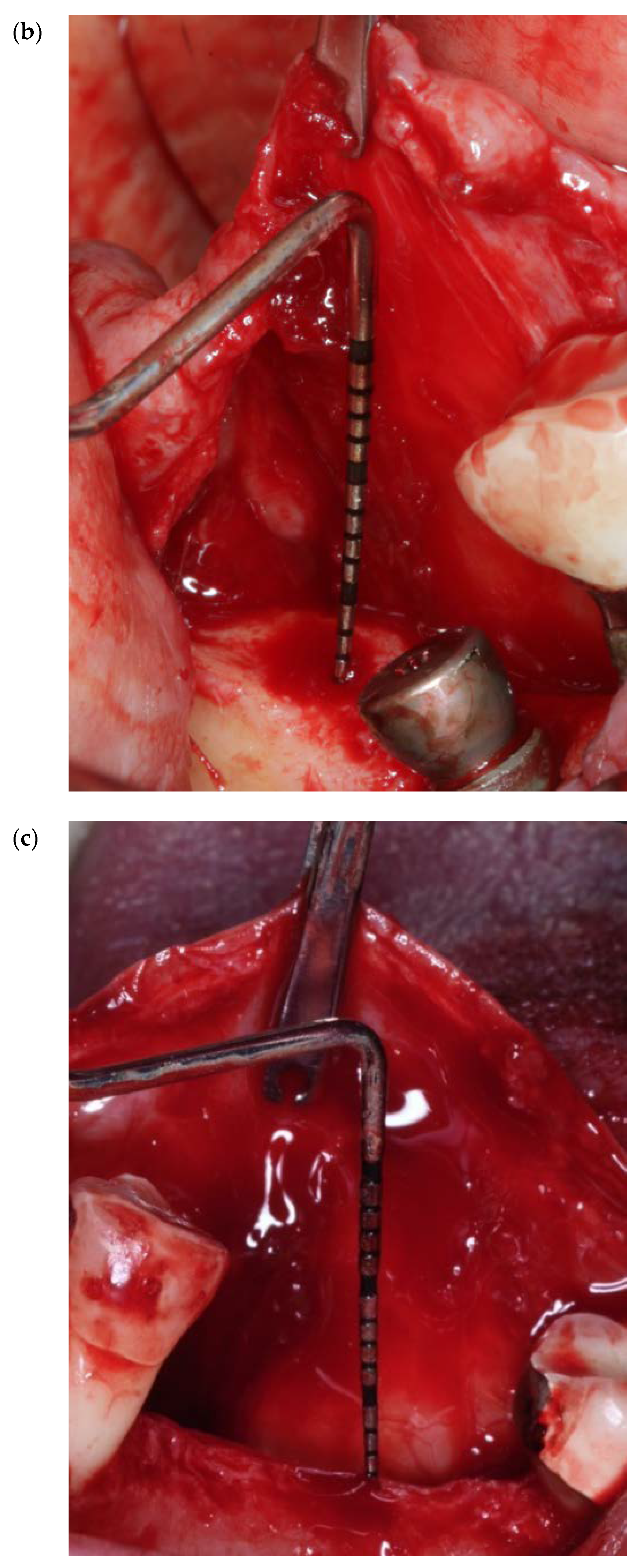
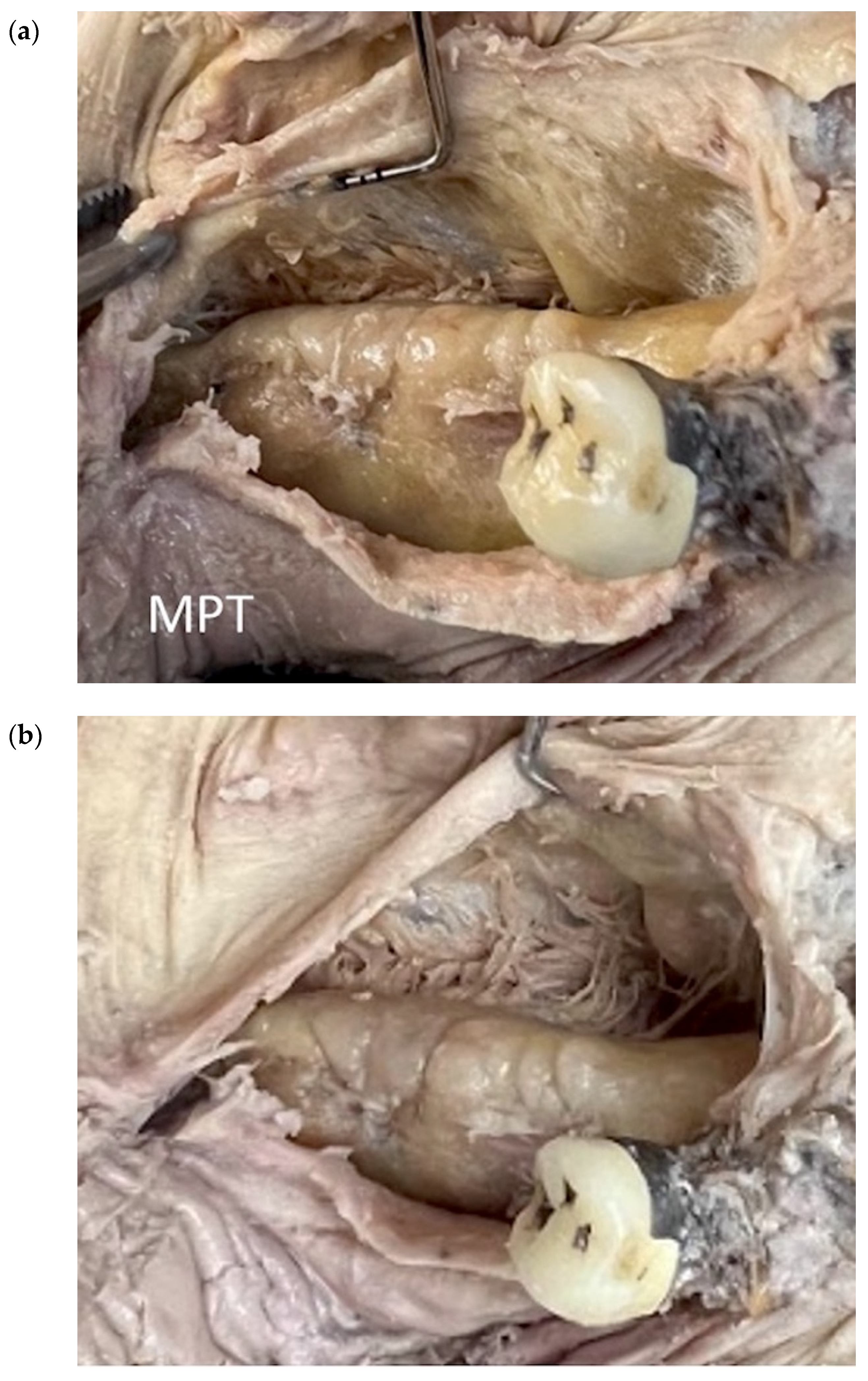
2.3. Mylohyoid Preservation Technique (MPT) in Three Key Anatomical Zones [6,13,14]

The flap design in this technique consists of a full-thickness reflection extending distally within 2 mm of the retromolar pad. Then a distal oblique vertical incision is made towards the coronoid process of the mandible. A vertical incision is made, mesial and buccal, two teeth away from the area of anticipated regeneration. Mesial and lingual, a 3–4 mm incision is performed at the mesial lingual line angle of the most distal tooth in front of the area of regeneration.
This technique considers three key anatomical zones: (i) tunneling and lifting of the retromolar pad (zone I), with the use of a periosteal elevator, the retromolar pad is reflected and pulled in a coronal direction; (ii) flap separation with mylohyoid muscle preservation (zone II, Figure 12a), as the soft tissue superior to the muscle insertion, with the use of a blunt instrument, is pushed in a lingual direction; and (iii) anterior, semi blunt periosteal release (zone III, Figure 12b), in which, with the use of a 15c blade in a perpendicular angle, an incision is performed in a sweeping motion. The goals of this technique are to include the retromolar pad into the lingual flap, which allows for maximum flap release; to separate the flap from the superior muscle fibers; and to provide flap flexibility, which will prevent any wound dehiscence. Similar to the previously discussed techniques, the lingual flap release could reach more than 30 mm from the crest in a vertical direction and 6–10 mm in a horizontal direction towards the buccal aspect.

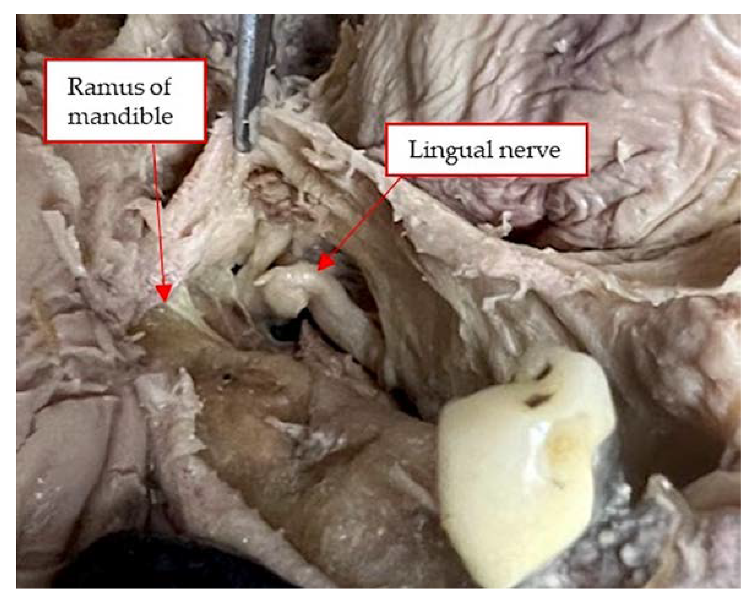
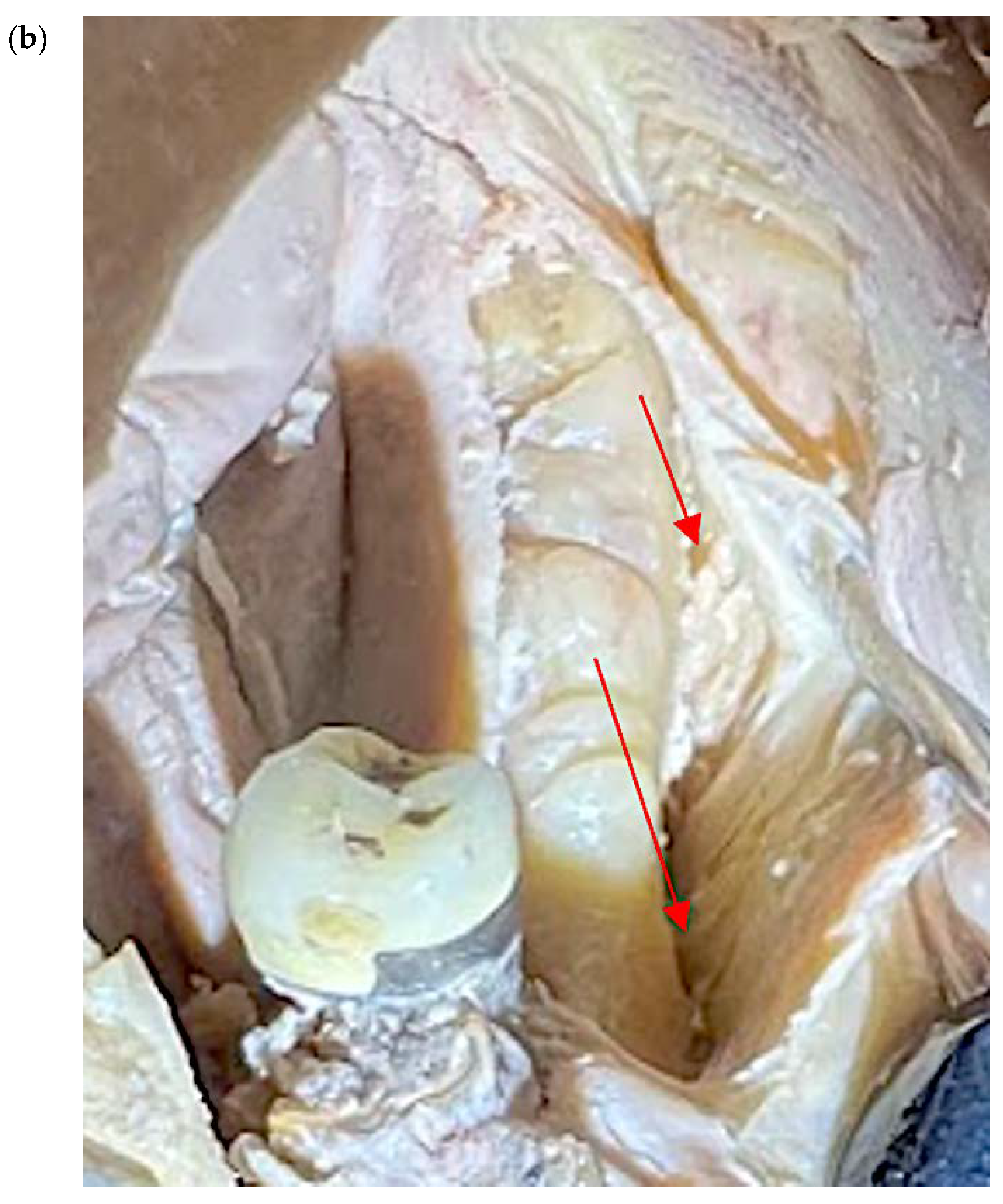
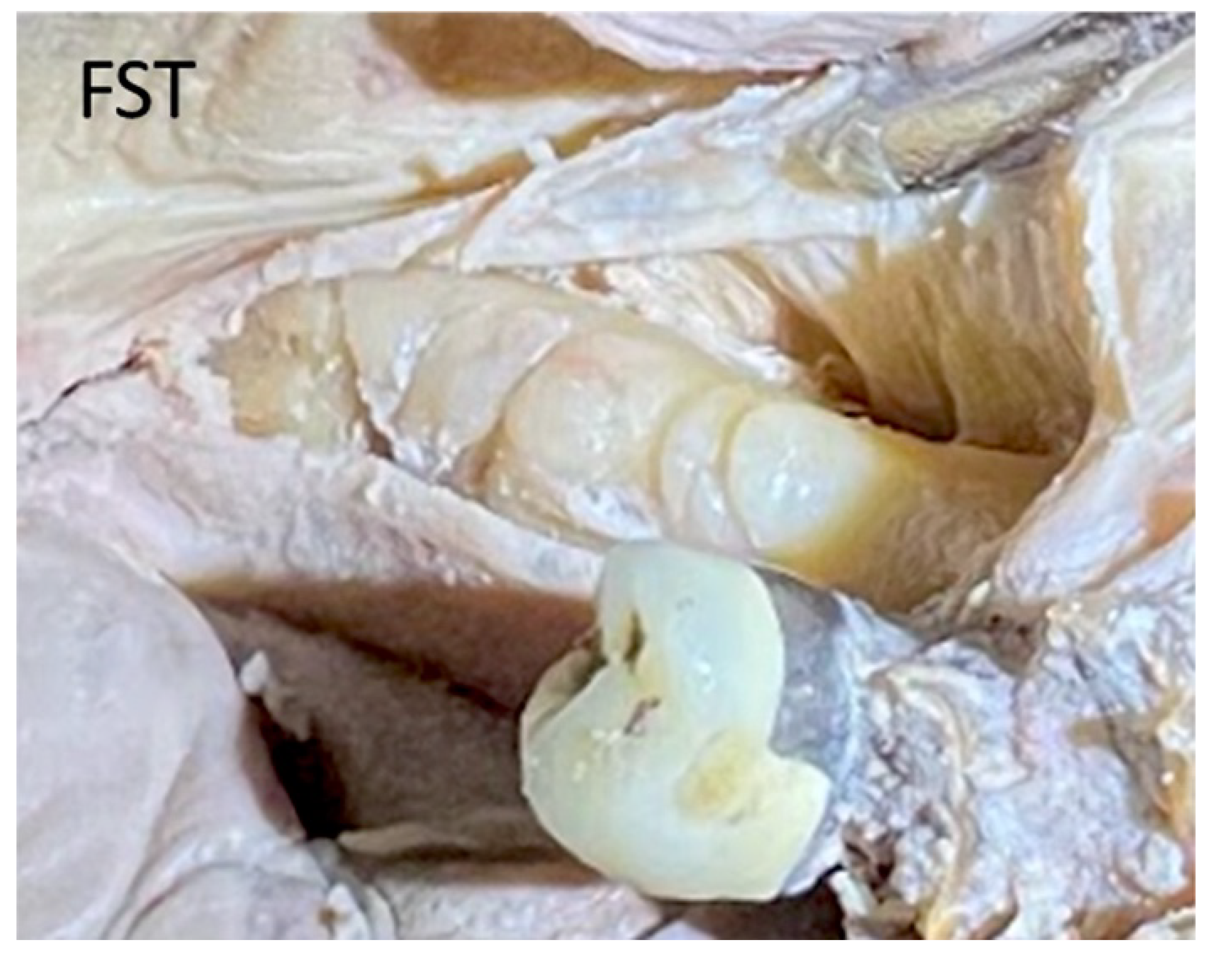
3. Cadaver Analyses of the Techniques

Blunt dissection with finger pressure, as described with the FST, thins the periosteal layer to expose and release the immediately underlying superficial mylohyoid muscle fibers. More posteriorly located, the lingual nerve is observed traversing parallel to the ramus, dropping apically distal to the location of a third molar. The MMALF technique describes initial penetration through the periosteum (at the level of the first molar) within the superior layers of the mylohyoid fiber attachment. Vital structures including the lingual nerve, artery, and submandibular salivary ducts are located apical and medial to the dissection. The MPT describes a semi-blunt penetration within the periosteum at the premolar zone. The mylohyoid muscle attachment to the mandible is more apically located in the premolar zone as compared to the attachment at the molar zone. All three techniques are considered safe (provided that the clinicians are experienced and properly trained) since they do not approximate any vital structures located below the mylohyoid muscle.

4. Discussion

It is of paramount importance to follow the prescribed surgical principles for buccal and lingual flap management to predictably attain and maintain tension-free primary closure during the healing period and avoid any post-operative complications. During the lingual flap management, severe complications may occur, including the risk of damaging the lingual nerve, the sublingual artery, the Wharton’s duct, the sublingual and submandibular glands, and perforating/overthinning the lingual flap [7,14,15,28,29,30]. These events may result in hematoma, which can be a life-threatening complication due to possible respiratory obstruction [31].
Urban et al. compared in a fresh human cadaver study FST and MMALF (control groups) with MPT (test group) [13]. The authors did not explain how many specimens in the control group were divided between the FST and the MMALF. Between the control and test groups, the test group technique showed statistically significant higher flap advancement in all three zones [13]. However, there is no study in the literature directly comparing the three techniques.
Ronda et al. in 2011, performed MMALF in a study with 69 VRA surgeries with the consequent placement of 187 implants [15]. The study reported four sites with signs of infections 2 weeks postoperatively, where the implants and the bone grafting materials had to be removed but not related to the lingual flap management. No membrane exposure was observed, or hemorrhagic issues such as hematoma, for the rest of the cases [15]. Ronda et al. in 2014, used in a similarly designed study the same technique in patients with atrophic posterior mandibles. Minor complications were reported regarding the lingual flap management, such as edema and hematoma [28].
Several studies described vertical releasing incisions as part of the management of the lingual flap. Flap retraction and membrane exposure were noted in four cases where the membranes were removed, and no regeneration was achieved [32,33,34]. Some studies did not report any complications with the use of FST [9,19,22,27]. Other studies for VRA did not report the technique was performed regarding the lingual flap management [7,35,36].
All three techniques are not free from limitations and complications. FST may create a communication between the surgical site and the submandibular or sublingual space. In case of infection, a severe medical complication may occur [19,22,27]. Removing the connective tissue band with the use of MMALF may lead to overthinning and necrosis of the lingual flap, which may cause exposure of the graft in the early healing period [15,28]. The MPT is a more complex and technique-sensitive approach, compared to the other two, in addition to the use of a 15c blade over the mesial aspect of the lingual flap, risking a possible perforation [6,13].
Soldatos and Weltman concluded that (i) flap management, with the use of MMR and buccal periosteal release, and (ii) the initial defect morphology are two very important key factors for successful GBR outcomes, especially in VRA [37]. Although flap management is an important principle for GBR procedures, a case report showed 2–5 mm VRA around previously placed dental implants with the use of a dense-PTFE membrane, which was left exposed to heal in a secondary intention [11].
Finally, the mandibular lingual releasing approach of the mylohyoid muscle is used in cases of oral and oropharyngeal carcinomas. The mylohyoid muscle is completely detached to give visual access to the providers [38,39]. In addition, the use of mandibular lingual release was associated with significantly lower frequency of maxillofacial pain and quality of life in these specific patients [39]. To the best of the authors’ knowledge, this is the first study reviewing, analyzing and discussing the three different techniques regarding MMR in VRA and HRA surgeries on a clinical and human cadaver level.

5. Conclusions

All three techniques, although using different approaches, achieve similar amounts of horizontal and vertical MMR. The techniques are not free of limitations and complications, due to the proximity to sensitive anatomical areas, but are considered safe, provided that they are performed only by highly experienced and trained clinicians.

Author Contributions

N.S. conceptualized the study, drafted, and critically reviewed the manuscript, and provided the clinical pictures; J.I. and R.W. provided the human cadaver pictures; G.R. and R.W. provided critical revision of the manuscript. All authors have read and agreed to the published version of the manuscript.

Funding

This research received no external funding.

Institutional Review Board Statement

IRB was not necessary for the conduction of the present narrative review study.

Informed Consent Statement

No separate consent is required for individual projects.

Data Availability Statement

No data were collected or analyzed.

Conflicts of Interest

The authors declare no conflict of interest.

References

  1. Seibert, J.S. Reconstruction of deformed, partially edentulous ridges, using full thickness onlay grafts. Part I. Technique and wound healing. Compend Contin Educ. Dent 1983, 4, 437–453. [Google Scholar] [PubMed]
  2. Wang, H.L.; Al-Shammari, K. HVC ridge deficiency classification: A therapeutically oriented classification. Int. J. Periodontics Restor. Dent 2002, 22, 335–343. [Google Scholar]
  3. Urban, I.; Monje, A.; Lozada, J.; Wang, H.L. Vertical ridge augmentation and soft tissue reconstruction of the anterior atrophic maxillae: A case series. Int. J. Periodontics Restor. Dent 2015, 35, 613–623. [Google Scholar] [CrossRef] [PubMed]
  4. Schropp, L.; Wenzel, A.; Kostopoulos, L.; Karring, T. Bone healing and soft tissue contour changes following single-tooth extraction: A clinical and radiographic 12-month prospective study. Int. J. Periodontics Restor. Dent 2003, 23, 313–323. [Google Scholar]
  5. Dahlin, C.; Linde, A.; Gottlow, J.; Nyman, S. Healing of bone defects by guided tissue regeneration. Plast. Reconstr. Surg. 1988, 81, 672–676. [Google Scholar] [CrossRef]
  6. Urban, I.; Monje, A.; Lozada, J.; Wang, H.-L. Principles for vertical ridge augmentation in the atrophic posterior mandible: A technical review. Int. J. Periodontics Restor. Dent 2017, 37, 639–645. [Google Scholar] [CrossRef]
  7. Simion, M.; Jovanovic, S.A.; Trisi, P.; Scarano, A.; Piattelli, A. Vertical ridge augmentation around dental implants using a membrane technique and autogenous bone or allografts in humans. Int. J. Periodontics Restor. Dent 1998, 18, 8–23. [Google Scholar]
  8. Simion, M.; Jovanovic, S.A.; Tinti, C.; Benfenati, S. Long term evaluation of osseointegrated implants inserted at the time or after vertical ridge augmentation. A retrospective study on 123 implants with 1-5-year follow-up. Clin. Oral Implants Res. 2001, 12, 35–45. [Google Scholar] [CrossRef]
  9. Soldatos, N.; Nelson-Rabe, L.; Ferguson, B.; Soldatos, K.; Weltman, R. Vertical ridge augmentation (VRA) with the use of cross-linked resorbable membrane, tenting screws, and a combination grafting technique: A report of three cases. Quintessence Int. 2021, 52, 328–339. [Google Scholar]
  10. Soldatos, N.K.; Stylianou, P.; Koidou, V.P.; Angelov, N.; Yukna, R.; Romanos, G.E. Limitations and options using resorbable versus resorbable membranes for successful guided bone regeneration procedures. Quintessence Int. 2017, 48, 131–147. [Google Scholar]
  11. Soldatos, N.; Garcia, M.; Umoh, E.; Irizarry, A.; Weltman, R. Vertical ridge augmentation around dental implants with the use of a dense PTFE membrane to correct previously failed augmentations. Clin. Adv. Periodontics 2022, 12, 51–56. [Google Scholar] [CrossRef]
  12. Jepsen, S.; Schwarz, F.; Cordaro, L.; Derks, J.; Hämmerle, C.H.; Heitz-Mayfield, L.J.; Hernández Alfaro, F.; Meijer, H.J.; Naenni, N.; Ortiz-Vigón, A.; et al. Regeneration of alveolar ridge defects. Consensus report of group 4 of the 15th European Workshop on Periodontology on Bone Regeneration. J. Clin. Periodontol 2019, 46, 277–286. [Google Scholar] [CrossRef]
  13. Urban, I.; Traxler, H.; Romero-Bustillos, M.; Farkasdi, S.; Bartee, B.; Baksa, G.; Avila-Ortiz, G. Effectiveness of two different lingual flap advancing techniques for vertical bone augmentation in the posterior mandible: A comparative, split-mouth cadaver study. Int. J. Periodontics Restor. Dent 2018, 38, 35–40. [Google Scholar] [CrossRef]
  14. Urban, I.; Monje, A.; Wang, H.-L.; Gerber, G. Mandibular regional anatomical landmarks, and clinical implications for ridge augmentation. Int. J. Periodontics Restor. Dent 2017, 37, 347–353. [Google Scholar] [CrossRef]
  15. Ronda, M.; Stacchi, C. Management of coronally advanced lingual flap in regenerative osseous surgery: A case series introducing a novel technique. Int. J. Periodontics Restor. Dent 2011, 31, 505–513. [Google Scholar]
  16. Machtei, E.E. The effect of membrane exposure on the outcome of regenerative procedures in humans: A meta-analysis. J. Periodontol. 2001, 72, 512–516. [Google Scholar] [CrossRef]
  17. Retzepi, M.; Donos, N. Guided Bone Regeneration: Biological principle and therapeutic applications. Clin. Oral Imp. Res. 2010, 21, 567–576. [Google Scholar] [CrossRef]
  18. Wang, H.L.; Boyapati, L. "PASS" principles for predictable bone regeneration. Implant Dent 2006, 15, 8–17. [Google Scholar] [CrossRef]
  19. Romanos, G.E. Periosteal releasing incision for successful coverage of augmented sites. A technical note. J. Oral Implantol. 2010, 36, 25–30. [Google Scholar] [CrossRef]
  20. Bunyaratavej, P.; Wang, H.L. Collagen membranes: A review. J. Periodontol. 2001, 72, 215–229. [Google Scholar] [CrossRef]
  21. Chasioti, E.; Tat Fai, C.; Drew, H. Maintaining space in localized ridge augmentation using guided bone regeneration with tenting screw technology. Quintessence Int. 2013, 44, 763–771. [Google Scholar] [PubMed]
  22. Pikos, M.A. Atrophic posterior maxilla, and mandible: Alveolar ridge reconstruction with mandibular block autografts. Alpha Omegan 2005, 98, 34–45. [Google Scholar] [PubMed]
  23. Simion, M.; Fontana, F.; Rasperini, G.; Maiorana, C. Vertical ridge augmentation by expanded-polytetrafluoroethylene membrane and a combination of intraoral autogenous bone graft and deproteinized anorganic bovine bone. Clin. Oral Implant. Res. 2007, 18, 620–629. [Google Scholar] [CrossRef] [PubMed]
  24. Greenstein, G.; Greenstein, B.; Cavallaro, J.; Tarnow, D. The role of decortication in enhancing the results of guided bone regeneration: A literature review. J. Periodontol. 2009, 80, 175–189. [Google Scholar] [CrossRef] [PubMed]
  25. Burkhardt, R.; Lang, N.P. Role of flap tension in primary wound closure of mucoperiosteal flaps: A prospective cohort study. Clin. Oral Imp. Res. 2010, 21, 50–54. [Google Scholar] [CrossRef]
  26. Herring, M.J.; Fehrenbach, S.W. Illustrated Anatomy of the Head and Neck, 4th ed.; Elsevier/Saunders: St. Louis, MO, USA, 2013. [Google Scholar]
  27. Pikos, M.A. Atrophic posterior mandibular reconstruction utilizing mandibular block autografts: Risk management. Int. J. Oral Maxillofac. Implant. 2003, 18, 765–766. [Google Scholar]
  28. Ronda, M.; Rebaudi, A.; Torelli, L.; Stacchi, C. Expanded vs. dense polytetrafluoroethylene membranes in vertical ridge augmentation around dental implants: A prospective randomized controlled clinical trial. Clin. Oral Implant. Res. 2014, 25, 859–866. [Google Scholar] [CrossRef]
  29. Simion, M.; Trisi, P.; Piattelli, A. Vertical ridge augmentation using a membrane technique associated with osseointegrated implants. Int. J. Periodontics Restor. Dent 1994, 14, 496–511. [Google Scholar]
  30. Urban, I.A.; Lozada, J.L.; Jovanovic, S.A.; Nagursky, H.; Nagy, K. Vertical ridge augmentation with titanium- reinforced dense PTFE membranes and a combination of particulated autogenous bone and anorganic bovine bone-derived mineral: A prospective case series in 19 patients. Int. J. Oral Maxillofac. Implant. 2014, 29, 185–193. [Google Scholar] [CrossRef]
  31. Niamtu, J., 3rd. Near-fatal airway obstruction after routine implant placement. Oral Surg Oral Med. Oral Pathol. Oral Radiol. Endod. 2001, 92, 597–600. [Google Scholar] [CrossRef]
  32. Tinti, C.; Parma-Benfenati, S.; Polizzi, G. Vertical ridge augmentation: What is the limit? Int. J. Periodontics Restor. Dent 1996, 16, 221–229. [Google Scholar]
  33. Tinti, C.; Parma-Benfenati, S. Vertical ridge augmentation: Surgical protocol and retrospective evaluation of 48 consecutively inserted implants. Int. J. Periodontics Restor. Dent 1998, 18, 435–443. [Google Scholar]
  34. Fugazzotto, P. Maintaining primary closure after guided bone regeneration procedures: Introduction of a new flap design and preliminary results. J. Periodontol. 2006, 77, 1452–1457. [Google Scholar] [CrossRef]
  35. Merli, M.; Lombardini, F.; Esposito, M. Vertical ridge augmentation with autogenous bone grafts 3 years after loading: Resorbable barriers versus titanium-reinforced barriers. A randomized controlled clinical trial. Int. J. Oral Maxillofac. Implant. 2010, 25, 801–807. [Google Scholar]
  36. Fugazzotto, P. Maintenance of soft tissue closure following guided bone regeneration: Technical considerations and report of 723 Cases. J. Periodontol. 1999, 70, 1085–1097. [Google Scholar] [CrossRef]
  37. Soldatos, N.; Weltman, R. Vertical ridge augmentation (VRA): Success and predictability. Curr. Oral Health Rep. 2022, 9, 197–204. [Google Scholar] [CrossRef]
  38. Stanley, R. Mandibular lingual releasing approach to oral and oropharyngeal carcinomas. Laryngoscope 1984, 94, 596–600. [Google Scholar] [CrossRef]
  39. Li, H.; Li, J.; Yang, B.; Su, M.; Xing, R.; Han, Z. Mandibular lingual release versus mandibular lip-split approach for expanded resection of middle-late tongue cancer: A case-control study. J. Cranio-Maxillo-Facial Surg. 2015, 43, 1054–1058. [Google Scholar] [CrossRef]
Figure 1. Twenty-three (23) mm of buccal periosteal release in a vertical direction.
Genes 14 00595 g001
Figure 2. (a,b) Ten (10) mm of buccal periosteal release in a horizontal direction.
Genes 14 00595 g002
Figure 3. (a,b) In a human cadaver model, the mylohyoid muscle attachment to the mandible is more apically located in the premolar zone as compared to the attachment at the molar zone. Should the surgical dissection during MMR, extend deeper into the mylohyoid muscle fiber attachment, the vital structures as described above are positioned apical and medial to the mylohyoid muscle.
Genes 14 00595 g003
Figure 4. More posteriorly located, the lingual nerve, in a human cadaver, is observed traversing parallel to the ramus dropping apically distal to the location of a third molar.
Genes 14 00595 g004
Figure 5. View of the superficial fibers and the attachment of the mylohyoid muscle to the mandible, forming the floor of the mouth: clinically (a) and in a human cadaver model [(b)-red arrows].
Genes 14 00595 g005aGenes 14 00595 g005b
Figure 6. (ac) Clinical view of MMR in a vertical direction, ranging from 29–32 mm.
Genes 14 00595 g006aGenes 14 00595 g006b
Figure 7. (ac) Six to ten (6–10) mm of MMR in a horizontal direction.
Genes 14 00595 g007aGenes 14 00595 g007b
Figure 8. The design of the lingual flap of the FST in a human cadaver model, showing the release of the superficial fibers of the mylohyoid muscle.
Genes 14 00595 g008
Figure 9. Clinical overview of the connective tissue band of 1–2 cm at the area of the first molar (black arrows), where the mylohyoid muscle attaches to the lingual flap.
Genes 14 00595 g009
Figure 10. (a,b) Clinical overview of the placement of two different blunt instruments under the attachment of the mylohyoid muscle at the lingual flap. The connective tissue attachment ranges from 1–2 cm.
Genes 14 00595 g010aGenes 14 00595 g010b
Figure 11. A human cadaver overview of a 23 mm release of the mylohyoid muscle using the MMALF technique.
Genes 14 00595 g011
Figure 12. MMR using the MPT technique in a human cadaver model. (a) The soft tissue superior to the muscle insertion is pushed in a lingual direction (zone II). (b) In the anterior section of the lingual flap, a semi-blunt periosteal release is performed in a sweeping motion with a 15c blade (zone III).
Genes 14 00595 g012
Table 1. Summary of all three techniques for MMR.
Techniques MethodsLimitations
FST
(Pikos 2005 [22],
Romanos 2010 [19])
Blunt dissection/separation of the superficial from the deep mylohyoid muscle fibers without any
sharp dissection with the use
of the index finger.
Creation of communication between the surgical site and the submandibular or sublingual space.
MMALF
(Ronda et al. 2011 [15])
A blunt instrument is inserted under the mylohyoid muscular insertion at the lingual flap. With a gentle traction in a coronal direction, the connective tissue band is detached.Overthinning of the lingual flap, which may lead to flap necrosis and exposure of the graft in the
early healing period.
MPT
(Urban et al. 2018 [13])
1. Tunneling and lifting of the retromolar pad (zone I).
2. Flap separation with mylohyoid muscle preservation (zone II).
3. Anterior, semi blunt periosteal release (zone III) with the use of a 15c blade in a perpendicular angle.
More complex and technique sensitive approach, especially in zone III; the use of a 15c blade over the mesial aspect of the lingual flap endangers a possible flap perforation.
Disclaimer/Publisher’s Note: The statements, opinions and data contained in all publications are solely those of the individual author(s) and contributor(s) and not of MDPI and/or the editor(s). MDPI and/or the editor(s) disclaim responsibility for any injury to people or property resulting from any ideas, methods, instructions or products referred to in the content.

