Review

Towards a Cure for HARS Disease

1
Department of Biochemistry, The University of Western Ontario, London, ON N6A 5C1, Canada
2
Department of Chemistry, The University of Western Ontario, London, ON N6A 5C1, Canada
*
Author to whom correspondence should be addressed.
Genes 2023, 14(2), 254; https://doi.org/10.3390/genes14020254
Submission received: 21 December 2022 / Revised: 11 January 2023 / Accepted: 13 January 2023 / Published: 18 January 2023
(This article belongs to the Special Issue Emerging Roles of tRNAs in Health and Disease)

Abstract

:
Histidyl-tRNA synthetase (HARS) ligates histidine to its cognate transfer RNA (tRNAHis). Mutations in HARS cause the human genetic disorders Usher syndrome type 3B (USH3B) and Charcot-Marie-Tooth syndrome type 2W (CMT2W). Treatment for these diseases remains symptomatic, and no disease specific treatments are currently available. Mutations in HARS can lead to destabilization of the enzyme, reduced aminoacylation, and decreased histidine incorporation into the proteome. Other mutations lead to a toxic gain-of-function and mistranslation of non-cognate amino acids in response to histidine codons, which can be rescued by histidine supplementation in vitro. We discuss recent advances in characterizing HARS mutations and potential applications of amino acid and tRNA therapy for future gene and allele specific therapy.

1. Aminoacyl-tRNA Synthetases in Translation

During translation, genetic information carried by each messenger RNA (mRNA) is used as a template to synthesize the corresponding sequence of amino acids of a particular protein. Protein synthesis can be divided into three steps: chain initiation, elongation, and termination. In translation initiation, mRNA is targeted by the translation machinery, which involves small (40S) and large (60S) ribosomal subunits, methionyl transfer RNA (Met-tRNA) decoding of the typical AUG start codon, and eukaryotic initiation factors (eIF) [1]. In chain elongation, an mRNA codon is paired with an anticodon of an aminoacyl-tRNA bound to the elongation factor, resulting in the transfer of one amino acid at a time from the tRNA molecule to the nascent chain of amino acids. The ribosome moves down the mRNA in the 5′ to 3′ direction until it reaches one of the three terminator codons (UAA, UAG, or UGA) resulting in the binding of a release factor, chain termination, and ribosome disassembly. The completed polypeptide is then released from the ribosome [2,3].
The fidelity of the genetic code is determined by the accuracy of these steps, and most significantly, by the accuracy of charging the transfer RNAs (tRNAs) with their cognate amino acid. In eukaryotes, there is a specific aminoacyl-tRNA synthetase (aaRS) for each of the 20 standard amino acids. The aaRSs belong to two independently evolved enzyme families, called class I and class II aaRSs [4]. Class I synthetases are usually monomeric with a Rossman fold including α-helices connecting a five-stranded parallel β sheet, whereas class II synthetases are usually dimeric or multimeric with a seven-stranded anti-parallel β-sheet fold flanked by α-helices. Within each class, subclasses are further divided to some extent according to their capacity to recognize chemically related amino acids [5].
Aminoacylation of tRNAs occurs in two steps (Figure 1). In the catalytic domain, adenosine triphosphate (ATP) and the cognate amino acid are bound, a high-energy phosphoanhydride bond in ATP is hydrolyzed to form aminoacyl adenylate-AMP, and inorganic pyrophosphate (PPi) is released. Next, the cognate tRNA is recruited to the aaRS, and the amino acid is transferred to the 3′ end of the tRNA at the acceptor stem, forming an aminoacyl-ester bond [6]. The aminoacyl-tRNAs then bind to the elongation factor and are competent substrates for protein synthesis on the ribosome.
All aaRSs contain a catalytic domain. aaRS amino acid specificity is determined by the composition and size of the amino acid binding pocket, and binding of the tRNA acceptor stem in the aaRS catalytic domain is a prerequisite to aminoacylation. Most aaRSs also contain a tRNA binding domain for the recognition of the tRNA anticodon loop. tRNA recognition relies on tRNA-specific recognition element nucleotides that are called identity elements. While many aaRSs recognize the anticodon as the primary identity element, bases in the acceptor stem and the variable loop are also common identity elements [6]. Additionally, some aaRSs contain a helix-turn-helix motif (WHEP domain), which is thought to function in protein-protein interactions between synthetases as well as high-affinity interactions with the tRNA [7]. In humans, protein-protein interactions occur between certain cytoplasmic aaRSs that form a large 11-subunit multisynthetase complex (MSC) composed of 8 aaRSs and 3 aaRS-interacting multifunctional proteins (AIMPs), which increases protein synthesis efficiency and is important for other non-canonical aaRS functions [8]. The specificity of each aaRS towards its cognate amino acid and tRNA gives tRNA-charging its necessary high fidelity, however, this is not the only mechanism, as some aaRSs have additional proofreading capabilities carried out in their editing domain. Editing domains are responsible for cleaving incorrectly paired aminoacyl-tRNA molecules to revert misaminoacylation and prevent mistranslation [5].

2. Translational Fidelity Guards the Genetic Code

Mutations in an aaRS that cause disease phenotypes can be broadly summarized as loss-of-function and gain-of-function mechanisms. Loss-of-function describes mutations leading to a significant decrease in the original activity of the aaRS. Examples of loss-of-function include mutations in aaRS signal peptides that cause defects in localization; mutations of residues that result in improper folding or dimerization of the aaRS; as well as mutations that impair ATP, tRNA, or amino acid binding and subsequent loss of aminoacylation activity or tRNA release [9]. These mutations lead to a decrease or elimination of the catalytic activity of the aaRS, reduced translational fidelity, and impaired cell growth.
Gain-of-function describes mutations leading to increased or error-prone enzyme activity. Gain-of-function mutations in aaRSs can cause aminoacylation defects, allowing for the ligation of the non-cognate amino acid to the cognate tRNA or the aminoacylation of a cognate amino acid to a non-cognate tRNA [5]. As a result, misaminoacylated tRNA is produced, and the mRNA message is mistranslated to produce mutated proteins. Mistranslation can lead to protein misfolding, aggregation, and a defective proteome [10]. Surprisingly, recent studies have shown that eukaryotic systems can withstand a significant level of mistranslation caused by single nucleotide variants in tRNA molecules. In fact, the abundance of highly tolerable mistranslating tRNAs in the human population indicates that their contribution to disease might be coincidental with other cellular defects [10,11].
Genes encoding aaRSs are essential and play an indispensable role in protein synthesis. Thus, disease-causing mutations in aaRS have become of increasing interest [12]. The aaRS genes are encoded in the human nuclear genome, including 18 aaRS with functions localized to the cytoplasm only, 17 aaRS localized to mitochondria only, and 2 aaRS that function in both the cytoplasm and mitochondria. To date, 56 genetic diseases have been associated with unique mutations in aaRS proteins [13]. The causative role of aaRS mutations in rare human diseases emphasizes the significance and criticality of the aminoacylation step by aaRS enzymes in maintaining translation fidelity and protein homeostasis [6].

3. Human Histidyl-tRNA Synthetase

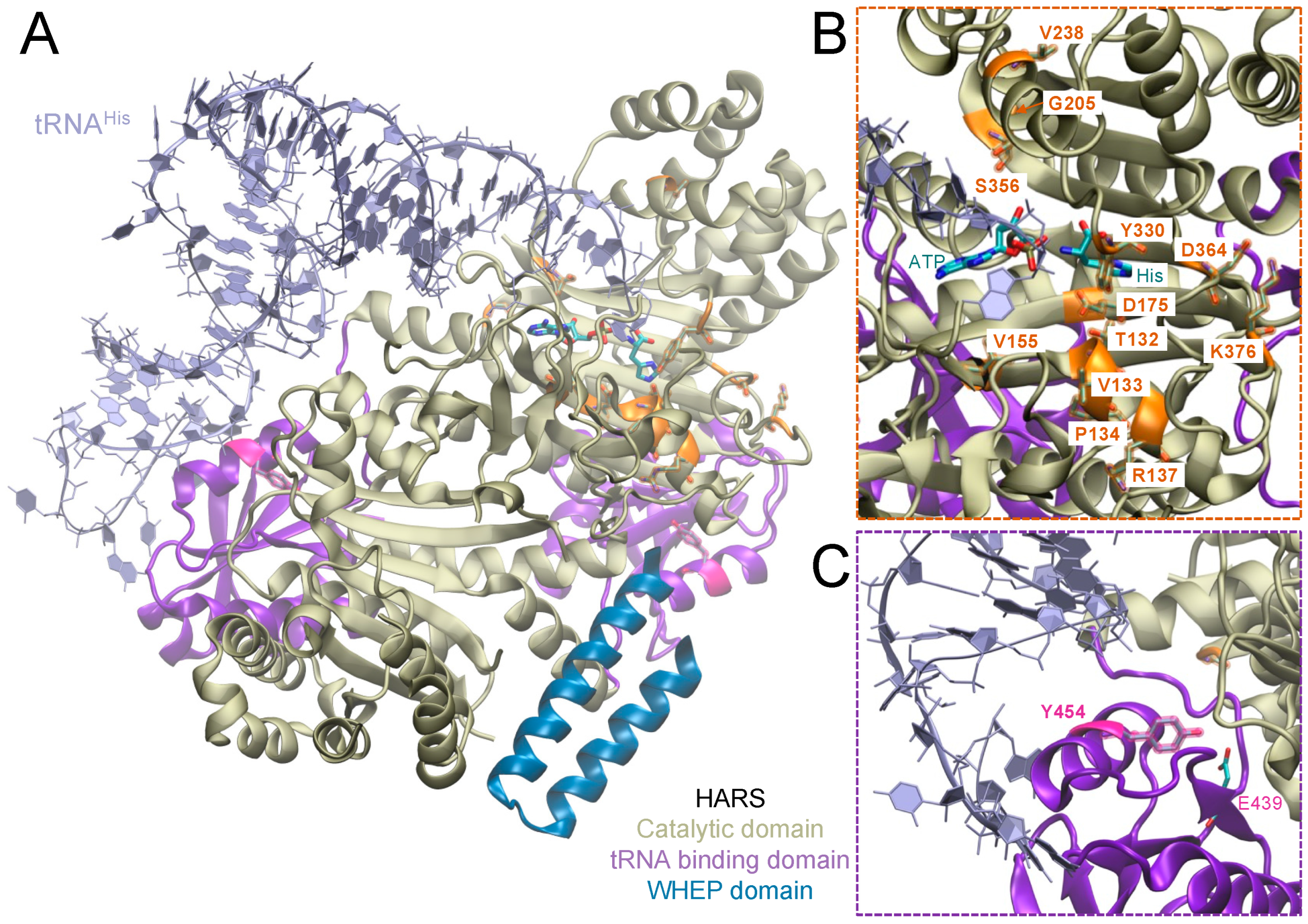
The human cytosolic histidyl-tRNA synthetase (HARS, HisRS) is a homodimeric class II aminoacyl-tRNA synthetase. HARS is a 13-exon, 17.4 kb gene located on chromosome 5 [14,15] and encodes 509 amino acids in a 57.4 kDa polypeptide. HARS consists of an N-terminal WHEP domain, a catalytic domain, and a C-terminal tRNA binding domain (Figure 2) [16]. HARS ligates tRNAHis to its cognate amino acid, histidine, producing His-tRNAHis, which is recruited for the synthesis of cellular proteins. Besides the essential function of HARS in aminoacylation, a non-canonical function of HARS is associated with cell proliferation. Knockdown of HARS in a zebrafish model resulted in increased cell death and reduced proliferation of retinal progenitor cells, suggesting a link between HARS and neuronal progenitor cell survival and proliferation during early development [17]. Furthermore, a splice variant of HARS (HARS∆CD), which lacks the catalytic domain and thus has no aminoacylation function, was found circulating in the bloodstream of patients positive for anti-Jo1 syndrome, an anti-synthetase autoimmune disease. Epitopes in the WHEP domain in particular were targeted by anti-Jo antibodies [18,19].
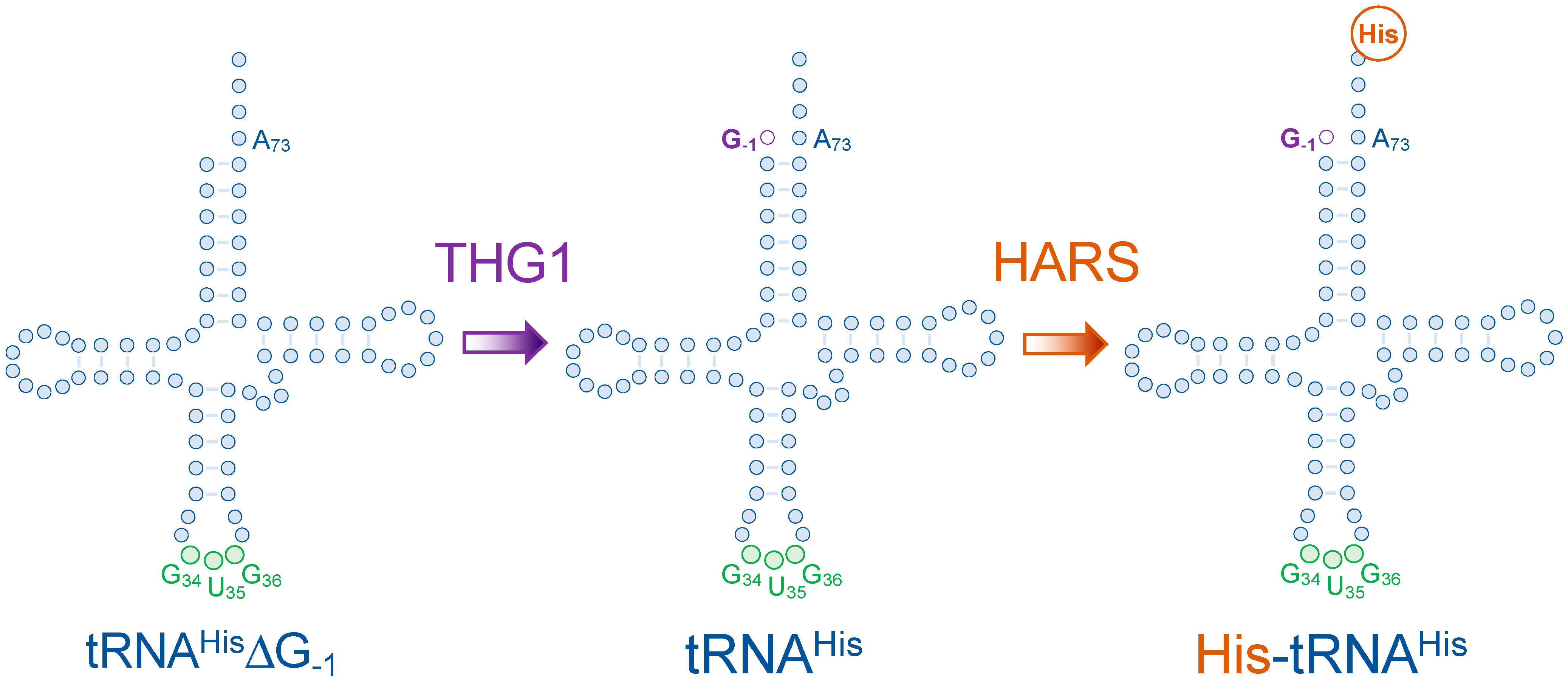
The identity element of tRNAHis is quite unique, as it involves an additional protein factor that aids in the production of mature tRNAHis. Eukaryotic tRNAHis undergoes post-transcriptional modifications that lead to the addition of its primary identity element: a guanylate residue at position -1 [20,21,22]. Eukaryotic pre-tRNAHis lacks this residue as it is not encoded in the genome (tRNAHis∆G-1) [20,21,23,24,25]. Ribonuclease P (RNase P) processes the 5′- end of pre-tRNAHis by cleaving at position +1. Then, tRNAHis guanylyl transferase (Thg1) adds the guanylate residue at position -1, through its 3′ to 5′ RNA polymerization function, providing mature tRNAHis with its primary G-1 identity element (Figure 3). Thg1 discriminates between tRNAHis and other tRNA molecules by recognizing its anticodon and the discriminator base N73. In some bacteria and archaea, G-1 is paired with a C73 in the acceptor stem of tRNAHis but is mispaired with an A73 in eukaryotes [21]. Eventually, most bacterial, yeast, or human HARSs recognize the G-1 residue, which marks tRNAHis for the subsequent histidylation (Figure 3) [26,27,28].

4. Disease-Causing HARS Mutations

While the aminoacylation and tRNA recognition in bacterial HARS have been studied in detail [32,33,34,35,36], relatively few studies have focused on the functionality and pathogenicity of the human enzyme [37]. Several HARS gene mutations have been reported to date as disease-causing (Figure 2, Table 1). Most of these mutations fall within the HARS catalytic domain and are associated with an autosomal dominant neurological disorder, Charcot-Marie-Tooth disease (CMT-type 2W) (Figure 2B) [38,39,40,41,42]. Interestingly, a single missense substitution, Tyr454Ser (Y454S), in the tRNA binding domain of HARS was found to cause Usher syndrome type IIIB (Figure 2C) [43].
Disease-causing mutations in aaRSs can lead to a loss of function, impaired aaRS stability, and/or reduced aminoacylation activity. Gain-of-function mutations can lead to new activities with off-target tRNAs or amino acids, causing misaminoacylation of the cognate tRNA (Figure 1B). Because the ribosome cannot distinguish correctly charged tRNAs from mischarged tRNAs [44], misaminoacylation leads to subsequent mistranslation across the proteome [10,45]. Mistranslation caused by mutations in tRNAs [10,21,46,47,48,49], aaRSs [46], or other components of the protein quality machinery, is increasingly associated with neurodegenerative diseases [10,50], including their ability to genetically modify causative alleles for protein aggregation in Huntington’s disease [48]. Of note, human disease-causing aaRS mutants usually retain a level of activity that is sufficient to sustain the essential functions of protein synthesis [13]. To our knowledge, no disease-causing aaRS mutants with a complete loss of function have been described. Since most of the associated autosomal dominant aaRS diseases affect neuronal activity [13], it is conceivable that dominant gain-of-function mutations and mistranslation might predominantly affect neuronal cells, such as motor neurons in CMT, but the specific CMT-causing mechanism for the aaRS mutants remains to be elucidated.

4.1. aaRS Mutations in Charcot Marie Tooth Disease

More than 150 genes are linked to CMT, with at least 59 known disease-causing alleles occurring in six different aaRSs (Table 2). CMT is a heterogeneous genetic disorder that leads to progressive chronic neuropathy affecting the motor and sensory nerves. CMT is the most common inherited neurological disease, affecting 1 in 2500 people. The onset of disease typically occurs in the first or second decade of life [54]. Several types of CMT can be distinguished using electrophysiological studies [55]. The two major CMT types are CMT1 and CMT2. CMT1 causes demyelination and is characterized by slower median or ulnar nerve conduction velocity (NCV), which measures how fast an electrical impulse moves through the nerves. CMT2 is characterized by normal NCV but direct damage to the nerve axon, resulting in reduced muscle action potential, which is the first step of events leading to muscle contraction [55].
All types of CMT present with similar classical clinical features, which include weakness, disappearance of muscle stretch reflexes starting distally from the feet and ascending through the body, as well as bone deformities [56]. Additionally, loss of sensation to pain, temperature, or vibration in the legs often occurs [57]. Patterns of genetic inheritance for CMT include autosomal dominant, autosomal recessive, and X-linked; however, a portion of mutations arise de novo [58]. The 59 mutations in six different aaRSs associated with CMT peripheral neuropathy [59] are found in glycyl-[60], tyrosyl-[61], alanyl-[62], histidyl-[39], methionyl-[63], and tryptophanyl-tRNA [29] synthetases (GARS, YARS, AARS, HARS, MARS, and WARS) (Table 2). CMT-associated aaRSs are mostly dimeric and are not associated with the MSC [64]. MARS presents the exception as being both monomeric and MSC associated, but no CMT mutations are thought to be relevant to MSC complex formation.
Recent data showed that both loss-of-function and gain-of-function of different aaRSs can lead to CMT phenotypes, and both mechanisms may not be mutually exclusive. Loss-of-function in aaRSs may arise from the failure of the enzyme to dimerize or mutations impairing ATP, tRNA, or amino acid binding [7]. A loss of dimerization inhibits the aaRSs from forming functional complexes, resulting in a reduction in aminoacylation activity. Loss of catalytic activity along with impairment of tRNA or amino acid binding may lead to reduced translational fidelity, stalled protein synthesis, and impaired cell growth.
HARS mutations V155G, Y330C, and S356N are associated with CMT2W and show a reduction in aminoacylation activity. Interestingly, all three were found to form stable dimers [34]. Mutations T132I, P134H, D175E, and D364Y are thought to induce a conformation-derived gain-of-function, rather than a loss of aminoacylation phenotype [29]. Finally, V133F, V155G, Y330C, and S356N are associated with mistranslation and insoluble protein aggregation, as we demonstrated in a novel yeast model for HARS disease [51]. The gain-of-function mutations in aaRSs are thought to be caused by aminoacylation defects, which allow for an incorrect amino acid to be ligated to a tRNAHis, leading to mistranslation [3]. These mutations may also result in a loss of specificity for binding of the cognate tRNA. The resulting misacylated tRNA produced by these aaRS is still recognized at the ribosome for protein synthesis as the codon-amino acid pairing is not impaired, and the mRNA message is translated into a mutated protein. Mistranslation resulting from CMT-causing aaRS alleles has a strong potential to cause proteome-wide errors leading to dysfunctional, misfolded, and aggregated proteins that contribute to disease. Not surprisingly, we recently showed the mistranslation of histidine to glutamine and threonine by CMT-causing HARS variants V155G and S356N. Misincorporation of these amino acids was demonstrated by mass spectrometry and was shown to cause a perturbation of the proteome due to threonine and glutamine incorporation at histidine codons [51].

4.2. Usher Syndrome Type IIIB

Usher syndrome is an autosomal recessive disease that is associated with severely affected hearing, sight, and balance. This disease has been classified into three categories (Usher syndrome (USH) types I, II, and III) based on the age of onset, severity, and progression of sensorineural hearing loss (SNHL), vision loss due to retinitis pigmentosa (RP), and vestibular dysfunction affecting balance [65,66].
USH-type I is the most severe and is characterized by profound congenital hearing impairment and vestibular dysfunction since birth, and first-decade RP. Usher syndrome is genetically heterogeneous. To date, a total of 14 genes have been associated with Usher syndrome (Table 3) [67]. Six USH1 genes have been identified in proteins that play a role in the development and maintenance of cochlear hair cell bundles. Of these, Harmonin (USH1C), Usher syndrome type-1G protein (USH1G), Cadherin-related 23 (CDH23), Myosin-VIIa (MYO7A/USH1B), and potentially Protocadherin-15 (PCDH15) are thought to form a functional network that mediates mechano-transduction in cochlear hair cells [68]. In a separate pathway to disease, Calcium and integrin-binding protein 2 (CIB2) is expressed in cochlear cells, and mutations in CIB2 are associated with USH1J [69]. USH-type II is the most common among Usher syndrome patients and presents with moderate to severe hearing loss, adolescent-onset RP, and normal vestibular function. USH2 is caused by mutations in three different proteins, including Usherin (USH2A), Adhesion G protein-coupled receptor V1 (GPR98/USH2C/ADGRV1), and Whirlin (DFNB31) [70]. Usherin is thought to play a role in extracellular matrix formation [71], and directly interacts with whirlin and GPR98 to form a protein complex in auditory hair cells and photoreceptor cells [72]. USH-type III is characterized by progressive loss of hearing in the first decade, RP in the second decade, and variable progressive vestibular dysfunction [66,67].
USH-type III is the rarest among Usher syndrome patients. USH-type III is classified into USH3A (OMIM #276902) and USH3B (OMIM #614504) based on the genetic mutation underlying each subtype. USH3A was associated with the gene CLARIN I (CLRN1) in 1995 [73,74,75]. Clarin-1 is an essential protein in the morphogenesis of auditory hair cells [76], and an intronic Clarin-1 mutation leads to aberrant splicing [77] and disrupted Clarin-1 function. Despite Clarin-1 and HARS being the only two proteins associated with USH3, no functional association between these proteins has been identified to date. The genetic mutation underlying USH3B was first identified in 2012 in the Plains populations of Pennsylvania and an Old Order Amish family in Ontario, Canada [43]. A single missense mutation in HARS [c.1361A > C], which is inherited in an autosomal recessive manner, leads to a Y454S substitution in patients and is causative for USH3B [43]. Infants with USH3B exhibit normal growth during infancy but experience progressive loss of hearing and sight in the first two decades of life. Rapid clinical deteriorations often occur in response to febrile illnesses. Most patients experience episodic hallucinations due to acute viral infections [43].
HARS Y454S is likely the best clinically characterized disease-causing mutation in human HARS [43,51,53], with a dedicated clinical trial nearing completion (Clinical Trial ID: NCT02924935). The crystal structures of human HARS show that Y454S is located in the interface between the tRNA binding domain and the catalytic domain of the opposing monomer (Figure 2C), suggesting that it may alter anticodon recognition or catalytic activity [30,78]. The substitution of tyrosine to serine at 454 creates a gap between Glu439 and Ser454, destabilizing the hydrogen bonding interaction between both residues (Figure 2C) [43]. Consequently, it was expected for Y454S to influence tRNA binding, aminoacylation, and perhaps the dimerization of the enzyme. A study on murine IMCD3 cells showed, however, only a minor decrease in aminoacylation by mutant murine HARS and unaffected localization and dimerization with endogenous wild-type HARS [43]. Furthermore, a recent study on USH3B patient fibroblasts has shown that the Y454S mutation does not alter the binding specificity to tRNAHis, nor does it affect the overall catalytic activity of HARS. On the other hand, recombinant Y454S HARS protein was shown to be thermolabile due to a significant decrease in melting temperature when compared to wild-type HARS, as well as its failure in incorporating histidine into the proteome of patients’ fibroblasts at elevated temperatures (43 °C) [53]. It is therefore likely that HARS Y454S leads to HARS destabilization and reduced catalytic activity, but it is also feasible that other functions, including non-canonical functions that lead to circulating HARS fragments in the bloodstream, may be impacted by the Y454S mutation [18,19].

5. Pathways to a Cure

Both USH3B and CMT2W are debilitating diseases, yet no current cure is available. Common treatments, such as braces and walking aids, alleviate CMT symptoms, but neither prevent disease onset nor halt or reverse disease progression. For USH3B, amino acid supplementation as a treatment is currently being considered in a clinical trial, and with the astonishing progress in RNA therapeutic technology, treatment of patients with tRNA supplements may also be possible in the near future.
Several non-canonical functions of GARS and KARS are associated with neurodegenerative disorders [79]. These functions and aaRS as therapeutic targets were recently reviewed in detail [5]. Another aaRS-related disease, anti-synthetase syndrome, is a rare autoimmune disorder where the production of antibodies against aaRS leads to myositis, interstitial lung disease, or arthritis [80]. Auto-antibodies against eight aminoacyl-tRNA synthetases have been associated with the clinical manifestation of anti-synthetase syndrome, with the Jo-1 antibody, which is specific for HARS, being the most frequent one [80]. No disease-specific treatment for anti-synthetase syndrome is currently available, and the 5-year survival rate of anti-Jo syndrome with immunosuppressive drugs is estimated at approximately 90% [80].
Recent advances in the treatment of CMT and other aaRS-linked diseases have focused on amino acid supplementation. Several studies show that prolonged, high doses of alanine [81], histidine [82], and glycine [83] supplementation are regarded as safe. Excitingly, and as mentioned above, histidine supplementation is being explored as a treatment option for USH3B patients in a clinical trial. Histidine, like many amino acids, is a readily available supplement in health food stores at a low cost. We found that mistranslation or HARS deficiency caused by CMT-associated aaRS mutations can be alleviated by histidine or tRNAHis supplementation in vitro [51]. For example, in a yeast model of CMT2W with the HARS mutations V133F, V155G, Y330C, or S356N, histidine supplementation rescued the growth defect and prevented insoluble protein accumulation in two of these mutants (V155G and S356N), indicating that histidine supplementation may be a viable treatment option for these patients [51].
While HARS V155G and S356N were rescued by histidine supplementation, the same treatment increased the toxicity of human HARS V1333F and Y330C mutant alleles in yeast cells, indicating that histidine is not an adequate treatment option and other avenues need to be explored. Several CMT-associated mutations in GARS and MARS are localized in the tRNA binding sites of the proteins (Table 1) and may affect tRNA binding. Some mutants affect amino acid specificity or aminoacylation efficiency, whereas other mutants lead to altered tRNA binding to the aaRS, which may be causative of a CMT phenotype. Interestingly, tRNA overexpression compensates for increased tRNA binding by a CMT-GARS mutant in mouse models [84]. In this case, mutations near the GARS tRNA binding domains resulted in altered tRNA specificity or binding, which was compensated for by overexpression of cognate tRNAs [84]. In several recent breakthrough studies, tRNA delivery was used to treat cystic fibrosis [85], mucopolysaccharidosis type I [86], and CMT [84] in mouse models. With recent advances in RNA delivery [87], tRNA medicine is a promising approach to curing CMT-causing phenotypes and will enable next-generation personalized treatments tailored to an individual’s particular CMT-causing allele.

Author Contributions

Conceptualization, R.K. and I.U.H.; writing—original draft preparation, S.D.P.W., R.K., Y.Q., P.O. and I.U.H.; writing—review and editing, S.D.P.W., R.K., Y.Q., P.O. and I.U.H.; visualization, S.D.P.W., R.K. and I.U.H.; supervision, I.U.H.; funding acquisition, I.U.H. All authors have read and agreed to the published version of the manuscript.

Funding

This work was supported from the Natural Sciences and Engineering Research Council of Canada [04776 to I.U.H; 04282 to P.O.]; Canada Research Chairs [232341 to P.O.]; and the Canadian Institutes of Health Research [165985 to P.O.]; Ontario Ministry of Research and Innovation [ER-18-14-183 to I.U.H] and the Rare Disease Models and Mechanisms network.

Data Availability Statement

Data sharing not applicable.

Acknowledgments

We thank Victoria Siu for her leadership in USH3B research and for insightful discussions.

Conflicts of Interest

The authors declare no conflict of interest. The funders had no role in the design of the study; in the collection, analyses, or interpretation of data; in the writing of the manuscript, or in the decision to publish the results.

References

  1. Schuller, A.P.; Green, R. Roadblocks and resolutions in eukaryotic translation. Nat. Rev. Mol. Cell Biol. 2018, 19, 526–541. [Google Scholar] [CrossRef] [PubMed]
  2. Schreiber, G. Translation of genetic information on the ribosome. Angew. Chem. Int. Ed. 1971, 10, 638–651. [Google Scholar] [CrossRef] [PubMed]
  3. Moldave, K. Eukaryotic protein synthesis. Annu. Rev. Biochem. 1985, 54, 1109–1149. [Google Scholar] [CrossRef]
  4. O’Donoghue, P.; Luthey-Schulten, Z. On the evolution of structure in aminoacyl-tRNA synthetases. Microbiol. Mol. Biol. Rev. 2003, 67, 550–573. [Google Scholar] [CrossRef] [Green Version]
  5. Kwon, N.H.; Fox, P.L.; Kim, S. Aminoacyl-tRNA synthetases as therapeutic targets. Nat. Rev. Drug Discov. 2019, 18, 629–650. [Google Scholar] [CrossRef]
  6. Ibba, M.; Söll, D. Aminoacyl-tRNA synthesis. Annu. Rev. Biochem. 2000, 69, 617–650. [Google Scholar] [CrossRef]
  7. Chang, C.-Y.; Chien, C.-I.; Chang, C.-P.; Lin, B.-C.; Wang, C.-C. A WHEP Domain Regulates the Dynamic Structure and Activity of Caenorhabditis elegans Glycyl-tRNA Synthetase. J. Biol. Chem. 2016, 291, 16567–16575. [Google Scholar] [CrossRef] [PubMed] [Green Version]
  8. Nathanson, L.; Deutscher, M.P. Active aminoacyl-tRNA synthetases are present in nuclei as a high molecular weight multienzyme complex. J. Biol. Chem. 2000, 275, 31559–31562. [Google Scholar] [CrossRef] [Green Version]
  9. Gile, G.H.; Moog, D.; Slamovits, C.H.; Maier, U.-G.; Archibald, J.M. Dual Organellar Targeting of Aminoacyl-tRNA Synthetases in Diatoms and Cryptophytes. Genome Biol. Evol. 2015, 7, 1728–1742. [Google Scholar] [CrossRef] [Green Version]
  10. Lant, J.T.; Berg, M.D.; Heinemann, I.U.; Brandl, C.J.; O’Donoghue, P. Pathways to disease from natural variations in human cytoplasmic tRNAs. J. Biol. Chem. 2019, 294, 5294–5308. [Google Scholar] [CrossRef]
  11. Hasan, F.; Lant, J.T.; O’Donoghue, P. Perseverance of protein homeostasis despite mistranslation of glycine codons with alanine. Phil. Trans. R. Soc. B 2023, 378. [Google Scholar] [CrossRef] [PubMed]
  12. Meyer-Schuman, R.; Antonellis, A. Emerging mechanisms of aminoacyl-tRNA synthetase mutations in recessive and dominant human disease. Hum. Mol. Genet. 2017, 26, R114–R127. [Google Scholar] [CrossRef] [PubMed] [Green Version]
  13. Turvey, A.K.; Horvath, G.A.; Cavalcanti, A.R.O. Aminoacyl-tRNA synthetases in human health and disease. Front. Physiol. 2022, 13. [Google Scholar] [CrossRef] [PubMed]
  14. O’Hanlon, T.P.; Miller, F. Genomic organization, transcriptional mapping, and evolutionary implications of the human bi-directional histidyl-tRNA synthetase locus (HARS/HARSL). Biochem. Biophys. Res. Commun. 2002, 294, 609–614. [Google Scholar] [CrossRef] [Green Version]
  15. Wasmuth, J.J.; Carlock, L.R. Chromosomal localization of human gene for histidyl-tRNA synthetase: Clustering of genes encoding aminoacyl-tRNA synthetases on human chromosome 5. Somat. Cell Mol. Genet. 1986, 12, 513–517. [Google Scholar] [CrossRef]
  16. Tsui, F.W.; Siminovitch, L. Isolation, structure and expression of mammalian genes for histidyl-tRNA synthetase. Nucleic Acids Res. 1987, 15, 3349–3367. [Google Scholar] [CrossRef] [Green Version]
  17. Waldron, A.; Wilcox, C.; Francklyn, C.; Ebert, A. Knock-Down of Histidyl-tRNA Synthetase Causes Cell Cycle Arrest and Apoptosis of Neuronal Progenitor Cells in vivo. Front. Cell Dev. Biol. 2019, 7, 67. [Google Scholar] [CrossRef]
  18. Xu, Z.; Wei, Z.; Zhou, J.J.; Ye, F.; Lo, W.-S.; Wang, F.; Lau, C.-F.; Wu, J.; Nangle, L.A.; Chiang, K.P.; et al. Internally deleted human tRNA synthetase suggests evolutionary pressure for repurposing. Structure 2012, 20, 1470–1477. [Google Scholar] [CrossRef] [Green Version]
  19. Zampieri, S.; Ghirardello, A.; Iaccarino, L.; Tarricone, E.; Gambari, P.F.; Doria, A. Anti-Jo-1 antibodies. Autoimmunity 2005, 38, 73–78. [Google Scholar] [CrossRef]
  20. Chen, A.W.; Jayasinghe, M.I.; Chung, C.Z.; Rao, B.S.; Kenana, R.; Heinemann, I.U.; Jackman, J.E. The Role of 3′ to 5′ Reverse RNA Polymerization in tRNA Fidelity and Repair. Genes 2019, 10, 250. [Google Scholar] [CrossRef]
  21. Heinemann, I.U.; Nakamura, A.; O’Donoghue, P.; Eiler, D.; Söll, D. tRNAHis-guanylyltransferase establishes tRNAHis identity. Nucleic Acids Res. 2011, 40, 333–344. [Google Scholar] [CrossRef] [Green Version]
  22. Jackman, J.E.; Gott, J.M.; Gray, M.W. Doing it in reverse: 3′-to-5′ polymerization by the Thg1 superfamily. RNA 2012, 18, 886–899. [Google Scholar] [CrossRef] [Green Version]
  23. Desai, R.; Kim, K.; Büchsenschütz, H.C.; Chen, A.W.; Bi, Y.; Mann, M.R.; Turk, M.; Chung, C.; Heinemann, I.U. Minimal requirements for reverse polymerization and tRNA repair by tRNAHis guanylyltransferase. RNA Biol. 2017, 15, 614–622. [Google Scholar] [CrossRef] [PubMed] [Green Version]
  24. Heinemann, I.U.; O’Donoghue, P.; Madinger, C.; Benner, J.; Randau, L.; Noren, C.J.; Söll, D. The appearance of pyrrolysine in tRNAHis guanylyltransferase by neutral evolution. Proc. Natl. Acad. Sci. USA 2009, 106, 21103–21108. [Google Scholar] [CrossRef] [Green Version]
  25. Heinemann, I.U.; Randau, L.; Tomko, R.J.; Söll, D. 3′-5′ tRNAHis guanylyltransferase in bacteria. FEBS Lett. 2010, 584, 3567–3572. [Google Scholar] [CrossRef] [Green Version]
  26. Connolly, S.A.; Rosen, A.E.; Musier-Forsyth, K.; Francklyn, C.S. G−1:C73 recognition by an arginine cluster in the active site of Escherichia coli histidyl-tRNA synthetase. Biochemistry 2004, 43, 962–969. [Google Scholar] [CrossRef] [PubMed]
  27. Rao, B.S.; Jackman, J.E. Life without post-transcriptional addition of G-1: Two alternatives for tRNAHis identity in Eukarya. RNA 2014, 21, 243–253. [Google Scholar] [CrossRef] [Green Version]
  28. Rudinger, J.; Florentz, C.; Giegé, R. Histidylation by yeast HisRS of tRNA or tRNA-like structure relies on residues–1 and 73 but is dependent on the RNA context. Nucleic Acids Res. 1994, 22, 5031–5037. [Google Scholar] [CrossRef] [PubMed]
  29. Blocquel, D.; Sun, L.; Matuszek, Z.; Li, S.; Weber, T.; Kuhle, B.; Kooi, G.; Wei, N.; Baets, J.; Pan, T.; et al. CMT disease severity correlates with mutation-induced open conformation of histidyl-tRNA synthetase, not aminoacylation loss, in patient cells. Proc. Natl. Acad. Sci. USA 2019, 116, 19440–19448. [Google Scholar] [CrossRef] [PubMed] [Green Version]
  30. Tian, Q.; Wang, C.; Liu, Y.; Xie, W. Structural basis for recognition of G-1-containing tRNA by histidyl-tRNA synthetase. Nucleic Acids Res. 2015, 43, 2980–2990. [Google Scholar] [CrossRef]
  31. Humphrey, W.; Dalke, A.; Schulten, K. VMD: Visual molecular dynamics. J. Mol. Graph. 1996, 14, 33–38. [Google Scholar] [CrossRef] [PubMed]
  32. Himeno, H.; Hasegawa, T.; Ueda, T.; Watanabe, K.; Miura, K.-I.; Shimizu, M. Role of the extra G-C pair at the end of the acceptor stem of tRNAHb in aminoacylation. Nucleic Acids Res. 1989, 17, 7855–7863. [Google Scholar] [CrossRef] [PubMed]
  33. Hawko, S.A.; Francklyn, C.S. Covariation of a specificity-determining structural motif in an aminoacyl-tRNA synthetase and a tRNA identity element. Biochemistry 2001, 40, 1930–1936. [Google Scholar] [CrossRef] [PubMed]
  34. Guth, E.C.; Francklyn, C.S. Kinetic discrimination of tRNA identity by the conserved motif 2 loop of a class II aminoacyl-tRNA synthetase. Mol. Cell 2007, 25, 531–542. [Google Scholar] [CrossRef] [PubMed] [Green Version]
  35. Guth, E.; Farris, M.; Bovee, M.; Francklyn, C.S. Asymmetric amino acid activation by class II histidyl-tRNA synthetase from Escherichia coli. J. Biol. Chem. 2009, 284, 20753–20762. [Google Scholar] [CrossRef] [Green Version]
  36. Guth, E.; Connolly, S.H.; Bovee, M.; Francklyn, C.S. A substrate-assisted concerted mechanism for aminoacylation by a class II aminoacyl-tRNA synthetase. Biochemistry 2005, 44, 3785–3794. [Google Scholar] [CrossRef]
  37. Lee, J.S.; Park, S.G.; Park, H.; Seol, W.; Lee, S.; Kim, S. Interaction network of human aminoacyl-tRNA synthetases and subunits of elongation factor 1 complex. Biochem. Biophys. Res. Commun. 2002, 291, 158–164. [Google Scholar] [CrossRef]
  38. Vester, A.; Velez-Ruiz, G.; McLaughlin, H.M.; Program, N.C.S.; Lupski, J.R.; Talbot, K.; Vance, J.M.; Züchner, S.; Roda, R.H.; Fischbeck, K.H.; et al. A loss-of-function variant in the human histidyl-tRNA synthetase (HARS) gene is neurotoxic in vivo. Hum. Mutat. 2012, 34, 191–199. [Google Scholar] [CrossRef] [Green Version]
  39. Brozkova, D.S.; Deconinck, T.; Griffin, L.B.; Ferbert, A.; Haberlova, J.; Mazanec, R.; Lassuthova, P.; Roth, C.; Pilunthanakul, T.; Rautenstrauss, B.; et al. Loss of function mutations in HARS cause a spectrum of inherited peripheral neuropathies. Brain 2015, 138, 2161–2172. [Google Scholar] [CrossRef] [Green Version]
  40. Royer-Bertrand, B.; Tsouni, P.; Mullen, P.; Campos Xavier, B.; Mittaz Crettol, L.; Lobrinus, A.J.; Ghika, J.; Baumgartner, M.R.; Rivolta, C.; Superti-Furga, A.; et al. Peripheral neuropathy and cognitive impairment associated with a novel monoallelic HARS variant. Ann. Clin. Transl. Neurol. 2019, 6, 1072–1080. [Google Scholar] [CrossRef]
  41. Abbott, J.A.; Meyer-Schuman, R.; Lupo, V.; Feely, S.; Mademan, I.; Oprescu, S.N.; Griffin, L.B.; Alberti, M.A.; Casasnovas, C.; Aharoni, S.; et al. Substrate interaction defects in histidyl-tRNA synthetase linked to dominant axonal peripheral neuropathy. Hum. Mutat. 2017, 39, 415–432. [Google Scholar] [CrossRef] [PubMed]
  42. Lupo, V.; García-García, F.; Sancho, P.; Tello, C.; García-Romero, M.; Villarreal, L.; Alberti, A.; Sivera, R.; Dopazo, J.; Pascual-Pascual, S.I.; et al. Assessment of Targeted Next-Generation Sequencing as a Tool for the Diagnosis of Charcot-Marie-Tooth Disease and Hereditary Motor Neuropathy. J. Mol. Diagn. 2016, 18, 225–234. [Google Scholar] [CrossRef] [PubMed]
  43. Puffenberger, E.G.; Jinks, R.N.; Sougnez, C.; Cibulskis, K.; Willert, R.A.; Achilly, N.P.; Cassidy, R.P.; Fiorentini, C.J.; Heiken, K.F.; Lawrence, J.J.; et al. Genetic mapping and exome sequencing identify variants associated with five novel diseases. PLoS ONE 2012, 7, e28936. [Google Scholar] [CrossRef] [PubMed] [Green Version]
  44. O’Donoghue, P.; Ling, J.; Wang, Y.-S.; Söll, D. Upgrading protein synthesis for synthetic biology. Nat. Chem. Biol. 2013, 9, 594–598. [Google Scholar] [CrossRef] [PubMed]
  45. Heinemann, I.U.; Rovner, A.J.; Aerni, H.R.; Rogulina, S.; Cheng, L.; Olds, W.; Fischer, J.T.; Söll, D.; Isaacs, F.J.; Rinehart, J. Enhanced phosphoserine insertion during Escherichia coli protein synthesis via partial UAG codon reassignment and release factor 1 deletion. FEBS Lett. 2012, 586, 3716–3722. [Google Scholar] [CrossRef] [Green Version]
  46. Guo, L.-T.; Wang, Y.-S.; Nakamura, A.; Eiler, D.; Kavran, J.M.; Wong, M.; Kiessling, L.L.; Steitz, T.A.; O’Donoghue, P.; Söll, D. Polyspecific pyrrolysyl-tRNA synthetases from directed evolution. Proc. Natl. Acad. Sci. USA 2014, 111, 16724–16729. [Google Scholar] [CrossRef] [Green Version]
  47. Lant, J.T.; Berg, M.; Sze, D.H.W.; Hoffman, K.S.; Akinpelu, I.C.; Turk, M.; Heinemann, I.U.; Duennwald, M.L.; Brandl, C.J.; O’Donoghue, P. Visualizing tRNA-dependent mistranslation in human cells. RNA Biol. 2017, 15, 567–575. [Google Scholar] [CrossRef] [Green Version]
  48. Lant, J.T.; Kiri, R.; Duennwald, M.L.; O’Donoghue, P. Formation and persistence of polyglutamine aggregates in mistranslating cells. Nucleic Acids Res. 2021, 49, 11883–11899. [Google Scholar] [CrossRef]
  49. Rozik, P.; Szabla, R.; Lant, J.T.; Kiri, R.; Wright, D.E.; Junop, M.; O’Donoghue, P. A novel fluorescent reporter sensitive to serine mis-incorporation. RNA Biol. 2022, 19, 221–233. [Google Scholar] [CrossRef]
  50. Kapur, M.; Monaghan, C.E.; Ackerman, S.L. Regulation of mRNA Translation in Neurons—A Matter of Life and Death. Neuron 2017, 96, 616–637. [Google Scholar] [CrossRef]
  51. Qiu, Y.; Kenana, R.; Beharry, A.; Wilhelm, S.D.P.; Hsu, S.Y.; Siu, V.M.; Duennwald, M.; Heinemann, I.U. Histidine supplementation can escalate or rescue HARS deficiency in a Charcot–Marie–Tooth Disease model. Hum. Mol. Genet. 2022, ddac239. [Google Scholar] [CrossRef]
  52. Mullen, P.; Abbott, J.A.; Wellman, T.; Aktar, M.; Fjeld, C.; Demeler, B.; Ebert, A.M.; Francklyn, C.S. Neuropathy-associated histidyl-tRNA synthetase variants attenuate protein synthesis in vitro and disrupt axon outgrowth in developing zebrafish. FEBS J. 2020, 288, 142–159. [Google Scholar] [CrossRef] [PubMed]
  53. Abbott, J.A.; Guth, E.; Kim, C.; Regan, C.; Siu, V.M.; Rupar, C.A.; Demeler, B.; Francklyn, C.S.; Robey-Bond, S.M. The Usher Syndrome Type IIIB Histidyl-tRNA Synthetase Mutation Confers Temperature Sensitivity. Biochemistry 2017, 56, 3619–3631. [Google Scholar] [CrossRef] [PubMed]
  54. Theadom, A.; Roxburgh, R.; Macaulay, E.; O’Grady, G.; Burns, J.; Parmar, P.; Jones, K.; Rodrigues, M. Prevalence of Charcot-Marie-Tooth disease across the lifespan: A population-based epidemiological study. BMJ Open 2019, 9, e029240. [Google Scholar] [CrossRef]
  55. Barreto, L.C.L.S.; Oliveira, F.S.; Nunes, P.S.; de França Costa, I.M.P.; Garcez, C.A.; Goes, G.M.; Neves, E.L.A.; Quintans, J.D.S.S.; de Souza Araújo, A.A. Epidemiologic Study of Charcot-Marie-Tooth Disease: A Systematic Review. Neuroepidemiology 2016, 46, 157–165. [Google Scholar] [CrossRef] [PubMed]
  56. Pareyson, D.; Marchesi, C. Diagnosis, natural history, and management of Charcot-Marie-Tooth disease. Lancet Neurol. 2009, 8, 654–667. [Google Scholar] [CrossRef]
  57. Szigeti, K.; Lupski, J.R. Charcot-Marie-Tooth disease. Eur. J. Hum. Genet. 2009, 17, 703–710. [Google Scholar] [CrossRef] [Green Version]
  58. Boerkoel, C.F.; Takashima, H.; Garcia, C.A.; Olney, R.K.; Johnson, J.; Berry, K.; Russo, P.; Kennedy, S.; Teebi, A.S.; Scavina, M.; et al. Charcot-Marie-Tooth disease and related neuropathies: Mutation distribution and genotype-phenotype correlation. Ann. Neurol. 2001, 51, 190–201. [Google Scholar] [CrossRef]
  59. Wei, N.; Zhang, Q.; Yang, X.-L. Neurodegenerative Charcot-Marie-Tooth disease as a case study to decipher novel functions of aminoacyl-tRNA synthetases. J. Biol. Chem. 2019, 294, 5321–5339. [Google Scholar] [CrossRef] [Green Version]
  60. Antonellis, A.; Ellsworth, R.E.; Sambuughin, N.; Puls, I.; Abel, A.; Lee-Lin, S.-Q.; Jordanova, A.; Kremensky, I.; Christodoulou, K.; Middleton, L.T.; et al. Glycyl tRNA synthetase mutations in Charcot-Marie-Tooth disease type 2D and distal spinal muscular atrophy type V. Am. J. Hum. Genet. 2003, 72, 1293–1299. [Google Scholar] [CrossRef]
  61. Jordanova, A.; Irobi, J.; Thomas, F.P.; Van Dijck, P.; Meerschaert, K.; Dewil, M.; Dierick, I.; Jacobs, A.; De Vriendt, E.; Guergueltcheva, V.; et al. Disrupted function and axonal distribution of mutant tyrosyl-tRNA synthetase in dominant intermediate Charcot-Marie-Tooth neuropathy. Nat. Genet. 2006, 38, 197–202. [Google Scholar] [CrossRef] [PubMed] [Green Version]
  62. Latour, P.; Thauvin-Robinet, C.; Baudelet-Méry, C.; Soichot, P.; Cusin, V.; Faivre, L.; Locatelli, M.-C.; Mayençon, M.; Sarcey, A.; Broussolle, E.; et al. A major determinant for binding and aminoacylation of tRNAAla in cytoplasmic alanyl-tRNA synthetase is mutated in dominant axonal Charcot-Marie-Tooth disease. Am. J. Hum. Genet. 2010, 86, 77–82. [Google Scholar] [CrossRef] [Green Version]
  63. Gonzalez, M.; McLaughlin, H.; Houlden, H.; Guo, M.; Yo-Tsen, L.; Hadjivassilious, M.; Speziani, F.; Yang, X.-L.; Antonellis, A.; Reilly, M.M.; et al. Exome sequencing identifies a significant variant in methionyl-tRNA synthetase (MARS) in a family with late-onset CMT2. J. Neurol. Neurosurg. Psychiatry 2013, 84, 1247–1249. [Google Scholar] [CrossRef] [PubMed] [Green Version]
  64. Khan, K.; Long, B.; Gogonea, V.; Deshpande, G.M.; Vasu, K.; Fox, P.L. Multimodal cotranslational interactions direct assembly of the human multi-tRNA synthetase complex. Proc. Natl. Acad. Sci. USA 2022, 119, e2205669119. [Google Scholar] [CrossRef] [PubMed]
  65. Millán, J.M.; Aller, E.; Jaijo, T.; Blanco-Kelly, F.; Gimenez-Pardo, A.; Ayuso, C. An Update on the Genetics of Usher Syndrome. J. Ophthalmol. 2010, 2011, 417217. [Google Scholar] [CrossRef] [PubMed] [Green Version]
  66. Aparisi, M.J.; Aller, E.; Fuster-García, C.; García-García, G.; Rodrigo, R.; Vazquez-Manrique, R.P.; Blanco-Kelly, F.; Ayuso, C.; Roux, A.-F.; Jaijo, T.; et al. Targeted next generation sequencing for molecular diagnosis of Usher syndrome. Orphanet J. Rare Dis. 2014, 9, 168. [Google Scholar] [CrossRef] [Green Version]
  67. Whatley, M.; Francis, A.; Ng, Z.Y.; Khoh, X.E.; Atlas, M.D.; Dilley, R.J.; Wong, E.Y.M. Usher Syndrome: Genetics and Molecular Links of Hearing Loss and Directions for Therapy. Front. Genet. 2020, 11, 565216. [Google Scholar] [CrossRef]
  68. Li, J.; He, Y.; Lu, Q.; Zhang, M. Mechanistic Basis of Organization of the Harmonin/USH1C-Mediated Brush Border Microvilli Tip-Link Complex. Dev. Cell 2016, 36, 179–189. [Google Scholar] [CrossRef] [Green Version]
  69. Wang, Y.; Li, J.; Yao, X.; Li, W.; Du, H.; Tang, M.; Xiong, W.; Chai, R.; Xu, Z. Loss of CIB2 Causes Profound Hearing Loss and Abolishes Mechanoelectrical Transduction in Mice. Front. Mol. Neurosci. 2017, 10, 401. [Google Scholar] [CrossRef] [Green Version]
  70. García-García, G.; Besnard, T.; Baux, D.; Vaché, C.; Aller, E.; Malcolm, S.; Claustres, M.; Millan, J.M.; Roux, A.-F. The contribution of GPR98 and DFNB31 genes to a Spanish Usher syndrome type 2 cohort. Mol. Vis. 2013, 19, 367–373. [Google Scholar]
  71. Eudy, J.D.; Weston, M.D.; Yao, S.; Hoover, D.M.; Rehm, H.L.; Ma-Edmonds, M.; Yan, D.; Ahmad, I.; Cheng, J.J.; Ayuso, C.; et al. Mutation of a gene encoding a protein with extracellular matrix motifs in usher syndrome type IIa. Science 1998, 280, 1753–1757. [Google Scholar] [CrossRef] [PubMed]
  72. Zou, J.; Chen, Q.; Almishaal, A.; Mathur, P.D.; Zheng, T.; Tian, C.; Zheng, Q.Y.; Yang, J. The roles of USH1 proteins and PDZ domain-containing USH proteins in USH2 complex integrity in cochlear hair cells. Hum. Mol. Genet. 2016, 26, 624–636. [Google Scholar] [CrossRef] [PubMed]
  73. Sankila, E.-M.; Pakarinen, L.; Kääriäinen, H.; Aittomäki, K.; Karjalainen, S.; Sistonen, P.; de la Chapelle, A. Assignment of an Usher syndrome type III (USH3) gene to chromosome 3q. Hum. Mol. Genet. 1995, 4, 93–98. [Google Scholar] [CrossRef] [PubMed]
  74. Joensuu, T.; Hämäläinen, R.; Yuan, B.; Johnson, C.; Tegelberg, S.; Gasparini, P.; Zelante, L.; Pirvola, U.; Pakarinen, L.; Lehesjoki, A.-E.; et al. Mutations in a novel gene with transmembrane domains underlie Usher syndrome type 3. Am. J. Hum. Genet. 2001, 69, 673–684. [Google Scholar] [CrossRef] [Green Version]
  75. Isosomppi, J.; Västinsalo, H.; Geller, S.F.; Heon, E.; Flannery, J.G.; Sankila, E.-M. Disease-causing mutations in the CLRN1 gene alter normal CLRN1 protein trafficking to the plasma membrane. Mol. Vis. 2009, 15, 1806–1818. [Google Scholar] [PubMed]
  76. Dulon, D.; Papal, S.; Patni, P.; Cortese, M.; Vincent, P.F.; Tertrais, M.; Emptoz, A.; Tlili, A.; Bouleau, Y.; Michel, V.; et al. Clarin-1 gene transfer rescues auditory synaptopathy in model of Usher syndrome. J. Clin. Investig. 2018, 128, 3382–3401. [Google Scholar] [CrossRef]
  77. Khan, A.O.; Becirovic, E.; Betz, C.; Neuhaus, C.; Altmüller, J.; Riedmayr, L.M.; Motameny, S.; Nürnberg, G.; Nürnberg, P.; Bolz, H.J. A deep intronic CLRN1 (USH3A) founder mutation generates an aberrant exon and underlies severe Usher syndrome on the Arabian Peninsula. Sci. Rep. 2017, 7, 1411. [Google Scholar] [CrossRef] [Green Version]
  78. Koh, C.Y.; Wetzel, A.B.; de van der Schueren, W.J.; Hol, W.G. Comparison of histidine recognition in human and trypanosomatid histidyl-tRNA synthetases. Biochimie 2014, 106, 111–120. [Google Scholar] [CrossRef] [Green Version]
  79. Boczonadi, V.; Jennings, M.J.; Horvath, R. The role of tRNA synthetases in neurological and neuromuscular disorders. FEBS Lett. 2018, 592, 703–717. [Google Scholar] [CrossRef] [Green Version]
  80. Bauhammer, J.; Fiehn, C. Antisynthetase syndrome. Z. Rheumatol. 2019, 78, 645–655. [Google Scholar] [CrossRef]
  81. Soto-Mota, A.; Norwitz, N.G.; Evans, R.D.; Clarke, K. Exogenous d-β-hydroxybutyrate lowers blood glucose in part by decreasing the availability of L-alanine for gluconeogenesis. Endocrinol. Diabetes Metab. 2021, 5, e00300. [Google Scholar] [CrossRef]
  82. Thalacker-Mercer, A.E.; Gheller, M.E. Benefits and Adverse Effects of Histidine Supplementation. J. Nutr. 2020, 150, 2588S–2592S. [Google Scholar] [CrossRef] [PubMed]
  83. Shibui, Y.; Miwa, T.; Yamashita, M.; Chin, K.; Kodama, T. A 4-week Repeated Dose Toxicity Study of Glycine in Rats by Gavage Administration. J. Toxicol. Pathol. 2013, 26, 405–412. [Google Scholar] [CrossRef] [Green Version]
  84. Zuko, A.; Mallik, M.; Thompson, R.; Spaulding, E.L.; Wienand, A.R.; Been, M.; Tadenev, A.L.D.; van Bakel, N.; Sijlmans, C.; Santos, L.A.; et al. tRNA overexpression rescues peripheral neuropathy caused by mutations in tRNA synthetase. Science 2021, 373, 1161–1166. [Google Scholar] [CrossRef] [PubMed]
  85. Albers, S.; Beckert, B.; Matthies, M.C.; Mandava, C.S.; Schuster, R.; Seuring, C.; Riedner, M.; Sanyal, S.; Torda, A.E.; Wilson, D.N.; et al. Repurposing tRNAs for nonsense suppression. Nat. Commun. 2021, 12, 3850. [Google Scholar] [CrossRef] [PubMed]
  86. Wang, J.; Zhang, Y.; Mendonca, C.A.; Yukselen, O.; Muneeruddin, K.; Ren, L.; Liang, J.; Zhou, C.; Xie, J.; Li, J.; et al. AAV-delivered suppressor tRNA overcomes a nonsense mutation in mice. Nature 2022, 604, 343–348. [Google Scholar] [CrossRef]
  87. Paunovska, K.; Loughrey, D.; Dahlman, J.E. Drug delivery systems for RNA therapeutics. Nat. Rev. Genet. 2022, 23, 265–280. [Google Scholar] [CrossRef]
Figure 1. Aminoacylation of tRNAs occurs in a two-step process. Step 1: In the catalytic domain of the aminoacyl-tRNA synthetase (aaRS), a reaction between an amino acid and an ATP molecule results in an aminoacyl-AMP intermediate and releases a pyrophosphate (PPi). Step 2: tRNA recognition is aaRS-specific and involves binding of the acceptor stem, as well as tRNA identity elements such as the anticodon loop, variable loop, or other identity elements in the tRNA. In the aaRS catalytic domain, the amino acid is transferred from the aminoacyl-AMP intermediate to the cognate tRNA molecule. The aminoacyl-tRNA product is released from the enzyme.
Genes 14 00254 g001
Figure 2. Localization of CMT and USH3B causing HARS mutations. (A) Crystal structure of human HARS with CMT and USH3B-causing mutations. The human HARS dimer is shown (pdb:6O76) [29] with the WHEP domain in blue, catalytic domain in tan, and RNA binding domain in purple. The structure was superimposed with the crystal structure from Thermus thermophilus (pdb:4RDX) [30], and the tRNA (lavender) is shown. Histidine and ATP are highlighted in the active site. Magnification of (B) the catalytic domain with CMT causing mutations depicted in orange, and (C) the RNA binding domain with the USH3B mutation Y454S in magenta. The amino acid E439 forms a salt bridge with Y454 that is disrupted in a Y454S mutation. The structure was illustrated using VMD [31].
Genes 14 00254 g002
Figure 3. The post-transcriptional addition of the tRNAHis identity element (G-1) by tRNAHis guanylyl transferase (Thg1). Processing of the 5′ end of tRNAHis by RNase P leads to the formation of tRNAHis∆G-1. tRNAHis guanylyl transferase (Thg1) inserts a guanylyl residue at position −1, opposing to A73 in eukaryotes to form mature tRNAHis. G-1 is the identity element in which histidyl tRNA synthetase (HARS) recognizes tRNAHis for aminoacylation to form His-tRNAHis.
Genes 14 00254 g003
Table 1. Disease-causing mutations in human HARS.
HARS MutationPublication
T132ISafka Brozkova et al., 2015 [39], Blocquel et al., 2019 [29]
T132SSafka Brozkova et al., 2015 [39]
V133FRoyer-Bertrand et al., 2019 [40], Qiu et al., 2022 [51]
P134HSafka Brozkova et al., 2015 [39], Blocquel et al., 2019 [29]
R137QVester et al., 2013 [38], Mullen et al., 2021 [52]
V155GAbbott et al., 2018 [41], Mullen et al., 2021 [52], Qiu et al., 2022 [51]
D175ESafka Brozkova et al., 2015 [39], Blocquel et al., 2019 [29]
G205DVester et al., 2013 [38]
V238AVester et al., 2013 [38]
Y330CAbbott et al., 2018 [41], Mullen et al., 2021 [52], Qiu et al., 2022 [51]
S356NAbbott et al., 2018 [41], Qiu et al., 2022 [51]
D364YSafka Brozkova et al., 2015 [39], Blocquel et al., 2019 [29]
K376RVester et al., 2013 [38]
Y454SPuffenberger et al., 2012 [43], Abbott et al., 2017 [53], Qiu et al., 2022 [51]
P505SVester et al., 2013 [38]
Table 2. CMT disease-associated mutations in aaRSs.
Active Site tRNA Binding DomainOther Domains
AARSN71Y, G102R, R326W, R329H, E337K S627L, E688G, E778A, D893N
GARSE71G, L129P, D167Y, D146N, C157R, H162R, C201R, S211F, H216R, L218Q, P224L P234K/Y, M238R, G240R, P244L, S265Y, S273R, E279D, I280F, G327R, P336H/R, H418R, H472R, D500N, K510Q, G526RM555V, S581L, G625R K27R, K27P, A57V
HARST132I, P134H, R137Q, V155G, D175E, Y330C, S356N, D364YY454S (USH3B)
MARSA397TR618C, R737W, P800T
YARSG41R, D81I, D(153–156), E196K, E196Q
WARSH257R, D314G
Table 3. Genes and loci associated with Usher syndrome. Loci are denoted in italics.
Associated genes/lociUSH-Type IUSH-Type IIUSH-Type III
MYO7A/USH1BUSH2ACLRN1
CDH23/USH1DGPR98/USH2CHARS
PCDH15DFNB31/USH2D
USH1C
USH1E
USH1G
USH1H
USH1K
CIB2
Disclaimer/Publisher’s Note: The statements, opinions and data contained in all publications are solely those of the individual author(s) and contributor(s) and not of MDPI and/or the editor(s). MDPI and/or the editor(s) disclaim responsibility for any injury to people or property resulting from any ideas, methods, instructions or products referred to in the content.