Brief Report

Alternative Genetic Diagnoses in Axenfeld–Rieger Syndrome Spectrum

1
Department of Ophthalmology and Visual Sciences, Medical College of Wisconsin, Milwaukee, WI 53226, USA
2
Department of Pediatrics and Children’s Research Institute, Medical College of Wisconsin and Children’s Wisconsin, Milwaukee, WI 53226, USA
3
Murdoch Children’s Research Institute, Department of Paediatrics, University of Melbourne, Parkville, VIC 3052, Australia
4
Division of Endocrinology, Diabetes, and Metabolic Diseases, Sidney Kimmel Medical College at Thomas Jefferson University, Philadelphia, PA 19107, USA
5
Division of Genetics and Metabolism, MassGeneral Hospital for Children, Boston, MA 02114, USA
6
Department of Pediatrics, University of Wisconsin School of Medicine and Public Health, Madison, WI 53726, USA
*
Author to whom correspondence should be addressed.
Genes 2023, 14(10), 1948; https://doi.org/10.3390/genes14101948
Submission received: 23 September 2023 / Revised: 12 October 2023 / Accepted: 14 October 2023 / Published: 17 October 2023
(This article belongs to the Special Issue Feature Papers in Human Genomics and Genetic Diseases 2023)

Abstract

:
Axenfeld–Rieger anomaly (ARA) is a specific ocular disorder that is frequently associated with other systemic abnormalities. PITX2 and FOXC1 variants explain the majority of individuals with Axenfeld–Rieger syndrome (ARS) but leave ~30% unsolved. Here, we present pathogenic/likely pathogenic variants in nine families with ARA/ARS or similar phenotypes affecting five different genes/regions. USP9X and JAG1 explained three families each. USP9X was recently linked with syndromic cognitive impairment that includes hearing loss, dental defects, ventriculomegaly, Dandy–Walker malformation, skeletal anomalies (hip dysplasia), and other features showing a significant overlap with FOXC1-ARS. Anterior segment anomalies are not currently associated with USP9X, yet our cases demonstrate ARA, congenital glaucoma, corneal neovascularization, and cataracts. The identification of JAG1 variants, linked with Alagille syndrome, in three separate families with a clinical diagnosis of ARA/ARS highlights the overlapping features and high variability of these two phenotypes. Finally, intragenic variants in CDK13, BCOR, and an X chromosome deletion encompassing HCCS and AMELX (linked with ocular and dental anomalies, correspondingly) were identified in three additional cases with ARS. Accurate diagnosis has important implications for clinical management. We suggest that broad testing such as exome sequencing be applied as a second-tier test for individuals with ARS with normal results for PITX2/FOXC1 sequencing and copy number analysis, with attention to the described genes/regions.

1. Introduction

Axenfeld–Rieger anomaly/syndrome (ARA/ARS) is a specific type of anterior segment disorder characterized by the triad of posterior embryotoxon (prominent annular white line near the limbus at the level of the Descemet membrane), iridocorneal adhesions, and iris anomalies including iris hypoplasia, corectopia, and/or polycoria [1,2]. The ocular features are frequently associated with additional nonocular anomalies with two distinct but highly variable syndromes described. In PITX2-assocated ARS (type 1), the ocular anomalies are almost universally associated with dental (missing and malformed teeth) and umbilical (redundant umbilical skin, umbilical hernia, and/or omphalocele) features [3]. FOXC1-associated ARS (type 3) causes much more variable nonocular features, with some individuals having isolated ocular features and others affected with a variety of additional anomalies including hearing loss, congenital heart defects, enamel hypoplasia/dental crowding, hip dysplasia and other skeletal anomalies, hypotonia/early delay, and white matter hyperintensities [3]. At the most severe end of the spectrum for FOXC1 disruption is De Hauwere syndrome, characterized by anterior segment anomalies, hypertelorism, short stature, hearing loss, hydrocephalus, joint hyperlaxity, and skeletal anomalies including hip dysplasia [3,4,5]. While Axenfeld–Rieger anomaly (ARA) is the most common ocular diagnosis in both types of ARS, a wide range of anterior segment ocular phenotypes have been reported for both genes but especially for FOXC1 [3]. Approximately 70% of ARS can be explained by sequence or copy number variants affecting PITX2 or FOXC1, indicating that additional genetic causes remain to be elucidated [3].
Other genetic variants have been occasionally reported in individuals with a clinical diagnosis of ARS or isolated ARA, including dominant variants in COL4A1 in multiple families [6,7], recessive variants in CPAMD8, ADAMTS17, and CYP1B1 reported in one family each [8,9], and dominant variants in histone methyltransferases SETD1A and KMT2F in one family each [10]. Large deletions affecting 13q were reported in three individuals with ARA with no causative gene identified [11,12,13]. PAX6, typically associated with aniridia, can present with overlapping anterior segment ocular phenotypes but is not a cause of ARA specifically [14].

2. Materials and Methods

This human study was approved by the Institutional Review Boards of the Medical College of Wisconsin and University of Iowa. We reviewed our cohort of unexplained individuals with ARA/ARS or similar phenotypes without sequence or copy number variants in PITX2 or FOXC1 [3]. Exome sequencing was performed by Psomagen (Rockville, MD, USA) and analyzed using VarSeq™ (Golden Helix, Bozeman, MT, USA), as previously described [10]. We reviewed variants with a frequency <0.001 in gnomAD v2.1.1 for pathogenic/likely pathogenic variants in all other genes associated with anterior segment disorders and other OMIM genes associated with pediatric syndromes. Copy number analysis was performed on exome data using the VS-CNV® caller. Pathogenicity was determined using ACMG/AMP criteria [15,16]. Variants were confirmed by Sanger sequencing (research and/or clinical confirmation), TaqMan assays (clinical), or independent exome analysis. Parents were tested when available by either exome analysis or Sanger sequencing.

3. Results

3.1. Identification of UPS9X and JAG1 Variants in Multiple Families with ARS

Pathogenic/likely pathogenic variants were identified in nine families affecting five genes (Table 1). All variants were absent from gnomAD. Two genes, UPS9X and JAG1, explained multiple cases within our cohort. The most novel of the two was USP9X, with three unrelated individuals carrying causative variants in this gene, which has not been recognized as a cause of anterior segment phenotypes.
Individual 1 is a 14-year-old White (U.S.) female diagnosed with ARS by her ophthalmologist. Ocular anomalies include ARA, early signs of glaucoma, and myopia. Nonocular anomalies include choanal atresia with deviated nasal septum, high arched palate with bilateral membranous palate anomaly, chronic sinusitis and otitis media (six sets of ear tubes, ear drum reconstruction, and subsequent mild hearing loss), dysmorphic facial features (long, thin face; down-slanting, short palpebral fissures; long, prominent nose that is thin at the nasal tip and pinched at the alar region), abnormal skin pigmentation along lines of Blaschko, and precocious puberty (menses at 9 years old). She has a history of early delays, especially gross motor, but transitioned out of intervention by first grade and is now on the honor roll with adjusted workload, and a history of depression. Height, weight, and head circumference are all normal (50th centile). Exome sequencing identified a likely pathogenic variant in USP9X, NM_001039590.2:c.1314+2T>C. Parental samples were not available. In silico analysis revealed a strong prediction of loss of the donor splice site (0.73 by SpliceAI). There is a weak prediction (0.04) of donor gain which would result in 91bp deletion from exon 10. Exon skipping would result in 153bp deletion of exon 10, c.1162_1315del p.(Glu388_Gln438del). Exon 10 is highly conserved with a very low rate of benign missense variants in gnomAD, so this change is likely to be deleterious.
Individual 2 (Figure 1A) is a 4-year-old Black (U.S.) female with features overlapping ARS including anterior segment dysgenesis, maxillary hypoplasia, unilateral sensorineural hearing loss, congenital heart defect (bicuspid and dysplastic aortic valve), enamel hypoplasia with severe caries and two congenitally missing secondary teeth, umbilical hernia, left developmental hip dysplasia, and Dandy–Walker malformation. Specific ocular anomalies in the left eye include buphthalmos; opaque, thickened, vascularized cornea; and corneal ectasia with spontaneous corneal perforation at 6 months followed by enucleation. Pathology revealed thickened cornea with hypercellular fibrosis and vascularization, cataractous changes in the lens, and only a small remnant of the ciliary body and iris, along with retinal atrophy with thinning of the inner nuclear layer. The right eye has a history of corneal ulcer with residual scarring, hypopigmented wedge in iris, and mild myopia (−1.75). Additional nonocular anomalies include small, posteriorly rotated ears; broad nose with short columella and pointed, depressed tip; flat philtrum; brachycephaly; low anterior hairline; and bilateral coxa valga. She has a history of poor growth with height < 1st centile and weight 3–5th centile and significant global developmental delay (nonverbal). Exome sequencing identified a likely pathogenic variant in USP9X, c.121G>T; p.(Glu41*), which was not present in the mother (father not available).
Individual 3 is a 9-year-old White (U.S.) female with features overlapping ARS including enamel hypoplasia and dental crowding, bilateral hip developmental dysplasia, congenital heart defects (interrupted aortic arch type B with large ventricular septal defect, left superior vena cava to coronary sinus, and bicuspid aortic valve), and history of feeding difficulties in infancy. Ocular anomalies include phlyctenular keratoconjunctivitis and superficial punctate keratitis, subtle scalloped scarring of the cornea with neovascularization, juvenile cataract (small dot multilayer but mostly cortical, not visually significant), mild myopia (−1.25; −1.00), amblyopia, and blepharokeratitis. Additional nonocular anomalies include high arched palate, low-set posteriorly rotated ears with overfolded helices, frontal bossing, anteverted nares, scoliosis with tethered cord, vesicoureteral reflux, left vocal cord paralysis, hypopigmentation of skin with cutis marmorata, eczema, and gross/fine motor delays but normal cognition. Height and weight are within the normal range (22nd and 70th centile, respectively). Exome sequencing identified a de novo likely pathogenic variant in USP9X, c.1603dupA p.(Ile535Asnfs*11); she was reported in a previous cohort at 2 years of age [17].
Individual 4 is a 20-year-old White (Australia) female referred with diagnosis of ARS based on ARA, bilateral sensorineural hearing loss, midface hypoplasia, hypertelorism, and dental crowding. Specific ocular anomalies consisted of posterior embryotoxon, iridocorneal adhesions, and iris hypoplasia along with hyperopia. Additional nonocular features included prominent forehead, short philtrum, arachnodactyly, marfanoid habitus, postural orthostatic tachycardia syndrome (POTS), and auditory/visual processing deficit with average cognition. Echocardiogram was normal. Exome sequencing identified a likely pathogenic variant in JAG1, NM_000214.3:c.59dupT p.(Leu21Alafs*52). Parental samples were not available.
Individual 5 (Figure 1B) is a 39-year-old White (U.S.) female referred with a diagnosis of ARS including ARA, narrow teeth and hypodontia (missing at least four teeth) along with frequent caries, and ventricular septal defect. Specific ocular anomalies consisted of bilateral Axenfeld anomaly with severe myopia and glaucoma diagnosed at 39 years of age. Additional nonocular anomalies include irritable bowel syndrome, pituitary macroadenoma (prolactinoma), and mildly elevated total bilirubin, AST and ALT. CNV analysis of exome sequencing data identified a pathogenic 3.01 Mb deletion of 20:10256130-13269324 (hg19) including seven genes (BTBD3, ISM1, JAG1, MKKS, SLX4IP, SNAP25, and SPTLC3), of which JAG1 is pathogenic. Parental samples were not available.
Individual 6 (Figure 1C,D) is a 38-year-old female clinically diagnosed with ARS in childhood by her ophthalmologist. She has posterior embryotoxon, iris hypoplasia, significant corectopia, small optic cup, and hyperopia (+4.25, +3.75). Nonocular anomalies include dental enamel defects, heart murmur, and polycystic ovary syndrome. Her 5-month-old son was born with congenital heart defects (pulmonary stenosis, atrial septal defect, and patent ductus arteriosus), neonatal jaundice and biliary atresia, and slightly small left kidney (no eye exam but no obvious iris anomalies). Exome sequencing identified a pathogenic variant in JAG1, c.2419G>T p.(Glu807*) in the proband and her son.

3.2. Identification of Variants in BCOR and CDK13 and chr X Deletion in Three Unrelated Cases

Variants in two other genes (CDK13, BCOR) were identified in a single family each, one of which (CDK13) had not been previously linked with anterior segment anomalies. Individual 7 is an 11-year-old White (U.S.) female with a clinical diagnosis of ARS including ARA and widely spaced eyes. Specific ocular features include bilateral posterior embryotoxon and mild iris hypoplasia with transillumination defects, megalocornea with normal intraocular pressure, and mild myopia (−1.00 OU). Additional nonocular anomalies include broad nasal tip, small mouth, history of recurrent otitis media, nasal speech, developmental delay, moderate hypotonia, and cognitive impairment. Height and weight were normal (75th–90th centile). Her 14-year-old brother, affected with cognitive impairment, mild hypotonia, hypospadias, cryptorchidism, history of feeding difficulties, nasal speech, bipolar disorder, behavior problems, and mild dysmorphic features (thin upper lip, telecanthus, right short fifth finger) was thought to have a different genetic syndrome from the proband. Both parents have a history of developmental delay with special education services in school but were unavailable for testing. The mother also has schizophrenia and an extensive family history of learning disabilities and psychiatric illness. Exome sequencing identified a likely pathogenic variant in the proband and her brother within the protein kinase domain of CDK13, NM_003718.5:c.2252G>A p.(Arg751Gln) with high CADD (31) and REVEL (0.697) scores. The same variant was reported previously in an unrelated individual within the Deciphering Developmental Disorders cohort [18].
Individual 8 is a 20-year-old White (U.S.) female referred with a clinical diagnosis of ARS based on ocular anomalies, peg-shaped and small teeth with dental crowding, maxillary hypoplasia, hearing loss, and redundant periumbilical skin. Specific ocular anomalies include bilateral congenital cataract, glaucoma diagnosed at 3 years of age, optic nerve hypoplasia, mild microphthalmia (left smaller than right), and amblyopia of the left eye. Additional nonocular anomalies include patent foramen ovale, nasal deviation, asymmetric and posteriorly rotated ears, narrow palate with thickened ridges, and fifth finger clinodactyly. Height and weight are normal (10–25th centile), as is cognition. Exome sequencing identified a likely pathogenic variant in BCOR NM_001123385.2: c.3350_3360dupCAGACCAGGTG p.(Ala1121GlnfsTer42). Parental samples were not available.
The final case was explained by a combination of two disrupted genes. Individual 9 is a 57-year-old White (U.S.) female who was diagnosed with ARS in early childhood based on ocular features and dental anomalies including small, mispositioned teeth, enamel hypoplasia, and extra teeth. Specific ocular features include bilateral congenital glaucoma with surgery in infancy, polycoria, microphthalmia (right smaller than left), corneal opacity with failed corneal transplant in adulthood, and small congenital cataract which did not require removal until 30 years of age. Additional nonocular anomalies include hearing loss (40s), rheumatoid arthritis, irritable bowel syndrome, connective tissue disease, and migraines. Height/weight and cognitive development are normal. Exome sequencing identified a pathogenic 4.08 Mb deletion of X:7370404-11445756 (hg19) including 17 genes (AMELX, ANOS1, ARHGAP6, CLCN4, CLDN34, FAM9A, FAM9B, GPR143, HCCS, MID1, PNPLA4, SHROOM2, TBL1X, VCX, VCX2, VCX3B, WWC3), of which HCCS and AMELX are pathogenic. Parental samples were not available.

4. Discussion

Axenfeld–Rieger syndrome is associated with significant variability in both ocular and nonocular features [3]. For FOXC1 in particular, a wide range of clinical presentations has been reported. While the majority of cases are caused by disruption of the two major genes, PITX2 and FOXC1, a significant portion remains unexplained.
Dominant loss-of-function and missense variants in USP9X are well-recognized as a cause of syndromic cognitive impairment [19,20]. Both male and female cohorts have been reported, with females showing a broader spectrum of syndromic anomalies [19]. While anterior segment ocular anomalies are not frequently reported in previous cohorts, the syndromic features of hearing loss, ventriculomegaly, Dandy–Walker malformation, short stature, skeletal anomalies including hip dysplasia, and dental defects show significant overlap with FOXC1-associated phenotypes, especially De Hauwere syndrome. Ocular phenotypes including cataract and myopia have been reported in some females, and corneal opacity was noted in one other girl [19,20]. Features unique to USP9X include abnormal pigment distribution along lines of Blaschko, choanal atresia, polydactyly, agenesis of the corpus callosum, and a higher rate of cognitive impairment. Interestingly, two of the three females reported here had a history of early delay but with normal cognitive abilities at older ages. Two of the three had significant anterior segment phenotypes consistent with FOXC1 disruption, and the third has subtle signs of corneal neovascularization, which has also been reported in human patients and mice with Foxc1 disruption [21]. In support of a role for USP9X in the developing eye, Usp9x is highly expressed in the mouse eye, similar to the level seen in the brain [22]. USP9X has been shown to play a role in the WNT pathway [23,24], which is known to be downstream of PITX2 and important in the ocular phenotype associated with this gene [25]. Additionally, loss of Usp9x resulted in reduced expression of N-cadherin (Cdh2) [24], also associated with a corneal phenotype in humans [26].
Alagille syndrome, caused by variants in JAG1 or NOTCH2, is a well-recognized phenotype characterized by highly penetrant posterior embryotoxon and liver dysfunction along with cardiac and vertebral anomalies [27]. While isolated posterior embryotoxon is the characteristic ocular anomaly associated with Alagille syndrome, iris hypoplasia was also present in 20–40% of those undergoing thorough eye exam [28,29]. The diagnosis of Alagille syndrome in three separate families with a clinical diagnosis of ARS was somewhat surprising and highlights the extreme variability of this phenotype. None of the patients meet the characteristic clinical criteria requiring involvement of three out of five organ systems (liver, cardiac, skeletal, ocular, facial). The lack of severe liver involvement likely complicated the clinical recognition of Alagille syndrome in these probands, since bile duct paucity is considered one of the defining features; two of the three individuals reported here had no known liver dysfunction and the third had only mildly abnormal liver function tests discovered through blood work. All three individuals also reported additional features more typical of ARS including dental anomalies (3) and hearing loss (2), though hearing loss has been occasionally reported with JAG1 variants [30].
Similar to UPS9X, disruption of CDK13 is associated with syndromic cognitive impairment [18,31]. The majority of variants are missense and almost all are clustered within the protein kinase domain of CDK13. Cognitive impairment and behavioral problems are the most consistent features with variable dysmorphic facial features, congenital heart defects, and structural brain anomalies. Eye anomalies are recognized but primarily consist of strabismus, so the association with ARA is novel. Similar to Usp9x, expression of Cdk13 in the mouse eye is seen at a similar level to the brain [22].
BCOR is well recognized with loss-of-function variants linked to X-linked dominant Oculofaciocardiodental (OFCD) syndrome in females, while missense variants are occasionally reported in males, typically with a more severe syndromic microphthalmia phenotype [32,33]. OFCD is highly variable and shows substantial overlap with combined features of the two types of ARS including missing teeth (PITX2) and hearing loss and congenital heart defects (FOXC1); while noted to be rare, umbilical hernia has occasionally been reported as well [34,35]. Congenital cataracts with microphthalmia are highly penetrant in OFCD but less commonly seen in ARS; anterior segment phenotypes including iris synechiae, iris coloboma, and posterior embryotoxon are occasionally reported with BCOR variants [33]. Nonocular features specific to BCOR include 2–3 toe syndactyly, extra teeth, or radiculomegaly, and developmental delay/cognitive impairment is more common with BCOR but highly variable [33].
The final case is an interesting example of a contiguous gene syndrome whose combined features resulted in a phenotype overlapping ARS. HCCS was first recognized as a cause of X-linked dominant linear skin defects with multiple congenital anomalies, with microphthalmia, sclerocornea, heart defects, structural brain anomalies, and linear skin defects on the face and neck [36]. Subsequent studies revealed a highly variable phenotype in affected individuals, with isolated ocular features in some females [37]. The addition of AMELX within the deleted region resulted in amelogenesis imperfecta, type 1E [38], with significant overlap with the dental features of ARS including severe enamel hypoplasia.
All of the genetic diagnoses identified here showed substantial overlap with the ARS phenotypic spectrum. Accurate diagnosis has important implications for clinical management and family planning. We suggest that broad testing such as exome sequencing be applied as a second-tier test for individuals with ARS with normal sequencing/copy number results for PITX2/FOXC1.

Author Contributions

Conceptualization, E.V.S.; methodology, E.V.S. and L.M.R.; formal analysis, L.M.R.; resources, D.J.A., R.A.H., C.B.N., K.M.K.-N. and S.A.C.; writing—original draft preparation, L.M.R. and E.V.S.; writing—review and editing, D.J.A., R.A.H., C.B.N., K.M.K.-N. and S.A.C.; funding acquisition, E.V.S. All authors have read and agreed to the published version of the manuscript.

Funding

This work was supported by NIH grants R01EY015518 and R01EY025718 as well as funds provided by the Children’s Research Institute Foundation at Children’s Wisconsin.

Institutional Review Board Statement

This study was conducted according to the guidelines of the Declaration of Helsinki and approved by the Institutional Review Board of Medical College of Wisconsin (PRO45954) and University of Iowa (199804080).

Informed Consent Statement

Informed consent was obtained from all subjects (and/or legal guardians) involved in this study, including permission to publish.

Data Availability Statement

There are no other data associated with this manuscript.

Acknowledgments

We are grateful to the patients and their families for participation in this study and to Jeffrey C. Murray, (University of Iowa) for expert guidance.

Conflicts of Interest

The authors declare no conflict of interest.

References

  1. Shields, M.B. Axenfeld-Rieger syndrome: A theory of mechanism and distinctions from the iridocorneal endothelial syndrome. Trans. Am. Ophthalmol. Soc. 1983, 81, 736–784. [Google Scholar] [PubMed]
  2. Shields, M.B.; Buckley, E.; Klintworth, G.K.; Thresher, R. Axenfeld-Rieger syndrome. A spectrum of developmental disorders. Surv. Ophthalmol. 1985, 29, 387–409. [Google Scholar] [CrossRef] [PubMed]
  3. Reis, L.M.; Maheshwari, M.; Capasso, J.; Atilla, H.; Dudakova, L.; Thompson, S.; Zitano, L.; Lay-Son, G.; Lowry, R.B.; Black, J.; et al. Axenfeld-Rieger syndrome: More than meets the eye. J. Med. Genet. 2023, 60, 368–379. [Google Scholar] [CrossRef] [PubMed]
  4. Reis, L.M.; Tyler, R.C.; Volkmann Kloss, B.A.; Schilter, K.F.; Levin, A.V.; Lowry, R.B.; Zwijnenburg, P.J.; Stroh, E.; Broeckel, U.; Murray, J.C.; et al. PITX2 and FOXC1 spectrum of mutations in ocular syndromes. Eur. J. Hum. Genet. 2012, 20, 1224–1233. [Google Scholar] [CrossRef]
  5. Garza Flores, A.; Nordgren, I.; Pettersson, M.; Dias-Santagata, D.; Nilsson, D.; Hammarsjo, A.; Lindstrand, A.; Batkovskyte, D.; Wiggs, J.; Walton, D.S.; et al. Case report: Extending the spectrum of clinical and molecular findings in FOXC1 haploinsufficiency syndrome. Front. Genet. 2023, 14, 1174046. [Google Scholar] [CrossRef] [PubMed]
  6. Coupry, I.; Sibon, I.; Mortemousque, B.; Rouanet, F.; Mine, M.; Goizet, C. Ophthalmological features associated with COL4A1 mutations. Arch. Ophthalmol. 2010, 128, 483–489. [Google Scholar] [CrossRef]
  7. Sibon, I.; Coupry, I.; Menegon, P.; Bouchet, J.P.; Gorry, P.; Burgelin, I.; Calvas, P.; Orignac, I.; Dousset, V.; Lacombe, D.; et al. COL4A1 mutation in Axenfeld-Rieger anomaly with leukoencephalopathy and stroke. Ann. Neurol. 2007, 62, 177–184. [Google Scholar] [CrossRef]
  8. Ma, A.; Yousoof, S.; Grigg, J.R.; Flaherty, M.; Minoche, A.E.; Cowley, M.J.; Nash, B.M.; Ho, G.; Gayagay, T.; Lai, T.; et al. Revealing hidden genetic diagnoses in the ocular anterior segment disorders. Genet. Med. 2020, 22, 1623–1632. [Google Scholar] [CrossRef]
  9. Tanwar, M.; Dada, T.; Dada, R. Axenfeld-Rieger Syndrome Associated with Congenital Glaucoma and Cytochrome P4501B1 Gene Mutations. Case Rep. Med. 2010, 2010, 212656. [Google Scholar] [CrossRef]
  10. Reis, L.M.; Atilla, H.; Kannu, P.; Schneider, A.; Thompson, S.; Bardakjian, T.; Semina, E.V. Distinct Roles of Histone Lysine Demethylases and Methyltransferases in Developmental Eye Disease. Genes 2023, 14, 216. [Google Scholar] [CrossRef]
  11. Stathacopoulos, R.A.; Bateman, J.B.; Sparkes, R.S.; Hepler, R.S. The Rieger syndrome and a chromosome 13 deletion. J. Pediatr. Ophthalmol. Strabismus 1987, 24, 198–203. [Google Scholar] [CrossRef] [PubMed]
  12. Roche, A.; Mora, J.; Perez Mdel, M.; Gean, E.; Perez, B.; O’Callaghan, M.; Catala, J.; De Torres, C.; Cruz, O.; Prat, J.; et al. Axenfeld-Rieger ocular anomaly and retinoblastoma caused by constitutional chromosome 13q deletion. Pediatr. Blood Cancer 2010, 54, 480–482. [Google Scholar] [CrossRef] [PubMed]
  13. Akazawa, K.; Yamane, S.; Shiota, H.; Naito, E. A case of retinoblastoma associated with Rieger’s anomaly and 13q deletion. Jpn. J. Ophthalmol. 1981, 25, 321–325. [Google Scholar]
  14. Riise, R.; D’Haene, B.; De Baere, E.; Gronskov, K.; Brondum-Nielsen, K. Rieger syndrome is not associated with PAX6 deletion: A correction to Acta Ophthalmol Scand 2001: 79: 201–203. Acta Ophthalmol. 2009, 87, 923. [Google Scholar] [CrossRef] [PubMed]
  15. Richards, S.; Aziz, N.; Bale, S.; Bick, D.; Das, S.; Gastier-Foster, J.; Grody, W.W.; Hegde, M.; Lyon, E.; Spector, E.; et al. Standards and guidelines for the interpretation of sequence variants: A joint consensus recommendation of the American College of Medical Genetics and Genomics and the Association for Molecular Pathology. Genet. Med. 2015, 17, 405–424. [Google Scholar] [CrossRef] [PubMed]
  16. Riggs, E.R.; Andersen, E.F.; Cherry, A.M.; Kantarci, S.; Kearney, H.; Patel, A.; Raca, G.; Ritter, D.I.; South, S.T.; Thorland, E.C.; et al. Technical standards for the interpretation and reporting of constitutional copy-number variants: A joint consensus recommendation of the American College of Medical Genetics and Genomics (ACMG) and the Clinical Genome Resource (ClinGen). Genet. Med. 2020, 22, 245–257. [Google Scholar] [CrossRef]
  17. Jolly, L.A.; Taylor, V.; Wood, S.A. USP9X enhances the polarity and self-renewal of embryonic stem cell-derived neural progenitors. Mol. Biol. Cell 2009, 20, 2015–2029. [Google Scholar] [CrossRef]
  18. Hamilton, M.J.; Caswell, R.C.; Canham, N.; Cole, T.; Firth, H.V.; Foulds, N.; Heimdal, K.; Hobson, E.; Houge, G.; Joss, S.; et al. Heterozygous mutations affecting the protein kinase domain of CDK13 cause a syndromic form of developmental delay and intellectual disability. J. Med. Genet. 2018, 55, 28–38. [Google Scholar] [CrossRef]
  19. Jolly, L.A.; Parnell, E.; Gardner, A.E.; Corbett, M.A.; Perez-Jurado, L.A.; Shaw, M.; Lesca, G.; Keegan, C.; Schneider, M.C.; Griffin, E.; et al. Missense variant contribution to USP9X-female syndrome. NPJ Genom. Med. 2020, 5, 53. [Google Scholar] [CrossRef]
  20. Reijnders, M.R.; Zachariadis, V.; Latour, B.; Jolly, L.; Mancini, G.M.; Pfundt, R.; Wu, K.M.; van Ravenswaaij-Arts, C.M.; Veenstra-Knol, H.E.; Anderlid, B.M.; et al. De Novo Loss-of-Function Mutations in USP9X Cause a Female-Specific Recognizable Syndrome with Developmental Delay and Congenital Malformations. Am. J. Hum. Genet. 2016, 98, 373–381. [Google Scholar] [CrossRef]
  21. Seo, S.; Singh, H.P.; Lacal, P.M.; Sasman, A.; Fatima, A.; Liu, T.; Schultz, K.M.; Losordo, D.W.; Lehmann, O.J.; Kume, T. Forkhead box transcription factor FoxC1 preserves corneal transparency by regulating vascular growth. Proc. Natl. Acad. Sci. USA 2012, 109, 2015–2020. [Google Scholar] [CrossRef] [PubMed]
  22. Wu, C.; Orozco, C.; Boyer, J.; Leglise, M.; Goodale, J.; Batalov, S.; Hodge, C.L.; Haase, J.; Janes, J.; Huss, J.W., III; et al. BioGPS: An extensible and customizable portal for querying and organizing gene annotation resources. Genome Biol. 2009, 10, R130. [Google Scholar] [CrossRef] [PubMed]
  23. Nielsen, C.P.; Jernigan, K.K.; Diggins, N.L.; Webb, D.J.; MacGurn, J.A. USP9X Deubiquitylates DVL2 to Regulate WNT Pathway Specification. Cell Rep. 2019, 28, 1074–1089.e1075. [Google Scholar] [CrossRef]
  24. Premarathne, S.; Murtaza, M.; Matigian, N.; Jolly, L.A.; Wood, S.A. Loss of Usp9x disrupts cell adhesion, and components of the Wnt and Notch signaling pathways in neural progenitors. Sci. Rep. 2017, 7, 8109. [Google Scholar] [CrossRef] [PubMed]
  25. Hendee, K.E.; Sorokina, E.A.; Muheisen, S.S.; Reis, L.M.; Tyler, R.C.; Markovic, V.; Cuturilo, G.; Link, B.A.; Semina, E.V. PITX2 deficiency and associated human disease: Insights from the zebrafish model. Hum. Mol. Genet. 2018, 27, 1675–1695. [Google Scholar] [CrossRef]
  26. Reis, L.M.; Houssin, N.S.; Zamora, C.; Abdul-Rahman, O.; Kalish, J.M.; Zackai, E.H.; Plageman, T.F., Jr.; Semina, E.V. Novel variants in CDH2 are associated with a new syndrome including Peters anomaly. Clin. Genet. 2020, 97, 502–508. [Google Scholar] [CrossRef]
  27. Ayoub, M.D.; Kamath, B.M. Alagille Syndrome: Current Understanding of Pathogenesis, and Challenges in Diagnosis and Management. Clin. Liver Dis. 2022, 26, 355–370. [Google Scholar] [CrossRef]
  28. da Palma, M.M.; Igelman, A.D.; Ku, C.; Burr, A.; You, J.Y.; Place, E.M.; Wang, N.K.; Oh, J.K.; Branham, K.E.; Zhang, X.; et al. Characterization of the Spectrum of Ophthalmic Changes in Patients With Alagille Syndrome. Investig. Ophthalmol. Vis. Sci. 2021, 62, 27. [Google Scholar] [CrossRef]
  29. Hingorani, M.; Nischal, K.K.; Davies, A.; Bentley, C.; Vivian, A.; Baker, A.J.; Mieli-Vergani, G.; Bird, A.C.; Aclimandos, W.A. Ocular abnormalities in Alagille syndrome. Ophthalmology 1999, 106, 330–337. [Google Scholar] [CrossRef]
  30. Le Caignec, C.; Lefevre, M.; Schott, J.J.; Chaventre, A.; Gayet, M.; Calais, C.; Moisan, J.P. Familial deafness, congenital heart defects, and posterior embryotoxon caused by cysteine substitution in the first epidermal-growth-factor-like domain of jagged 1. Am. J. Hum. Genet. 2002, 71, 180–186. [Google Scholar] [CrossRef]
  31. van den Akker, W.M.R.; Brummelman, I.; Martis, L.M.; Timmermans, R.N.; Pfundt, R.; Kleefstra, T.; Willemsen, M.H.; Gerkes, E.H.; Herkert, J.C.; van Essen, A.J.; et al. De novo variants in CDK13 associated with syndromic ID/DD: Molecular and clinical delineation of 15 individuals and a further review. Clin. Genet. 2018, 93, 1000–1007. [Google Scholar] [CrossRef] [PubMed]
  32. Ng, D.; Thakker, N.; Corcoran, C.M.; Donnai, D.; Perveen, R.; Schneider, A.; Hadley, D.W.; Tifft, C.; Zhang, L.; Wilkie, A.O.; et al. Oculofaciocardiodental and Lenz microphthalmia syndromes result from distinct classes of mutations in BCOR. Nat. Genet. 2004, 36, 411–416. [Google Scholar] [CrossRef] [PubMed]
  33. Ragge, N.; Isidor, B.; Bitoun, P.; Odent, S.; Giurgea, I.; Cogne, B.; Deb, W.; Vincent, M.; Le Gall, J.; Morton, J.; et al. Expanding the phenotype of the X-linked BCOR microphthalmia syndromes. Hum. Genet. 2019, 138, 1051–1069. [Google Scholar] [CrossRef] [PubMed]
  34. Lozic, B.; Ljubkovic, J.; Panduric, D.G.; Saltvig, I.; Kutsche, K.; Krzelj, V.; Zemunik, T. Oculo-facio-cardio-dental syndrome in three succeeding generations: Genotypic data and phenotypic features. Braz. J. Med. Biol. Res. 2012, 45, 1315–1319. [Google Scholar] [CrossRef]
  35. Verma, G.; Singh, G.K.; Tandon, P.; Verma, S.L. A rare syndrome with unusual dental findings: Oculo-facio-cardio-dental syndrome. J. Oral. Maxillofac. Pathol. 2014, 18, 331. [Google Scholar] [CrossRef]
  36. Wimplinger, I.; Morleo, M.; Rosenberger, G.; Iaconis, D.; Orth, U.; Meinecke, P.; Lerer, I.; Ballabio, A.; Gal, A.; Franco, B.; et al. Mutations of the mitochondrial holocytochrome c-type synthase in X-linked dominant microphthalmia with linear skin defects syndrome. Am. J. Hum. Genet. 2006, 79, 878–889. [Google Scholar] [CrossRef]
  37. Weh, E.; Reis, L.M.; Happ, H.C.; Levin, A.V.; Wheeler, P.G.; David, K.L.; Carney, E.; Angle, B.; Hauser, N.; Semina, E.V. Whole exome sequence analysis of Peters anomaly. Hum. Genet. 2014, 133, 1497–1511. [Google Scholar] [CrossRef]
  38. Lagerstrom, M.; Dahl, N.; Nakahori, Y.; Nakagome, Y.; Backman, B.; Landegren, U.; Pettersson, U. A deletion in the amelogenin gene (AMG) causes X-linked amelogenesis imperfecta (AIH1). Genomics 1991, 10, 971–975. [Google Scholar] [CrossRef]
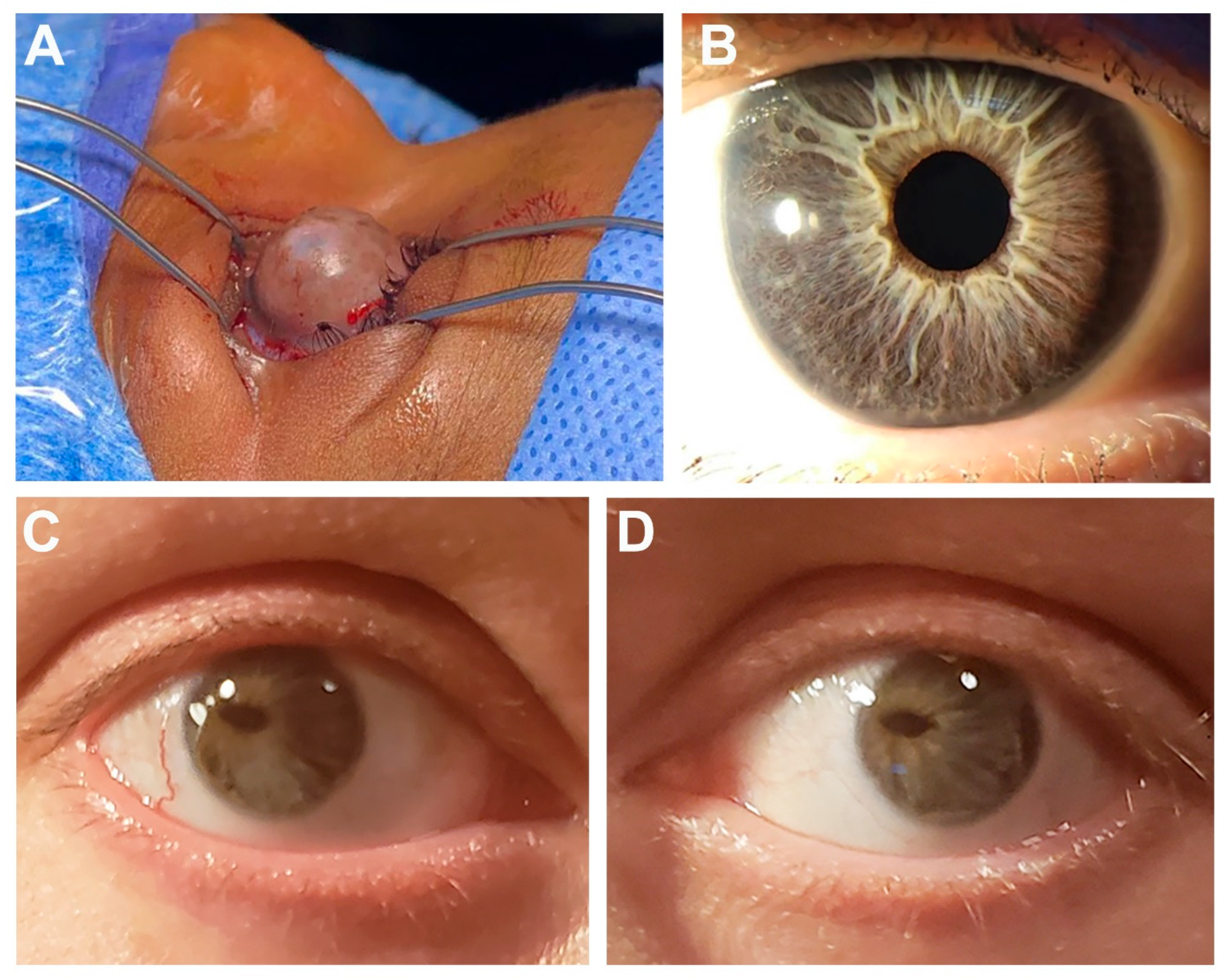
Figure 1. Selected ocular images demonstrating anterior segment phenotypes. (A) Right eye of Individual 2 showing corneal ectasia with large bulbous anterior extension. (B) Eye of Individual 5 showing posterior embryotoxon and mild iris hypoplasia (C,D) Bilateral eye images of Individual 6 showing severe corectopia, posterior embryotoxon and iris hypoplasia.
Genes 14 01948 g001
Table 1. Clinical features of presented individuals.
IndividualOcularOtherGeneVariantACMG Criteria
1ARA, GL, MYMild delay but normal cognition, dysmorphic facial features, palate anomaly, choanal atresia, lines of Blaschko, precocious pubertyUSP9XNM_001039590.2:
c.1314+2T>C
LP: PVS1, PM2_supp
2Left: ASD, severe IH, CAT, buphthalmos, corneal neovascularization, corneal ectasia with perforation Right: corneal ulcer/scarring, MY, iris hypopigmentationDA, HL, dysmorphic facial features, CHD, Dandy–Walker malformation, hip dysplasia, umbilical hernia, global delay, short statureUSP9Xc.121G>T; p.(Glu41*)LP: PVS1, PM2_supp
3Corneal scarring/neovascularization, CAT, MYDA, dysmorphic facial features, CHD, hip dysplasia, feeding difficulties, palate anomaly, vesicoureteral reflux, skin pigment anomaly, scoliosis, gross motor delay but normal cognitionUSP9Xc.1603dupA
p.(Ile535Asnfs*11)
LP: PVS1, PM2_supp
4ARADA, dysmorphic facial features, HL, connective tissue anomaly, arachnodactylyJAG1NM_000214.3:c.59dupT p.(Leu21Alafs*52)LP: PVS1, PM2_supp
5ARA, MYDA, HL, CHD, liver dysfunction, pituitary macroadenoma (prolactinoma)JAG1Gene deletion: 3.01 Mb deletion of 20:10256130-13269324 (hg19)P: 2A
6ARA, CGL, corectopia, HYDA, heart murmur, PCOSJAG1NM_000214.3: c.2419G>T p.(Glu807*)P: PVS1, PM2_supp, PP1
7ARA, megalocorneaDysmorphic facial features, cognitive impairmentCDK13NM_003718.5:c.2252G>A p.(Arg751Gln)LP: PS1, PM1, PM2_supp, PP3
8GL, CAT, ONH, mild MIDA, HL, dysmorphic facial features, RU, CHD, palate anomalyBCORNM_001123385.2:c.3350_3360dup p.(Ala1121Glnfs*42)LP: PVS1, PM2_supp
9CGL, IH, polycoria, CAT, mild MIDA, HL, rheumatoid arthritis, connective tissue anomalyHCCS and AMELXGene deletions: 4.08 Mb deletion of X:7370404-11445756 (hg19)P: 2A
ARA: Axenfeld–Reiger anomaly; ASD: anterior segment dysgenesis; CAT: cataract; CGL: congenital glaucoma; GL: glaucoma; HY: hyperopia; MI: microphthalmia; MY: myopia; DA: dental anomalies; HL: hearing loss; PCOS: polycystic ovarian syndrome; RU: redundant umbilicus. ACMG criteria [15,16]. Features in bold are typical for ARS types 1 or 3.
Disclaimer/Publisher’s Note: The statements, opinions and data contained in all publications are solely those of the individual author(s) and contributor(s) and not of MDPI and/or the editor(s). MDPI and/or the editor(s) disclaim responsibility for any injury to people or property resulting from any ideas, methods, instructions or products referred to in the content.