Abstract
With the advances in genomic sequencing, many organisms with novel biological properties are ripe for use as emerging model organisms. However, to make full use of them, transformation methods need to be developed to permit genome editing. Here, we present the development of transformation for the fungus fly Bradysia (Sciara) coprophila; this may serve as a paradigm for the development of transformation for other emerging systems, especially insects. Bradysia (Sciara) has a variety of unique biological features, including locus-specific developmentally regulated DNA amplification, chromosome imprinting, a monopolar spindle in male meiosis I, non-disjunction of the X chromosome in male meiosis II, X chromosome elimination in early embryogenesis, germ-line-limited (L) chromosomes and high resistance to radiation. Mining the unique biology of Bradysia (Sciara) requires a transformation system to test mutations of DNA sequences that may play roles for these features. We describe a Bradysia (Sciara) transformation system using a modified piggyBac transformation vector and detailed protocols we have developed to accommodate Bradysia (Sciara) specific requirements. This advance will provide a platform for us and others in the growing Bradysia (Sciara) community to take advantage of this unique biological system. In addition, the versatile piggyBac vectors described here and transformation methods will be useful for other emerging model systems.
1. Introduction
Many advances in biological understanding have come from the elucidation of principles in unique biological systems. For example, the protozoan ciliate Tetrahymena served as the experimental material for discoveries that led to two different Nobel prizes (self-splicing, telomerase) [1]. Similarly, the nematode worm Caenorhabditis elegans has been the experimental organism that has garnered several Nobel prizes (programmed cell death, RNA interference). Recognition of the value of the breadth of knowledge from a large number of model systems [2] has led to efforts to sequence genomes from a diverse array of organisms, including the i5K project to sequence the genomes of 5000 arthropods, including all the phylogenetic branches of insects (http://arthropodgenomes.org/wiki/i5K, access on 1 January 2022). In order to reap the full advantage of such information, a transformation methodology is needed that has broad applicability. The recent advent of CRISPR/Cas9 has provided a technology for genomic mutations [3,4,5]. However, since Cas9 cleavage creates blunt ends, it is not suitable for insertion of large DNA (such as genes) using the non-homologous end-joining pathway that is preferred in most organisms over homologous recombination. In contrast, piggyBac, with its broad phylogenetic range, allows a widely adaptable means for genomic insertion of large DNA. Here, we present the first report of a method using piggyBac to transform the fungus fly Bradysia (Sciara) coprophila with no prior transformation system, and this may offer applications to other emerging model organisms, especially insects.
The fungus fly Bradysia (Sciara) [6] represents the largest known eukaryotic genus in the world, with over 700 species. Some years ago, it was renamed Bradysia coprophila [7], but we also retain the earlier name of Sciara to be consistent with the large body of published literature on this organism. The novel biological strategies employed by Bradysia (Sciara) led it to be on the short list as a premier model organism in the 1930s at a Cold Spring Harbor meeting, when Bradysia (Sciara) competed with Drosophila melanogaster, and in the 1970s, when Sydney Brenner compared the merits of Bradysia (Sciara) with the worm C. elegans. The many unique features of Bradysia (Sciara) (reviewed in Refs. [8,9,10,11]) include:
- Locus-specific DNA amplification in larval salivary gland polytene chromosomes;
- Chromosome imprinting;
- A monopolar spindle in male meiosis I;
- Non-disjunction of the X chromosome in male meiosis II;
- Chromosome elimination in early embryogenesis;
- Germ-line-limited (L) chromosomes;
- High resistance to radiation.
Recently, the Bradysia (Sciara) genome has been sequenced, annotated and assembled [12]. This serves as the foundation for genome editing, where a transformation system for Bradysia (Sciara) is required to facilitate experiments to understand the molecular basis for this extraordinary number of unique biological features. Such molecular tools would allow manipulation of the Bradysia (Sciara) genome to assay the role of specific DNA sequences for these unique biological features.
Historically, transposable elements have permitted the integration of DNA sequences into insect genomes. P-elements were the first of these [13], but they lacked a broad host range [14,15,16]. In contrast, piggyBac has been used to transform many organisms, including a broad range of insects from Diptera, Lepidoptera and Coeoptera (reviewed in Refs. [17,18]). Moreover, P-elements preferentially insert into the promoters of genes, whereas piggyBac has evolved to reduce the deleterious effects in host gene expression [19]. The piggyBac transposon, derived from the cabbage looper moth Trichoplusia ni, is a 2472 bp Class II transposon with 13 bp inverted terminal repeats [20,21]. PiggyBac contains a single ORF that encodes a 594-amino-acid transposase that mediates the insertion of this transposon into a TTAA target site in the genome, resulting in target-site duplication that flanks the transposable element [21,22]. The piggyBac transposase is active in a broad number of insects [17] and other animals, ranging from planaria to mammalian cells [23].
Given the extensive host range of piggyBac, we have developed a transformation system for Bradysia (Sciara) based on the piggyBac transformation vector pBac [3xP3-ECFPafm] [24,25], which is designed to screen transgenic animals using the ECFP fluorescent marker driven by the highly conserved P3 promoter. Our subsequent modifications included the use of TagYFP and blasticidin resistance as markers for screening. For future versatility to allow site-specific insertions of large DNA by a variety of means, we included an artificial zinc finger nuclease target (ZFN-T) site [26] and the phiC31 attP site [27]. We also report here the development of methods for the various steps in Bradysia (Sciara) transformation. These vectors and procedures will serve as the foundation for future experiments on Bradysia (Sciara) by the growing scientific community. Moreover, they should be helpful for the transformation of other non-model organisms.
2. Materials and Methods
All materials, vectors and reagents are freely available upon request to the authors or the Bradysia (Sciara) Stock Center (https://sites.brown.edu/sciara/, access on 1 January 2022), with permission needed from Dr. Craig J. Coates for the piggyBac transposase construct.
2.1. Bradysia (Sciara) Injection
2.1.1. Egg Collection and Injection
The sterilized vials used for rearing the fungus gnat Sciara coprophila (syn. Bradysia coprophila or Bradysia tilicola) are 29 mm inner diameter and 9.5 cm tall (Thermo Fisher Scientific, Waltham, MA, USA # 03-339-26H or Wilmad LabGlass, Vineland, NJ, USA), filled with 1.3 inches of 2.2% (w/v) bacteriological agar and topped with non-absorbent cotton-filled gauze plugs. One day before injection, 5 vials were set up with mass matings, with each vial containing 10 males and 10 females that are male producers from the HoLo2 line that was derived from the 7298 stock (X’ chromosome for female producers marked with the dominant allele for Wavy wings) but lacks the polymorphism noted for 7298 [28]. Previously, the larvae should be well-fed to provide fat adults with large eggs from the females. All the following steps for egg collection and transformation were performed in a room at 18 °C with several humidifiers for at least 70% humidity and preferably >90% humidity. We used three humidifiers in a small temperature-controlled room with the recirculating air duct blocked, and we monitored the temperature and humidity level. One day (18 h) after mating, the adult females were collected by anesthesia on a CO2 pad, and their thorax is gently squeezed (avoiding injury to the abdomen) to induce their wings to extend upright. They were then picked up by their wings with a forceps for placement on a 100-mm-diameter Petri dish containing 2.2% (w/v) agar, with their wings inserted into the agar. Egg laying was induced by gently squeezing the head with a forceps until the female fly showed seizure-like movements. The Petri dish was then covered with the lid that was moistened with a water-dampened Kimwipe to prevent electrostatic attraction of the eggs to the lid. After 30 min, the eggs were transferred with a dampened (but not overly wet) fine-bristle paint-brush onto the egg sorting agar, gently using forceps to dislodge any embryos stuck on the brush when transferring. It is important to proceed quickly so that injections are completed within a window of 1–3 h after egg-laying, before the pole cells form and while the embryos are still a syncytial blastoderm [29,30,31,32].
Egg sorting Petri plates (50 mm diameter and 9 mm height; Falcon Plastics, Brookings, SD, USA) were made with 2.2% (w/v) agar darkened by adding green or blue food dye so that the nuclei inside the eggs could be visualized with a strong focused light. The colored agar was removed with a metal spatula from the Petri dish and placed upside down on top of the lid. A straight edge (about 4 cm long) was made by cutting the agar with a razor blade 1/5 from the edge of the Petri dish. Re-usable glass spatulas were made in advance by heating with rotation the long stem of a Pasteur pipette in a Bunsen burner flame, removing it from the flame to pull and break the melted tip and heat-sealing the tip (close, typically 3 cm, to the thicker body of the pipette) in the flame so that the sealed tip is the width of one embryo. The sealed tip is round and not flat. The eggs were aligned close to each other with the glass spatula, with the anterior end closest to the cut edge of the agar. The eggs should not be right at the edge nor protrude beyond the agar cut edge as they will dry out. The posterior part of the embryo is opaque; the anterior part is more transparent and the nuclei can be visualized. Embryos with a small clearing at the anterior and posterior ends should not be used as they are too old.
Bradysia (Sciara) appeared to be extremely sensitive to the glues commonly used for Drosophila embryo injection. Instead, Kwik-Cast glue (World Precision Instruments, Inc., Sarasota, FL, USA) was used as a non-toxic substitute. A small amount of Kwik-Cast yellow and blue substances were squeezed from the tubes and mixed on a glass microscope slide with a pipetteman tip to turn the mixture to green. A dab of the green Kwik-Cast mixture was scooped up with a pipetteman tip and spread evenly on the long edge of a fresh, ethanol-washed glass microscope slide. The thickness of the Kwik-Cast is crucial for successful injection. Embryos must be lightly covered with Kwik-Cast. However, if it is too thick, it will interfere with the injection. Kwik-Cast cures in 3–5 min at 18 °C. Moreover, for successful transfer of the embryos, the viscosity of the Kwik-Cast is important. If it is too soft, the embryos will remain on the agar plate. Conversely, if it is too hard, it will not cover the embryos enough. The viscosity of the Kwik-Cast glue was monitored by touching the Kwik-Cast mixture that remained on the first slide with a pipetteman tip. When the viscosity was acceptable (~3 min after mixing the Kwik-Cast), the microscope slide with the long line of Kwik-Cast was inverted and held against the embryos that were aligned on the colored agar to transfer them to the Kwik-Cast, with the posterior end of the aligned embryos at the edge of the slide. Any excess glue between the embryos and the edge of the slide can be removed with a razor blade. The embryos on the Kwik-Cast glue stripe were then covered lightly with halocarbon 27 oil (CAS#9002-83-9; Halocarbon Products Corporation, Peachtree Corners, GA, USA) and placed in a humid chamber for at least 3 min to let the glue cure completely. The humid chamber was a shallow plastic box (245 mm × 245 mm; Corning) with 2–3 layers of 3 MM paper soaked with water; the microscope slides were elevated on plastic spacer bars to avoid direct contact with water. Note that halocarbon 27 oil is a medium-weight polymer of chlorotrifluoroethylene (PCTFE) rather than the high-molecular-weight PCTFE polymer used for Drosophila injections.
Bradysia (Sciara) embryos were injected within 2 h of egg laying, using an injection apparatus similar to that used for Drosophila, with needles drawn out from borosilicate glass capillaries (1B100-4; World Precision Instruments, Inc., Sarasota, FL, USA) and filled with a Microfil 28 needle (MF28G67-5 4; World Precision Instruments, Inc., Sarasota, FL, USA). The needles were made using a Vertical Pipet Puller Model 700C (David Kopf Instruments, Tujunga, CA, USA) with a one-stage pull and were straight-tapered. Bradysia (Sciara) embryos are smaller than Drosophila embryos, so the needle is pulled with a much longer straight taper towards the tip to make the needle’s diameter as narrow as possible where it is inserted in the embryos. This reduces the escape of cytoplasm due to surface tension during injection. However, since the needle is inserted through the cured elastic mounting medium, if the needle is too narrow, it tends to bend too much and makes it very difficult to penetrate through the medium. It is crucial to find the fine balance between the stability and sharpness of the needle.
The chorion of Bradysia (Sciara) is very thin and does not need to be removed for injection; however, the Bradysia (Sciara) embryos need to be maintained in a humid environment to avoid desiccation. PiggyBac vector DNA (final concentration in injection solution of 1 μg/μL water) was mixed with hr5-ie1 piggyBac transposase plasmid (400 ng/μL) and IE1 transactivator plasmid (pIE1/153) (4 ng/μL) for the injection [33]. The inverted bright field microscope used for the injections rested on an anti-vibration platform (World Precision Instruments, Inc., Sarasota, FL, USA). The filled needle was placed in the injector and lowered to contact the edge of a broken coverslip that was affixed with double-sided tape to the microscope slide to break the needle tip, allowing droplets of the injection fluid to flow continuously from the tip. The needle was lowered to be in line with the embryos on the microscope slide, and the slide was then moved towards the needle for sequential injection into the posterior end of each embryo. During the injection procedure, the pressure of liquid in the needle was adjusted to maintain a continuous flow; too little pressure results in cytoplasm from the embryo backing up into the needle. Alternatively, pulse injections were used with a time gated setting for the injector.
After injection, the slide with affixed embryos was held vertically for 5 min on a paper towel to drain off the excess oil. The slide was examined under a dissecting microscope to confirm that just enough oil was left to cover the eggs by surface tension. If the embryos are completely submerged in too much oil, they will not survive due to lack of oxygen. Bradysia (Sciara) seems to be very sensitive to the oxygen level. Excess oil was blotted off with a rolled up Kimwipe; the slide was placed in a humid chamber box (described above) that was wrapped in Saran wrap and incubated 5 days at 18 °C.
After 5 days at 18 °C and development into late embryonic stages (before pigmentation of the jaw appears), the embryos were excised from the Kwik-Cast using the tip of a 23G needle to cut the edge of the glue surrounding the embryos. This allowed the oil to seep under the embryos and pop them off the Kwik-Cast glue. The embryos were then transferred with a glass spatula onto a 100-mm-diameter Petri dish containing 2.2% agar (w/v) (without antibiotics) and placed in a humid chamber at 21 °C. It is important to do this before the embryos emerge as first instar larvae are more prone to damage by the transfer procedure than the embryos. Once the larvae emerged, they were fed on alternating days 3× per week. Bradysia (Sciara) food is one part Brewer’s yeast mixed with 4 parts (by volume) of an autoclaved mixture of 2 parts very finely ground oat straw, 1 part mushroom powder and 1 part spinach or nettle powder (all are insecticide-free). If the larvae try to crawl out of the Petri dish (regularly monitor this), the Petri dish containing them should be moved from the humid chamber to a dry chamber at 21 °C. After the 4th instar and pupation, when the body color of the pupae had begun to darken, the pupae were transferred from the Petri dish into a glass vial containing 2.2% agar (w/v) and incubated at 21 °C until the adult flies emerged. About 300 Bradysia (Sciara) embryos were injected in one day to become ~60 G0 flies based on the observation that, typically, there are 20–50% survivors at 5 days after injection. Once they hatch to the first instar, typically more than 80% of the larvae reach the pupal stage; some (~10%) may not complete pupation. In general, there is a higher transformation rate among the injected flies that are less fertile; we adjust the concentration of the DNA in the injection mixture to obtain at least 60% fertility in the G0 crosses. Moreover, as little solution as possible is injected at the posterior end of the embryos.
After emerging as 1st instar larvae, at least 60–70% develop into fertile adults. The injection is repeated on sequential days with the goal of obtaining 100 fertile G0 adults that will yield 1–2 transformants. The transformation efficiency for the pBac [3XP3-ECFPafm] attP-ZFN-T vector was about 2–3% for fertile G0 crosses and was 0.3% for the pBac [3XP3-TagYFP, hr5-ie1-BlasR, su(Hw)BS] attP, ZFN-T vector.
2.1.2. Subsequent Crosses with Fluorescent Marker Selection
When the G0 male flies emerged, each was placed in a vial containing 2.2% agar (w/v) with 5 female producer female flies (HoLo 2 line). Alternatively, each male fly was crossed with 3 females on day one and then on day two the male was moved to a fresh vial containing 2 new female producer female flies. Therefore, each single G0 male was crossed with a total of 5 female producer females. The vials were incubated at 21 °C until the larva were 3rd instar, when the larvae from a single G0 male were transferred to a 100-mm-diameter Petri dish containing 2.2% agar (w/v) with a maximum density of 50 larvae per Petri dish. Since food in the larval gut can create autofluorescence, the larvae were left overnight at 18 °C on the Petri dish with no food to clear the gut contents. Larvae with fluorescent signals were selected under a fluorescence stereo dissecting microscope (Zeiss Stemi SV11 Apo, equipped with a standard EYFP, EGFP, ECFP fluorescent filter set and with an X-Cite 120 light source) and fed 3× per week on the agar Petri dishes at 21 °C until they pupated. Fluorescent screening was completed as soon as feasible (generally for 3rd rather than 4th instar larvae). After the fluorescent larvae from a single G0 male emerged as pupae and started to show strong eye pigmentation (and the fluorescent signal may no longer be visible), they were transferred to a single vial containing 2.2% agar (w/v) for development into adult female flies. To amplify the transformant lines, matings were completed of transformed female-producer females with males from the Bradysia (Sciara) HoLo 2 line using three males per female in a single vial. Thus, the transgene is transmitted through the female in each generation, avoiding the problem that male-derived chromosomes are eliminated on the monopolar spindle in meiosis I of spermatogenesis (where the transgene could be lost).
2.1.3. Subsequent Crosses with Antibiotic Selection
When the G0 male flies emerged, each was crossed with 5 female-producing female flies (HoLo 2 line) as in Section 2.1.2. One day after each cross, the females were placed onto 100-mm-diameter Petri dishes containing 2.2% agar (w/v) with 20 µg/mL blasticidin S (A.G. Scientific, Inc., San Diego, CA, USA), diluted from a stock solution of 10 mg/mL blasticidin in 20 mM HEPES, pH 7.2. The Petri dishes were incubated in a humid chamber at 21 °C, with removal of the dead adult females after 2–4 days. As a control, non-transformed female-producing females (HoLo 2 line) were mated with HoLo 2 males for egg laying on Petri dishes containing 2.2% agar (w/v) with 20 μg/mL blasticidin. The Petri dishes with the control and experimental larvae were maintained without feeding in a humid chamber at 21 °C until the control larvae died (generally 3rd instar). At that point, the blasticidin-resistant larvae were transferred to Petri dishes containing 2.2% agar (w/v) but no blasticidin and were maintained in a dry chamber at 21 °C with feeding 3× per week. When the larvae became dark pupae, they were transferred from the Petri dishes to vials containing 2.2% agar (w/v). When the adult flies emerged, the female producer female flies were crossed with HoLo 2 males to maintain the transgene through the female lineage and to amplify the transformant lines.
2.2. Construction of piggyBac Vectors
2.2.1. Construction of pBac [3XP3-ECFPafm] attP-ZFN-T
A 231 bp DNA fragment containing the attP sequence (AATAATGATTTTATTTTGACTGATAGTGACCTGTTCGTTGCAACAAATTGATGAGCAATGCTTTTTTATAATGCCAACTTTGTACAAAAAAGCTGAACGAGAAACGTAAAATGATATAAATATCAATATATTAAATTAGATTTTGCATAAAAAACAGACTACATAATACTGTAAAACACAACATATCCAGTCACTATGAATCAACTACTTAGATGGTATTAGTGACCTGTA) with PstI sites flanking each end was synthesized by Integrated DNA Technologies (IDT, Redwood City, CA, USA) and cloned into the PstI site of pBac [3XP3-ECFPafm] attP [24] that was a kind gift from Dr. Alfred M. Handler, USDA. The synthesized fragment was cloned into the PCR-Blunt II-TOPO vector (Invitrogen, Carlsbad, CA, USA).
Next, a linker was added that included the same artificial zinc finger nuclease (ZFN) target site (CGCTACCCGACCATGAAGCAGCA) as used in Ref. [26]. The FseI sites flanking the linker were created by annealing two oligos (FTEAF 5′and FTEAF 3′). The sequences of these oligos were:
FTEAF 5′: CCCGCTACCCCGACCATGAAGCAGCAGAATTCGGCGCGCCGGCCGG;
FTEAF 3′: CCGGCGCGCCGAATTCTGCTGCTTCATGGTCGGGGTAGCGGGCCGG.
This FseI-bounded linker containing the ZFN target site was then cloned into the FseI site of (pBac [3XP3-ECFPafm] attP) to create (pBac [3XP3-ECFPafm] attP-ZFN-T).
2.2.2. Construction of pBac [3XP3-TagYFP, hr5-ie1-BlasR, su(Hw)BS] attP-ZFN-T
Since the introduction of TagYFP involved PstI restriction digestion, attP was initially removed from the PstI site and later re-introduced into that site. The removal of attP converted the plasmid pBac [3XP3-ECFPafm] attP-ZFN-T described in the preceding Section 2.2.1 to pBac [3XP3-ECFPafm] ZFN-T as the starting vector. As described in detail below (also see Supplementary Figure S3), two su(Hw)BS were introduced, and then ECFP was replaced with TagYFP, followed by addition of the attP site.
The suppressor of Hairy wing binding site (su(Hw)BS) was introduced into the plasmid in a two-step procedure as follows. First, the plasmid pBac [3xP3-ECFP afm] ZFN-T was linearized by PCR using the primer sets pBac 3Fp and pBac 3R:
pBac 3Fp: phos-GATGTTCCCACTGGCCTGGAGCG
pBac 3R: GATGTTTTGTTTTGACGGACCCCTT
The 362 bp sequence for su(Hw)BS was amplified from the pYES vector [34] (kindly provided by Dr. John Tower, University of Southern California) using primer sets 20-1-1 F5p and YY59-1:
20-1-1-F5p: phos-AGTTACTCTTCCGGCTAAATGGTATGGCAAGAAAAGGTATGCAATAT
YY59-1: GGGCGAATTGGGTACCCTATTCGC
The amplified su(Hw)BS sequence was then blunt-end-ligated into the linearized vector pBac [3xP3-ECFP afm] ZFN-T noted above at a position just in front of 3XP3-ECFP. This clone was subsequently linearized by PCR using primer pairs pBac 5F and pBac5Rp:
pBac 5F: GACAATGTTCAGTGCAGAGACTCG
pBac 5Rp: phos-AGATCTGTCATGATGATCATTGCAATT
Subsequently, the same amplified su(Hw)BS sequence noted above was again blunt-end-ligated, in this case just downstream of ZFN-T in the plasmid being constructed.
Next, the ECFP gene was replaced by TagYFP in the plasmid under construction, named pBac [3XP3-ECFP, su(Hw)BS] ZFN-T. The 750 bp TagYFP coding sequence was amplified from pTagYFP-C (Evrogen, Moscow, Russia) using primer pairs P3-TagYFP F1 and TagYFP 3R1:
P3-TagYFP F1:
gcccgggatccaccggtcgccaccATGGTTAGCAAAGGCGAGGAGCTGTTCGCCGGC
(The TagYFP coding sequence is underlined and the 3xP3 is in lower case letters)
TagYFP 3R1:
AGAGTCGCGGCCGCTTTACCGGTACAGCTCGTCCATGCCGTGGGTGTGGC
(The NotI site is underlined)
Subsequently, the 1.4 kb fragment (including the 5′ piggyBac arm and 3XP3-TATA) was amplified from the plasmid noted above, named pBac [3XP3-ECFP, su(Hw)BS] ZFN-T, using primer pairs PstI F1 and P3-TagYFP R1:
PstI F1:
CCTACTGCAGGTCATCACAGAACACATTTGGTCTAGCGTGTCCACTCCGCC
(The PstI site is underlined)
P3-TagYFP R1: (contains 3′ end of P3 promoter and 5′ end of TagYFP coding sequence)
GGCGGAGTGGACACGCTAGACCAAATGTGTTCTGTGATGACCTGCCGTAGG
The 1.4 kb fragment and 750 bp TagYFP fragment (both described above) were mixed together and amplified with primer sets PstI F1 (sequence given above) and TagYFP 3R1-2:
TagYFP 3R1-2:
GCGCCTGTAGCCACACCCACGGCATGGACGAGCTGTACCGGTAAAGCGGCCGCAAGAATTCTTGCGGCCGCTTTACCGGTACAGCTCGTCCATGCCGTGGGTGTGGCTACAGGCGC
The PstI site of the TagYFP coding sequence was eliminated by replacing CTGCAG with CTGTAG to keep the same codon (cysteine) at this position. (The Not I site is in italics and the complementary sequence of the destroyed PstI site is underlined.) The resulting 2.15 kb product was purified, digested with PstI and NotI and then cloned into the PstI and NotI sites of pBac [3XP3-ECFP, su(Hw)BS] ZFN-T.
The attP sequence was introduced into the plasmid as described in Section 2.2.1. Subsequently, this DNA fragment was inserted into the PstI site of plasmid pBac [3XP3-TagYFP, su(Hw)BS] ZFN-T.
The blasticidin resistance gene was introduced as follows. The blasticidin resistance gene coding sequence was amplified by PCR with the primers BlasS SacII F1 and BlasS BamHI R3 from pcDNA6/V5-His-A (Invitrogen, Carlsbad, CA, USA):
BlasS SacII F1:
AATTACCGCGGATAAAATGGCCAAGCCTTTGTCTCAAGAAGAATCCACCCTCATT
BlasS BamHI R3:
AATTGGATCCGCCCTCCCACACATAACCAGAGGGCAGCAATTCACGA
The amplified PCR product was then cloned into the SacII-BamHI site of the pIE1-3 carrying piggyBac transposase (kindly provided by Dr. Craig J. Coates, Texas A&M University). The Hr5-ie1-blasiticidin coding sequence with polyA of this clone was amplified with primer set pIE AscI F1 and pIE AscI-R1:
pIE AscI F1:
ATATATGGCGCGCCCACAATCAAGTATGAGTCATAAGCTGATGTCATGTTTTGC
pIE AscI-R1:
TTAATTGGCGCGCCAAGCTTAAAAGTAGGAGGAACGGGCATACTCTTGGCCACCGGCGGC
This PCR amplified product was then cloned into the AscI site of pBac [3XP3-TagYFP, su(Hw)BS] attP, ZFN-T.
2.3. PCR
Larvae were homogenized in Lysis Buffer (100 mM Tris-HCl (pH 8.0), 500 mM NaCl, 1% SDS, 0.15 mM spermine, 0.5 mM spermidine), and genomic DNA was isolated by phenol/chloroform and resuspended in TE buffer. All PCRs were carried out using the following conditions: step 1: 103 °C 1 min; step 2: 103 °C 10 s; step 3: 55 °C 10 s; step 4: 72 °C 45 s; step 5: repeat steps 2–4 40 times; step 6: 72 °C 2 min; step 7: 4 °C.
2.3.1. Genomic PCR for pBac [3XP3-ECFPafm] attP-ZFN-T
The 700 bp fragment from larvae transformed with pBac [3XP3-ECFPafm] attP-ZFN-T was amplified using the primers 16-1-2 F2 and 5-1-1-F2-R1:
16-1-2 F2: CAACCACTACCTGAGCACCC
5-1-1-F2-R1: GTGCAGCCAACGTCAAGCGG
2.3.2. Genomic PCR for pBac [3XP3-TagYFP, hr5-ie1-BlasR, su(Hw)BS] attP, ZFN-T
Genomic PCRs of DNA from larvae transformed with pBac [3XP3-TagYFP, hr5-ie1-BlasR, su(Hw)BS] attP, ZFN-T were carried out using primer pairs TagYFP code F1 and TagYFP code R1 for PCR 1 and BlasR code F1 and BlasR code R1 for PCR 2:
TagYFP code F1: ATGGTTAGCAAAGGCGAGGAGCTGTTCGCCGGCATCGTGCCCG
TagYFP code R1: CTCCAGCTTGTGGCCCAGGATGTTGCCG
BlasR code F1: ATGGCCAAGCCTTTGTCTCAAGAAGAATCCACCCTCATT
BlasR code R1: TTAGCCCTCCCACACATAACCAGAGGGCA
2.3.3. Inverse PCR
Inverse PCR was carried out on DNA from Bradysia (Sciara) transformed with pBac [3XP3-ECFPafm] attP-ZFN-T following the Berkeley Drosophila Genome Project (BDGP) protocol (Roger Hoskins and Martha Evans-Holm, 11 June 2004 FlyBase http://www.fruitfly.org/about/methods/inverse.pcr.html, access on 1 January 2022, E. Jay Rehm, 2014 and http://www.fruitfly.org/, access on 1 January 2022) with minor modification: the Sau3A digested genomic DNA was cleaned with one phenol/chloroform extraction prior to the self-ligation step.
2.4. DNA Sequencing
Amplified DNA fragments were gel-purified and sequenced directly using the same primers as used for the PCR. All sequencing was completed by Genewiz (Cambridge, MA, USA).
3. Results
3.1. Handling Bradysia (Sciara) for Transformation
To develop a transformation system, it was necessary to establish the protocols for the successful injection of Bradysia (Sciara) embryos (see Materials and Methods for specific details). We designed a procedure for mass mating. One day after mating, the adult female flies were impaled on agar plates and the head was crushed to induce egg laying. This is advantageous to accumulate synchronized newly laid and fertilized eggs. It is important to inject embryos before the pole cells form and while the nuclei are still in a syncytium so that the DNA injected into the cytoplasm can easily enter the nucleus. In Bradysia (Sciara), the first eleven cleavage divisions are syncytial and occur in the first nine hours at 22 °C after egg laying [32], although the timescale seemed somewhat faster in our hands after induced egg-laying. We injected within two hours after egg-laying, which is before cleavage cycle 5 when the elimination of germ-line-limited L chromosomes occurs in the somatic nuclei [29,30,31,32]. Moreover, at cleavage cycle 5, two nuclei enter the germplasm at the posterior of the egg and undergo rapid divisions to form the pole cells [29,30]. For germline integration of DNA, it is important to inject the posterior of the embryo, and, for this, it is necessary to be able to distinguish the anterior and posterior ends of the embryo. The micropyle, which is a canal piercing the chorion and through which the sperm enters, is a brown disc at the anterior end of the egg [29,30], but it is usually hard to see. Instead, we have made use of the fact that the cytoplasm is less dense at the anterior end, allowing the nuclei to be more readily visible when the embryos are on a dark background (Supplementary Figure S1). The embryos should all be aligned so that their posterior ends will be at the edge of the microscope slide for easy access for injection (Supplementary Figure S2). As a control for accuracy in determining the posterior end, the aligned embryos can be left on the agar Petri dish for a week in a humid chamber to allow the black jaw at the anterior end to become visible, thus confirming the alignment. Initially, the embryos were correctly aligned 90% of the time, and, with practice, 99% of the time.
After embryo alignment, embryos less than 2 h after egg-laying were used for injection. We utilized an injection apparatus that is the same for standard Drosophila embryo injections. However, Bradysia (Sciara) embryos are extremely sensitive to environmental factors, such as oxygen level, humidity and chemical contamination. Consequently, we needed to develop a protocol to accommodate Bradysia (Sciara) specific requirements using special material and handling conditions. The chorion of Bradysia (Sciara) embryos is thin, and it was not necessary to remove it prior to injection. Bradysia (Sciara) is extremely sensitive to the glues typically used for Drosophila transformation. Therefore, Kwik-Cast adhesive (World Precision Instruments, Inc., Sarasota, FL, USA) was used instead as the glue to affix the Bradysia (Sciara) embryos onto microscope slides. Kwik-Cast has ultra-low toxicity and was a crucial factor for successful injection. The embryos were covered with halocarbon oil and injected at their posterior end, where the future germ cells will reside; most of the oil was removed after the injection in order not to suffocate the embryos. Bradysia (Sciara) coprophila is monogenic, and females will have only sons or only daughters. We chose to inject male embryos so that the G0 male flies that emerged could be mated with five female flies, thereby amplifying the number of F1 progeny. With these procedures, we were able to inject up to 500 Bradysia (Sciara) embryos per day, with a high yield of fertile adults. The non-injected embryos have 80% development to fertile adults, whereas the injected embryos had 60–70% development to fertile adults in the best cases.
3.2. Genomic Integration of pBac [3XP3-ECFPafm] attP-ZFN-T
Prior to the injections for Bradysia (Sciara) piggyBac transformation, we carried out trial experiments in Drosophila and injected in vitro transcripts of mRNA encoding piggyBac transposase. However, there was much variability with each preparation of mRNA, and each had to be tested to find the maximum concentration that provided good transformation efficiency but was not too high to compromise the viability and fertility of the injected embryos. Therefore, for the piggyBac transformation of Bradysia (Sciara), only helper plasmid DNA encoding transposase rather than transposase mRNA was used. We utilized the Autographa carifornica nuclear polyhedrosis baculovirus immediate early gene (ie1) flanked by the hr5 enhancer [33] instead of the hsp70 promoter [35] to drive piggyBac transposase; the hr5-ie1 promoter–enhancer dramatically increases transposase expression ~500 to 1000-fold. We also constructed the same helper plasmid with a hyperactive piggyBac transposase [36] (Wellcome Trust Sanger Institute, Hinxton, Cambridgeshire, UK), but it did not further improve the transformation efficiency in a pilot experiment conducted in Drosophila, probably because the codon usage for this transposase had been optimized for mammals rather than insects. Therefore, the standard piggyBac transposase was used for Bradysia (Sciara) transformation.
The piggyBac plasmid needs to carry a dominant marker to select for positive transformants. Fluorescence markers have been widely used [37]. The initial piggyBac plasmid that we used was pBac [3XP3-ECFPafm] attP-ZFN-T, which carries the ECFP selectable reporter gene [24] driven by the highly conserved constitutive promoter containing three eyeless binding sites (3XP3) [38,39] activated by the phylogenetically conserved transcription factor Pax-6 to identify positive transformants by visualizing the ECFP fluorescence of the nervous system through the larval cuticle. For future flexibility, we added the target binding site for a zinc finger nuclease (ZFN-T) [26] and also the phiC31 attP site [27], either of which could be used for site-specific integration of large DNA into the ectopic piggyBac locus. We have previously reported ZFN-mediated site-specific insertion of large pieces of DNA in Bradysia (Sciara) [40].
Based on the pilot experiments above, we injected Bradysia (Sciara) embryos with (i) helper plasmid DNA encoding the hr5-ie1-driven piggyBac transposase along with (ii) a plasmid encoding the IE1 protein that binds to the hr5-ie1 enhancer to activate the promoter [33] as well as (iii) the plasmid pBac [3XP3-ECFPafm] attP-ZFN-T (Figure 1A) that contains piggyBac, the selectable marker ECFP driven by the 3XP3 promoter and targeting sites of attP and ZFN for future insertion of large DNA.
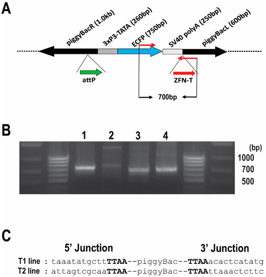
Figure 1.
Confirmation of integration into the Bradysia (Sciara) genome by piggyBac [3xP3-ECFPafm] attP-ZFN-T. (A) Schematic presentation of piggyBac [3xP3-ECFPafm] attP-ZFN-T. The left (L) and right (R) arms of the piggyBac vector are indicated, as is the selectable marker for ECFP driven by the 3XP3 promoter. The target sites for future insertion of large DNA are shown for phiC31 attP (attP, thick green arrow) and for a zinc finger nuclease (ZFN-T, thick red arrow). The PCR primer set to amplify a 700 bp fragment from this vector is indicated by thin red arrows. (B) Gel photo of genomic PCR products using internal primer pairs for the vector: (lane 1) Drosophila genomic DNA from larvae transformed with the same vector (positive control); (lane 2) genomic DNA from non-transformed wild type Bradysia (Sciara) larvae (negative control); genomic DNA from Bradysia (Sciara) transformant line T1 (lane 3) and T2 (lane 4). The 700 bp PCR product is present only in the Drosophila positive control (lane 1) and in the two Bradysia (Sciara) transformant lines T1 and T2 (lanes 3 and 4, respectively). (C) Sequence of the junction between the transformation vector and genomic DNA recovered from Bradysia (Sciara) genomic DNA from transformant lines T1 and T2 after using inverse PCR. The TTAA target site (in bold font) flanking the piggyBac vector is present in both Bradysia (Sciara) transformant lines (T1 and T2).
Out of the 230 successful G0 crosses for Bradysia (Sciara) transformants, we recovered two independent clones (T1 line and T2 line), which had strong ECFP expression in neural tissue, as expected from the 3XP3 promoter. Strong expression of ECFP was observed in the brain of the first instar through the fourth instar Bradysia (Sciara) larvae (Figure 2A).
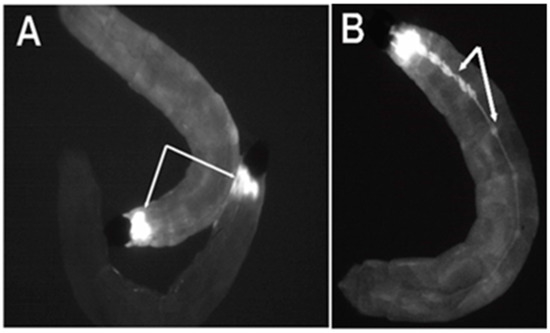
Figure 2.
Expression of the ECFP fluorescent marker in flies transformed with pBac [3XP3-ECFPafm] attP-ZFN-T. Panels (A,B) are figures of third instar Bradysia (Sciara) larvae from transformant lines T1 and T2. (A) Strong ECFP expression is observed in the larval brain (dorsal view). (B) Strong expression of ECFP is observed in the central ganglion along the length of the larval body (ventral view) in addition to signal in the brain (not visualized here since that is on the dorsal side).
From the late second instar through the fourth instar, strong expression became visible in the central ganglia on the ventral side of the larvae (Figure 2B). No non-neural ECFP signal was observed in the Bradysia (Sciara) larval transformants, indicating the specificity of the 3XP3 promoter. The ECFP signal was not detectable in adult Bradysia (Sciara) flies, probably because the black pigmentation of the adult fly body masks the ECFP signal.
To confirm the integration of the pBac [3XP3-ECFPafm] attP-ZFN-T vector into the Bradysia (Sciara) genome, we carried out a genomic PCR using a primer set to amplify a 700 bp internal sequence of the vector (Figure 1A). The same 700 bp fragment amplified from control Drosophila transformants was recovered from the Bradysia (Sciara) transformant lines T1 and T2 but not observed in non-transformed wild type Bradysia (Sciara) (Figure 1B). The flanking genomic sequences around the integrated vector were recovered by an inverse PCR from Bradysia (Sciara) transformant lines T1 and T2 for DNA sequencing. As is typical for piggyBac transformation, a duplicated TTAA sequence adjacent to the piggyBac inverted repeats was found in both the T1 and T2 transformant lines (Figure 1C).
3.3. Genomic Integration of pBac [3XP3-TagYFP, hr5-ie1-BlasR, su(Hw)BS] attP-ZFN-T
Although we have successfully recovered transformants using pBac [3XP3-EGFP afm] attP-ZFN-T, we proceeded to add some modifications to this original vector to make it more suitable for our experiments. Specifically, we (i) replaced ECFP with TagYFP, (ii) added blasticidin resistance as an additional marker for screening and (iii) added the insulator element su(Hw)BS to combat position effects. The construction of this plasmid is described in Materials and Methods and summarized in Supplementary Figure S3.
Wild type GFP has two disadvantages for its use as a transformation marker: it is relatively insoluble and its excitation by UV light can be injurious to the transformed organisms [37]. For these reasons, the more soluble blue-light-shifted mutant GFP variants, such as enhanced CFP (ECFP), have been used [24,37]. Nonetheless, we observed that strong expression of ECFP can have negative effects on the viability of the Bradysia (Sciara) transformants. Therefore, we replaced ECFP with TagYFP (Evrogen, Moscow, Russia) which is more soluble, very stable and not toxic to cells. We kept the 3XP3 promoter to drive the expression of the TagYFP gene.
Though feasible, screening using just the fluorescent marker alone was extremely time-consuming and labor-intensive. To overcome this problem, we have used antibiotic resistance as a second marker. We compared the lethality curves for neomycin (G418) to blasticidin and found that a 10× greater dose was needed for the former than the latter. Therefore, it was less costly to use blasticidin, which is routinely used for the selection of cultured cells. Moreover, the blasticidin coding sequence is only 399 bp, which is less than half the size of G418, thus allowing a smaller length of vector. We introduced into the piggyBac plasmid the blasticidin resistance gene driven by the hr5-ie1 promoter and have developed the screening protocol using antibiotic selection (see Section 2.1.3). After a titration experiment (Supplementary Figure S4), we chose a dose of 20 ug/mL blasticidin in the 2.2% (w/v) agar as optimal since this is the minimal concentration to kill all non-transformed larvae by the fourth larval instar. Antibiotic resistance made it possible for us to screen a larger number of G1 progeny routinely than would be feasible with just a fluorescence marker.
We wanted to avoid position effects that not only dampen gene expression but also can interfere with the cis-regulatory elements that drive DNA amplification [41,42]. Insulator elements are DNA sequences first identified in transcription experiments [43]. Insulators placed between enhancers and promoters can block their interaction; insulators placed adjacent to a transgene can protect the transgenes from chromosomal position effects on transcription. The suppressor of the Hairy wing protein binding site [su(Hw)BS] from the gypsy transposon is a strong transcriptional insulator [44,45,46,47]. The su(Hw)BS insulator also protects chorion gene locus constructs from position effects on DNA amplification in Drosophila, thereby allowing detailed studies on cis-regulatory elements and sequence elements important for DNA amplification [48,49,50].
To insulate the piggyBac transgene, we introduced su(Hw)BS insulators flanking the two selection markers, as shown in Figure 3A. As also noted by others [51], we found that placement of su(Hw)BS in tandem as an inverted repeat (using the FseI or AscI cloning sites) without any spacer between two copies of su(Hw)BS resulted in instability in the construct in E. coli during the cloning process. The su(Hw)BS inverted repeats seem to be a hot spot of transposition of the bacterial transposon Tn1000 (our unpublished data); this can be mitigated in part by using E.coli lacking an F’ factor. We overcame the instability problem by first putting the markers in the construct and then sequentially adding su(Hw)BS to flank the markers.
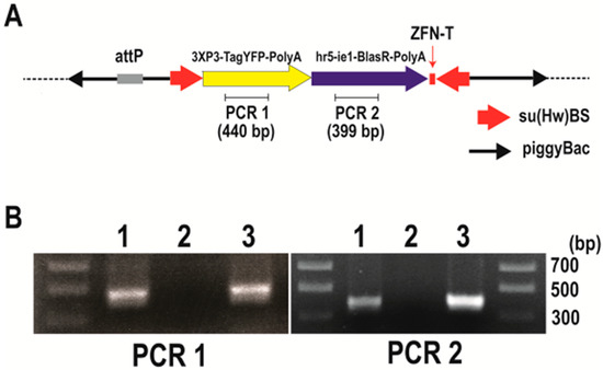
Figure 3.
Confirmation of integration into the Bradysia (Sciara) genome by pBac [3XP3-TagYFP, hr5-ie1-BlasR, su(Hw)BS] attP-ZFN-T. (A) Schematic depiction of pBac [3XP3-TagYFP, hr5-ie1-BlasR, su(Hw)BS] attP-ZFN-T. (B) Genomic PCR to amplify the TagYFP coding sequence (PCR1) and blasticidin resistance gene coding sequence (PCR2). The gel lanes contain genomic DNA from Drosophila transformants carrying pBac [3XP3-TagYFP, hr5-ie1-BlasR, su(Hw)BS] attP-ZFN-T (lane 1), genomic DNA from uninjected Bradysia (Sciara) larva as a negative control (lane 2) and genomic DNA from Bradysia (Sciara) transformant line 1 (lane 3).
We co-injected the Bradysia (Sciara) embryos with the same piggyBac transposase plasmid and the transactivator plasmid as used above but with the new plasmid construct pBac [3XP3-TagYFP, hr5-ie1-BlasR, su(Hw)BS] attP-ZFN-T (Figure 3A). Moreover, as a control, the same mixture of plasmids was injected into Drosophila and the transformants were screened by the TagYFP marker. Among all eight independent Drosophila transformant lines that we recovered, TagYFP was expressed strongly in the central nervous system, eye promordium, midgut and anal pad of third instar larvae (Figure 4, animal #1), and there was no obvious variation in the expression level of TagYFP among the lines. In contrast, variation in the gut and anal pad TagYFP intensity was observed between the various Drosophila lines that had been transformed with the unbuffered plasmid pBac [3XP3-TagYFP, hr5-ie1-BlasR] attP-ZFN-T that lacks su(Hw)BS (Figure 4, animals #2 and #3). This suggested that the su(Hw)BS insulator was working in the eight transformant lines where it was employed. Moreover, these Drosophila transformants with the su(Hw)BS insulator were also resistant to the blasticidin antibiotic used as the second selectable marker. Wild type uninjected Drosophila larvae do not survive in the presence of 10 μg/mL of blasticidin mixed into the food, but all the Drosophila homozygous transformant lines survived in the presence of 80 μg/mL of blasticidin. Therefore, su(Hw)BS efficiently insulated against the position effect for hr5-ie1-driven blasticidin selection marker gene expression as well as the expression of 3XP3-TATA-TagYFP-PolyA in Drosophila.
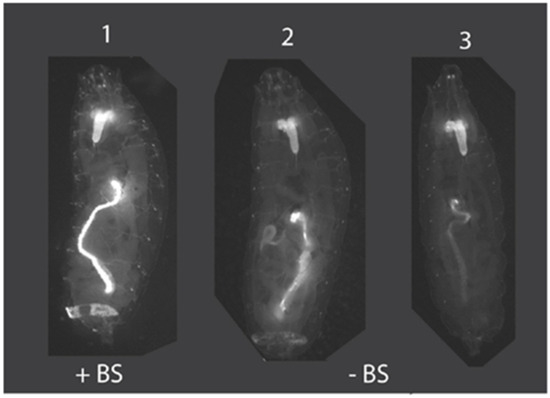
Figure 4.
Insulation of position effects by su(Hw)BS. Animal #1 is an example of Drosophila transformed with pBac [3XP3-TagYFP, hr5-ie1-BlasR, su(Hw)BS] attP-ZFN-T, where the su(Hw)BS insulator (“+BS”) prevents position effects and TagYFP is strongly expressed in the central nervous system, eye primordium, midgut and anal pad of third instar larvae. This strong expression pattern with no variability was observed for all eight Drosophila transformant lines. In contrast, animals #2 and 3 are Drosophila transformed with the unbuffered construct pBac [3XP3-TagYFP, hr5-ie1-BlasR] attP-ZFN-T that lacks the su(Hw)BS insulator (“−BS”) and that shows TagYFP expression in the brain in all third instar larval transformants but variable expression of TagYFP in the gut and anal pad, with expression levels differing between different transformant lines due to position effects.
After the injection of the Bradysia (Sciara) embryos with pBac [3XP3-TagYFP, hr5-ie1-BlasR, su(Hw)BS] attP-ZFN-T, we recovered three independent blasticidin resistance lines of Bradysia (Sciara) transformants (lines 1 to 3). To confirm the integration of the plasmid into the Bradysia (Sciara) genome, a genomic PCR was carried out using primer sets to amplify 440 bp of the TagYFP coding sequence (PCR1) and primer sets to amplify 399 bp of the blasticidin resistance gene (PCR2) (Figure 3A). The PCR products from the genomic DNA of Bradysia (Sciara) transformant line 1 are shown in Figure 3B. Both the PCR1 and PCR2 products are present in the genomic DNA from the Drosophila transformants and Bradysia (Sciara) line 1 transformants but not in the genomic DNA of the negative control of uninjected Bradysia (Sciara) (Figure 3B). Exactly the same results were obtained from Bradysia (Sciara) transformant line 2 and line 3. The entire TagYFP marker gene was amplified by the PCR and the complete TagYFP sequence was confirmed by DNA sequencing to be present in the genomic DNA of all three Bradysia (Sciara) transformant lines. However, TagYFP expression was not observed in the transformed Bradysia (Sciara), although it was visible after the transformation of Drosophila with the same construct.
4. Discussion
We present here our systematic development of a method for the transformation of the lower dipteran fly Bradysia (Sciara) coprophila. The set of experiments can be used as a paradigm for the development of transformation for other emerging model systems where no prior transformation methodology has existed. To begin, it was necessary to understand the biology of the organism, such as identification of the posterior end of the embryo for injection, a means to recover synchronous embryos, an understanding of when blastoderm cellularization occurs and sensitivities to toxic conditions during handling. For example, Kwik-Cast is a very-low-toxicity silicone elastomer developed for neuroscience applications (World Precision Instruments). The Kwik-Cast adhesive appeared to have low toxicity and enough oxygen permeability to be used for Bradysia (Sciara) injection, and, so far, this is the only adhesive in our hands that provided a good survival rate. It could be very beneficial for other experimental organisms as well that are sensitive to environmental factors or chemicals.
We used piggyBac as the vector since it can integrate into the genomes of a wide range of insects [17] and organisms [23]. In Drosophila, the piggyBac vector often integrates into the non-coding region of a gene without causing any apparent phenotype associated with it [19]. For future flexibility, we added the target binding site for a zinc finger nuclease (ZFN-T) [26] and also the phiC31 attP site [27], either of which could be used for site-specific integration of large DNA into the ectopic piggyBac locus once transformants are established.
It was also necessary to develop a method for the selection of transformants. This can be challenging, especially as no prior phenotypic mutants are available for use as selectable markers. As one selection marker, we used fluorescence of the nervous system; initially, we employed ECFP and later switched to the less toxic TagYFP. In both cases, they were driven by the highly conserved constitutive promoter containing three eyeless binding sites (3XP3) [38,39] activated by the phylogenetically conserved transcription factor Pax-6. Although this worked, it was not feasible for scale-up for high throughput. Therefore, we also employed resistance to the antibiotic blasticidin for use in an initial screen, with subsequent confirmation by the fluorescent marker. As the promoter to drive the blasticidin resistance gene, we chose the Autographa carifornica nuclear polyhedrosis baculovirus immediate early gene (ie1) promoter flanked by the hr5 enhancer since this drives 500–1000× stronger expression than other promoters [33]. To minimize position effects that could reduce the expression of the selectable markers, su(Hw)BS can be used as an insulator [44,45,46,47]. However, since su(Hw)BS increases the size of the vector and, therefore, could reduce the integration efficiency, its use is optional. Alternatively, transformants without su(Hw)BS that express the selectable markers would be indicative of insertion into the genome at sites not impacted by position effects.
Supplementary Materials
The following supporting information can be downloaded at: https://www.mdpi.com/2073-4425/13/7/1108/s1, Supplementary Figure S1: Visualization of the anterior end of Bradysia (Sciara) embryos; Supplementary Figure S2: Schematic drawing of alignment of Bradysia (Sciara) embryos; Supplementary Figure S3: Construction of pBac [3XP3-TagYFP, su(Hw)BS] attP, ZFN-T; Supplementary Figure S4: Lethality curves for neomycin and blasticidin.
Author Contributions
Y.Y. designed and executed the experiments, analyzed the results, prepared the figures and contributed to writing this paper. S.A.G. oversaw the project and wrote the paper. All authors have read and agreed to the published version of the manuscript.
Funding
We are grateful for research funding to S.A.G. by NIH GM121455 currently and previously from NIH GM35929.
Institutional Review Board Statement
Not applicable.
Informed Consent Statement
Not applicable.
Data Availability Statement
The data are presented in this paper.
Acknowledgments
We thank Craig J. Coates for the starting materials of the piggyBac vector, piggyBac transposase plasmid and IE1 transactivator plasmid (pIE1/153), Alfred M. Handler for piggyBac [3xP3-ECFPafm] and John Tower for the pYES vector. We also thank Fyodor D. Urnov for kindly supplying the ZFN clones for ZFN-T. Heidi Smith and Jacob Bliss are thanked for help with Bradysia (Sciara) maintenance, and Heidi Smith also for her great help in screening the transformants.
Conflicts of Interest
The authors declare no conflict of interest. The funders had no role in the design of the study; in the collection, analyses, or interpretation of data; in the writing of the manuscript or in the decision to publish the results.
References
- Pederson, T. An Olympian protozoan. Nucleus 2010, 1, 2–3. [Google Scholar] [CrossRef]
- Sullivan, W. The institute for the study of non-model organisms and other fantasies. Mol. Biol. Cell. 2015, 26, 387–389. [Google Scholar] [CrossRef] [PubMed]
- Jinek, M.; Chylinski, K.; Fonfara, I.; Hauer, M.; Doudna, J.; Charpentier, E. A programmable dual-RNA-guided DNA endonuclease in adaptive bacterial immunity. Science 2012, 337, 816–821. [Google Scholar] [CrossRef] [PubMed]
- Mali, P.; Esvelt, K.; Church, G. Cas9 as a versatile tool for engineering biology. Nat. Methods 2013, 10, 957–963. [Google Scholar] [CrossRef] [PubMed]
- Esvelt, K.; Smidler, A.; Catteruccia, F.; Church, G. Concerning RNA-guided gene drives for the alteration of wild populations. eLife 2014, 17, e03401. [Google Scholar] [CrossRef] [PubMed]
- Meigen, J.W. Versuch einer neuen Gattungs Einheilung der europäischen zweiflügligen insekten. Mag. Insecktenk. 1803, 2, 259–281. [Google Scholar]
- Steffan, W.A. A generic revision of the family Sciaridae (Diptera) of America north of Mexico. Univ. Calif. Publ. Entomol. 1966, 44, 1–77. [Google Scholar]
- Gerbi, S.A. Unusual chromosome movements in Sciarid flies. In Results and Problems in Cell Differentiation; Hennig, W., Ed.; Germ Line–Soma Differentiation; Springer: Berlin/Heidelberg, Germany, 1986; Volume 13, pp. 71–104. [Google Scholar]
- Gerbi, S.A.; Urnov, F.D. Differential DNA replication in insects. In DNA Replication in Eukaryotic Cells; DePamphilis, M.L., Ed.; Cold Spring Harbor Laboratory Press: Cold Spring Harbor, NY, USA, 1996; pp. 947–969. [Google Scholar]
- Gerbi, S.A.; Strezoska, Z.; Waggener, J.M. Initiation of DNA replication in multicellular eukaryotes. J. Struct. Biol. 2002, 140, 17–30. [Google Scholar] [CrossRef]
- Gerbi, S.A. Non-random chromosome segregation and chromosome eliminations in the fly Bradysia (Sciara). Chromosome Res. 2022; in press. [Google Scholar]
- Urban, J.M.; Foulk, M.S.; Bliss, J.E.; Coleman, C.M.; Lu, N.; Mazloom, R.; Brown, S.J.; Spradling, A.C.; Gerbi, S.A. High contiguity de novo genome assembly and DNA modification analyses for the fungus fly, Sciara coprophila, using single-molecule sequencing. BMC Genom. 2021, 22, 643. [Google Scholar] [CrossRef]
- Rubin, G.M.; Spradling, A.C. Genetic transformation of Drosophila with transposable element vectors. Science 1982, 218, 348–353. [Google Scholar] [CrossRef] [PubMed]
- Handler, A.M.; Gomez, S.P.; O’Brochta, D.A. A functional analysis of the P-element gene-transfer vector in insects. Arch. Insect Biochem. Physiol. 1993, 22, 373–384. [Google Scholar] [CrossRef] [PubMed]
- O’Brochta, D.A.; Atkinson, P.W. Transposable elements and gene transformation in non-drosophilid insects. Insect Biochem. Mol. Biol. 1996, 26, 739–753. [Google Scholar] [CrossRef]
- Handler, A.M.; McCombs, S.D.; Fraser, M.J.; Saul, S.H. The lepidopteran transposon vector, piggyBac, mediates germ-line transformation in the Mediterranean fruit fly. Proc. Natl. Acad. Sci. USA 1998, 95, 7520–7525. [Google Scholar] [CrossRef]
- Handler, A.M. Use of the piggyBac transposon for germ-line transformation of insects. Insect Biochem. Mol. Biol. 2002, 32, 1211–1220. [Google Scholar] [CrossRef]
- Sarkar, A.; Sim, C.; Hong, Y.S.; Hogan, J.R.; Fraser, M.J.; Robertson, H.M.; Collins, F.H. Molecular evolutionary analysis of the widespread piggyBac transposon family and related “domesticated” sequences. Mol. Genet. Genom. 2003, 270, 173–180. [Google Scholar] [CrossRef]
- Bellen, H.J.; Levis, R.W.; He, Y.; Carlson, J.W.; Evans-Holm, M.; Bae, E.; Kim, J.; Metaxakis, A.; Savakis, C.; Schulze, K.L.; et al. The Drosophila gene disruption project: Progress using transposons with distinctive site specificities. Genetics 2011, 188, 731–743. [Google Scholar] [CrossRef]
- Cary, L.C.; Goebel, M.; Corsaro, B.G.; Wang, H.G.; Rosen, E.; Fraser, M.J. Transposon mutagenesis of baculoviruses: Analysis of Trichoplusia ni transposon IFP2 insertions within the FP-locus of nuclear polyhedrosis viruses. Virology 1989, 172, 156–169. [Google Scholar] [CrossRef]
- Fraser, M.J.; Ciszczon, T.; Elick, T.; Bauser, C. Precise excision of TTAA-specific lepidopteran transposons piggyBac (IFP2) and tagalong (TFP3) from the baculovirus genome in cell lines from two species of Lepidoptera. Insect Mol. Biol. 1996, 5, 141–151. [Google Scholar] [CrossRef]
- Fraser, M.J.; Cary, L.; Boonvisudhi, K.; Wang, H.G. Assay for movement of lepidopteran transposon IFP2 in insect cells using a baculovirus genome as a target DNA. Virology 1995, 211, 397–407. [Google Scholar] [CrossRef]
- Lobo, N.F.; Fraser, T.S.; Adams, J.A.; Fraser, M.J., Jr. Interplasmid transposition demonstrates piggyBac mobility in vertebrate species. Genetica 2006, 128, 347–357. [Google Scholar] [CrossRef] [PubMed]
- Horn, C.; Wimmer, E.A. A versatile vector set for animal transgenesis. Dev. Genes Evol. 2000, 210, 630–637. [Google Scholar] [CrossRef] [PubMed]
- Horn, C.; Jaunich, B.; Wimmer, E.A. Highly sensitive, fluorescent transformation marker for Drosophila transgenesis. Dev. Genes Evol. 2000, 210, 623–629. [Google Scholar] [CrossRef] [PubMed]
- Urnov, F.D.; Miller, J.C.; Lee, Y.L.; Beausejour, C.M.; Rock, J.M.; Augustus, S.; Jamieson, A.C.; Porteus, M.H.; Gregory, P.D.; Holmes, M.C. Highly efficient endogenous human gene correction using designed zinc-finger nucleases. Nature 2005, 435, 646–651. [Google Scholar] [CrossRef]
- Groth, A.C.; Fish, M.; Nusse, R.; Calos, M.P. Construction of transgenic Drosophila by using the site-specific integrase from phage phiC31. Genetics 2004, 166, 1775–1782. [Google Scholar]
- Yamamoto, Y.; Gustafson, E.A.; Foulk, M.S.; Smith, H.S.; Gerbi, S.A. Anatomy and evolution of a DNA replication origin. Chromosoma 2021, 130, 199–214. [Google Scholar] [CrossRef]
- Du Bois, A.M. Elimination of chromosomes during cleavage in the eggs of Sciara (Diptera). Proc. Natl. Acad. Sci. USA 1932, 18, 352–356. [Google Scholar] [CrossRef]
- Du Bois, A.M. A contribution to the embryology of Sciara (Diptera). J. Morphol. 1932, 54, 161–191. [Google Scholar] [CrossRef]
- Du Bois, A.M. Chromosome behavior during cleavage in the eggs of Sciara coprophila (Diptera) in the relation to the problem of sex determination. Zellforschung 1933, 19, 595–614. [Google Scholar] [CrossRef]
- de Saint Phalle, B.; Sullivan, W. Incomplete sister chromatid separation is the mechanism of programmed chromosome elimination during early Sciara coprophila embryogenesis. Development 1996, 122, 3775–3784. [Google Scholar] [CrossRef]
- Mohammed, A.; Coates, C.J. Promoter and piggyBac activities within embryos of the potato tuber moth, Phthorimaea operculella, Zeller (Lepidoptera: Gelechiidae). Gene 2004, 342, 293–301. [Google Scholar] [CrossRef] [PubMed]
- Patton, J.S.; Gomes, X.V.; Geyer, P.K. Position-independent germline transformation in Drosophila using a cuticle pigmentation gene as a selectable marker. Nucleic Acids Res. 1992, 20, 5859–5860. [Google Scholar] [CrossRef] [PubMed]
- Handler, A.M.; Harrell, R.A., 2nd. Germline transformation of Drosophila melanogaster with the piggyBac transposon vector. Insect Mol. Biol. 1999, 8, 449–457. [Google Scholar] [CrossRef]
- Yusa, K.; Zhou, L.; Li, M.A.; Bradley, A.; Craig, N.L. A hyperactive piggyBac transposase for mammalian applications. Proc. Natl. Acad. Sci. USA 2011, 108, 1531–1536. [Google Scholar] [CrossRef]
- Horn, C.; Schmid, B.; Pogoda, F.S.; Wimmer, E.A. Fluorescent transformation markers for insect transgenesis. Insect Biochem. Mol. Biol. 2002, 32, 1221–1236. [Google Scholar] [CrossRef]
- Noll, M. Evolution and role of Pax genes. Curr. Opin. Genet. Dev. 1993, 3, 595–605. [Google Scholar] [CrossRef]
- Berghammer, A.J.; Klingler, M.; Wimmer, E.A. A universal marker for transgenic insects. Nature 1999, 402, 370–371. [Google Scholar] [CrossRef]
- Yamamoto, Y.; Bliss, J.; Gerbi, S.A. Whole organism genome editing: Targeted large DNA insertion via ObLiGaRe nonhomologous end-joining in vivo capture. G3 Genes Genomes Genet. 2015, 5, 1843–1847. [Google Scholar] [CrossRef]
- Delidakis, C.; Kafatos, F.C. Amplification of a chorion gene cluster in Drosophila is subject to multiple cis-regulatory elements and to long-range position effects. J. Mol. Biol. 1987, 197, 11–26. [Google Scholar] [CrossRef]
- Delidakis, C.; Kafatos, F.C. Amplification enhancers and replication origins in the autosomal chorion gene cluster of Drosophila. EMBO J. 1989, 8, 891–901. [Google Scholar] [CrossRef]
- Gerasimova, T.I.; Corces, V.G. Chromatin insulators and boundaries: Effects on transcription and nuclear organization. Annu. Rev. Genet. 2001, 35, 193–208. [Google Scholar] [CrossRef] [PubMed]
- Geyer, P.K.; Corces, V.G. DNA position-specific repression of transcription by a Drosophila zinc finger protein. Genes Dev. 1992, 6, 1865–1873. [Google Scholar] [CrossRef] [PubMed]
- Corces, V.G. Keeping enhancers under control. Nature 1995, 376, 462–463. [Google Scholar] [CrossRef] [PubMed]
- Mallin, D.R.; Myung, J.S.; Patton, J.S.; Geyer, P.K. Polycomb group repression is blocked by the suppressor of Hairy-wing [su(Hw)] insulator. Genetics 1998, 148, 331–339. [Google Scholar] [CrossRef] [PubMed]
- Zorin, I.D.; Gerasimova, T.I.; Corces, V.G. The lawc gene is a new member of the trithorax-group that affects the function of the gypsy insulator of Drosophila. Genetics 1999, 152, 1045–1055. [Google Scholar] [CrossRef]
- Lu, L.; Tower, J. A transcriptional insulator element, the su(Hw) binding site, protects a chromosomal DNA replication origin from position effects. Mol. Cell. Biol. 1997, 17, 2202–2206. [Google Scholar] [CrossRef]
- Lu, L.; Zhang, H.; Tower, J. Functionally distinct, sequence-specific replicator and origin elements are required for Drosophila chorion gene amplification. Genes Dev. 2001, 15, 134–146. [Google Scholar] [CrossRef]
- Zhang, H.; Tower, J. Sequence requirements for function of the Drosophila chorion gene locus ACE3 replicator and ori-β origin elements. Development 2004, 131, 2089–2099. [Google Scholar] [CrossRef]
- Sarkar, A.; Atapattu, A.; Belikoff, E.J.; Heinrich, J.C.; Li, X.; Horn, C.; Wimmer, E.A.; Scott, M.J. Insulated piggyBac vectors for insect transgenesis. BMC Biotechnol. 2006, 6, 27. [Google Scholar] [CrossRef]


Publisher’s Note: MDPI stays neutral with regard to jurisdictional claims in published maps and institutional affiliations. |
© 2022 by the authors. Licensee MDPI, Basel, Switzerland. This article is an open access article distributed under the terms and conditions of the Creative Commons Attribution (CC BY) license (https://creativecommons.org/licenses/by/4.0/).