Article

Screening of Differentially Expressed Genes and miRNAs in Hypothalamus and Pituitary Gland of Sheep under Different Photoperiods

1
College of Animal Science and Technology, Anhui Agricultural University, Hefei 230036, China
2
Key Laboratory of Animal Genetics, Breeding and Reproduction of Ministry of Agriculture and Rural Affairs, Institute of Animal Science, Chinese Academy of Agricultural Sciences, Beijing 100193, China
*
Authors to whom correspondence should be addressed.
Genes 2022, 13(6), 1091; https://doi.org/10.3390/genes13061091
Submission received: 23 May 2022 / Revised: 15 June 2022 / Accepted: 16 June 2022 / Published: 19 June 2022
(This article belongs to the Special Issue Gene Regulation of Development and Reproduction in Mammals)

Abstract

:
The reproduction of sheep is affected by many factors such as light, nutrition and genetics. The Hypothalamic-pituitary-gonadal (HPG) axis is an important pathway for sheep reproduction, and changes in HPG axis-related gene expression can affect sheep reproduction. In this study, a model of bilateral ovarian removal and estrogen supplementation (OVX + E2) was applied to screen differentially expressed genes and miRNAs under different photoperiods using whole transcriptome sequencing and reveal the regulatory effects of the photoperiod on the upstream tissues of the HPG axis in sheep. Whole transcriptome sequencing was performed in ewe hypothalamus (HYP) and distal pituitary (PD) tissues under short photoperiod 21st day (SP21) and long photoperiod 21st day (LP21). Compared to the short photoperiod, a total of 1813 differential genes (up-regulation 966 and down-regulation 847) and 145 differential miRNAs (up-regulation 73 and down-regulation 72) were identified in the hypothalamus of long photoperiod group. Similarly, 2492 differential genes (up-regulation 1829 and down-regulation 663) and 59 differential miRNAs (up-regulation 49 and down-regulation 10) were identified in the pituitary of long photoperiod group. Subsequently, GO and KEGG enrichment analysis revealed that the differential genes and target genes of differential miRNA were enriched in GnRH, Wnt, ErbB and circadian rhythm pathways associated with reproduction. Combined with sequence complementation and gene expression correlation analysis, several miRNA-mRNA target combinations (e.g., LHB regulated by novel-414) were obtained. Taken together, these results will help to understand the regulatory effect of the photoperiod on the upstream tissues of HPG in sheep.

1. Introduction

Animals can adapt themselves to their survival environment by regulating themselves, for example, females will choose to breed in specific seasons to ensure that they give birth in the most appropriate season, which will facilitate lactation and early pup development, and a stable physiological mechanism has evolved over the years [1]. Light perception is one of the important mechanisms that trigger the response of organisms and many physiological processes in mammals are influenced by the photoperiod [2]; a long photoperiod induces circadian transcription factor BMAL2 in pars tuberalis (PT) and triggers summer biological activity through the EYA3/TSH pathway. In contrast, the continuous prolongation of a short photoperiod leads to melatonin secretion inducing circadian repressors, including DEC1, which inhibit BMAL2 and EYA3/TSH pathways, into winter biological activity, and circadian rhythms are usually involved in some way in photoperiod-induced physiological activity [3]. Seasonal changes in light have profound effects on the behavior and physiological functions of many animals, and changes in the light-dark cycle can also induce anxiety and depressive behavior in adult rodents [4,5]. The development of the visual system is affected by light, and mice kept in the dark show alterations in a retinal synaptic organization and visual function [6]. In short photoperiods, birds can meet increased energy demands in winter by increasing their body weight, digestible energy intake and digestive tract size [7], and photoperiods also induce migratory behavior in birds [8]. Photoperiodic changes also influence the neuroendocrine action in fish [9] and horse deer [10]. Circadian clock genes are involved in seasonal activity, and clock genes are expressed in PT and ventricular canal cells within the hypothalamus [11], for example, the ability to regulate seasonal changes in reproductive dormancy is disrupted in drosophila, which is deficient in the EYA gene [12]. Many studies on photoperiodic effects on reproduction have been carried out on quail and hamsters [13,14,15].
Reproduction in sheep is a complex biological process influenced by many factors such as light, nutrition, genetics, reproductive hormones, and reproductive techniques. The reproduction of mammals was controlled by the hypothalamic-pituitary-gonadal (HPG) axis regulation network of the reproductive axis. The supraoptic nucleus receives light signals to the pituitary gland, where light signaling factors and cell morphology in the pituitary nodules are transformed, leading to morphological changes in hypothalamic GnRH neurons and ultimately to changes in the levels of gonadotropins secreted by the distal pituitary gland, which ultimately induce estrus and estrus in sheep [16,17,18,19]. Early recognition of the role of the hypothalamus as an endocrine organ and as a site of integration of autonomic and behavioral responses dates back centuries, and although the hypothalamus represents only 2% of the total volume of the brain, it is a key regulator of pituitary function and homeostatic balance, - it therefore has many functions, including food and fluid intake, lactation, thermoregulation, circadian rhythms and sleep-wake cycles [20]. The pituitary gland is targeted and regulated by the hypothalamic fine cell neuroendocrine system through a hormonal feedback mechanism. The anterior pituitary is a key organ involved in the control of a variety of physiological functions, including growth, reproduction, and metabolic functions. To perform its functions, the pituitary receives and processes signals from the central and peripheral sources and transmits them appropriately to several key endocrine and non-endocrine organs [21]. These cells produce prolactin (PRL), growth hormone (GH), corticotropin (ACTH), gonadotropin, thyroid-stimulating hormone, and follicle-stimulating hormone (FSH)/luteinizing hormone (LH), which act on the organism. Therefore, it is important to investigate the genetic changes in the hypothalamus and pituitary gland related to estrus and circadian rhythms for reproduction in sheep.
With the development and application of whole transcriptomics sequencing technology, many miRNAs have been identified in various tissues and organs of sheep. In recent years, a large number of miRNAs are involved in reproduction in both sheep [22,23] and other species [24,25,26]. The lncRNA of different light-induced reproduction in sheep have been mined in our laboratory in the previous stage using the classical model of the OVX + E2 model [27,28]. The study used this model to explore the regulatory role of the upstream tissues of the HPG axis in sheep and to screen the hypothalamic and pituitary differential genes and miRNAs in sheep with different photoperiods, which will lay the theoretical foundation for revealing the molecular mechanism of photoperiodic regulation of estrus in sheep.

2. Materials and Methods

2.1. Animals and Sample Collection

A group of 21 Small Tail Han ewes (3 y, clinically normal, and non-pregnant) were selected from Heilongjiang Province, China. All ewes were fed ad libitum and had free access to water. After removal of the ovaries (OVX) from the experimental Small Tail Han ewes with good estrous traits as described above, estrogen silicone tubes were implanted subcutaneously, and the sheep were penned into artificially controlled light sheds after the hormone concentration was stabilized at 7.23 ± 2.50 pg/mL. OVX + E2 sheep and light control rooms were constructed as previously described [27]. After 21 days of short photoperiod (8/16 h light-dark), six were euthanized (intravenous pentobarbital 100 mg/kg). After slaughtering, hypothalamic and distal pituitary tissues were taken and were labeled as SP21−HYP and SP21−PD, respectively. Six other Small-tailed Sheep were transferred to a long photoperiod (16/8 h light-dark) sheep barn and after 21 days of rearing, tissues were slaughtered and collected and labeled LP21−HYP and LP21−PD. All collected tissue was snap-frozen at liquid nitrogen, and then brought back to the laboratory and stored in a −80 °C refrigerator for long-term storage.

2.2. RNA Extraction, Library Construction, Sequencing and Raw Data Processing

Total RNA from each sample was isolated using the TRIzol reagent (Invitrogen, Carlsbad, CA, USA). then the Nanodrop 2000 instrument was used to detect the concentration and purity of the extracted RNA, and the integrity of RNA was detected by agarose gel electrophoresis. After the samples were tested and passed, cDNA and sRNA library construction were performed according to the previous method [29]. To finally obtain valid data, we filtered the raw sequencing data to obtain clean reads. Clean reads were aligned to the sheep reference genome using bowtie (Oar_rambouillet_v1.0).

2.3. Differential Expression Analysis of mRNAs

Quantitative analysis was performed using StringTie software to obtain the number of Reads per sample compared to each transcript, which was converted to FPKM (Fragments Per Kilobase per Million bases). Quantitative statistical analysis of transcript expression was performed to filter out transcripts with significantly different expression levels in the samples. We used DESeq2 to analyze the significance of transcript expression differences. The differentially expressed transcripts were screened from two levels of fold change (FC) and corrected significance level (padj/q-value). In addition, |log2FC| > 1 and q-value < 0.05 were considered DEGs.

2.4. Differential Expression Analysis of miRNAs and Target Gene Prediction

Identification of known miRNAs was performed using bowtie software by comparing sRNAs to miRNA sequences from the specified range of miRBase (v22) database, and prediction of new miRNAs was performed using mirEvo and miRDeep software and expression information was obtained. The expression of miRNAs represented by TPM was obtained by comparing to the reads of miRNA precursor and mature body regions. After calculating the expression, differential analysis was performed using DESeq2, and the screening threshold for differential miRNAs was set to |log2FC|> 1, p value < 0.05.
The previously obtained known miRNAs and new miRNAs were analyzed for miRNA target gene prediction. The miRanda and qTar software were selected for target gene prediction, and the intersection of the two software was taken as the final result.

2.5. Functional Annotation and Enrichment Analysis of Target Genes of DE miRNAs and DE mRNAs

The DEGs and the predicted target genes of DEMs were analyzed by Gene Ontology (GO) (referred to as GO, http://www.geneontology.org/, accessed on 6 January 2022), which includes biological process, molecular function and cellular component, and Kyoto Encyclopedia of Genes and Genomes (KEGG). GO items or KEGG pathways with a hypergeometric p-value < 0.05 were those that were significantly enriched.

2.6. Construction of Integral miRNA-mRNA Interaction Networks

To accurately identify the key association with reproductive DEMs and DEGs, based on miRNA function, the mRNAs that were negatively related to miRNAs were screened out, and the miRNA-mRNA interaction networks were built by using Cytoscape (v3.8.2, http://www.cytoscape.org/, accessed on 1 April 2022).

2.7. Quantitative PCR Validation

To verify the accuracy of sequencing data, 7mRNAs including hypothalamus (DIO2, SIX1, RASD1, KCNH3) and distal pituitary (OXT, THUMPD1, LOC101114319) were randomly selected. The first primers were synthesized by Bioengineering (Shanghai) Co., Ltd. For the qPCR analysis of mRNAs, reverse transcription was performed using the PrimeScriptTM RT reagent kit (TaKaRa, Dalian, China). Furthermore, qPCR with the SYBR Green qPCR Mix (TaKaRa, Dalian, China) was conducted with a RocheLight Cycler480® Ⅱ system (Roche Applied Science, Mannheim, Germany) as follows: initial denaturation at 95 °C for 5 min, followed by 40 cycles of denaturation at 95 °C for 5 s, then annealing at 60 °C for 30 s. The sequences of qPCR primers are listed in Table 1.
Eight miRNAs were randomly selected, including hypothalamus (oar-miR-200a, novel_10, novel_101, novel_4, oar-miR-143) and distal pituitary (novel_142, oar-miR-181a, oar-miR-323b). For the qPCR analysis of miRNAs, reverse transcription was performed using miRcute Plus miRNAs First-Strand cDNA Kit (TIANGEN, Beijing, China). Then, qPCR was conducted miRcute Plus miRNA qPCR Kit (TIANGEN, Beijing, China) using a RocheLight Cycler480® II system (Roche AppliedScience, Mannheim, Germany) in the following procedure: initial denaturation at 95 °C for 15 min, followed by 40 cycles of denaturation at 94 °C for 20 s, then annealing at 60 °C for 34 s. The primers specific to miRNAs were synthesized by Beijing Tianyi Huiyuan Biotechnology Co., Ltd. (Beijing, China). The primer design information is shown in Table 2.
In addition, RPL19 (for mRNA) and U6 small nuclear RNA (for miRNA) were utilized as reference gene-miRNA to calculate the relative expression level with the method of 2−ΔΔCt.

3. Results

3.1. Summary of Sequencing Data for mRNA and miRNA

RNA-seq for mRNA generated about 673 million (hypothalamus) and 640 million (distal pituitary) clean reads after data filtering, and more than 94% of the clean reads were located in the genome. The GC content of clean reads was between 47.97 and 53.37%, and the clean reads quality scores of Q20 and Q30 were above 96.29% and 89.87%, respectively, indicating that the reliability and quality of the sequencing data were sufficient for further analysis (Supplementary Table S1). Regarding the expression level of mRNAs, details are provided in Supplementary Table S2, Figure 1A,C. In addition, the chromosomal distribution of mRNA on both tissues showed that genes were mostly present on chromosomes 1, 2 and 3, occupying 27.53% and 27.46%, respectively, (Supplementary Table S2, Figure 1B,D).
RNA-seq for miRNAs generated about 140 million (hypothalamus) and 132 million (distal pituitary) clean reads after data filtering, and in the range of 92.99–98.65% of the clean reads, which were located in the genome (Supplementary Table S1), with the length between 21–24nt (Supplementary Table S3, Figure 2B,D). The average Q20 content was 98% (hypothalamus) and 96% (distal pituitary) (Supplementary Table S1). In addition, a variety of non-coding RNAs (ncRNAs) were also identified, including transfer RNAs (tRNAs), snRNAs and miRNAs (Supplementary Table S3, Figure 2A,C). The known miRNAs accounted for only a part of all identified ncRNAs, 29.85% and 32.73%, respectively. However, most of them are newly identified miRNAs. Totally, 902 new miRNAs and 149 known miRNAs were included in the hypothalamus, 628 new miRNAs and 152 known miRNAs in the distal pituitary (Supplementary Table S3).

3.2. Identification of Differential Expressed Genes and miRNAs

A total of 1813 DEGs were identified from SP21−HYP vs. LP21−HYP comparison. Compared to the short photoperiods,966 genes were up-regulated, and 847 genes were down-regulated in the long photoperiods (Supplementary Table S4, Figure 3A). Similarly, a total of 2492 DEGs were identified from SP21−PD vs. LP21−PD comparison. Among these DEGs, 1829 genes were up-regulated, and 663 genes were down-regulated in the long photoperiods (Supplementary Table S5, Figure 3C). The 10 mRNAs with the most significant difference are shown in Table 2.
A total of 145 DEMs were detected among the 1051 identified miRNAs. Compared to the short photoperiods, 73 were up-regulated and 72 were down-regulated in the long photoperiods (Supplementary Table S4, Figure 3B). A total of 59 DEMs were detected among the 1051 identified miRNAs, of which 49 were up-regulated and 10 were down-regulated (Supplementary Table S5, Figure 3D). The 10 mRNAs with most significant difference was shown in Table 3, the 10 miRNAs with most significant difference was shown in Table 4, Sequence details of miRNAs are available at Supplementary Table S5.

3.3. Functional Enrichment Analysis of the DEGs

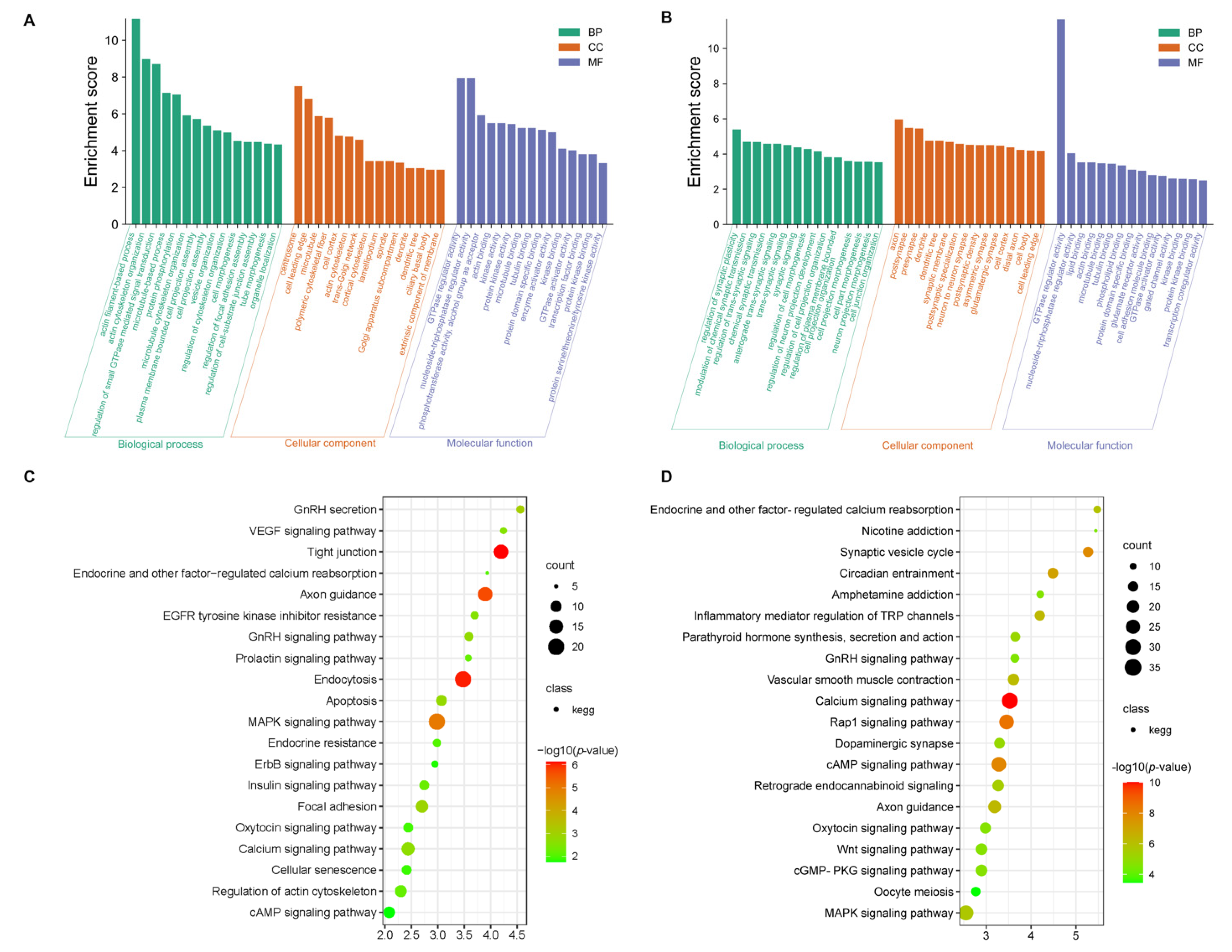
GO and KEGG analyses were conducted for the differentially expressed mRNAs. In GO terms, the most enriched terms in SP21−HYP vs. LP21−HYP are actin cytoskeleton organization and protein phosphorylation (Supplementary Table S6, Figure 4A). KEGG analysis showed that MAPK signaling pathway, GnRH signaling pathway, and insulin signaling pathway were significantly enriched (Supplementary Table S6, Figure 4C).
In GO terms, the most enriched terms in SP21−PD vs. LP21−PD are modulation of chemical synaptic transmission and regulation of trans-synaptic signaling (Supplementary Table S7, Figure 4C). All of these pathways are related to the basic activities of life, such as circadian entrainment, oxytocin signaling pathway and GnRH signaling pathway, and are associated with reproduction (Supplementary Table S7, Figure 4D).

3.4. miRNA-mRNA Interaction Network

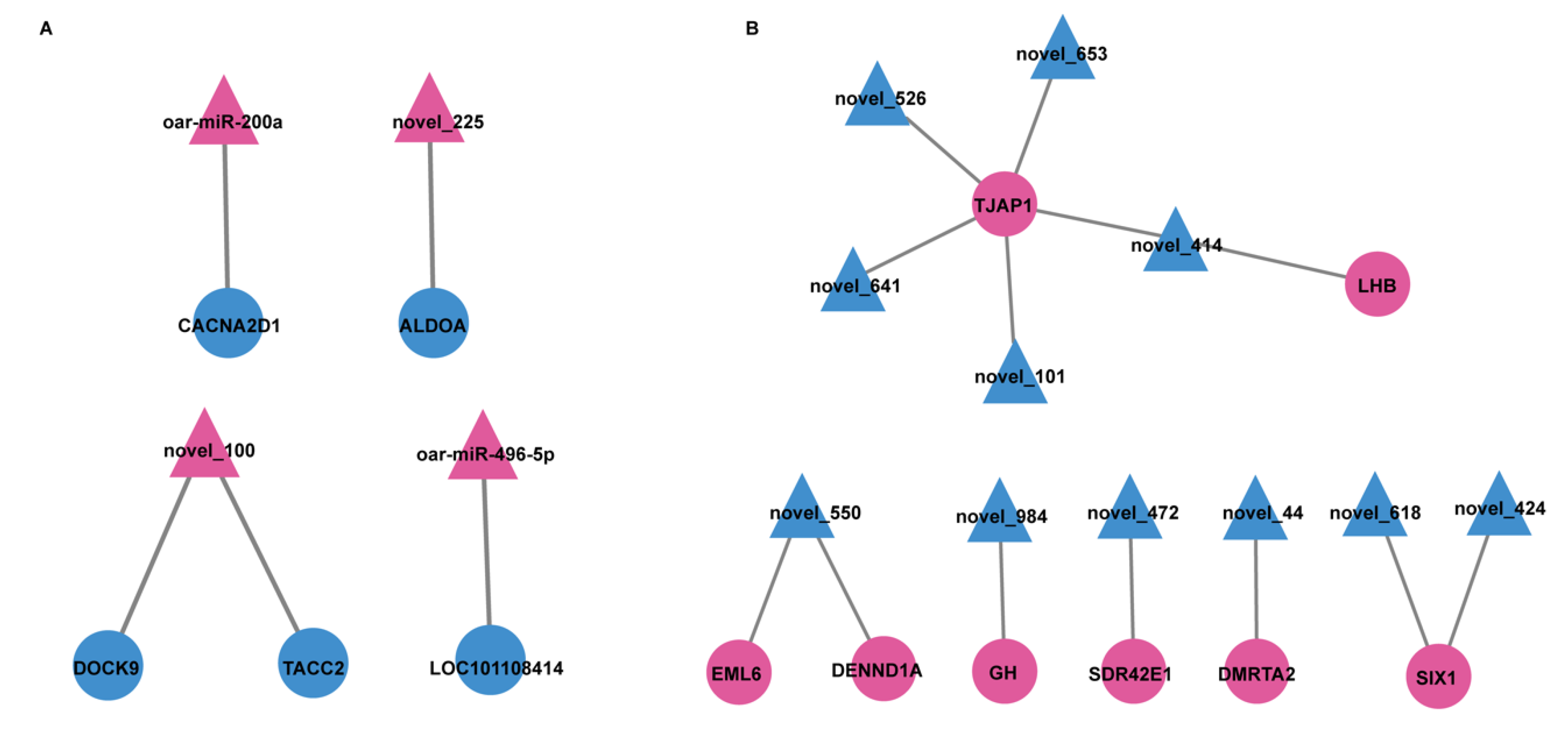
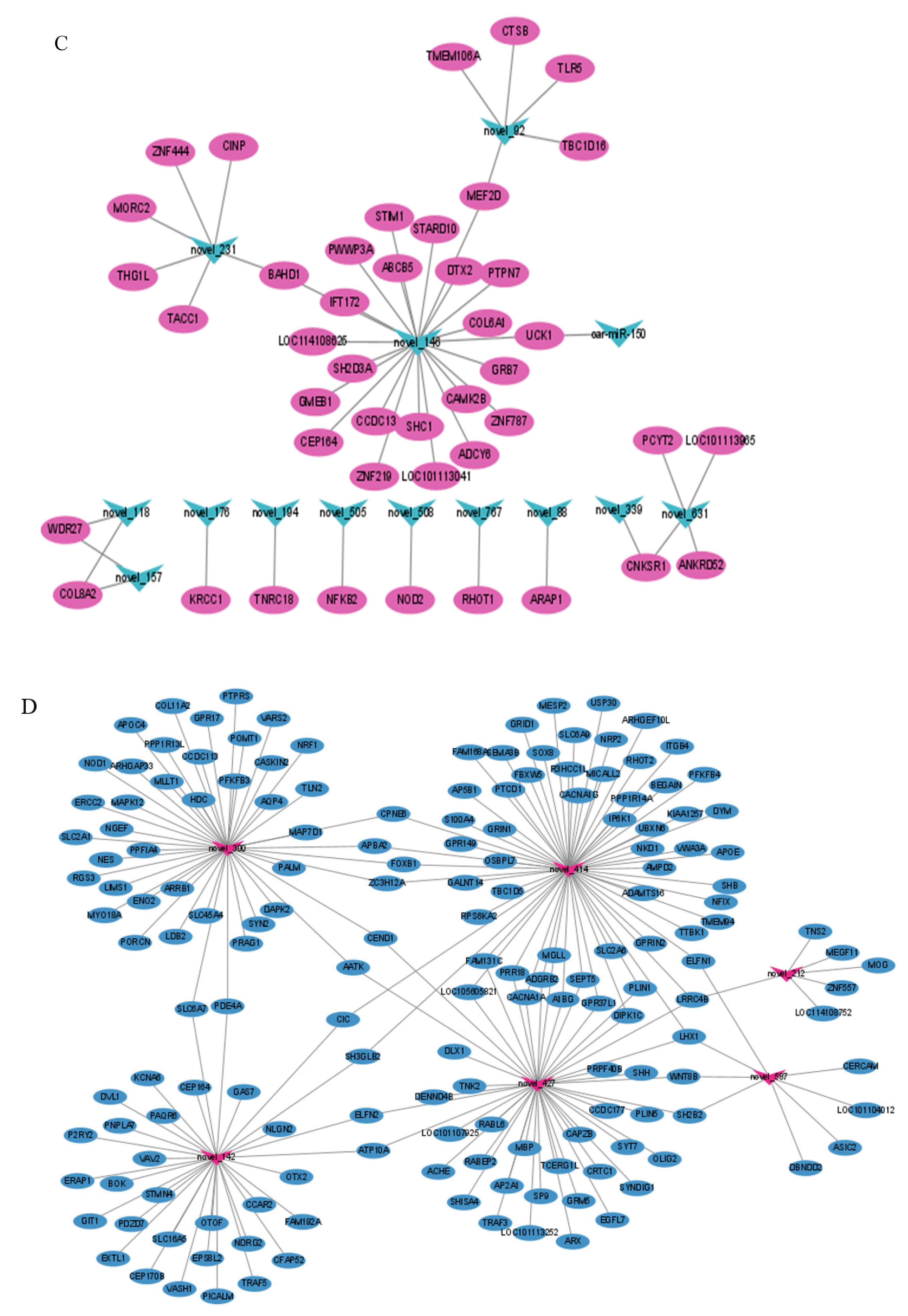
To further analyze the relationship between miRNAs and mRNAs, we constructed a genomic network of interactions using potential target genes of DEMs with DEGs obtained by RNA-Seq. Crossing the identified potential target genes of DEMs with the DEGs obtained from RNA-Seq, there were 468 hypothalamic overlapping genes and 426 pituitary overlapping genes (Figure 5). Then, Cytoscape was used to construct a co-regulatory network of DE miRNA-mRNA pairs with multiple targeting relationships (Figure 6, Supplementary Tables S10 and S11).
To further determine the role of genes and miRNAs in the regulation of the gonadal axis by light conversion. GO and KEGG analysis was undertaken for the above overlapping genes (i.e., simultaneous differential expression of miRNAs and target genes). The two most enriched entries of GO are the regulatory activity of GTPase regulator activity and nucleoside-triphosphatase regulator activity in SP21−HYP vs. LP21−HYP (Supplementary Table S8, Figure 7A). GO results found that positive regulation of glial cell production was significantly enriched in SP21−PD vs. LP21−PD (Supplementary Table S9, Figure 7B). The pathways of KEGG enrichment of target genes in DEMs and DEGs were similar in both groups (Supplementary Table S8, Figure 7C, Supplementary Table S9, Figure 7D).

3.5. Data Validation

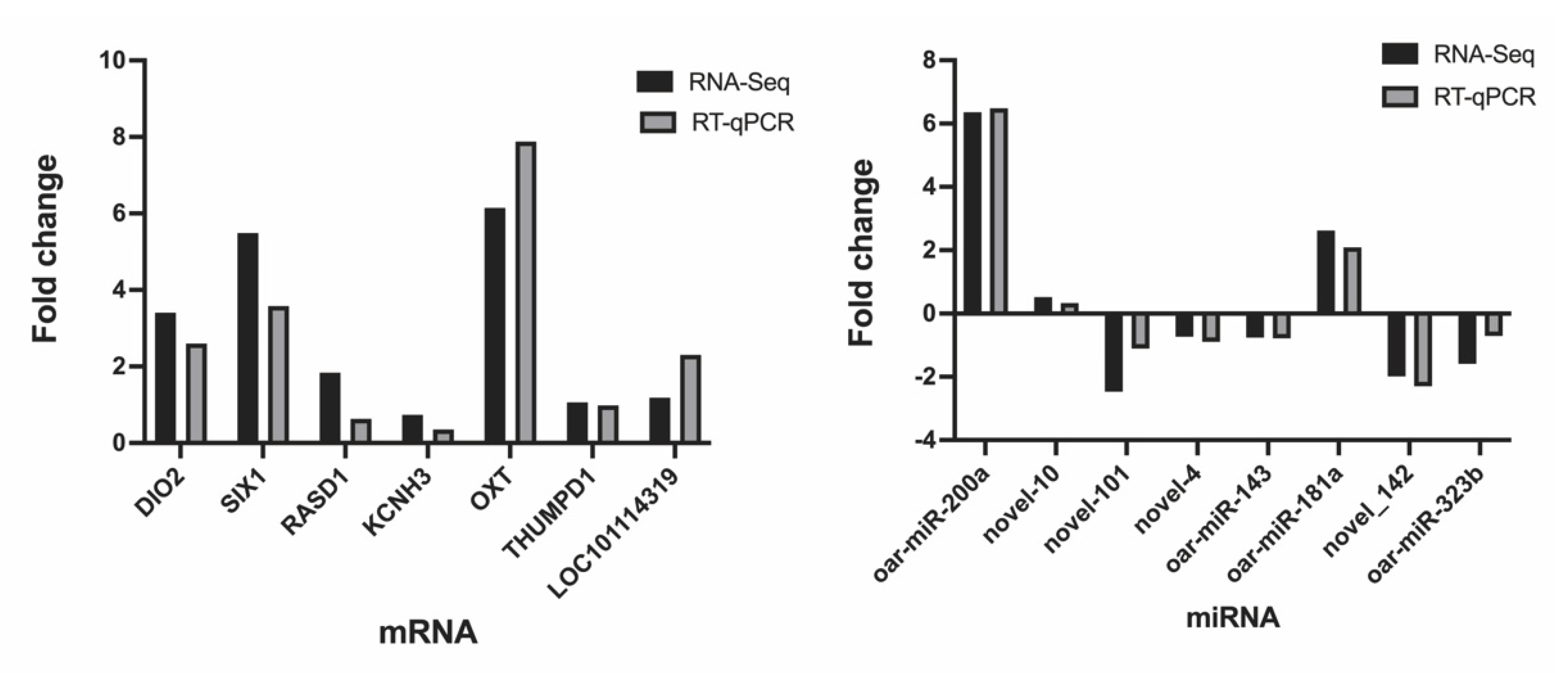
In order to assess the accuracy of sequencing, mRNAs and miRNAs were selected randomly for qPCR validation. We measured the gene expression level and compared it with the RNA-seq data. The results demonstrated that RNA-seq data and qPCR data showed similar patterns (Figure 8), which indicate the reliability of the data generated from RNA-seq.

4. Discussion

In most animals, reproduction is regulated by the hypothalamic-pituitary-gonadal axis [30]. The hypothalamus and pituitary gland are important upstream tissues that regulate HPG in sheep and control a wide range of animal behaviors [31,32]. To better probe the effects of the photoperiod on hypothalamic and pituitary function, we applied the OVX + E2 sheep model, which is often used to study the response of seasonally breeding animals to photoperiodic and reproductive endocrine changes [33,34,35]. Therefore, this study used transcriptomic approaches to mine differential genes and miRNA expression patterns associated with reproduction in sheep with different photoperiods.

4.1. Identification and Analysis of mRNAs and miRNAs Data in SP21− HYP vs. LP21− HYP

A total of 145 DEMs were identified for SP21−HYP vs. LP21−HYP, including miR-200 [36]. Family members were identified multiple times in seasonal and non-seasonal estrus ovaries of sheep. Among them, miR-200a is a member of the miR-200 family, which is associated with ovarian development in goats [37]. One study showed that miR-494-3p significantly inhibited apoptosis of endometrial epithelial cells [38], and oar-miR-485-3p can regulate the developmental quality of oocytes [39]. From the above, we can speculate that DEMs target and regulate genes related to reproductive estrus, which may be responsible for the reproductive variation in sheep due to different photoperiodic changes.
A total of 1813 DEGs were identified in SP21−HYP vs. LP21−HYP, including APAF1, CASP3 associated with apoptosis, and DIO2, LHB, and TSHB associated with estrous reproduction in sheep. Some studies have demonstrated that haplotype H2 and diploid H2/H4 of TSHB may be associated with year-round estrus [40]. GO was enriched for many terms associated with intercellular communication, and differential genes expressed may influence changes in reproductive traits in sheep by affecting the expression of intercellular communication-related receptor proteins. KEGG pathways include enriched ErbB signaling pathway; GnRH signaling pathway (PTK2B and CAM2B), gonadotropin secretion (LHB), and prolactin signaling pathway were also significantly enriched; these pathways have been studied and proven to be associated with reproductive activity [27,41].

4.2. Analysis of miRNA-mRNA Interaction Network in SP21−HPY vs. LP21−HPY

To better understand the function of miRNAs, the SP21−HPY and LP21−HPY interaction negative network was constructed. In the upregulated network, a key oar-miR-200a targeting CACNA2D1 was identified, and it has been shown that miR-200a-3p plays an important role in chicken [42] and goat [37] reproductive regulation, and some other miRNAs target genes that regulate cell proliferation apoptosis-related biological processes [43].
Among the down-regulated networks, the important DEGs and DEMs were selected to construct the reciprocal network, in which DENND1A was the target gene of novel_550, and it was shown that DENND1A gene was expressed higher in the low yield group than in the high yield group in ovarian and showed an opposite expression trend with chi-miR-324-3p. CCK-8 assay showed that chi-miR-324-3p overexpression significantly inhibited the proliferation of GCs and knockdown of chi-miR-324-3p promoted GC proliferation [44], thus affecting ovulation and reproduction in ewes, while novel_550 inhibited DENND1A gene expression in this study. We speculate that novel_550 and chi-miR-324-3p act similarly. It has also been reported that DENND1A is a susceptibility gene for polycystic ovaries, which is essential for ovarian and embryonic development [45,46,47] and can affect the reproductive process. Another target gene of novel_550, EML6 was also shown to be associated with reproduction. The production of high-quality eggs in mouse reproduction requires normal segregation during oocyte meiosis, and EML6 is highly expressed in oocytes and is responsible for the accurate segregation of homologous chromosomes [48]. GH is a target gene of novel_984; GH plays an inductive role in ovulatory reproduction [49], and is elevated during growth hormone secretion during major transitions in reproductive status such as puberty and pregnancy [50]. It is an important regulator of female reproduction, and is involved in gonadal steroidogenesis, gametogenesis and ovulation [51], and alterations to the GH axis can have a reproduction. In growth regulation, it is mainly used with growth hormone (GH) to promote cellular metabolism, promote growth and regulate reproduction [52]. Novel_414 targets LHB, luteinizing hormone (LH), also known as luteinizing hormone, which is released by gonadotropins and promotes blood flow and secretion of progesterone in the ovary, affecting ovulation [53]. SIX1 is a common target gene of novel_618 and novel_424, and long light signaling molecules are mainly mediated by EYA3 and TSHβ. EYA3 can form a complex with thyrotropic embryonic factor (TEF) and SIX1, which synergistically promotes TSHβ transcription upon binding to the TSHβ promoter D element [54]. In summary, in the down-regulated network, the expression of the above DEGs in this study was significantly higher during the shift from short to long photoperiods, and we hypothesize that DEMs such as chi-miR-324-3p, novel_550, and novel_414 lead to alterations in reproductive traits in sheep by negatively regulating and reproduction-related genes.

4.3. Identification and Analysis of mRNAs and miRNAs Data in SP21−PD vs. LP21−PD

A total of 2942 DEGs were identified for SP21−PD vs. LP21−PD, including CREB3L1 and OXT. CREB3L1 acts as a downstream effector of TSH to regulate the expression of transport proteins and increase the ability of secretory amplification [55]. Under short photoperiods, the pituitary nodules secrete the thyrotropic hormone, which stimulates T3 in the hypothalamus, which in turn affects the secretion of gonadotropin-releasing hormone and gonadotropin. Studies have shown that OXT and its receptor (OXTR) play a central role in reproduction and metabolism, especially in females, by functioning in an estrogen-dependent manner in rats [56] and are associated with human social and emotional behavior, as well as physical and mental health and disease [57,58,59]. In addition, we performed functional enrichment analysis of target genes of DEGs and DEMs in sheep, respectively. The results of KEGG pathway enrichment in both showed many common pathways in LP and SP comparison, among which the most interesting reproductive pathways included GnRH signaling pathway, Wnt signaling pathway, ErbB signaling pathway, and MAPK signaling pathway. Circadian rhythm-related pathways included circadian and TNF signaling pathways, and these results are consistent with the Kazakh sheep [60], suggesting that reproduction in Small-tailed Sheep is also influenced by the photoperiod. In KEGG analysis, enriched for pathways related to calcium regulation, light influences vitamin D synthesis and thus changes in calcium processing expression. It was shown that the rapid, light-dependent changes in melatonin levels in the pineal organ are apparently regulated by novel calcium signaling pathways [61,62].
We found that among the identified miRNAs, the most highly expressed ones, oar-miR-148a and oar-miR-379-5p, oar-miR-495-3p, oar-miR-143, oar-miR-106b and oar-miR-218a, have been shown to play important roles in regulating GnRH release [63]. In addition, a variety of miRNAs are expressed in the sheep pituitary and at different levels under different light conditions. However, some miRNAs are species-specific, which is also likely to lead to different sensitivity to photoperiods in different animals and different PD secretion of hormones leading to differences in reproductive cyclicity. A total of 6 of the 59 DEMs are known, of which oar-let-7c, oar-let-7a, and oar-let-7b are of the same miRNA family, in animal genomes. The high conservation of let-7 in different animal species suggests that they may play important (and possibly similar) roles in biological processes in various organisms [64,65]. The effect of miR-150 on GC cell apoptosis by targeting the STAR, thereby affecting ovarian function and subsequently hormone secretion related to estrus [66].

4.4. Analysis of miRNA-mRNA Interaction Network in SP−PD vs. LP−PD

In the up-regulated network, containing 45 mRNAs and 14 miRNAs negative interactors, of which ZNF787 and CAMK2 are both target genes of novel_146, CAMK2b is one of the most prominent isoforms of CAMK2 [67], and calcium/calmodulin-dependent protein kinase II (CAMK2) is a key player in synaptic plasticity and memory formation. It has been shown that CAMK2b is associated with apoptosis and is involved in the protection of neurons from homocysteine-induced apoptosis through the HIF-1α signaling pathway [68] and that ZNF787 is a neuronal inhibitory molecule [69].
The down-regulated network contained 169 mRNAs and 6 miRNAs. Bok was targeted by novel_142, a pro-apoptotic Bcl-2 protein, and the loss of Bok significantly reduced susceptibility to apoptosis and had an important role in ovarian and follicular maturation and apoptosis in humans [70] and mice [71]. Therefore, we hypothesize that miRNAs are affecting estrus-related hormone activity by inhibiting apoptosis and thus ultimately estrus. Interestingly, novel_414, novel_427 and novel_587 collectively target Lhx1, and Lhx1 deficiency causes severe loss of circadian rhythm and sleeps’ light control in mice [72], and in the present study, Lhx1 expression was elevated during long photoperiods, in which we hypothesize that light sensitivity in sheep is reduced through miRNA regulation of Lhx1.
The two pathways that regulate seasonal estrus in sheep, are the hypothalamic-pituitary-gonadal axis and KISS1/GPR54. In birds [73] and sheep [74] TSHβ, a molecular signature of long photoperiods, increased in expression in the shift from short to long photoperiods. Under long photoperiods t, high levels of TSHβ bind to TSHR and then drive the conversion of DIO3 to DIO2 through the cAMP signaling pathway, which regulates GnRH neurons, in addition to higher levels of KISS1 expression in the sheep hypothalamus than in the non-breeding season [75]. In the present study, we did a collated comparison of differential genes in the comparison groups of hypothalamus and distal pituitary, and found consistent trends in KISS1 and DIO3 (Supplementary Table S12). In this study, during the short to long photoperiod shift, TSHR expression increased, KISS1 gene expression decreased, DIO2 significantly increased, and DIO3 was decreasing, consistent with the above studies. LHB expression decreased and PRL increased, consistent with the trend of change during the shift from short to long photoperiods in seasonal estrus [60,75], but FSHR expression was slightly decreased, and these changes may also be the reason for the Small Tail Han sheep.

5. Conclusions

The present study provides, for the first time, the complete miRNA-mRNA network of sheep under photoperiodic regulation from the hypothalamus and pars tuberalis. We identified many DEGs (e.g., LHB, TSHB) and miRNA-mRNA pairs (e.g., LHB is a targeting gene of novel_414) from the RNA-seq data obtained from hypothalamus. We identifiedmany DEGs (e.g., OXT, GH and Lhx1) and miRNA-mRNA pairs (e.g., Lhx1 is a targeting gene of novel_414, novel_427, and novel_587) from the RNA-seq data obtained from pars tuberalis. These data provide significant data for further exploring the reproduction of sheep.

Supplementary Materials

The following supporting information can be downloaded at: https://www.mdpi.com/article/10.3390/genes13061091/s1. Table S1: Overview of quality control of mRNA and miRNAs reads. Table S2: Introduction of mRNA. Table S3: Introduction of miRNA. Table S4: Differentially expressed mRNAs in long photoperiods to short photoperiods were identified in hypothalamic and distal pituitary. tissues. Table S5: Differentially expressed miRNAs in long photoperiods to short photoperiods were identified in hypothalamic and pituitary tissues. Table S6: GO and KEGG enrichment annotation for mRNAs in hypothalamus. Table S7: GO and KEGG enrichment annotation for mRNAs in distal pituitary. Table S8: Crossover genes GO and KEGG between predicted target genes of DEGs and DEMs in the hypothalamus. Table S9: Crossover genes GO and KEGG between predicted target genes of DEGs and DEMs in the distal pituitary. Table S10: Construction of miRNA-mRNA Interaction Network in hypothalamus. Table S11: Construction of miRNA-mRNA Interaction Network in distal pituitary. Table S12: Summary of changes in the genes of the hypothalamus and pituitary gland.

Author Contributions

Conceptualization, M.C. and Z.Z.; methodology, Q.L. and R.D.; investigation, C.R., X.H., X.W. and Q.X.; data curation, Q.L., R.D., Z.Z. and M.C.; writing—original draft preparation, Q.L., R.D. and Z.Z.; writing—review and editing, M.C. and Z.Z.; visualization, Q.L.; supervision, M.C. and Z.Z.; project administration, Z.Z. and M.C; funding acquisition, Z.Z. and M.C. All authors have read and agreed to the published version of the manuscript.

Funding

This research was funded by National Natural Science Foundation of China (32172704 and 31861143012), Anhui Provincial Joint Research and Development Project for Animal Breeding (340000211260001000431), China Agriculture Research System of MOF and MARA (CARS-38), Central Public-Interest Scientific Institution Basal Research Fund (2021-YWF-ZYSQ-14), Agricultural Science and Technology Innovation Program of China (CAAS-ZDRW202106 and ASTIP-IAS13), Open Project of Jiangsu Key Laboratory of Animal Genetic Breeding and Molecular Design (AGBMD2020).

Institutional Review Board Statement

Not applicable.

Informed Consent Statement

Not applicable.

Data Availability Statement

Not applicable.

Conflicts of Interest

The authors declare no conflict of interest.

References

  1. Matthews, C.D.; Guerin, M.V.; Deed, J.R. Melatonin and photoperiodic time measurement: Seasonal breeding in the sheep. J. Pineal Res. 1993, 14, 105–116. [Google Scholar] [CrossRef] [PubMed] [Green Version]
  2. Brenna, A.; Ripperger, J.A.; Saro, G.; Glauser, D.A.; Yang, Z.; Albrecht, U. PER2 mediates CREB-dependent light induction of the clock gene Per1. Sci. Rep. 2021, 11, 21766. [Google Scholar] [CrossRef] [PubMed]
  3. Pittendrigh, C.S. Circadian surfaces and the diversity of possible roles of circadian organization in photoperiodic induction. Proc. Natl. Acad. Sci. USA 1972, 69, 2734–2737. [Google Scholar] [CrossRef] [PubMed] [Green Version]
  4. Dulcis, D.; Jamshidi, P.; Leutgeb, S.; Spitzer, N.C. Neurotransmitter switching in the adult brain regulates behavior. Science 2013, 340, 449–453. [Google Scholar] [CrossRef]
  5. Landgraf, D.; Long, J.E.; Proulx, C.D.; Barandas, R.; Malinow, R.; Welsh, D.K. Genetic Disruption of Circadian Rhythms in the Suprachiasmatic Nucleus Causes Helplessness, Behavioral Despair, and Anxiety-like Behavior in Mice. Biol Psychiatry 2016, 80, 827–835. [Google Scholar] [CrossRef] [Green Version]
  6. Tian, N.; Copenhagen, D.R. Visual deprivation alters development of synaptic function in inner retina after eye opening. Neuron 2001, 32, 439–449. [Google Scholar] [CrossRef] [Green Version]
  7. Wang, R.M.; Wu, Y.N.; Liang, H.J.; Lin, L.; Zheng, W.H.; Liu, J.S. Effects of temperature and photoperiod on body mass, energy budget and digestive tract morphology in Pycnonotus sinensis. J. Appl. Ecol. 2016, 27, 1959–1967. [Google Scholar]
  8. Cerasale, D.J.; Zajac, D.M.; Guglielmo, C.G. Behavioral and physiological effects of photoperiod-induced migratory state and leptin on a migratory bird, Zonotrichia albicollis: I. Anorectic effects of leptin administration. Gen. Comp. Endocrinol. 2011, 174, 276–286. [Google Scholar] [CrossRef]
  9. Nisembaum, L.G.; Martin, P.; Lecomte, F.; Falcón, J. Melatonin and osmoregulation in fish: A focus on Atlantic salmon Salmo salar smoltification. J. Neuroendocrinol. 2021, 33, e12955. [Google Scholar] [CrossRef]
  10. Webster, J.R.; Corson, I.D.; Littlejohn, R.P.; Stuart, S.K.; Suttie, J.M. Effects of photoperiod on the cessation of growth during autumn in male red deer and growth hormone and insulin-like growth factor-I secretion. Gen. Comp. Endocrinol. 1999, 113, 464–477. [Google Scholar] [CrossRef]
  11. Ikegami, K.; Yoshimura, T. Circadian clocks and the measurement of daylength in seasonal reproduction. Mol. Cell. Endocrinol. 2012, 349, 76–81. [Google Scholar] [CrossRef] [PubMed]
  12. Abrieux, A.; Xue, Y.; Cai, Y.; Lewald, K.M.; Nguyen, H.N.; Zhang, Y.; Chiu, J.C. EYES ABSENT and TIMELESS integrate photoperiodic and temperature cues to regulate seasonal physiology in Drosophila. Proc. Natl. Acad. Sci. USA 2020, 117, 15293–15304. [Google Scholar] [CrossRef] [PubMed]
  13. Smith, J.T. The role of kisspeptin and gonadotropin inhibitory hormone in the seasonal regulation of reproduction in sheep. Domest. Anim. Endocrinol. 2012, 43, 75–84. [Google Scholar] [CrossRef] [PubMed]
  14. Harter, C.J.L.; Kavanagh, G.S.; Smith, J.T. The role of kisspeptin neurons in reproduction and metabolism. J. Endocrinol. 2018, 238, R173–R183. [Google Scholar] [CrossRef] [Green Version]
  15. He, X.; Liu, Q.; Li, X.; Guo, X.; Wang, X.; Hu, W.; Di, R.; Chu, M. Molecular cloning and epigenetic change detection of Kiss1 during seasonal reproduction in Chinese indigenous sheep. Reprod. Fertil. Dev. 2018, 30, 734–743. [Google Scholar] [CrossRef]
  16. Gao, X.; Ye, J.; Yang, C.; Luo, L.; Liu, Y.; Ding, J.; Zhang, Y.; Ling, Y.; Huang, W.; Zhang, X.; et al. RNA-seq analysis of lncRNA-controlled developmental gene expression during puberty in goat & rat. BMC Genet. 2018, 19, 19. [Google Scholar]
  17. Hill, J.W.; Elias, C.F. Neuroanatomical Framework of the Metabolic Control of Reproduction. Physiol. Rev. 2018, 98, 2349–2380. [Google Scholar] [CrossRef] [Green Version]
  18. Yamamura, T.; Hirunagi, K.; Ebihara, S.; Yoshimura, T. Seasonal morphological changes in the neuro-glial interaction between gonadotropin-releasing hormone nerve terminals and glial endfeet in Japanese quail. Endocrinology 2004, 145, 4264–4267. [Google Scholar] [CrossRef] [Green Version]
  19. Dardente, H.; Hazlerigg, D.G.; Ebling, F.J. Thyroid hormone and seasonal rhythmicity. Front. Endocrinol. 2014, 5, 19. [Google Scholar] [CrossRef] [Green Version]
  20. Toni, R.; Malaguti, A.; Benfenati, F.; Martini, L. The human hypothalamus: A morpho-functional perspective. J. Endocrinol. Investig. 2004, 27, 73–94. [Google Scholar]
  21. Langmead, B.; Salzberg, S.L. Fast gapped-read alignment with Bowtie 2. Nat. Methods 2012, 9, 357–359. [Google Scholar] [CrossRef] [PubMed] [Green Version]
  22. Yuan, H.; Lu, J.; Xiao, S.Y.; Han, X.Y.; Song, X.T.; Qi, M.Y.; Liu, G.S.; Yang, C.X.; Yao, Y.C. miRNA expression analysis of the sheep follicle during the prerecruitment, dominant, and mature stages of development under FSH stimulation. Theriogenology 2022, 181, 161–169. [Google Scholar] [CrossRef] [PubMed]
  23. Gu, B.; Liu, H.; Han, Y.; Chen, Y.; Jiang, H. Integrated analysis of miRNA and mRNA expression profiles in 2-, 6-, and 12-month-old Small Tail Han Sheep ovaries reveals that oar-miR-432 downregulates RPS6KA1 expression. Gene 2019, 710, 76–90. [Google Scholar] [CrossRef] [PubMed]
  24. Xing, K.; Gao, M.; Li, X.; Feng, Y.; Ge, Y.; Qi, X.; Wang, X.; Ni, H.; Guo, Y.; Sheng, X. An integrated analysis of testis miRNA and mRNA transcriptome reveals important functional miRNA-targets in reproduction traits of roosters. Reprod. Biol. 2020, 20, 433–440. [Google Scholar] [CrossRef]
  25. Reza, A.; Choi, Y.J.; Han, S.G.; Song, H.; Park, C.; Hong, K.; Kim, J.H. Roles of microRNAs in mammalian reproduction: From the commitment of germ cells to peri-implantation embryos. Biol. Rev. Camb. Philos. Soc. 2019, 94, 415–438. [Google Scholar] [CrossRef]
  26. Wang, X.; Zhang, P.; Li, L.; Che, D.; Li, T.; Li, H.; Li, Q.; Jia, H.; Tao, S.; Hua, J.; et al. miRNA editing landscape reveals miR-34c regulated spermatogenesis through structure and target change in pig and mouse. Biochem. Biophys. Res. Commun. 2018, 502, 486–492. [Google Scholar] [CrossRef]
  27. He, X.; Tao, L.; Zhong, Y.; Di, R.; Xia, Q.; Wang, X.; Guo, X.; Gan, S.; Zhang, X.; Zhang, J.; et al. Photoperiod induced the pituitary differential regulation of lncRNAs and mRNAs related to reproduction in sheep. PeerJ. 2021, 9, e10953. [Google Scholar] [CrossRef]
  28. He, X.; Di, R.; Guo, X.; Cao, X.; Zhou, M.; Li, X.; Xia, Q.; Wang, X.; Zhang, J.; Zhang, X.; et al. Transcriptomic changes of photoperiodic response in the hypothalamus were identified in ovariectomized and estradiol-treated sheep. Front. Mol. Biosci. 2022, 9, 848144. [Google Scholar] [CrossRef]
  29. Liang, C.; Han, M.; Zhou, Z.; Liu, Y.; He, X.; Jiang, Y.; Ouyang, Y.; Hong, Q.; Chu, M. Hypothalamic transcriptome analysis reveals the crucial microRNAs and mRNAs affecting litter size in goats. Front. Vet. Sci. 2021, 8, 747100. [Google Scholar] [CrossRef]
  30. Greives, T.J.; Mason, A.O.; Scotti, M.A.; Levine, J.; Ketterson, E.D.; Kriegsfeld, L.J.; Demas, G.E. Environmental control of kisspeptin: Implications for seasonal reproduction. Endocrinology 2007, 148, 1158–1166. [Google Scholar] [CrossRef] [Green Version]
  31. Di, R.; He, J.; Song, S.; Tian, D.; Liu, Q.; Liang, X.; Ma, Q.; Sun, M.; Wang, J.; Zhao, W.; et al. Characterization and comparative profiling of ovarian microRNAs during ovine anestrus and the breeding season. BMC Genom. 2014, 15, 899. [Google Scholar] [CrossRef] [PubMed] [Green Version]
  32. Hou, Y.; Hu, M.; Zhou, H.; Li, C.; Li, X.; Liu, X.; Zhao, Y.; Zhao, S. Neuronal Signal Transduction-Involved Genes in Pig Hypothalamus Affect Feed Efficiency as Revealed by Transcriptome Analysis. BioMed Res. Int. 2018, 2018, 5862571. [Google Scholar] [CrossRef] [PubMed]
  33. Legan, S.J.; Peng, X.; Yun, C.; Duncan, M.J. Effect of arousing stimuli on circulating corticosterone and the circadian rhythms of luteinizing hormone (LH) surges and locomotor activity in estradiol-treated ovariectomized (ovx + EB) Syrian hamsters. Horm. Behav. 2015, 72, 28–38. [Google Scholar] [CrossRef] [PubMed] [Green Version]
  34. Scotti, M.A.; Place, N.J.; Demas, G.E. Short-day increases in aggression are independent of circulating gonadal steroids in female Siberian hamsters (Phodopus sungorus). Horm. Behav. 2007, 52, 183–190. [Google Scholar] [CrossRef]
  35. Jackson, L.M.; Mytinger, A.; Roberts, E.K.; Lee, T.M.; Foster, D.L.; Padmanabhan, V.; Jansen, H.T. Developmental programming: Postnatal steroids complete prenatal steroid actions to differentially organize the GnRH surge mechanism and reproductive behavior in female sheep. Endocrinology 2013, 154, 1612–1623. [Google Scholar] [CrossRef] [Green Version]
  36. Zhai, M.; Xie, Y.; Liang, H.; Lei, X.; Zhao, Z. Comparative profiling of differentially expressed microRNAs in estrous ovaries of Kazakh sheep in different seasons. Gene 2018, 664, 181–191. [Google Scholar] [CrossRef]
  37. Zou, X.; Lu, T.; Zhao, Z.; Liu, G.; Lian, Z.; Guo, Y.; Sun, B.; Liu, D.; Li, Y. Comprehensive analysis of mRNAs and miRNAs in the ovarian follicles of uniparous and multiple goats at estrus phase. BMC Genom. 2020, 21, 267. [Google Scholar] [CrossRef]
  38. Yuan, L.; Feng, F.; Mao, Z.; Huang, J.Z.; Liu, Y.; Li, Y.L.; Jiang, R.X. Regulation mechanism of miR-494-3p on endometrial receptivity in mice via PI3K/AKT/mTOR pathway. Gen. Physiol. Biophys. 2021, 40, 351–363. [Google Scholar] [CrossRef]
  39. Zhang, X.; Dong, C.; Yang, J.; Li, Y.; Feng, J.; Wang, B.; Zhang, J.; Guo, X. The Roles of the miRNAome and Transcriptome in the Ovine Ovary Reveal Poor Efficiency in Juvenile Superovulation. Animals 2021, 11, 239. [Google Scholar] [CrossRef]
  40. Huang, D.W.; Wang, J.X.; Liu, Q.Y.; Chu, M.X.; Di, R.; He, J.N.; Cao, G.L.; Fang, L.; Feng, T.; Li, N. Analysis on DNA sequence of TSHB gene and its association with reproductive seasonality in goats. Mol. Biol. Rep. 2013, 40, 1893–1904. [Google Scholar] [CrossRef]
  41. Chen, S.; Guo, X.; He, X.; Di, R.; Zhang, X.; Zhang, J.; Wang, X.; Chu, M. Transcriptome analysis reveals differentially expressed genes and long non-coding RNAs associated with fecundity in sheep hypothalamus with different FecB genotypes. Front. Cell Dev. Biol. 2021, 9, 633747. [Google Scholar] [CrossRef] [PubMed]
  42. Wu, N.; Gaur, U.; Zhu, Q.; Chen, B.; Xu, Z.; Zhao, X.; Yang, M.; Li, D. Expressed microRNA associated with high rate of egg production in chicken ovarian follicles. Anim. Genet. 2017, 48, 205–216. [Google Scholar] [CrossRef] [PubMed]
  43. Tarnopolsky, M.A. Myopathies Related to Glycogen Metabolism Disorders. Neurotherapeutics 2018, 15, 915–927. [Google Scholar] [CrossRef] [PubMed] [Green Version]
  44. Liu, Y.; Chen, Y.; Zhou, Z.; He, X.; Tao, L.; Jiang, Y.; Lan, R.; Hong, Q.; Chu, M. chi-miR-324-3p Regulates Goat Granulosa Cell Proliferation by Targeting DENND1A. Front. Vet. Sci. 2021, 8, 732440. [Google Scholar] [CrossRef]
  45. 4Shi, J.; Gao, Q.; Cao, Y.; Fu, J. Dennd1a, a susceptibility gene for polycystic ovary syndrome, is essential for mouse embryogenesis. Dev. Dyn. 2019, 248, 351–362. [Google Scholar]
  46. Zhu, Y.N.; Zhang, Y.T.; Liu, Q.; Shen, S.M.; Zou, X.; Cao, Y.X.; Wang, W.J.; Yi, L.; Gao, Q.; Yang, W.D.; et al. Association analysis between the tag single nucleotide polymorphisms of DENND1A and the risk of polycystic ovary syndrome in Chinese Han women. BMC Med. Genet. 2020, 21, 14. [Google Scholar] [CrossRef]
  47. Zheng, J.; Deng, T.; Jiang, E.; Li, J.; Wijayanti, D.; Wang, Y.; Ding, X.; Lan, X. Genetic variations of bovine PCOS-related DENND1A gene identified in GWAS significantly affect female reproductive traits. Gene 2021, 802, 145867. [Google Scholar] [CrossRef]
  48. Yin, H.; Hou, X.; Zhang, T.; Shi, L.; Su, Y.Q. Participation of EML6 in the regulation of oocyte meiotic progression in mice. J. Biomed. Res. 2019, 34, 44–53. [Google Scholar]
  49. Blumenfeld, Z.; Amit, T. The role of growth hormone (GH), GH-receptor and GH-binding protein in reproduction and ovulation induction. J. Pediatric Endocrinol. Metab. 1996, 9, 145–162. [Google Scholar]
  50. Hull, K.L.; Harvey, S. GH as a co-gonadotropin: The relevance of correlative changes in GH secretion and reproductive state. J. Endocrinol. 2002, 172, 1–19. [Google Scholar] [CrossRef] [Green Version]
  51. Hull, K.L.; Harvey, S. Growth hormone: Roles in female reproduction. J. Endocrinol. 2001, 168, 1–23. [Google Scholar] [CrossRef] [PubMed]
  52. Valentinis, B.; Baserga, R. IGF-I receptor signalling in transformation and differentiation. Mol. Pathol. 2001, 54, 133–137. [Google Scholar] [CrossRef] [PubMed]
  53. Gomez-León, V.E.; Ginther, O.J.; Guimarães, J.D.; Wiltbank, M.C. Hormonal mechanisms regulating follicular wave dynamics II: Progesterone decreases diameter at follicle selection regardless of whether circulating FSH or LH are decreased or elevated. Theriogenology 2020, 143, 148–156. [Google Scholar] [CrossRef] [PubMed]
  54. Dupré, S.M.; Miedzinska, K.; Duval, C.V.; Yu, L.; Goodman, R.L.; Lincoln, G.A.; Davis, J.R.; McNeilly, A.S.; Burt, D.D.; Loudon, A.S. Identification of Eya3 and TAC1 as long-day signals in the sheep pituitary. Curr. Biol. 2010, 20, 829–835. [Google Scholar] [CrossRef] [PubMed] [Green Version]
  55. García, I.A.; Torres Demichelis, V.; Viale, D.L.; Di Giusto, P.; Ezhova, Y.; Polishchuk, R.S.; Sampieri, L.; Martinez, H.; Sztul, E.; Alvarez, C. CREB3L1-mediated functional and structural adaptation of the secretory pathway in hormone-stimulated thyroid cells. J. Cell Sci. 2017, 130, 4155–4167. [Google Scholar] [CrossRef] [PubMed] [Green Version]
  56. Neumann, I.D. Brain oxytocin: A key regulator of emotional and social behaviours in both females and males. J. Neuroendocrinol. 2008, 20, 858–865. [Google Scholar] [CrossRef]
  57. Jurek, B.; Neumann, I.D. The Oxytocin Receptor: From Intracellular Signaling to Behavior. Physiol. Rev. 2018, 98, 1805–1908. [Google Scholar] [CrossRef]
  58. Neumann, I.D.; Slattery, D.A. Oxytocin in General Anxiety and Social Fear: A Translational Approach. Biol. Psychiatry 2016, 79, 213–221. [Google Scholar] [CrossRef] [Green Version]
  59. Olazábal, D.E. Role of oxytocin in parental behaviour. J. Neuroendocrinol. 2018, 30, e12594. [Google Scholar] [CrossRef]
  60. Yang, H.; Fu, L.; Luo, Q.; Li, L.; Zheng, F.; Liu, X.; Zhao, Z.; Wang, Z.; Xu, H. Comparative analysis and identification of differentially expressed microRNAs in the hypothalamus of Kazakh sheep exposed to different photoperiod conditions. Biochem. Biokhimiia 2021, 86, 1315–1325. [Google Scholar] [CrossRef]
  61. Korf, H.W.; Schomerus, C.; Maronde, E.; Stehle, J.H. Signal transduction molecules in the rat pineal organ: Ca2+, pCREB, and ICER. Naturwissenschaften 1996, 83, 535–543. [Google Scholar] [CrossRef] [PubMed]
  62. Kroeber, S.; Meissl, H.; Maronde, E.; Korf, H.W. Analyses of signal transduction cascades reveal an essential role of calcium ions for regulation of melatonin biosynthesis in the light-sensitive pineal organ of the rainbow trout (Oncorhynchus mykiss). J. Neurochem. 2000, 74, 2478–2489. [Google Scholar] [CrossRef] [PubMed] [Green Version]
  63. Zhang, Z.; Tang, J.; Di, R.; Liu, Q.; Wang, X.; Gan, S.; Zhang, X.; Zhang, J.; Chu, M.; Hu, W. Integrated hypothalamic transcriptome profiling reveals the reproductive roles of mRNAs and miRNAs in sheep. Front. Genet. 2019, 10, 1296. [Google Scholar] [CrossRef] [PubMed] [Green Version]
  64. Pasquinelli, A.E.; Reinhart, B.J.; Slack, F.; Martindale, M.Q.; Kuroda, M.I.; Maller, B.; Hayward, D.C.; Ball, E.E.; Degnan, B.; Müller, P.; et al. Conservation of the sequence and temporal expression of let-7 heterochronic regulatory RNA. Nature 2000, 408, 86–89. [Google Scholar] [CrossRef]
  65. Hertel, J.; Bartschat, S.; Wintsche, A.; Otto, C.; Stadler, P.F. Evolution of the let-7 microRNA family. RNA Biol. 2012, 9, 231–241. [Google Scholar] [CrossRef] [Green Version]
  66. Zhou, R.; Miao, Y.; Li, Y.; Li, X.; Xi, J.; Zhang, Z. MicroRNA-150 promote apoptosis of ovine ovarian granulosa cells by targeting STAR gene. Theriogenology 2019, 127, 66–71. [Google Scholar] [CrossRef]
  67. Kool, M.J.; Proietti Onori, M.; Borgesius, N.Z.; van de Bree, J.E.; Elgersma-Hooisma, M.; Nio, E.; Bezstarosti, K.; Buitendijk, G.H.S.; Aghadavoud Jolfaei, M.; Demmers, J.A.A.; et al. CAMK2-Dependent Signaling in Neurons Is Essential for Survival. J. Neurosci. 2019, 39, 5424–5439. [Google Scholar] [CrossRef] [PubMed] [Green Version]
  68. Fang, M.; Feng, C.; Zhao, Y.X.; Liu, X.Y. Camk2b protects neurons from homocysteine-induced apoptosis with the involvement of HIF-1α signal pathway. Int. J. Clin. Exp. Med. 2014, 7, 1659–1668. [Google Scholar]
  69. Kutsche, L.K.; Gysi, D.M.; Fallmann, J.; Lenk, K.; Petri, R.; Swiersy, A.; Klapper, S.D.; Pircs, K.; Khattak, S.; Stadler, P.F.; et al. Combined experimental and system-level analyses reveal the complex regulatory network of miR-124 during human neurogenesis. Cell Syst. 2018, 7, 438–452.e438. [Google Scholar] [CrossRef] [Green Version]
  70. Jääskeläinen, M.; Nieminen, A.; Pökkylä, R.M.; Kauppinen, M.; Liakka, A.; Heikinheimo, M.; Vaskivuo, T.E.; Klefström, J.; Tapanainen, J.S. Regulation of cell death in human fetal and adult ovaries—Role of Bok and Bcl-X(L). Mol. Cell. Endocrinol. 2010, 330, 17–24. [Google Scholar] [CrossRef] [Green Version]
  71. Govindaraj, V.; Rao, A.J. Comparative proteomic analysis of primordial follicles from ovaries of immature and aged rats. Syst. Biol. Reprod. Med. 2015, 61, 367–375. [Google Scholar] [CrossRef] [PubMed] [Green Version]
  72. Bedont, J.L.; LeGates, T.A.; Buhr, E.; Bathini, A.; Ling, J.P.; Bell, B.; Wu, M.N.; Wong, P.C.; Van Gelder, R.N.; Mongrain, V.; et al. An LHX1-regulated transcriptional network controls sleep/wake coupling and thermal resistance of the central circadian clockworks. Curr. Biol. 2017, 27, 128–136. [Google Scholar] [CrossRef] [Green Version]
  73. Hanon, E.A.; Lincoln, G.A.; Fustin, J.M.; Dardente, H.; Masson-Pévet, M.; Morgan, P.J.; Hazlerigg, D.G. Ancestral TSH mechanism signals summer in a photoperiodic mammal. Curr. Biol. 2008, 18, 1147–1152. [Google Scholar] [CrossRef] [PubMed] [Green Version]
  74. Radwańska, P.; Kosior-Korzecka, U. Relationships between leptin, KiSS-1/GPR54 expression and TSH secretion from pituitary cells of pubertal ewes in vitro. Res. Vet. Sci. 2016, 105, 180–187. [Google Scholar] [CrossRef] [PubMed]
  75. Caraty, A.; Lomet, D.; Sébert, M.E.; Guillaume, D.; Beltramo, M.; Evans, N.P. Gonadotrophin-releasing hormone release into the hypophyseal portal blood of the ewe mirrors both pulsatile and continuous intravenous infusion of kisspeptin: An insight into kisspeptin’s mechanism of action. J. Neuroendocrinol. 2013, 25, 537–546. [Google Scholar] [CrossRef]
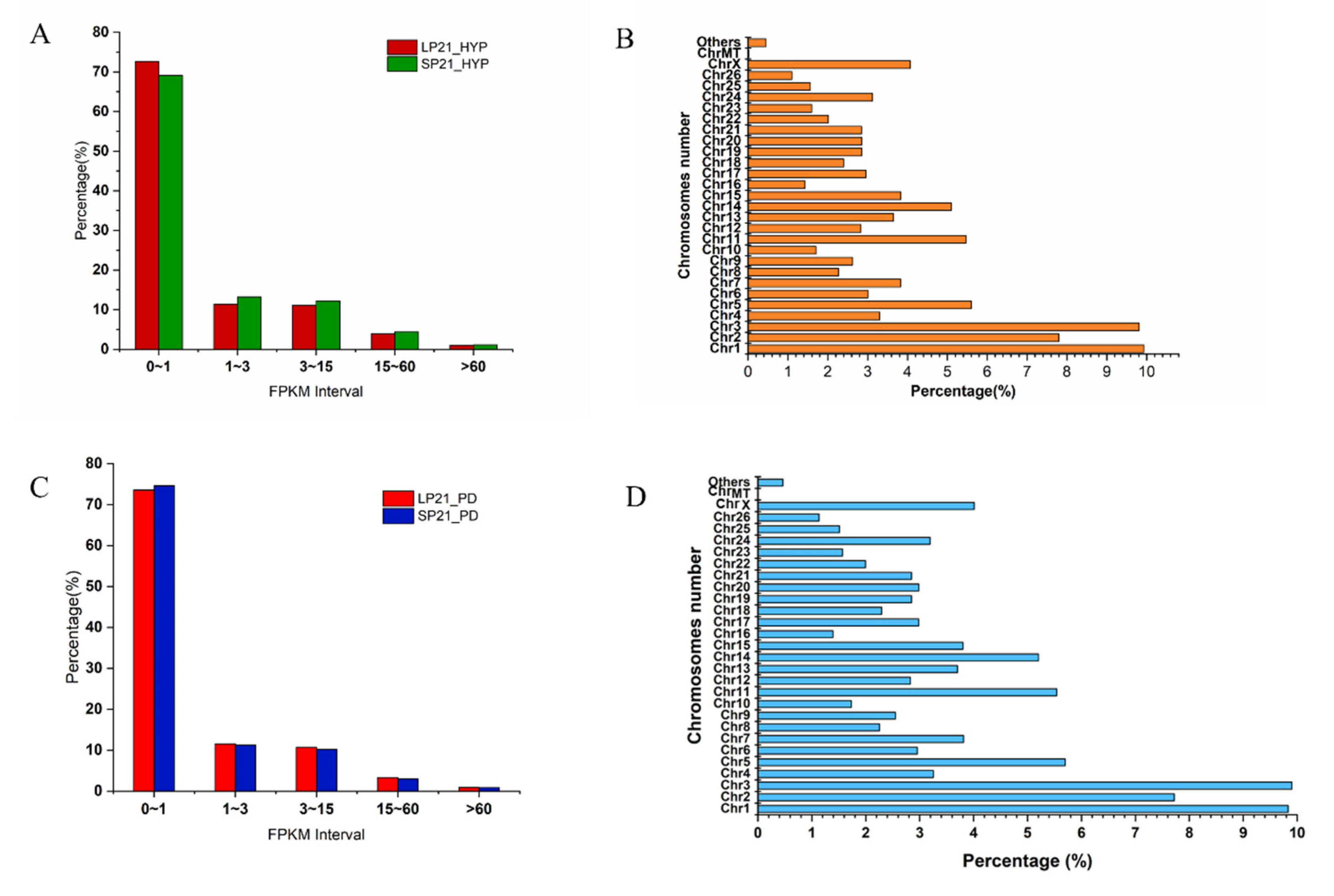
Figure 1. FPKM (A) and Chromosome (B) distribution of identified g expressed genes in SP21−HYP vs. LP21−HYP; FPKM (C) and Chromosome (D) distribution of identified expressed genes in SP21−PD vs. LP21−PD.
Genes 13 01091 g001
Figure 2. Types of non-coding rRNAs (A)and length distribution of sRNAs (B) from SP21−HYP vs. LP21−HYP, (C,D) from SP21−PD vs. LP21−PD.
Genes 13 01091 g002
Figure 3. DEGs and DEMs analyses. Volcano plot of identified genes (A) and miRNAs (B) in SP21−HYP vs. LP21−HYP, where red and blue represent up or down-regulation, respectively. Volcano plot of identified genes (C) and miRNAs (D) in SP21−PD vs. LP21−PD, where red and green represent up or down-regulation, respectively.
Genes 13 01091 g003
Figure 4. TOP enriched gene ontology (GO) study of differential expressed genes between short-photoperiod and long-photoperiod in the hypothalamus and distal pituitary of sheep. (A) GO enrichment terms for DEGs in SP21−HYP vs. LP21−HYP. (B) GO enrichment terms for DEGs in SP21−PD vs. LP21−PD. KEGG pathway analysis for DEGs between short photoperiod and long photoperiod in hypothalamus and PD of Small Tail Han sheep. (C) 20 enrichment pathways in SP21−HYP vs. LP21−HYP. (D) 20 enrichment pathways in SP21−PD vs. LP21−PD. Rich factors are defined as the amount of differentially expressed genes enriched in the pathway/amount of all genes in the background gene set.
Genes 13 01091 g004
Figure 5. Overview of mRNA-miRNA networks. (A) Overlapping genes in SP21−HYP vs. LP21−HYP between DEGs and predicted target genes by miRNAs. (B) Overlapping genes in SP21−PD vs. LP21−PD between DEGs and predicted target genes by miRNAs.
Genes 13 01091 g005
Figure 6. Regulatory networks of DE miRNA-mRNA in hypothalamus and distal pituitary of sheep. (A) The network contained 4 up-regulated miRNAs and 4 target genes in SP21−HYP vs. LP21−HYP. (B) The network contained 10 down-regulated miRNAs and 8 target genes in SP21−HYP vs. LP21−HYP. Red and blue indicate up or down-regulation, respectively. (C) The network contained 14 upregulated miRNAs and target genes in SP21−PD vs. LP21−PD. (D) The network contained 6 downregulated miRNAs and target genes in SP21−PD vs. LP21−PD. Blue and Red indicate up or down-regulation, respectively.
Genes 13 01091 g006aGenes 13 01091 g006b
Figure 7. TOP enriched gene ontology (GO) study of target genes for differential expressed miRNAs between short-photoperiod and long-photoperiod in the hypothalamus and distal pituitary of sheep. (A) GO enrichment terms for target genes of differentially expressed miRNAs in SP21−HYP vs. LP21−HYP. (B) GO enrichment terms for target genes of differentially expressed miRNAs in SP21 −PD vs. LP21−PD. KEGG pathway analysis for target genes of differentially expressed miRNAs between short photoperiod and long photoperiod in hypothalamus and distal pituitary of sheep. (C) 20 enrichment pathways in SP21−HYP vs. LP21−HYP. (D) 20 enrichment pathways in SP21−PD vs. LP21−PD. Rich factors are defined as the amount of differentially expressed genes enriched in the pathway/amount of all genes in the background gene set.
Genes 13 01091 g007
Figure 8. qPCR validation of mRNAs and miRNAs identified by RNA−seq.
Genes 13 01091 g008
Table 1. The Details of Primers of mRNA.
GenePrimer Sequences (5′-3′)Product Size (bp)Tm (℃)
KCNH3F:CGCAGAACACCTTCCTGGACAC12260
R:GCAGAAGCCATCAGAGCAGTAGAC
SIX1F:CATCGTTCGGCTTCACACAGGAG14260
R:GCCTTGAGCACGCTTTCATTCTTG
DIO2F:CCAGAGCTGTTCCAAGGCAAGTC10960
R:CTCCAGTGCTGCTGTCCAAGATG
RASD1F: GGAGACGTGTTCATCCTGGTGTTC8660
R: GTGTCGAGAATCTGCCGCTTGAG
OXTF:GCCTTCTCCCAGCACTGAGA8160
R: TCCTGGGGATGATTACAGAGGGA
THUMPD1F:TCAACGAATACGGCGACGACATG10860
R:TCTTCAAGGCAGCTTCCACATCATC
LOC101114319F:GTACACCTTGGTCTTGACAGATCCG12160
R: GAGAACCGTGCCACTGCTGATG
RPL19F:ATCGCCAATGCCAACTC15460
R: CCTTTCGCTTACCTATACC
Table 2. The Details of Primers of miRNAs.
The Name of the PrimerPrimer SequenceTm (℃)
oar-miR-200aF:GCTGCAACACTGTCTGGTAACGAT60
novel_10F:CGCTTCACAGTGGCTAAGTTCTGC60
novel_101F:AGCTCTGGGTCTGTGGGGA60
novel_4F:CCGCGTCTTTGGTTATCTAGCTGTATG60
oar-miR-143F:CGCTGAGATGAAGCACTGTAGCTC60
novel_142F:CCACCTCCCCTGCAAACG60
oar-miR-181aF:TGCGAACATTCAACGCTGTCGGTGAG60
oar-miR-323bF:AGGCCTCCCAATACACGGTCGATCTC60
U6F:CAAGGATGACACGCAAATTCG60
Note: The reverse primer for miRNA is the universal primer of the miRcute Plus miRNA qPCR Kit (TIANGEN, Beijing, China).
Table 3. mRNAs with the Most Significant Differences in Expression of Hypothalamus and Distal Pituitary.
GroupmRNAlog2FoldChangeq-ValueUp/Down
SP21−HYP vs. LP21−HYPCOMMD5−7.483.93 × 10−72down
EHBP1−6.301.54 × 1051down
LOC1141087523.922.39 × 1048up
LOC1141160525.908.05 × 1045up
MAPK6−14.075.16 × 1024down
MAX1.932.30 × 1023up
ASPSCR16.502.49 × 1023up
STK385.08-2.49 × 10²³up
SV2A−9.163.40 × 1023down
RBM4213.673.76 × 1021up
SP21−PD vs. LP21−PDLOC1141160525.434.39 × 1080up
LOC1141087523.841.15 × 1043up
PRUNE29.876.13 × 1038up
AKAP915.959.79 × 1036up
GGNBP26.591.94 × 1026up
BROX−13.022.59 × 1021down
RAPGEF613.832.03 × 1018up
PTEN−4.09-6.83 × 10¹⁷down
PLEC13.596.83 × 1017up
CEP112−8.201.14 × 1016down
Table 4. miRNAs with the Most Significant Differences in Expression of Hypothalamus and Distal Pituitary.
GroupmiRNAlog2FoldChangep-ValueUp/Down
SP21−HYP vs. LP21−HYPnovel_1547.341.17 × 10−5up
oar-miR-3956-5p−2.321.33 × 10−5down
oar-miR-544-5p3.863.61 × 10−5up
novel_1564.646.50 × 10−5up
oar-miR-3956-3p−3.681.61 × 10−4down
oar-miR-376e-3p4.851.63 × 10−4up
oar-miR-376b-3p5.551.70 × 10−4up
oar-miR-374a3.851.77 × 10−4up
novel_2343.863.66 × 10−4up
novel_1995.676.00 × 10−4up
SP21−PD vs. LP21−PDnovel_58.831.02 × 10−9up
novel_2005.593.06 × 10−9up
novel_629.671.85 × 10−6up
novel_6525.475.10 × 10−6up
novel_1726.387.94 × 10−6up
novel_1234.852.04 × 10−5up
novel_2035.694.00 × 10−5up
novel_5054.121.18 × 10−4up
novel_3576.411.78 × 10−4up
novel_833.285.61 × 10−4up
Publisher’s Note: MDPI stays neutral with regard to jurisdictional claims in published maps and institutional affiliations.