Review

Achromobacter xylosoxidans and Stenotrophomonas maltophilia: Emerging Pathogens Well-Armed for Life in the Cystic Fibrosis Patients’ Lung

1
HydroSciences Montpellier, CNRS, IRD, Univ Montpellier, 34093 Montpellier, France
2
HydroSciences Montpellier, CNRS, IRD, Univ Montpellier, Department d’Hygiène Hospitalière, CHU Montpellier, 34093 Montpellier, France
3
HydroSciences Montpellier, Université de Montpellier, CNRS, IRD, Centre de Ressources et de Compétences de la Mucoviscidose, CHU de Montpellier, 34093 Montpellier, France
4
HydroSciences Montpellier, CNRS, IRD, Univ Montpellier, Service de Microbiologie et Hygiène Hospitalière, CHU Nîmes, 34093 Nîmes, France
5
UMR 5151 HydroSciences Montpellier, Equipe Pathogènes Hydriques Santé Environnements, U.F.R. des Sciences Pharmaceutiques et Biologiques, Université de Montpellier, 15, Avenue Charles Flahault, BP 14491, CEDEX 5, 34093 Montpellier, France
*
Author to whom correspondence should be addressed.
Genes 2021, 12(5), 610; https://doi.org/10.3390/genes12050610
Submission received: 1 March 2021 / Revised: 6 April 2021 / Accepted: 19 April 2021 / Published: 21 April 2021
(This article belongs to the Special Issue Phenotypic Variability of Cystic Fibrosis: New Challenges)

Abstract

:
In patients with cystic fibrosis (CF), the lung is a remarkable ecological niche in which the microbiome is subjected to important selective pressures. An inexorable colonization by bacteria of both endogenous and environmental origin is observed in most patients, leading to a vicious cycle of infection–inflammation. In this context, long-term colonization together with competitive interactions among bacteria can lead to over-inflammation. While Pseudomonas aeruginosa and Staphylococcus aureus, the two pathogens most frequently identified in CF, have been largely studied for adaptation to the CF lung, in the last few years, there has been a growing interest in emerging pathogens of environmental origin, namely Achromobacter xylosoxidans and Stenotrophomonas maltophilia. The aim of this review is to gather all the current knowledge on the major pathophysiological traits, their supporting mechanisms, regulation and evolutionary modifications involved in colonization, virulence, and competitive interactions with other members of the lung microbiota for these emerging pathogens, with all these mechanisms being major drivers of persistence in the CF lung. Currently available research on A. xylosoxidans complex and S. maltophilia shows that these emerging pathogens share important pathophysiological features with well-known CF pathogens, making them important members of the complex bacterial community living in the CF lung.

1. Introduction

Cystic fibrosis (CF) is the most commonly, still fatal, inherited genetic disease in Caucasian populations, caused by a mutation in the cystic fibrosis transmembrane conductance regulator (CFTR) gene coding for a transmembrane channel allowing the transport of chloride ions. Different organs are affected, including the respiratory tract, where thick mucus, mucociliary clearance defects, and a decrease in anti-microbial defenses favoring bacterial colonization are observed [1]. The main causes of morbidity and mortality in CF patients are recurrent respiratory infections. From early childhood, colonization is often initiated by Staphylococcus aureus or Haemophilus influenzae. Later, the microbiology of the lung becomes more complex, as Pseudomonas aeruginosa becomes dominant, and the lung may also be colonized by other microorganisms such as nontuberculous mycobacteria (NTM), Burkholderia cepacia complex, Achromobacter xylosoxidans, and Stenotrophomonas maltophilia [2,3].

2. Emerging Pathogens and Colonization of the Lungs of Cystic Fibrosis Patients

Sm, Axc, and NTM are considered as emergent in CF [2] based on the increase in patients colonized over 20 years reported in the French CF registry (10.3% of the patients in 2018 versus 4.7% in 1999 for Sm, 6.7% versus 3.1% for Axc, and 3.1% versus 0.5% for NTM) [4]. Similar trends were reported in the American CF registry [5].
Hereafter, the review has been limited to the aerobic, non-fermentative, Gram-negative rods, ubiquitously distributed in moist environments and soil, Achromobacter xylosoxidans and Stenotrophomonas maltophilia. We will refer to Achromobacter xylosoxidans complex (Axc) to consider Achromobacter xylosoxidans sensu stricto and related species identified in CF whose distinction requires specific molecular-based methods. However, although Stenotrophomonas maltophilia (Sm) was also considered as a complex due to its large genetic diversity and its genetic organization in genogroups, we will refer to S. maltophilia as the speciation status of the sub-lineages within S. maltophilia has still to be clarified [6].
The determinants for emergence are many and varied, related to patient management and the intrinsic characteristics of bacteria [7]. First, emerging pathogens have been better recognized over time, with better patient follow-up, greater life expectancy, an increased use of aggressive eradication therapy for initial infection with “typical” CF pathogens like P. aeruginosa, and advances in bacterial identification methods in microbiology laboratories; second, the bacterial characteristics, including significant innate and acquired antimicrobial resistance, as well as their ability to implement various adaptive strategies, contribute to their persistence in the patients’ respiratory tract.
While the clinical impact of P. aeruginosa and S. aureus, particularly methicillin-resistant S. aureus, is proven in the colonization and infection of CF patients, the clinical significance of Axc and Sm in CF has been a matter of debate, despite the fact that both of them are well-known opportunistic pathogens causing a variety of severe infections (bacteremia, pneumonia, endocarditis, peritonitis, etc.), especially in immunocompromised hosts and patients with underlying diseases [8,9,10,11,12,13]. The implication of Axc in the decline of CF lung function is still controversial, although A. xylosoxidans has been shown to induce a level of inflammation similar to that caused by P. aeruginosa in chronically infected patients [14]. So far, large multicenter studies have yet to decipher the clinical burden of Axc colonization, taking into consideration variability factors of previous small cohort studies, i.e., Axc species involved, age at first colonization, type of colonization, definition of chronic colonization, and co-colonizer microorganisms [14,15,16,17]. However, one retrospective case-control study on 36 adult patients in France has demonstrated that Axc isolation (at least one sputum culture positive for Axc) is associated with a greater decline in respiratory function and a higher number of exacerbations compared with control CF patients uninfected by Axc [17]. In a study on 34 Canadian adult patients, Edwards et al. also found that patients were more likely to experience pulmonary exacerbation at the time of Axc isolation [16]. A focus on patients with chronic colonization showed that the 23 French patients included in the study by Tetard et al. had more exacerbations than intermittently colonized patients in the 3 years after Axc colonization [17], a finding not observed in other studies including 15 and 10 chronically colonized patients showing no long-term significant difference in lung function [14,16], except for one subgroup of patients with a rapid increase in Axc-specific antibodies [14]. For Sm, an increased rate of pulmonary exacerbations and hospitalizations has been demonstrated in colonized CF patients [18,19,20] but the decline in lung function has been differentially observed according to the studies [18,19,21]. The species also elicited significant in vitro (airway epithelial and macrophage cells) and in vivo (BALB/cJ mice) inflammatory response, suggestive of its contribution to patient airway inflammation [22,23]. Nevertheless, Axc and Sm are carefully considered and treated accordingly by CF clinicians, with both pathogens being recognized as markers of more severe lung disease in affected patients [17,24] and sharing important pathophysiological features with the emblematic CF pathogen, P. aeruginosa.
After initial colonization, the success of a strain’s persistence can vary depending on whether its colonization is sporadic, intermittent, or chronic. Chronic colonization is usually defined as the isolation of the bacterial species in more than 50% of sputum samples collected during the last 12 months, with a minimum of four sputum samples analyzed during that period [25]. Persistence of opportunistic environmental bacteria is a frequent observation in the airways of CF patients. In 2018, the French CF registry reported 21% of patients with chronic P. aeruginosa colonization [26]. Various other Gram-negative bacilli of environmental origin have also been isolated in CF patients over long periods of time, up to the death of the patient, e.g., Burkholderia cepacia complex, Pandoraea sp., Inquilinus sp., etc. [27,28,29]. For Axc, chronic colonization was reported in 11% to 30% of CF patients after the initial colonization [30]. To our knowledge, no similar data are available for Sm yet, despite its recognized ability to persist in the CF lung. However, a recently published monocentric study has suggested that intermittent Sm colonization is dominant in CF patients, with lower rates of chronically colonized patients (about 3%) observed in this study [31]. During initial colonization, and more markedly during persistence, emerging pathogens are subjected to various significant selective pressures in the lungs of CF patients, just like other members of the CF lung microbiota. CF lung is indeed a remarkable ecological niche in which biotic selective pressures (interactions among microorganisms living within the same ecosystem or with the host immune response) and abiotic selective pressures (non-living factors such as high levels of antibiotic use, modified acidity and salinity of the surrounding environment, oxygen deprivation in mucus, altered antimicrobial peptide production, etc.) continually shape the microbial community.
Based on the recent interest in emerging pathogens in CF and the aforementioned characteristics of Axc and Sm, the aim of this review is to gather the current knowledge on the major bacterial pathophysiological traits of Axc and Sm involved in colonization, virulence, and competitive interactions with other members of the lung microbiota, their supporting mechanisms, regulation, and evolutionary modifications during persistence in the airways of CF patients. A focus is made on biofilm biosynthesis, motility, antimicrobial resistance, secretion systems, and diffusible virulence or signal factors, competitive interactions impacting the above parameters, and genotypic adaptive modifications (gene expression, hypermutation, and microevolution). Indeed, all these abilities are considered as major drivers of pathoadaptation and persistence in the CF lung [32]. As largely reported for the “Swiss army knife” P. aeruginosa [33,34], this review highlights that the two emerging pathogens under consideration are well-armed for life in the lungs of CF patients, no doubt a key for their success in this setting.

3. Hypermutation and Genomic Within-Host Evolution

3.1. Hypermutation

Hypermutability is just one of the ways in which bacteria adapt to the diverse surrounding pressures in the CF lung. Due to a deficiency in DNA mismatch repair (nonsynonymous mutations in mutS, mutL, mutH, and/or uvrD genes) that provokes an even higher rate of mutation, hypermutation leads to increased bacterial adaptability.
Well-characterized in P. aeruginosa, only a few works have investigated hypermutation in Axc [35,36,37] and Sm [38,39,40,41]. These studies found strains that undergo mutations at a high rate (mutation frequency ≥ 4.0 × 10⁻⁷) in both Axc and Sm CF populations and considered hypermutation as a key adaptive pathway of both pathogens.
Axc and Sm hypermutators have been shown to present distinct alterations of DNA mismatch repair (MMR) with a large deletion in the mutS gene demonstrated in two independent studies on Axc [36,37] and a H683P amino acid change observed in the Sm MutS protein, which impaired its activity [38]. Hypermutation was prevalent among CF clinical isolates of Axc, being observed in 44% of the Axc isolates (40 of 90 isolates) and 26% of the patients (11 of 42 patients) [36]. The hypermutator phenotype was exclusively associated with chronic infections and over-represented in strains from the Danish epidemic strain of Achromobacter ruhlandii [36]. For Sm, 31% of CF isolates were hypermutators (28 of 90 isolates) in the studies of Vidigal et al. and Esposito et al., and 16.7% in the study of Turrientes et al. [38,39,40]. However, these percentages must be considered with caution as studies usually included several strains per patient or clonally related strains. One study of 174 Sm isolates from clinical (48 strains from 13 CF patients and 66 from 53 non-CF patients) and environmental (60 strains) origins showed that hypermutators were exclusively observed among clinical isolates, mainly originating from CF patients (8 of 48 isolates, 16.7%) compared with non-CF patients (2 of 66 isolates, 3%) [38]. This result indicates that clinical environments might select bacterial populations with high mutation frequencies. Finally, significantly higher mutation frequencies were observed for Sm strains of Sequence Type 184 compared with other genotypes, suggesting that some genetic backgrounds may be more prone to hypermutation [40]. Hypermutation was thus differentially observed among strains and species of Axc and Sm, but also among clonally related adaptive variants in a patient or in a sample, as isolates presenting different mutation frequencies were shown to coexist within a patient.
Being hypermutable can confer a transient advantage to fluctuating environmental conditions, but mutation status usually evolves over time to a less mutator phenotype due to fitness cost associated with hypermutation [39,41]. However, studies have reported CF patients with successively recovered isolates displaying increased mutation frequencies during persistence [38,40,42], suggesting that the dynamics of hypermutators also depend on specific conditions linked to the host/strain partnership and that the fitness cost of hypermutable Sm strains may not systematically impede long-standing CF lung infection.

3.2. Diversified Populations and Within-Host Genome Evolution

The genomes of Axc and Sm are highly dynamic, and numerous alterations in the genomic content of Axc and Sm, named microevolution, have been demonstrated during their persistence in the CF lung, as previously studied in P. aeruginosa [43,44]. During Acx or Sm persistence, studies based on pulsed-field gel electrophoresis (PFGE) first led to the recognition of “true” chronic colonization as well as intra-clonal evolution of PFGE profiles over time through genome rearrangements [15,39,40,41,42,45]. As for P. aeruginosa, microevolution was related to lung habitat pathoadaptation by the authors. Further investigation of the within-host evolution through the analysis of whole genome sequences of clonal lineages of Axc showed that microevolution occurred through small mutations in a high variety of genes, mostly in genes involved in the general metabolism but also in genes involved in virulence, antimicrobial resistance, transcription and translation, cell wall, and stress response [46,47,48]. In Sm, genes identified as undergoing adaptive evolution were found to be involved in virulence and resistance to antibiotics and heavy metals [49].
Another level of complexity was brought to light by the discovery of highly complex Axc and Sm populations composed of a variety of sub-clonal variants, so-called diversified populations, in a patient or sputum sample [40,42,45]. Again, these observations were confirmed by whole genome sequence-based studies showing that a wide majority of mutations were isolate-specific within a clonal lineage in Axc [46]. Divergent evolution forming several subpopulations from the original infecting isolate had previously been observed in P. aeruginosa and was linked to the heterogeneity of the CF lung, wherein bacteria are submitted to distinct selective pressures leading to distinct evolutionary processes [44]. Here, we would like to elucidate a broad study analyzing 552 Sm genomes from 23 sites of the lungs of a CF patient that led to the most complete description of a bacterial population diversified into multiple coexisting lineages for an emerging pathogen in CF [49]. In this study, lineage-separating mutations were found in genes known to be important for virulence. The authors identified mutations arising independently in lineages that colocalize in space, providing evidence for location-specific selection. This also suggested that certain genotypes can locally proliferate because of site-specific selection and that such site-specific selection might contribute to long-term maintenance of diversity in CF chronic infections that could impede therapy [49].
Microevolution thus leads to an important genotypic heterogeneity related to an equally important phenotypic diversity. We previously conducted a study on multiple Axc isolates from a sputum sample of several chronically colonized CF patients and observed significant genomic diversity in PFGE associated with a striking diversity in antimicrobial resistance patterns, not predictable from colonial morphology, that may contribute to compromise the antibiotic management of CF airway infections by Axc [45]. Similar observations are available for Sm whose pathoadaptation to the CF lung is associated with populational genotypic and phenotypic heterogeneity in numerous phenotypical traits, i.e., growth rate, antibiotic resistance, biofilm formation, and both in vivo and in vitro pathogenicity [40,42].
More generally, an adapted, diversified population will encompass numerous sub-clonal variants that may vary on all characteristics listed within this review. The existence of these variants supports long-term survival of the pathogen in the CF lung, as it appears to quickly adapt to changing selective pressures through the selective advantage given to one or several of these variants by the new environmental conditions. This has previously been described as “the insurance hypothesis” [43].

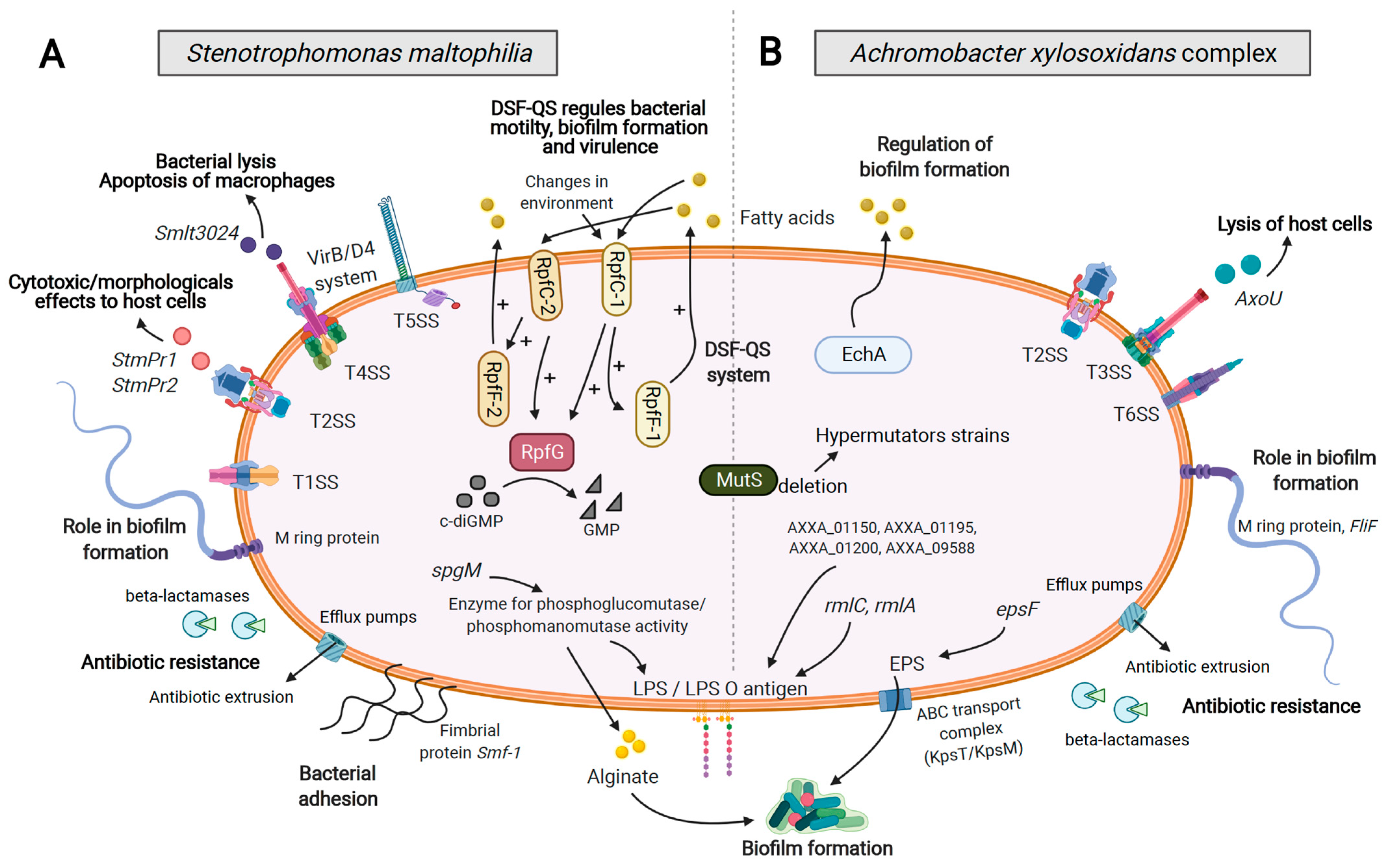
4. Virulence Factors and Secretion Systems

Bacteria possess specialized protein secretion systems allowing them to deliver a wide variety of proteins either in the extracellular space or directly into other cells. Classified into nine groups, from T1SS to T9SS (Type X Secretion System), these secretion systems are involved in the interaction between bacteria and their environment, a host organism, or other bacteria [50,51,52,53,54,55]. In the context of CF, these systems secrete effectors, allowing bacterial pathogens to escape from the host immune system but also to compete with other bacteria of the airway microbiota. Both Axc and Sm were shown to possess different secretion systems, common to many Gram-negative bacilli such as Escherichia coli, B. cepacia complex, or P. aeruginosa [51,56,57]. The TSSs harbored by Axc and Sm are shown in Figure 1.
First of all, Axc and Sm harbor T2SS, which contributes to virulence in many pathogens; as examples, T2SS-dependent substrates include the cholera toxin from Vibrio cholerae and exotoxin A from P. aeruginosa [51]. It may also be involved in host cell adhesion, biofilm formation, and intracellular infection of macrophages [58,59,60]. T2SS was identified in the sequenced and annotated genomes of Axc strain NH44784-1996 and strain Ax8 from CF patients [61]. Sm clinical strain K279a also possesses a T2SS that mediates the secretion of at least seven proteins and three proteolytic activities, and causes damage to lung epithelial cells, mostly through the secretion of serine proteases StmPr1 and StmPr2, which induce cytotoxic and morphological effects on host cells [62,63].
Thirteen and twelve genes encoding for T3SS and T6SS respectively, were identified in the genome of Axc strain NH44784-1996 [61]. T3SS and T6SS mediate an effector transport by direct contact with the target cells [64] and T6SS has been shown to deliver lethal toxins into bacterial cells [65,66]. A complete T3SS locus has been observed in 72% of the 65 Axc genomes examined [67]. An important T3SS substrate is the phospholipase A2 AxoU, similar to the ExoU of P. aeruginosa, encoded by 98% of T3SS-positive Axc strains [67,68]. AxoU exhibits toxicity against eukaryotic cells by cleavage of membrane phospholipids, leading to lysis of the host cells, particularly phagocytic cells, and inhibits the innate immune response [67,68]. AxoU/T3SS-mediated cytotoxicity should thus be considered as an important virulence trait of Axc; however, further investigation is still required to fully elucidate this virulence mechanism, as bacterial internalization appears to be required for cytotoxicity [67].
A T4SS, homologous to the T4SS of the plant pathogens Agrobacterium tumefaciens and Xanthomonas citri, has also been identified in the genome of the clinical strain Sm K279a. T4SS mediates effector delivery in the extracellular medium or, in a contact-dependent manner, directly in the target cells, and has been involved in interbacterial antagonism and the delivery of lethal toxins into bacterial cells [64,65,66,69]. Sm K279a T4SS is encoded by a cluster of chromosomally located genes, virB1-virB11 and virD4, called the virB/D4 system. Highly conserved among environmental and clinical strains of Sm, T4SS inhibits apoptosis in infected human epithelial cells, promotes apoptosis in infected mammalian macrophages, bacterial competition, and the death of multiple species, including P. aeruginosa [70]. In Sm, T4SS requires a direct contact with the target cells and the panel of affected cells, and its deleterious effects suggest that the VirB/D4 system is capable of interacting with a range of different receptors/membranes and then injecting diverse protein effectors into the target cells [70]. Among these, Smlt3024, an effector whose function is yet to be completely characterized, has shown the ability to reduce the growth rate of E. coli cells [69].
Finally, T1SS and T5SS have also been identified in the genome of the clinical strain Sm K279a [71].
Additionally, clinical Axc and Sm CF strains produce siderophores and secrete other virulence factors such as proteases, phospholipases, lipases, and nucleases, which contribute to host colonization and persistence through adhesion to and degradation of host tissues and iron uptake essential for bacterial growth [70,72,73,74,75,76].

5. Pathoadaptive Traits

5.1. Virulence Attenuation

Beyond chronic colonization, virulence attenuation is a pathoadaptive trait mostly described in P. aeruginosa (loss of T3SS and flagellum, reduced production of proteases, pyocyanins, and other virulence factors) [77] that was also reported for Axc and Sm CF strains. Regarding Axc, a strong decrease in the expression of genes coding for T3SS and motility [78], as well as mutations in virulence-associated genes [46], have also been observed for strains involved in late infection episodes compared with strains involved in early infections from the same patient. For Sm, a lower motility, a decrease or the loss of some virulence factors, and decreased virulence both in human cells and in an invertebrate model have been demonstrated [41,42]. All these observations underline that both Axc and Sm have the capacity to modulate their pathogenicity over time. Less exposed to the host immune system, this represents an additional key to bacterial persistence in the CF lung.

5.2. Adhesion, Motility, and Biofilm Formation

Axc isolates exhibited affinity to bind mucin, collagen, and fibronectin [79], while Sm was shown to be able to adhere to A549 respiratory epithelial cells and IB3-1 bronchial cells [80,81]. After adhesion, the ability to invade CF-derived IB3-1 bronchial epithelial cells was reported for Sm only, to date [81], and it represents a way to persist in the CF airways, as intracellular bacteria represent a less accessible reservoir of pathogens for both immune system and antimicrobial agents. Genes, such as the smf-1 gene coding for the fimbrial protein Smf-1, have a crucial role in the bacterial adhesion [82] and further biofilm formation as only Sm isolates containing the smf-1 gene were found to produce biofilm [83] (Figure 1). These observations are important for the initial CF patient airway colonization by Axc and Sm via adhesion to the surface of airway mucosae. Motility, particularly swimming motility, is also critical for this first step of adhesion and subsequent biofilm development and was found to be a constant characteristic of CF Axc and Sm strains [84,85]. Motility has indeed shown positive correlations with the Sm strains’ capacity for mucin-adhesion and biofilm formation [85] and the loss of flagella has been shown to significantly decrease Sm adhesiveness [81]. In Axc, more than 20 genes are involved in flagellum synthesis and most of them are downregulated during the sessile phase [76,86]. Among them, inactivation of the flif gene coding for the flagellar M ring protein leads to a reduction in biofilm formation [86]. This protein is also present in the flagellum of Sm and could have the same importance in biofilm formation (Figure 1). The early biofilm formation stages are thus mediated by flagella and fimbriae, allowing adhesion to epithelial cells [71].
After this initial pivotal stage of adhesion, both Axc and Sm have the ability to form biofilm, a highly organized multicellular structure in which bacterial cells are embedded in a self-produced extracellular matrix developing after transition of bacteria from their planktonic form to a sessile stage [14,79,84,87,88,89], with two distinct morphotypes observed in the CF lung for Axc, i.e., a surface-attached biofilm morphotype of small aggregates and an unattached biofilm morphotype of large suspended aggregates [90]. Congruent with previous genomic evidence [61], the ability to form biofilm has been observed in 97.1% to 100% of CF Axc strains [79,84,87] and is shared by several species of the Axc, i.e., A. xylosoxidans (Ax), A. dolens/A. ruhlandii, A. insolitus, A. insuavis, and A. piechaudii [79,84], with one third to 58% of isolates being strong biofilm producers. A slightly lower percentage of CF Sm strains are able to form biofilm, although this remains a characteristic presented by 87.5% to 90.2% of strains [91,92], with a high level of variability observed both among strains analyzed and in biofilm architecture [93]. This ability to form biofilm favors persistence and dissemination through the maturation of the biofilm and subsequent detachment of bacterial cells from the mature biofilm, in accordance with the general stages of biofilm development [94].
During the biofilm maturation phase, bacteria secrete various polysaccharides which form an exopolysaccharide (EPS) matrix in which they are embedded [95]. The main EPS secreted is alginate, which consists of mannuronic and glucuronic acid [96]. In Axc, the ABC transport complex (KpsT/KpsM) exports the polysaccharides through the cytoplasmic membrane [97]. According to Pompilio et al., the genes spgM, rmlC, and rmLa are associated with biofilm formation as well as 4 AXXA proteins (01150, 01195, 01200, 09588) [91] (Figure 1). In Sm, the spgM gene codes for a bifunctional enzyme with phosphoglucomutase and phosphomannomutase activity related to the metabolic process of carbohydrates and is involved in the lipopolysaccharide (LPS) and alginate biosynthesis, as observed for the homologous algC gene found in P. aeruginosa [89,98], whereas in Axc, rmlC, and rmlA genes, as well as Ax’s, AXXA-encoding genes are involved in the biosynthesis of the LPS O antigen [76,89] (Figure 1). It should be noted that mucoid phenotype related to EPS production is one of the most studied pathoadaptations of P. aeruginosa in CF but has been rarely reported in Axc and Sm subject to an accurate identification for older studies [99,100,101].
Numerous specificities are associated with the growth of biofilm (adaptation to hypoxic and acidic conditions, quiescent forms, high inoculum, gene expression modifications, etc.), all of which are important determinants for persistence. Axc and Sm undergo numerous metabolic and structural changes between the planktonic and sessile phases. The expression of the epsF gene coding for the Axc exopolysaccharides’ synthesis is increased by a factor of 6.4 during the sessile phase. In addition, the capsular ABC transporters of polysaccharides KpsT, KpsE, and KpsM are increased by 5.1 to 8.7 times [86]. For some Axc strains, a modification in the expression of genes involved in the stress response is also described during the sessile state. These genes code for diguanylate cyclase and phosphodiesterase synthesis [102]. In addition, an increase in the efflux pump activity during the sessile stage of Axc is observed compared to the planktonic stage. Indeed, the AxyA efflux pump regulated by the AxyAB-OprM operon has 7.4 times more activity in the sessile stage [86]. This efflux pump may be involved in the Axc biofilm metabolism, in addition to its role in antibiotic tolerance.
In Sm, factors identified as influencing biofilm formation are cell motility, genes involved in lipopolysaccharide/exopolysaccharide biosynthesis, SmeYZ and MacABCsm efflux pumps, iron availability, histidin kinase, the two-component signal transduction system BfmAK, and the diffusible signal factor (DSF) quorum sensing (QS) system [93,103,104]. This is supported and completed by recent transcriptomic analyses revealing that a small set of commonly regulated genes are involved in the biofilm lifestyle of Sm, with 6% to 9% of all genes being upregulated and 1% to 3% of all genes being downregulated in biofilms versus planktonic cells [93]. Commonly, upregulated genes show a large functional distribution, and they are mostly involved in transcription and translation followed by a remarkably high number of genes involved in iron acquisition, then in metabolism/biosynthesis, membrane proteins/transporters, and respiration/energy [93]. In addition, the authors identified an extracellular protease activity up to 40 times higher in Sm biofilm compared with planktonic cultures; however, although such an activity has previously been associated with virulence and biofilm formation, they could not link the increased protease activity observed with any particular virulence or biofilm-forming profile [93].
When looking at specific characteristics that might be presented by CF strains, the available studies showed no difference in biofilm formation between clinical and environmental Sm isolates [91], unlike Axc clinical strains which tend to produce more biofilm than environmental strains [90]. A focus on clinical strains has shown a similar proportion of Axc clinical isolates with the ability to form biofilm in CF and non-CF panels of strains [79]. For Sm, the proportion of CF and non-CF clinical strains able to form biofilm depends on the study, with some studies showing either no difference, congruent with the absence of difference in the presence of rmlA, rpfF, and spgM biofilm-associated genes [85,92], or a significantly higher proportion of biofilm-forming strains among non-CF strains [91]. In the latter study, the genes associated with biofilm formation, particularly the spgM gene, were differently expressed in Sm strains from CF and non-CF patients [91]. Nevertheless, CF Sm strains were shown to produce less biofilm biomass levels and exhibit a significantly higher proportion of biofilm-forming strains only when the moderate-biofilm production category was considered [91]. On the whole, they did not display any specific characteristics regarding their ability to form biofilm compared with other populations.
Considering the type of colonization, no difference in biofilm-forming capacity was observed between populations of Axc isolates from chronic or sporadic infection [84]. However, during persistence, a downward trend in biofilm biomass formed by clonally related, temporally isolated Sm and Axc strains from chronically infected CF patients was observed compared to the initial isolates obtained from the same patient as a consequence of Sm or Axc adaptation to the stressful lung environment [42,90]. For Sm, this was associated with a decrease in the biofilm’s ability to adhere and modifications in the biofilm structure that became distorted in the later infection stages compared with the early stages of infection where the Sm biofilm was well-structured with many juxtaposed layers [75].
Altogether, biofilm is an emblematic structure which allows Axc and Sm to survive under stressful conditions, making them able to persistently colonize the airways of CF patients. However, its production may greatly vary according to strains and conditions. Its regulation involves different structures such as flagella, fimbriae, efflux pumps, or transporters. Many metabolic changes and gene modulations take place during biofilm formation, with many genes activated or inhibited. Interestingly, similar pathways and genes were found involved in biofilm production in Axc and Sm. Finally, it should be pointed out that the biofilm structure confers a protection against various antimicrobial strategies, whether host defenses or antibiotic regimens [102], with Axc and Sm grown in biofilms exhibiting higher tolerance to various antibiotics [42,105,106]. Considered together with high levels of innate and acquired resistance presented by both pathogens (as presented below), antimicrobial tolerance conferred by the biofilm lifestyle adds to the difficulty of eradicating Axc and Sm through conventional antimicrobial strategies.

5.3. Antimicrobial Resistance

Axc and Sm have recently been shown to be more resistant than P. aeruginosa to airway antimicrobial peptides, with Ax being the most resistant species [107]. Both Axc and Sm are also innately multidrug-resistant (MDR) microorganisms able to increase their basal level of resistance through the acquisition of additional resistance mechanisms. In both species, resistance, either innate or acquired, is conferred by two major mechanisms: extrusion of the antibiotics through efflux pumps and enzymatic degradation (Figure 1).
A variety of efflux pumps and enzymes have been identified as contributing to antimicrobial resistance in Axc and Sm and are presented in Table 1.
Considering antibiotics, the most widely used in the management of bacterial infections, resistance in Axc affects most cephalosporins except ceftazidime via the production of an inducible chromosomal β-lactamase, aztreonam, and aminoglycosides, while Sm is usually resistant to all β-lactams except ticarcillin-clavulanate and ceftazidime via the production of two inducible β-lactamases, including a carbapenemase (metallo-β-lactamase L1), aminoglycosides, and colistin. Antibiotics available to treat MDR Axc and Sm infections are thus limited, with co-trimoxazole usually considered as the best option for treatment when patients meet the criteria for treatment initiation [147,148,149]. Otherwise, therapy should be guided by antimicrobial susceptibility testing results. For Axc, the most active agents beside co-trimoxazole are minocycline, meropenem, or imipenem, piperacillin-tazobactam, ceftazidime, and/or chloramphenicol, depending on the study [150,151,152,153]. For Sm, chloramphenicol, minocycline, or doxycycline can be considered for the treatment, as well as ticarcillin-clavulanate, ceftazidime, and levofloxacin, for which clinical breakpoints are available. However, the use of both first-line and second-line treatments may be compromised when infection involves exogenous strains which have acquired additional resistance up to being extensively drug-resistant or strains with in vivo acquisition of resistance during chronic colonization [31,37,42,78,79,112,113].
Due to the highly selective surrounding conditions encountered by pathogens in the CF lung, including significant exposure to antibiotics, it should be noted that bacterial populations displayed specific resistance characteristics. One recent study showed that Sm CF strains were more resistant to piperacillin-tazobactam and to cotrimoxazole compared to non-CF strains, with MDR strains being significantly more prevalent in CF and hypermutable CF strains showing a high number of antibiotic resistances [92]. In addition, it is worth noting that small-colony-variants (SCV) of Sm have been identified in samples of CF patients [49,154] and that SCV phenotype can be selected during the selective pressure of the first-line cotrimoxazole antimicrobial therapy [154]. SCV morphotype is usually associated with slow-growing subpopulations of bacteria displaying distinctive phenotypic and pathogenic traits of which increased resistance to antibiotics and contribute to persistent or recurrent infections.
Altogether, antimicrobial resistance makes the antibiotic treatment of Axc and Sm infections challenging in CF patients, particularly if we consider that both pathogens may also be protected from antimicrobial drugs in the biofilm formed in the CF lung, where higher antibiotic minimal inhibitory concentrations are usually observed compared with the planktonic forms [105,106].

6. Competitive Interactions

Whereas P. aeruginosa and S. aureus have both been greatly studied for their interspecies competitive behavior in the context of CF, Axc and Sm have received far less attention despite their increased prevalence in a restricted niche represented by CF lungs and their frequent co-isolation with other pathogens, suggesting that they also have to compete for space and nutrients, just like other members of the resident microbial community [155]. Several of the Axc and Sm characteristics listed above, especially biofilm formation, are also well-known traits influencing competitive or cooperative bacterial interactions [156].
A few studies included Sm CF clinical strains and have revealed complex, reciprocal interferences with P. aeruginosa CF clinical strains or reference strains. Most studies revealed that P. aeruginosa inhibited Sm growth but that it confers to Sm an increased tolerance to antibiotics, such as ciprofloxacin and tobramycin within dual biofilms. In return, Sm displays the ability to modulate P. aeruginosa virulence in mixed biofilms [23,155,157,158]. These observations support the existence of a cooperative pathogenicity between the two species, as proposed by Pompilio et al. [158]. These interactions appear to require a direct contact between Sm and P. aeruginosa as Sm growth inhibition was not observed when P. aeruginosa culture supernatant was tested, suggesting that P. aeruginosa T6SS was involved [159]. Other modifications highlighted during SmP. aeruginosa co-cultures are modifications in P. aeruginosa motility and pigment production [157,158]. Altogether, about one third of the SmP. aeruginosa pairs of strains tested (31%, 18 of 59) displayed at least one modification affecting growth, motility, and/or pigment production when tested in co-culture compared with the corresponding monocultures [157]. Divergent competitive ability was, however, observed according to the study conditions regarding dual biofilm biomass and P. aeruginosa motility, either decreased or increased [155,157,158], at least partly linked to the tested strains, i.e., reference or clinical strain, CF or non-CF isolates, time to first colonization for CF isolates, and the planktonic or sessile growth conditions of the assays. Pompilio et al. identified several P. aeruginosa virulence genes whose expression was significantly modified by co-culture with Sm, with protease and alginate-encoding aprA and aglD genes being upregulated and the QS-related rhlR and lasI genes being downregulated [158]. A first recent in vivo study has brought new knowledge of cooperativity between Sm and P. aeruginosa, showing the co-localization of both pathogens in the lungs of BALB/cJ mice and the integration of Sm in P. aeruginosa biofilm, both observations being factors favoring interaction between the two species. Increased Sm loads, correlated with P. aeruginosa loads, were observed in lung homogenate and broncho-alveolar lavage fluid samples in the presence of P. aeruginosa [23]. Viable P. aeruginosa cells were necessary to confer a significant persistence benefit to Sm, showing that this persistence was not only mediated by passive protection in biofilm but also required an active cellular process [23].
Regarding the interaction between Sm and other species, Sm motility and/or growth alterations have occasionally been observed in SmAxc and SmS. aureus co-cultures [157]. Sm can also establish mixed biofilm with Aspergillus fumigatus which can be co-isolated from the airways of CF patients [160]. In SmA. fumigatus co-culture biofilms, the morphology of A. fumigatus changes and its growth decreases, whereas antimicrobial susceptibility of both partners is modulated (increased A. fumigatus susceptibility to amphotericin B and increased Sm tolerance to levofloxacin) [161,162]. In poly-species culture, Sm has been shown to confer some protection to Burkholderia cenocepacia against hydrogen cyanide, a toxic metabolite released by P. aeruginosa [163]. Finally, Sm also demonstrates the ability to lyse CF bacterial competitors such as P. aeruginosa using T4SS [70] or to disrupt the hyphal transition and biofilm formation of Candida albicans through its DSF QS system [164].
The competitive ability of Axc has been even more rarely studied than that of Sm. The first evidence of Axc’s competitive ability revealed that nearly half of the AxcP. aeruginosa pairs of strains tested (49%, 26/53) displayed at least one modification affecting growth, motility, and/or pigment production when tested in co-culture compared with the corresponding monocultures [157]. Some Axc clinical strains were able to outcompete clinical as well as environmental strains of P. aeruginosa by inhibiting the above major P. aeruginosa pathophysiological traits. In addition, Axc has the ability to outcompete Sm by decreasing its growth and/or motility. Finally, interactions affecting Axc motility were also observed on an intra-species level, with one Axc strain being able to influence the motility of another, even when co-isolated from a unique sample [157].
As previously recognized for major CF pathogens, interactions involving emerging CF pathogens appear complex and varied and probably act synergistically. Bacterial competition affects the major bacterial pathophysiological traits represented by growth, motility, biofilm formation, antimicrobial tolerance, and/or P. aeruginosa pigment production, considered as an indirect marker of P. aeruginosa virulence. However, additional studies using well-documented clinical strains and in vivo models are still needed to better delineate the importance of these interactions in shaping the CF lung polymicrobial community and decipher the mechanisms involved in these interactions.

7. Quorum Sensing Regulation

Bacterial populations use quorum sensing (QS) molecules for intercellular signaling [165], which allow coordination of gene expression when they reach a critical cell density. QS is a unique regulatory mechanism that acts through the production, detection, and response to extracellular signaling molecules called autoinducers. Autoinducers accumulate in the environment as bacterial populations become concentrated. When they reach a high local concentration, the cells “detect” the change in their number and the population responds with coordinated expression of specific target genes [165,166]. This coordination allows the bacterium to regulate various mechanisms involved in colonization or virulence, such as biofilm formation, motility, and the production of siderophores and other virulence factors. This system strongly contributes to the bacterial establishment and persistence in the host and is long recognized as a key parameter in bacterial virulence and pathogenesis. Indeed, pathogens with QS pathways are usually considered as able to infect host organisms more effectively. With regard to bacteria that utilize QS as part of their pathogenic lifestyle, P. aeruginosa is the best understood in terms of the virulence factors regulated and the role quorum sensing plays in pathogenicity. Despite increased knowledge presented hereafter for Sm and Axc, research is still needed to decipher the role that QS plays in their pathogenicity in CF patients.

7.1. Diffusible Signal Factor (DSF) System

This QS system is present in various Gram-negative bacteria, including P. aeruginosa [165]. It is also characterized in strain K279a, a reference Sm strain. The QS system is based on the diffusible signal factor (DSF), with cis-11-methyl-2-dodecenoic acid as the main signaling factor [167]. Its production is controlled by the rpf (regulation of pathogenicity factors) cluster, and two types of rpf clusters have been demonstrated in clinical strains: rpfF-1 (60%, 47 of 78 strains) and rpf-2 (40%, 31 of 78 strains), with established links between the type of DSF-based QS system and the phenotypes of biofilm formation, virulence, and antimicrobial resistance of the strains [168,169]. rpf-1-type Sm strains basically produce DSF in response to diverse environmental changes, whereas rpf-2-type Sm strains are deficient for basal DSF production while retaining their ability to detect DSF produced by other strains and subsequently produce DSF [168]. The rpf cluster encodes for RpfF synthase (which has different N-terminal regions according to the rpf cluster type) and the RpfC/RpfG two-component system responsible for perception and transduction of the DSF. Activated RpfG converts cyclic diguanylate monophosphate (c-di-GMP) to GMP, thereby controlling the expression of genes, which regulate biofilm formation, virulence, and bacterial motility in rpfF-1 strains [170] (Figure 1). DSF-mediated regulation has been associated with a decrease in motility, siderophores, protease level, and virulence in different models [170,171], and either with an increase [168] or a decrease [171] in biofilm production, depending on the study. In the Sm rpf-2 system, RpfC-2 blocks RpfF-2, which in turn stops DSF synthesis. Exogenous DSF signals released by surrounding bacteria (e.g., rpf-1 strain) are detected by RpfC-2, liberating active RpfF-2 to produce DSF (Figure 1), thus stimulating bacterial virulence, as demonstrated by experiments in the zebrafish model [172].
Other than its regulatory role, the Sm DSF system plays a role in inter-microbial interference and competition and, as stated above, has been shown to disrupt the hyphal transition and biofilm formation of C. albicans [164].
With regard to Axc QS systems, nothing had ever been described before the study by Cameron et al., showing that Enoyl CoA hydratase (echA) plays a central role in the biosynthesis of cis-2 fatty acid signaling molecules by A. xylosoxidans (Figure 1). As described in Sm, these molecules are DSF, which regulate biofilm formation and tolerance to antimicrobials [102].

7.2. Other Quorum Sensing Factors

A small protein Ax21 was first described as a QS-related virulence factor in Xanthomonas species pathogenic for plants. Then, later identified in clinical strains of Sm, its production was linked with several important pathophysiological traits in Sm, such as virulence (both in zebrafish and larval Galleria mellonella infection models), antibiotic tolerance/resistance, biofilm formation, and motility [173,174,175]. Secreted within the outer membrane vesicles, Ax21 is thought to mediate intraspecies communication. However, as synthesis and secretion of Ax21 can be influenced by the DSF-QS system, further investigations are required to find out whether Ax21 acts indirectly through the influence of the DSF [176]. Finally, although Sm does not have a complete Acyl Homoserin Lactone (AHL)-mediated QS system (LuxI/LuxR family) like P. aeruginosa, it possesses the LuxR regulator element of this system, known as SmoR [177]. This LuxR solo SmoR confers Sm the ability to detect AHL signals produced by other microorganisms, with AHL produced by P. aeruginosa being able to increase the swarming motility of Sm, for example, and thus represents another way for interspecific communication [177].

8. Conclusions

A review of the research on S. maltophilia and A. xylosoxidans complex shows that both emerging pathogens in CF share important pathophysiological features with other well-known CF pathogens such as P. aeruginosa, making them important members of the complex bacterial community living in the CF lung and successful CF pathogens. Indeed, the summarized literature highlights that both Sm and Axc are well-armed for the colonization and subsequent persistence in the hostile environment represented by the CF lung, as they harbor a large panel of adaptive strategies allowing them to face adverse, fluctuating conditions. Most of the pathogenic phenotypes described in this review are interconnected with each other, either positively or negatively, and are subjected to rapidly changing surrounding selective pressures in the host. Their evolutionary dynamics are thus highly complex, intimately linked to the patient airway colonization history and the specific host/pathogen partnership shaping the progress of bacterial pathoadaptation in a bacteria–host evolutionary arms race. In this context, the increased knowledge of major pathophysiological traits presented by both Sm and Axc and their regulation by QS systems opens up new perspectives for the care of CF patients, with novel antimicrobial strategies targeting either directly Sm or Axc characteristics (anti-biofilm) or interfering with their QS communication signals (quorum quenching) as an anti-virulence strategy [75,149,178,179,180].

Author Contributions

Writing—original draft preparation, Q.M., P.S., C.D., and H.M.; writing—review and editing, C.D., R.C., E.J.-B., and H.M.; supervision, H.M. All authors have read and agreed to the published version of the manuscript.

Funding

This research received no external funding.

Institutional Review Board Statement

Not applicable.

Informed Consent Statement

Not applicable.

Data Availability Statement

Not applicable.

Acknowledgments

The authors gratefully acknowledge Teresa Sawyers, English medical writer at Nîmes University Hospital, for editing this paper.

Conflicts of Interest

The authors declare no conflict of interest.

References

  1. Valenza, G.; Tappe, D.; Turnwald, D.; Frosch, M.; König, C.; Hebestreit, H.; Abele-Horn, M. Prevalence and antimicrobial susceptibility of microorganisms isolated from sputa of patients with cystic fibrosis. J. Cyst. Fibros. 2008, 7, 123–127. [Google Scholar] [CrossRef]
  2. Parkins, M.D.; Floto, R.A. Emerging bacterial pathogens and changing concepts of bacterial pathogenesis in cystic fibrosis. J. Cyst. Fibros. 2015, 14, 293–304. [Google Scholar] [CrossRef] [PubMed] [Green Version]
  3. Bhagirath, A.Y.; Li, Y.; Somayajula, D.; Dadashi, M.; Badr, S.; Duan, K. Cystic fibrosis lung environment and Pseudomonas aeruginosa infection. BMC Pulm. Med. 2016, 16, 174. [Google Scholar] [CrossRef] [PubMed] [Green Version]
  4. Menetrey, Q.; Dupont, C.; Chiron, R.; Marchandin, H. Emerging bacteria in cystic fibrosis and non-cystic fibrosis bronchiectasis from a microbiologist’s perspective. Rev. Mal. Respir. 2020, 37, 561–571. [Google Scholar] [CrossRef]
  5. Cystic Fibrosis Foundation Patient Registry 2019 Annual Data Report. Available online: https://www.cff.org/Research/Researcher-Resources/Patient-Registry/2019-Patient-Registry-Annual-Data-Report.Pdf (accessed on 20 February 2021).
  6. Gröschel, M.I.; Meehan, C.J.; Barilar, I.; Diricks, M.; Gonzaga, A.; Steglich, M.; Conchillo-Solé, O.; Scherer, I.-C.; Mamat, U.; Luz, C.F.; et al. The phylogenetic landscape and nosocomial spread of the multidrug-resistant opportunist Stenotrophomonas maltophilia. Nat. Commun. 2020, 11, 2044. [Google Scholar] [CrossRef]
  7. Green, H.; Jones, A.M. The microbiome and emerging pathogens in cystic fibrosis and non-cystic fibrosis bronchiectasis. Semin. Respir. Crit. Care Med. 2015, 36, 225–235. [Google Scholar] [CrossRef] [PubMed]
  8. Tzanetou, K.; Triantaphillis, G.; Tsoutsos, D.; Petropoulou, D.; Ganteris, G.; Malamou-Lada, E.; Ziroyiannis, P. Stenotrophomonas maltophilia peritonitis in CAPD patients: Susceptibility to antibiotics and treatment outcome: A report of five cases. Perit. Dial. Int. 2004, 24, 401–404. [Google Scholar] [CrossRef] [PubMed]
  9. Araoka, H.; Baba, M.; Yoneyama, A. Risk factors for mortality among patients with Stenotrophomonas maltophilia bacteremia in Tokyo, Japan, 1996. Eur. J. Clin. Microbiol. Infect. Dis. 2010, 29, 605–608. [Google Scholar] [CrossRef]
  10. Hauser, A.R.; Jain, M.; Bar-Meir, M.; McColley, S.A. Clinical significance of microbial infection and adaptation in cystic fibrosis. Clin. Microbiol. Rev. 2011, 24, 29–70. [Google Scholar] [CrossRef] [Green Version]
  11. Brooke, J.S. Stenotrophomonas maltophilia: An emerging global opportunistic pathogen. Clin. Microbiol. Rev. 2012, 25, 2–41. [Google Scholar] [CrossRef] [Green Version]
  12. Liu, C.; Pan, F.; Guo, J.; Yan, W.; Jin, Y.; Liu, C.; Qin, L.; Fang, X. Hospital acquired pneumonia due to Achromobacter spp. in a geriatric ward in China: Clinical characteristic, genome variability, biofilm production, antibiotic resistance and integron in isolated strains. Front. Microbiol. 2016, 7, 621. [Google Scholar] [CrossRef] [PubMed]
  13. Barragán, E.P.; Pérez, J.S.; Corbella, L.; Orellana, M.Á.; Fernández-Ruiz, M. Achromobacter xylosoxidans bacteremia: Clinical and microbiological features in a 10-year case series. Rev. Esp. Quimioter. 2018, 31, 268–273. [Google Scholar]
  14. Hansen, C.R.; Pressler, T.; Nielsen, K.G.; Jensen, P.Ø.; Bjarnsholt, T.; Høiby, N. Inflammation in Achromobacter xylosoxidans infected cystic fibrosis patients. J. Cyst. Fibros. 2010, 9, 51–58. [Google Scholar] [CrossRef] [Green Version]
  15. Lambiase, A.; Catania, M.R.; del Pezzo, M.; Rossano, F.; Terlizzi, V.; Sepe, A.; Raia, V. Achromobacter xylosoxidans respiratory tract infection in cystic fibrosis patients. Eur. J. Clin. Microbiol. Infect. Dis. 2011, 30, 973–980. [Google Scholar] [CrossRef] [PubMed] [Green Version]
  16. Edwards, B.D.; Greysson-Wong, J.; Somayaji, R.; Waddell, B.; Whelan, F.J.; Storey, D.G.; Rabin, H.R.; Surette, M.G.; Parkins, M.D. Prevalence and outcomes of Achromobacter species infections in adults with cystic fibrosis: A North American Cohort Study. J. Clin. Microbiol. 2017, 55, 2074–2085. [Google Scholar] [CrossRef] [Green Version]
  17. Tetart, M.; Wallet, F.; Kyheng, M.; Leroy, S.; Perez, T.; Le Rouzic, O.; Wallaert, B.; Prevotat, A. Impact of Achromobacter xylosoxidans isolation on the respiratory function of adult patients with cystic fibrosis. ERJ Open Res. 2019, 5. [Google Scholar] [CrossRef] [PubMed] [Green Version]
  18. Waters, V.; Yau, Y.; Prasad, S.; Lu, A.; Atenafu, E.; Crandall, I.; Tom, S.; Tullis, E.; Ratjen, F. Stenotrophomonas maltophilia in cystic fibrosis: Serologic response and effect on lung disease. Am. J. Respir. Crit. Care Med. 2011, 183, 635–640. [Google Scholar] [CrossRef] [PubMed]
  19. Barsky, E.E.; Williams, K.A.; Priebe, G.P.; Sawicki, G.S. Incident Stenotrophomonas maltophilia infection and lung function decline in cystic fibrosis. Pediatr. Pulmonol. 2017, 52, 1276–1282. [Google Scholar] [CrossRef]
  20. Wettlaufer, J.; Klingel, M.; Yau, Y.; Stanojevic, S.; Tullis, E.; Ratjen, F.; Waters, V. Longitudinal study of Stenotrophomonas maltophilia antibody levels and outcomes in cystic fibrosis patients. J. Cyst. Fibros. 2017, 16, 58–63. [Google Scholar] [CrossRef] [PubMed] [Green Version]
  21. Goss, C.H.; Mayer-Hamblett, N.; Aitken, M.L.; Rubenfeld, G.D.; Ramsey, B.W. Association between Stenotrophomonas maltophilia and lung function in cystic fibrosis. Thorax 2004, 59, 955–959. [Google Scholar] [CrossRef] [Green Version]
  22. Waters, V.J.; Gómez, M.I.; Soong, G.; Amin, S.; Ernst, R.K.; Prince, A. Immunostimulatory properties of the emerging pathogen Stenotrophomonas maltophilia. Infect. Immun. 2007, 75, 1698–1703. [Google Scholar] [CrossRef] [PubMed] [Green Version]
  23. McDaniel, M.S.; Schoeb, T.; Swords, W.E. Cooperativity between Stenotrophomonas maltophilia and Pseudomonas aeruginosa during polymicrobial airway infections. Infect. Immun. 2020, 88. [Google Scholar] [CrossRef] [PubMed]
  24. Hansen, S.K.; Rau, M.H.; Johansen, H.K.; Ciofu, O.; Jelsbak, L.; Yang, L.; Folkesson, A.; Jarmer, H.Ø.; Aanæs, K.; von Buchwald, C.; et al. Evolution and diversification of Pseudomonas aeruginosa in the paranasal sinuses of cystic fibrosis children have implications for chronic lung infection. ISME J. 2012, 6, 31–45. [Google Scholar] [CrossRef] [Green Version]
  25. Gade, S.S.; Nørskov-Lauritsen, N.; Ridderberg, W. Prevalence and species distribution of Achromobacter sp. cultured from cystic fibrosis patients attending the Aarhus centre in Denmark. J. Med. Microbiol. 2017, 66, 686–689. [Google Scholar] [CrossRef]
  26. Registre Français de la Mucoviscidose –Bilan des Données 2018, Vaincre la Mucoviscidose Paris, décembre. Available online: https://www.vaincrelamuco.org/sites/default/files/registre_francais_de_la_mucoviscidose_bilan_donnees_2018.pdf (accessed on 20 February 2021).
  27. Chiron, R.; Marchandin, H.; Counil, F.; Jumas-Bilak, E.; Freydière, A.-M.; Bellon, G.; Husson, M.-O.; Turck, D.; Brémont, F.; Chabanon, G.; et al. Clinical and microbiological features of Inquilinus Sp. isolates from five patients with cystic fibrosis. J. Clin. Microbiol. 2005, 43, 3938–3943. [Google Scholar] [CrossRef] [Green Version]
  28. Coutinho, C.P.; Dos Santos, S.C.; Madeira, A.; Mira, N.P.; Moreira, A.S.; Sá-Correia, I. Long-term colonization of the cystic fibrosis lung by Burkholderia cepacia complex bacteria: Epidemiology, clonal variation, and genome-wide expression alterations. Front. Cell. Infect. Microbiol. 2011, 1, 12. [Google Scholar] [CrossRef] [PubMed] [Green Version]
  29. Dupont, C.; Aujoulat, F.; Chiron, R.; Condom, P.; Jumas-Bilak, E.; Marchandin, H. Highly diversified Pandoraea pulmonicola population during chronic colonization in cystic fibrosis. Front. Microbiol. 2017, 8, 1892. [Google Scholar] [CrossRef]
  30. Cools, P.; Ho, E.; Vranckx, K.; Schelstraete, P.; Wurth, B.; Franckx, H.; Ieven, G.; Van Simaey, L.; Van Daele, S.; Verhulst, S.; et al. Epidemic Achromobacter xylosoxidans strain among Belgian cystic fibrosis patients and review of literature. BMC Microbiol. 2016, 16, 122. [Google Scholar] [CrossRef] [Green Version]
  31. Capaldo, C.; Beauruelle, C.; Saliou, P.; Rault, G.; Ramel, S.; Héry-Arnaud, G. Investigation of Stenotrophomonas maltophilia epidemiology in a French cystic fibrosis center. Respir. Med. Res. 2020, 78, 100757. [Google Scholar] [CrossRef]
  32. Cullen, L.; McClean, S. Bacterial adaptation during chronic respiratory infections. Pathogens 2015, 4, 66–89. [Google Scholar] [CrossRef] [Green Version]
  33. Hogardt, M.; Heesemann, J. Adaptation of Pseudomonas aeruginosa during persistence in the cystic fibrosis lung. Int. J. Med. Microbiol. 2010, 300, 557–562. [Google Scholar] [CrossRef]
  34. Aujoulat, F.; Roger, F.; Bourdier, A.; Lotthé, A.; Lamy, B.; Marchandin, H.; Jumas-Bilak, E. From environment to man: Genome evolution and adaptation of human opportunistic bacterial pathogens. Genes 2012, 3, 191–232. [Google Scholar] [CrossRef] [Green Version]
  35. Veschetti, L.; Sandri, A.; Patuzzo, C.; Melotti, P.; Malerba, G.; Lleò, M.M. Mobilome analysis of Achromobacter spp. isolates from chronic and occasional lung infection in cystic fibrosis patients. Microorganisms 2021, 9, 130. [Google Scholar] [CrossRef] [PubMed]
  36. Ridderberg, W.; Jensen Handberg, K.; Nørskov-Lauritsen, N. Prevalence of hypermutator isolates of Achromobacter spp. from cystic fibrosis patients. Int. J. Med. Microbiol. 2020, 310, 151393. [Google Scholar] [CrossRef]
  37. Gabrielaite, M.; Bartell, J.A.; Nørskov-Lauritsen, N.; Pressler, T.; Nielsen, F.C.; Johansen, H.K.; Marvig, R.L. Transmission and antibiotic resistance of Achromobacter in cystic fibrosis. J. Clin. Microbiol. 2021, 59, e02911-20. [Google Scholar] [CrossRef] [PubMed]
  38. Turrientes, M.C.; Baquero, M.R.; Sánchez, M.B.; Valdezate, S.; Escudero, E.; Berg, G.; Cantón, R.; Baquero, F.; Galán, J.C.; Martínez, J.L. Polymorphic mutation frequencies of clinical and environmental Stenotrophomonas maltophilia populations. Appl. Environ. Microbiol. 2010, 76, 1746–1758. [Google Scholar] [CrossRef] [PubMed] [Green Version]
  39. Vidigal, P.G.; Dittmer, S.; Steinmann, E.; Buer, J.; Rath, P.-M.; Steinmann, J. Adaptation of Stenotrophomonas maltophilia in cystic fibrosis: Molecular diversity, mutation frequency and antibiotic resistance. Int. J. Med. Microbiol. 2014, 304, 613–619. [Google Scholar] [CrossRef] [PubMed]
  40. Esposito, A.; Pompilio, A.; Bettua, C.; Crocetta, V.; Giacobazzi, E.; Fiscarelli, E.; Jousson, O.; Di Bonaventura, G. Evolution of Stenotrophomonas maltophilia in cystic fibrosis lung over chronic infection: A genomic and phenotypic population study. Front. Microbiol. 2017, 8, 1590. [Google Scholar] [CrossRef]
  41. Alcaraz, E.; Centrón, D.; Camicia, G.; Quiroga, M.P.; Di Conza, J.; Passerini de Rossi, B. Stenotrophomonas maltophilia phenotypic and genotypic features through 4-year cystic fibrosis lung colonization. J. Med. Microbiol. 2021, 70, 001281. [Google Scholar] [CrossRef]
  42. Pompilio, A.; Crocetta, V.; Ghosh, D.; Chakrabarti, M.; Gherardi, G.; Vitali, L.A.; Fiscarelli, E.; Di Bonaventura, G. Stenotrophomonas maltophilia phenotypic and genotypic diversity during a 10-year colonization in the lungs of a cystic fibrosis patient. Front. Microbiol. 2016, 7, 1551. [Google Scholar] [CrossRef] [Green Version]
  43. Hogardt, M.; Heesemann, J. Microevolution of Pseudomonas aeruginosa to a chronic pathogen of the cystic fibrosis lung. Curr. Top. Microbiol. Immunol. 2013, 358, 91–118. [Google Scholar] [CrossRef]
  44. Winstanley, C.; O’Brien, S.; Brockhurst, M.A. Pseudomonas aeruginosa evolutionary adaptation and diversification in cystic fibrosis chronic lung infections. Trends Microbiol. 2016, 24, 327–337. [Google Scholar] [CrossRef] [PubMed] [Green Version]
  45. Dupont, C.; Michon, A.-L.; Jumas-Bilak, E.; Nørskov-Lauritsen, N.; Chiron, R.; Marchandin, H. Intrapatient diversity of Achromobacter spp. involved in chronic colonization of cystic fibrosis airways. Infect. Genet. Evol. 2015, 32, 214–223. [Google Scholar] [CrossRef] [PubMed]
  46. Ridderberg, W.; Nielsen, S.M.; Nørskov-Lauritsen, N. Genetic adaptation of Achromobacter sp. during persistence in the lungs of cystic fibrosis patients. PLoS ONE 2015, 10, e0136790. [Google Scholar] [CrossRef] [Green Version]
  47. Ormerod, K.L.; George, N.M.; Fraser, J.A.; Wainwright, C.; Hugenholtz, P. Comparative genomics of non-pseudomonal bacterial species colonising paediatric cystic fibrosis patients. PeerJ 2015, 3, e1223. [Google Scholar] [CrossRef]
  48. Jeukens, J.; Freschi, L.; Vincent, A.T.; Emond-Rheault, J.-G.; Kukavica-Ibrulj, I.; Charette, S.J.; Levesque, R.C. A pan-genomic approach to understand the basis of host adaptation in Achromobacter. Genome Biol. Evol. 2017, 9, 1030–1046. [Google Scholar] [CrossRef]
  49. Chung, H.; Lieberman, T.D.; Vargas, S.O.; Flett, K.B.; McAdam, A.J.; Priebe, G.P.; Kishony, R. Global and local selection acting on the pathogen Stenotrophomonas maltophilia in the human lung. Nat. Commun. 2017, 8, 14078. [Google Scholar] [CrossRef]
  50. Kostakioti, M.; Newman, C.L.; Thanassi, D.G.; Stathopoulos, C. Mechanisms of protein export across the bacterial outer membrane. J. Bacteriol. 2005, 187, 4306–4314. [Google Scholar] [CrossRef] [Green Version]
  51. Douzi, B.; Filloux, A.; Voulhoux, R. On the path to uncover the bacterial type II secretion system. Philos. Trans. R. Soc. Lond. B. Biol. Sci. 2012, 367, 1059–1072. [Google Scholar] [CrossRef] [PubMed] [Green Version]
  52. Brunet, Y.R.; Espinosa, L.; Harchouni, S.; Mignot, T.; Cascales, E. Imaging type VI secretion-mediated bacterial killing. Cell Rep. 2013, 3, 36–41. [Google Scholar] [CrossRef]
  53. Abby, S.S.; Cury, J.; Guglielmini, J.; Néron, B.; Touchon, M.; Rocha, E.P.C. Identification of protein secretion systems in bacterial genomes. Sci. Rep. 2016, 6, 23080. [Google Scholar] [CrossRef] [Green Version]
  54. Cao, S.-Y.; Liu, W.-B.; Tan, Y.-F.; Yang, H.-Y.; Zhang, T.-T.; Wang, T.; Wang, X.-Y.; Song, Y.-J.; Yang, R.-F.; Du, Z.-M. An interaction between the inner rod protein Ysci and the needle protein Yscf is required to assemble the needle structure of the Yersinia type three secretion system. J. Biol. Chem. 2017, 292, 5488–5498. [Google Scholar] [CrossRef] [PubMed] [Green Version]
  55. Lasica, A.M.; Ksiazek, M.; Madej, M.; Potempa, J. The Type IX Secretion System (T9SS): Highlights and recent insights into its structure and function. Front. Cell. Infect. Microbiol. 2017, 7, 215. [Google Scholar] [CrossRef] [PubMed]
  56. Sajjan, S.U.; Carmody, L.A.; Gonzalez, C.F.; LiPuma, J.J. A type IV secretion system contributes to intracellular survival and replication of Burkholderia cenocepacia. Infect. Immun. 2008, 76, 5447–5455. [Google Scholar] [CrossRef] [PubMed] [Green Version]
  57. Navarro-Garcia, F.; Ruiz-Perez, F.; Cataldi, Á.; Larzábal, M. Type VI secretion system in pathogenic Escherichia coli: Structure, role in virulence, and acquisition. Front. Microbiol. 2019, 10, 1965. [Google Scholar] [CrossRef] [PubMed] [Green Version]
  58. Cianciotto, N.P. Type II secretion: A protein secretion system for all seasons. Trends Microbiol. 2005, 13, 581–588. [Google Scholar] [CrossRef]
  59. McCoy-Simandle, K.; Stewart, C.R.; Dao, J.; DebRoy, S.; Rossier, O.; Bryce, P.J.; Cianciotto, N.P. Legionella pneumophila type II secretion dampens the cytokine response of infected macrophages and epithelia. Infect. Immun. 2011, 79, 1984–1997. [Google Scholar] [CrossRef] [Green Version]
  60. Baldi, D.L.; Higginson, E.E.; Hocking, D.M.; Praszkier, J.; Cavaliere, R.; James, C.E.; Bennett-Wood, V.; Azzopardi, K.I.; Turnbull, L.; Lithgow, T.; et al. The type II secretion system and its ubiquitous lipoprotein substrate, SslE, are required for biofilm formation and virulence of enteropathogenic Escherichia coli. Infect. Immun. 2012, 80, 2042–2052. [Google Scholar] [CrossRef] [Green Version]
  61. Jakobsen, T.H.; Hansen, M.A.; Jensen, P.Ø.; Hansen, L.; Riber, L.; Cockburn, A.; Kolpen, M.; Rønne Hansen, C.; Ridderberg, W.; Eickhardt, S.; et al. Complete genome sequence of the cystic fibrosis pathogen Achromobacter xylosoxidans NH44784-1996 complies with important pathogenic phenotypes. PLoS ONE 2013, 8, e68484. [Google Scholar] [CrossRef] [Green Version]
  62. Karaba, S.M.; White, R.C.; Cianciotto, N.P. Stenotrophomonas maltophilia encodes a type II protein secretion system that promotes detrimental effects on lung epithelial cells. Infect. Immun. 2013, 81, 3210–3219. [Google Scholar] [CrossRef] [Green Version]
  63. DuMont, A.L.; Karaba, S.M.; Cianciotto, N.P. Type II secretion-dependent degradative and cytotoxic activities mediated by Stenotrophomonas maltophilia serine proteases Stmpr1 and Stmpr. Infect. Immun. 2015, 83, 3825–3837. [Google Scholar] [CrossRef] [Green Version]
  64. Hayes, C.S.; Aoki, S.K.; Low, D.A. Bacterial contact-dependent delivery systems. Annu. Rev. Genet. 2010, 44, 71–90. [Google Scholar] [CrossRef]
  65. Souza, D.P.; Oka, G.U.; Alvarez-Martinez, C.E.; Bisson-Filho, A.W.; Dunger, G.; Hobeika, L.; Cavalcante, N.S.; Alegria, M.C.; Barbosa, L.R.S.; Salinas, R.K.; et al. Bacterial killing via a type IV secretion system. Nat. Commun. 2015, 6, 6453. [Google Scholar] [CrossRef] [Green Version]
  66. Hood, R.D.; Singh, P.; Hsu, F.; Güvener, T.; Carl, M.A.; Trinidad, R.R.S.; Silverman, J.M.; Ohlson, B.B.; Hicks, K.G.; Plemel, R.L.; et al. A type VI secretion system of Pseudomonas aeruginosa targets a toxin to bacteria. Cell Host Microbe 2010, 7, 25–37. [Google Scholar] [CrossRef] [Green Version]
  67. Pickrum, A.M.; DeLeon, O.; Dirck, A.; Tessmer, M.H.; Riegert, M.O.; Biller, J.A.; Ledeboer, N.A.; Kirby, J.R.; Frank, D.W. Achromobacter xylosoxidans cellular pathology is correlated with activation of a type III secretion system. Infect. Immun. 2020, 88, e00136-20. [Google Scholar] [CrossRef]
  68. Tessmer, M.H.; Anderson, D.M.; Pickrum, A.M.; Riegert, M.O.; Frank, D.W. Identification and verification of ubiquitin-activated bacterial phospholipases. J. Bacteriol. 2019, 201, e00623-18. [Google Scholar] [CrossRef] [Green Version]
  69. Bayer-Santos, E.; Cenens, W.; Matsuyama, B.Y.; Oka, G.U.; Di Sessa, G.; Mininel, I.D.V.; Alves, T.L.; Farah, C.S. The opportunistic pathogen Stenotrophomonas maltophilia utilizes a type IV secretion system for interbacterial killing. PLoS Pathog. 2019, 15, e1007651. [Google Scholar] [CrossRef] [PubMed] [Green Version]
  70. Nas, M.Y.; White, R.C.; DuMont, A.L.; Lopez, A.E.; Cianciotto, N.P. Stenotrophomonas maltophilia encodes a VirB/VirD4 type IV secretion system that modulates apoptosis in human cells and promotes competition against heterologous bacteria, including Pseudomonas aeruginosa. Infect. Immun. 2019, 87, e00457-19. [Google Scholar] [CrossRef] [PubMed] [Green Version]
  71. Crossman, L.C.; Gould, V.C.; Dow, J.M.; Vernikos, G.S.; Okazaki, A.; Sebaihia, M.; Saunders, D.; Arrowsmith, C.; Carver, T.; Peters, N.; et al. The complete genome, comparative and functional analysis of Stenotrophomonas maltophilia reveals an organism heavily shielded by drug resistance determinants. Genome Biol. 2008, 9, R74. [Google Scholar] [CrossRef] [PubMed] [Green Version]
  72. Mantovani, R.P.; Levy, C.E.; Yano, T. A heat-stable cytotoxic factor produced by Achromobacter xylosoxidans isolated from Brazilian patients with CF is associated with in vitro increased proinflammatory cytokines. J. Cyst. Fibros. 2012, 11, 305–311. [Google Scholar] [CrossRef] [PubMed] [Green Version]
  73. Figueirêdo, P.M.S.; Furumura, M.T.; Santos, A.M.; Sousa, A.C.T.; Kota, D.J.; Levy, C.E.; Yano, T. Cytotoxic activity of clinical Stenotrophomonas maltophilia. Lett. Appl. Microbiol. 2006, 43, 443–449. [Google Scholar] [CrossRef] [PubMed]
  74. Swenson, C.E.; Sadikot, R.T. Achromobacter respiratory infections. Ann. Am. Thorac. Soc. 2015, 12, 252–258. [Google Scholar] [CrossRef] [PubMed]
  75. Trifonova, A.; Strateva, T. Stenotrophomonas maltophilia—A low-grade pathogen with numerous virulence factors. Infect. Dis. 2019, 51, 168–178. [Google Scholar] [CrossRef] [PubMed]
  76. Li, X.; Hu, Y.; Gong, J.; Zhang, L.; Wang, G. Comparative genome characterization of Achromobacter members reveals potential genetic determinants facilitating the adaptation to a pathogenic lifestyle. Appl. Microbiol. Biotechnol. 2013, 97, 6413–6425. [Google Scholar] [CrossRef]
  77. Sousa, A.M.; Pereira, M.O. Pseudomonas aeruginosa diversification during infection development in cystic fibrosis lungs—A review. Pathogens 2014, 3, 680–703. [Google Scholar] [CrossRef] [Green Version]
  78. Nielsen, S.M.; Meyer, R.L.; Nørskov-Lauritsen, N. Differences in gene expression profiles between early and late isolates in monospecies Achromobacter biofilm. Pathogens 2017, 6, 20. [Google Scholar] [CrossRef] [Green Version]
  79. Filipic, B.; Malesevic, M.; Vasiljevic, Z.; Lukic, J.; Novovic, K.; Kojic, M.; Jovcic, B. Uncovering differences in virulence markers associated with Achromobacter species of CF and non-CF origin. Front. Cell. Infect. Microbiol. 2017, 7, 224. [Google Scholar] [CrossRef] [Green Version]
  80. Di Bonaventura, G.; Prosseda, G.; Del Chierico, F.; Cannavacciuolo, S.; Cipriani, P.; Petrucca, A.; Superti, F.; Ammendolia, M.G.; Concato, C.; Fiscarelli, E.; et al. Molecular characterization of virulence determinants of Stenotrophomonas maltophilia strains isolated from patients affected by cystic fibrosis. Int. J. Immunopathol. Pharmacol. 2007, 20, 529–537. [Google Scholar] [CrossRef] [Green Version]
  81. Pompilio, A.; Crocetta, V.; Confalone, P.; Nicoletti, M.; Petrucca, A.; Guarnieri, S.; Fiscarelli, E.; Savini, V.; Piccolomini, R.; Di Bonaventura, G. Adhesion to and biofilm formation on IB3-1 bronchial cells by Stenotrophomonas maltophilia isolates from cystic fibrosis patients. BMC Microbiol. 2010, 10, 102. [Google Scholar] [CrossRef] [Green Version]
  82. de Oliveira-Garcia, D.; Dall’Agnol, M.; Rosales, M.; Azzuz, A.C.G.S.; Alcántara, N.; Martinez, M.B.; Girón, J.A. Fimbriae and adherence of Stenotrophomonas maltophilia to epithelial cells and to abiotic surfaces. Cell Microbiol. 2003, 5, 625–636. [Google Scholar] [CrossRef]
  83. Gallo, S.W.; Figueiredo, T.P.; Bessa, M.C.; Pagnussatti, V.E.; Ferreira, C.A.S.; Oliveira, S.D. Isolation and characterization of Stenotrophomonas maltophilia isolates from a Brazilian hospital. Microb. Drug Resist. N 2016, 22, 688–695. [Google Scholar] [CrossRef] [PubMed]
  84. Pereira, R.H.V.; Leão, R.S.; Carvalho-Assef, A.P.; Albano, R.M.; Rodrigues, E.R.A.; Firmida, M.C.; Folescu, T.W.; Plotkowski, M.C.; Bernardo, V.G.; Marques, E.A. Patterns of virulence factor expression and antimicrobial resistance in Achromobacter xylosoxidans and Achromobacter ruhlandii isolates from patients with cystic fibrosis. Epidemiol. Infect. 2017, 145, 600–606. [Google Scholar] [CrossRef] [PubMed] [Green Version]
  85. Madi, H.; Lukić, J.; Vasiljević, Z.; Biočanin, M.; Kojić, M.; Jovčić, B.; Lozo, J. Genotypic and phenotypic characterization of Stenotrophomonas maltophilia strains from a pediatric tertiary care hospital in Serbia. PLoS ONE 2016, 11, e0165660. [Google Scholar] [CrossRef] [PubMed]
  86. Nielsen, S.M.; Penstoft, L.N.; Nørskov-Lauritsen, N. Motility, biofilm formation and antimicrobial efflux of sessile and planktonic cells of Achromobacter xylosoxidans. Pathogens 2019, 8, 14. [Google Scholar] [CrossRef] [Green Version]
  87. Trancassini, M.; Iebba, V.; Citerà, N.; Tuccio, V.; Magni, A.; Varesi, P.; De Biase, R.V.; Totino, V.; Santangelo, F.; Gagliardi, A.; et al. Outbreak of Achromobacter xylosoxidans in an italian cystic fibrosis center: Genome variability, biofilm production, antibiotic resistance, and motility in isolated strains. Front. Microbiol. 2014, 5, 138. [Google Scholar] [CrossRef] [PubMed]
  88. Firmida, M.C.; Marques, E.A.; Leão, R.S.; Pereira, R.H.V.; Rodrigues, E.R.A.; Albano, R.M.; Folescu, T.W.; Bernardo, V.; Daltro, P.; Capone, D.; et al. Achromobacter xylosoxidans infection in cystic fibrosis siblings with different outcomes: Case reports. Respir. Med. Case Rep. 2017, 20, 98–103. [Google Scholar] [CrossRef]
  89. Flores-Treviño, S.; Bocanegra-Ibarias, P.; Camacho-Ortiz, A.; Morfín-Otero, R.; Salazar-Sesatty, H.A.; Garza-González, E. Stenotrophomonas maltophilia biofilm: Its role in infectious diseases. Expert Rev. Anti Infect. Ther. 2019, 17, 877–893. [Google Scholar] [CrossRef]
  90. Nielsen, S.M.; Nørskov-Lauritsen, N.; Bjarnsholt, T.; Meyer, R.L. Achromobacter species Isolated from cystic fibrosis patients reveal distinctly different biofilm morphotypes. Microorganisms 2016, 4, 33. [Google Scholar] [CrossRef] [Green Version]
  91. Pompilio, A.; Pomponio, S.; Crocetta, V.; Gherardi, G.; Verginelli, F.; Fiscarelli, E.; Dicuonzo, G.; Savini, V.; D’Antonio, D.; Di Bonaventura, G. Phenotypic and genotypic characterization of Stenotrophomonas maltophilia isolates from patients with cystic fibrosis: Genome diversity, biofilm formation, and virulence. BMC Microbiol. 2011, 11, 159. [Google Scholar] [CrossRef] [Green Version]
  92. Pompilio, A.; Savini, V.; Fiscarelli, E.; Gherardi, G.; Di Bonaventura, G. Clonal diversity, biofilm formation, and antimicrobial resistance among Stenotrophomonas maltophilia strains from cystic fibrosis and Non-cystic fibrosis patients. Antibiotics 2020, 9, 15. [Google Scholar] [CrossRef] [Green Version]
  93. Alio, I.; Gudzuhn, M.; Pérez García, P.; Danso, D.; Schoelmerich, M.C.; Mamat, U.; Schaible, U.E.; Steinmann, J.; Yero, D.; Gibert, I.; et al. Phenotypic and transcriptomic analyses of seven clinical Stenotrophomonas maltophilia isolates identify a small set of shared and commonly regulated genes involved in the biofilm lifestyle. Appl. Environ. Microbiol. 2020, 86, e02038-20. [Google Scholar] [CrossRef] [PubMed]
  94. Wimpenny, J. Ecological determinants of biofilm formation. Biofouling 1996, 10, 43–63. [Google Scholar] [CrossRef] [PubMed]
  95. Ryder, C.; Byrd, M.; Wozniak, D.J. Role of polysaccharides in Pseudomonas aeruginosa biofilm development. Curr. Opin. Microbiol. 2007, 10, 644–648. [Google Scholar] [CrossRef] [PubMed] [Green Version]
  96. Mann, E.E.; Wozniak, D.J. Pseudomonas biofilm matrix composition and niche biology. FEMS Microbiol. Rev. 2012, 36, 893–916. [Google Scholar] [CrossRef] [PubMed] [Green Version]
  97. Whitfield, C. Biosynthesis and assembly of capsular polysaccharides in Escherichia coli. Annu. Rev. Biochem. 2006, 75, 39–68. [Google Scholar] [CrossRef] [PubMed]
  98. McKay, G.A.; Woods, D.E.; MacDonald, K.L.; Poole, K. Role of phosphoglucomutase of Stenotrophomonas maltophilia in lipopolysaccharide biosynthesis, virulence, and antibiotic resistance. Infect. Immun. 2003, 71, 3068–3075. [Google Scholar] [CrossRef] [Green Version]
  99. Chester, B.; Cooper, L.H. Achromobacter species (CDC Group VD): Morphological and biochemical characterization. J. Clin. Microbiol. 1979, 9, 425–436. [Google Scholar]
  100. Irifune, K.; Ishida, T.; Shimoguchi, K.; Ohtake, J.; Tanaka, T.; Morikawa, N.; Kaku, M.; Koga, H.; Kohno, S.; Hara, K. Pneumonia caused by Stenotrophomonas maltophilia with a mucoid phenotype. J. Clin. Microbiol. 1994, 32, 2856–2857. [Google Scholar] [CrossRef] [Green Version]
  101. Cescutti, P.; Cuzzi, B.; Liut, G.; Segonds, C.; Di Bonaventura, G.; Rizzo, R. A novel highly charged exopolysaccharide produced by two Strains of Stenotrophomonas maltophilia recovered from patients with cystic fibrosis. Carbohydr. Res. 2011, 346, 1916–1923. [Google Scholar] [CrossRef]
  102. Cameron, L.C.; Bonis, B.; Phan, C.Q.; Kent, L.A.; Lee, A.K.; Hunter, R.C. A putative enoyl-CoA hydratase contributes to biofilm formation and the antibiotic tolerance of Achromobacter xylosoxidans. NPJ Biofilms Microbiomes 2019, 5, 20. [Google Scholar] [CrossRef] [Green Version]
  103. Lin, Y.-T.; Huang, Y.-W.; Chen, S.-J.; Chang, C.-W.; Yang, T.-C. The SmeYZ efflux pump of Stenotrophomonas maltophilia contributes to drug resistance, virulence-related characteristics, and virulence in mice. Antimicrob. Agents Chemother. 2015, 59, 4067–4073. [Google Scholar] [CrossRef] [Green Version]
  104. Zheng, L.; Wang, F.-F.; Ren, B.-Z.; Liu, W.; Liu, Z.; Qian, W. Systematic mutational analysis of histidine kinase genes in the nosocomial pathogen Stenotrophomonas maltophilia identifies BfmAK system control of biofilm development. Appl. Environ. Microbiol. 2016, 82, 2444–2456. [Google Scholar] [CrossRef] [Green Version]
  105. Wu, K.; Yau, Y.C.W.; Matukas, L.; Waters, V. Biofilm compared to conventional antimicrobial susceptibility of Stenotrophomonas maltophilia isolates from cystic fibrosis patients. Antimicrob. Agents Chemother. 2013, 57, 1546–1548. [Google Scholar] [CrossRef] [Green Version]
  106. Tom, S.K.; Yau, Y.C.W.; Beaudoin, T.; LiPuma, J.J.; Waters, V. Effect of high-dose antimicrobials on biofilm growth of Achromobacter species isolated from cystic fibrosis patients. Antimicrob. Agents Chemother. 2016, 60, 650–652. [Google Scholar] [CrossRef] [Green Version]
  107. Cabak, A.; Hovold, G.; Petersson, A.-C.; Ramstedt, M.; Påhlman, L.I. Activity of airway antimicrobial peptides against cystic fibrosis pathogens. Pathog. Dis. 2020, 78, ftaa048. [Google Scholar] [CrossRef]
  108. Bador, J.; Amoureux, L.; Duez, J.-M.; Drabowicz, A.; Siebor, E.; Llanes, C.; Neuwirth, C. First description of an RND-type multidrug efflux pump in Achromobacter xylosoxidans, AxyABM. Antimicrob. Agents Chemother. 2011, 55, 4912–4914. [Google Scholar] [CrossRef] [Green Version]
  109. Bador, J.; Amoureux, L.; Blanc, E.; Neuwirth, C. Innate aminoglycoside resistance of Achromobacter xylosoxidans is due to AxyXY-OprZ, an RND-type multidrug efflux pump. Antimicrob. Agents Chemother. 2013, 57, 603–605. [Google Scholar] [CrossRef] [Green Version]
  110. Bador, J.; Neuwirth, C.; Liszczynski, P.; Mézier, M.-C.; Chrétiennot, M.; Grenot, E.; Chapuis, A.; de Curraize, C.; Amoureux, L. Distribution of innate efflux-mediated aminoglycoside resistance among different Achromobacter species. New Microbes New Infect. 2016, 10, 1–5. [Google Scholar] [CrossRef] [Green Version]
  111. Doi, Y.; Poirel, L.; Paterson, D.L.; Nordmann, P. Characterization of a naturally occurring class D β-lactamase from Achromobacter xylosoxidans. Antimicrob. Agents Chemother. 2008, 52, 1952–1956. [Google Scholar] [CrossRef] [Green Version]
  112. Neuwirth, C.; Freby, C.; Ogier-Desserrey, A.; Perez-Martin, S.; Houzel, A.; Péchinot, A.; Duez, J.-M.; Huet, F.; Siebor, E. VEB-1 in Achromobacter xylosoxidans from cystic fibrosis patient, France. Emerg. Infect. Dis. 2006, 12, 1737–1739. [Google Scholar] [CrossRef] [PubMed]
  113. Traglia, G.M.; Almuzara, M.; Merkier, A.K.; Adams, C.; Galanternik, L.; Vay, C.; Centrón, D.; Ramírez, M.S. Achromobacter xylosoxidans: An emerging pathogen carrying different elements involved in horizontal genetic transfer. Curr. Microbiol. 2012, 65, 673–678. [Google Scholar] [CrossRef] [Green Version]
  114. Vali, P.; Shahcheraghi, F.; Seyfipour, M.; Zamani, M.A.; Allahyar, M.R.; Feizabadi, M.M. Phenotypic and genetic characterization of carbapenemase and ESBLs producing Gram-Negative Bacteria (GNB) isolated from patients with Cystic Fibrosis (CF) in Tehran hospitals. J. Clin. Diagn. Res. 2014, 8, 26–30. [Google Scholar] [CrossRef]
  115. Riccio, M.L.; Pallecchi, L.; Fontana, R.; Rossolini, G.M. In70 of plasmid PAX22, a bla(VIM-1)-containing integron carrying a new aminoglycoside phosphotransferase gene cassette. Antimicrob. Agents Chemother. 2001, 45, 1249–1253. [Google Scholar] [CrossRef] [Green Version]
  116. Shin, K.S.; Han, K.; Lee, J.; Hong, S.B.; Son, B.R.; Youn, S.J.; Kim, J.; Shin, H.S. Imipenem-resistant Achromobacter xylosoxidans carrying blaVIM-2-containing class 1 integron. Diagn. Microbiol. Infect. Dis. 2005, 53, 215–220. [Google Scholar] [CrossRef]
  117. Sofianou, D.; Markogiannakis, A.; Metzidie, E.; Pournaras, S.; Tsakris, A. VIM-2 metallo-β-lactamase in Achromobacter xylosoxidans in Europe. Eur. J. Clin. Microbiol. Infect. Dis. 2005, 24, 854–855. [Google Scholar] [CrossRef]
  118. El Salabi, A.; Borra, P.S.; Toleman, M.A.; Samuelsen, Ø.; Walsh, T.R. Genetic and biochemical characterization of a novel metallo-β-lactamase, TMB-1, from an Achromobacter xylosoxidans strain isolated in Tripoli, Libya. Antimicrob. Agents Chemother. 2012, 56, 2241–2245. [Google Scholar] [CrossRef] [Green Version]
  119. Lilić, B.; Filipić, B.; Malešević, M.; Novović, K.; Vasiljević, Z.; Kojić, M.; Jovčić, B. Fluoroquinolone-resistant Achromobacter xylosoxidans clinical isolates from Serbia: High prevalence of the aac-(6′)-Ib-cr gene among resistant isolates. Folia Microbiol. 2019, 64, 153–159. [Google Scholar] [CrossRef]
  120. Furlan, J.P.R.; Sanchez, D.G.; Gallo, I.F.L.; Stehling, E.G. Replicon typing of plasmids in environmental Achromobacter sp. producing quinolone-resistant determinants. APMIS 2018, 126, 864–869. [Google Scholar] [CrossRef]
  121. Chang, L.-L.; Chen, H.-F.; Chang, C.-Y.; Lee, T.-M.; Wu, W.-J. Contribution of integrons, and SmeABC and SmeDEF efflux pumps to multidrug resistance in clinical isolates of Stenotrophomonas maltophilia. J. Antimicrob. Chemother. 2004, 53, 518–521. [Google Scholar] [CrossRef] [Green Version]
  122. Zhang, L.; Li, X.Z.; Poole, K. SmeDEF multidrug efflux pump contributes to intrinsic multidrug resistance in Stenotrophomonas maltophilia. Antimicrob. Agents Chemother. 2001, 45, 3497–3503. [Google Scholar] [CrossRef] [Green Version]
  123. Zhao, J.; Liu, Y.; Liu, Y.; Wang, D.; Ni, W.; Wang, R.; Liu, Y.; Zhang, B. Frequency and genetic determinants of tigecycline resistance in clinically isolated Stenotrophomonas maltophilia in Beijing, China. Front. Microbiol. 2018, 9, 549. [Google Scholar] [CrossRef]
  124. Wu, C.-J.; Lu, H.-F.; Lin, Y.-T.; Zhang, M.-S.; Li, L.-H.; Yang, T.-C. Substantial contribution of SmeDEF, SmeVWX, SmQnr, and heat shock response to fluoroquinolone resistance in clinical isolates of Stenotrophomonas maltophilia. Front. Microbiol. 2019, 10, 822. [Google Scholar] [CrossRef]
  125. Li, L.-H.; Zhang, M.-S.; Wu, C.-J.; Lin, Y.-T.; Yang, T.-C. Overexpression of SmeGH contributes to the acquired MDR of Stenotrophomonas maltophilia. J. Antimicrob. Chemother. 2019, 74, 2225–2229. [Google Scholar] [CrossRef] [PubMed]
  126. Huang, Y.-W.; Liou, R.-S.; Lin, Y.-T.; Huang, H.-H.; Yang, T.-C. A linkage between SmeIJK efflux pump, cell envelope integrity, and σE-mediated envelope stress response in Stenotrophomonas maltophilia. PLoS ONE 2014, 9, e111784. [Google Scholar] [CrossRef] [Green Version]
  127. Lin, C.-W.; Huang, Y.-W.; Hu, R.-M.; Yang, T.-C. SmeOP-TolCSm efflux pump contributes to the multidrug resistance of Stenotrophomonas maltophilia. Antimicrob. Agents Chemother. 2014, 58, 2405–2408. [Google Scholar] [CrossRef] [Green Version]
  128. García-León, G.; Ruiz de Alegría Puig, C.; García de la Fuente, C.; Martínez-Martínez, L.; Martínez, J.L.; Sánchez, M.B. High-level quinolone resistance is associated with the overexpression of SmeVWX in Stenotrophomonas maltophilia clinical isolates. Clin. Microbiol. Infect. 2015, 21, 464–467. [Google Scholar] [CrossRef] [Green Version]
  129. Blanco, P.; Corona, F.; Martínez, J.L. Biolog phenotype microarray is a tool for the identification of multidrug resistance efflux pump inducers. Antimicrob. Agents Chemother. 2018, 62, e01263-18. [Google Scholar] [CrossRef] [Green Version]
  130. Wu, C.-J.; Huang, Y.-W.; Lin, Y.-T.; Ning, H.-C.; Yang, T.-C. Inactivation of SmeSyRy two-component regulatory system inversely regulates the expression of SmeYZ and SmeDEF efflux pumps in Stenotrophomonas maltophilia. PLoS ONE 2016, 11, e0160943. [Google Scholar] [CrossRef]
  131. Lin, Y.-T.; Huang, Y.-W.; Liou, R.-S.; Chang, Y.-C.; Yang, T.-C. MacABCsm, an ABC-type tripartite efflux pump of Stenotrophomonas maltophilia involved in drug resistance, oxidative and envelope stress tolerances and biofilm formation. J. Antimicrob. Chemother. 2014, 69, 3221–3226. [Google Scholar] [CrossRef]
  132. Huang, Y.-W.; Hu, R.-M.; Chu, F.-Y.; Lin, H.-R.; Yang, T.-C. Characterization of a major facilitator superfamily (MFS) tripartite efflux pump EmrCABsm from Stenotrophomonas maltophilia. J. Antimicrob. Chemother. 2013, 68, 2498–2505. [Google Scholar] [CrossRef] [Green Version]
  133. Hu, R.-M.; Liao, S.-T.; Huang, C.-C.; Huang, Y.-W.; Yang, T.-C. An inducible fusaric acid tripartite efflux pump contributes to the fusaric acid resistance in Stenotrophomonas maltophilia. PLoS ONE 2012, 7, e51053. [Google Scholar] [CrossRef]
  134. Al-Hamad, A.; Upton, M.; Burnie, J. Molecular cloning and characterization of SmrA, a novel ABC multidrug efflux pump from Stenotrophomonas maltophilia. J. Antimicrob. Chemother. 2009, 64, 731–734. [Google Scholar] [CrossRef] [Green Version]
  135. Okazaki, A.; Avison, M.B. Induction of L1 and L2 β-lactamase production in Stenotrophomonas maltophilia is dependent on an AmpR-type regulator. Antimicrob. Agents Chemother. 2008, 52, 1525–1528. [Google Scholar] [CrossRef] [Green Version]
  136. Crowder, M.W.; Walsh, T.R.; Banovic, L.; Pettit, M.; Spencer, J. Overexpression, purification, and characterization of the cloned metallo-β-lactamase L1 from Stenotrophomonas maltophilia. Antimicrob. Agents Chemother. 1998, 42, 921–926. [Google Scholar] [CrossRef] [PubMed] [Green Version]
  137. Walsh, T.R.; MacGowan, A.P.; Bennett, P.M. Sequence analysis and enzyme kinetics of the L2 serine β-lactamase from Stenotrophomonas maltophilia. Antimicrob. Agents Chemother. 1997, 41, 1460–1464. [Google Scholar] [CrossRef] [Green Version]
  138. Avison, M.B.; von Heldreich, C.J.; Higgins, C.S.; Bennett, P.M.; Walsh, T.R. A TEM-2 β-lactamase encoded on an active Tn1-like transposon in the genome of a clinical isolate of Stenotrophomonas maltophilia. J. Antimicrob. Chemother. 2000, 46, 879–884. [Google Scholar] [CrossRef] [Green Version]
  139. Al Naiemi, N.; Duim, B.; Bart, A. A CTX-M extended-spectrum β-lactamase in Pseudomonas aeruginosa and Stenotrophomonas maltophilia. J. Med. Microbiol. 2006, 55, 1607–1608. [Google Scholar] [CrossRef] [PubMed]
  140. Lambert, T.; Ploy, M.C.; Denis, F.; Courvalin, P. Characterization of the chromosomal aac(6′)-Iz gene of Stenotrophomonas maltophilia. Antimicrob. Agents Chemother. 1999, 43, 2366–2371. [Google Scholar] [CrossRef] [PubMed] [Green Version]
  141. Li, X.-Z.; Zhang, L.; McKay, G.A.; Poole, K. Role of the acetyltransferase aac(6′)-Iz modifying enzyme in iminoglycoside resistance in Stenotrophomonas maltophilia. J. Antimicrob. Chemother. 2003, 51, 803–811. [Google Scholar] [CrossRef] [PubMed] [Green Version]
  142. Okazaki, A.; Avison, M.B. Aph(3′)-IIc, an aminoglycoside resistance determinant from Stenotrophomonas maltophilia. Antimicrob. Agents Chemother. 2007, 51, 359–360. [Google Scholar] [CrossRef] [Green Version]
  143. Tada, T.; Miyoshi-Akiyama, T.; Dahal, R.K.; Mishra, S.K.; Shimada, K.; Ohara, H.; Kirikae, T.; Pokhrel, B.M. Identification of a novel 6′-N-aminoglycoside acetyltransferase, AAC(6′)-Iak, from a multidrug-resistant clinical isolate of Stenotrophomonas maltophilia. Antimicrob. Agents Chemother. 2014, 58, 6324–6327. [Google Scholar] [CrossRef] [PubMed] [Green Version]
  144. Shimizu, K.; Kikuchi, K.; Sasaki, T.; Takahashi, N.; Ohtsuka, M.; Ono, Y.; Hiramatsu, K. Smqnr, a new chromosome-carried quinolone resistance gene in Stenotrophomonas maltophilia. Antimicrob. Agents Chemother. 2008, 52, 3823–3825. [Google Scholar] [CrossRef] [PubMed] [Green Version]
  145. Sánchez, M.B.; Martínez, J.L. SmQnr contributes to intrinsic resistance to quinolones in Stenotrophomonas maltophilia. Antimicrob. Agents Chemother. 2010, 54, 580–581. [Google Scholar] [CrossRef] [Green Version]
  146. Chang, Y.-C.; Tsai, M.-J.; Huang, Y.-W.; Chung, T.-C.; Yang, T.-C. SmQnrR, a DeoR-type transcriptional regulator, negatively regulates the expression of Smqnr and SmtcrA in Stenotrophomonas maltophilia. J. Antimicrob. Chemother. 2011, 66, 1024–1028. [Google Scholar] [CrossRef] [Green Version]
  147. Abbott, I.J.; Peleg, A.Y. Stenotrophomonas, Achromobacter, and nonmelioid Burkholderia species: Antimicrobial resistance and therapeutic strategies. Semin. Respir. Crit. Care Med. 2015, 36, 99–110. [Google Scholar] [CrossRef] [Green Version]
  148. Chang, Y.-T.; Lin, C.-Y.; Chen, Y.-H.; Hsueh, P.-R. Update on infections caused by Stenotrophomonas maltophilia with particular attention to resistance mechanisms and therapeutic options. Front. Microbiol. 2015, 6, 893. [Google Scholar] [CrossRef]
  149. Adegoke, A.A.; Stenström, T.A.; Okoh, A.I. Stenotrophomonas maltophilia as an emerging ubiquitous pathogen: Looking beyond contemporary antibiotic therapy. Front. Microbiol. 2017, 8, 2276. [Google Scholar] [CrossRef] [Green Version]
  150. Amoureux, L.; Sauge, J.; Sarret, B.; Lhoumeau, M.; Bajard, A.; Tetu, J.; Bador, J.; Neuwirth, C.; MucoMicrobes Group. Study of 109 Achromobacter spp. isolates from 9 French CF centres reveals the circulation of a multiresistant clone of A. xylosoxidans belonging to ST137. J. Cyst. Fibros. 2019, 18, 804–807. [Google Scholar] [CrossRef]
  151. Díez-Aguilar, M.; Ekkelenkamp, M.; Morosini, M.-I.; Merino, I.; de Dios Caballero, J.; Jones, M.; van Westreenen, M.; Tunney, M.M.; Cantón, R.; Fluit, A.C. Antimicrobial susceptibility of non-fermenting Gram-negative pathogens isolated from cystic fibrosis patients. Int. J. Antimicrob. Agents 2019, 53, 84–88. [Google Scholar] [CrossRef] [Green Version]
  152. Okoliegbe, I.N.; Hijazi, K.; Cooper, K.; Ironside, C.; Gould, I.M. Longitudinal surveillance and combination antimicrobial susceptibility testing of multidrug-resistant Achromobacter species from cystic fibrosis patients. Antimicrob. Agents Chemother. 2020, 64, e01467-20. [Google Scholar] [CrossRef] [PubMed]
  153. Isler, B.; Kidd, T.J.; Stewart, A.G.; Harris, P.; Paterson, D.L. Achromobacter infections and treatment options. Antimicrob. Agents Chemother. 2020, 64, e01025-20. [Google Scholar] [CrossRef]
  154. Anderson, S.W.; Stapp, J.R.; Burns, J.L.; Qin, X. Characterization of small-colony-variant Stenotrophomonas maltophilia isolated from the sputum specimens of five patients with cystic fibrosis. J. Clin. Microbiol. 2007, 45, 529–535. [Google Scholar] [CrossRef] [Green Version]
  155. Magalhães, A.P.; Lopes, S.P.; Pereira, M.O. Insights into cystic fibrosis polymicrobial consortia: The role of species interactions in biofilm development, phenotype, and response to in-use antibiotics. Front. Microbiol. 2017, 7, 2146. [Google Scholar] [CrossRef] [Green Version]
  156. O’Brien, S.; Fothergill, J.L. The role of multispecies social interactions in shaping Pseudomonas aeruginosa pathogenicity in the cystic fibrosis lung. FEMS Microbiol. Lett. 2017, 364, fnx128. [Google Scholar] [CrossRef]
  157. Menetrey, Q.; Dupont, C.; Chiron, R.; Jumas-Bilak, E.; Marchandin, H. High occurrence of bacterial competition among clinically documented opportunistic pathogens including Achromobacter xylosoxidans in cystic fibrosis. Front. Microbiol. 2020, 11, 558160. [Google Scholar] [CrossRef]
  158. Pompilio, A.; Crocetta, V.; De Nicola, S.; Verginelli, F.; Fiscarelli, E.; Di Bonaventura, G. Cooperative pathogenicity in cystic fibrosis: Stenotrophomonas maltophilia modulates Pseudomonas aeruginosa virulence in mixed biofilm. Front. Microbiol. 2015, 6, 951. [Google Scholar] [CrossRef] [PubMed]
  159. Tashiro, Y.; Yawata, Y.; Toyofuku, M.; Uchiyama, H.; Nomura, N. Interspecies interaction between Pseudomonas aeruginosa and other microorganisms. Microbes Environ. 2013, 28, 13–24. [Google Scholar] [CrossRef] [PubMed] [Green Version]
  160. Melloul, E.; Luiggi, S.; Anaïs, L.; Arné, P.; Costa, J.-M.; Fihman, V.; Briard, B.; Dannaoui, E.; Guillot, J.; Decousser, J.-W.; et al. Characteristics of Aspergillus fumigatus in association with Stenotrophomonas maltophilia in an in vitro model of mixed biofilm. PLoS ONE 2016, 11, e0166325. [Google Scholar] [CrossRef]
  161. Melloul, E.; Roisin, L.; Durieux, M.-F.; Woerther, P.-L.; Jenot, D.; Risco, V.; Guillot, J.; Dannaoui, E.; Decousser, J.-W.; Botterel, F. Interactions of Aspergillus fumigatus and Stenotrophomonas maltophilia in an in vitro mixed biofilm model: Does the strain matter? Front. Microbiol. 2018, 9, 2850. [Google Scholar] [CrossRef] [Green Version]
  162. Roisin, L.; Melloul, E.; Woerther, P.-L.; Royer, G.; Decousser, J.-W.; Guillot, J.; Dannaoui, E.; Botterel, F. Modulated response of Aspergillus fumigatus and Stenotrophomonas maltophilia to antimicrobial agents in polymicrobial biofilm. Front. Cell. Infect. Microbiol. 2020, 10, 574028. [Google Scholar] [CrossRef] [PubMed]
  163. Bernier, S.P.; Workentine, M.L.; Li, X.; Magarvey, N.A.; O’Toole, G.A.; Surette, M.G. Cyanide toxicity to Burkholderia cenocepacia is modulated by polymicrobial communities and environmental factors. Front. Microbiol. 2016, 7, 725. [Google Scholar] [CrossRef] [PubMed] [Green Version]
  164. de Rossi, B.P.; García, C.; Alcaraz, E.; Franco, M. Stenotrophomonas maltophilia interferes via the DSF-mediated quorum sensing system with Candida albicans filamentation and its planktonic and biofilm modes of growth. Rev. Argent. Microbiol. 2014, 46, 288–297. [Google Scholar] [CrossRef] [Green Version]
  165. Papenfort, K.; Bassler, B.L. Quorum sensing signal-response systems in Gram-negative bacteria. Nat. Rev. Microbiol. 2016, 14, 576–588. [Google Scholar] [CrossRef]
  166. Waters, C.M.; Bassler, B.L. Quorum sensing: Cell-to-cell communication in bacteria. Annu. Rev. Cell Dev. Biol. 2005, 21, 319–346. [Google Scholar] [CrossRef] [PubMed] [Green Version]
  167. Huang, T.-P.; Wong, A.C.L. A cyclic AMP receptor protein-regulated cell-cell communication system mediates expression of a FecA homologue in Stenotrophomonas maltophilia. Appl. Environ. Microbiol. 2007, 73, 5034–5040. [Google Scholar] [CrossRef] [Green Version]
  168. Huedo, P.; Yero, D.; Martínez-Servat, S.; Estibariz, I.; Planell, R.; Martínez, P.; Ruyra, A.; Roher, N.; Roca, I.; Vila, J.; et al. Two different Rpf clusters distributed among a population of Stenotrophomonas maltophilia clinical strains display differential diffusible signal factor production and virulence regulation. J. Bacteriol. 2014, 196, 2431–2442. [Google Scholar] [CrossRef] [Green Version]
  169. Yero, D.; Huedo, P.; Conchillo-Solé, O.; Martínez-Servat, S.; Mamat, U.; Coves, X.; Llanas, F.; Roca, I.; Vila, J.; Schaible, U.E.; et al. Genetic variants of the DSF quorum sensing system in Stenotrophomonas maltophilia influence virulence and resistance phenotypes among genotypically diverse clinical isolates. Front. Microbiol. 2020, 11, 1160. [Google Scholar] [CrossRef] [PubMed]
  170. Huedo, P.; Kumar, V.P.; Horgan, C.; Yero, D.; Daura, X.; Gibert, I.; O’Sullivan, T.P. Sulfonamide-based diffusible signal factor analogs interfere with quorum sensing in Stenotrophomonas maltophilia and Burkholderia cepacia. Future Med. Chem. 2019, 11, 1565–1582. [Google Scholar] [CrossRef]
  171. Alcaraz, E.; García, C.; Friedman, L.; de Rossi, B.P. The Rpf/DSF signalling system of Stenotrophomonas maltophilia positively regulates biofilm formation, production of virulence-associated factors and β-Lactamase induction. FEMS Microbiol. Lett. 2019, 366, fnz069. [Google Scholar] [CrossRef]
  172. Huedo, P.; Yero, D.; Martinez-Servat, S.; Ruyra, À.; Roher, N.; Daura, X.; Gibert, I. Decoding the genetic and functional diversity of the DSF quorum-sensing system in Stenotrophomonas maltophilia. Front. Microbiol. 2015, 6, 761. [Google Scholar] [CrossRef] [Green Version]
  173. Ferrer-Navarro, M.; Planell, R.; Yero, D.; Mongiardini, E.; Torrent, G.; Huedo, P.; Martínez, P.; Roher, N.; Mackenzie, S.; Gibert, I.; et al. Abundance of the quorum-sensing factor Ax21 in four strains of Stenotrophomonas maltophilia correlates with mortality rate in a new zebrafish model of infection. PLoS ONE 2013, 8, e67207. [Google Scholar] [CrossRef] [Green Version]
  174. Han, S.-W.; Lee, S.-W.; Ronald, P.C. Secretion, modification, and regulation of AxCurr. Opin. Microbiol. 2011, 14, 62–67. [Google Scholar] [CrossRef] [PubMed] [Green Version]
  175. An, S.-Q.; Tang, J.-L. The Ax21 Protein influences virulence and biofilm formation in Stenotrophomonas maltophilia. Arch. Microbiol. 2018, 200, 183–187. [Google Scholar] [CrossRef] [Green Version]
  176. Devos, S.; Van Oudenhove, L.; Stremersch, S.; Van Putte, W.; De Rycke, R.; Van Driessche, G.; Vitse, J.; Raemdonck, K.; Devreese, B. The effect of imipenem and diffusible signaling factors on the secretion of outer membrane vesicles and associated Ax21 proteins in Stenotrophomonas maltophilia. Front. Microbiol. 2015, 6, 298. [Google Scholar] [CrossRef] [Green Version]
  177. Martínez, P.; Huedo, P.; Martinez-Servat, S.; Planell, R.; Ferrer-Navarro, M.; Daura, X.; Yero, D.; Gibert, I. Stenotrophomonas maltophilia responds to exogenous AHL signals through the LuxR Solo SmoR (Smlt1839). Front. Cell. Infect. Microbiol. 2015, 5, 41. [Google Scholar] [CrossRef] [Green Version]
  178. Huedo, P.; Coves, X.; Daura, X.; Gibert, I.; Yero, D. Quorum sensing signaling and quenching in the multidrug-resistant pathogen Stenotrophomonas maltophilia. Front. Cell Infect. Microbiol. 2018, 8, 122. [Google Scholar] [CrossRef] [PubMed]
  179. Kim, H.R.; Lee, D.; Eom, Y.B. Anti-biofilm and anti-virulence efficacy of celastrol against Stenotrophomonas maltophilia. Int. J. Med. Sci. 2018, 15, 617–627. [Google Scholar] [CrossRef] [Green Version]
  180. Scoffone, V.C.; Trespidi, G.; Chiarelli, L.R.; Barbieri, G.; Buroni, S. Quorum sensing as antivirulence target in cystic fibrosis pathogens. Int. J. Mol. Sci. 2019, 20, 1838. [Google Scholar] [CrossRef] [PubMed] [Green Version]
Figure 1. Schematic representation of major characteristics and mechanisms involved in colonization, persistence, and virulence of Stenotrophomonas maltophilia (A) and Achromobacter xylosoxidans (the most studied species in the A. xylosoxidans complex) (B): biofilm formation, antiobiotic resistance, hypermutation, secretion systems (TXSS: Type X Secretion System) and their associated molecules, Quorum Sensing (QS) system. In bold type: the role associated with each factor/mechanism. LPS, lipopolysaccharides; EPS, exopolysaccharides; DSF, diffusible signal factor (Figure created with BioRender.com, last accessed on 20 April 2021).
Genes 12 00610 g001
Table 1. Main antibiotic resistance determinants in Achromobacter xylosoxidans complex and Stenotrophomonas maltophilia and their contribution to intrinsic or acquired antimicrobial resistance.
Bacteria/Types of MechanismsResistance MechanismsSpectrumType of ResistanceReference
A. xylosoxidans complex
Efflux pumpsAxyABMCephalosporins (except cefuroxime and cefepime), aztreonam Int[108]
AxyXY-OprZAminoglycosides, tetracyclines including tigecycline, fluoroquinolones, erythromycin, cefepime, carbapenems Int[109,110]
AxyEF-OprNSome fluoroquinolones, tetracyclines, carpabenemsInt?[86]
β-lactamasesOXA-114Piperacillin, ticarcillin, benzylpenicillin, cephalothinInt [111]
ESBL (CTX-M, VEB-1); AmpC (CMY-2, AmpC)All β-lactams except carbapenemsAcq[112,113,114]
Plasmidic (IMP, VIM and KPC) and chromosomal carbapenemase (TMB-1)All β-lactams except aztreonam (VIM-2 strains resistant to aztreonam) Int and Acq[115,116,117,118]
Othersaac(6′)Ib-cr, qnrA, oqxA, oqxBFluoroquinolones, aminoglycosides Acq[113,119]
gyrA, parCFluoroquinolonesAcq[120]
S. maltophilia
Efflux pumpsSmeABCAminoglycosides, β-lactams, fluoroquinolones Acq[121]
SmeDEFTetracycline, chloramphenicol, macrolides, fluoroquinolones, sulfamethoxazole, trimethoprim, trimethoprim/sulfamethoxazole, tigecycline Int and Acq [121,122,123,124]
SmeGHFluoroquinolones, β-lactams, tetracycline, polymyxin B Int and Acq[71,125]
SmeIJKAminoglycosides, tetracycline, minocycline, ciprofloxacin, levofloxacinInt and Acq[126]
SmeOPNalidixic acid, doxycycline, aminoglycosides, macrolidesInt [127]
SmeVWZQuinolones, chloramphenicol, trimethoprim/sulfamethoxazole Acq[124,128,129]
SmeYZAminoglycosides, tetracycline, trimethoprim/sulfamethoxazole Int and Acq[103,129,130]
MacABCsmAminoglycosides, macrolides, polymyxins Int[131]
EmrCABsmNalidixic acid, erythromycin Int[132]
FuaABCFusaric acidInt[133]
SmrA Fluoroquinolones, tetracyclineInt and Acq?[123,134]
β-lactamasesL1 Class B3 Zn2+ -dependent metallo-β-lactamase β-lactams (except monobactams) Int[135,136]
L2 Class A clavulanic acid- susceptible cephalosporinase β-lactamsInt[135,137]
TEM-2 penicillinaseAmpicillin, piperacillinInt[138]
CTX-M-1 β-lactamase (ESBL)β-lactamsAcq[139]
Othersaac(6′)-Iz, aph(3′)-IIc, aac(6′)-IakAminoglycosidesInt [140,141,142,143]
Smqnr QuinolonesInt and Acq[124,144,145,146]
Int, intrinsic resistance; acq, acquired resistance; ?, suspected role in intrinsic or acquired resistance. Bacterial names are indicated in bold on a grey background.
Publisher’s Note: MDPI stays neutral with regard to jurisdictional claims in published maps and institutional affiliations.

