
Prenatal Diagnosis by Array Comparative Genomic Hybridization in Fetuses with Cardiac Abnormalities

 , , , , , , , ,
Abstract
:1. Introduction
2. Materials and Methods
2.1. Sample Types and DNA Isolation
2.2. Genomic Array Platform (Array Comparative Genomic Hybridization (Array CGH) Analysis and Interpretation)
2.3. CNV Classification
- Pathogenic aberrations: CNV is classified as pathogenic if it is a big aberration of several Mb, or it is one of the recurrent genomic disorders and known microdeletion/microduplication syndromes, or it contains known genes involved in a particular pathology and was previously described in specific clinical disorders.
- Likely pathogenic aberrations: CNVs that have not yet been described or have been described infrequently and contain some gene/genes whose function is known and may be responsible for the patient’s clinical features. Likely pathogenic aberrations overlap genomic regions that are associated with intellectual disability, dysmorphic features, and/or congenital malformations.
- Variants of unknown significance (VOUS): This category includes all CNVs that have no clearly defined clinical relevance at the time the test result was released. These aberrations have not been reported in prenatal results, because the function of genes in this region is unknown or difficult to associate with the ultrasound abnormalities. These aberrations were not verified in parents, although it might help to further interpret them as likely benign or likely pathogenic.
- Likely benign aberrations: CNVs that have not been described but are present in healthy people and have only been described in a few cases in the general population but do not represent a common polymorphism. CNVs interpreted as likely benign were not reported.
- Benign aberrations: These CNVs do not affect the phenotype (polymorphisms found in the general population), which include aberrations in the region of segmental duplication, aberrations that do not contain genes, aberrations in areas containing dose-insensitive genes often recurring in the Polish population, and aberrations known as copy number variants described in the Database of Genomic Variants database (http://dgv.tcag.ca/dgv/app/home accessed on 6 September 2021) (track: DGV Gold Standard Variants). Known polymorphic CNVs were interpreted as benign and not reported.
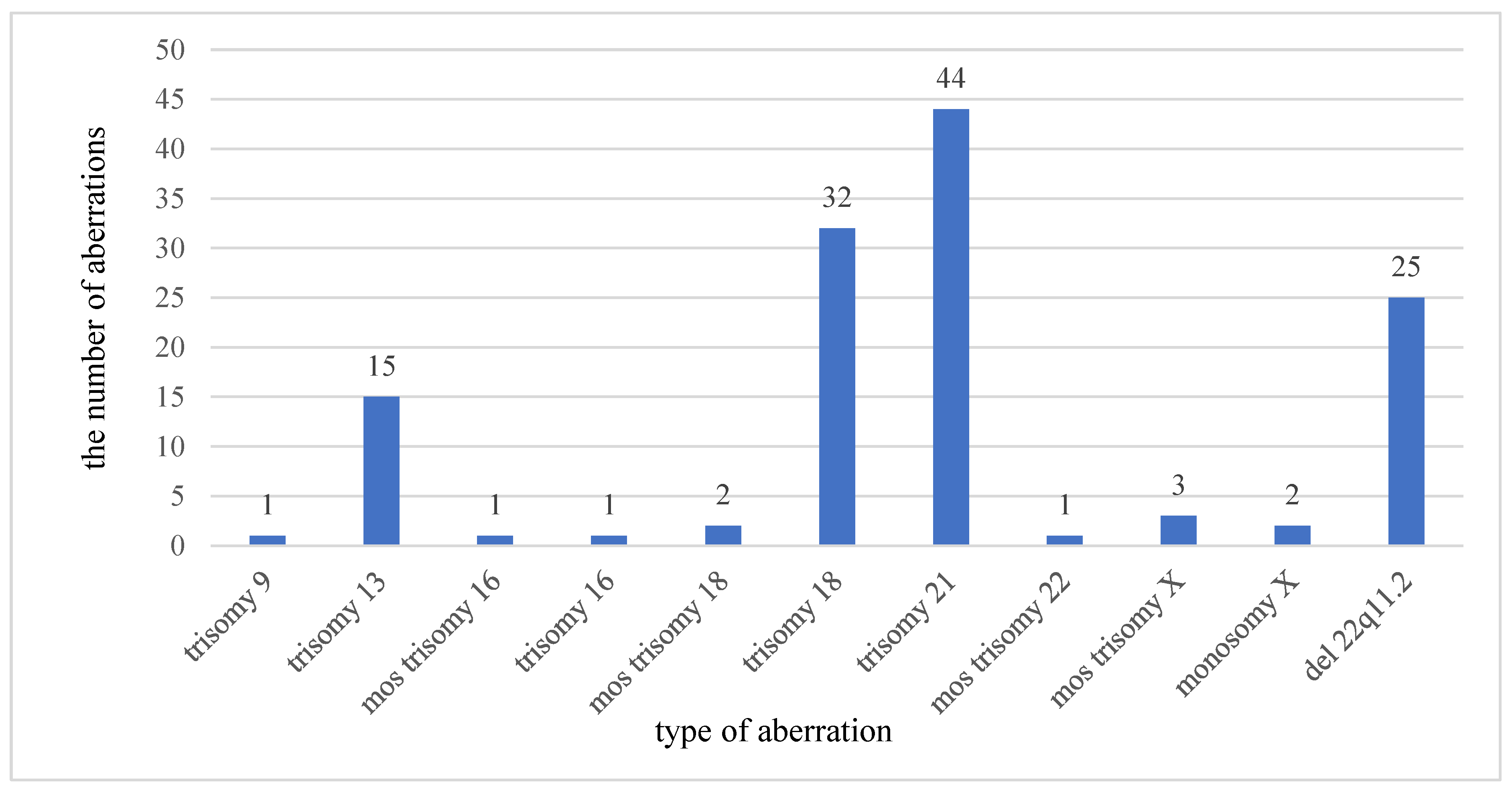
3. Results
4. Discussion
Author Contributions
Funding
Institutional Review Board Statement
Informed Consent Statement
Data Availability Statement
Conflicts of Interest
References
- Xia, Y.; Yang, Y.; Huang, S.; Wu, Y.; Li, P.; Zhuang, J. Clinical application of chromosomal microarray analysis for the prenatal diagnosis of chromosomal abnormalities and copy number variations in fetuses with congenital heart disease. Prenat. Diagn. 2018, 38, 406–413. [Google Scholar] [CrossRef]
- Costain, G.; Silversides, C.K.; Bassett, A.S. The importance of copy number variation in congenital heart disease. NPJ Genom. Med. 2016, 1, 16031. [Google Scholar] [CrossRef] [PubMed] [Green Version]
- Stosic, M.; Levy, B.; Wapner, R. The Use of Chromosomal Microarray Analysis in Prenatal Diagnosis. Obstet. Gynecol. Clin. North Am. 2018, 45, 55–68. [Google Scholar] [CrossRef] [PubMed]
- Robson, S.C.; Chitty, L.S.; Morris, S.; Verhoef, T.; Ambler, G.; Wellesley, D.G.; Graham, R.; Leader, C.; Fisher, J.; Crolla, J.A. Evaluation of Array Comparative Genomic Hybridisation in Prenatal Diagnosis of Fetal Anomalies: A Multicentre Cohort Study with Cost Analysis and Assessment of Patient, Health Professional and Commissioner Preferences for Array Comparative Genomic Hybridisation; NIHR Journals Library: Southampton, UK, 2017. [Google Scholar]
- Mademont-Soler, I.; Morales, C.; Soler, A.; Martínez-Crespo, J.M.; Shen, Y.; Margarit, E.; Clusellas, N.; Obón, M.; Wu, B.L.; Sánchez, A. Prenatal diagnosis of chromosomal abnormalities in fetuses with abnormal cardiac ultrasound findings: Evaluation of chromosomal microarray-based analysis. Ultrasound Obstet. Gynecol. 2013, 41, 375–382. [Google Scholar] [CrossRef] [PubMed]
- Fiorentino, F.; Napoletano, S.; Caiazzo, F.; Sessa, M.; Bono, S.; Spizzichino, L.; Gordon, A.; Nuccitelli, A.; Rizzo, G.; Baldi, M. Chromosomal microarray analysis as a first-line test in pregnancies with a priori low risk for the detection of submicroscopic chromosomal abnormalities. Eur. J. Hum. Genet. 2013, 21, 725–730. [Google Scholar] [CrossRef] [PubMed]
- Committee on Genetics and the Society for Maternal-Fetal Medicine. Committee Opinion No. 682: Microarrays and Next-Generation Sequencing Technology: The Use of Advanced Genetic Diagnostic Tools in Obstetrics and Gynecology. Obstet. Gynecol. 2016, 128, e262–e268. [Google Scholar] [CrossRef]
- Armour, C.M.; Dougan, S.D.; Brock, J.A.; Chari, R.; Chodirker, B.N.; DeBie, I.; Evans, J.A.; Gibson, W.T.; Kolomietz, E.; Nelson, T.N.; et al. Practice guideline: Joint CCMG-SOGC recommendations for the use of chromosomal microarray analysis for prenatal diagnosis and assessment of fetal loss in Canada. J. Med Genet. 2018, 55, 215–221. [Google Scholar] [CrossRef]
- Silva, M.; de Leeuw, N.; Mann, K.; Schuring-Blom, H.; Morgan, S.; Giardino, D.; Rack, K.; Hastings, R. European guidelines for constitutional cytogenomic analysis. Eur. J. Hum. Genet. 2019, 27, 1–16. [Google Scholar] [CrossRef]
- Hay, S.B.; Sahoo, T.; Travis, M.K.; Hovanes, K.; Dzidic, N.; Doherty, C.; Strecker, M.N. ACOG and SMFM guidelines for prenatal diagnosis: Is karyotyping really sufficient? Prenat. Diagn. 2018, 38, 184–189. [Google Scholar] [CrossRef] [Green Version]
- Brady, P.D.; Delle Chiaie, B.; Christenhusz, G.; Dierickx, K.; Van Den Bogaert, K.; Menten, B.; Janssens, S.; Defoort, P.; Roets, E.; Sleurs, E.; et al. A prospective study of the clinical utility of prenatal chromosomal microarray analysis in fetuses with ultrasound abnormalities and an exploration of a framework for reporting unclassified variants and risk factors. Genet. Med. 2014, 16, 469–476. [Google Scholar] [CrossRef] [Green Version]
- Dugoff, L.; Norton, M.E.; Kuller, J.A. The use of chromosomal microarray for prenatal diagnosis. Am. J. Obstet. Gynecol. 2016, 215, B2–B9. [Google Scholar] [CrossRef] [Green Version]
- Vanakker, O.; Vilain, C.; Janssens, K.; Van der Aa, N.; Smits, G.; Bandelier, C.; Blaumeiser, B.; Bulk, S.; Caberg, J.H.; De Leener, A.; et al. Implementation of genomic arrays in prenatal diagnosis: The Belgian approach to meet the challenges. Eur. J. Med. Genet. 2014, 57, 151–156. [Google Scholar] [CrossRef] [PubMed]
- Tonni, G.; Palmisano, M.; Perez Zamarian, A.C.; Rabachini Caetano, A.C.; Santana, E.F.M.; Peixoto, A.B.; Armbruster-Moraes, E.; Ruano, R.; Araujo Júnior, E. Phenotype to genotype characterization by array-comparative genomic hydridization (a-CGH) in case of fetal malformations: A systematic review. Taiwan. J. Obstet. Gynecol. 2019, 58, 15–28. [Google Scholar] [CrossRef]
- Song, T.; Wan, S.; Li, Y.; Xu, Y.; Dang, Y.; Zheng, Y.; Li, C.; Zheng, J.; Chen, B.; Zhang, J. Detection of copy number variants using chromosomal microarray analysis for the prenatal diagnosis of congenital heart defects with normal karyotype. J. Clin. Lab. Anal. 2019, 33, e22630. [Google Scholar] [CrossRef] [Green Version]
- Lee, M.Y.; Won, H.S.; Han, Y.J.; Ryu, H.M.; Lee, D.E.; Jeong, B.D. Clinical value of chromosomal microarray analysis in prenatally diagnosed dextro-transposition of the great arteries. J. Matern. Fetal Neonatal Med. 2020, 33, 1480–1485. [Google Scholar] [CrossRef] [PubMed]
- Yan, Y.; Wu, Q.; Zhang, L.; Wang, X.; Dan, S.; Deng, D.; Sun, L.; Yao, L.; Ma, Y.; Wang, L. Detection of submicroscopic chromosomal aberrations by array-based comparative genomic hybridization in fetuses with congenital heart disease. Ultrasound Obstet. Gynecol. 2014, 43, 404–412. [Google Scholar] [CrossRef] [Green Version]
- Wu, X.L.; Li, R.; Fu, F.; Pan, M.; Han, J.; Yang, X.; Zhang, Y.L.; Li, F.T.; Liao, C. Chromosome microarray analysis in the investigation of children with congenital heart disease. BMC Pediatr. 2017, 17, 117. [Google Scholar] [CrossRef] [Green Version]
- Oneda, B.; Rauch, A. Microarrays in prenatal diagnosis. Best Pract. Res. Clin. Obstet. Gynaecol. 2017, 42, 53–63. [Google Scholar] [CrossRef]
- Meyer, R.E.; Liu, G.; Gilboa, S.M.; Ethen, M.K.; Aylsworth, A.S.; Powell, C.M.; Flood, T.J.; Mai, C.T.; Wang, Y.; Canfield, M.A.; et al. Survival of children with trisomy 13 and trisomy 18: A multi-state population-based study. Am. J. Med. Genet. A 2016, 170, 825–837. [Google Scholar] [CrossRef] [Green Version]
- Kaneko, Y.; Kobayashi, J.; Yamamoto, Y.; Yoda, H.; Kanetaka, Y.; Nakajima, Y.; Endo, D.; Tsuchiya, K.; Sato, H.; Kawakami, T. Intensive cardiac management in patients with trisomy 13 or trisomy 18. Am. J. Med. Genet. A 2008, 146, 1372–1380. [Google Scholar] [CrossRef] [PubMed]
- Martin, C.L.; Kirkpatrick, B.E.; Ledbetter, D.H. Copy number variants, aneuploidies, and human disease. Clin. Perinatol. 2015, 42, 227–242. [Google Scholar] [CrossRef] [PubMed] [Green Version]
- Morales-Demori, R. Congenital heart disease and cardiac procedural outcomes in patients with trisomy 21 and Turner syndrome. Congenit. heart Dis. 2017, 12, 820–827. [Google Scholar] [CrossRef]
- Jansen, F.A.; Blumenfeld, Y.J.; Fisher, A.; Cobben, J.M.; Odibo, A.O.; Borrell, A.; Haak, M.C. Array comparative genomic hybridization and fetal congenital heart defects: A systematic review and meta-analysis. Ultrasound Obstet. Gynecol. 2015, 45, 27–35. [Google Scholar] [CrossRef] [Green Version]
- Levyv, B.; Wapner, R. Prenatal diagnosis by chromosomal microarray analysis. Fertil. Steril. 2018, 109, 201–212. [Google Scholar] [CrossRef] [PubMed] [Green Version]
- Ciaccio, C.; Fontana, L.; Milani, D.; Tabano, S.; Miozzo, M.; Esposito, S. Fragile X syndrome: A review of clinical and molecular diagnoses. Ital. J. Pediatr. 2017, 43, 39. [Google Scholar] [CrossRef] [PubMed] [Green Version]
- Lee, C.N.; Lin, S.Y.; Lin, C.H.; Shih, J.C.; Lin, T.H.; Su, Y.N. Clinical utility of array comparative genomic hybridisation for prenatal diagnosis: A cohort study of 3171 pregnancies. BJOG 2012, 119, 614–625. [Google Scholar] [CrossRef]
- Fiorentino, F.; Caiazzo, F.; Napolitano, S.; Spizzichino, L.; Bono, S.; Sessa, M.; Nuccitelli, A.; Biricik, A.; Gordon, A.; Rizzo, G.; et al. Introducing array comparative genomic hybridization into routine prenatal diagnosis practice: A prospective study on over 1000 consecutive clinical cases. Prenat. Diagn. 2011, 31, 1270–1282. [Google Scholar] [CrossRef]
- Breman, A.; Pursley, A.N.; Hixson, P.; Bi, W.; Ward, P.; Bacino, C.A.; Shaw, C.; Lupski, J.R.; Beaudet, A.; Patel, A.; et al. Prenatal chromosomal microarray analysis in a diagnostic laboratory; experience with >1000 cases and review of the literature. Prenat. Diagn. 2012, 32, 351–361. [Google Scholar] [CrossRef] [PubMed]
- Rooryck, C.; Toutain, J.; Cailley, D.; Bouron, J.; Horovitz, J.; Lacombe, D.; Arveiler, B.; Saura, R. Prenatal diagnosis using array-CGH: A French experience. Eur. J. Med. Genet. 2013, 56, 341–345. [Google Scholar] [CrossRef] [PubMed]
- Jinxiu, L.; Shuimei, L.; Ming, X.; Jonathan, L.C.; Xiangju, L.; Wenyuan, D. Wiedemann-steiner syndrome with a de novo mutation in KMT2A: A case report. Medicine 2020, 99, e19813. [Google Scholar] [CrossRef] [PubMed]
- Reuter, M.S.; Jobling, R.; Chaturvedi, R.R.; Manshaei, R.; Costain, G.; Heung, T.; Curtis, M.; Hosseini, S.M.; Liston, E.; Lowther, C.; et al. Haploinsufficiency of vascular endothelial growth factor related signaling genes is associated with tetralogy of Fallot. Genet. Med. 2019, 21, 1001–1007. [Google Scholar] [CrossRef] [PubMed] [Green Version]
- Xie, H.M.; Werner, P.; Stambolian, D.; Bailey-Wilson, J.E.; Hakonarson, H.; White, P.S.; Taylor, D.M.; Goldmuntz, E. Rare copy number variants in patients with congenital conotruncal heart defects. Birth defects Res. 2017, 109, 271–295. [Google Scholar] [CrossRef] [PubMed] [Green Version]
- Chen, L.; Wang, L.; Yin, D.; Zeng, Y.; Tang, F.; Wang, J. Influence of the detection of parent-of-origin on the pregnancy outcomes of fetuses with copy number variation of unknown significance. Sci. Rep. 2020, 10, 8864. [Google Scholar] [CrossRef]
- Gajecka, M.; Saitta, S.C.; Gentles, A.J.; Campbell, L.; Ciprero, K.; Geiger, E.; Catherwood, A.; Rosenfeld, J.A.; Shaikh, T.; Shaffer, L.G. Recurrent interstitial 1p36 deletions: Evidence for germline mosaicism and complex rearrangement breakpoints. Am. J. Med. Genet. A 2010, 152, 3074–3083. [Google Scholar] [CrossRef] [Green Version]
- Aarabi, M.; Sniezek, O.; Jiang, H.; Saller, D.N.; Bellissimo, D.; Yatsenko, S.A.; Rajkovic, A. Importance of complete phenotyping in prenatal whole exome sequencing. Hum. Genet. 2018, 137, 175–181. [Google Scholar] [CrossRef]
- Downie, L.; Halliday, J.L.; Burt, R.A.; Lunke, S.; Lynch, E.; Martyn, M.; Poulakis, Z.; Gaff, C.; Sung, V.; Wake, M.; et al. A protocol for whole-exome sequencing in newborns with congenital deafness: A prospective population-based cohort. BMJ Paediatr. Open 2017, 1, e000119. [Google Scholar] [CrossRef] [PubMed]
- Jelin, A.C.; Vora, N. Whole Exome Sequencing: Applications in Prenatal Genetics. Obstet. Gynecol. Clin. North Am. 2018, 45, 69–81. [Google Scholar] [CrossRef] [PubMed]
- Yang, Y.; Muzny, D.M.; Reid, J.G.; Bainbridge, M.N.; Willis, A.; Ward, P.A.; Braxton, A.; Beuten, J.; Xia, F.; Niu, Z.; et al. Clinical whole-exome sequencing for the diagnosis of mendelian disorders. N. Engl. J. Med. 2013, 369, 1502–1511. [Google Scholar] [CrossRef] [Green Version]
- Kalynchuk, E.J.; Althouse, A.; Parker, L.S.; Saller, D.N., Jr.; Rajkovic, A. Prenatal whole-exome sequencing: Parental attitudes. Prenat. Diagn. 2015, 35, 1030–1036. [Google Scholar] [CrossRef]
- Vora, N.L.; Powell, B.; Brandt, A.; Strande, N.; Hardisty, E.; Gilmore, K.; Foreman, A.K.M.; Wilhelmsen, K.; Bizon, C.; Reilly, J.; et al. Prenatal exome sequencing in anomalous fetuses: New opportunities and challenges. Genet. Med. 2017, 19, 1207–1216. [Google Scholar] [CrossRef] [Green Version]
- Drury, S.; Williams, H.; Trump, N.; Boustred, C.; GOSGene; Lench, N.; Scott, R.H.; Chitty, L.S. Exome sequencing for prenatal diagnosis of fetuses with sonographic abnormalities. Prenat. Diagn. 2015, 35, 1010–1017. [Google Scholar] [CrossRef] [PubMed] [Green Version]


| Aneuploidy | Heart Defects in Ultrasound |
|---|---|
| Trisomy 21 | AVSD, VSD, TOF, ASD |
| Trisomy 18 | VSD, AVSD, DORV |
| Trisomy 13 | VSD, ASD |
| Monosomy X | VSD |
| Patient | Prenatal Diagnosis | Aberration (Inheritance—If It Has Been Identified) | Size |
|---|---|---|---|
| 1166 | Cleft palate, VSD, foot deformation | 1p36.33p36.22(779733_9620926)x1,5p15.33(22149_2274755)x3 | 8.8 Mb; 2.2 Mb |
| 254 | cardiomegaly | 1q21.1q44(142491666_246928498)x3 | 104 Mb |
| 2054 | HLHS | 2p25.3(21191_3062258)x1,12q24.13q24.33(113023613_133773393)x3 | 3 Mb; 21 Mb |
| 1220 | TOF | 1q42.12q44(226703815_249203359)x3, 9q34.3(138907844_141018976)x1 | 22.2 Mb; 2.1 Mb |
| 653 | VSD | 3p22.2(37646228_38961056)x1,3q24q25.32(145448788_158594702)x1 | 1.3 Mb; 13 Mb |
| 1214 | AVSD | 3p24.1p22.3(27228808_33971880)x1 | 6.7 Mb |
| 551 | VSD | 5p15.33p12(22149_45362363)x3 | 45 Mb |
| 13 | TOF | 6q25.3q26(156813910_162033274)x3 dn | 5.2 Mb |
| 322 | VSD | 6q26q27(163436214_170847447)x1 dn | 7.4 Mb |
| 589 | ASD | 7p14.3p14.1(31773017_42738664)x1 | 11 Mb |
| 1478 | VSD | 8p23.1(7113656_12454089)x1 | 5.34 Mb |
| 784 | AVSD, TOF | 8p23.1p21.3(6224261_21242145)x1 | 15 Mb |
| 1258 | Cleft palate, AVSD | 8p23.3p21.2(191605_24918147)x3,9p24.3q21.32(204090_84386182)x3 | 27 Mb; 84 Mb |
| 1006 | AVSD | 8p23.3p23.1(191605_12454089)x1 mat, 18p11.32p11.31(149089_7094765)x3 mat | 12 mb; 6 Mb |
| 983 | VSD | 10q11.22q26.3(46426869_135404550)x3 | 89 Mb |
| 1262 | ASD | 11p15.5p11.2(113082_46371104)x3 | 46 Mb |
| 173 | VSD, CoAo | 12p13.33p11.1(100698_34647463)x3 | 34.5 Mb |
| 1148 | CoAo | 13q21.1q21.32(57950814_67755631)x3 dn | 9.8 Mb |
| 1065 | IUGR, VSD, ARSA | 14q24.3q32.31(79087813_102919927)x1 | 23.8 Mb |
| 1306 | VSD | 15q11.1q11.2(20686203_23586302)x1,(18)x3 | 2.9 Mb; 80.7 Mb |
| 1995 | cardiomegaly | 16p11.2q24.3(34202297_90252496)x3 | 56 Mb |
| 404 | HLHS | 17p13.3p13.2(1656_5534353)x1 | 5.53 Mb |
| Patient | Prenatal Diagnosis | Aberration (Inheritance—If It Has Been Identified) | Size |
|---|---|---|---|
| 383 | ARSA | 1q32.1(197684386_198909224)x3 mat,3p26.3(69430_2062244)x1 mat | 1.22 Mb; 1.99 Mb |
| 1009 | HLHS | 2p13.1(73763801_74194368)x3 pat | 430 kb |
| 698 | VSD, Dandy-Walker syndrome | 2p15(61632727_62017908)x3 pat | 385 kb |
| 986 | AVSD | 2p16.3(50880241_50949412)x1 (Additionally, this patient had trisomy of chromosome 13) | 64 kb |
| 1736 | VSD | 3p12.3(77192875_79219598)x1 pat | 2 Mb |
| 608 | AVSD | 4p15.32(16064173_16813206)x3 mat | 989 kb |
| 395 | AVSD | 5q35.3(177068821_178058571)x3 pat | 900 kb |
| 447 | HLHS | 5q35.3(177956887_178917587)x3 mat | 1 Mb |
| 2155 | VSD, ARSA | 8p23.1(11550005_11558331)x3 | 8.3 kb |
| 978 | mitral regurgitation | 11q23.3(118363939_118367204)x3 | 3.26 kb |
| 888 | AVSD | 14q23.3q32.33(67146824_107287708)x3 dn, Xp21.1(31699053_31805802)x1 dn | 40 Mb; 106 kb |
| 1199 | AVSD, HLHS | 16p11.2(28318123_29182200)x3 pat | 864 kb |
| 1122 | AVSD | 17p12(14111754_14423151)x3,17p12(14911841_15322595)x3 | 311 kb; 411 kb |
| 948 | HLHS | 17q12(34652173_36290311)x1 pat | 1.68 Mb |
| 851 | HLHS | 18q11.1(18542080_18672140)x1 pat | 130 kb |
| 1658 | VSD | Xp22.2(11600766_12080374)x3 mat | 479 kb |
| 2195 | VSD | Xq28(153324080_153362472)x3 dn | 30 kb |
| 5528 | TOF | 5q35.3(179950554_180152423)x1 mat | 202 kb |
| Patient | Prenatal Diagnosis | Locus | Size | Gene | Associated Anomalies |
|---|---|---|---|---|---|
| 584 | cardiac ectopy | 13q13.3(37145323_37351415)x3 | 206 kb | SERTM1 | Serine Rich and Transmembrane Domain Containing 1 protein with high expression in cancer tissue. |
| 674 | tricuspid valve regurgitation | 2p16.3(48059806_48500445)x3 | 440 kb | FBXO11 | FBXO11 mutations were also identified in human cancers, such as colon, lung, ovary, and head and neck tumors. In mice, a homozygous mutation of FBXO11 results in cleft palate defects, facial clefting, and dysmorphic features |
| 765 | atrioventricular septal defect (AVSD) | 11q22.1(101436248_101756583)x3 | 320 kb | ex 1 TRPC6 | TRPC6 encodes Transient Receptor Potential Cation Channel Subfamily C Member 6. Mutations in the TRPC6 cation channel causes familial focal segmental glomerulosclerosis. TRPC6 is a known factor in cardiac hypertrophy and heart failure. |
| 1045 | Ebstein Syndrome | 21q11.2(15824276_16137741)x3 | 313 kb | SAMSN1 | SAMSN1 is a member of a novel gene family of putative adaptors and scaffold proteins containing SH3 and SAM (sterile α motif) domains. SAMSN1 act as a cytoplasmic adaptor to mediate a signaling pathway |
| 1093 | aberrant right subclavian artery (ARSA) | 13q31.3(92065636_92299097)x3 | 233 kb | ex 2 GCP5 | This gene has been tested for association to diseases (Colitis, Ulcerative; Crohn Disease; Lymphoma). Proteins are expected to have molecular function (heparan sulfate proteoglycan binding) and to localize in various compartments (integral to plasma membrane) extracellular space, anchored to membrane, extracellular region, proteinaceous extracellular matrix) |
| 1165 | common arterial trunk (CAT) | 9q21.32q21.33(86825588_87161409)x3 | 335 kb | SLC28A3 | SLC28A3 encodes Solute Carrier Family 28 Member 3, which plays a role in multiple cellular processes, including neurotransmission, vascular tone, adenosine concentration in the vicinity of cell surface receptors, and transport and metabolism of nucleoside drugs |
| 1278 | abnormal heart rotation | 1p36.32(2633351_3161118)x3 | 522 kb | ex 1-3 PRDM16 | PRDM16 acts as a transcription coregulator that controls the development of brown adipocytes in brown adipose tissue. The protein encoded by this gene is a zinc finger transcription factor. PRDM16 controls the cell fate between muscle and brown fat cells |
| 1280 | atrioventricular septal defect (AVSD) | 2q14.2(121549137_121659393)x3 | 110 kb | GLI2 | Heterozygous mutation in the GLI2 gene was described in patients with Culler–Jones syndrome |
| 2093 | Ebstein Syndrome | 10q26.12(122509983_122668106)x3 | 158 kb | WDR11 | WDR11 is a member of the WD repeat-containing protein family. Heterozygous mutation in the WDR11 gene was described in patients with congenital idiopathic hypogonadotropic hypogonadism (IHH). |
Publisher’s Note: MDPI stays neutral with regard to jurisdictional claims in published maps and institutional affiliations. |
© 2021 by the authors. Licensee MDPI, Basel, Switzerland. This article is an open access article distributed under the terms and conditions of the Creative Commons Attribution (CC BY) license (https://creativecommons.org/licenses/by/4.0/).
Share and Cite
Kowalczyk, K.; Bartnik-Głaska, M.; Smyk, M.; Plaskota, I.; Bernaciak, J.; Kędzior, M.; Wiśniowiecka-Kowalnik, B.; Jakubów-Durska, K.; Braun-Walicka, N.; Barczyk, A.; et al. Prenatal Diagnosis by Array Comparative Genomic Hybridization in Fetuses with Cardiac Abnormalities. Genes 2021, 12, 2021. https://doi.org/10.3390/genes12122021
Kowalczyk K, Bartnik-Głaska M, Smyk M, Plaskota I, Bernaciak J, Kędzior M, Wiśniowiecka-Kowalnik B, Jakubów-Durska K, Braun-Walicka N, Barczyk A, et al. Prenatal Diagnosis by Array Comparative Genomic Hybridization in Fetuses with Cardiac Abnormalities. Genes. 2021; 12(12):2021. https://doi.org/10.3390/genes12122021
Chicago/Turabian StyleKowalczyk, Katarzyna, Magdalena Bartnik-Głaska, Marta Smyk, Izabela Plaskota, Joanna Bernaciak, Marta Kędzior, Barbara Wiśniowiecka-Kowalnik, Krystyna Jakubów-Durska, Natalia Braun-Walicka, Artur Barczyk, and et al. 2021. "Prenatal Diagnosis by Array Comparative Genomic Hybridization in Fetuses with Cardiac Abnormalities" Genes 12, no. 12: 2021. https://doi.org/10.3390/genes12122021
APA StyleKowalczyk, K., Bartnik-Głaska, M., Smyk, M., Plaskota, I., Bernaciak, J., Kędzior, M., Wiśniowiecka-Kowalnik, B., Jakubów-Durska, K., Braun-Walicka, N., Barczyk, A., Geremek, M., Castañeda, J., Kutkowska-Kaźmierczak, A., Własienko, P., Dębska, M., Kucińska-Chahwan, A., Roszkowski, T., Kozłowski, S., Mikulska, B., ... Nowakowska, B. A. (2021). Prenatal Diagnosis by Array Comparative Genomic Hybridization in Fetuses with Cardiac Abnormalities. Genes, 12(12), 2021. https://doi.org/10.3390/genes12122021