Review

Atypical NF1 Microdeletions: Challenges and Opportunities for Genotype/Phenotype Correlations in Patients with Large NF1 Deletions

by
Hildegard Kehrer-Sawatzki
1,*,
Ute Wahlländer
2,
David N. Cooper
3 and
Victor-Felix Mautner
4
1
Institute of Human Genetics, University of Ulm, 89081 Ulm, Germany
2
Kliniken des Bezirks Oberbayern (KBO), Children Clinical Center Munich, 81377 Munich, Germany
3
Institute of Medical Genetics, Cardiff University, Heath Park, Cardiff CF14 4XN, UK
4
Department of Neurology, University Hospital Hamburg Eppendorf, 20246 Hamburg, Germany
*
Author to whom correspondence should be addressed.
Genes 2021, 12(10), 1639; https://doi.org/10.3390/genes12101639
Submission received: 13 September 2021 / Revised: 30 September 2021 / Accepted: 14 October 2021 / Published: 19 October 2021
(This article belongs to the Section Human Genomics and Genetic Diseases)

Abstract

:
Patients with neurofibromatosis type 1 (NF1) and type 1 NF1 deletions often exhibit more severe clinical manifestations than patients with intragenic NF1 gene mutations, including facial dysmorphic features, overgrowth, severe global developmental delay, severe autistic symptoms and considerably reduced cognitive abilities, all of which are detectable from a very young age. Type 1 NF1 deletions encompass 1.4 Mb and are associated with the loss of 14 protein-coding genes, including NF1 and SUZ12. Atypical NF1 deletions, which do not encompass all 14 protein-coding genes located within the type 1 NF1 deletion region, have the potential to contribute to the delineation of the genotype/phenotype relationship in patients with NF1 microdeletions. Here, we review all atypical NF1 deletions reported to date as well as the clinical phenotype observed in the patients concerned. We compare these findings with those of a newly identified atypical NF1 deletion of 698 kb which, in addition to the NF1 gene, includes five genes located centromeric to NF1. The atypical NF1 deletion in this patient does not include the SUZ12 gene but does encompass CRLF3. Comparative analysis of such atypical NF1 deletions suggests that SUZ12 hemizygosity is likely to contribute significantly to the reduced cognitive abilities, severe global developmental delay and facial dysmorphisms observed in patients with type 1 NF1 deletions.

1. Introduction

It has been estimated that 5–11% of all NF1 patients have large deletions which include the NF1 gene and its flanking regions at 17q11.2 [1,2,3,4]. These so called ‘NF1 microdeletions’ are frequently associated with severe clinical manifestations, giving rise to the NF1 microdeletion syndrome (MIM#613675), which occurs with an estimated incidence of 1:60,000 (reviewed by [5]). Four types of large NF1 deletion (type 1, 2 and 3 and atypical) have been identified, which are distinguishable in terms of their frequency, size and breakpoint location, by the number of genes deleted and by the frequency of somatic mosaicism with normal cells without the deletion. Most frequent are the type 1 NF1 deletions which encompass 1.4 Mb and include 14 protein-coding genes as well as five microRNA genes [6,7,8]. Approximately 70–80% of all large NF1 deletions are of type 1, and in most instances, they occur as germline deletions that are present in all cells of the affected patients [9,10]. The vast majority of type 1 NF1 deletions result from interchromosomal non-allelic homologous recombination (NAHR) during maternal meiosis [11,12]. NAHR causing type 1 NF1 deletions occurs between the low-copy repeats NF1-REPa and NF1-REPc. Most type 1 deletions exhibit recurrent breakpoints within the NF1-REPs located in two NAHR hotspots [13,14,15,16,17,18].
Much less frequent than type 1 deletions are those of type 2; approximately 10–15% of large NF1 deletions are type 2 [9]. Type 2 deletions encompass 1.2 Mb and are associated with the loss of 13 protein-coding genes. In the majority of cases, they are mediated by NAHR and their breakpoints are located within SUZ12 and its pseudogene SUZ12P [19,20]. Type 2 NF1 deletions are frequently of postzygotic origin, mediated by mitotic NAHR, and hence may occur as mosaic deletions alongside normal cells [19,21,22,23]. Type 3 NF1 deletions are very rare: they occur in only 1–4% of all patients with large NF1 deletions. Type 3 deletions encompass 1.0 Mb and are caused by NAHR between NF1-REPb and NF1-REPc [24,25,26].
In contrast to type 1, 2 and 3 NF1 deletions, atypical large NF1 deletions do not exhibit recurrent breakpoints and are quite heterogeneous in terms of their size and the number of genes located within the deleted region [1,4,6,7,20,25,27,28,29,30,31,32,33,34,35,36,37,38,39,40,41,42,43,44,45,46,47,48,49,50]. It has been estimated that 8–10% of all large NF1 deletions are atypical [9,25]. Atypical NF1 deletions may occur as germline mutations but can also be of postzygotic origin [43]. Atypical NF1 deletions are not only heterogeneous in terms of their length but also in terms of the underlying mutational mechanisms including DNA double strand break repair, aberrant replication and retrotransposon-mediated mechanisms [43]. So far, the clinical phenotype associated with NF1 microdeletions has mostly been studied in patients with type 1 NF1 deletions. These patients frequently exhibit more severe clinical manifestations of NF1 as well as features that are not frequently seen in patients with intragenic NF1 mutations. The type 1 NF1 deletion-associated phenotype includes facial dysmorphic features, severe global developmental delay, significantly lower full-scale IQ scores, higher neurofibroma burden and an increased risk of malignant peripheral nerve sheath tumours (MPNSTs) as compared to patients with intragenic NF1 mutations [5,51,52,53,54]. It has been postulated that some of the genes located within the type 1 NF1 microdeletion region and co-deleted with NF1 exert an influence on the clinical manifestation of the disease in NF1 deletion patients. Atypical NF1 deletions, which encompass only a subset of the 14 protein-coding genes located within the type 1 NF1 microdeletion region, are of particular interest since the phenotypes expressed by affected patients may facilitate genotype/phenotype correlations in patients with different types of NF1 microdeletion. However, as yet, only a few patients with atypical NF1 deletions encompassing a subset of the 14 protein-coding genes have been clinically characterised in any detail. Here, we report on a young patient with an atypical NF1 deletion that encompasses only 9 of the 14 genes present in the type 1 NF1 microdeletion region. The clinical phenotype of this patient is potentially informative in the context of genotype/phenotype relationships and the putative modifier role of genes located within the NF1 microdeletion region. Further, we compare the extent of the deletion in this patient with all atypical NF1 deletions published to date and the clinical findings in patients with atypical NF1 deletions that encompass only a subset of the 14 protein-coding genes located within the type 1 NF1 microdeletion interval.

2. Patient and Methods

2.1. Genetic Analysis

Male patient 310221 was genetically investigated at the age of 4 years because he exhibited multiple café-au-lait spots, axillary freckling and developmental delay. An atypical NF1 deletion was detected in patient 310221 by means of CytoScan™ HD array analysis (Affymetrix, Santa Clara, CA, USA). Genomic DNA isolated from the blood of this patient was digested, ligated, amplified, fragmented, labelled and hybridised on the array according the manufacturer’s instructions. The raw data were analysed by means of the Chromosome Analysis Suite software (ChAS, V.3.1.0.15) (Affymetrix, Santa Clara, CA, USA). The deletion was confirmed by multiplex ligation-dependent probe amplification (MLPA) analysis using the SALSA MLPA Probemix P122 (version D2) NF1-area and Probemix P081-D1 NF1 mix 1 as well as P082-C2 NF1 mix 2, (MRC-Holland, Amsterdam, the Netherlands). For MLPA analysis, 100 ng denatured genomic DNA isolated from the blood of the patient was used. MLPA was performed according to the instructions provided by MRC-Holland. MLPA fragments were separated using an ABI Prism 3100 Genetic Analyzer (Applied Biosystems, Foster City, CA, USA). MLPA data analysis was performed with the Coffalyser.Net™ v.1 software (MRC-Holland, Amsterdam, the Netherlands). A reduction of ~0.5 in the peak area values was considered indicative of a deletion of the genomic region represented by the corresponding MLPA probes.
Written informed consent was obtained from the parents of the patient as his legal guardians/representatives. The study was approved by the respective institutional review boards in Hamburg and München and performed in accordance with the Helsinki Declaration and its later amendments.

2.2. Investigation of the Cognitive Abilities of Patient 310221

From the age of 3.5 years onwards until the age of 7, the male patient was clinically investigated every 6–9 months at the Children Clinical Center of the Kliniken des Bezirks Oberbayern (KBO) in München, Germany, involving multidimensional field diagnostics in social paediatrics as well as a special consultation for patients with NF1.
The tests performed to investigate the patient’s cognitive abilities and development were the Münchener Funktionelle Entwicklungsdiagnostik for the second and third years of life (MFED) [55,56], the Wechsler Preschool and Primary Scale of Intelligence (WPPSI-III and WPPSI-IV) [57], the Kaufman Assessment Battery for Children (KABC-II) [58], the Reynell Developmental Language Scales (RDLS) [59], the language development tests SETK2 and SETK3–5 [60,61], the Active Vocabulary Test (AWST-R) for children aged 3–5 years [62] and the Beery–Buktenica Developmental Test of Visual–Motor Integration (Beery VMI) [63].

3. Results

3.1. Clinical Investigation of Patient 310221

The male patient was born spontaneously at term as the second child of healthy, unrelated parents. At birth, his weight was 3520 g (P 56), his length was 49 cm (P 11) and his head circumference was 35 cm (P 42). Results of newborn screening as well as hearing tests were normal. Muscular hypotonia or feeding problems were absent during the neonatal period. The patient was able to sit on his own by the age of 6–7 months, started to crawl at the age of 10 months and to walk at the age of 14 months in a timely manner. However, developmental delay affecting primarily speech and coordination was suspected when the patient was approximately 2 years old. He spoke his first specific words at the age of 18–24 months and sentences comprising two words at the age of 2 years. His progress in acquiring vocabulary was slow. The patient had multiple café-au-lait spots over 5 mm in diameter, disseminated mainly on his trunk and not restricted to one body segment. Additionally, axillary freckling was noted on both sides and hence the patient fulfilled the NIH diagnostic criteria for NF1. Nevus anemicus was observed on his chest and upper arms. From the age of 3.5 years onwards until the age of 7 years, the boy was clinically investigated every 6–9 months. Neither neurofibromas nor xanthogranulomas were detected at the age of 3.5 years. He had mild pectus excavatum and pes planovalgus of both feet. Generalised muscular hypotonia was diagnosed at the age of 3.5 years, which persisted in later years. The patient was investigated by cranial MRI at the age of 3.5 and 7 years, which did not disclose intracranial tumours, inflammation, structural brain abnormalities, macrolesions or foci of abnormal signal intensity (FASI). Neither thickening of the optic nerves nor optic gliomas were detected by cranial MRI. The subarachnoid spaces were of normal size. Audiological evaluation of the patient indicated normal hearing.
At the age of 3.5 and 4.6 years, the patient was investigated by an ophthalmologist using optical coherence tomography (OCT) and slit lamp examination. Neither Lisch nodules nor choroidal anomalies were detected. The patient had macrocephaly, and from the age of 4.6 years onwards, small body size was noted, which persisted in later years (Supplementary Table S1).
At the age of 5.7 years, spinal MRI was performed. Neither intraspinal nor paravertebral tumours were detected. Mild scoliosis was noted as well as a kyphosis of the pelvic spine. Osseous lesions such as sphenoid wing dysplasia and anterolateral bowing of the tibia were not observed.
Abdominal ultrasound investigation performed at the age of 6.1 years did not indicate internal tumours. However, at this age, a small cutaneous neurofibroma was detected on his chest. The patient had mild hypertelorism, thinning of the eyebrows at the inner roots and a slightly low hairline. However, he did not exhibit facial dysmorphic features frequently seen in patients with type 1 NF1 deletions, including coarse facial appearance, downward-slanting palpebral fissures, long philtrum, micrognathia and broad nasal bridge. At the time of writing this report, the patient was 7 years old.

3.2. Development and Cognitive Abilities

Delays in the development of language as well as gross motor skills were first suspected when the patient attended a day nursery at the age of 2 years. To assess this more thoroughly, he was examined by developmental tests performed at the age of 3.5 years (MFED and WPPSI-III). The subtests assessing his performance skills revealed an IQ of 93, indicating average nonverbal cognitive abilities for his age. However, in subtests assessing his passive vocabulary, he showed below average performance. The patient was delayed in his development of expressive and receptive language by one year according to RDLS for children at the age of 1–6 years. The SETK2 language development test, performed when the patient was 3.5 years old, indicated a delay in vocabulary acquisition of 0.5–1 years. The patient also exhibited deficits in grammar, articulation and oral motor function. Attention deficits and inadequate emotional control in situations outside of his immediate family environment were also noted. To support his development, the patient started to attend an integrative kindergarten for disabled and nondisabled children and received comprehensive ergotherapy, special education as well as speech therapy once per week.
In response to the therapeutic intervention, the patient made good progress over the following year. The language development tests SETK3–5 and AWST-R, performed when the patient was 4.2 years old, indicated considerable improvements in receptive language development and vocabulary acquisition. His capabilities in these areas were estimated to be equal those of children aged 3.6–4 years, indicating that his developmental delay in these skills had been reduced. He was raised to be bilingual, with German being the main language and English his second language. Nevertheless, at the age of 4.2 years, he still showed deficits in expressive language skills, including grammar and articulation. The orofacial hypotonia persisted.
At the age of 4.6 years, his mental processing and cognitive abilities were evaluated by means of KABC-II (Supplementary Table S2). The Fluid-Crystallised Index (FCI), the general intelligence composite score of the patient, was 87, which is at the lower end of the range of normal FCI values (85–115) determined for healthy children at the age of 3–6 years. The adjusted mean FCI in 505 children at the age of 3–6 years was 101.6 [58]. The patient’s Mental Processing Index (MPI), the global intelligence score based on the Luria model of the KABC-II, was 91, which is within the normal range. The adjusted mean MPI in 505 healthy children was 101 [58]. The evaluation of the boy’s drawing capabilities revealed a developmental delay of approximately one year. Taken together, the tests performed when the patient was 4.2–4.6 years old indicated normal cognitive abilities. His skills in receptive language had improved considerably, but he still showed mild developmental delays in expressive language, fine-motor skills and visuospatial functioning. Attention deficit problems and hyperactivity persisted, which impaired his performance.
The patient continued to attend the integrative kindergarten and received special education and speech therapy on a continual basis. At the age of 5.5 years, his cognitive development was assessed by means of WPPSI-IV. The full-scale IQ of the patient was 99, which is clearly within the normal range. Considerable variation was, however, observed in the results of the subtests (Supplementary Table S3). The language development tests SETK3–5 and AWST-R indicated receptive and expressive language skills in the normal range according to his age. In the subtest of the KABC-II, which assesses auditory short-term memory including number recall, the patient performed well for his age. Hence, the patient had caught up in his development of expressive and receptive language. Indeed, the progress made by the patient was rated as very positive. All in all, his intellectual and language abilities were stable at an average level. However, mild deficits were still observed in fine motor and graphomotor skills as determined by the Beery VMI test. Although his overall assessment at the age of 5.5 years was very positive, distractibility and attention deficits persisted. Furthermore, deficits in controlling impulses and social integration were noted.
At the age of 6.1 years, the patient was investigated again by means of the KABC-II developmental test. The values obtained on the different scales varied considerably (Supplementary Table S2). The FCI of the patient was 88, which is at the lower boundary of the range of normal FCI values (85–115). Deficits in attention and executive functions impaired his performance. The patient performed well in the kindergarten; his socioemotional competence had improved, but he showed deficits in certain aspects of social contact behaviour with other children. He was successful in preschool tasks, was able to write several words and spoke in English and German.
At the age of 6.7 years, the patient started to attend primary school in a class that included the provision of special needs education. He performed well, was successfully integrated into the class and was eager to learn. However, he was easily distractible and showed self-centred and sometimes socially restrained behaviour, which raised the suspicion of the development of an autism spectrum disorder.

3.3. Characterisation of the Deletion in Patient 310221

An atypical NF1 deletion encompassing the NF1 gene and its flanking regions was identified by microarray analysis, performed when the patient was 4 years old. According to this analysis, the deletion extends over position 28,997,893–29,695,563 (Human Genome Build GRCh37/hg19) and encompasses 698 kb (ISCN 2020: arr[GRCh37] 17q11.2(28997893-29695563)x1). The centromeric deletion breakpoint is located within NF1-REPa, and the telomeric deletion breakpoint within intron 57 of the NF1 gene, as confirmed by MLPA analysis. The deletion identified in patient 310221 encompasses the NF1 gene, its three embedded genes and the five genes located centromeric to NF1, namely CRLF3, ATAD5, TEFM, ADAP2 and RNF135 (Figure 1). Three of these nine genes (namely NF1, OMG and ATAD5) are loss-of-function intolerant, as determined by the metric ‘probability of being loss-of-function intolerant (pLI)’ (Table 1). The pLI score partitions genes into loss-of-function intolerant (pLI ≥ 0.9) or tolerant (pLI ≤ 0.1) [64]. For ATAD5 and NF1, the pLI score is 1.00, indicating extreme intolerance to loss-of-function variants.
Neither array analysis nor MLPA analysis of blood and saliva-derived genomic DNA of the patient indicated the presence of somatic mosaicism with normal cells. The deletion of patient 310221 was not identified in the blood of his parents or his healthy sister and is hence considered to have occurred de novo.

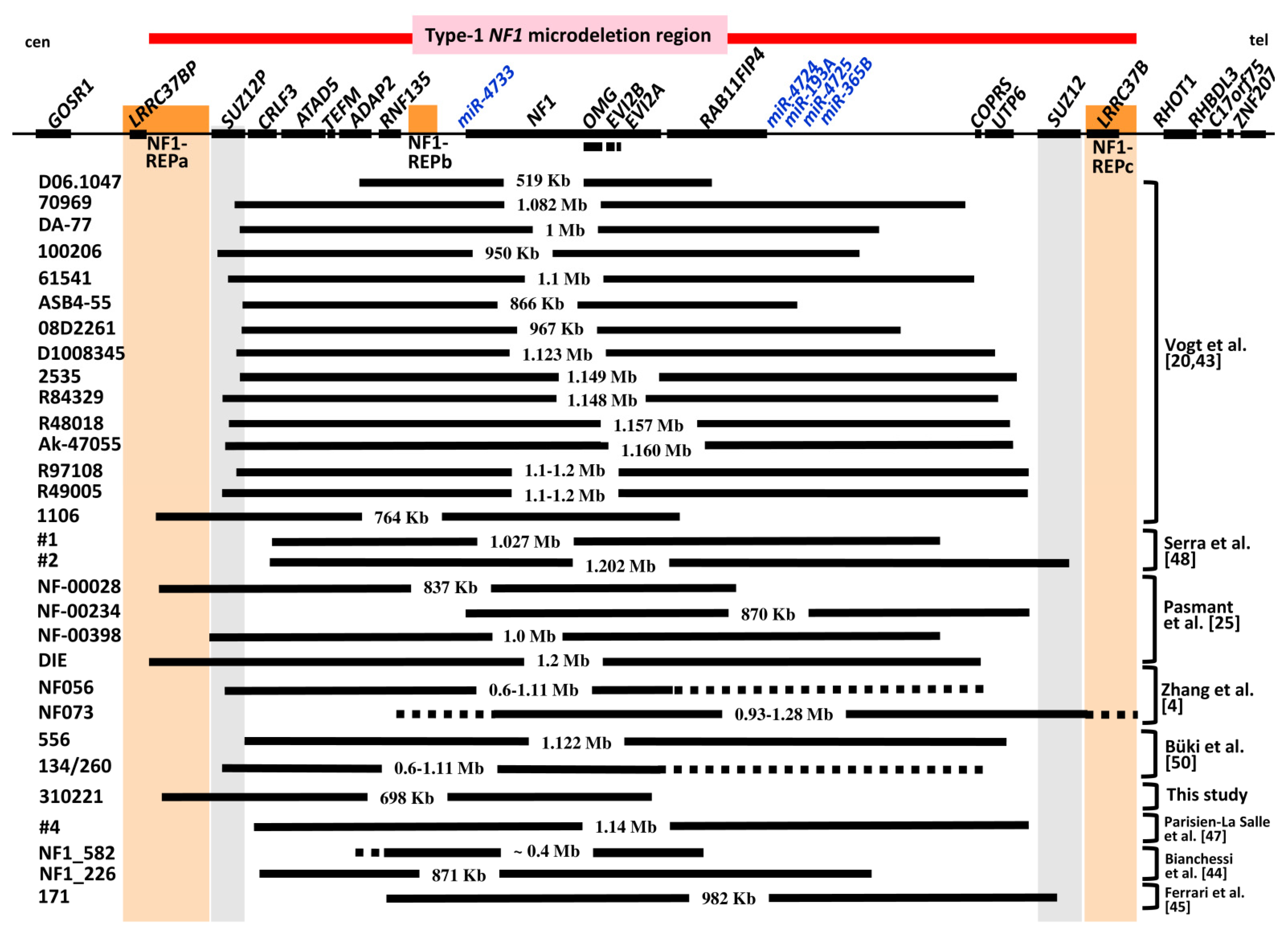
3.4. Comparison of the Deletion in Patient 310221 with Previously Reported Atypical NF1 Deletions

So far, 61 atypical NF1 deletions have been reported [1,4,6,7,20,25,27,28,29,30,31,32,33,34,35,36,37,38,39,40,41,42,43,44,45,46,47,48,49,50]. These 61 atypical NF1 deletions can be classified into two groups: The first encompasses very large deletions extending beyond one or both of the boundaries of the 1.4 Mb type 1 NF1 microdeletion region which is flanked by NF1-REPa and NF1-REPc. In total, 31 such large atypical NF1 deletions have been identified, which together constitute group #1 (Table 2).
The second group of atypical NF1 deletions, termed group #2 deletions, exhibit breakpoints that are located within the type 1 NF1 microdeletion region. The deletion of patient 310221 analysed in this study belongs to this group #2 atypical NF1 deletions, which are smaller than 1.4 Mb in size and not associated with the loss of all 14 protein-coding genes located within the type 1 NF1 microdeletion interval. Thus, group #2 atypical deletions encompass only a subset of these 14 genes. Taken together, 30 group #2 atypical NF1 deletions have been reported to date, including patient 310221 (Figure 1, Table 3). Remarkably, 15 (50%) of these 30 group #2 atypical NF1 deletions exhibit centromeric breakpoints located within the SUZ12P pseudogene. A total of 5 of the 30 group #2 atypical NF1 deletions have centromeric breakpoints located within NF1-REPa, including the deletion of patient 310221 reported here. Thus, SUZ12P and NF1-REPa represent genomic regions predisposed to chromosomal breakage causing atypical NF1 deletions.
Table 1. Protein-coding genes located within the 1.4 Mb type 1 NF1 microdeletion region. The genes rendered hemizygous in patient 310221 as a consequence of the 698 kb atypical deletion are marked in bold type.
Official HGNC Gene SymbolMIM#Official Gene NamepLI Score
CRLF3614853cytokine receptor like factor 30
ATAD5609534ATPase family, AAA domain containing 51.00
TEFM616422transcription elongation factor, mitochondrial0.51
ADAP2608635ArfGAP with dual PH domains 20.00
RNF135611358ring finger protein 1350.00
NF1162200neurofibromin 1.00
OMG164345oligodendrocyte myelin glycoprotein0.97
EVI2B158381ecotropic viral integration site 2B0.06
EVI2A158380ecotropic viral integration site 2A0.00
RAB11FIP4611999RAB11 family interacting protein 40.99
COPRS616477coordinator of PRMT5 and differentiation stimulator0.25
UTP6-UTP6 small subunit processome component0
SUZ12613675SUZ12 polycomb repressive complex 2 subunit1.00
LRRC37B616558leucine rich repeat containing 37B0.01
According to GnomAD v2.1.1/GnomAD SVs v2.1. The gnomAD browser (https://gnomad.broadinstitute.org/ accessed on 15 October 2021) provides the constraint metric termed ‘probability of loss-of-function’ (pLI). To determine the pLI metric, the observed and expected variant counts for a given gene are considered. The closer the pLI value is to 1, the more loss-of-function intolerant the gene appears to be. A pLI score ≥ 0.9 is indicative of genes that are predicted to be intolerant of loss-of-function variants [64].
Table 2. Large atypical NF1 deletions with one or both breakpoints located beyond the boundaries of the type 1 NF1 microdeletion region as defined by flanking NF1-REPa and NF1-REPc (group #1 atypical NF1 deletions).
Patient IDDeletion Size in MbReferences
UWA 106-33.2–3.7[6,27,28,31]
UWA 155-12.1–2.7[6,31]
ID806~7[29,37]
3724A2.0–3.1[1,31]
UWA 113-1~1[6]
BUD4.7[7,31,50]
BL3[30]
63[32]
1181–2[33]
4422[35,50]
8065.5[25,36]
T165>2.2[36,37]
282775>1.33[36,37]
T1451.61–1.75[36,37]
SNF1-2~1.3[38]
SNF1-31.84–2.8[38]
5522.7[39]
DUB7.6[40]
NF003581.2[41]
D05.26785.9[43]
D08015872[43]
6193[43,51]
ID not specified2.8[42]
NF0401.27–1.46[5]
NF0761.26–1.63[5]
NF1_311.8[44]
NF1_5051.1[44]
NF1_724>1.6[44]
ID not specified1.695[46]
20191.26–1.63[49]
1251.6[50]
Table 3. Features of the 30 atypical deletions of group #2. Indicated are the locations of the deletion breakpoints, the methods used to analyse the deletions and the number of functional SUZ12 gene copies in the respective patients. nd: not determined; BS-PCR: breakpoint-spanning PCR; MLPA: multiplex ligation-dependent probe amplification.
Patient IDCentromeric BreakpointTelomeric BreakpointMethod of AnalysisSUZ12 CopiesDeletion SizeMosaicReference
D06.1047ADAP2RAB11FIP4BS-PCR, MLPA2519,291 bpyes[43]
70969SUZ12PRAB11FIP4 and COPRSBS-PCR, MLPA21,082,491 bpno[43]
DA-77SUZ12Pbetween RAB11FIP4 and COPRSBS-PCR, MLPA21,001,546 bpyes[43]
100206SUZ12Pbetween RAB11FIP4 and COPRSBS-PCR, MLPA2950,940 bpyes[43]
61541SUZ12Pbetween COPRS and UTP6BS-PCR, MLPA21,105,242 bpyes[43]
ASB4-55SUZ12Pbetween RAB11FIP4 and COPRSBS-PCR, MLPA2866,769 bpyes[43]
08D2261SUZ12Pbetween RAB11FIP4 and COPRSBS-PCR, MLPA2976,455 bpyes[43]
D1008345SUZ12PUTP6BS-PCR, MLPA21,123,78 bpno[43]
2535SUZ12Pbetween UTP6 and SUZ12BS-PCR, MLPA21149,077 bpno[43]
R84329SUZ12PUTP6BS-PCR, MLPA21,148,828 bpyes[43]
R48018SUZ12Pbetween UTP6 and SUZ12BS-PCR, MLPA21,157,378 bpno[43]
Ak-47055SUZ12Pbetween UTP6 and SUZ12BS-PCR, MLPA21,160,989 bpyes[43]
R97108SUZ12Pbetween UTP6 and SUZ12MLPA21.1–1.2 Mbnd[20]
R49005SUZ12Pbetween UTP6 and SUZ12MLPA21.1–1.2 Mbnd[20]
1106NF1REPa RAB11FIP4BS-PCR, MLPA2764,080 bpno[43]
#1CRLF328 kb centromeric to COPRSmicroarray21,027,355 bpno[48]
#2CRLF3SUZ12microarray11,202,659 bpno[48]
NF00028LRRC37BPRAB11FIP4microarray2837 Kbnd[25]
NF00234RNF135between UTP6 and SUZ12microarray2870 Kbnd[25]
NF00398LRRC37BPbetween RAB11FIP4 and COPRSmicroarray21.0 Mbnd[25]
DIENF1-REPabetween COPRS and UTP6microarray21.2 Mbnd[25]
NF056SUZ12Pbetween NF1 exon 57 and UTP6MLPA20.6–1.11 Mbnd[4]
NF073between RNF135 and NF1 exon 1between LRRC37B and ZNF207MLPA10.93–1.28 Mbnd[4]
556between SUZ12P and CRLF3UTP6microarray, MLPA21,122,447 bpyes[50]
134/260SUZ12Pbetween NF1 exon 58 and UTP6MLPA20.6–1.11 Mbno[50]
310221NF1-REPaintron 57 of NF1microarray, MLPA2698 Kbnothis study
#4CRLF3between UTP6 and SUZ12microarray 21,144,007 bpnd[47]
NF_582between ADAP2 and RNF135between NF1 exons 57 and 58MLPA2~0.4 Mbnd[44]
NF_226CRLF3between RAB11FIP4 and COPRSmicroarray2871 Kbnd[44]
171RNF135SUZ12microarray1981,763 bpnd[45]
Only 2 of the 30 known group #2 atypical NF1 deletions may be associated with the loss of the same nine genes as observed in patient 310221, namely the deletions in patients 134/260 and NF056 (Figure 1). However, the deletions in these patients were analysed exclusively by MLPA, and the telomeric deletion breakpoints cannot be assigned with any accuracy because of the large gap between the MLPA probes located at the end of the NF1 gene and the UTP6 gene. It is therefore unclear whether the deletions in these patients also encompass the RAB11FIP4 and COPRS genes (Figure 1). Patients 134 and 260 possess the same 0.6–1.11 Mb deletion. Patient 134 is a 40-year-old woman who passed on the deletion to her 8-year-old son, patient 260. However, patient 134 is also likely to have inherited the deletion since her mother was affected by NF1 [50]. The deletion identified in patients 134 and 260 differs from the 698 kb deletion of patient 310221 in terms of the location of the centromeric breakpoint. Two further group #2 atypical NF1 deletions, those in patients 1 and 556, are of interest in terms of comparing their extent with the deletion in patient 310221, since clinical data from the respective patients have been published. Patient 1, reported by Serra et al. [48], harbours an atypical NF1 deletion of 1.027 Mb encompassing 10 genes (Figure 1). The deletion in patient 1 leads to hemizygosity of the same nine genes as observed in patient 310221. However, in addition to these nine genes, the RAB11FIP4 gene is also deleted in patient 1. Patient 556, reported by Büki et al. [50], harbours an atypical NF1 deletion of 1.12 Mb associated with the loss of 13 genes. The SUZ12 gene is not included in the deletion interval in this patient. If all 30 atypical group #2 deletions are considered, only three of them are associated with the loss of one copy of SUZ12 (Table 3).

3.5. Genotype/Phenotype Correlations in Patients with Atypical NF1 Deletions of Group #2

Group #2 atypical NF1 deletions can inform genotype/phenotype correlations if they are associated with only some of the clinical features frequently observed in patients with the larger type 1 NF1 deletions. However, details of the clinical phenotype are only available for 9 of the 30 patients with group #2 atypical NF1 deletions (Table 4). In six of these nine patients, the deletion does not encompass SUZ12, whereas in three patients, SUZ12 is either present only in one copy due to the deletion or functionally inactivated by the telomeric deletion breakpoint (Table 3 and Table 4).
Developmental delay was observed in patient 310221 reported here as well as in patient 1 analysed by Serra et al. [48]. Patient 1 was investigated at the age of 4 and 6 years. She attended primary school, and dysgraphia was noticed after several months. Hyperactivity was also evident. She had a full-scale IQ of 88 which is in the normal range, but heterogeneous results were observed in the different subtests. Her performance IQ was 122, which is in the high normal range. However, her verbal IQ was 71, which is well below average. The speech impairment of patient 1 was more severe than the delay in the development of receptive and expressive language of patient 310221. Nevertheless, neither patient showed the global, very severe developmental delay in numerous areas as frequently observed in children with type 1 NF1 deletions.
In addition, patients 556 and 134 with group #2 deletions did not exhibit severe developmental delay (Table 4). By contrast, patient 260 who had the same deletion as his mother (patient 134), showed severe delay in cognitive development at the age of 8 years [50]. The severity of the developmental delay in patient 260 is, however, difficult to estimate. For this patient, neither IQ values nor detailed information about specific tests performed to assess different areas of developmental delay have been reported. It is also unknown whether patient 260 received any therapy to improve his skills. As shown for the patient 310221 reported here, therapeutic intervention may help to reduce developmental delays quite considerably.
Mild hypertelorism was noted in patient 310221, and hypertelorism was also observed in patients 134 and 260. However, other facial dysmorphic features frequently seen in patients with type 1 NF1 deletions, including coarse face, downward-slanting palpebral fissures and broad nasal bridge, were not observed in patient 310221 or in any other patient with an atypical NF1 deletion of group #2 and two copies of the SUZ12 gene (Table 4).
Importantly, severe cognitive impairment associated with an IQ < 70 was not observed in any of those patients with a group #2 atypical NF1 deletion who had been clinically analysed in greater detail and whose deletion did not encompass SUZ12. For three of these patients with atypical group #2 deletions that did not encompass SUZ12, IQ values were assessed and found to be in the normal range (Table 4). By contrast, patient 2, reported by Serra et al. [48], had severe global developmental delay and an FSIQ of 55. The telomeric breakpoint of the deletion in patient 2 is located within the SUZ12 gene. Hence, this deletion is associated with the functional inactivation of SUZ12.
Table 4. Clinical features observed in patients with group #2 atypical NF1 deletions.
Clinical FeaturesPatients
556134260310221#1#2NF056NF073171
m, 10yf, 40ym, 8ym, 7yf, 4yf, 3yf, 60yf, 25ym, 3y
Broad nasal bridge ndndnd+ndndnd
Downward slanting palpebral fissuresndndndndndndnd
Hypertelorism+++, mild+ndndnd
Facial asymmetryndndndnd
Coarse facendndndnd
Micrognathiandndndndndndnd
Broad neck+ndndnd
Large hand and feetndndndnd
Excess soft tissue on handsndndndnd
Café-au-lait spots+++++++++
Freckling+++++++++
Lisch nodules+ndndnd
Tall stature/overgrowthndnd+
Subcutaneous neurofibromas+ndndnd
Cutaneous neurofibromas++nd
Externally visible plexiform neurofibromasndndnd
Spinal neurofibromasndndndndndndnd
Delay in development+++global, +ndnd+
Learning difficulties+++ndndnd
Speech difficulties++ndndndnd
IQ89ndnd938855ndndnd
Attention deficit and hyperactivity++ndndndnd
Autism spectrum disorderndndnd+ndndndndnd
Scoliosis++, mild+, severendnd
Pectus excavatum++ndnd
Bone cystsndnd
Other bone abnormalitiespes planusbilateral calcaneovalgusgenu valgum, pes planusnd
Joint hyperflexibilityndndndndndnd
Macrocephaly++nd+ndnd
Muscular hypotonia++ndnd+
MPNSTsnd
T2 hyperintensitiesnd++ndnd+
Optic gliomas++ndndnd
SUZ12copy number222221211
nd: not determined; −: absent; +: present; y: years; m: male; f: female. MPNST: malignant peripheral nerve sheath tumour. Patients 556, 134 and 260 were reported by Büki et al. [50], patients 1 and 2 by Serra et al. [48], patients NF056 and NF073 by Zhang et al. [4], patient 171 by Ferrari et al. [45]. The telomeric breakpoints of the deletions in patient 2 and 171 are located within the SUZ12 gene. Hence, these deletions were associated with the functional inactivation of SUZ12. Patient 2 exhibited severe global developmental delay and dysmorphic features, including broad forehead, dysplastic and low-set ears with thick helix, synophrys, receding orbital roof with exophthalmus, malar hypoplasia and long and prominent philtrum.

4. Discussion

In contrast to type 1 NF1 deletions, atypical NF1 deletions are much less frequent. In total, 61 atypical NF1 deletions have been reported to date, including the patient described here (Table 2, Figure 1). Atypical NF1 deletions are either larger or smaller than type 1 NF1 deletions. In total, 31 large atypical NF1 deletions have been reported which extend beyond one or both boundaries of the type 1 NF1 microdeletion region (group #1; Table 2). The deletion of additional genes located beyond the boundaries of the type 1 NF1 microdeletion region is expected to give rise to additional clinical symptoms or an even more complex clinical phenotype than generally observed in patients with type 1 NF1 deletions. More informative with regard to genotype/phenotype correlations are those atypical NF1 deletions, which are smaller than 1.4 Mb, each encompassing only a subset of the 14 protein-coding genes located in the type 1 NF1 microdeletion region. As yet, 30 such atypical NF1 deletions have been identified; these are termed group #2 atypical deletions (Figure 1, Table 3 and Table 4). Unfortunately, clinical data are only available for nine of the patients with these deletions, and only five of them have been characterized in any detail (Table 4).
The atypical deletion of patient 310221 reported here is remarkable since it spans only 698 kb and encompasses nine genes. Patient 310221 exhibited multiple café-au-lait spots and freckling as well as developmental delay first noted when he was 2 years old. At the age of 3.5 years, he showed developmental delay in expressive as well as receptive language and, to a minor degree, in motor skills. He was considered to be delayed in his development by approximately one year. However, therapeutic intervention helped him to catch up, and his deficits diminished considerably over the following years. The developmental delay observed in patient 310221 was much less severe than the global developmental delay frequently observed in children with type 1 or very large atypical NF1 microdeletions (reviewed in [5]). Severe global developmental delay in language and motor skills has been observed in 28 (93%) of 30 children with NF1 microdeletions [51]. These delays were already apparent at a young age (1–3 years) and persisted during later childhood. A total of 2 of the 30 children had very large atypical NF1 deletions, extending beyond the boundaries of the NF1 microdeletion region, whereas 28 children had type 1 deletions [51]. These findings imply that severe global developmental delay is frequent in children with type 1 NF1 microdeletions. By contrast, the developmental delay observed in patient 310221 reported here was restricted to specific areas and therapeutic intervention helped to reduce the delay so that he could catch up with his peers.
It should be appreciated that developmental delays are not rare among children in the general NF1 population [65,66,67,68,69]. Delays in at least one of eight areas, including fine motor, gross motor, receptive language, expressive language, math/premath ability, reading/pre-reading, self-help and socioemotional development, were observed in 68% of children with NF1 unselected for NF1 mutation type [65]. Significant developmental abnormalities were found in the areas of fine motor (35%), gross motor (52%) and math/premath ability (31%) [65]. At least 54% of children with NF1 older than 4 years exhibited lower scores for motor proficiency indicating poorer motor development [70]. In a study of 39 toddlers (21–30 months of age), psychomotor scores were found to be significantly lower for those with NF1 compared to those without NF1 [71]. Hence, developmental delays often present early in development in children with NF1.
Wessel et al. [66] investigated the progression of deficits caused by developmental delays in children with NF1 over time. Analysis of the total delays in several areas revealed that the number of areas delayed increased with age. The mean proportion of areas delayed was 22% in infants, 28% in preschool and 47% in school-age children with NF1 [66]. Closely related to motor performance is visuospatial functioning, which is frequently impaired in children with NF1 (reviewed by [72]). Numerous studies have indicated cognitive impairments and learning disabilities in 30–65% of all children with NF1 [72]. Children with NF1 frequently exhibit impairment in motor control and learning accompanied by substantial problems in visuomotor integration [73,74]. Nevertheless, the developmental delays observed in children with NF1 and intragenic NF1 mutations are much less severe than the global developmental delays affecting multiple areas seen in many children with large NF1 microdeletions [51]. The developmental delay in patient 310221 reported here appears to be within the range of developmental delays frequently seen in children with NF1 and intragenic NF1 mutations. Although patient 310221 showed a delay in the development of expressive and receptive language, he responded well to therapeutic intervention and was able to improve his deficits considerably, acquiring language skills within the normal range for his age. He also improved in terms of his gross motor skills, but mild deficits in fine motor skills have persisted.
Not much is known about the areas of developmental delay and their progression over time in other patients with atypical NF1 deletions of group #2, which encompass only a subset of genes within the type 1 NF1 microdeletion region. Only for seven patients with group #2 atypical NF1 deletions has information about the presence of developmental delay been published (Table 4). Remarkably, female patient 1 with a 1.027 Mb atypical NF1 deletion reported by Serra et al. [48], also exhibited developmental delay mainly associated with speech impairment. Her FSIQ was in the normal range. The cognitive abilities of patient 310221 reported here were also in the normal range as measured by several tests (Supplementary Table S3). The full-scale IQ (FSIQ) of patient 310221 at the age of 5.5 years was 99 whereas the mean FSIQ in 470 children with intragenic NF1 mutations was 87.7 [52]. Previous studies have indicated that the FSIQ in children from the general NF1 population is in the normal range, although somewhat lower than in children of comparison groups (usually around 90, instead of the normative mean of 100) [72,75]. By contrast, patients with type 1 NF1 deletions have significantly lower FSIQ scores than individuals carrying intragenic pathogenic NF1 variants or the general NF1 population [51,52,76,77]. The mean FSIQ of patients with type 1 NF1 deletions analysed in these studies ranged from 71.2–77.9 (Supplementary Table S4).
The normal cognitive abilities of patient 310221 and his comparatively moderate developmental delay are suggestive of a possible genotype/phenotype correlation. Thus, genes located telomeric to NF1, and not encompassed by the 698 kb atypical NF1 deletion of patient 310221, may contribute to the significantly reduced FSIQ and severe global developmental delays frequently seen in patients with type 1 NF1 deletions. The deletion in patient 310221 does not encompass the five genes RAB11FIP4, COPRS, UTP6, SUZ12 and LRRC37B. These genes are located telomeric to NF1 and are included in the type 1 NF1 microdeletion region (Figure 1). Remarkably, only two of these five genes, RAB11FIP4 and SUZ12, are loss-of-function intolerant, implying that their haploinsufficiency in patients with NF1 microdeletions is highly likely to have severe pathological consequences. RAB11FIP4 expression is highest in the brain, particularly in the cortex and frontal cortex [78]. However, it is still unknown if and how RAB11FIP4 hemizygosity contributes to the phenotype associated with large NF1 microdeletions.
In contrast to RAB11FIP4, much more information is available for SUZ12 which renders SUZ12 an important putative modifier of the type 1 NF1 microdeletion-associated phenotype. SUZ12 has a pLI score of 1.00, indicative of extreme intolerance to loss-of-function variants (Table 1) [64]. Hemizygosity of SUZ12 has been shown to be associated with an increased risk of MPNSTs in patients with type 1 NF1 microdeletions [79,80,81]. Importantly, patients with pathogenic variants located within SUZ12 exhibit pre- and postnatal overgrowth, facial dysmorphic features, musculoskeletal abnormalities and developmental delay/intellectual disability [82,83]. SUZ12 is one of the core components of the polycomb repressive complex 2 (PRC2), an epigenetic regulator with H3K27 methyltransferase activity. PRC2 is involved in gene silencing and the control of many different processes during cellular differentiation (reviewed by [84]). Mutations in the genes encoding other components of PRC2, namely EZH2 and EED, also lead to overgrowth, macrocephaly, advanced bone age, variable intellectual disability and distinctive facial features [84]. Since intellectual disability is quite common in patients with mutations of genes encoding PRC2 components, it is likely that the loss of SUZ12 contributes to the cognitive disabilities and severe developmental delay seen in patients with type 1 NF1 deletions hemizygous for SUZ12. The normal cognitive abilities of patient 310221 and other patients with atypical group #2 deletions who are not hemizygous for SUZ12 are in accordance with this putative genotype/phenotype correlation (Table 4).
Remarkably, patients with intragenic SUZ12 mutations exhibit an overgrowth phenotype [82,83,84]. Childhood overgrowth is also frequent in patients with large NF1 deletions. In contrast to the short stature observed in most patients with intragenic NF1 mutations, tall stature in adults and overgrowth during childhood have been reported in patients with NF1 microdeletions [5,25,77,85,86,87,88]. The loss of the RNF135 gene located centromeric to NF1 within the type 1 NF1 deletion region has been suggested to be associated with the overgrowth seen in patients with NF1 microdeletions [89]. However, tall stature and childhood overgrowth have also been reported in patients with NF1 deletions that do not encompass the RNF135 gene but are associated with the loss of SUZ12 [24,25,26,89]. Thus, it may be inferred that the heterozygous loss of SUZ12 resulting in SUZ12 haploinsufficiency is likely to contribute to the overgrowth phenotype observed in patients with type 1 NF1 microdeletions. This conclusion is supported by the observation that SUZ12 is not included within the deletion interval in patient 310221 who is small for age. Overgrowth or tall stature was also not reported for other patients with atypical NF1 deletions of group #2 (Table 4).
Patients with intragenic SUZ12 mutations exhibit facial dysmorphic features similar to those observed in many patients with type 1 NF1 deletions including hypertelorism, broad nasal bridge and downward-slanting palpebral fissures [82,83]. Importantly, facial dysmorphic features are rare in patients with intragenic NF1 mutations. Hemizygosity of SUZ12 is likely to contribute to the facial dysmorphic features of patients with type 1 NF1 deletions. This conclusion is supported by the absence of facial dysmorphism, as frequently seen in patients with type 1 NF1 deletions, in patient 310221 who has two copies of SUZ12. However, facial dysmorphism is not apparent in all patients with type 1 NF1 deletions or at least not to the same extent. It follows that the causes of facial dysmorphism in patients with type 1 NF1 deletions are likely to be complex, being influenced by SUZ12 hemizygosity as well as additional factors.
Many studies have identified impairment of executive functions and working memory in patients with NF1 [90]. An increased prevalence of attention deficit hyperactivity disorder (ADHD) in children with NF1 has been reported [91,92,93,94,95]. ADHD impairs the daily life functioning of many children with NF1 quite considerably [96]. ADHD is also frequent in patients with type 1 NF1 deletions [77] and has been diagnosed in 15 (88%) of 17 children and adolescents with type 1 NF1 deletions [51]. Patient 310221, reported here, exhibited attention deficits which impaired his learning over and above the direct impact on his cognitive and adaptive skills. The frequency of attention deficits in patients with atypical NF1 deletions has not so far been investigated in any detail and further studies are required to evaluate the impact of these deficits on performance and development in this group of patients.
In addition to ADHD, a high incidence of autism has been observed in patients with NF1 [92,97,98,99,100]. Autism spectrum disorder (ASD), with a phenotypic profile similar to idiopathic autism, is observed in at least 25% of children with NF1, whereas an additional 20% of children with NF1 exhibit partial ASD features [101]. Autism-related symptoms also appear to be very frequent in patients with NF1 microdeletions. In a previous study, we observed mild to moderate autistic symptomatology in 15 (71%) of 21 children with type 1 NF1 deletions, which was significantly more frequent than in the general NF1 population [51]. Autistic symptomatology and hyperactivity were also noticed in patient 310221. At the age of 7 years, the onset of an autism spectrum disorder was suspected.
A specific role in the development of the NF1 microdeletion-associated phenotype and in particular autism has recently been demonstrated for the cytokine receptor-like factor 3 (CRLF3) gene located within the NF1 microdeletion region. CRLF3 is deleted in patient 310221 reported here (Figure 1). Induced pluripotent stem cell forebrain cerebral organoids (hCOs), isolated from patients with type 1 NF1 microdeletions, display both neural stem cell proliferation and elevated neuronal abnormalities such as dendritic maturation deficits. Whilst increased neuronal stem cell proliferation has been shown to result from decreased NF1/RAS regulation, the neuronal differentiation, survival and maturation defects of these hCOs are caused by reduced CRLF3 expression and impaired RhoA signalling [102]. This role of CRLF3 was confirmed by the analysis of hCOs, isolated from a patient with an atypical NF1 deletion which did not include the CRLF3 gene. These hCOs did not show abnormalities of neuronal survival, differentiation and maturation [102]. Further, these authors identified 7 of 17 NF1 patients with an increased autistic trait burden who harboured a germline putatively pathogenic missense variant within the CRLF3 gene (c.1166T > C, p.Leu389Pro) present in addition to pathogenic variants in NF1. Taken together, these findings suggest an essential role for CRLF3 in both human brain development and autism [102]. Consequently, the loss of the CRLF3 gene in the patient described here may have contributed to his autistic symptoms. Further analyses of patients with different types of NF1 microdeletion will confirm or refute the possible contribution of the CRLF3 gene to the deletion-associated phenotype.
Atypical NF1 deletions of group #2, such as the 698 kb deletion observed in patient 310221, are potentially very informative in terms of genotype/phenotype correlations. However, the possibility that atypical NF1 deletions can be associated with mosaicism with normal cells not harbouring the deletion must be considered. Vogt et al. [43] observed mosaicism with normal cells in 10 (59%) of 17 patients with atypical NF1 deletions. In 6 of these 10 patients, mosaicism was analysed by FISH which allowed the determination of the proportion of cells harbouring the deletion. In the blood samples of these six patients, the proportion of cells with the deletions was 70%, 75%, 80%, 93%, 96% and 98%, respectively. In three (50%) of the six patients, mosaicism was also detected by MLPA, which is considered to have an intrinsic detection limit of ~10% (reviewed in [10]). In other words, low-grade mosaicism with normal cells present at a proportion lower than ~10% cannot be detected by MLPA. Since FISH on blood cells was not performed in patient 310221, low-grade mosaicism with normal cells cannot be unequivocally excluded even though MLPA and microarray analysis did not give any indication of mosaicism with normal cells. Patient 310221 does not show features frequently seen in patients with type 1 NF1 deletions, such as global severe developmental delay, cognitive disability, overgrowth and facial dysmorphism, which are all detectable at a very young age. Even though we cannot completely exclude the possibility that normal cells lacking the deletion are present in low proportions and could have influenced the clinical phenotype in patient 310221, his comparatively mild clinical phenotype is likely to be attributable to the presence of two copies of the SUZ12 gene, which do not reside within the deletion interval in this patient.

5. Conclusions

Group #2 atypical NF1 deletions, encompassing only a subset of the 14 protein-coding genes located within the type 1 NF1 microdeletion region, have the potential to inform genotype/phenotype correlations and facilitate the identification of modifying genes. Patient 310221 analysed here harbours a group #2 atypical NF1 deletion which encompasses the CRLF3 gene but not SUZ12. The relatively mild clinical phenotype of this patient suggests that the retention of SUZ12 may have ameliorated what might otherwise have been a more severe clinical phenotype. Indeed, the relatively mild clinical phenotype of group #2 atypical NF1 deletion patients with two copies of SUZ12 suggests that SUZ12 hemizygosity is highly likely to influence the more severe clinical phenotype observed in patients with type 1 NF1 microdeletions characterized by facial dysmorphic features, reduced cognitive abilities and severe global developmental delay, all of which were absent in patient 310221. The co-deletion of CRLF3 may contribute to the clinical manifestations (particularly the autistic symptomatology) in those patients whose NF1 microdeletions encompass this gene. However, further studies are necessary to investigate this in greater detail. To date, genotype/phenotype correlations for group #2 atypical NF1 deletions are limited by the scarcity of clinical details available for many of the reported patients and by limited breakpoint definition due to the use of MLPA instead of microarray analysis. Moreover, information about possible mosaicism with cells not harbouring the deletion is not available for many patients with atypical NF1 deletions. The future analysis of genetically and clinically well-characterized patients with atypical NF1 deletions will serve to further clarify the role of the loss of genes such as SUZ12 and CRLF3 for the NF1 microdeletion-associated phenotype.

Supplementary Materials

The following are available online at https://www.mdpi.com/article/10.3390/genes12101639/s1, Table S1: Physical measurements of patient 310221; Table S2: Patient 310221 was investigated by means of the Kaufman Assessment Battery for Children (KABC-II). The KABC-II measures 5 scales according to the Cattell-Horn-Carroll (CHC) model. The results of the subtests are indicated as index-values of the patient. The summary of all index-values from the scales is represented by the Fluid-Crystallised Index (FCI), the general intelligence composite score of KABC-II according to the CHC model; Table S3: The cognitive abilities of patient 310221 were evaluated by the Kaufman Assessment Battery for Children (KABC-II) and the Wechsler Preschool and Primary Scale of Intelligence. FCI: Fluid-Crystallised Index (FCI), the general intelligence composite score of KABC-II according to the CHC model; Table S4: Full scale IQ (FSIQ) in patients with NF1 microdeletions analysed by Descheemaeker et al., Mautner et al., Ottenhoff et al. and Kehrer-Sawatzki et al. The 17 patients with NF1 microdeletions analysed by Ottenhoff et al. indicated in this table do not include the patients analysed by Descheemaeker et al. Six patients included in the study of Kehrer-Sawatzki et al. had already been analysed previously by Mautner et al.; these six patients are not included in the 17 NF1 microdeletion patients analysed by Mautner et al. listed below.

Author Contributions

H.K.-S., D.N.C. and V.-F.M. contributed to the conception and design of the study. U.W. and V.-F.M. investigated the patients and reviewed all similar cases. H.K.-S. and D.N.C. wrote the first draft of the manuscript. V.-F.M. and U.W. wrote sections of the manuscript. All authors have read and agreed to the published version of the manuscript.

Funding

This work has been funded by the Deutsche Forschungsgemeinschaft (DFG) Grant KE 724/12-2 given to H.K.-S.

Institutional Review Board Statement

The study was conducted according to the guidelines of the Declaration of Helsinki and approved by the Institutional Review Board of the University of Ulm (219/10) and the University Hospital Hamburg Eppendorf (PV3291).

Informed Consent Statement

Written informed consent for publication was obtained from the parents of the patient as his legal guardians/representatives.

Data Availability Statement

Data are contained within the article or Supplementary Material.

Conflicts of Interest

The authors declare no conflict of interest.

References

  1. Cnossen, M.H.; van der Est, M.N.; Breuning, M.H.; van Asperen, C.J.; Breslau-Siderius, E.J.; van der Ploeg, A.T.; de Goede-Bolder, A.; van den Ouweland, A.M.; Halley, D.J.; Niermeijer, M.F. Deletions spanning the neurofibromatosis type 1 gene: Implications for genotype-phenotype correlations in neurofibromatosis type 1? Hum. Mutat. 1997, 9, 458–464. [Google Scholar] [CrossRef]
  2. Rasmussen, S.A.; Colman, S.D.; Ho, V.T.; Abernathy, C.R.; Arn, P.H.; Weiss, L.; Schwartz, C.; Saul, R.A.; Wallace, M.R. Constitutional and mosaic large NF1 gene deletions in neurofibromatosis type 1. J. Med. Genet. 1998, 35, 468–471. [Google Scholar] [CrossRef] [Green Version]
  3. Kluwe, L.; Siebert, R.; Gesk, S.; Friedrich, R.E.; Tinschert, S.; Kehrer-Sawatzki, H.; Mautner, V.-F. Screening 500 unselected neurofibromatosis 1 patients for deletions of the NF1 gene. Hum. Mutat. 2004, 23, 111–116. [Google Scholar] [CrossRef]
  4. Zhang, J.; Tong, H.; Fu, X.; Zhang, Y.; Liu, J.; Cheng, R.; Liang, J.; Peng, J.; Sun, Z.; Liu, H.; et al. Molecular characterization of NF1 and neurofibromatosis type 1 genotype-phenotype correlations in a Chinese population. Sci. Rep. 2015, 5, 11291. [Google Scholar] [CrossRef] [PubMed] [Green Version]
  5. Kehrer-Sawatzki, H.; Mautner, V.-F.; Cooper, D.N. Emerging genotype–phenotype relationships in patients with large NF1 deletions. Hum. Genet. 2017, 136, 349–376. [Google Scholar] [CrossRef] [PubMed] [Green Version]
  6. Dorschner, M.O.; Sybert, V.P.; Weaver, M.; Pletcher, B.A.; Stephens, K. NF1 microdeletion breakpoints are clustered at flanking repetitive sequences. Hum. Mol. Genet. 2000, 9, 35–46. [Google Scholar] [CrossRef] [PubMed] [Green Version]
  7. Jenne, D.E.; Tinschert, S.; Reimann, H.; Lasinger, W.; Thiel, G.; Hameister, H.; Kehrer-Sawatzki, H. Molecular characterization and gene content of breakpoint boundaries in patients with neurofibromatosis type 1 with 17q11.2 microdeletions. Am. J. Hum. Genet. 2001, 69, 516–527. [Google Scholar] [CrossRef] [PubMed] [Green Version]
  8. López-Correa, C.; Dorschner, M.; Brems, H.; Lázaro, C.; Clementi, M.; Upadhyaya, M.; Dooijes, D.; Moog, U.; Kehrer-Sawatzki, H.; Rutkowski, J.L.; et al. Recombination hotspot in NF1 microdeletion patients. Hum. Mol. Genet. 2001, 10, 1387–1392. [Google Scholar] [CrossRef] [Green Version]
  9. Messiaen, L.; Vogt, J.; Bengesser, K.; Fu, C.; Mikhail, F.; Serra, E.; Garcia, C.L.; Cooper, D.N.; Lazaro, C.; Kehrer-Sawatzki, H. Mosaic type-1 NF1 microdeletions as a cause of both generalized and segmental neurofibromatosis type-1 (NF1). Hum. Mutat. 2011, 32, 213–219. [Google Scholar] [CrossRef] [Green Version]
  10. Summerer, A.; Schäfer, E.; Mautner, V.-F.; Messiaen, L.; Cooper, D.N.; Kehrer-Sawatzki, H. Ultra-deep amplicon sequencing indicates absence of low-grade mosaicism with normal cells in patients with type-1 NF1 deletions. Hum. Genet. 2018, 138, 73–81. [Google Scholar] [CrossRef] [Green Version]
  11. Correa, C.L.; Brems, H.; Lázaro, C.; Marynen, P.; Legius, E. Unequal meiotic crossover: A frequent cause of NF1 microdeletions. Am. J. Hum. Genet. 2000, 66, 1969–1974. [Google Scholar] [CrossRef] [PubMed] [Green Version]
  12. Neuhäusler, L.; Summerer, A.; Cooper, D.N.; Mautner, V.-F.; Kehrer-Sawatzki, H. Pronounced maternal parent-of-origin bias for type-1 NF1 microdeletions. Hum. Genet. 2018, 137, 365–373. [Google Scholar] [CrossRef]
  13. Forbes, S.H.; Dorschner, M.O.; Le, R.; Stephens, K. Genomic context of paralogous recombination hotspots mediating recurrent NF1 region microdeletion. Genes Chromosom. Cancer 2004, 41, 12–25. [Google Scholar] [CrossRef]
  14. De Raedt, T.; Stephens, M.; Heyns, I.; Brems, H.; Thijs, D.; Messiaen, L.; Stephens, K.; Lazaro, C.; Wimmer, K.; Kehrer-Sawatzki, H.; et al. Conservation of hotspots for recombination in low-copy repeats associated with the NF1 microdeletion. Nat. Genet. 2006, 38, 1419–1423. [Google Scholar] [CrossRef]
  15. Bengesser, K.; Vogt, J.; Mussotter, T.; Mautner, V.-F.; Messiaen, L.; Cooper, D.N.; Kehrer-Sawatzki, H. Analysis of crossover breakpoints yields new insights into the nature of the gene conversion events associated with large NF1 deletions mediated by nonallelic homologous recombination. Hum. Mutat. 2013, 35, 215–226. [Google Scholar] [CrossRef]
  16. Hillmer, M.; Wagner, D.; Summerer, A.; Daiber, M.; Mautner, V.-F.; Messiaen, L.; Cooper, D.N.; Kehrer-Sawatzki, H. Fine mapping of meiotic NAHR-associated crossovers causing large NF1 deletions. Hum. Mol. Genet. 2015, 25, 484–496. [Google Scholar] [CrossRef] [Green Version]
  17. Hillmer, M.; Summerer, A.; Mautner, V.-F.; Högel, J.; Cooper, D.N.; Kehrer-Sawatzki, H. Consideration of the haplotype diversity at nonallelic homologous recombination hotspots improves the precision of rearrangement breakpoint identification. Hum. Mutat. 2017, 38, 1711–1722. [Google Scholar] [CrossRef]
  18. Summerer, A.; Mautner, V.-F.; Upadhyaya, M.; Claes, K.; Högel, J.; Cooper, D.N.; Messiaen, L.; Kehrer-Sawatzki, H. Extreme clustering of type-1 NF1 deletion breakpoints co-locating with G-quadruplex forming sequences. Hum. Genet. 2018, 137, 511–520. [Google Scholar] [CrossRef]
  19. Steinmann, K.; Cooper, D.N.; Kluwe, L.; Chuzhanova, N.A.; Senger, C.; Serra, E.; Lazaro, C.; Gilaberte, M.; Wimmer, K.; Mautner, V.-F.; et al. Type 2 NF1 deletions are highly unusual by virtue of the absence of nonallelic homologous recombination hotspots and an apparent preference for female mitotic recombination. Am. J. Hum. Genet. 2007, 81, 1201–1220. [Google Scholar] [CrossRef] [PubMed] [Green Version]
  20. Vogt, J.; Mussotter, T.; Bengesser, K.; Claes, K.; Högel, J.; Chuzhanova, N.; Fu, C.; van den Ende, J.; Mautner, V.-F.; Cooper, D.N.; et al. Identification of recurrent type-2 NF1 microdeletions reveals a mitotic nonallelic homologous recombination hotspot underlying a human genomic disorder. Hum. Mutat. 2012, 33, 1599–1609. [Google Scholar] [CrossRef] [PubMed]
  21. Kehrer-Sawatzki, H.; Kluwe, L.; Sandig, C.; Kohn, M.; Wimmer, K.; Krammer, U.; Peyrl, A.; Jenne, D.; Hansmann, I.; Mautner, V.-F. High frequency of mosaicism among patients with neurofibromatosis type 1 (NF1) with microdeletions caused by somatic recombination of the JJAZ1 gene. Am. J. Hum. Genet. 2004, 75, 410–423. [Google Scholar] [CrossRef] [PubMed] [Green Version]
  22. Roehl, A.C.; Vogt, J.; Mussotter, T.; Zickler, A.N.; Spöti, H.; Högel, J.; Chuzhanova, N.; Wimmer, K.; Kluwe, L.; Mautner, V.-F.; et al. Intrachromosomal mitotic nonallelic homologous recombination is the major molecular mechanism underlying type-2 NF1 deletions. Hum. Mutat. 2010, 31, 1163–1173. [Google Scholar] [CrossRef] [PubMed] [Green Version]
  23. Roehl, A.C.; Mussotter, T.; Cooper, D.N.; Kluwe, L.; Wimmer, K.; Högel, J.; Zetzmann, M.; Vogt, J.; Mautner, V.F.; Kehrer-Sawatzki, H. Tissue-specific differences in the proportion of mosaic large NF1 deletions are suggestive of a selective growth advantage of hematopoietic del(+/−) stem cells. Hum. Mutat. 2012, 33, 541–550. [Google Scholar] [CrossRef] [PubMed]
  24. Bengesser, K.; Cooper, D.N.; Steinmann, K.; Kluwe, L.; Chuzhanova, N.; Wimmer, K.; Tatagiba, M.; Tinschert, S.; Mautner, V.-F.; Kehrer-Sawatzki, H. A novel third type of recurrent NF1 microdeletion mediated by nonallelic homologous recombination between LRRC37B-containing low-copy repeats in 17q11.2. Hum. Mutat. 2010, 31, 742–751. [Google Scholar] [CrossRef] [PubMed] [Green Version]
  25. Pasmant, E.; Sabbagh, A.; Spurlock, G.; Laurendeau, I.; Grillo, E.; Hamel, M.J.; Martin, L.; Barbarot, S.; Leheup, B.; Rodriguez, D.; et al. NF1 microdeletions in neurofibromatosis type 1: From genotype to phenotype. Hum. Mutat. 2010, 31, 1506–1518. [Google Scholar] [CrossRef] [Green Version]
  26. Zickler, A.M.; Hampp, S.; Messiaen, L.; Bengesser, K.; Mussotter, T.; Roehl, A.C.; Wimmer, K.; Mautner, V.-F.; Kluwe, L.; Upadhyaya, M.; et al. Characterization of the nonallelic homologous recombination hotspot PRS3 associated with type-3 NF1 deletions. Hum. Mutat. 2012, 33, 372–383. [Google Scholar] [CrossRef] [PubMed]
  27. Kayes, L.M.; Riccardi, V.M.; Burke, W.; Bennett, R.L.; Stephens, K. Large de novo DNA deletion in a patient with sporadic neurofibromatosis 1, mental retardation, and dysmorphism. J. Med. Genet. 1992, 29, 686–690. [Google Scholar] [CrossRef]
  28. Kayes, L.M.; Burke, W.; Riccardi, V.M.; Bennett, R.; Ehrlich, P.; Rubenstein, A.; Stephens, K. Deletions spanning the neurofibromatosis 1 gene: Identification and phenotype of five patients. Am. J. Hum. Genet. 1994, 54, 424–436. [Google Scholar]
  29. Upadhyaya, M.; Roberts, S.H.; Maynard, J.; Sorour, E.; Thompson, P.W.; Vaughan, M.; Wilkie, A.; Hughes, H.E. A cytogenetic deletion, del(17)(q11.22q21.1), in a patient with sporadic neurofibromatosis type 1 (NF1) associated with dysmorphism and developmental delay. J. Med. Genet. 1996, 33, 148–152. [Google Scholar] [CrossRef]
  30. Riva, P.; Corrado, L.; Natacci, F.; Castorina, P.; Wu, B.-L.; Schneider, G.H.; Clementi, M.; Tenconi, R.; Korf, B.R.; Larizza, L. NF1 microdeletion syndrome: Refined FISH characterization of sporadic and familial deletions with locus-specific probes. Am. J. Hum. Genet. 2000, 66, 100–109. [Google Scholar] [CrossRef] [Green Version]
  31. Kehrer-Sawatzki, H.; Tinschert, S.; Jenne, D.E. Heterogeneity of breakpoints in non-LCR-mediated large constitutional deletions of the 17q11.2 NF1 tumor suppressor region. J. Med. Genet. 2003, 40, E116. [Google Scholar] [CrossRef] [PubMed] [Green Version]
  32. Gervasini, C.; Orzan, F.N.; Bentivegna, A.; Colapietro, P.; Friso, A.; Tenconi, R.; Upadhyaya, M.; Larizza, L.; Corrado, L.; Venturin, M.; et al. Evidence for non-homologous end joining and non-allelic homologous recombination in atypical NF1 microdeletions. Hum. Genet. 2004, 115, 69–80. [Google Scholar] [CrossRef]
  33. Venturin, M.; Guarnieri, P.; Natacci, F.; Stabile, M.; Tenconi, R.; Clementi, M.; Hernandez, C.; Thompson, P.; Upadhyaya, M.; Larizza, L.; et al. Mental retardation and cardiovascular malformations in NF1 microdeleted patients point to candidate genes in 17q11.2. J. Med. Genet. 2004, 41, 35–41. [Google Scholar] [CrossRef] [PubMed] [Green Version]
  34. Gervasini, C.; Venturin, M.; Orzan, F.N.; Friso, A.; Clementi, M.; Tenconi, R.; Larizza, L.; Riva, P. Uncommon Alu-mediated NF1 microdeletion with a breakpoint inside the NF1 gene. Genomics 2005, 85, 273–279. [Google Scholar] [CrossRef]
  35. Kehrer-Sawatzki, H.; Kluwe, L.; Mautner, V.-F. Extensively high load of internal tumors determined by whole body MRI scanning in a patient with neurofibromatosis type 1 and a non-LCR-mediated 2-Mb deletion in 17q11.2. Hum. Genet. 2005, 116, 466–475. [Google Scholar] [CrossRef]
  36. Mantripragada, K.K.; Thuresson, A.-C.; Piotrowski, A.; De Ståhl, T.D.; Menzel, U.; Grigelionis, G.; Ferner, R.E.; Griffiths, S.; Bolund, L.; Mautner, V.; et al. Identification of novel deletion breakpoints bordered by segmental duplications in the NF1 locus using high resolution array-CGH. J. Med. Genet. 2005, 43, 28–38. [Google Scholar] [CrossRef] [Green Version]
  37. Wimmer, K.; Yao, S.; Claes, K.; Kehrer-Sawatzki, H.; Tinschert, S.; De Raedt, T.; Legius, E.; Callens, T.; Beiglböck, H.; Maertens, O.; et al. Spectrum of single- and multiexon NF1 copy number changes in a cohort of 1,100 unselected NF1 patients. Genes Chromosom. Cancer 2006, 45, 265–276. [Google Scholar] [CrossRef]
  38. Maertens, O.; De Schepper, S.; Vandesompele, J.; Brems, H.; Heyns, I.; Janssens, S.; Speleman, F.; Legius, E.; Messiaen, L. Molecular dissection of isolated disease features in mosaic neurofibromatosis type 1. Am. J. Hum. Genet. 2007, 81, 243–251. [Google Scholar] [CrossRef] [PubMed] [Green Version]
  39. Kehrer-Sawatzki, H.; Schmid, E.; Fünsterer, C.; Kluwe, L.; Mautner, V.-F. Absence of cutaneous neurofibromas in an NF1 patient with an atypical deletion partially overlapping the common 1.4 Mb microdeleted region. Am. J. Med. Genet. Part A 2008, 146, 691–699. [Google Scholar] [CrossRef]
  40. Pasmant, E.; De Saint-Trivier, A.; Laurendeau, I.; Dieux-Coeslier, A.; Parfait, B.; Vidaud, M.; Vidaud, M.; Bieche, I. Characterization of a 7.6-Mb germline deletion encompassing the NF1 locus and about a hundred genes in an NF1 contiguous gene syndrome patient. Eur. J. Hum. Genet. 2008, 16, 1459–1466. [Google Scholar] [CrossRef] [Green Version]
  41. Pasmant, E.; Sabbagh, A.; Masliah-Planchon, J.; Haddad, V.; Hamel, M.-J.; Laurendeau, I.; Soulier, J.; Parfait, B.; Wolkenstein, P.; Bièche, I.; et al. Detection and characterization of NF1 microdeletions by custom high resolution array CGH. J. Mol. Diagn. 2009, 11, 524–529. [Google Scholar] [CrossRef] [Green Version]
  42. Taylor Tavares, A.L.; Willatt, L.; Armstrong, R.; Simonic, I.; Park, S.-M. Mosaic deletion of the NF1 gene in a patient with cognitive disability and dysmorphic features but without diagnostic features of NF1. Am. J. Med. Genet. Part A 2013, 161, 1185–1188. [Google Scholar] [CrossRef] [PubMed]
  43. Vogt, J.; Bengesser, K.; Claes, K.; Wimmer, K.; Mautner, V.-F.; Van Minkelen, R.; Legius, E.; Brems, H.; Upadhyaya, M.; Högel, J.; et al. SVA retrotransposon insertion-associated deletion represents a novel mutational mechanism underlying large genomic copy number changes with non-recurrent breakpoints. Genome Biol. 2014, 15, R80. [Google Scholar] [CrossRef] [Green Version]
  44. Bianchessi, D.; Morosini, S.; Saletti, V.; Ibba, M.C.; Natacci, F.; Esposito, S.; Cesaretti, C.; Riva, D.; Finocchiaro, G.; Eoli, M. 126 novel mutations in Italian patients with neurofibromatosis type 1. Mol. Genet. Genom. Med. 2015, 3, 513–525. [Google Scholar] [CrossRef] [PubMed]
  45. Ferrari, L.; Scuvera, G.; Tucci, A.; Bianchessi, D.; Rusconi, F.; Menni, F.; Battaglioli, E.; Milani, D.; Riva, P. Identification of an atypical microdeletion generating the RNF135-SUZ12 chimeric gene and causing a position effect in an NF1 patient with overgrowth. Hum. Genet. 2017, 136, 1329–1339. [Google Scholar] [CrossRef]
  46. Al-Araimi, M.; Hamza, N.; Al Yahmadi, A.; Al Mazrooey, H.; Elsheikh, A.; Al Amri, A.; Al Harrasi, S.; Hausdorf, L.; Mula-Abed, W. Rare NF1 microdeletion syndrome in an Omani patient. Clin. Case Rep. 2018, 6, 2424–2426. [Google Scholar] [CrossRef] [Green Version]
  47. Parisien-La Salle, S.; Dumas, N.; Rondeau, G.; Latour, M.; Bourdeau, I. Isolated pheochromocytoma in a 73-year-old man with no clinical manifestations of type 1 neurofibromatosis carrying an unsuspected deletion of the entire NF1 gene. Front Endocrinol 2019, 10, 546. [Google Scholar] [CrossRef]
  48. Serra, G.; Antona, V.; Corsello, G.; Zara, F.; Piro, E.; Falsaperla, R. NF1 microdeletion syndrome: Case report of two new patients. Ital. J. Pediatr. 2019, 45, 1–7. [Google Scholar] [CrossRef]
  49. Kluwe, L.; Friedrich, R.E.; Farschtschi, S.C.; Hagel, C.; Kehrer-Sawatzki, H.; Mautner, V. Null phenotype of neurofibromatosis type 1 in a carrier of a heterozygous atypical NF1 deletion due to mosaicism. Hum. Mutat. 2020, 41, 1226–1231. [Google Scholar] [CrossRef] [PubMed]
  50. Büki, G.; Zsigmond, A.; Czakó, M.; Szalai, R.; Antal, G.; Farkas, V.; Fekete, G.; Nagy, D.; Széll, M.; Tihanyi, M.; et al. Genotype-phenotype associations in patients with type-1, type-2, and atypical NF1 microdeletions. Front. Genet. 2021, 12, 673025. [Google Scholar] [CrossRef]
  51. Kehrer-Sawatzki, H.; Kluwe, L.; Salamon, J.; Well, L.; Farschtschi, S.; Rosenbaum, T.; Mautner, V.-F. Clinical characterization of children and adolescents with NF1 microdeletions. Child’s Nerv. Syst. 2020, 36, 2297–2310. [Google Scholar] [CrossRef] [PubMed]
  52. Ottenhoff, M.M.J.; Msc, A.B.R.; Mous, S.E.; Plasschaert, E.; Msc, D.G.; Brems, H.; Oostenbrink, R.; Van Minkelen, R.; Nellist, M.; Schorry, E.; et al. Examination of the genetic factors underlying the cognitive variability associated with neurofibromatosis type 1. Genet. Med. 2020, 22, 889–897. [Google Scholar] [CrossRef] [PubMed] [Green Version]
  53. Pacot, L.; Vidaud, D.; Sabbagh, A.; Laurendeau, I.; Briand-Suleau, A.; Coustier, A.; Maillard, T.; Barbance, C.; Morice-Picard, F.; Sigaudy, S.; et al. Severe phenotype in patients with large deletions of NF1. Cancers 2021, 13, 2963. [Google Scholar] [CrossRef] [PubMed]
  54. Well, L.; Döbel, K.; Kluwe, L.; Bannas, P.; Farschtschi, S.; Adam, G.; Mautner, V.F.; Salamon, J. Genotype-phenotype correlation in neurofibromatosis type-1: NF1 whole gene deletions lead to high tumor-burden and increased tumor-growth. PLoS Genet 2021, 17, e1009517. [Google Scholar] [CrossRef]
  55. Hellbrügge, T. Münchener Funktionelle Entwicklungsdiagnostik, Zweites und Drittes Lebensjahr. Durchführungs-, Beurteilungs- und Interpretationshinweise; 4. korrigierte und Erweiterte Auflage; Deutsche Akademie für Entwicklungsrehabilitation: München, Germany, 1994. [Google Scholar]
  56. Köhler, G.; Egelkraut, H. Münchener Funktionelle Entwicklungsdiagnostik für das Zweite und Dritte Lebensjahr, Handanweisung; Universität München, Institut für Soziale Pädiatrie und Jugendmedizin: München, Germany, 1984. [Google Scholar]
  57. Wechsler, D. The Wechsler Preschool and Primary Scale of Intelligence, 4rd ed.; The Psychological Corporation: San Antonio, TX, USA, 2012. [Google Scholar]
  58. Kaufman, A.S.; Kaufman, N.L. Kaufman Assessment Battery for Children: Second Edition (KABC-II); AGS Publishing: Circle Pines, MN, USA, 2004. [Google Scholar]
  59. Edwards, S.; Fletcher, P.; Garman, M.; Hughes, A.; Letts, C.; Sinka, I. Reynell Developmental Language Scales III—The University of Reading Edition; GL Assessments: London, UK, 1997. [Google Scholar]
  60. Grimm, H. Sprachentwicklungstest für Zweijährige Kinder. SETK-2; Hogrefe: Göttingen, Germany, 2000. [Google Scholar]
  61. Grimm, H. Sprachentwicklungstest für drei- bis Fünfjährige Kinder—SETK 3-5; Hogrefe: Göttingen, Germany, 2001. [Google Scholar]
  62. Kiese-Himmel, C. AWST-R—Aktiver Wortschatztest für 3- bis 5-Jährige Kinder: Manual, Neuaufl. ed.; Hogrefe Verlag: Göttingen, Germany, 2005; ISBN 13-978-3801719777. [Google Scholar]
  63. Beery, K.; Buktenica, N.; Beery, N. Beery-Buktenica Developmental Test of Visual-Motor Integration Sixth Edition (Beery VMI) Visual Perception Forms; Psychological Corporation Ltd.: Tokyo, Japan, 2010; Volume 25, p. 46246. ISBN 978-0-7491-6028-9. [Google Scholar]
  64. Lek, M.; Karczewski, K.J.; Minikel, E.V.; Samocha, K.E.; Banks, E.; Fennell, T.; O’Donnell-Luria, A.H.; Ware, J.S.; Hill, A.J.; Cummings, B.B.; et al. Analysis of protein-coding genetic variation in 60,706 humans. Nature 2016, 536, 285–291. [Google Scholar] [CrossRef] [Green Version]
  65. Soucy, E.A.; Gao, F.; Gutmann, D.H.; Dunn, C.M. Developmental delays in children with neurofibromatosis type 1. J. Child Neurol. 2012, 27, 641–644. [Google Scholar] [CrossRef]
  66. Wessel, L.; Gao, F.; Gutmann, D.H.; Dunn, C.M. Longitudinal analysis of developmental delays in children with neurofibromatosis type 1. J. Child Neurol. 2013, 28, 1689–1693. [Google Scholar] [CrossRef]
  67. Kolesnik, A.M.; Jones, E.J.H.; Garg, S.; Green, J.; Charman, T.; Johnson, M.H.; EDEN-BASIS Team. Early development of infants with neurofibromatosis type 1: A case series. Mol. Autism 2017, 8, 62. [Google Scholar] [CrossRef] [Green Version]
  68. Arnold, S.S.; Payne, J.M.; Lorenzo, J.; North, K.N.; Barton, B. Preliteracy impairments in children with neurofibromatosis type 1. Dev. Med. Child Neurol. 2018, 60, 703–710. [Google Scholar] [CrossRef] [PubMed] [Green Version]
  69. Thompson, H.L. Evaluation of phonological processing skills of young children with neurofibromatosis type 1. Dev. Med. Child Neurol. 2018, 60, 642. [Google Scholar] [CrossRef] [PubMed] [Green Version]
  70. Johnson, B.A.; MacWilliams, B.; Carey, J.C.; Viskochil, D.H.; D’Astous, J.L.; Stevenson, D.A. Motor proficiency in children with neurofibromatosis type 1. Pediatr. Phys. Ther. 2010, 22, 344–348. [Google Scholar] [CrossRef] [Green Version]
  71. Lorenzo, J.; Barton, B.; Acosta, M.T.; North, K. Mental, motor, and language development of toddlers with neurofibromatosis Type 1. J. Pediatr. 2011, 158, 660–665. [Google Scholar] [CrossRef]
  72. Lehtonen, A.; Howie, E.; Trump, D.; Huson, S.M. Behaviour in children with neurofibromatosis type 1: Cognition, executive function, attention, emotion, and social competence. Dev. Med. Child Neurol. 2012, 55, 111–125. [Google Scholar] [CrossRef]
  73. Krab, L.C.; De Goede-Bolder, A.; Aarsen, F.K.; Moll, H.A.; De Zeeuw, C.I.; Elgersma, Y.; Van Der Geest, J.N. Motor learning in children with neurofibromatosis type I. Cerebellum 2010, 10, 14–21. [Google Scholar] [CrossRef] [PubMed] [Green Version]
  74. Debrabant, J.; Plasschaert, E.; Caeyenberghs, K.; Vingerhoets, G.; Legius, E.; Janssens, S.; Van Waelvelde, H. Deficient motor timing in children with neurofibromatosis type 1. Res. Dev. Disabil. 2014, 35, 3131–3138. [Google Scholar] [CrossRef] [PubMed]
  75. Plasschaert, E.; Van Eylen, L.; Descheemaeker, M.; Noens, I.; Legius, E.; Steyaert, J. Executive functioning deficits in children with neurofibromatosis type 1: The influence of intellectual and social functioning. Am. J. Med. Genet. Part B Neuropsychiatr. Genet. 2016, 171, 348–362. [Google Scholar] [CrossRef] [PubMed] [Green Version]
  76. Descheemaeker, M.; Roelandts, K.; De Raedt, T.; Brems, H.; Fryns, J.; Legius, E. Intelligence in individuals with a neurofibromatosis type 1 microdeletion. Am. J. Med. Genet. 2004, 131, 325–326. [Google Scholar] [CrossRef]
  77. Mautner, V.-F.; Kluwe, L.; Friedrich, R.E.; Roehl, A.C.; Bammert, S.; Hogel, J.; Spori, H.; Cooper, D.N.; Kehrer-Sawatzki, H. Clinical characterisation of 29 neurofibromatosis type-1 patients with molecularly ascertained 1.4 Mb type-1 NF1 deletions. J. Med. Genet. 2010, 47, 623–630. [Google Scholar] [CrossRef] [Green Version]
  78. Uechi, L.; Jalali, M.; Wilbur, J.D.; French, J.L.; Jumbe, N.L.; Meaney, M.J.; Gluckman, P.D.; Karnani, N.; Sakhanenko, N.A.; Galas, D.J.; et al. Complex genetic dependencies among growth and neurological phenotypes in healthy children: Towards deciphering developmental mechanisms. PLoS ONE 2020, 15, e0242684. [Google Scholar] [CrossRef]
  79. De Raedt, T.; Beert, E.; Pasmant, E.; Luscan, A.; Brems, H.; Ortonne, N.; Helin, K.; Hornick, J.L.; Mautner, V.; Kehrer-Sawatzki, H.; et al. PRC2 loss amplifies Ras-driven transcription and confers sensitivity to BRD4-based therapies. Nature 2014, 514, 247–251. [Google Scholar] [CrossRef]
  80. Lee, W.; Teckie, S.; Wiesner, T.; Ran, L.; Granada, C.N.P.; Lin, M.; Zhu, S.; Cao, Z.; Liang, Y.; Sboner, A.; et al. PRC2 is recurrently inactivated through EED or SUZ12 loss in malignant peripheral nerve sheath tumors. Nat. Genet. 2014, 46, 1227–1232. [Google Scholar] [CrossRef] [Green Version]
  81. Zhang, M.; Wang, Y.; Jones, S.; Sausen, M.; McMahon, K.; Sharma, R.; Wang, Q.; Belzberg, A.J.; Chaichana, K.; Gallia, G.L.; et al. Somatic mutations of SUZ12 in malignant peripheral nerve sheath tumors. Nat. Genet. 2014, 46, 1170–1172. [Google Scholar] [CrossRef] [Green Version]
  82. Imagawa, E.; Albuquerque, E.V.; Isidor, B.; Mitsuhashi, S.; Mizuguchi, T.; Miyatake, S.; Takata, A.; Miyake, N.; Boguszewski, M.C.; Boguszewski, C.; et al. Novel SUZ12 mutations in Weaver-like syndrome. Clin. Genet. 2018, 94, 461–466. [Google Scholar] [CrossRef]
  83. Cyrus, S.S.; Cohen, A.S.A.; Agbahovbe, R.; Avela, K.; Yeung, K.S.; Chung, B.H.Y.; Luk, H.; Tkachenko, N.; Choufani, S.; Weksberg, R.; et al. Rare SUZ12 variants commonly cause an overgrowth phenotype. Am. J. Med. Genet. Part C Semin. Med. Genet. 2019, 181, 532–547. [Google Scholar] [CrossRef]
  84. Cyrus, S.; Burkardt, D.; Weaver, D.D.; Gibson, W. PRC2-complex related dysfunction in overgrowth syndromes: A review of EZH2, EED, and SUZ12 and their syndromic phenotypes. Am. J. Med. Genet. Part C Semin. Med. Genet. 2019, 181, 519–531. [Google Scholar] [CrossRef]
  85. van Asperen, C.J.; Overweg-Plandsoen, W.C.; Cnossen, M.H.; van Tijn, D.A.; Hennekam, R.C. Familial neurofibromatosis type 1 associated with an overgrowth syndrome resembling Weaver syndrome. J. Med. Genet. 1998, 35, 323–327. [Google Scholar] [CrossRef] [Green Version]
  86. Spiegel, M.; Oexle, K.; Horn, D.; Windt, E.; Buske, A.; Albrecht, B.; Prott, E.-C.; Seemanová, E.; Seidel, J.; Rosenbaum, T.; et al. Childhood overgrowth in patients with common NF1 microdeletions. Eur. J. Hum. Genet. 2005, 13, 883–888. [Google Scholar] [CrossRef] [PubMed] [Green Version]
  87. Mensink, K.A.; Ketterling, R.; Flynn, H.C.; Knudson, R.A.; Lindor, N.M.; Heese, B.A.; Spinner, R.J.; Babovic-Vuksanovic, D. Connective tissue dysplasia in five new patients with NF1 microdeletions: Further expansion of phenotype and review of the literature. J. Med. Genet. 2005, 43, e08. [Google Scholar] [CrossRef] [PubMed] [Green Version]
  88. Ning, X.; Farschtschi, S.; Jones, A.; Kehrer-Sawatzki, H.; Mautner, V.-F.; Friedman, J.M. Growth in neurofibromatosis 1 microdeletion patients. Clin. Genet. 2015, 89, 351–354. [Google Scholar] [CrossRef] [PubMed]
  89. Douglas, J.; Cilliers, D.; Coleman, K.; Tatton-Brown, K.; Barker, K.; Bernhard, B.; Burn, J.; Huson, S.; Josifova, D.; Lacombe, D.; et al. Mutations in RNF135, a gene within the NF1 microdeletion region, cause phenotypic abnormalities including overgrowth. Nat. Genet. 2007, 39, 963–965. [Google Scholar] [CrossRef] [PubMed]
  90. Pobric, G.; Taylor, J.R.; Ramalingam, H.M.; Pye, E.; Robinson, L.; Vassallo, G.; Jung, J.; Bhandary, M.; Szumanska-Ryt, K.; Theodosiou, L.; et al. Cognitive and electrophysiological correlates of working memory impairments in neurofibromatosis type 1. J. Autism Dev. Disord. 2021, 8, 1–17. [Google Scholar] [CrossRef]
  91. Mautner, V.F.; Kluwe, L.; Thakker, S.D.; Leark, R.A. Treatment of ADHD in neurofibromatosis type 1. Dev. Med. Child Neurol. 2002, 44, 164–170. [Google Scholar] [CrossRef] [PubMed]
  92. Garg, S.; Lehtonen, A.; Huson, S.M.; Emsley, R.; Trump, D.; Evans, D.G.; Green, J. Autism and other psychiatric comorbidity in neurofibromatosis type 1: Evidence from a population-based study. Dev. Med. Child Neurol. 2013, 55, 139–145. [Google Scholar] [CrossRef] [PubMed]
  93. Isenberg, J.C.; Templer, A.; Gao, F.; Titus, J.B.; Gutmann, D.H. Attention skills in children with neurofibromatosis type 1. J. Child Neurol. 2012, 28, 45–49. [Google Scholar] [CrossRef]
  94. Heimgärtner, M.; Granström, S.; Haas-Lude, K.; Leark, R.A.; Mautner, V.F.; Lidzba, K. Attention deficit predicts intellectual functioning in children with neurofibromatosis type 1. Int. J. Pediatr. 2019, 10, 9493837. [Google Scholar] [CrossRef]
  95. Lion-François, L.; Herbillon, V.; Peyric, E.; Mercier, C.; Gérard, D.; Ginhoux, T.; Coutinho, V.; Kemlin, I.; Kassai, B.; Desportes, V.; et al. Attention and executive disorders in neurofibromatosis 1: Comparison between NF1 with ADHD symptomatology (NF1 + ADHD) and ADHD per se. J. Atten. Disord. 2017, 24, 1807–1823. [Google Scholar] [CrossRef]
  96. Payne, J.M.; Haebich, K.M.; MacKenzie, R.; Walsh, K.; Hearps, S.J.C.; Coghill, D.; Barton, B.; Pride, N.A.; Ullrich, N.J.; Tonsgard, J.H.; et al. Cognition, ADHD symptoms, and functional impairment in children and adolescents with neurofibromatosis type 1. J. Atten. Disord. 2021, 25, 1177–1186. [Google Scholar] [CrossRef]
  97. Garg, S.; Heuvelman, H.; Huson, S.; Tobin, H.; Green, J. Northern UK NF1 Research Network. Sex bias in autism spectrum disorder in neurofibromatosis type 1. J. Neurodev. Disord. 2016, 8, 26. [Google Scholar] [CrossRef] [Green Version]
  98. Eijk, S.; Mous, S.E.; Dieleman, G.C.; Dierckx, B.; Rietman, A.B.; De Nijs, P.F.A.; Hoopen, L.W.T.; Van Minkelen, R.; Elgersma, Y.; Catsman-Berrevoets, C.E.; et al. Autism spectrum disorder in an unselected cohort of children with neurofibromatosis type 1 (NF1). J. Autism Dev. Disord. 2018, 48, 2278–2285. [Google Scholar] [CrossRef] [Green Version]
  99. Morris, S.M.; Gutmann, D.H. A genotype—phenotype correlation for quantitative autistic trait burden in neurofibromatosis 1. Neurol. 2018, 90, 377–379. [Google Scholar] [CrossRef]
  100. Haebich, K.M.; Pride, N.A.; Walsh, K.; Chisholm, A.; Rouel, M.; Maier, A.; Anderson, V.; Barton, B.; Silk, T.; Korgaonkar, M.; et al. Understanding autism spectrum disorder and social functioning in children with neurofibromatosis type 1: Protocol for a cross-sectional multimodal study. BMJ Open 2019, 9, e030601. [Google Scholar] [CrossRef] [PubMed] [Green Version]
  101. Morris, S.M.; Acosta, M.T.; Garg, S.; Green, J.; Huson, S.; Legius, E.; North, K.N.; Payne, J.M.; Plasschaert, E.; Frazier, T.W.; et al. Disease burden and symptom structure of autism in neurofibromatosis type 1: A study of the International NF1-ASD Consortium Team (INFACT). JAMA Psychiatry 2016, 73, 1276–1284. [Google Scholar] [CrossRef] [PubMed]
  102. Wegscheid, M.L.; Anastasaki, C.; Hartigan, K.A.; Cobb, O.M.; Papke, J.B.; Traber, J.N.; Morris, S.M.; Gutmann, D.H. Patient-derived iPSC-cerebral organoid modeling of the 17q11.2 microdeletion syndrome establishes CRLF3 as a critical regulator of neurogenesis. Cell Rep. 2021, 36, 109315. [Google Scholar] [CrossRef] [PubMed]
Figure 1. Schema of the type 1 NF1 microdeletion region, which includes 14 protein-coding genes as well as the SUZ12P pseudogene and 5 microRNA genes. The relative locations of these genes are indicated by black rectangles. The low-copy repeats NF1-REPa and NF1-REPc (shaded) are located at the boundaries of the type 1 NF1 microdeletion region. Indicated also is the extent of the known atypical NF1 deletions of group #2, which are represented by black horizontal bars. The atypical NF1 deletions of group #2, which exhibit breakpoints located within the boundaries of the type 1 NF1 microdeletion region, are smaller than the much more frequent type 1 NF1 deletions and do not encompass all of the 14 protein-coding genes located within the type 1 NF1 microdeletion region. A total of 30 group #2 atypical NF1 deletions have been reported to date, inclusive of patient 310221 described in this study (patient acronyms are given on the left). The extent of these deletions is indicated by horizontal bars. Details of the clinical phenotypes associated with the atypical NF1 deletions included in the studies of Pasmant et al. [25], Vogt et al. [20,43] and Bianchessi et al. [44] were not reported.
Genes 12 01639 g001
Publisher’s Note: MDPI stays neutral with regard to jurisdictional claims in published maps and institutional affiliations.

