Ultrasound Therapy: Experiences and Perspectives for Regenerative Medicine


Abstract
1. Introduction
2. Biological Effects of Ultrasound
2.1. Proliferation and Viability
2.2. Adhesion
2.3. Extracellular Matrix Production
2.4. Migration
2.5. Homing
2.6. Differentation
2.7. Regenerative Effects
2.8. Angiogenic Effects
2.9. Anti-Inflammatory Effects
2.10. Anti-Degenerative Effects
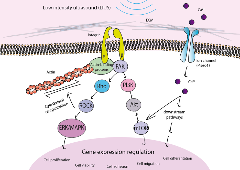
3. Mechanism of Action
4. Therapeutic Applications of Ultrasound
4.1. Search Strategy
4.2. Preclinical Studies with Ultrasound
4.2.1. Preclinical Studies in Bone Healing
4.2.2. Preclinical Studies in Other Tissues
4.3. Clinical Trials with Ultrasound
5. Limitations
6. Conclusions
Supplementary Materials
Author Contributions
Funding
Acknowledgments
Conflicts of Interest
References
- Baldari, S.; Di Rocco, G.; Piccoli, M.; Pozzobon, M.; Muraca, M.; Toietta, G. Challenges and Strategies for Improving the Regenerative Effects of Mesenchymal Stromal Cell-Based Therapies. Int. J. Mol. Sci. 2017, 18, 2087. [Google Scholar] [CrossRef] [PubMed]
- Reisman, M.; Adams, K.T. Stem cell therapy: A look at current research, regulations, and remaining hurdles. P T A Peer Rev. J. Formul. Manag. 2014, 39, 846–857. [Google Scholar]
- Sennoga, C.A.; Kanbar, E.; Auboire, L.; Dujardin, P.A.; Fouan, D.; Escoffre, J.M.; Bouakaz, A. Microbubble-mediated ultrasound drug-delivery and therapeutic monitoring. Expert Opin. Drug Deliv. 2017, 14, 1031–1043. [Google Scholar] [CrossRef] [PubMed]
- Miller, D.L.; Smith, N.B.; Bailey, M.R.; Czarnota, G.J.; Hynynen, K.; Makin, I.R.; Bioeffects Committee of the American Institute of Ultrasound in Medicine. Overview of therapeutic ultrasound applications and safety considerations. J. Ultrasound Med. Off. J. Am. Inst. Ultrasound Med. 2012, 31, 623–634. [Google Scholar] [CrossRef] [PubMed]
- Siedek, F.; Yeo, S.Y.; Heijman, E.; Grinstein, O.; Bratke, G.; Heneweer, C.; Puesken, M.; Persigehl, T.; Maintz, D.; Grull, H. Magnetic Resonance-Guided High-Intensity Focused Ultrasound (MR-HIFU): Technical Background and Overview of Current Clinical Applications (Part 1). RoFo Fortschr. Geb. Rontgenstrahlen Nukl. 2019, 191, 522–530. [Google Scholar] [CrossRef]
- Rubin, C.; Bolander, M.; Ryaby, J.P.; Hadjiargyrou, M. The use of low-intensity ultrasound to accelerate the healing of fractures. J. Bone Jt. Surg. Am. Vol. 2001, 83, 259–270. [Google Scholar] [CrossRef]
- Li, L.; Wu, S.; Liu, Z.; Zhuo, Z.; Tan, K.; Xia, H.; Zhuo, L.; Deng, X.; Gao, Y.; Xu, Y. Ultrasound-Targeted Microbubble Destruction Improves the Migration and Homing of Mesenchymal Stem Cells after Myocardial Infarction by Upregulating SDF-1/CXCR4: A Pilot Study. Stem Cells Int. 2015, 2015, 691310. [Google Scholar] [CrossRef]
- Cheung, W.H.; Chin, W.C.; Wei, F.Y.; Li, G.; Leung, K.S. Applications of exogenous mesenchymal stem cells and low intensity pulsed ultrasound enhance fracture healing in rat model. Ultrasound Med. Biol. 2013, 39, 117–125. [Google Scholar] [CrossRef]
- Liu, D.D.; Ullah, M.; Concepcion, W.; Dahl, J.J.; Thakor, A.S. The role of ultrasound in enhancing mesenchymal stromal cell-based therapies. Stem Cells Transl. Med. 2020, 9, 850–866. [Google Scholar] [CrossRef]
- Ziadloo, A.; Burks, S.R.; Gold, E.M.; Lewis, B.K.; Chaudhry, A.; Merino, M.J.; Frenkel, V.; Frank, J.A. Enhanced homing permeability and retention of bone marrow stromal cells by noninvasive pulsed focused ultrasound. Stem Cells 2012, 30, 1216–1227. [Google Scholar] [CrossRef]
- Haake, M.; Buch, M.; Schoellner, C.; Goebel, F.; Vogel, M.; Mueller, I.; Hausdorf, J.; Zamzow, K.; Schade-Brittinger, C.; Mueller, H.H. Extracorporeal shock wave therapy for plantar fasciitis: Randomised controlled multicentre trial. BMJ 2003, 327, 75. [Google Scholar] [CrossRef] [PubMed]
- Stania, M.; Juras, G.; Chmielewska, D.; Polak, A.; Kucio, C.; Krol, P. Extracorporeal Shock Wave Therapy for Achilles Tendinopathy. Biomed Res. Int. 2019, 2019, 3086910. [Google Scholar] [CrossRef] [PubMed]
- Li, L.; Yang, Z.; Zhang, H.; Chen, W.; Chen, M.; Zhu, Z. Low-intensity pulsed ultrasound regulates proliferation and differentiation of osteoblasts through osteocytes. Biochem. Biophys. Res. Commun. 2012, 418, 296–300. [Google Scholar] [CrossRef] [PubMed]
- Gleizal, A.; Ferreira, S.; Lavandier, B.; Simon, B.; Beziat, J.L.; Bera, J.C. The impact of low intensity pulsed ultrasound on mouse skull bone osteoblast cultures. Rev. Stomatol. Chir. Maxillo Faciale 2010, 111, 280–285. [Google Scholar] [CrossRef]
- Wu, L.; Lin, L.; Qin, Y.X. Enhancement of cell ingrowth, proliferation, and early differentiation in a three-dimensional silicon carbide scaffold using low-intensity pulsed ultrasound. Tissue Eng. Part A 2015, 21, 53–61. [Google Scholar] [CrossRef]
- Ikeda, K.; Takayama, T.; Suzuki, N.; Shimada, K.; Otsuka, K.; Ito, K. Effects of low-intensity pulsed ultrasound on the differentiation of C2C12 cells. Life Sci. 2006, 79, 1936–1943. [Google Scholar] [CrossRef]
- Kusuyama, J.; Nakamura, T.; Ohnishi, T.; Eiraku, N.; Noguchi, K.; Matsuguchi, T. Low-Intensity Pulsed Ultrasound (LIPUS) Promotes BMP9-Induced Osteogenesis and Suppresses Inflammatory Responses in Human Periodontal Ligament-Derived Stem Cells. J. Orthop. Trauma 2017, 31, S4. [Google Scholar] [CrossRef]
- Gao, Q.; Walmsley, A.D.; Cooper, P.R.; Scheven, B.A. Ultrasound Stimulation of Different Dental Stem Cell Populations: Role of Mitogen-activated Protein Kinase Signaling. J. Endod. 2016, 42, 425–431. [Google Scholar] [CrossRef]
- Hu, B.; Zhang, Y.; Zhou, J.; Li, J.; Deng, F.; Wang, Z.; Song, J. Low-intensity pulsed ultrasound stimulation facilitates osteogenic differentiation of human periodontal ligament cells. PLoS ONE 2014, 9, e95168. [Google Scholar] [CrossRef]
- Ren, L.; Yang, Z.; Song, J.; Wang, Z.; Deng, F.; Li, W. Involvement of p38 MAPK pathway in low intensity pulsed ultrasound induced osteogenic differentiation of human periodontal ligament cells. Ultrasonics 2013, 53, 686–690. [Google Scholar] [CrossRef]
- Inubushi, T.; Tanaka, E.; Rego, E.B.; Kitagawa, M.; Kawazoe, A.; Ohta, A.; Okada, H.; Koolstra, J.H.; Miyauchi, M.; Takata, T.; et al. Effects of ultrasound on the proliferation and differentiation of cementoblast lineage cells. J. Periodontol. 2008, 79, 1984–1990. [Google Scholar] [CrossRef] [PubMed]
- Teo, A.; Morshedi, A.; Wang, J.C.; Zhou, Y.; Lim, M. Enhancement of Cardiomyogenesis in Murine Stem Cells by Low-Intensity Ultrasound. J. Ultrasound Med. Off. J. Am. Inst. Ultrasound Med. 2017, 36, 1693–1706. [Google Scholar] [CrossRef] [PubMed]
- Appleford, M.R.; Oh, S.; Cole, J.A.; Protivinsky, J.; Ong, J.L. Ultrasound effect on osteoblast precursor cells in trabecular calcium phosphate scaffolds. Biomaterials 2007, 28, 4788–4794. [Google Scholar] [CrossRef] [PubMed]
- He, R.; Chen, J.; Jiang, J.; Liu, B.; Liang, D.; Zhou, W.; Chen, W.; Wang, Y. Synergies of accelerating differentiation of bone marrow mesenchymal stem cells induced by low intensity pulsed ultrasound, osteogenic and endothelial inductive agent. Artif. Cells Nanomed. Biotechnol. 2019, 47, 674–684. [Google Scholar] [CrossRef]
- Amini, A.; Chien, S.; Bayat, M. Impact of Ultrasound Therapy on Stem Cell Differentiation, A Systematic Review. Curr Stem Cell Res Ther. 2020, 15, 462–472. [Google Scholar] [CrossRef]
- Angle, S.R.; Sena, K.; Sumner, D.R.; Virdi, A.S. Osteogenic differentiation of rat bone marrow stromal cells by various intensities of low-intensity pulsed ultrasound. Ultrasonics 2011, 51, 281–288. [Google Scholar] [CrossRef]
- Yoon, J.H.; Roh, E.Y.; Shin, S.; Jung, N.H.; Song, E.Y.; Lee, D.S.; Han, K.S.; Kim, J.S.; Kim, B.J.; Jeon, H.W.; et al. Introducing pulsed low-intensity ultrasound to culturing human umbilical cord-derived mesenchymal stem cells. Biotechnol. Lett. 2009, 31, 329–335. [Google Scholar] [CrossRef]
- Xu, P.; Gul-Uludag, H.; Ang, W.T.; Yang, X.; Huang, M.; Marquez-Curtis, L.; McGann, L.; Janowska-Wieczorek, A.; Xing, J.; Swanson, E.; et al. Low-intensity pulsed ultrasound-mediated stimulation of hematopoietic stem/progenitor cell viability, proliferation and differentiation in vitro. Biotechnol. Lett. 2012, 34, 1965–1973. [Google Scholar] [CrossRef]
- Budhiraja, G.; Sahu, N.; Subramanian, A. Low-Intensity Ultrasound Upregulates the Expression of Cyclin-D1 and Promotes Cellular Proliferation in Human Mesenchymal Stem Cells. Biotechnol. J. 2018, 13, e1700382. [Google Scholar] [CrossRef]
- Ling, L.; Wei, T.; He, L.; Wang, Y.; Wang, Y.; Feng, X.; Zhang, W.; Xiong, Z. Low-intensity pulsed ultrasound activates ERK1/2 and PI3K-Akt signalling pathways and promotes the proliferation of human amnion-derived mesenchymal stem cells. Cell Prolif. 2017, 50. [Google Scholar] [CrossRef]
- Carina, V.; Costa, V.; Raimondi, L.; Pagani, S.; Sartori, M.; Figallo, E.; Setti, S.; Alessandro, R.; Fini, M.; Giavaresi, G. Effect of low-intensity pulsed ultrasound on osteogenic human mesenchymal stem cells commitment in a new bone scaffold. J. Appl. Biomater. Funct. Mater. 2017, 15, e215–e222. [Google Scholar] [CrossRef] [PubMed]
- Zhou, S.; Schmelz, A.; Seufferlein, T.; Li, Y.; Zhao, J.; Bachem, M.G. Molecular mechanisms of low intensity pulsed ultrasound in human skin fibroblasts. J. Biol. Chem. 2004, 279, 54463–54469. [Google Scholar] [CrossRef] [PubMed]
- Lv, Y.; Zhao, P.; Chen, G.; Sha, Y.; Yang, L. Effects of low-intensity pulsed ultrasound on cell viability, proliferation and neural differentiation of induced pluripotent stem cells-derived neural crest stem cells. Biotechnol. Lett. 2013, 35, 2201–2212. [Google Scholar] [CrossRef]
- Man, J.; Shelton, R.M.; Cooper, P.R.; Landini, G.; Scheven, B.A. Low intensity ultrasound stimulates osteoblast migration at different frequencies. J. Bone Miner. Metab. 2012, 30, 602–607. [Google Scholar] [CrossRef] [PubMed]
- Iwanabe, Y.; Masaki, C.; Tamura, A.; Tsuka, S.; Mukaibo, T.; Kondo, Y.; Hosokawa, R. The effect of low-intensity pulsed ultrasound on wound healing using scratch assay in epithelial cells. J. Prosthodont. Res. 2016, 60, 308–314. [Google Scholar] [CrossRef]
- Ning, G.Z.; Song, W.Y.; Xu, H.; Zhu, R.S.; Wu, Q.L.; Wu, Y.; Zhu, S.B.; Li, J.Q.; Wang, M.; Qu, Z.G.; et al. Bone marrow mesenchymal stem cells stimulated with low-intensity pulsed ultrasound: Better choice of transplantation treatment for spinal cord injury: Treatment for SCI by LIPUS-BMSCs transplantation. CNS Neurosci. Ther. 2019, 25, 496–508. [Google Scholar] [CrossRef]
- Lim, K.; Kim, J.; Seonwoo, H.; Park, S.H.; Choung, P.H.; Chung, J.H. In vitro effects of low-intensity pulsed ultrasound stimulation on the osteogenic differentiation of human alveolar bone-derived mesenchymal stem cells for tooth tissue engineering. Biomed Res. Int. 2013, 2013, 269724. [Google Scholar] [CrossRef]
- Lee, H.J.; Choi, B.H.; Min, B.H.; Park, S.R. Low-intensity ultrasound inhibits apoptosis and enhances viability of human mesenchymal stem cells in three-dimensional alginate culture during chondrogenic differentiation. Tissue Eng. 2007, 13, 1049–1057. [Google Scholar] [CrossRef]
- Park, S.R.; Choi, B.H.; Min, B.H. Low-Intensity Ultrasound (LIUS) as an Innovative Tool for Chondrogenesis of Mesenchymal Stem Cells (MSCs). Organogenesis 2007, 3, 74–78. [Google Scholar] [CrossRef]
- Feril, L.B., Jr.; Kondo, T.; Cui, Z.G.; Tabuchi, Y.; Zhao, Q.L.; Ando, H.; Misaki, T.; Yoshikawa, H.; Umemura, S. Apoptosis induced by the sonomechanical effects of low intensity pulsed ultrasound in a human leukemia cell line. Cancer Lett. 2005, 221, 145–152. [Google Scholar] [CrossRef]
- Shi, M.; Liu, B.; Liu, G.; Wang, P.; Yang, M.; Li, Y.; Zhou, J. Low intensity-pulsed ultrasound induced apoptosis of human hepatocellular carcinoma cells in vitro. Ultrasonics 2016, 64, 43–53. [Google Scholar] [CrossRef] [PubMed]
- Xiao, W.; Xu, Q.; Zhu, Z.; Li, L.; Chen, W. Different performances of CXCR4, integrin-1beta and CCR-2 in bone marrow stromal cells (BMSCs) migration by low-intensity pulsed ultrasound stimulation. Biomed. Technik. Biomed. Eng. 2017, 62, 89–95. [Google Scholar] [CrossRef]
- Roper, J.; Harrison, A.; Bass, M.D. Induction of adhesion-dependent signals using low-intensity ultrasound. J. Vis. Exp. 2012, 63, e4024. [Google Scholar] [CrossRef] [PubMed]
- Choi, W.H.; Choi, B.H.; Min, B.H.; Park, S.R. Low-intensity ultrasound increased colony forming unit-fibroblasts of mesenchymal stem cells during primary culture. Tissue Eng. Part C Methods 2011, 17, 517–526. [Google Scholar] [CrossRef]
- Costa, V.; Carina, V.; Fontana, S.; De Luca, A.; Monteleone, F.; Pagani, S.; Sartori, M.; Setti, S.; Faldini, C.; Alessandro, R.; et al. Osteogenic commitment and differentiation of human mesenchymal stem cells by low-intensity pulsed ultrasound stimulation. J. Cell. Physiol. 2018, 233, 1558–1573. [Google Scholar] [CrossRef]
- Zhang, X.; Hu, Z.; Hao, J.; Shen, J. Low Intensity Pulsed Ultrasound Promotes the Extracellular Matrix Synthesis of Degenerative Human Nucleus Pulposus Cells Through FAK/PI3K/Akt Pathway. Spine 2016, 41, E248–E254. [Google Scholar] [CrossRef]
- Xia, P.; Ren, S.; Lin, Q.; Cheng, K.; Shen, S.; Gao, M.; Li, X. Low-Intensity Pulsed Ultrasound Affects Chondrocyte Extracellular Matrix Production via an Integrin-Mediated p38 MAPK Signaling Pathway. Ultrasound Med. Biol. 2015, 41, 1690–1700. [Google Scholar] [CrossRef]
- Choi, B.H.; Choi, M.H.; Kwak, M.G.; Min, B.H.; Woo, Z.H.; Park, S.R. Mechanotransduction pathways of low-intensity ultrasound in C-28/I2 human chondrocyte cell line. Proc. Inst. Mech. Eng. Part H J. Eng. Med. 2007, 221, 527–535. [Google Scholar] [CrossRef]
- Bernal, A.; Perez, L.M.; De Lucas, B.; Martin, N.S.; Kadow-Romacker, A.; Plaza, G.; Raum, K.; Galvez, B.G. Low-Intensity Pulsed Ultrasound Improves the Functional Properties of Cardiac Mesoangioblasts. Stem Cell Rev. Rep. 2015, 11, 852–865. [Google Scholar] [CrossRef]
- Chen, J.; Jiang, J.; Wang, W.; Qin, J.; Chen, J.; Chen, W.; Wang, Y. Low intensity pulsed ultrasound promotes the migration of bone marrow- derived mesenchymal stem cells via activating FAK-ERK1/2 signalling pathway. Artif. Cells Nanomed. Biotechnol. 2019, 47, 3603–3613. [Google Scholar] [CrossRef]
- Wang, Y.; Li, J.; Qiu, Y.; Hu, B.; Chen, J.; Fu, T.; Zhou, P.; Song, J. Low intensity pulsed ultrasound promotes periodontal ligament stem cell migration through TWIST1mediated SDF1 expression. Int. J. Mol. Med. 2018, 42, 322–330. [Google Scholar] [CrossRef] [PubMed]
- Leng, X.; Shang, J.; Gao, D.; Wu, J. Low-intensity pulsed ultrasound promotes proliferation and migration of HaCaT keratinocytes through the PI3K/AKT and JNK pathways. Braz. J. Med. Biol. Res. Rev. Bras. Pesqui. Med. Biol. 2018, 51, e7862. [Google Scholar] [CrossRef] [PubMed]
- Atherton, P.; Lausecker, F.; Harrison, A.; Ballestrem, C. Low-intensity pulsed ultrasound promotes cell motility through vinculin-controlled Rac1 GTPase activity. J. Cell Sci. 2017, 130, 2277–2291. [Google Scholar] [CrossRef] [PubMed]
- Wei, F.Y.; Leung, K.S.; Li, G.; Qin, J.; Chow, S.K.; Huang, S.; Sun, M.H.; Qin, L.; Cheung, W.H. Low intensity pulsed ultrasound enhanced mesenchymal stem cell recruitment through stromal derived factor-1 signaling in fracture healing. PLoS ONE 2014, 9, e106722. [Google Scholar] [CrossRef] [PubMed]
- Burks, S.R.; Ziadloo, A.; Hancock, H.A.; Chaudhry, A.; Dean, D.D.; Lewis, B.K.; Frenkel, V.; Frank, J.A. Investigation of cellular and molecular responses to pulsed focused ultrasound in a mouse model. PLoS ONE 2011, 6, e24730. [Google Scholar] [CrossRef] [PubMed]
- Tebebi, P.A.; Kim, S.J.; Williams, R.A.; Milo, B.; Frenkel, V.; Burks, S.R.; Frank, J.A. Improving the therapeutic efficacy of mesenchymal stromal cells to restore perfusion in critical limb ischemia through pulsed focused ultrasound. Sci. Rep. 2017, 7, 41550. [Google Scholar] [CrossRef]
- Matsumoto, K.; Shimo, T.; Kurio, N.; Okui, T.; Ibaragi, S.; Kunisada, Y.; Obata, K.; Masui, M.; Pai, P.; Horikiri, Y.; et al. Low-intensity pulsed ultrasound stimulation promotes osteoblast differentiation through hedgehog signaling. J. Cell. Biochem. 2018, 119, 4352–4360. [Google Scholar] [CrossRef]
- Harrison, A.; Lin, S.; Pounder, N.; Mikuni-Takagaki, Y. Mode & mechanism of low intensity pulsed ultrasound (LIPUS) in fracture repair. Ultrasonics 2016, 70, 45–52. [Google Scholar] [CrossRef]
- Kusuyama, J.; Bandow, K.; Shamoto, M.; Kakimoto, K.; Ohnishi, T.; Matsuguchi, T. Low intensity pulsed ultrasound (LIPUS) influences the multilineage differentiation of mesenchymal stem and progenitor cell lines through ROCK-Cot/Tpl2-MEK-ERK signaling pathway. J. Biol. Chem. 2014, 289, 10330–10344. [Google Scholar] [CrossRef]
- Ying, Z.M.; Lin, T.; Yan, S.G. Low-intensity pulsed ultrasound therapy: A potential strategy to stimulate tendon-bone junction healing. J. Zhejiang Univ. Sci. B 2012, 13, 955–963. [Google Scholar] [CrossRef]
- An, Y.; Song, Y.; Wang, Z.; Wang, J.; Wu, G.; Zhu, G.; Chen, L. Effect of low-intensity pulsed ultrasound on the biological behaviors of bone marrow mesenchymal stem cells on titanium with different surface topographies. Am. J. Transl. Res. 2018, 10, 67–76. [Google Scholar] [PubMed]
- Zhou, X.; Castro, N.J.; Zhu, W.; Cui, H.; Aliabouzar, M.; Sarkar, K.; Zhang, L.G. Improved Human Bone Marrow Mesenchymal Stem Cell Osteogenesis in 3D Bioprinted Tissue Scaffolds with Low Intensity Pulsed Ultrasound Stimulation. Sci. Rep. 2016, 6, 32876. [Google Scholar] [CrossRef] [PubMed]
- Chiu, C.Y.; Tsai, T.L.; Vanderby, R., Jr.; Bradica, G.; Lou, S.L.; Li, W.J. Osteoblastogenesis of Mesenchymal Stem Cells in 3-D Culture Enhanced by Low-Intensity Pulsed Ultrasound through Soluble Receptor Activator of Nuclear Factor Kappa B Ligand. Ultrasound Med. Biol. 2015, 41, 1842–1852. [Google Scholar] [CrossRef]
- Lai, C.H.; Chen, S.C.; Chiu, L.H.; Yang, C.B.; Tsai, Y.H.; Zuo, C.S.; Chang, W.H.; Lai, W.F. Effects of low-intensity pulsed ultrasound, dexamethasone/TGF-beta1 and/or BMP-2 on the transcriptional expression of genes in human mesenchymal stem cells: Chondrogenic vs. osteogenic differentiation. Ultrasound Med. Biol. 2010, 36, 1022–1033. [Google Scholar] [CrossRef] [PubMed]
- Aliabouzar, M.; Lee, S.J.; Zhou, X.; Zhang, G.L.; Sarkar, K. Effects of scaffold microstructure and low intensity pulsed ultrasound on chondrogenic differentiation of human mesenchymal stem cells. Biotechnol. Bioeng. 2018, 115, 495–506. [Google Scholar] [CrossRef]
- Aliabouzar, M.; Zhang, L.G.; Sarkar, K. Lipid Coated Microbubbles and Low Intensity Pulsed Ultrasound Enhance Chondrogenesis of Human Mesenchymal Stem Cells in 3D Printed Scaffolds. Sci. Rep. 2016, 6, 37728. [Google Scholar] [CrossRef]
- Cui, J.H.; Park, S.R.; Park, K.; Choi, B.H.; Min, B.H. Preconditioning of mesenchymal stem cells with low-intensity ultrasound for cartilage formation in vivo. Tissue Eng. 2007, 13, 351–360. [Google Scholar] [CrossRef]
- Lee, H.J.; Choi, B.H.; Min, B.H.; Son, Y.S.; Park, S.R. Low-intensity ultrasound stimulation enhances chondrogenic differentiation in alginate culture of mesenchymal stem cells. Artif. Organs 2006, 30, 707–715. [Google Scholar] [CrossRef]
- Schumann, D.; Kujat, R.; Zellner, J.; Angele, M.K.; Nerlich, M.; Mayr, E.; Angele, P. Treatment of human mesenchymal stem cells with pulsed low intensity ultrasound enhances the chondrogenic phenotype in vitro. Biorheology 2006, 43, 431–443. [Google Scholar]
- Ebisawa, K.; Hata, K.; Okada, K.; Kimata, K.; Ueda, M.; Torii, S.; Watanabe, H. Ultrasound enhances transforming growth factor beta-mediated chondrocyte differentiation of human mesenchymal stem cells. Tissue Eng. 2004, 10, 921–929. [Google Scholar] [CrossRef]
- Fu, N.; Yang, X.; Ba, K.; Fu, Y.; Wei, X.; Yue, Y.; Li, G.; Yao, Y.; Chen, J.; Cai, X.; et al. Low-intensity pulsed ultrasound induced enhanced adipogenesis of adipose-derived stem cells. Cell Prolif. 2013, 46, 312–319. [Google Scholar] [CrossRef] [PubMed]
- Li, F.; Liu, Y.; Cai, Y.; Li, X.; Bai, M.; Sun, T.; Du, L. Ultrasound Irradiation Combined with Hepatocyte Growth Factor Accelerate the Hepatic Differentiation of Human Bone Marrow Mesenchymal Stem Cells. Ultrasound Med. Biol. 2018, 44, 1044–1052. [Google Scholar] [CrossRef] [PubMed]
- Liu, S.H.; Lai, Y.L.; Chen, B.L.; Yang, F.Y. Ultrasound Enhances the Expression of Brain-Derived Neurotrophic Factor in Astrocyte Through Activation of TrkB-Akt and Calcium-CaMK Signaling Pathways. Cereb. Cortex 2017, 27, 3152–3160. [Google Scholar] [CrossRef] [PubMed]
- Cui, J.H.; Park, K.; Park, S.R.; Min, B.H. Effects of low-intensity ultrasound on chondrogenic differentiation of mesenchymal stem cells embedded in polyglycolic acid: An in vivo study. Tissue Eng. 2006, 12, 75–82. [Google Scholar] [CrossRef]
- Xia, P.; Wang, X.; Qu, Y.; Lin, Q.; Cheng, K.; Gao, M.; Ren, S.; Zhang, T.; Li, X. TGF-beta1-induced chondrogenesis of bone marrow mesenchymal stem cells is promoted by low-intensity pulsed ultrasound through the integrin-mTOR signaling pathway. Stem Cell Res. Ther. 2017, 8, 281. [Google Scholar] [CrossRef]
- Wang, X.; Lin, Q.; Zhang, T.; Wang, X.; Cheng, K.; Gao, M.; Xia, P.; Li, X. Low-intensity pulsed ultrasound promotes chondrogenesis of mesenchymal stem cells via regulation of autophagy. Stem Cell Res. Ther. 2019, 10, 41. [Google Scholar] [CrossRef]
- Zhang, Z.; Ma, Y.; Guo, S.; He, Y.; Bai, G.; Zhang, W. Low-intensity pulsed ultrasound stimulation facilitates in vitro osteogenic differentiation of human adipose-derived stem cells via up-regulation of heat shock protein (HSP)70, HSP90, and bone morphogenetic protein (BMP) signaling pathway. Biosci. Rep. 2018, 38. [Google Scholar] [CrossRef]
- Wu, S.; Kawahara, Y.; Manabe, T.; Ogawa, K.; Matsumoto, M.; Sasaki, A.; Yuge, L. Low-intensity pulsed ultrasound accelerates osteoblast differentiation and promotes bone formation in an osteoporosis rat model. Pathobiol. J. Immunopathol. Mol. Cell. Biol. 2009, 76, 99–107. [Google Scholar] [CrossRef]
- Cho, S.E.; Kim, Y.M.; Jeong, J.S.; Seo, Y.K. The effect of ultrasound for increasing neural differentiation in hBM-MSCs and inducing neurogenesis in ischemic stroke model. Life Sci. 2016, 165, 35–42. [Google Scholar] [CrossRef]
- Xia, B.; Zou, Y.; Xu, Z.; Lv, Y. Gene expression profiling analysis of the effects of low-intensity pulsed ultrasound on induced pluripotent stem cell-derived neural crest stem cells. Biotechnol. Appl. Biochem. 2017, 64, 927–937. [Google Scholar] [CrossRef]
- Xia, B.; Chen, G.; Zou, Y.; Yang, L.; Pan, J.; Lv, Y. Low-intensity pulsed ultrasound combination with induced pluripotent stem cells-derived neural crest stem cells and growth differentiation factor 5 promotes sciatic nerve regeneration and functional recovery. J. Tissue Eng. Regen. Med. 2019, 13, 625–636. [Google Scholar] [CrossRef] [PubMed]
- Kusuyama, J.; Hwan Seong, C.; Ohnishi, T.; Bandow, K.; Matsuguchi, T. 10. Low-Intensity Pulsed Ultrasound (LIPUS) Stimulation Helps to Maintain the Differentiation Potency of Mesenchymal Stem Cells by Induction in Nanog Protein Transcript Levels and Phosphorylation. J. Orthop. Trauma 2016, 30, S4–S5. [Google Scholar] [CrossRef] [PubMed]
- Hasuike, A.; Sato, S.; Udagawa, A.; Ando, K.; Arai, Y.; Ito, K. In vivo bone regenerative effect of low-intensity pulsed ultrasound in rat calvarial defects. Oral Surg. Oral Med. Oral Pathol. Oral Radiol. Endod. 2011, 111, e12–e20. [Google Scholar] [CrossRef] [PubMed]
- Wang, Y.; Qiu, Y.; Li, J.; Zhao, C.; Song, J. Low-intensity pulsed ultrasound promotes alveolar bone regeneration in a periodontal injury model. Ultrasonics 2018, 90, 166–172. [Google Scholar] [CrossRef]
- Yamaguchi, S.; Aoyama, T.; Ito, A.; Nagai, M.; Iijima, H.; Tajino, J.; Zhang, X.; Wataru, K.; Kuroki, H. Effect of Low-Intensity Pulsed Ultrasound after Mesenchymal Stromal Cell Injection to Treat Osteochondral Defects: An In Vivo Study. Ultrasound Med. Biol. 2016, 42, 2903–2913. [Google Scholar] [CrossRef]
- Hui, C.F.; Chan, C.W.; Yeung, H.Y.; Lee, K.M.; Qin, L.; Li, G.; Leung, K.S.; Hu, Y.Y.; Cheng, J.C. Low-intensity pulsed ultrasound enhances posterior spinal fusion implanted with mesenchymal stem cells-calcium phosphate composite without bone grafting. Spine 2011, 36, 1010–1016. [Google Scholar] [CrossRef]
- Lv, Y.; Nan, P.; Chen, G.; Sha, Y.; Xia, B.; Yang, L. In vivo repair of rat transected sciatic nerve by low-intensity pulsed ultrasound and induced pluripotent stem cells-derived neural crest stem cells. Biotechnol. Lett. 2015, 37, 2497–2506. [Google Scholar] [CrossRef]
- Peng, D.Y.; Reed-Maldonado, A.B.; Lin, G.T.; Xia, S.J.; Lue, T.F. Low-intensity pulsed ultrasound for regenerating peripheral nerves: Potential for penile nerve. Asian J. Androl. 2020, 22, 335–341. [Google Scholar] [CrossRef]
- Shindo, T.; Ito, K.; Ogata, T.; Hatanaka, K.; Kurosawa, R.; Eguchi, K.; Kagaya, Y.; Hanawa, K.; Aizawa, K.; Shiroto, T.; et al. Low-Intensity Pulsed Ultrasound Enhances Angiogenesis and Ameliorates Left Ventricular Dysfunction in a Mouse Model of Acute Myocardial Infarction. Arterioscler. Thromb. Vasc. Biol. 2016, 36, 1220–1229. [Google Scholar] [CrossRef]
- Higashi, Y.; Azuma, N.; Takeishi, Y.; Minamino, T.; Kihara, Y.; Node, K.; Sata, M.; Fukumoto, Y.; Origasa, H.; Matsuo, H.; et al. Effect of a Low-Intensity Pulsed Ultrasound Device, SX-1001, on Clinical Symptoms in Buerger Disease With Limb Ischemia. Int. Heart J. 2015, 56, 632–638. [Google Scholar] [CrossRef]
- Ramli, R.; Reher, P.; Harris, M.; Meghji, S. The effect of ultrasound on angiogenesis: An in vivo study using the chick chorioallantoic membrane. Int. J. Oral Maxillofac. Implant 2009, 24, 591–596. [Google Scholar]
- Mizrahi, N.; Seliktar, D.; Kimmel, E. Ultrasound-induced angiogenic response in endothelial cells. Ultrasound Med. Biol. 2007, 33, 1818–1829. [Google Scholar] [CrossRef] [PubMed]
- Barzelai, S.; Sharabani-Yosef, O.; Holbova, R.; Castel, D.; Walden, R.; Engelberg, S.; Scheinowitz, M. Low-intensity ultrasound induces angiogenesis in rat hind-limb ischemia. Ultrasound Med. Biol. 2006, 32, 139–145. [Google Scholar] [CrossRef] [PubMed]
- Kang, P.L.; Huang, H.H.; Chen, T.; Ju, K.C.; Kuo, S.M. Angiogenesis-promoting effect of LIPUS on hADSCs and HUVECs cultured on collagen/hyaluronan scaffolds. Mater. Sci. Eng. C Mater. Biol. Appl. 2019, 102, 22–33. [Google Scholar] [CrossRef] [PubMed]
- Hanawa, K.; Ito, K.; Aizawa, K.; Shindo, T.; Nishimiya, K.; Hasebe, Y.; Tuburaya, R.; Hasegawa, H.; Yasuda, S.; Kanai, H.; et al. Low-intensity pulsed ultrasound induces angiogenesis and ameliorates left ventricular dysfunction in a porcine model of chronic myocardial ischemia. PLoS ONE 2014, 9, e104863. [Google Scholar] [CrossRef]
- Zheng, C.; Wu, S.M.; Lian, H.; Lin, Y.Z.; Zhuang, R.; Thapa, S.; Chen, Q.Z.; Chen, Y.F.; Lin, J.F. Low-intensity pulsed ultrasound attenuates cardiac inflammation of CVB3-induced viral myocarditis via regulation of caveolin-1 and MAPK pathways. J. Cell. Mol. Med. 2019, 23, 1963–1975. [Google Scholar] [CrossRef]
- Zhao, X.; Zhao, G.; Shi, Z.; Zhou, C.; Chen, Y.; Hu, B.; Yan, S. Low-intensity pulsed ultrasound (LIPUS) prevents periprosthetic inflammatory loosening through FBXL2-TRAF6 ubiquitination pathway. Sci. Rep. 2017, 7, 45779. [Google Scholar] [CrossRef]
- Zhang, B.; Chen, H.; Ouyang, J.; Xie, Y.; Chen, L.; Tan, Q.; Du, X.; Su, N.; Ni, Z.; Chen, L. SQSTM1-dependent autophagic degradation of PKM2 inhibits the production of mature IL1B/IL-1beta and contributes to LIPUS-mediated anti-inflammatory effect. Autophagy 2020, 16, 1262–1278. [Google Scholar] [CrossRef]
- Nakamura, T.; Fujihara, S.; Yamamoto-Nagata, K.; Katsura, T.; Inubushi, T.; Tanaka, E. Low-intensity pulsed ultrasound reduces the inflammatory activity of synovitis. Ann. Biomed. Eng. 2011, 39, 2964–2971. [Google Scholar] [CrossRef]
- Li, H.; Deng, Y.; Tan, M.; Feng, G.; Kuang, Y.; Li, J.; Song, J. Low-intensity pulsed ultrasound upregulates osteogenesis under inflammatory conditions in periodontal ligament stem cells through unfolded protein response. Stem Cell Res. Ther. 2020, 11, 215. [Google Scholar] [CrossRef]
- Sato, M.; Kuroda, S.; Mansjur, K.Q.; Khaliunaa, G.; Nagata, K.; Horiuchi, S.; Inubushi, T.; Yamamura, Y.; Azuma, M.; Tanaka, E. Low-intensity pulsed ultrasound rescues insufficient salivary secretion in autoimmune sialadenitis. Arthritis Res. Ther. 2015, 17, 278. [Google Scholar] [CrossRef] [PubMed]
- Yang, F.Y.; Lu, W.W.; Lin, W.T.; Chang, C.W.; Huang, S.L. Enhancement of Neurotrophic Factors in Astrocyte for Neuroprotective Effects in Brain Disorders Using Low-intensity Pulsed Ultrasound Stimulation. Brain Stimul. 2015, 8, 465–473. [Google Scholar] [CrossRef] [PubMed]
- Zhou, J.X.; Liu, Y.J.; Chen, X.; Zhang, X.; Xu, J.; Yang, K.; Wang, D.; Lin, S.; Ye, J. Low-Intensity Pulsed Ultrasound Protects Retinal Ganglion Cell From Optic Nerve Injury Induced Apoptosis via Yes Associated Protein. Front. Cell. Neurosci. 2018, 12, 160. [Google Scholar] [CrossRef] [PubMed]
- Padilla, F.; Puts, R.; Vico, L.; Raum, K. Stimulation of bone repair with ultrasound: A review of the possible mechanic effects. Ultrasonics 2014, 54, 1125–1145. [Google Scholar] [CrossRef]
- Sato, M.; Nagata, K.; Kuroda, S.; Horiuchi, S.; Nakamura, T.; Karima, M.; Inubushi, T.; Tanaka, E. Low-intensity pulsed ultrasound activates integrin-mediated mechanotransduction pathway in synovial cells. Ann. Biomed. Eng. 2014, 42, 2156–2163. [Google Scholar] [CrossRef]
- Qiu, Z.; Guo, J.; Kala, S.; Zhu, J.; Xian, Q.; Qiu, W.; Li, G.; Zhu, T.; Meng, L.; Zhang, R.; et al. The Mechanosensitive Ion Channel Piezo1 Significantly Mediates In Vitro Ultrasonic Stimulation of Neurons. iScience 2019, 21, 448–457. [Google Scholar] [CrossRef]
- De Lucas, B.; Perez, L.M.; Galvez, B.G. Importance and regulation of adult stem cell migration. J. Cell. Mol. Med. 2018, 22, 746–754. [Google Scholar] [CrossRef]
- Choi, B.H.; Kim, K.H.; Karmacharya, M.B.; Min, B.-H.; Park, S.R. Low-Intensity Ultrasound in Stem Cells and Tissue Engineering. Cell Mol. Biol. Imaging Stem Cells 2014, 45–65. [Google Scholar] [CrossRef]
- Wu, C. Focal adhesion: A focal point in current cell biology and molecular medicine. Cell Adhes. Migr. 2007, 1, 13–18. [Google Scholar] [CrossRef]
- Takeuchi, R.; Ryo, A.; Komitsu, N.; Mikuni-Takagaki, Y.; Fukui, A.; Takagi, Y.; Shiraishi, T.; Morishita, S.; Yamazaki, Y.; Kumagai, K.; et al. Low-intensity pulsed ultrasound activates the phosphatidylinositol 3 kinase/Akt pathway and stimulates the growth of chondrocytes in three-dimensional cultures: A basic science study. Arthritis Res. Ther. 2008, 10, R77. [Google Scholar] [CrossRef]
- Prieto, M.L.; Firouzi, K.; Khuri-Yakub, B.T.; Maduke, M. Activation of Piezo1 but Not NaV1.2 Channels by Ultrasound at 43 MHz. Ultrasound Med. Biol. 2018, 44, 1217–1232. [Google Scholar] [CrossRef] [PubMed]
- Gao, Q.; Cooper, P.R.; Walmsley, A.D.; Scheven, B.A. Role of Piezo Channels in Ultrasound-stimulated Dental Stem Cells. J. Endod. 2017, 43, 1130–1136. [Google Scholar] [CrossRef] [PubMed]
- Sugimoto, A.; Miyazaki, A.; Kawarabayashi, K.; Shono, M.; Akazawa, Y.; Hasegawa, T.; Ueda-Yamaguchi, K.; Kitamura, T.; Yoshizaki, K.; Fukumoto, S.; et al. Piezo type mechanosensitive ion channel component 1 functions as a regulator of the cell fate determination of mesenchymal stem cells. Sci. Rep. 2017, 7, 17696. [Google Scholar] [CrossRef] [PubMed]
- Han, Y.; Liu, C.; Zhang, D.; Men, H.; Huo, L.; Geng, Q.; Wang, S.; Gao, Y.; Zhang, W.; Zhang, Y.; et al. Mechanosensitive ion channel Piezo1 promotes prostate cancer development through the activation of the Akt/mTOR pathway and acceleration of cell cycle. Int. J. Oncol. 2019, 55, 629–644. [Google Scholar] [CrossRef]
- Pilla, A.A.; Mont, M.A.; Nasser, P.R.; Khan, S.A.; Figueiredo, M.; Kaufman, J.J.; Siffert, R.S. Non-invasive low-intensity pulsed ultrasound accelerates bone healing in the rabbit. J. Orthop. Trauma 1990, 4, 246–253. [Google Scholar] [CrossRef]
- Garg, P.; Mazur, M.M.; Buck, A.C.; Wandtke, M.E.; Liu, J.; Ebraheim, N.A. Prospective Review of Mesenchymal Stem Cells Differentiation into Osteoblasts. Orthop. Surg. 2017, 9, 13–19. [Google Scholar] [CrossRef]
- Yang, B.; Li, M.; Lei, H.; Xu, Y.; Li, H.; Gao, Z.; Guan, R.; Xin, Z. Low Intensity Pulsed Ultrasound Influences the Myogenic Differentiation of Muscle Satellite Cells in a Stress Urinary Incontinence Rat Model. Urology 2019, 123, 297.e1–297.e8. [Google Scholar] [CrossRef]
- Sato, M.; Motoyoshi, M.; Shinoda, M.; Iwata, K.; Shimizu, N. Low-intensity pulsed ultrasound accelerates nerve regeneration following inferior alveolar nerve transection in rats. Eur. J. Oral Sci. 2016, 124, 246–250. [Google Scholar] [CrossRef]
- Wang, F.; Li, Y.; Yang, Z.; Lu, K.; Zuo, J.; Zhou, Z. Effect of Low-Intensity Pulsed Ultrasound on a Rat Model of Dentin-Dental Pulp Injury and Repair. Ultrasound Med. Biol. 2017, 43, 163–175. [Google Scholar] [CrossRef]
- Maeda, T.; Masaki, C.; Kanao, M.; Kondo, Y.; Ohta, A.; Nakamoto, T.; Hosokawa, R. Low-intensity pulsed ultrasound enhances palatal mucosa wound healing in rats. J. Prosthodont. Res. 2013, 57, 93–98. [Google Scholar] [CrossRef]
- El-Bialy, T.; El-Shamy, I.; Graber, T.M. Repair of orthodontically induced root resorption by ultrasound in humans. Am. J. Orthod. Dentofac. Orthop. 2004, 126, 186–193. [Google Scholar] [CrossRef] [PubMed]
- Young, S.R.; Dyson, M. The effect of therapeutic ultrasound on angiogenesis. Ultrasound Med. Biol. 1990, 16, 261–269. [Google Scholar] [CrossRef]
- Poolman, R.W.; Agoritsas, T.; Siemieniuk, R.A.; Harris, I.A.; Schipper, I.B.; Mollon, B.; Smith, M.; Albin, A.; Nador, S.; Sasges, W.; et al. Low intensity pulsed ultrasound (LIPUS) for bone healing: A clinical practice guideline. BMJ 2017, 356, j576. [Google Scholar] [CrossRef] [PubMed]
- Tarride, J.E.; Hopkins, R.B.; Blackhouse, G.; Burke, N.; Bhandari, M.; Johal, H.; Guyatt, G.H.; Busse, J.W. Low-intensity pulsed ultrasound for treatment of tibial fractures: An economic evaluation of the TRUST study. Bone Jt. J. 2017, 99-B, 1526–1532. [Google Scholar] [CrossRef] [PubMed]
- TRUST Investigators writing group; Busse, J.W.; Bhandari, M.; Einhorn, T.A.; Schemitsch, E.; Heckman, J.D.; Tornetta, P., III; Leung, K.S.; Heels-Ansdell, D.; Makosso-Kallyth, S.; et al. Re-evaluation of low intensity pulsed ultrasound in treatment of tibial fractures (TRUST): Randomized clinical trial. BMJ 2016, 355, i5351. [Google Scholar] [CrossRef][Green Version]
- Zhou, X.Y.; Zhang, X.X.; Yu, G.Y.; Zhang, Z.C.; Wang, F.; Yang, Y.L.; Li, M.; Wei, X.Z. Effects of Low-Intensity Pulsed Ultrasound on Knee Osteoarthritis: A Meta-Analysis of Randomized Clinical Trials. Biomed Res. Int. 2018, 2018, 7469197. [Google Scholar] [CrossRef]
- Cruz, J.M.; Hauck, M.; Cardoso Pereira, A.P.; Moraes, M.B.; Martins, C.N.; da Silva Paulitsch, F.; Plentz, R.D.; Peres, W.; Vargas da Silva, A.M.; Signori, L.U. Effects of Different Therapeutic Ultrasound Waveforms on Endothelial Function in Healthy Volunteers: A Randomized Clinical Trial. Ultrasound Med. Biol. 2016, 42, 471–480. [Google Scholar] [CrossRef]
- Yang, K.H.; Parvizi, J.; Wang, S.J.; Lewallen, D.G.; Kinnick, R.R.; Greenleaf, J.F.; Bolander, M.E. Exposure to low-intensity ultrasound increases aggrecan gene expression in a rat femur fracture model. J. Orthop. Res. Off. Publ. Orthop. Res. Soc. 1996, 14, 802–809. [Google Scholar] [CrossRef]


| Parameters | Applications | ||
|---|---|---|---|
| Therapeutic Ultrasound (<5 MHz) | Traditional | 0.1–3 W/cm2 SATA, frequency 1–3 MHz | tendonitis, osteoarthritis and pain relief |
| LIPUS | 30–100 mW/cm2 SATA, frequency 1.5 MHz, 1 kHz, duty cycle of 20% | Bone fracture healing, soft tissue regeneration, anti-inflammatory effects… | |
| cLIUS | 30–100 mW/cm2 SATA, frequency 1.5 MHz, 1 kHz, duty cycle of 100% | ||
| pFUS | 133 W/cm2 SATA, frequency 1 MHz, 5 Hz, duty cycle of 5% | Chemoattractant local and temporal gradient for homing process | |
| HIFU | 400–10,000 W/cm2 SATA, frequency 0.8–4 MHz | non-invasive thermal or mechanical ablation of benign and malignant tissue | |
| Shock waves | 300–3000 pulses, energy 0.05–0.12 mJ/mm2 | kidney stone lithotripsy, physical therapy | |
| Biological Effects | Ultrasound Parameters | References |
|---|---|---|
| Proliferation and viability | frequency 1/1.5 MHz; 30/50/100 mW/cm2 SATA; 20%/100% duty cycle; 10/20/30 min/day | [28,30,32,33,34,36,39,41] |
| Adhesion | frequency 1.5 MHz; 30/100 mW/cm2 SATA; 20%/100% duty cycle; 10/20 min/day | [42,43,44] |
| Extracellular matrix production | frequency 1/1.5/3 MHz; 30/200 mW/cm2 SATA; 20% duty cycle; 15/20 min/day for 5/7 consecutive days | [45,46,47,48] |
| Migration | frequency 1/1.5/3 MHz; 30/160/240 mW/cm2 SATA; 20%/100% duty cycle; 15/20 min/day | [34,35,42,51,53] |
| Homing | LIPUS: frequency 1.5 MHz; 30 mW/cm2 SATA; 20% duty cycle; 20 min/day for 3 days pFUS: 133 mW/cm2 SATA; frequency 1 MHz; 1/5 Hz; 5% duty cycle | [54,55,56] |
| Differentiation | frequency 1/1.5/2 MHz; 30/50/150/200/300/500 mW/cm2 SATA; 20% duty cycle; 10/20/30 min/day | [28,36,45,62,64,67,72,77,81] |
| Regenerative effects | frequency 1.5/1.6 MHz; 30/50/90 mW/cm2 SATA; 20% duty cycle; 20 min/day | [8,36,83,84,86] |
| Angiogenic effects | frequency 1/1875 MHz; 15/25 mW/cm2 SATA; 20% duty cycle; 20 min/day | [89,91,94,95] |
| Anti-inflammatory effects | frequency 1.5/3 MHz; 30/200 mW/cm2 SATA; 20% duty cycle; 15/20 min/day | [95,97,99,101] |
| Anti-degenerative effects | frequency 1 MHz; 50/110 mW/cm2 SATA; 20%/50% duty cycle; 10/15 min/day | [73,102,103] |
| Mouse | Rat | Rabbit | Others | Total | |
|---|---|---|---|---|---|
| LIPUS | |||||
| 1990–1999 | 0 | 1 | 1 | 0 | 2 |
| 2000–2009 | 2 | 5 | 4 | 3 | 14 |
| 2010–2020 | 5 | 23 | 13 | 1 | 42 |
| Shock Wave | - | - | - | - | 31 |
| Mouse | Rat | Rabbit | Other | Total | |
|---|---|---|---|---|---|
| LIPUS | 16 | ||||
| 1990–1999 | 1 | 13 | 0 | 2 | |
| Shock Wave | - | - | - | - | 19 |
| NCT | Condition | Treatment | Outcome |
|---|---|---|---|
| NCT03705039 | Knee osteoarthritis | Ultrasound (1 mW/cm2, frequency 1 MHz, 1 kHz, ratio 1:4, 10 min) | Synovial fluid and cartilage thickness |
| NCT03147313 | Peripheral nerve injury | Shock wave (300 or 500 pulses, frequency 3 Hz, energy 0.1 mJ/mm2) | regeneration of peripheral nerve injuries |
| NCT04123782 | Muscle injury | Focus Extracorporeal shock wave (3000 impulses at 0.12 mJ/mm2) | Muscle injuries recovery |
| NCT02042066 | Hypertension | Shock wave (0.09–0.1 mJ/mm2) | Increase tissue perfusion |
| NCT02800200 | Carpal tunnel syndrome | Plasma and Shock wave (100 MPa, 10 μs). | Regeneration of peripheral neuropathy |
| NCT03986359 | Erectile dysfunction | Shock wave (0.05 mJ/mm2, 3000 pulses) | Increases erection hardness score |
| NCT Number | Conditions | Interventions | Outcome |
|---|---|---|---|
| NCT00423956 | Root resorption | LIPUS | Induced inflammatory root resorption |
| NCT00744861 | Lumbar degenerative disc disease | LIPUS | Posterolateral fusion success |
| NCT00931749 | Knee osteoarthritis | LIPUS | Knee cartilage thickness and volume |
| NCT01623804 | Knee osteoarthritis | LIPUS | Pain and physical function |
| NCT02253212 | Glioblastoma | LIPUS + Drug | Blood–brain barrier opening |
| NCT02034409 | Osteoarthritis | LIPUS | Symptoms reduction |
| NCT02383160 | Fractures | LIPUS | Time to union of scaphoid non-unions |
| NCT00667849 | Tibial fractures | LIPUS | Healing of tibial fractures |
| NCT02872922 | Diabetes mellitus | LIPUS | Changes arterial endothelial function |
| NCT03251807 | Malocclusion | LIPUS | Dentoskeletal changes |
| NCT03119961 | Brain diseases | LIPUS | Blood–brain barrier opening |
| NCT03347084 | Brain diseases | LIFUP | Improvements in cognitive functioning |
| NCT03329482 | Low back pain | LIPUS | Pain intensity of patients |
| NCT03744026 | Glioblastoma | LIPUS + Drug | Blood–brain barrier opening |
| NCT03679507 | Osteoarthritis in the knee | LIPUS | Pain intensity measure |
| NCT03657056 | Temporal lobe epilepsy | LIFUP | Changes of the blood-oxygenation level |
| NCT03717922 | Brain diseases | LIFUP | Auditory verbal learning |
| NCT04021420 | Metastatic melanoma | LIPUS + Drug | Blood–brain barrier opening |
| NCT04131387 | Female stress urinary incontinence | LIPUS | Urinary incontinence questionaire |
| NCT03868293 | Drug resistant epilepsy | FOCCUS LIPUS | Reducing seizure frequency |
| NCT04406337 | Osteoarthritis | LIPUS | Visual analog scale (Pain) |
| NCT04339972 | Healthy adults | LIFUP | Analgesia |
© 2020 by the authors. Licensee MDPI, Basel, Switzerland. This article is an open access article distributed under the terms and conditions of the Creative Commons Attribution (CC BY) license (http://creativecommons.org/licenses/by/4.0/).
Share and Cite
de Lucas, B.; Pérez, L.M.; Bernal, A.; Gálvez, B.G. Ultrasound Therapy: Experiences and Perspectives for Regenerative Medicine. Genes 2020, 11, 1086. https://doi.org/10.3390/genes11091086
de Lucas B, Pérez LM, Bernal A, Gálvez BG. Ultrasound Therapy: Experiences and Perspectives for Regenerative Medicine. Genes. 2020; 11(9):1086. https://doi.org/10.3390/genes11091086
Chicago/Turabian Stylede Lucas, Beatriz, Laura M. Pérez, Aurora Bernal, and Beatriz G. Gálvez. 2020. "Ultrasound Therapy: Experiences and Perspectives for Regenerative Medicine" Genes 11, no. 9: 1086. https://doi.org/10.3390/genes11091086
APA Stylede Lucas, B., Pérez, L. M., Bernal, A., & Gálvez, B. G. (2020). Ultrasound Therapy: Experiences and Perspectives for Regenerative Medicine. Genes, 11(9), 1086. https://doi.org/10.3390/genes11091086