Article

A Novel Cosegregating DCTN1 Splice Site Variant in a Family with Bipolar Disorder May Hold the Key to Understanding the Etiology

by
André Hallen
1,* and
Arthur J. L. Cooper
2,*
1
Department of Molecular Sciences, Macquarie University, Sydney 2109, NSW, Australia
2
Department of Biochemistry and Molecular Biology, New York Medical College, Valhalla, NY 10595, USA
*
Authors to whom correspondence should be addressed.
Genes 2020, 11(4), 446; https://doi.org/10.3390/genes11040446
Submission received: 10 March 2020 / Revised: 8 April 2020 / Accepted: 15 April 2020 / Published: 18 April 2020
(This article belongs to the Special Issue Genetics of Psychiatric Disorders)

Abstract

:
A novel cosegregating splice site variant in the Dynactin-1 (DCTN1) gene was discovered by Next Generation Sequencing (NGS) in a family with a history of bipolar disorder (BD) and major depressive diagnosis (MDD). Psychiatric illness in this family follows an autosomal dominant pattern. DCTN1 codes for the largest dynactin subunit, namely p150Glued, which plays an essential role in retrograde axonal transport and in neuronal autophagy. A GT→TT transversion in the DCTN1 gene, uncovered in the present work, is predicted to disrupt the invariant canonical splice donor site IVS22 + 1G > T and result in intron retention and a premature termination codon (PTC). Thus, this splice site variant is predicted to trigger RNA nonsense-mediated decay (NMD) and/or result in a C-terminal truncated p150Glued protein (ct-p150Glued), thereby negatively impacting retrograde axonal transport and neuronal autophagy. BD prophylactic medications, and most antipsychotics and antidepressants, are known to enhance neuronal autophagy. This variant is analogous to the dominant-negative GLUED Gl1 mutation in Drosophila, which is responsible for a neurodegenerative phenotype. The newly identified variant may reflect an autosomal dominant cause of psychiatric pathology in this affected family. Factors that affect alternative splicing of the DCTN1 gene, leading to NMD and/or ct-p150Glued, may be of fundamental importance in contributing to our understanding of the etiology of BD as well as MDD.

1. Introduction

Bipolar disorder (BD) is a chronic and severe psychiatric disorder. In most cases, it is a recurrent psychiatric disorder characterized by oscillations between mania and major depressive episodes, although some patients may only experience mania. Mania in affected patients may or may not involve psychosis. BD is subdivided into two main subtypes, notably type I (BDI) and type II (BDII). BDI is generally regarded as more severe than BDII due to the presence of mania in BDI patients compared to an absence or decreased severity of mania in BDII affected individuals [1]. Cyclothymic disorder or bipolar disorder III (BDIII) is regarded as part of the BD spectrum of illnesses, sharing similar characteristics with other BDs. However, BDIII is rarely diagnosed because symptoms are generally below the threshold required for a definitive diagnosis of BD. Nevertheless, in many cases, individuals with cyclothymic disorder also go on later to experience episodes of mania and develop BDI [2]. BD often requires repeated hospitalizations and is an expensive illness to treat both for the affected individual and for society, often also resulting in significant unemployment [3]. The disorder is often associated with considerable morbidity and a substantial increased risk of suicide (10%–20%) [4]. BD is considered a complex illness, often suspected to involve interactions between genetic and environmental factors such as stressful circumstances as well as seasonal changes [5,6].
Our present understanding of the underlying molecular basis of BD has thus far been insufficient to explain its etiology and there are no existing biochemical- or genetic-based laboratory tests available to validate a diagnosis. Currently, the diagnoses are based on observations and history taking, and thus are not predictive of illness risk for individuals who may later develop illness. BDI and BDII have a lifetime world-wide prevalence of ~0.6%, and ~0.4% respectively, and the lifetime prevalence of subthreshold BD has been estimated to be ~1.4% [7]. Major depressive diagnosis (MDD), on the other hand, is more common with a lifetime world-wide prevalence of ~4%–5% [8]. BDI has long been known to be highly heritable (> 80%) [9]. As major depressive episodes are a notable feature of BD, it is not surprising that MDD is also a common feature in extended families with BD, implying a common etiology in these affected families [10]. The age of onset of mania in BDI patients shows a peak in early adulthood in a 35 year UK study [11]. These findings are corroborated by a large study in the Netherlands where the authors found two peaks in the age of onset for BD, one likewise in early adulthood (mainly for BDI), and another peak later in life [12]. Similarly, MDD patients having a family history of MDD, and experiencing recurrent depressive episodes, also exhibit a peak for age of onset in early adulthood [13].
Many genes and chromosomal regions have been implicated in BD. However, to this date, no single gene defect alone has been determined to be causative [14,15]. One of the primary difficulties in discovering candidate disease-causing variants is in defining a homogeneous cohort of patients. In this research study, we have attempted to address this problem by focusing on a single core family with two siblings diagnosed with BDI, a parent with BDIII and a family pedigree which strongly suggests that psychiatric illness follows an autosomal dominant inheritance pattern (Figure 1).
Both BDI siblings have a decades-long psychiatric history and have experienced multiple manic episodes, with psychotic features, and depressive episodes satisfying the DSM-5 (Diagnostic and Statistical Manual of Mental Disorders—Fifth Edition) criteria for BDI. It is known that there is a high risk of psychiatric disorders among the relatives of patients with BDI and MDD [16,17]. Thus, we did not discriminate among different psychiatric diagnoses in the extended family as the family pedigree historically includes individuals diagnosed with BDI, MDD, or BDIII (Figure 1). Apart from the three affected subjects, there were no other living individuals with a psychiatric diagnosis in the extended family. Genomic data from twelve related controls, and one unrelated control, were also used to filter variants to assist in the discovery of the novel cosegregating DCTN1 splice site variant described in this research.

2. Materials and Methods

2.1. Genome Sequencing

Whole exome sequencing (WES) was used in the present study, which included genetic analyses of three affected subjects, twelve related unaffected controls (Figure 1), and one unrelated unaffected control. This research project was approved by the Macquarie University Human Ethics Committee: “Bipolar Disorder I: a family-based genome sequencing study” (Ref#: 5201400393). In accordance with standard ethical practice all participants who were genetically tested provided their signed informed consent. Genomic DNA was extracted from whole blood samples using accepted published protocols and submitted to the relevant sequencing center for sequencing (Macrogen, South Korea). Paired-end reads of 100 bp were used. Agilent SureSelect All Exon V5 kit was used for NGS exome enrichment, and NGS was performed using Illumina HiSeq 4000 (Illumina, San Diego, CA, USA) at an average depth of 100×. Illumina sequencing files were deposited in the NCBI SRA repository (accession: PRJNA607165).

2.2. Genomic Alignment

All bioinformatic analyses were performed using the DNASTAR Lasergene Full Suite (DNASTAR, Madison, Wisconsin, USA, v17). Raw genomic data was aligned, and variants called and annotated using SeqMan NGen. Genomic raw FASTQ data was aligned to the GRCh37.p13-dbSNP150 genome template (preconfigured by DNASTAR) using the default low stringency layout option in order to maximize the true positive rate. A mer size of 21 nt and minimum match percentage of 93% was used. The default alignment settings in the Seqman NGen application were used and duplicate reads were combined (minimum alignment length = 35, minimum layout length = 21, maximum gap size = 30). Reads were auto scanned for adaptor sequences and auto trimmed prior to alignment. The Agilent SureSelect All Exon V5 targeted region BED file was used covering 21,522 genes and 357,999 targeted exomes.

2.3. Analytical Strategy

As the family pedigree suggests autosomal dominant inheritance is most likely (Figure 1), the strategy used herein was to search for novel cosegregating variants because these variants would be the most likely to be disease-causing. This strategy has consistently been shown to have validity [18,19]. Effective use was made of numerous related controls to narrow the search for candidate genes by filtering out confounding variants also present in controls. Comparative analysis was performed using the ArrayStar application within DNASTAR Lasergene.
  • Variant calls were restricted to coding and splice regions, and a minimal variant call filter of the probability of not being reference ≥ 90% was applied.
  • Variant calls with a minor allele frequency (MAF) ≥ 0.01 in 1000 genomes phase 3 were filtered.
  • Functional prediction filtering was applied where only variants that were predicted to be deleterious in at least one of the following bioinformatic resources included in the ArrayStar application were retained; LRT [20], MutationTaster [20], or SIFT [21].
  • Using the Venn diagram feature of ArrayStar, only cosegregating variants common to all three affected subjects and not present in any of the controls were retained.
  • The variant calls from the sequencing data of ~120,000 exomes within the Genome Aggregation Database v2.02 (gnomAD) [22] were loaded into ArrayStar and all variant calls present in gnomAD were removed by filtering.

2.4. Bioinformatic Predictions

In addition to bioinformatic predictive algorithms included in Arraystar, prediction of pathogenicity was also determined using the following bioinformatic resources: CADD [23], DANN [24], and FATHMM-XF [25]. Genomic evolutionary conservation was determined using GERP++RS [23], PhastCons100way_vertebrate [26], and PhyloP100way_vertebrate [26] from within ArrayStar. Predicted pathogenicity was also evaluated using Varsome, which utilizes multiple bioinformatic algorithms [27].

2.5. Sanger Sequencing Validation

Samples #1–7 were further validated by Sanger sequencing. These samples covered all five members of the core family under investigation, including all three with a psychiatric diagnosis (Figure 1, #1, 3, 4). As DNA samples were collected from different continents, not all control DNA was available for Sanger sequencing. Quality/quantity of the extracted DNA was evaluated by gel electrophoresis and Qubit dsDNA BR Assay Kit (Life Technologies). To evaluate the probability of DNA degradation, gel electrophoresis was carried out by loading 10 µL (500 ng) of extracted DNA onto a 1% agarose gel. The following PCR primers were used: Forward-primer: 5′-TCATACTCCCCCTCCTGCAT-3′; Reverse-primer 5′-AATGAGGGGCTACTTGTGGC-3′. PCR was performed in a 100 µL volume, consisting of: 10 µL Qiagen PCR Buffer (10×), 2 µL of 10 mM dNTPs, primer sets, 0.5 µL of Taq DNA polymerase, and 4 µL (200 ng) of genomic DNA. The amplification program consisted of one initial denaturation at 94 °C for 3 min followed by 30 cycles of 30 s at 94 °C for denaturation, 30 s at 65 °C for primer annealing, 30 s at 72 °C for extension and final extension at 72 °C for 5 min. Products were separated on a 1% SYBR Safe agarose gel and visualized by Safe Imager™ 2.0 Blue Light Transilluminator (Thermo Fisher).

3. Results

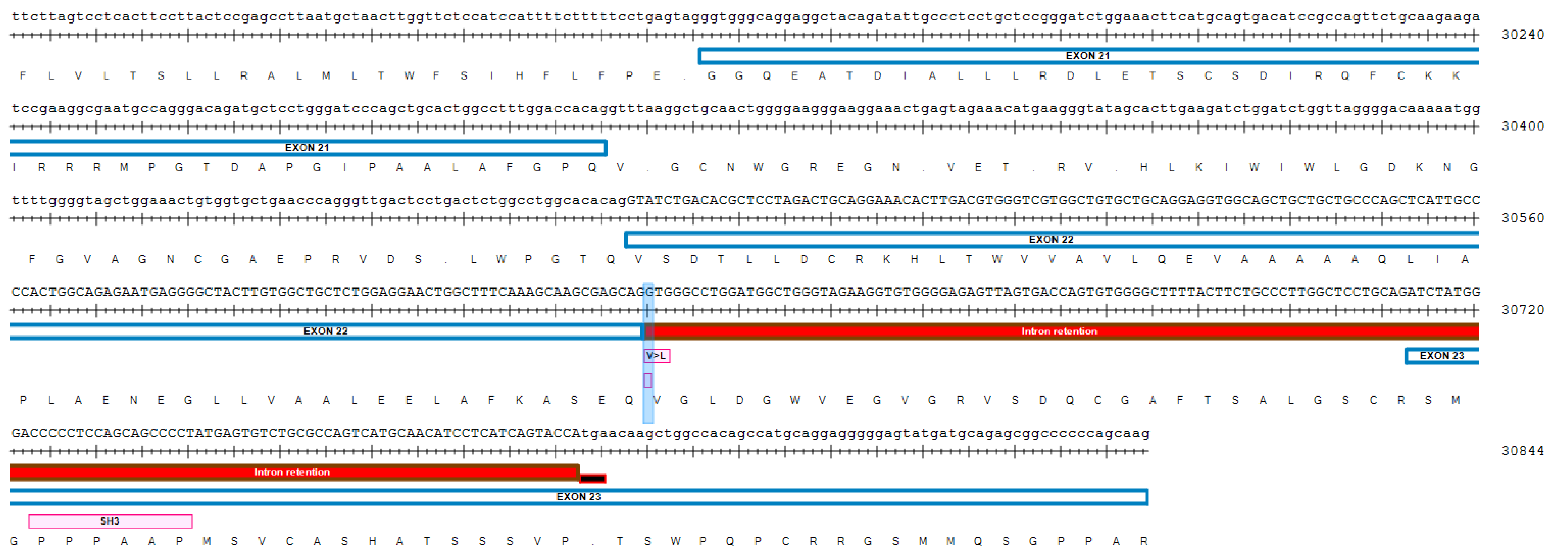
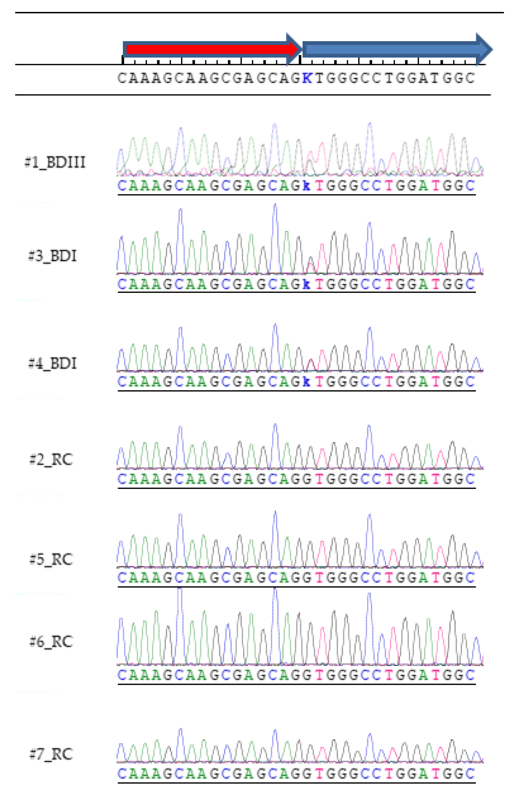
Only a single novel cosegregating variant, predicted to be deleterious, was identified in the affected family (Figure 1). When confounding variants were removed through filtering, a transversion splice site variant in the DCTN1 gene (IVS22 + 1G > T, GRCh37/hg19: 2:74,593,585C > A) was discovered using NGS (Table 1; File S1), and validated by Sanger sequencing (Figure 2, File S2). It was the only novel variant, in coding or splice regions, that was found to cosegregate with the affected cohort and not be present in any of the controls (File S1). This splice site variant is predicted to disrupt the canonical splice donor site at the invariant +1 position and result in retention of intron 22 and introduction of a subsequent PTC (Figure A1). The splice site variant is in a highly evolutionary conserved region (Table 2), and all functional prediction algorithms predicted the variant to be deleterious with no algorithms predicting the variant to be benign (Table 3, https://varsome.com/variant/hg19/2%3A74593585%3AC%3AA). There is a very rare alternative allele (n = 1) noted at this coordinate in the dbSNP database (rs75942278) which contains a C > T transition. However, a transition (where nucleotide ring structure remains constant) is less likely to impair the spliceosome mechanism and result in donor splice site disruption compared to a transversion (where nucleotide ring structure changes).
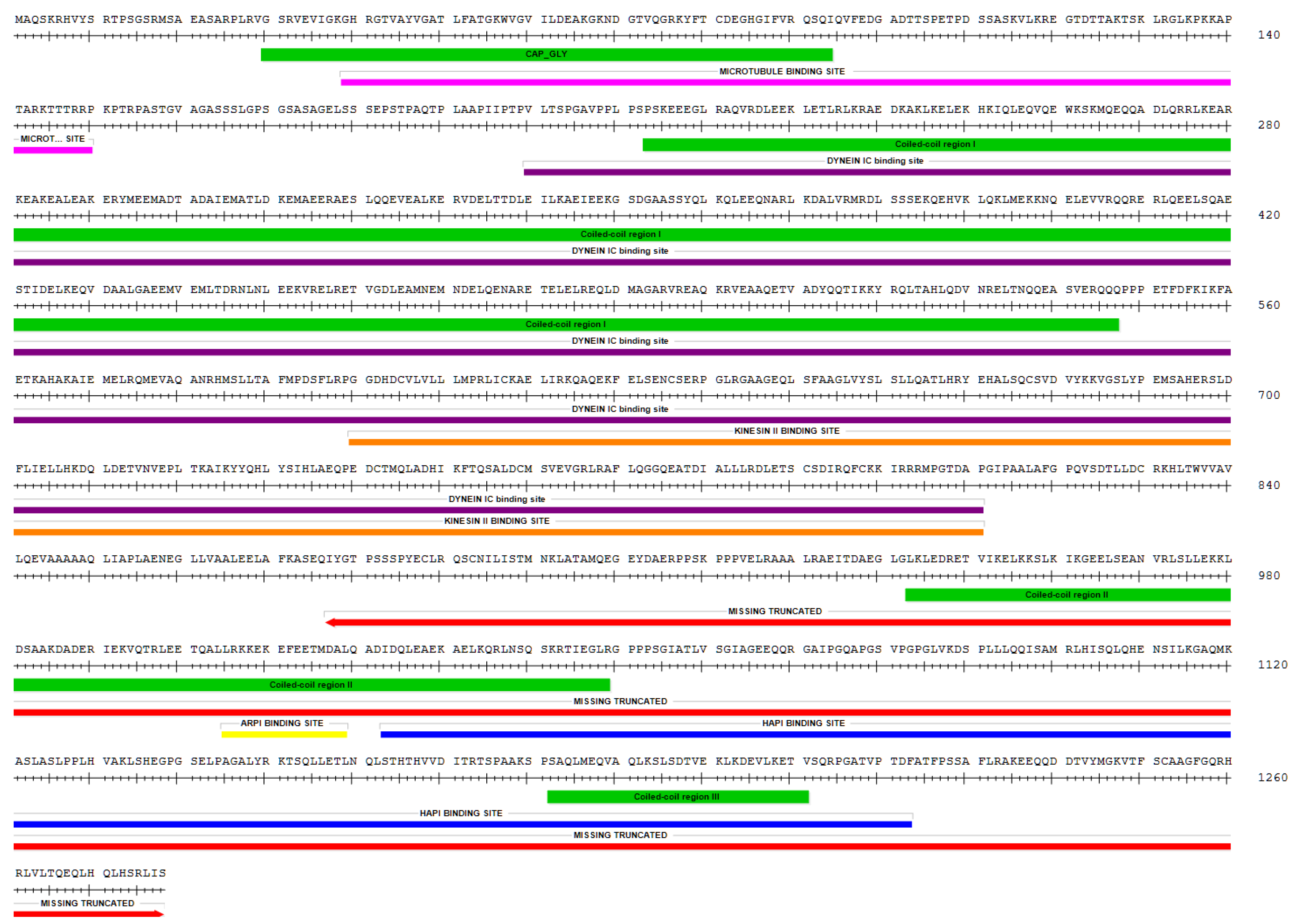
Retrograde axonal transport is the minus-end-directed transport from the distal axon towards the soma and is required for neuronal autophagy. Cytoplasmic dynein is essential for retrograde axonal transport in neurons. It is the only known protein motor involved in retrograde axonal transport and transports numerous cargoes including membrane-bound organelles, mRNA, vesicles, misfolded proteins, and protein aggregates. [28]. Dynactin is a large multiprotein complex which is required for dynein function, including linking cargoes to dynein [29]. The DCTN1 gene codes for the p150Glued protein that is highly expressed in mammalian brain and is the largest component of the dynactin complex [30,31]. In this manuscript, we specifically discuss autophagy in the context of neuronal autophagy. Rare autosomal-dominant disease-causing variants in DCTN1 are already known for Perry syndrome (PS) [32], motor neuron disease (ALS) [33,34], and distal spinal and bulbar muscular dystrophy (dHMN7B) [35]. Psychiatric symptoms, which include severe depression, are a noted feature of PS, which is characterized by onset of atypical Parkinson disease (PD) [36]. These variants result in disruption of retrograde axonal transport and neuronal autophagy. The variants are point mutations affecting the microtubule binding region towards the N-terminus, which contrasts with our research findings in which the three affected probands are shown to possess a novel variant that is predicted to affect the C-terminus (Figure A1 and Figure A2). Of note, there is no historical record of PS, ALS, any muscular dystrophy, or PD in the extended family under investigation. DCTN1 is located on 2p13.1, which is within a region previously found to show significant linkage in autosomal dominant models for BD (2p13–16) [37] and schizophrenia (2p13–14) [38]. DCTN1 is within a region showing suggestive linkage for BD (2p11–q14) [39], as well as significant linkage for MDD (2p11.2–p13.2) [40].

4. Discussion

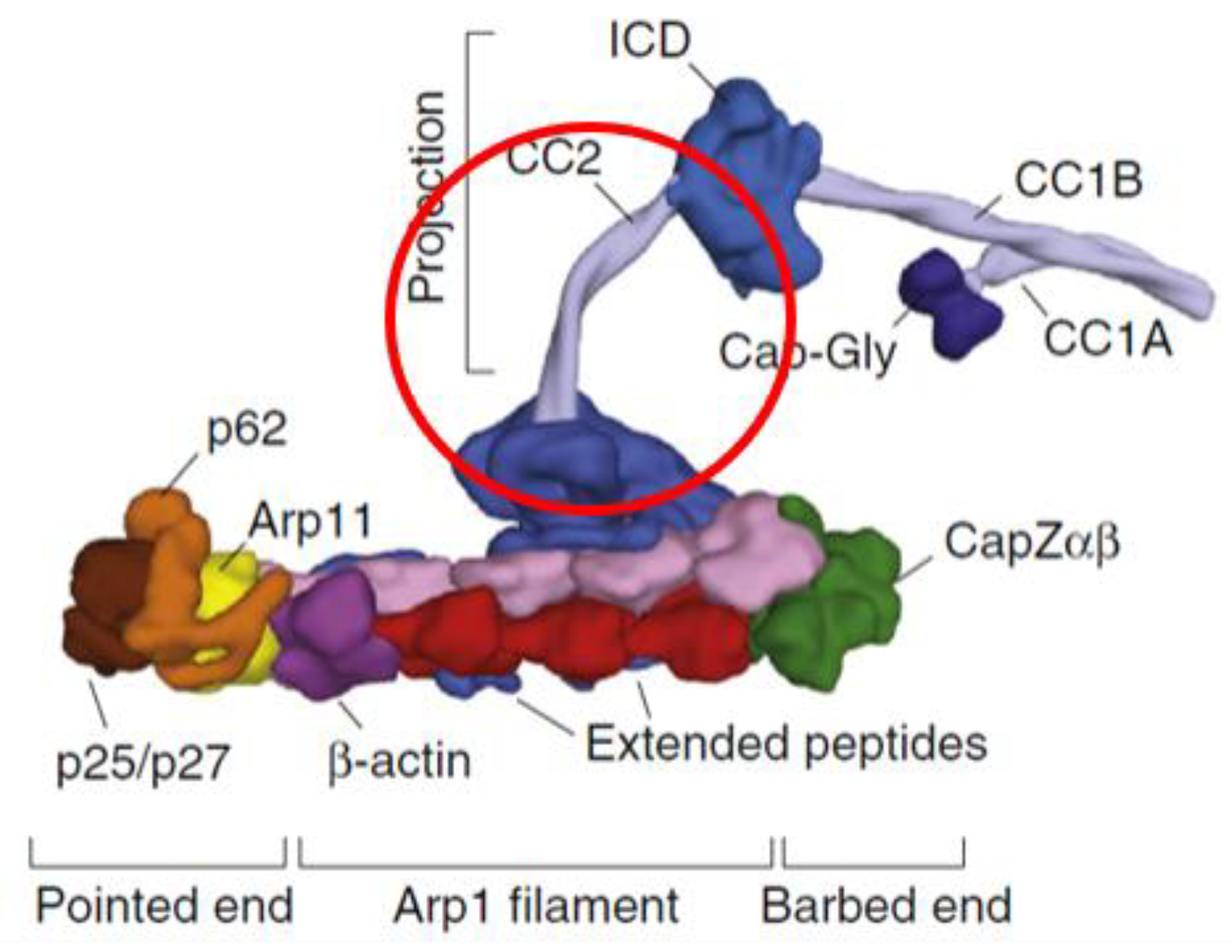
The novel splice site variant discovered in the current research is analogous to the heterozygous dominant-negative Gl1 mutation in the Drosophila Glued gene, which is the ortholog of vertebrate DCTN1 [41,42,43,44]. Homozygous mutations were found to be lethal in Drosophila. The Drosophila transposon-induced mutation results in a truncated transcript, which in turn results in a truncated p150Glued protein missing the C-terminus (ct-p150Glued) [45]. The ensuing ct-p150Glued fails to assemble into the dynactin complex with resulting deficits in retrograde axonal transport and neuronal autophagy [46,47]. Glued Gl1 mutations result in a neurodegenerative rough-eye phenotype and cause deleterious central nervous system effects in Drosophila, disrupting synapse formation and maturation [41]. This finding is consistent with research demonstrating that the dynactin complex is known to stabilize synapses [48]. The novel DCTN1 splice site variant discovered in the current research is predicted to result in a PTC and a truncated protein missing the C-terminal region (Figure A1 and Figure A2). Further evidence from recent structural biology work on mammalian dynein/dynactin complexes confirms that ct-p150Glued would be unable to assimilate into the dynactin complex as the C-terminus normally anchors p150Glued to the rest of the dynactin complex (Figure 3) [49].
If ct-p150Glued is indeed produced in humans, it would be contingent on the truncated DCTN1 transcript escaping NMD. As the PTC lies internally and is >55 nt from the last exon junction, it should be a very strong candidate to trigger NMD [50]. However, a ct-p150Glued, similar in size to the one predicted to result from the splice site variant in our current research, has previously been documented in human specimens, implying that alternative splicing in this region of DCTN1 may be under dynamic control. Fujiwara et al. discovered substantial amounts of ct-p150Glued in all investigated brain specimens obtained from Alzheimer disease (AD) patients (10%–70% of total p150Glued) [51]. ct-p150Glued was not readily observable in normal brain [51]. Although their sample size was small (n = 3 AD brains), the finding of Fujiwara et al. does, nonetheless, importantly confirm that a ct-p150Glued can exist in substantial amounts in vivo, and may represent an important feature of neurodegenerative disorders. AD is also strongly associated with psychiatric disorders, with psychosis being a common event [52]. Conversely, BD patients have a significantly higher risk of developing dementia later in life [53].
As a relationship between glutamate excitoxicity and AD has been widely reported [54], Fujiwara et al. suspected that this may be the cause of the observed accumulation of ct-p150Glued in AD brains. They subsequently demonstrated that glutamate excitotoxicity in primary neuronal cultures similarly resulted in the production of ct-p150Glued, strongly suggesting a causal relationship [51]. As was the case for Drosophila, this truncated protein was unable to assemble into the dynactin complex and interact with cargoes (β-amyloid protein in the case of human ct-p150Glued), and thus represents a loss of function, disrupting retrograde axonal transport and neuronal autophagy [55]. Glutamate excitotoxicity is coupled with increased intracellular calcium concentrations [56], which are an important negative regulator of NMD [57]. Therefore, it is quite likely that formation of ct-p150Glued is related to glutamate/calcium-induced impairment of NMD. Similarly, and with reference to this current research, disturbances of excitatory glutamatergic signaling are also implicated in affective disorders [58]. Indeed, many prophylactic drugs used to treat BD patients are known to modulate glutamatergic signaling [59]. BD patients in the current research cohort may be particularly sensitive to the effects of glutamatergic reinforcement of missplicing in this region of the DCTN1 gene.
A DCTN1 mRNA transcript of very low abundance is known (Ensembl transcript ID: DCTN1-210, ENST00000434055.5), which reflects low-level alternative splicing. This transcript is the result of alternative splicing at the identical splice junction identified in our current research and results in the same PTC. The DCTN1 exon 22/intron 22 region has characteristics that are common to many alternatively spliced regions, namely short exons with short flanking introns [60] and G/C-rich regions typical of cis-regulatory elements [61] (Figure 2). This transcript is also predicted to generate a similar sized ct-p150Glued and/or trigger NMD of the truncated transcript [62]. Possibly, tissue-specific ct-p150Glued is normally produced and maintained at low levels by NMD, with its formation being dependent on transient neuroexcitatory factors. Fujiwara et al. documented the presence of ct-p150Glued in AD brain tissue, but did not suspect alternative splicing as a possible mechanism [51,55]. However, as we have noted above, this may be related to glutamate/calcium-induced impairment of NMD. Likewise, this may also occur in BD by the same mechanism and occur even without the presence of damaging splice variants in the DCTN1 gene.
For the DCTN1 splice site variant, and the predicted resulting formation of ct-p150Glued and/or NMD, to be considered with any seriousness as being causally related to BD, it would have to satisfy a major requirement: it would need to be strongly correlated with BD prophylactic drugs, antipsychotics, and antidepressants, all of which are commonly used in the treatment of BD. As discussed above, impaired retrograde axonal transport and neuronal autophagy are already known to be associated with ct-p150Glued. Similarly, a depletion of DCTN1 mRNA transcripts by NMD would most likely downregulate p150Glued expression with autophagic deficits [63,64]. In this regard, previous ground-breaking research has definitively linked BD prophylactic drugs to neuronal autophagy [65] The BD prophylactic drugs lithium, valproate, and carbamazepine have all been shown to enhance neuronal autophagy and thus would compensate for any deficits that are predicted to result from this variant. They achieve this by a common mechanism, namely via negatively regulating phosphoinositide signaling through depletion of inositol and by reducing myo-inositol-1,4,5-trisphosphate (IP3) [65,66]. Apart from the aforementioned psychotropic agents, the majority of antipsychotics (with two notable exceptions, namely being the typical antipsychotic haloperidol and the atypical antipsychotic clozapine) [67,68,69] as well as antidepressants [70,71] are also known to enhance neuronal autophagy. In addition, the autophagy-inducers rapamycin and trehalose have both been shown to have therapeutic effects in rodent models of affective disorders [72,73]. All the previously mentioned compounds are also under active consideration as therapeutic options for use in neurodegenerative disorders, by their ability to enhance retrograde axonal transport and neuronal autophagy, and to enhance clearance of aggregated and toxic protein [68,74]. Axonal transport, and associated neuronal autophagy, is known to decline with age with two distinct periods of decline, in early adulthood and later in life in rodent studies [75]. Moreover, these findings correlate with peaks for the age of onset for BD and MDD alluded to previously, inferring a relationship between decreased autophagy and the disorders.
At least two different mutant mouse models are known that suggest that deficits in neuronal autophagy may be of critical importance in the etiology of affective disorders. Reduced expression of the lysosomal aspartyl protease cathepsin D (CTSD) is known to impair neuronal autophagy [76]. In this regard, heterozygous CTSD-deficient mice exhibit BD characteristics, such as mania-related behavior and stress-induced depression [77]. Importantly, chronic administration of the BD prophylactic drugs, lithium and valproate, were found to ameliorate their symptoms and thus strongly suggest a causal relationship. In a similar manner to CTSD-deficient mice, knockdown of DCTN5 in a murine model of psychiatric disorder susceptibility genes also results in abnormal mania-related hyperactivity [78]. DCTN5 codes for the p25 subunit of dynactin and thus, like p150Glued, it is intimately involved in retrograde axonal transport and neuronal autophagy [79]. It is also considered a susceptibility gene for BD [80,81]. Furthermore, deficits in neuronal autophagy are also common in lysosomal storage disorders [82], with psychiatric symptoms noted in many of these disorders [83]. For example, mania and/or psychosis has been noted in the adult forms of metachromatic leukodystrophy [84], Niemann–Pick type C disease [85] and Tay–Sachs disease [86].
Protein kinase C (PKC) has often been suggested as playing a key role in BD, as both lithium and valproate inhibit PKC signaling [87]. Thus, inhibitors of PKC have been considered as potential therapeutic agents [88]. In this regard, the selective PKC inhibitor chelerythrine, and the antiestrogenic PKC inhibitor tamoxifen, have both been shown to have antimanic effects in a murine model of mania [89]. Tamoxifen has also been shown to be effective in human trials for the treatment of mania [90]. PKCs are also known to suppress autophagy [91], and that includes Drosophila atypical PKC (aPKC) [92]. aPKCs differ from other PKC isozymes in that they are not dependent on calcium or diacylglycerol for their activity, but can nonetheless be activated by IP3 [93]. The neurodegenerative phenotype resulting from the Drosophila GLUED Gl1 mutation (which results in ct-p150Glued) has been shown to be rescued by mutant alleles of aPKC, thus implying that factors that negatively regulate aPKC activation (which includes reduced IP3 levels) alleviate the mutant phenotype [94]. Given the analogy with the observations of ct-p150Glued in AD brain samples discussed previously, and its predicted formation in the BD research cohort, this finding may provide some indication of the biochemical mechanisms involved in the beneficial effects of therapeutic interventions which negatively regulate IP3 signaling and PKC.
The ct-p150Glued predicted to occur in the current research contains a newly exposed C-terminal sequence, with a typical SH3 polyproline II helix binding motif (Pro-Pro-Pro-X-X-Pro) (Figure A1). Proline-rich regions such as these play an important role in protein–protein interactions which are central to cellular signaling [95]. This suggests that ct-p150Glued, or a cleavage product thereof, may have a physiological regulatory role, quite apart from the expected loss of function related to axonal transport. Pro-Pro-Pro-NH2, in a polyproline II helix conformation, has been shown to be an allosteric activator of dopamine D2 receptors, maintaining them in a high affinity state [96,97]. Thus, it is possible that ct-p150Glued may represent a gain of function, activating dopamine D2 receptors in a similar manner. As dopamine D2 receptors are positive regulators of autophagy [98], this gain of function may represent a compensatory mechanism to maintain neuronal autophagy. Interestingly, a convergent functional genomics study had previously linked increased DCTN1 gene expression with highly delusional states [99] and may be related to this proposed compensatory mechanism. The importance of dopamine in bipolar psychosis has been a central theme in the dopamine hypothesis of bipolar disorder for many decades and this proposed mechanism would be consistent with that hypothesis [100]. Dopamine D2 receptors in particular have been of intense interest as they are a common target of antipsychotic agents, and pathways leading to psychosis have been suggested to converge via high affinity state D2 dopamine receptors [101].
There is a strong likelihood that a transversion at the invariant +1 position of the splice donor site would disrupt splicing, as evidenced by previous examples of splice donor site transversions resulting in a disease phenotype (e.g., splice site disruption leading to hereditary thrombocythemia [102], in congenital afibrinogenemia [103], and mutations in CHD7 leading to CHARGE syndrome [104]). However, further research is required to validate the exact physiological effects of the splice site variant discovered in the current research cohort, and the relative contributions of NMD and ct-p150Glued formation. Of note is the fact that NMD demonstrates widely differing heritable efficiencies [105] and tissue-specific differences [106]. This may explain, at least in part, the phenotypical differences noted in the research cohort. The exon/intron 22 region of the DCTN1 gene, as well as factors that affect splicing efficiency, and alternative splicing, in this region deserve further intense scrutiny in psychiatric disorders. It would also be highly desirable to identify this splice variant in other affected families to further validate its significance and to obtain an estimate of relative frequency.

5. Conclusions

The DCTN1 splice site variant discovered in the current research is not only cosegregating, novel, and in a highly evolutionary conserved region, but it is also predicted to be deleterious. Taken together, all these qualities strongly suggest pathogenicity of the splice site variant. Furthermore, the neuronal autophagy deficits that are predicted to result are consistent with established knowledge regarding the therapeutic interventions used in BD and MDD. Previously discovered disease-causing variants in DCTN1 (e.g., in PS, ALS) are inherited in an autosomal-dominant fashion, and disease-causing variants in PS (resulting in atypical PD) are already known to also result in severe depression. Thus, there is a strong likelihood that the variant uncovered in the present research may also reflect a primary autosomal dominant cause of psychiatric pathology in the research cohort.
As noted previously, a ct-p150Glued, which is predicted to result from the splice site variant discovered in this research, has also been found in AD brain samples as well as in Drosophila mutants. In both cases it is related to deficits in neuronal autophagy. Furthermore, deficits in neuronal autophagy are implied as being causal in at least two different murine models of affective disorders. As also noted previously, axonal transport and associated neuronal autophagy similarly slow at ages that are likewise consistent with onset of illness in both BD as well as recurrent MDD. In addition, lysosomal storage diseases, which are characterized by deficits in neuronal autophagy, are also associated with psychiatric illness. Therefore, this novel cosegregating splice site variant, which is predicted to impair neuronal autophagy, may also hold an important key to the understanding of the pathological biochemical mechanisms involved in BD and MDD in general.
In summary, the proposed mechanisms are highly speculative, but nonetheless, they form a framework for future research in many areas of neuropsychiatric and neurodegenerative diseases.

Supplementary Materials

The following are available online at https://www.mdpi.com/2073-4425/11/4/446/s1 File S1, variant calls; S2, Sanger reads.

Author Contributions

A.H. initiated this research project, was responsible for experimental design, and performed all analyses. A.H. and A.J.L.C. wrote and edited the manuscript. All authors have read and agreed to the published version of the manuscript.

Funding

This research was partially funded by a small grant from Bioplatforms Australia, partially self-funded by A.H., and partially funded through a crowd funding campaign on GoFundMe.

Acknowledgments

Ole A. Andreassen (Psychosis Research Centre, Oslo University, Norway) and Srdjan Djurovic (Dept. Medical genetics, Oslo University Hospital, Norway) are thanked for sample collection from unaffected controls, subsequent genomic DNA extraction and assembly of informed consent forms, as well as shipment of the relevant DNA samples for subsequent sequencing. Andrew Carter (U. Cambridge, UK) is thanked for permission to use his graphic depicting the structure of the dynactin complex. The following are thanked for providing valuable feedback and/or proof-reading during the preparation of the manuscript: Jenny Vo (Teva Pharmaceuticals, Australia), Jan Fullerton (UNSW, Australia), Steve Ambrose (Ambrose Ecological, Australia), Philip Kuchel (U. Sydney, Australia), Fei Liu (Macquarie U., Australia), Daria Mochly-Rosen (Stanford U., USA), and Andrew Carter (U. Cambridge, UK). Guy Plunkett III and Eric Ma are thanked for providing DNASTAR Lasergene software technical support (DNASTAR, Madison, Wisconsin, USA).

Conflicts of Interest

The authors declare no conflict of interest.

Appendix A

Figure A1. The region of the DCTN1 gene illustrating the splice site variant (NG_008735.2: g.30630G > T) that is predicted to disrupt the invariant +1 splice donor site, resulting in intron retention and a premature termination codon (PTC). The transcript can be degraded by NMD and/or be translated to form a C-truncated p150Glued protein. The exons/introns in this region are short which is characteristic of alternatively spliced regions. The new C-terminus tail includes an amino acid variant with a predicted benign change V > L (V877L) and is characterized by a SH3 polyproline II helix binding motif (PPPxxP), which implies that the C-terminal truncated p150Glued protein may have a physiological role which is hypothesized to involve allosteric activation of dopamine D2 receptors.
Genes 11 00446 g0a1
Figure A2. p150Glued protein (UniProt ID: Q14203, 1278AA). The colored bars under the amino acid sequence illustrate the different regions: Red, the region predicted in this study to be missing in ct-p150Glued due to a splice site variant; Green, CAP_GLY and coiled-coil domains; Magenta, microtubule binding domain; Purple, dynein IC binding region; Orange, kinesin II binding region; Yellow, ARPI binding region; Blue, HAP1 binding region.
Genes 11 00446 g0a2

References

  1. De la Vega, D.; Pina, A.; Peralta, F.J.; Kelly, S.A.; Giner, L. A Review on the General Stability of Mood Disorder Diagnoses Along the Lifetime. Curr. Psychiatry Rep. 2018, 20, 29. [Google Scholar] [CrossRef]
  2. Van Meter, A.R.; Youngstrom, E.A.; Findling, R.L. Cyclothymic disorder: A critical review. Clin. Psychol. Rev. 2012, 32, 229–243. [Google Scholar] [CrossRef]
  3. Kleine-Budde, K.; Touil, E.; Moock, J.; Bramesfeld, A.; Kawohl, W.; Rossler, W. Cost of illness for bipolar disorder: A systematic review of the economic burden. Bipolar Disord. 2014, 16, 337–353. [Google Scholar] [CrossRef] [PubMed]
  4. Boardman, A.P.; Healy, D. Modelling suicide risk in affective disorders. Eur. Psychiatry 2001, 16, 400–405. [Google Scholar] [CrossRef]
  5. Marangoni, C.; Hernandez, M.; Faedda, G.L. The role of environmental exposures as risk factors for bipolar disorder: A systematic review of longitudinal studies. J. Affect. Disord. 2016, 193, 165–174. [Google Scholar] [CrossRef] [PubMed]
  6. Uher, R. Gene-environment interactions in severe mental illness. Front. Psychiatry 2014, 5, 48. [Google Scholar] [CrossRef] [Green Version]
  7. Merikangas, K.R.; Jin, R.; He, J.P.; Kessler, R.C.; Lee, S.; Sampson, N.A.; Viana, M.C.; Andrade, L.H.; Hu, C.; Karam, E.G.; et al. Prevalence and correlates of bipolar spectrum disorder in the world mental health survey initiative. Arch. Gen. Psychiatry 2011, 68, 241–251. [Google Scholar] [CrossRef]
  8. Patten, S.B.; Williams, J.V.; Lavorato, D.H.; Fiest, K.M.; Bulloch, A.G.; Wang, J. The prevalence of major depression is not changing. Can. J. Psychiatry 2015, 60, 31–34. [Google Scholar] [CrossRef] [Green Version]
  9. Barnett, J.H.; Smoller, J.W. The genetics of bipolar disorder. Neuroscience 2009, 164, 331–343. [Google Scholar] [CrossRef] [Green Version]
  10. Smith, D.J.; Harrison, N.; Muir, W.; Blackwood, D.H. The high prevalence of bipolar spectrum disorders in young adults with recurrent depression: Toward an innovative diagnostic framework. J. Affect. Disord. 2005, 84, 167–178. [Google Scholar] [CrossRef]
  11. Kennedy, N.; Everitt, B.; Boydell, J.; Van Os, J.; Jones, P.B.; Murray, R.M. Incidence and distribution of first-episode mania by age: Results from a 35-year study. Psychol. Med. 2005, 35, 855–863. [Google Scholar] [CrossRef]
  12. Kroon, J.S.; Wohlfarth, T.D.; Dieleman, J.; Sutterland, A.L.; Storosum, J.G.; Denys, D.; de Haan, L.; Sturkenboom, M.C. Incidence rates and risk factors of bipolar disorder in the general population: A population-based cohort study. Bipolar Disord. 2013, 15, 306–313. [Google Scholar] [CrossRef] [PubMed]
  13. Tozzi, F.; Prokopenko, I.; Perry, J.D.; Kennedy, J.L.; McCarthy, A.D.; Holsboer, F.; Berrettini, W.; Middleton, L.T.; Chilcoat, H.D.; Muglia, P. Family history of depression is associated with younger age of onset in patients with recurrent depression. Psychol. Med. 2008, 38, 641–649. [Google Scholar] [CrossRef] [PubMed]
  14. Budde, M.; Forstner, A.J.; Adorjan, K.; Schaupp, S.K.; Nothen, M.M.; Schulze, T.G. Genetics of bipolar disorder. Nervenarzt 2017, 88, 755–759. [Google Scholar] [CrossRef] [PubMed]
  15. Gordovez, F.J.A.; McMahon, F.J. The genetics of bipolar disorder. Mol. Psychiatry 2020, 25, 544–559. [Google Scholar] [CrossRef] [PubMed]
  16. Gershon, E.S.; Hamovit, J.; Guroff, J.J.; Dibble, E.; Leckman, J.F.; Sceery, W.; Targum, S.D.; Nurnberger, J.I., Jr.; Goldin, L.R.; Bunney, W.E., Jr. A family study of schizoaffective, bipolar I, bipolar II, unipolar, and normal control probands. Arch. Gen. Psychiatry 1982, 39, 1157–1167. [Google Scholar] [CrossRef]
  17. Gershon, E.S.; DeLisi, L.E.; Hamovit, J.; Nurnberger, J.I., Jr.; Maxwell, M.E.; Schreiber, J.; Dauphinais, D.; Dingman, C.W., 2nd; Guroff, J.J. A controlled family study of chronic psychoses. Schizophrenia and schizoaffective disorder. Arch. Gen. Psychiatry 1988, 45, 328–336. [Google Scholar] [CrossRef]
  18. Cirulli, E.T.; Goldstein, D.B. Uncovering the roles of rare variants in common disease through whole-genome sequencing. Nat. Rev. Genet. 2010, 11, 415–425. [Google Scholar] [CrossRef]
  19. Kryukov, G.V.; Pennacchio, L.A.; Sunyaev, S.R. Most rare missense alleles are deleterious in humans: Implications for complex disease and association studies. Am. J. Hum. Genet. 2007, 80, 727–739. [Google Scholar] [CrossRef] [Green Version]
  20. Adzhubei, I.A.; Schmidt, S.; Peshkin, L.; Ramensky, V.E.; Gerasimova, A.; Bork, P.; Kondrashov, A.S.; Sunyaev, S.R. A method and server for predicting damaging missense mutations. Nat. Methods 2010, 7, 248–249. [Google Scholar] [CrossRef] [Green Version]
  21. Kumar, P.; Henikoff, S.; Ng, P.C. Predicting the effects of coding non-synonymous variants on protein function using the SIFT algorithm. Nat. Protoc. 2009, 4, 1073–1081. [Google Scholar] [CrossRef] [PubMed]
  22. Lek, M.; Karczewski, K.J.; Minikel, E.V.; Samocha, K.E.; Banks, E.; Fennell, T.; O‘Donnell-Luria, A.H.; Ware, J.S.; Hill, A.J.; Cummings, B.B.; et al. Analysis of protein-coding genetic variation in 60,706 humans. Nature 2016, 536, 285–291. [Google Scholar] [CrossRef] [PubMed] [Green Version]
  23. Davydov, E.V.; Goode, D.L.; Sirota, M.; Cooper, G.M.; Sidow, A.; Batzoglou, S. Identifying a high fraction of the human genome to be under selective constraint using GERP++. PLoS Comput. Biol. 2010, 6, e1001025. [Google Scholar] [CrossRef] [PubMed] [Green Version]
  24. Quang, D.; Chen, Y.; Xie, X. DANN: A deep learning approach for annotating the pathogenicity of genetic variants. Bioinformatics 2015, 31, 761–763. [Google Scholar] [CrossRef] [Green Version]
  25. Rogers, M.F.; Shihab, H.A.; Mort, M.; Cooper, D.N.; Gaunt, T.R.; Campbell, C. FATHMM-XF: Accurate prediction of pathogenic point mutations via extended features. Bioinformatics 2018, 34, 511–513. [Google Scholar] [CrossRef] [PubMed] [Green Version]
  26. Pollard, K.S.; Hubisz, M.J.; Rosenbloom, K.R.; Siepel, A. Detection of nonneutral substitution rates on mammalian phylogenies. Genome Res. 2010, 20, 110–121. [Google Scholar] [CrossRef] [Green Version]
  27. Kopanos, C.; Tsiolkas, V.; Kouris, A.; Chapple, C.E.; Albarca Aguilera, M.; Meyer, R.; Massouras, A. VarSome: The human genomic variant search engine. Bioinformatics 2019, 35, 1978–1980. [Google Scholar] [CrossRef]
  28. Vallee, R.B.; Shpetner, H.S.; Paschal, B.M. The role of dynein in retrograde axonal transport. Trends Neurosci. 1989, 12, 66–70. [Google Scholar] [CrossRef]
  29. Kardon, J.R.; Vale, R.D. Regulators of the cytoplasmic dynein motor. Nat. Rev. Mol. Cell Biol. 2009, 10, 854–865. [Google Scholar] [CrossRef] [Green Version]
  30. Karki, S.; Holzbaur, E.L. Cytoplasmic dynein and dynactin in cell division and intracellular transport. Curr. Opin. Cell Biol. 1999, 11, 45–53. [Google Scholar] [CrossRef]
  31. Melloni, R.H., Jr.; Tokito, M.K.; Holzbaur, E.L. Expression of the p150Glued component of the dynactin complex in developing and adult rat brain. J. Comp. Neurol. 1995, 357, 15–24. [Google Scholar] [CrossRef] [PubMed]
  32. Farrer, M.J.; Hulihan, M.M.; Kachergus, J.M.; Dachsel, J.C.; Stoessl, A.J.; Grantier, L.L.; Calne, S.; Calne, D.B.; Lechevalier, B.; Chapon, F.; et al. DCTN1 mutations in Perry syndrome. Nat. Genet. 2009, 41, 163–165. [Google Scholar] [CrossRef] [PubMed]
  33. Munch, C.; Sedlmeier, R.; Meyer, T.; Homberg, V.; Sperfeld, A.D.; Kurt, A.; Prudlo, J.; Peraus, G.; Hanemann, C.O.; Stumm, G.; et al. Point mutations of the p150 subunit of dynactin (DCTN1) gene in ALS. Neurology 2004, 63, 724–726. [Google Scholar] [CrossRef] [PubMed]
  34. Stockmann, M.; Meyer-Ohlendorf, M.; Achberger, K.; Putz, S.; Demestre, M.; Yin, H.; Hendrich, C.; Linta, L.; Heinrich, J.; Brunner, C.; et al. The dynactin p150 subunit: Cell biology studies of sequence changes found in ALS/MND and Parkinsonian syndromes. J. Neural. Transm. 2013, 120, 785–798. [Google Scholar] [CrossRef]
  35. Puls, I.; Oh, S.J.; Sumner, C.J.; Wallace, K.E.; Floeter, M.K.; Mann, E.A.; Kennedy, W.R.; Wendelschafer-Crabb, G.; Vortmeyer, A.; Powers, R.; et al. Distal spinal and bulbar muscular atrophy caused by dynactin mutation. Ann. Neurol. 2005, 57, 687–694. [Google Scholar] [CrossRef]
  36. Perry, T.L.; Bratty, P.J.; Hansen, S.; Kennedy, J.; Urquhart, N.; Dolman, C.L. Hereditary mental depression and Parkinsonism with taurine deficiency. Arch. Neurol. 1975, 32, 108–113. [Google Scholar] [CrossRef]
  37. Liu, J.; Juo, S.H.; Dewan, A.; Grunn, A.; Tong, X.; Brito, M.; Park, N.; Loth, J.E.; Kanyas, K.; Lerer, B.; et al. Evidence for a putative bipolar disorder locus on 2p13-16 and other potential loci on 4q31, 7q34, 8q13, 9q31, 10q21-24, 13q32, 14q21 and 17q11-12. Mol. Psychiatry 2003, 8, 333–342. [Google Scholar] [CrossRef] [Green Version]
  38. Coon, H.; Myles-Worsley, M.; Tiobech, J.; Hoff, M.; Rosenthal, J.; Bennett, P.; Reimherr, F.; Wender, P.; Dale, P.; Polloi, A.; et al. Evidence for a chromosome 2p13-14 schizophrenia susceptibility locus in families from Palau, Micronesia. Mol. Psychiatry 1998, 3, 521–527. [Google Scholar] [CrossRef] [Green Version]
  39. Goes, F.S.; Zandi, P.P.; Miao, K.; McMahon, F.J.; Steele, J.; Willour, V.L.; Mackinnon, D.F.; Mondimore, F.M.; Schweizer, B.; Nurnberger, J.I., Jr.; et al. Mood-incongruent psychotic features in bipolar disorder: Familial aggregation and suggestive linkage to 2p11-q14 and 13q21-33. Am. J. Psychiatry 2007, 164, 236–247. [Google Scholar] [CrossRef]
  40. Bulayeva, K.; Lencz, T.; Glatt, S.; Takumi, T.; Gurgenova, F.; Kawakami, H.; Bulayev, O. Mapping genes related to early onset major depressive disorder in Dagestan genetic isolates. Turk. Psikiyatri Derg. 2012, 23, 161–170. [Google Scholar]
  41. Allen, M.J.; Shan, X.; Caruccio, P.; Froggett, S.J.; Moffat, K.G.; Murphey, R.K. Targeted expression of truncated Glued disrupts giant fiber synapse formation in Drosophila. J. Neurosci. 1999, 19, 9374–9384. [Google Scholar] [CrossRef] [PubMed] [Green Version]
  42. Waterman-Storer, C.M.; Holzbaur, E.L. The product of the Drosophila gene, Glued, is the functional homologue of the p150Glued component of the vertebrate dynactin complex. J. Biol. Chem. 1996, 271, 1153–1159. [Google Scholar] [CrossRef] [PubMed] [Green Version]
  43. Garen, A.; Miller, B.R.; Paco-Larson, M.L. Mutations affecting functions of the Drosophila gene glued. Genetics 1984, 107, 645–655. [Google Scholar] [PubMed]
  44. Harte, P.J.; Kankel, D.R. Genetic analysis of mutations at the Glued locus and interacting loci in Drosophila melanogaster. Genetics 1982, 101, 477–501. [Google Scholar]
  45. Swaroop, A.; Paco-Larson, M.L.; Garen, A. Molecular genetics of a transposon-induced dominant mutation in the Drosophila locus Glued. Proc. Natl. Acad. Sci. USA 1985, 82, 1751–1755. [Google Scholar] [CrossRef] [Green Version]
  46. McGrail, M.; Gepner, J.; Silvanovich, A.; Ludmann, S.; Serr, M.; Hays, T.S. Regulation of cytoplasmic dynein function in vivo by the Drosophila Glued complex. J. Cell Biol. 1995, 131, 411–425. [Google Scholar] [CrossRef]
  47. Fan, S.S.; Ready, D.F. Glued participates in distinct microtubule-based activities in Drosophila eye development. Development 1997, 124, 1497–1507. [Google Scholar]
  48. Eaton, B.A.; Fetter, R.D.; Davis, G.W. Dynactin is necessary for synapse stabilization. Neuron 2002, 34, 729–741. [Google Scholar] [CrossRef] [Green Version]
  49. Carter, A.P.; Diamant, A.G.; Urnavicius, L. How dynein and dynactin transport cargos: A structural perspective. Curr. Opin. Struct. Biol. 2016, 37, 62–70. [Google Scholar] [CrossRef]
  50. Maquat, L.E. Nonsense-mediated mRNA decay in mammals. J. Cell Sci. 2005, 118, 1773–1776. [Google Scholar] [CrossRef] [Green Version]
  51. Fujiwara, T.; Morimoto, K.; Kakita, A.; Takahashi, H. Dynein and dynactin components modulate neurodegeneration induced by excitotoxicity. J. Neurochem. 2012, 122, 162–174. [Google Scholar] [CrossRef] [PubMed]
  52. Morgan, S. Psychotic and Bipolar Disorders: Behavioral Disorders in Dementia. FP Essent. 2017, 455, 18–22. [Google Scholar] [PubMed]
  53. Diniz, B.S.; Teixeira, A.L.; Cao, F.; Gildengers, A.; Soares, J.C.; Butters, M.A.; Reynolds, C.F., 3rd. History of Bipolar Disorder and the Risk of Dementia: A Systematic Review and Meta-Analysis. Am. J. Geriatr. Psychiatry 2017, 25, 357–362. [Google Scholar] [CrossRef] [Green Version]
  54. Lewerenz, J.; Maher, P. Chronic Glutamate Toxicity in Neurodegenerative Diseases-What is the Evidence? Front. Neurosci. 2015, 9, 469. [Google Scholar] [CrossRef] [PubMed]
  55. Fujiwara, T.; Morimoto, K. Cooperative effect of p150Glued and microtubule stabilization to suppress excitotoxicity-induced axon degeneration. Biochem. Biophys. Res. Commun. 2012, 424, 82–88. [Google Scholar] [CrossRef] [PubMed]
  56. Choi, D.W. Glutamate neurotoxicity in cortical cell culture is calcium dependent. Neurosci. Lett. 1985, 58, 293–297. [Google Scholar] [CrossRef]
  57. Nickless, A.; Jackson, E.; Marasa, J.; Nugent, P.; Mercer, R.W.; Piwnica-Worms, D.; You, Z. Intracellular calcium regulates nonsense-mediated mRNA decay. Nat. Med. 2014, 20, 961–966. [Google Scholar] [CrossRef] [Green Version]
  58. Jun, C.; Choi, Y.; Lim, S.M.; Bae, S.; Hong, Y.S.; Kim, J.E.; Lyoo, I.K. Disturbance of the glutamatergic system in mood disorders. Exp. Neurobiol. 2014, 23, 28–35. [Google Scholar] [CrossRef] [Green Version]
  59. Rapoport, S.I.; Basselin, M.; Kim, H.W.; Rao, J.S. Bipolar disorder and mechanisms of action of mood stabilizers. Brain Res. Rev. 2009, 61, 185–209. [Google Scholar] [CrossRef] [Green Version]
  60. Sakabe, N.J.; de Souza, S.J. Sequence features responsible for intron retention in human. BMC Genom. 2007, 8, 59. [Google Scholar] [CrossRef] [Green Version]
  61. Wang, Z.; Burge, C.B. Splicing regulation: From a parts list of regulatory elements to an integrated splicing code. Rna 2008, 14, 802–813. [Google Scholar] [CrossRef] [PubMed] [Green Version]
  62. Hammesfahr, B.; Kollmar, M. Evolution of the eukaryotic dynactin complex, the activator of cytoplasmic dynein. BMC Evol. Biol. 2012, 12, 95. [Google Scholar] [CrossRef] [PubMed] [Green Version]
  63. Ikenaka, K.; Kawai, K.; Katsuno, M.; Huang, Z.; Jiang, Y.M.; Iguchi, Y.; Kobayashi, K.; Kimata, T.; Waza, M.; Tanaka, F.; et al. dnc-1/dynactin 1 knockdown disrupts transport of autophagosomes and induces motor neuron degeneration. PLoS ONE 2013, 8, e54511. [Google Scholar] [CrossRef] [PubMed] [Green Version]
  64. Ishikawa, K.I.; Saiki, S.; Furuya, N.; Imamichi, Y.; Tsuboi, Y.; Hattori, N. p150glued deficiency impairs effective fusion between autophagosomes and lysosomes due to their redistribution to the cell periphery. Neurosci. Lett. 2019, 690, 181–187. [Google Scholar] [CrossRef]
  65. Sarkar, S.; Rubinsztein, D.C. Inositol and IP3 levels regulate autophagy: Biology and therapeutic speculations. Autophagy 2006, 2, 132–134. [Google Scholar] [CrossRef] [Green Version]
  66. Tritsaris, K.; Gromada, J.; Jorgensen, T.D.; Nauntofte, B.; Dissing, S. Reduction in the rate of inositol 1,4,5-trisphosphate synthesis in rat parotid acini by lithium. Arch. Oral Biol. 2001, 46, 365–373. [Google Scholar] [CrossRef]
  67. Li, Y.; McGreal, S.; Zhao, J.; Huang, R.; Zhou, Y.; Zhong, H.; Xia, M.; Ding, W.X. A cell-based quantitative high-throughput image screening identified novel autophagy modulators. Pharmacol. Res. 2016, 110, 35–49. [Google Scholar] [CrossRef] [Green Version]
  68. Sarkar, S.; Rubinsztein, D.C. Small molecule enhancers of autophagy for neurodegenerative diseases. Mol. Biosyst. 2008, 4, 895–901. [Google Scholar] [CrossRef]
  69. Park, J.; Chung, S.; An, H.; Kim, J.; Seo, J.; Kim, D.H.; Yoon, S.Y. Haloperidol and clozapine block formation of autophagolysosomes in rat primary neurons. Neuroscience 2012, 209, 64–73. [Google Scholar] [CrossRef]
  70. Zschocke, J.; Rein, T. Antidepressants encounter autophagy in neural cells. Autophagy 2011, 7, 1247–1248. [Google Scholar] [CrossRef] [Green Version]
  71. Zschocke, J.; Zimmermann, N.; Berning, B.; Ganal, V.; Holsboer, F.; Rein, T. Antidepressant drugs diversely affect autophagy pathways in astrocytes and neurons--dissociation from cholesterol homeostasis. Neuropsychopharmacology 2011, 36, 1754–1768. [Google Scholar] [CrossRef] [PubMed]
  72. Kara, N.Z.; Toker, L.; Agam, G.; Anderson, G.W.; Belmaker, R.H.; Einat, H. Trehalose induced antidepressant-like effects and autophagy enhancement in mice. Psychopharmacology 2013, 229, 367–375. [Google Scholar] [CrossRef] [PubMed]
  73. Kara, N.Z.; Flaisher-Grinberg, S.; Anderson, G.W.; Agam, G.; Einat, H. Mood-stabilizing effects of rapamycin and its analog temsirolimus: Relevance to autophagy. Behav. Pharmacol. 2018, 29, 379–384. [Google Scholar] [CrossRef] [PubMed]
  74. Ravikumar, B.; Sarkar, S.; Rubinsztein, D.C. Clearance of mutant aggregate-prone proteins by autophagy. Methods Mol. Biol. 2008, 445, 195–211. [Google Scholar] [PubMed]
  75. Milde, S.; Adalbert, R.; Elaman, M.H.; Coleman, M.P. Axonal transport declines with age in two distinct phases separated by a period of relative stability. Neurobiol. Aging 2015, 36, 971–981. [Google Scholar] [CrossRef] [PubMed] [Green Version]
  76. Sarkar, C.; Zhao, Z.; Aungst, S.; Sabirzhanov, B.; Faden, A.I.; Lipinski, M.M. Impaired autophagy flux is associated with neuronal cell death after traumatic brain injury. Autophagy 2014, 10, 2208–2222. [Google Scholar] [CrossRef] [Green Version]
  77. Zhou, R.; Lu, Y.; Han, Y.; Li, X.; Lou, H.; Zhu, L.; Zhen, X.; Duan, S. Mice heterozygous for cathepsin D deficiency exhibit mania-related behavior and stress-induced depression. Prog. Neuropsychopharmacol. Biol. Psychiatry 2015, 63, 110–118. [Google Scholar] [CrossRef]
  78. MacLaren, E.J.; Charlesworth, P.; Coba, M.P.; Grant, S.G. Knockdown of mental disorder susceptibility genes disrupts neuronal network physiology in vitro. Mol. Cell. Neurosci. 2011, 47, 93–99. [Google Scholar] [CrossRef]
  79. Qiu, R.; Zhang, J.; Xiang, X. p25 of the dynactin complex plays a dual role in cargo binding and dynactin regulation. J. Biol. Chem. 2018, 293, 15606–15619. [Google Scholar] [CrossRef] [Green Version]
  80. Wellcome Trust Case Control, C. Genome-wide association study of 14,000 cases of seven common diseases and 3,000 shared controls. Nature 2007, 447, 661–678. [Google Scholar] [CrossRef] [Green Version]
  81. Rao, A.R.; Yourshaw, M.; Christensen, B.; Nelson, S.F.; Kerner, B. Rare deleterious mutations are associated with disease in bipolar disorder families. Mol. Psychiatry 2017, 22, 1009–1014. [Google Scholar] [CrossRef] [PubMed]
  82. Settembre, C.; Fraldi, A.; Jahreiss, L.; Spampanato, C.; Venturi, C.; Medina, D.; de Pablo, R.; Tacchetti, C.; Rubinsztein, D.C.; Ballabio, A. A block of autophagy in lysosomal storage disorders. Hum. Mol. Genet. 2008, 17, 119–129. [Google Scholar] [CrossRef] [PubMed]
  83. Staretz-Chacham, O.; Choi, J.H.; Wakabayashi, K.; Lopez, G.; Sidransky, E. Psychiatric and behavioral manifestations of lysosomal storage disorders. Am. J. Med. Genet. B Neuropsychiatr. Genet. 2010, 153, 1253–1265. [Google Scholar] [CrossRef] [PubMed]
  84. Wender, M.; Pruchnik-Wolinska, D.; Paprzycki, W.; Czartoryska, B. Familial metachromatic leukodystrophy as a cause of psychotic manifestations in young adults. Psychiatr. Pol. 1998, 32, 113–119. [Google Scholar] [PubMed]
  85. Maubert, A.; Hanon, C.; Metton, J.P. Adult onset Niemann-Pick type C disease and psychosis: Literature review. Encephale 2013, 39, 315–319. [Google Scholar] [CrossRef] [PubMed]
  86. MacQueen, G.M.; Rosebush, P.I.; Mazurek, M.F. Neuropsychiatric aspects of the adult variant of Tay-Sachs disease. J. Neuropsychiatry Clin. Neurosci. 1998, 10, 10–19. [Google Scholar] [CrossRef]
  87. Saxena, A.; Scaini, G.; Bavaresco, D.V.; Leite, C.; Valvassoria, S.S.; Carvalho, A.F.; Quevedo, J. Role of Protein Kinase C in Bipolar Disorder: A Review of the Current Literature. Mol. Neuropsychiatry 2017, 3, 108–124. [Google Scholar] [CrossRef]
  88. Zarate, C.A.; Manji, H.K. Protein kinase C inhibitors: Rationale for use and potential in the treatment of bipolar disorder. CNS Drugs 2009, 23, 569–582. [Google Scholar] [CrossRef]
  89. Abrial, E.; Betourne, A.; Etievant, A.; Lucas, G.; Scarna, H.; Lambas-Senas, L.; Haddjeri, N. Protein kinase C inhibition rescues manic-like behaviors and hippocampal cell proliferation deficits in the sleep deprivation model of mania. Int. J. Neuropsychopharmacol. 2014, 18, 31. [Google Scholar] [CrossRef]
  90. Yildiz, A.; Guleryuz, S.; Ankerst, D.P.; Ongur, D.; Renshaw, P.F. Protein kinase C inhibition in the treatment of mania: A double-blind, placebo-controlled trial of tamoxifen. Arch. Gen. Psychiatry 2008, 65, 255–263. [Google Scholar] [CrossRef] [Green Version]
  91. Jiang, H.; Cheng, D.; Liu, W.; Peng, J.; Feng, J. Protein kinase C inhibits autophagy and phosphorylates LC3. Biochem. Biophys. Res. Commun. 2010, 395, 471–476. [Google Scholar] [CrossRef] [Green Version]
  92. Jin, A.; Neufeld, T.P.; Choe, J. Kibra and aPKC regulate starvation-induced autophagy in Drosophila. Biochem. Biophys. Res. Commun. 2015, 468, 1–7. [Google Scholar] [CrossRef] [PubMed] [Green Version]
  93. Xiao, H.; Liu, M. Atypical protein kinase C in cell motility. Cell. Mol. Life Sci. 2013, 70, 3057–3066. [Google Scholar] [CrossRef] [PubMed]
  94. Ma, L.; Johns, L.A.; Allen, M.J. A modifier screen in the Drosophila eye reveals that aPKC interacts with Glued during central synapse formation. BMC Genet. 2009, 10, 77. [Google Scholar] [CrossRef] [PubMed] [Green Version]
  95. Adzhubei, A.A.; Sternberg, M.J.; Makarov, A.A. Polyproline-II helix in proteins: Structure and function. J. Mol. Biol. 2013, 425, 2100–2132. [Google Scholar] [CrossRef]
  96. Baures, P.W.; Pradhan, A.; Ojala, W.H.; Gleason, W.B.; Mishra, R.K.; Johnson, R.L. Synthesis and dopamine receptor modulating activity of unsubstituted and substituted triproline analogues of L-prolyl-L-leucyl-glycinamide (PLG). Bioorg. Med. Chem. Lett. 1999, 9, 2349–2352. [Google Scholar] [CrossRef]
  97. Raghavan, B.; Skoblenick, K.J.; Bhagwanth, S.; Argintaru, N.; Mishra, R.K.; Johnson, R.L. Allosteric modulation of the dopamine D2 receptor by Pro-Leu-Gly-NH2 peptidomimetics constrained in either a polyproline II helix or a type II β-turn conformation. J. Med. Chem. 2009, 52, 2043–2051. [Google Scholar] [CrossRef] [Green Version]
  98. Wang, D.; Ji, X.; Liu, J.; Li, Z.; Zhang, X. Dopamine Receptor Subtypes Differentially Regulate Autophagy. Int. J. Mol. Sci. 2018, 19, 1540. [Google Scholar] [CrossRef] [Green Version]
  99. Kurian, S.M.; Le-Niculescu, H.; Patel, S.D.; Bertram, D.; Davis, J.; Dike, C.; Yehyawi, N.; Lysaker, P.; Dustin, J.; Caligiuri, M.; et al. Identification of blood biomarkers for psychosis using convergent functional genomics. Mol. Psychiatry 2011, 16, 37–58. [Google Scholar] [CrossRef] [Green Version]
  100. Ashok, A.H.; Marques, T.R.; Jauhar, S.; Nour, M.M.; Goodwin, G.M.; Young, A.H.; Howes, O.D. The dopamine hypothesis of bipolar affective disorder: The state of the art and implications for treatment. Mol. Psychiatry 2017, 22, 666–679. [Google Scholar] [CrossRef]
  101. Seeman, P.; Schwarz, J.; Chen, J.F.; Szechtman, H.; Perreault, M.; McKnight, G.S.; Roder, J.C.; Quirion, R.; Boksa, P.; Srivastava, L.K.; et al. Psychosis pathways converge via D2high dopamine receptors. Synapse 2006, 60, 319–346. [Google Scholar] [CrossRef] [PubMed]
  102. Liu, K.; Kralovics, R.; Rudzki, Z.; Grabowska, B.; Buser, A.S.; Olcaydu, D.; Gisslinger, H.; Tiedt, R.; Frank, P.; Okon, K.; et al. A de novo splice donor mutation in the thrombopoietin gene causes hereditary thrombocythemia in a Polish family. Haematologica 2008, 93, 706–714. [Google Scholar] [CrossRef] [PubMed]
  103. Attanasio, C.; de Moerloose, P.; Antonarakis, S.E.; Morris, M.A.; Neerman-Arbez, M. Activation of multiple cryptic donor splice sites by the common congenital afibrinogenemia mutation, FGA IVS4 + 1 G-->T. Blood 2001, 97, 1879–1881. [Google Scholar] [CrossRef] [PubMed] [Green Version]
  104. Villate, O.; Ibarluzea, N.; Fraile-Bethencourt, E.; Valenzuela, A.; Velasco, E.A.; Grozeva, D.; Raymond, F.L.; Botella, M.P.; Tejada, M.I. Functional Analyses of a Novel Splice Variant in the CHD7 Gene, Found by Next Generation Sequencing, Confirm Its Pathogenicity in a Spanish Patient and Diagnose Him with CHARGE Syndrome. Front. Genet. 2018, 9, 7. [Google Scholar] [CrossRef] [PubMed] [Green Version]
  105. Seoighe, C.; Gehring, C. Heritability in the efficiency of nonsense-mediated mRNA decay in humans. PLoS ONE 2010, 5, e11657. [Google Scholar] [CrossRef] [PubMed] [Green Version]
  106. Zetoune, A.B.; Fontaniere, S.; Magnin, D.; Anczukow, O.; Buisson, M.; Zhang, C.X.; Mazoyer, S. Comparison of nonsense-mediated mRNA decay efficiency in various murine tissues. BMC Genet. 2008, 9, 83. [Google Scholar] [CrossRef] [Green Version]
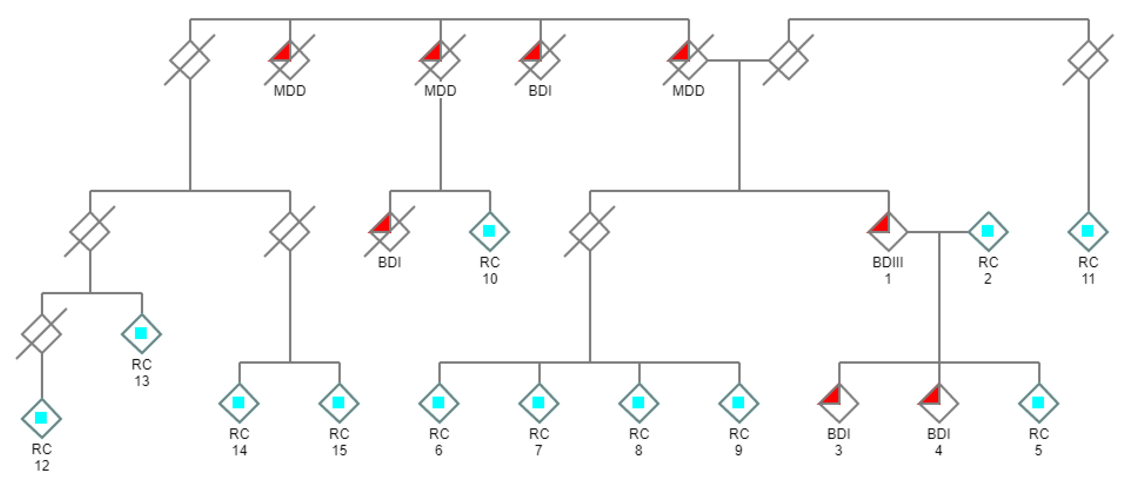
Figure 1. Pedigree of the family under investigation. Individuals with a psychiatric history are denoted with filled red triangles. Psychiatric illness in this extended family is inherited in a manner resembling autosomal dominance. WES was performed and subtractive analysis was used to discover novel cosegregating candidate gene variants in the three living affected individuals in the core group (#1, 3, 4), using related unaffected living relatives as negative controls (related controls/RC: light blue squares). (/ = deceased; and males and females are not distinguished for reasons of privacy).
Genes 11 00446 g001
Figure 2. The DCTN1 variant [NC_000002.11: g.74593585C > A (GRCh37)] was also verified using Sanger sequencing using both forward and reverse primers. Chromatograms using the forward primer are depicted. The chromatograms show heterozygous variant calls (k) for the three affected family members (Figure 1; #1, 3, 4), and a normal allele for related controls (Figure 1; #2, 5, 6, 7). The novel variant is predicted to disrupt the invariant donor splice site (IVS22 + 1G > T [NM_004082.4: c.2628 + 1G > T]) of the DCTN1 gene. The red arrow depicts exon 22 and the blue arrow intron 22. As the DCTN1 gene lies on the minus strand the reverse-complement alignment is shown to highlight the GT > TT splice site variant at the invariant +1 position.
Genes 11 00446 g002
Figure 3. The structure of mammalian dynactin illustrating the region (circled in red) predicted to be missing as a result of the splice site variant. The long projection of p150Glued is anchored to the rest of the dynactin complex by the C-terminal domain. Thus, ct-p150Glued would be unable to assimilate into the dynactin complex as it lacks this domain. In contrast, disease-causing variants responsible for PS and ALS are toward the N-terminal and primarily affect the region around the Cap-Gly microtubule-binding domain. Modified from [49] with permission by the authors and Publisher (Copyright Elsevier).
Genes 11 00446 g003
Table 1. Variant counts for the DCTN1 splice site variant (NC_000002.11: g.74593585C > A (GRCh37)) in affected subjects (#1, 3, 4); BDI, bipolar disorder I; BDIII, bipolar disorder III. Variant calls for all probands and controls are in File S1.
Subject #DiagnosisDCTN1 Count CDCTN1 Count A
1 BDIII3151
3 BDI5158
4 BDI7664
Table 2. Genomic evolutionary conservation data for the DCTN1 splice site variant.
GERP++RSPhastCons100way_VertebratePhyloP100way_Vertebrate
Score5.08 (range 1–6.18)1 (range 0–1)5.75 (range—20–10)
ConservationHighly conservedHighly conservedHighly conserved
Table 3. Functional predictions for the DCTN1 splice site variant.
CADDDANNFATHMM-XF5MutationTaster
Score25.6
[> 20 = 1% most pathogenic]
0.9946
[range 0–1]
0.996
[range 0–1]
1.0
[range 0–1]
PredictionDeleteriousDeleterious DeleteriousDeleterious