Article

The Molecular Evolution of Circadian Clock Genes in Spotted Gar (Lepisosteus oculatus)

1
School of Biology & Basic Medical Sciences, Medical College of Soochow University, Suzhou 215123, China
2
Center for Circadian Clocks, Soochow University, Suzhou 215123, China
3
Institute of Neuroscience, University of Oregon, Eugene, OR 97403, USA
*
Authors to whom correspondence should be addressed.
Genes 2019, 10(8), 622; https://doi.org/10.3390/genes10080622
Submission received: 3 July 2019 / Revised: 9 August 2019 / Accepted: 14 August 2019 / Published: 17 August 2019

Abstract

:
Circadian rhythms are biological rhythms with a period of approximately 24 h. While canonical circadian clock genes and their regulatory mechanisms appear highly conserved, the evolution of clock gene families is still unclear due to several rounds of whole genome duplication in vertebrates. The spotted gar (Lepisosteus oculatus), as a non-teleost ray-finned fish, represents a fish lineage that diverged before the teleost genome duplication (TGD), providing an outgroup for exploring the evolutionary mechanisms of circadian clocks after whole-genome duplication. In this study, we interrogated the spotted gar draft genome sequences and found that spotted gar contains 26 circadian clock genes from 11 families. Phylogenetic analysis showed that 9 of these 11 spotted gar circadian clock gene families have the same number of genes as humans, while the members of the nfil3 and cry families are different between spotted gar and humans. Using phylogenetic and syntenic analyses, we found that nfil3-1 is conserved in vertebrates, while nfil3-2 and nfil3-3 are maintained in spotted gar, teleost fish, amphibians, and reptiles, but not in mammals. Following the two-round vertebrate genome duplication (VGD), spotted gar retained cry1a, cry1b, and cry2, and cry3 is retained in spotted gar, teleost fish, turtles, and birds, but not in mammals. We hypothesize that duplication of core clock genes, such as (nfil3 and cry), likely facilitated diversification of circadian regulatory mechanisms in teleost fish. We also found that the transcription factor binding element (Ahr::Arnt) is retained only in one of the per1 or per2 duplicated paralogs derived from the TGD in the teleost fish, implicating possible subfuctionalization cases. Together, these findings help decipher the repertoires of the spotted gar’s circadian system and shed light on how the vertebrate circadian clock systems have evolved.

1. Introduction

Circadian clocks regulate various cellular and physiological activities and processes in organisms ranging from cyanobacteria to mammals, allowing for them to adapt to the day–night cycle on Earth [1]. A circadian oscillator exhibits persistent rhythmical activity with a near 24 h periodicity under constant conditions [2]. The universal mechanisms of circadian clocks are transcriptional/translational feedback loops [3]. In mammals, daily biological rhythms are generated at the molecular level through auto-regulatory positive and negative feedback loops of the core clock genes (e.g., Clock, bmal1, Period, Cryptochrome) [4,5]. CLOCK and BMAL1 form a heterodimer (CLOCK: BMAL1) and activate transcription of Period and Cryptochrome genes by binding to E-box motifs (CACGTG) in their promoter regions. PERIOD and CRYPTOCHROME then form another heterodimer to repress the transcriptional activities of CLOCK: BMAL1 heterodimers [6,7]. The circadian oscillators exhibit remarkable conservation of function across wide evolutionary time spans [1,8,9].
Circadian systems have undergone a genetic revolution. Like many other multigene families, the circadian genes that are found in single copies in invertebrates are duplicated in vertebrates [10]. Gene duplication is one of the most important mechanisms in the evolution of gene diversity, presumably because obtaining new functions by modifying preexisting genetic systems is easier than by generating them de novo [11,12]. Investigation of the molecular architecture of traits in vertebrates should take into consideration the past duplication events in this lineage. In humans and mice, CLOCK1 and NPAS2/CLOCK2 are two known paralogs that were generated by the 2R genome duplication event [13]. Zebrafish and fugu are known to have three copies of the clock family (clock1a, clock1b and clock2). Tetraodon has clock1a and clock1b genes, while medaka and stickleback have clock1 and clock2 genes. clock1a/ clock1b is are a duplicated pair that resulted from the third round of whole genome duplication that occurred within the ray-finned fish lineage prior to the radiation of the teleost fish (approximately 300 Mya) [13].
Teleost fish are attractive for the study of many evolutionary questions related to diverse aspects of biology. Fish show a remarkable level of diversity in their morphology, ecology, behavior, and genomes, as well as multiple other facets of their biology [14]. Whole genome sequencing analyses of several fish species have shed light on the organization and evolution of fish genomes and now allow for investigation of the evolutionary mechanisms underlying biodiversity in fish lineages [15,16,17]. Teleost genomes differ from mammalian genomes, however, by a whole-genome duplication event, the teleost genome duplication (TGD) [18,19,20,21]. While the TGD allows for the dissection of ancestral gene functions via the partitioning of ancestral subfunctions [22,23], it also obfuscates correlations between teleost disease models and their human counter-parts because of the difficulty of ortholog assignment after the lineage-specific loss of duplicated genes and the asymmetric evolution of gene duplicates [24]. Genomic resources from a ray-finned (Actinopterygian) fish that diverged from teleosts before the TGD would facilitate the connectivity of teleost and mammalian genomes. Analysis of a half dozen genes in spotted gar (L. o.), a large, air-breathing ray-finned North American fish, suggested that its lineage has diverged from the teleost lineage before the TGD [25,26]. Thus, spotted gar represents a genomic intermediary between teleost medical models and the human genome, providing a critical link between biological models in teleost fish, to which the spotted gar is biologically similar, and humans, to which spotted gar is genomically similar [16,24].
A major question in molecular evolution is how duplicate genes are retained in a genome. Several studies have attempted to account for the evolutionary fates of duplicate genes [10,22,27,28]. Genetic analyses have identified numerous mammalian homologs of circadian clock gene families, including Clock, Bmal, Per, Cry, Csnk1e, Nr1d, Ror, and Tim. A number of other transcription factors also thought to function in the circadian regulation of gene expression, including Dbp, Nfil3, and Dec [29,30,31]. We have discovered, in a previous study, that the spotted gar genome retained most of its circadian clock genes, which are similar in copy numbers with those of humans [16]. To further examine the evolution of circadian clock genes in teleost fish, we interrogated the spotted gar draft genome sequences and analyzed 11 families of the main clock genes (including bmal, clock, period, cry, dec, csnk1e, nr1d, ror, par, nfil3 and tim) involved in vertebrates’ circadian rhythm pathways. We found that spotted gar contains 26 circadian clock genes from the 11 families. Nine of these 11 families of spotted gar are the same as those of humans. However, the orthologs of nfil3 and cry are different between spotted gar and humans. These analyses strongly support the notion that spotted gar resemble humans to a great extent, without the TGD, but still contain extra copies of genes that mammals lack.

2. Materials and Methods

2.1. Data Sets and Phylogenetic Analysis

We selected 11 circadian clock gene families including period, clock, bmal, cry, dec, csnk1e, nr1d, ror, par, nfil3, and tim. These circadian clock genes from representative species were uncovered from Ensembl (Release 95) (http://www.ensembl.org/index.html). The final data set included 365 gene sequences (Supplementary Table S1). Based upon comparative genomic analysis, we have changed the names for some genes including cry and nfil3, except for tim (there is only one tim ortholog gene in each species of vertebrate), to better reflect their evolutionary history and increase genome connectivity. All of the old names and corresponding new names of these genes are listed in Supplementary Table S1. Multiple sequence alignments of protein sequences were performed with CLUSTAL W [32]. To explore the evolutionary pattern of the circadian rhythm genes, the neighbor-joining (NJ) algorithm was used to construct phylogenetic trees of these circadian clock genes with MEGA6 [33]. Each tree was a consensus tree derived from a heuristic search of 1000 bootstrap replicates. The abbreviated species names are as follows: Ac, Anolis carolinensis; Dm, Drosophila melanogaster; Dr, Danio rerio; Ga, Gasterosteus aculeatus; Gg, Gallus gallus; Hs, Homo sapiens; Lc, Latimeria chalumnae; Lo, L. o.; Mu, Mus musculus; Oa, Ornithorhynchus anatinus; Ol, Oryzias latipes; On, Oreochromis niloticus; Pm, Petromyzon marinus; Ps, Pelodiscus sinensis; Tg, Taeniopygia guttata; Tn, Tetraodon nigroviridis; Tr, Takifugu rubripes; and Xt, Xenopus tropicalis.

2.2. Exon Structural Analysis

Exonic boundaries of the coding regions of these genes were determined according to Ensembl. The exon–intron structures were presented using the FancyGene visualization software (http://bio.ieo.eu/fancygene/) [34]. The function domains were predicted with the online tool InterPro (http://www.ebi.ac.uk/interpro/).

2.3. Conserved Synteny Analysis

Gene synteny for the circadian clock genes was compared to the syntenic region using the Genomicus database (http://www.genomicus.biologie.ens.fr/genomicus) [35]. Spotted gar was used as a reference species for cross-species synteny, and because of its protein similarity, to highlight the conservation of synteny and the protein sequence in vertebrates. Synteny data and species images were downloaded from Genomicus. Consistent with the label in the database, the thick blue line between two genes is equivalent to a “gap” in the alignment of this extant species, i.e., the two genes are neighbours in this species but not in the reference species. The thin blue line is equivalent to a “break” in the continuity of the alignment, i.e., the two genes are linked in order, but at least one gene separates them in other species. The dotted line means no alignment, and the double-headed arrow under a block of genes means that the order of the genes shown was flipped around (reversed).

2.4. Regulatory Region Analysis

The upstream regions at approximately 3000 bp for per genes in vertebrates were obtained from Ensembl. The transcription factor binding elements were predicted using the JASPAR (http://jaspar.genereg.net/) dataset (profile score threshold 99%).

3. Results

3.1. Phylogenetic Analysis of Spotted Gar Circadian Clock Genes

Using zebrafish and human circadian clock genes to interrogate the spotted gar draft genome sequences, we found that spotted gar contains 26 circadian clock genes from 11 families, which are consistent with our previous report (Table 1) [16]. To help understand how the gar circadian genes link to the human genome, we constructed phylogenetic trees of the 11 families with 365 genes in vertebrates, and showed that eight of these 11 families of spotted gar circadian clock genes, including period, clock, bmal, tim, dec, csnk1e, nr1d, and ror, have the same number of genes as humans without extra duplicate copies (Figure 1). However, most of the circadian clock families except for tim were duplicated in teleost fish. The final data set, including gene sequences, is listed in Supplementary Table S1.
For the nfil3 gene family, spotted gar contains nfil3-1, nfil3-2, and nfil3-3. While nfil3-1 is the ortholog of the mammalian Nfil3 gene, nfil3-2 and nfil3-3 were duplicated to nfil3-2a/nfil3-2b, nfil3-3a /nfil3-3b, respectively, via TGD in teleost fish, including zebrafish. Some teleost fish possess additional nfil3-1b.1 and nfil3-1b.2 genes.
For the cry gene family, spotted gar contains two cry1 genes, cry1a, and cry1b, which are co-orthologs of mammalian Cry1, likely derived from tandem duplication in teleost fish. Zebrafish have four cry1 genes, cry1aa, cry1ab, cry1ba, and cry1bb, likely derived from TGD. These results are consistent with our previous study [36]. In addition, spotted gar also contain one cry2 gene, the ortholog of mammalian Cry2; and one cry3 gene, which mammals do not have.
In the par gene family, spotted gar has one tef and one hlf gene, like humans (both genes with TGD ohnologs). The potential ortholog of dbp is present as an unannotated sequences on an unassembled scaffold (JH591448.1: 146,984 to 147,190) in the current version (LepOcu1) of the spotted gar genome (reciprocal best blast hit with coelacanth dbp and nearest four neighbors on one side being orthologs of the four nearest neighbors of zebrafish dpba).

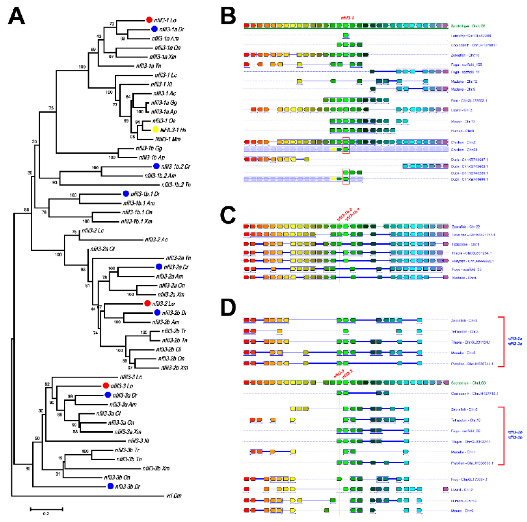
3.2. Evolution of nfil3 Genes

The phylogenetic analysis of nfil3 family genes showed that there is only one nfil3 ortholog in flies (Figure 2A), and this gene family can be classified into three main subclades in vertebrates. The nfil3-1 gene is shared among most chordates, with the exception of its loss in a few teleost fish (fugu and medaka). Almost all fish, including coelacanth and spotted gar, have the other two nfil3 members, nfil3-2 and nfil3-3. Intriguingly, lizards have only nfil3-2, while frogs have only nfil3-3. However, both nfil3-2 and nfil3-3 are completely lost in mammals and birds. Further, nfil3-2 and nfil3-3 were duplicated to nfil3-2a/nfil3-2b and nfil3-3a/nfil3-3b in teleost fish via TGD, respectively. As expected, some of these nfil3 duplicates were lost in some fish lineages—for instance, nfil3-2a and nfil3-3a in fugu, nfil3-3a in tetraodon, nfil3-3b in medaka, and nfil3-3b in cave fish were all gone. Moreover, zebrafish and cave fish possess additional nfil3-1b.1 and nfil3-1b.2 genes, while tilapia and platyfish only have nfil3-1b.1, and tetraodon only has nfil3-1b.2, which appears to derive from local tandem duplication and cannot be found in other species (Figure 2C). Finally, phylogenetic analysis also showed that Nfil3-1 was duplicated to Nfil3-1a and Nfil3-1b in chickens and ducks.
Two or more orthologous genes linked in a single chromosome or a chromosomal fragment in each of two or more different species define a conserved syntenic region [37,38]. The conserved syntenic analyses provide important evidence for the duplication of genes and genomes. To further study the evolutionary relationships of the nfil3 family members, we also conducted a syntenic analysis. Using the Genomicus Database, we determined the orthologs of nfil3 in vertebrates. nfil3-1 is conserved from spotted gar and coelacanth to humans (Figure 2B). We also observed highly conserved synteny in fugu and medaka genomes, although the two teleost fish do not possess the nfil3-1 gene. In addition, some teleosts contain nfil3-1b.1 and nfil3-1b.2 members, which are linked in the same chromosome. The conserved synteny structures are found in all teleost fish, although some species have lost their duplicates (Figure 2C). Moreover, nfil3-2 is linked to nfil3-3 in the same chromosome. A similarly conserved synteny is also observed in species from coelacanth to reptiles, although there is only one of nfil3-2 and nfil3-3 maintained in amphibians or reptiles (Figure 2D). Interestingly, even though mammals do not possess any nfil3-2 or nfil3-3 genes, we still observed highly conserved synteny in their genomes. On the other hand, the exon structures of the three nfil3 subclade genes are also similar, and most of the nfil3 genes have only one exon.

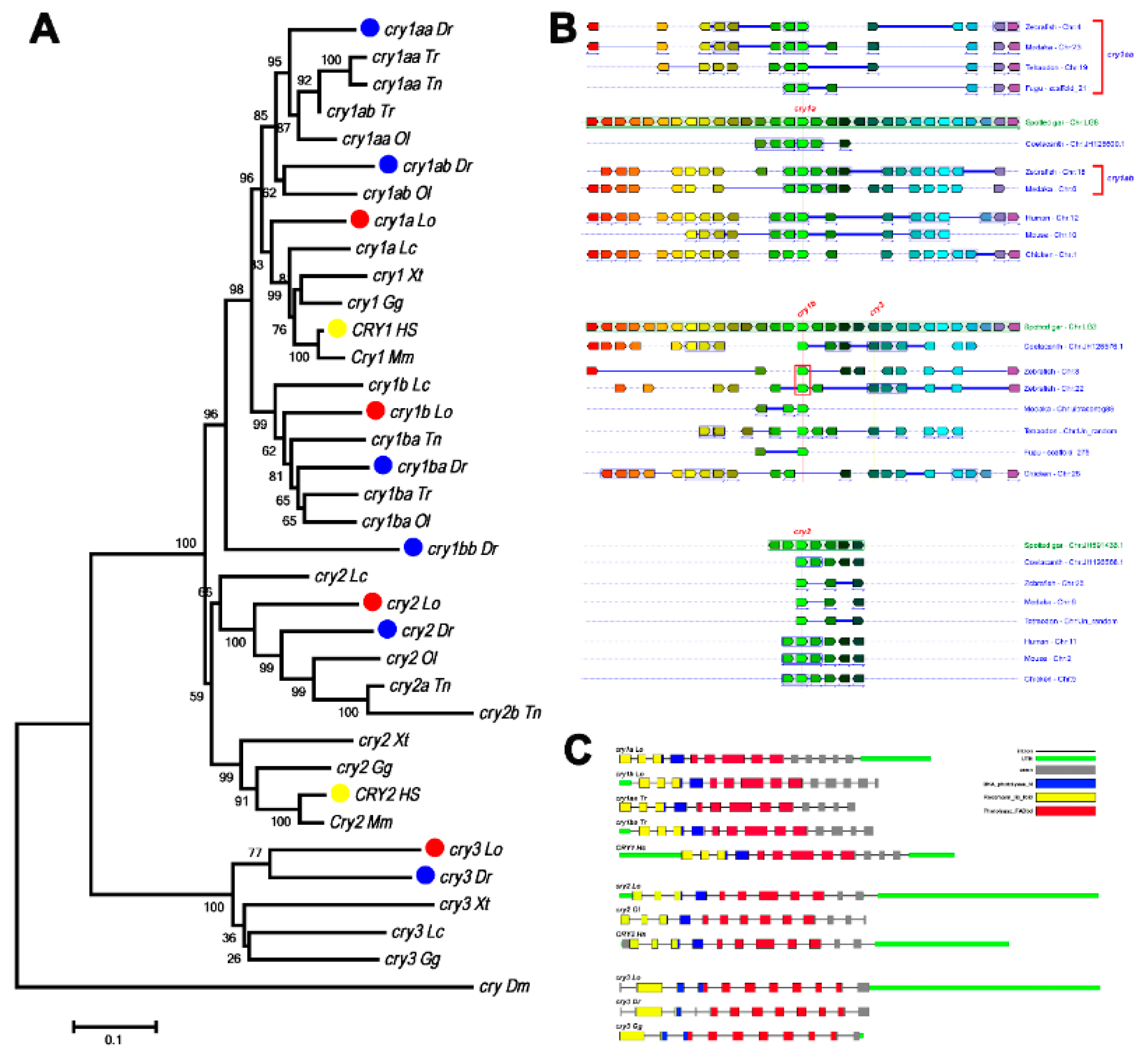
3.3. Evolution of Cry Genes

The cry1a and cry1b genes of spotted gar are clustered with human CRY1 genes (Figure 3A). Phylogenetic analysis showed that all teleost fish cry1 genes can be classified into two subclades, cry1a and cry1b. Further, cry1a and cry1b have been duplicated in most teleost fish, giving rise to cry1aa, cry1ab, zcry1ba, and cry1bb, as reported previously [36]. In addition to tetraodon, which has a cry2a/cry2b duplicate, all other fish each have only one cry2 gene, forming a monophyletic group with human CRY2 genes (Figure 3A). In a separate monophyletic clade, spotted gar, teleost fish, turtles, and birds each have one cry3, which cannot be found in mammals (Figure 3A).
We also observed conserved syntenic structures surrounding cry1a and cry1b genes in vertebrate genomes (Figure 3B). The exon–intron structures of cry1, cry2, and cry3 are highly conserved, with the same coding exon numbers and function domain location found in spotted gar, teleost fish, birds, and humans (Figure 3C).

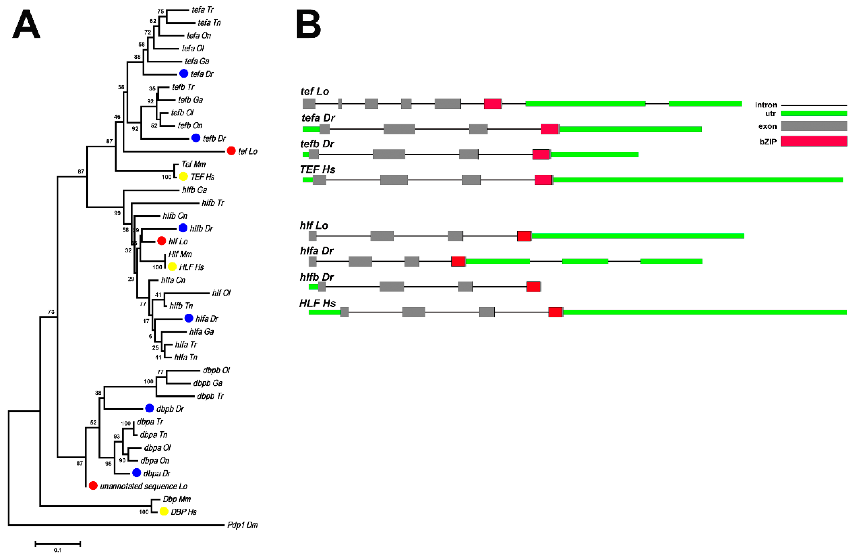
3.4. Evolution of the Par Family Genes

The par gene family contains three members: tef, hlf, and dbp. While spotted gar has one tef and one hlf orthologs as mammals, most of the teleost fish have two tef genes (tefa, tefb) and two hlf genes (hlfa, hlfb), which form monophyletic groups with spotted gar orthologs, respectively (Figure 4A). The potential gar ortholog of dbp is present as a partial unannotated sequence (Scaffold JH591448.1:146984-147190, 69aa) in the spotted gar genome (reciprocal best blast hit with coelacanth dbp and the nearest four neighbors on one side being orthologs of the four nearest neighbors of zebrafish dpba). Dbp orthologs are also conserved in most vertebrates. There is only one Dbp gene in mammals, which was duplicated to dbpa and dbpb in teleost fish.
The exon structures of hlf genes are conserved from spotted gar to humans (four coding exons), but the structure of tef in gar (six coding exons) is different between zebrafish and humans (four coding exons) (Figure 4B).

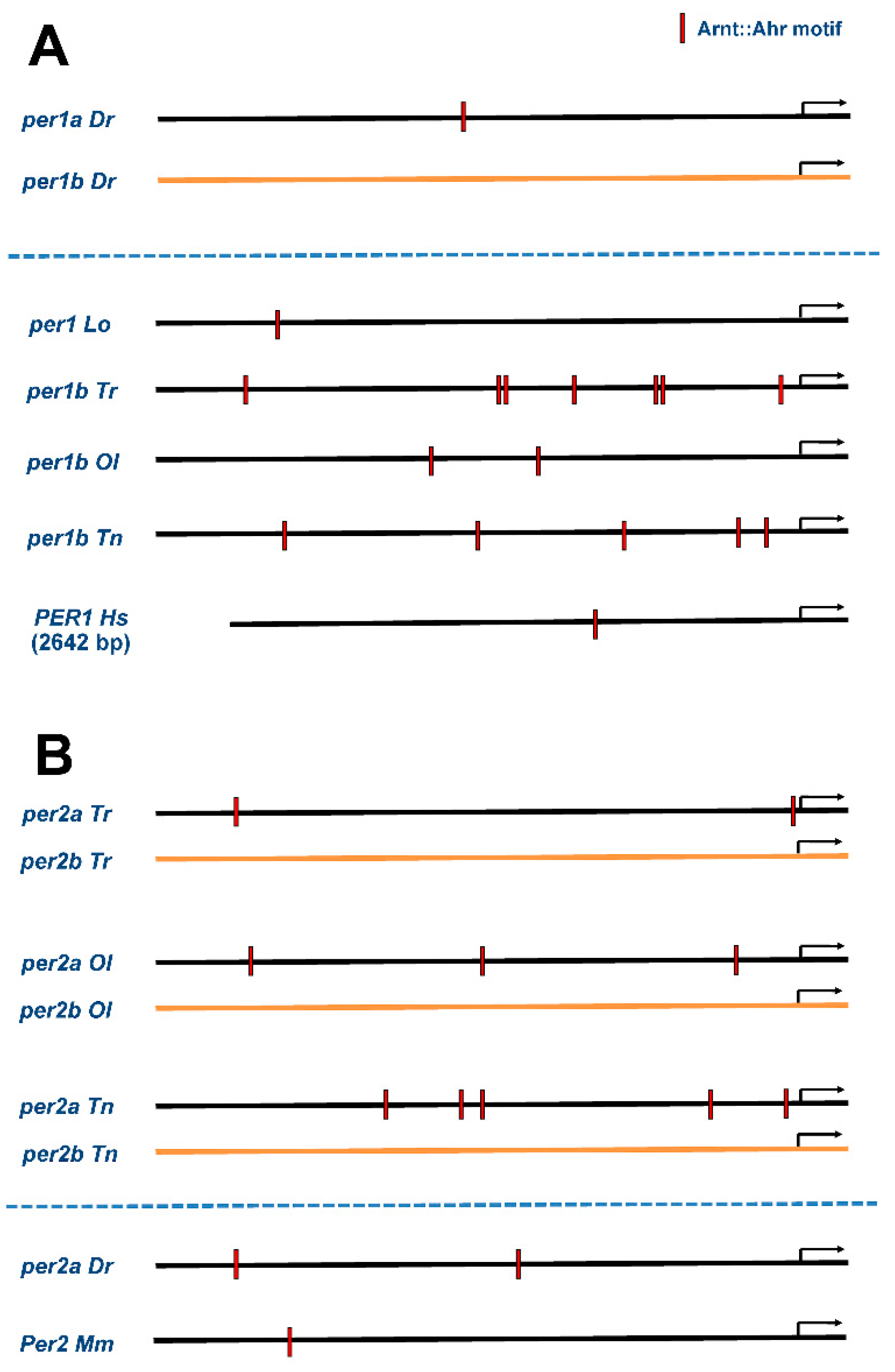
3.5. Preservation of Regulatory Elements in Duplicated Circadian Clock Genes

Although the genetic regulation of circadian rhythms appears to be similar between fish and mammals, the numbers of gene copies involved in this process vary among these groups. To further study the regulation of the duplicated genes, we compared the transcription factor binding elements of per genes between spotted gar, teleost fish, and mammals. There are some motifs, such as NFE2L1::MafG, Prrx2 and SOX10, in upstream regions of the orthologus genes from spotted gar to mammals. Interestingly, we found that the Ahr::Arnt motif exists in unduplicated per1 and per2 ortholog genes in spotted gar, some teleosts, and mammals, but this motif only exists in one of the per1 or per2 duplicated pairs in teleost fish (Figure 5). For instance, while per1a in zebrafish (Figure 5A) and per2a in fugu, medaka, and tetraodon (Figure 5B) have Ahr::Arnt motifs, the per1b (Figure 5A) and per2b (Figure 5B) genes in these species do not, thereby implicating possible cases predicted by the duplication-degeneration-complementation (DDC) model. The results of all regulatory elements in per1 and per2 genes are shown in Supplementary Tables S2 and S3.

4. Discussion

Teleost fish provide an attractive model for studying a multitude of questions related to evolution. This may be linked to the apparent, considerable, plasticity of their genome, manifested, for example, by a high variability in genome size and chromosome number [39]. Particularly, there is now substantial evidence that an ancient event of genome duplication provided the evolutionary framework for the diversification of gene functions and for speciation in fish [22,40]. Phylogenetic analyses have suggested that spotted gar occupy a clade of ancient ray-finned fish that diverged from the teleost lineage after the divergence of the bichir (Polypterus sp.) lineage [25,26,41,42,43]. Among species occupying this pre-TGD clade, spotted gar appear to be the most suitable for studies of development, genomics, and physiology [16,24,25]. Circadian rhythms are endogenous rhythms that are observed in a wide range of life forms, and circadian oscillators exhibit remarkable conservation of function across wide evolutionary time spans [44]. Our analyses of spotted gar circadian clock genes provide insight into the evolution and evolutionary impact of circadian clock genes in fish genomes and in vertebrates.
In this study, we studied the evolutional relationship of nfil3 genes in vertebrates. The phylogenetic and the syntenic analyses support the notion that the teleost fish and tetrapod nfil3 have a common ancestor. There is only one nfil3 ortholog in lampreys, and nfil3 genes have increased to three in coelacanth and spotted gar. During vertebrate evolution, the second round of vertebrate genome duplication (VGD) arose after the divergence between agnatha and teleost fish. The most parsimonious explanation is that the ancestral nfil3 gave rise to nfil3-1/2 and nfil3-3/4 by tandem duplication. Then, the clusters were duplicated to nfil3-1, nfil3-2, nfil3-3, and nfil3-4 genes in the VGD2, with the quick loss of some orthologs (Figure 6). The nfil3-1 is conserved in all vertebrates, with the exception of their loss in a few teleost fish. Although fugu and medaka do not possess nfil3-1, we still observed highly conserved synteny surrounding nfil3-1 in their genomes, implying that gene loss occurred in the two species. Further, the nfil3-1b.1/nfil3-1b.2 linkage was only found in teleost fish, and the conserved synteny surrounding nfil3-1b.1/nfil3-1b.2 was observed in all teleost fish. It seems that nfil3-1b.1 and nfil3-1b.2 were derived from local tandem duplication before teleost TGD. On the other hand, nfil3-2 is linked to nfil3-3 in the same chromosome in teleost fish and tetrapod lineage, except for mammals and birds. Amphibians retained only nfil3-3, while reptiles retained only nfil3-2. It is possible that one of the linked chromosome fragments has been lost in amphibians and reptiles, respectively. The highly conserved synteny surrounding nfil3-2 and nfil3-3 was also observed in mammalian genomes. Thus, it is likely that both nfil3-2 and nfil3-3 existed in tetrapod ancestors and were lost in mammals during evolution. Finally, the nfil3-2a/2b and nfil3-3a/3b in teleost fish were likely derived via the TGD (Figure 6).
The Cry genes had already evolved before the origin of eukaryotic organisms. The phylogenetic and syntenic analyses also suggested that the teleost fish and tetrapod Cry have a common ancestor. The vertebrate Cry1 group and Cry2 group form a sister clade, suggesting that they were derived from the duplication of a common ancestral gene. It seems that, during first round of VGD, the ancestral Cry gave rise to Cry12 and Cry34. During the second round of VGD, Cry12 gave rise to Cry1 and Cry2, while Cry34 gave rise Cry3 and Cry4, but Cry4 was quickly lost. Cry1 and Cry2 have been preserved in all vertebrates, while Cry3 genes are preserved only in the teleost and tetrapod lineages but not in mammals. In the teleost fish, cry1 generated cry1a and cry1b by local gene duplication before teleost genome duplication (TGD). The TGD generated extra copies of cry1a in teleost fish. For instance, zebrafish has retained cry1aa, cry1ab, cry1ba, cry1bb, cry2, and cry3 genes, which are consistent with our previous findings [36].
It is important to understand the evolutionary fates of circadian clock genes. According to the duplication-degeneration-complementation (DDC) model, one of the potential fates of duplicate gene pairs with multiple regulatory regions is that each duplicate may experience a loss or reduction of its expression for different subfunctions by degenerative mutations [22]. The combined action of both polygene copies is necessary to fulfill the requirements of the ancestral locus (subfunctionalization). If this happens, then complementation of the subfunctions between duplicate genes will preserve both partially degenerated copies [22]. The Ahr::Arnt motif DNA binds with protein interactions of the AHR/ARNT heterodimer [45]. ARNTL (BMAL) could interact with the PER protein. It seems that per1a/per1b or per2a/per2b have preserved some functions compared to their ancestors (subfunctionalization). Interesting, the duplicated copies of the per1 gene found in zebrafish showed distinct patterns of temporal and spatial expression [46]. The per1a gene is expressed in the retina and in both the telencephalon and the diencephalon of the forebrain in a dark environment, when there is no detectable expression of per1b. However, the expression of per1a is significantly up-regulated, and the per1b gene is constitutively expressed throughout the head region when the fish are in a light environment [46]. This pattern represents a clear example of subfunctionalization, in which each daughter copy adopts part of the function of the parental gene [27]. Our analysis of the regulatory elements in per genes may provide evidence that the mutations of these binding motifs caused gene subfunctionalization.
Compared to mammals, the extra copies of core clock genes in spotted gar were found primarily in the nfil3 and cry families. It appears that these genes duplicated before the TGD. In mammals, NFIL3 is a homologue of vrille (vri), which functions as a key negative component of the circadian clocks in Drosophila [47,48,49,50,51]. In mammals, NFIL3 was shown to interact with PER2 as well as CRY2, suggesting that NFIL3 binds to PER2 or CRY2 and contributes to mammalian circadian regulation [52,53]. Although there is a remarkable conservation of function in the circadian molecular machinery in vertebrates, studies have suggested differences in circadian regulation between fish and mammals [54,55,56]. Teleost fish exhibit an amazing level of biodiversity. In contrast to mammalian genomes, teleost genomes also contain multiple gene families. The ancient event of genome duplication could provide the evolutionary framework for the diversification of gene functions and for speciation in fish [14]. The duplication of a gene can relieve selective pressures from the duplicates because one copy can compensate for deleterious mutations in the other. The existence of paralogs enables individual members of the gene family to specialize and extend their original ancestral roles, thereby generating the potential for the evolution of complex regulatory mechanisms [44]. Hence, it is tempting to speculate that the duplication of core clock genes (such as nfil3 and cry) in teleost fish diversified the circadian regulatory mechanisms in fish.
In conclusion, we uncovered 26 circadian clock genes from 11 families among spotted gar, and nine of these 11 families of spotted gar circadian clock genes possessed the same number as humans, without an extra duplicate copy. The spotted gar contains nfil3-1, nfil3-2.1, and nfil3-2.2. While nfil3-1 is the ortholog of mammalian Nfil3-1, the nfil3-2 and nfil3-3 orthologs have been lost in mammals. For the cry gene family, spotted gar has two cry1 genes, cry1a, and cry1b, which likely derived from local (tandem) duplication in their ancestor and are co-orthologs of mammalian Cry1. Similar to teleosts, spotted gar also contain cry3, which mammals do not have. In addition, we also found that the transcription factor binding element (Ahr::Arnt) is retained only in one of the per1 or per2 paralogs in the teleost fish, suggesting that subfunctionalization occurred in the teleost fish per family. These results strongly support the notion that spotted gar genomically resembles humans to a great extent (without the TGD), but still contain extra copies of genes that mammals lack.

Supplementary Materials

The following are available online at https://www.mdpi.com/2073-4425/10/8/622/s1. Table S1: The 226 circadian clock genes used for phylogenetic analyses, Table S2: The regulatory elements in upstream regions of per1 genes, Table S3: The regulatory elements in upstream regions of per2 genes. Figure S1: The conserved syntenic analyses of (A) clock family, (B) tim family, (C) bmal family, (D) per family, (E) ror family and (F) dec family. The chromosome models of several organisms with genes are displayed. The chromosome number or linkage group number is shown to right of the chromosomes. The same colored rectangles represent the orthologous genes.

Author Contributions

Conceptualization, Y.S. and H.W.; methodology, Y.S. and C.L.; software, M.H. and J.H.; investigation, Y.S. and C.L.; data curation, C.L. (Changhong Liu) and J.Z.; writing—original draft preparation, Y.S. and C.L.; writing—review and editing, Y.S., H.W. and J.H.P.; visualization, M.H. and J.H.; project administration, Y.S. and H.W.; funding acquisition, Y.S. and H.W.

Funding

This work was supported by the National Natural Science Foundation of China (NSFC) (#31101641 and #31030062), the Natural Science Foundation of Jiangsu Province (#BK2012052), the National Basic Research Program of China (973 Program) (#2012CB947600), the National High Technology Research and Development Program of China (863 Program) (#2011AA100402-2), and the project Funded by the Priority Academic Program Development of Jiangsu Higher Education Institutions.

Acknowledgments

We thank members of the Wang lab for helpful discussion. We are also thankful for funding from the National Natural Science Foundation of China 31101641 and 31030062.

Conflicts of Interest

The authors declare no conflict of interest.

References

  1. Panda, S.; Hogenesch, J.B.; Kay, S.A. Circadian rhythms from flies to human. Nature 2002, 417, 329–335. [Google Scholar] [CrossRef] [PubMed]
  2. Reppert, S.M.; Weaver, D.R. Coordination of circadian timing in mammals. Nature 2002, 418, 935–941. [Google Scholar] [CrossRef] [PubMed]
  3. Ben-Shlomo, R.; Kyriacou, C.P. Circadian rhythm entrainment in flies and mammals. Cell Biochem. Biophys. 2002, 37, 141–156. [Google Scholar] [CrossRef]
  4. Hardin, P.E. The circadian timekeeping system of Drosophila. Curr. Biol. CB 2005, 15, R714–R722. [Google Scholar] [CrossRef] [PubMed]
  5. Ko, C.H.; Takahashi, J.S. Molecular components of the mammalian circadian clock. Hum. Mol. Genet. 2006, 15 (Suppl. 2), R271–R277. [Google Scholar] [CrossRef]
  6. Bell-Pedersen, D.; Cassone, V.M.; Earnest, D.J.; Golden, S.S.; Hardin, P.E.; Thomas, T.L.; Zoran, M.J. Circadian rhythms from multiple oscillators: Lessons from diverse organisms. Nat. Rev. Genet. 2005, 6, 544–556. [Google Scholar] [CrossRef]
  7. Paibomesai, M.I.; Moghadam, H.K.; Ferguson, M.M.; Danzmann, R.G. Clock genes and their genomic distributions in three species of salmonid fishes: Associations with genes regulating sexual maturation and cell cycling. BMC Res. Notes 2010, 3, 215. [Google Scholar] [CrossRef]
  8. Dunlap, J.C. Molecular bases for circadian clocks. Cell 1999, 96, 271–290. [Google Scholar] [CrossRef]
  9. Yu, W.; Hardin, P.E. Circadian oscillators of Drosophila and mammals. J. Cell Sci. 2006, 119, 4793–4795. [Google Scholar] [CrossRef]
  10. Layeghifard, M.; Rabani, R.; Pirhaji, L.; Yakhchali, B. Evolutionary mechanisms underlying the functional divergence of duplicate genes involved in vertebrates’ circadian rhythm pathway. Gene 2008, 426, 65–71. [Google Scholar] [CrossRef]
  11. Hughes, A.L. The evolution of functionally novel proteins after gene duplication. Proc. Biol. Sci. R. Soc. 1994, 256, 119–124. [Google Scholar] [CrossRef]
  12. Aguileta, G.; Bielawski, J.P.; Yang, Z. Gene conversion and functional divergence in the β-globin gene family. J. Mol. Evol. 2004, 59, 177–189. [Google Scholar] [CrossRef] [PubMed]
  13. Wang, H. Comparative analysis of teleost fish genomes reveals preservation of different ancient clock duplicates in different fishes. Mar. Genom. 2008, 1, 69–78. [Google Scholar] [CrossRef] [PubMed]
  14. Volff, J.N. Genome evolution and biodiversity in teleost fish. Heredity 2005, 94, 280–294. [Google Scholar] [CrossRef] [PubMed]
  15. Amemiya, C.T.; Alfoldi, J.; Lee, A.P.; Fan, S.; Philippe, H.; Maccallum, I.; Braasch, I.; Manousaki, T.; Schneider, I.; Rohner, N.; et al. The African coelacanth genome provides insights into tetrapod evolution. Nature 2013, 496, 311–316. [Google Scholar] [CrossRef] [PubMed] [Green Version]
  16. Braasch, I.; Gehrke, A.R.; Smith, J.J.; Kawasaki, K.; Manousaki, T.; Pasquier, J.; Amores, A.; Desvignes, T.; Batzel, P.; Catchen, J.; et al. The spotted gar genome illuminates vertebrate evolution and facilitates human-teleost comparisons. Nat. Genet. 2016, 48, 427–437. [Google Scholar] [CrossRef] [Green Version]
  17. Xu, P.; Zhang, X.; Wang, X.; Li, J.; Liu, G.; Kuang, Y.; Xu, J.; Zheng, X.; Ren, L.; Wang, G.; et al. Genome sequence and genetic diversity of the common carp, Cyprinus carpio. Nat. Genet. 2014, 46, 1212–1219. [Google Scholar] [CrossRef] [Green Version]
  18. Amores, A.; Force, A.; Yan, Y.L.; Joly, L.; Amemiya, C.; Fritz, A.; Ho, R.K.; Langeland, J.; Prince, V.; Wang, Y.L.; et al. Zebrafish HOX clusters and vertebrate genome evolution. Science 1998, 282, 1711–1714. [Google Scholar] [CrossRef]
  19. Postlethwait, J.H.; Yan, Y.L.; Gates, M.A.; Horne, S.; Amores, A.; Brownlie, A.; Donovan, A.; Egan, E.S.; Force, A.; Gong, Z.; et al. Vertebrate genome evolution and the zebrafish gene map. Nat. Genet. 1998, 18, 345–349. [Google Scholar] [CrossRef]
  20. Taylor, J.S.; Braasch, I.; Frickey, T.; Meyer, A.; Van de Peer, Y. Genome duplication, a trait shared by 22,000 species of ray-finned fish. Genome Res. 2003, 13, 382–390. [Google Scholar] [CrossRef]
  21. Jaillon, O.; Aury, J.M.; Brunet, F.; Petit, J.L.; Stange-Thomann, N.; Mauceli, E.; Bouneau, L.; Fischer, C.; Ozouf-Costaz, C.; Bernot, A.; et al. Genome duplication in the teleost fish Tetraodon nigroviridis reveals the early vertebrate proto-karyotype. Nature 2004, 431, 946–957. [Google Scholar] [CrossRef] [Green Version]
  22. Force, A.; Lynch, M.; Pickett, F.B.; Amores, A.; Yan, Y.L.; Postlethwait, J. Preservation of duplicate genes by complementary, degenerative mutations. Genetics 1999, 151, 1531–1545. [Google Scholar]
  23. Postlethwait, J.; Amores, A.; Cresko, W.; Singer, A.; Yan, Y.L. Subfunction partitioning, the teleost radiation and the annotation of the human genome. Trends Genet. TIG 2004, 20, 481–490. [Google Scholar] [CrossRef]
  24. Amores, A.; Catchen, J.; Ferrara, A.; Fontenot, Q.; Postlethwait, J.H. Genome evolution and meiotic maps by massively parallel DNA sequencing: Spotted gar, an outgroup for the teleost genome duplication. Genetics 2011, 188, 799–808. [Google Scholar] [CrossRef]
  25. Hoegg, S.; Brinkmann, H.; Taylor, J.S.; Meyer, A. Phylogenetic timing of the fish-specific genome duplication correlates with the diversification of teleost fish. J. Mol. Evol. 2004, 59, 190–203. [Google Scholar] [CrossRef]
  26. Crow, K.D.; Stadler, P.F.; Lynch, V.J.; Amemiya, C.; Wagner, G.P. The “fish-specific” HOX cluster duplication is coincident with the origin of teleosts. Mol. Biol. Evol. 2006, 23, 121–136. [Google Scholar] [CrossRef]
  27. Toloza-Villalobos, J.; Arroyo, J.I.; Opazo, J.C. The circadian clock of teleost fish: A comparative analysis reveals distinct fates for duplicated genes. J. Mol. Evol. 2015, 80, 57–64. [Google Scholar] [CrossRef]
  28. Zhang, J.; Rosenberg, H.F.; Nei, M. Positive Darwinian selection after gene duplication in primate ribonuclease genes. Proc. Natl. Acad. Sci. USA 1998, 95, 3708–3713. [Google Scholar] [CrossRef] [Green Version]
  29. Ueda, H.R.; Hayashi, S.; Chen, W.; Sano, M.; Machida, M.; Shigeyoshi, Y.; Iino, M.; Hashimoto, S. System-level identification of transcriptional circuits underlying mammalian circadian clocks. Nat. Genet. 2005, 37, 187–192. [Google Scholar] [CrossRef]
  30. Benna, C.; Scannapieco, P.; Piccin, A.; Sandrelli, F.; Zordan, M.; Rosato, E.; Kyriacou, C.P.; Valle, G.; Costa, R. A second timeless gene in Drosophila shares greater sequence similarity with mammalian tim. Curr. Biol. CB 2000, 10, R512–R513. [Google Scholar] [CrossRef] [Green Version]
  31. Kurien, P.; Hsu, P.K.; Leon, J.; Wu, D.; McMahon, T.; Shi, G.; Xu, Y.; Lipzen, A.; Pennacchio, L.A.; Jones, C.R.; et al. TIMELESS mutation alters phase responsiveness and causes advanced sleep phase. Proc. Natl. Acad. Sci. USA 2019, 116, 12045–12053. [Google Scholar] [CrossRef] [Green Version]
  32. Larkin, M.A.; Blackshields, G.; Brown, N.P.; Chenna, R.; McGettigan, P.A.; McWilliam, H.; Valentin, F.; Wallace, I.M.; Wilm, A.; Lopez, R.; et al. Clustal W and Clustal X version 2.0. Bioinformatics 2007, 23, 2947–2948. [Google Scholar] [CrossRef] [Green Version]
  33. Tamura, K.; Stecher, G.; Peterson, D.; Filipski, A.; Kumar, S. MEGA6: Molecular Evolutionary Genetics Analysis version 6.0. Mol. Biol. Evol. 2013, 30, 2725–2729. [Google Scholar] [CrossRef]
  34. Rambaldi, D.; Ciccarelli, F.D. FancyGene: Dynamic visualization of gene structures and protein domain architectures on genomic loci. Bioinformatics 2009, 25, 2281–2282. [Google Scholar] [CrossRef]
  35. Louis, A.; Muffato, M.; Roest Crollius, H. Genomicus: five genome browsers for comparative genomics in eukaryota. Nucleic Acids Res. 2013, 41, D700–D705. [Google Scholar] [CrossRef]
  36. Liu, C.; Hu, J.; Qu, C.; Wang, L.; Huang, G.; Niu, P.; Zhong, Z.; Hong, F.; Wang, G.; Postlethwait, J.H.; et al. Molecular evolution and functional divergence of zebrafish (Danio rerio) cryptochrome genes. Sci. Rep. 2015, 5, 8113. [Google Scholar] [CrossRef]
  37. Ehrlich, J.; Sankoff, D.; Nadeau, J.H. Synteny conservation and chromosome rearrangements during mammalian evolution. Genetics 1997, 147, 289–296. [Google Scholar]
  38. Woods, I.G.; Wilson, C.; Friedlander, B.; Chang, P.; Reyes, D.K.; Nix, R.; Kelly, P.D.; Chu, F.; Postlethwait, J.H.; Talbot, W.S. The zebrafish gene map defines ancestral vertebrate chromosomes. Genome Res. 2005, 15, 1307–1314. [Google Scholar] [CrossRef] [Green Version]
  39. Venkatesh, B. Evolution and diversity of fish genomes. Curr. Opin. Genet. Dev. 2003, 13, 588–592. [Google Scholar] [CrossRef]
  40. Glasauer, S.M.; Neuhauss, S.C. Whole-genome duplication in teleost fishes and its evolutionary consequences. Mol. Genet. Genom. MGG 2014, 289, 1045–1060. [Google Scholar] [CrossRef] [Green Version]
  41. Inoue, J.G.; Miya, M.; Venkatesh, B.; Nishida, M. The mitochondrial genome of Indonesian coelacanth Latimeria menadoensis (Sarcopterygii: Coelacanthiformes) and divergence time estimation between the two coelacanths. Gene 2005, 349, 227–235. [Google Scholar] [CrossRef]
  42. Katsu, Y.; Kohno, S.; Hyodo, S.; Ijiri, S.; Adachi, S.; Hara, A.; Guillette, L.J., Jr.; Iguchi, T. Molecular cloning, characterization, and evolutionary analysis of estrogen receptors from phylogenetically ancient fish. Endocrinology 2008, 149, 6300–6310. [Google Scholar] [CrossRef]
  43. Salaneck, E.; Larsson, T.A.; Larson, E.T.; Larhammar, D. Birth and death of neuropeptide Y receptor genes in relation to the teleost fish tetraploidization. Gene 2008, 409, 61–71. [Google Scholar] [CrossRef]
  44. Looby, P.; Loudon, A.S. Gene duplication and complex circadian clocks in mammals. Trends Genet. TIG 2005, 21, 46–53. [Google Scholar] [CrossRef]
  45. Swanson, H.I. DNA binding and protein interactions of the AHR/ARNT heterodimer that facilitate gene activation. Chem. Biol. Interact. 2002, 141, 63–76. [Google Scholar] [CrossRef]
  46. Wang, H. Comparative analysis of period genes in teleost fish genomes. J. Mol. Evol. 2008, 67, 29–40. [Google Scholar] [CrossRef]
  47. Blau, J.; Young, M.W. Cycling vrille expression is required for a functional Drosophila clock. Cell 1999, 99, 661–671. [Google Scholar] [CrossRef]
  48. Cyran, S.A.; Buchsbaum, A.M.; Reddy, K.L.; Lin, M.C.; Glossop, N.R.; Hardin, P.E.; Young, M.W.; Storti, R.V.; Blau, J. vrille, Pdp1, and dClock form a second feedback loop in the Drosophila circadian clock. Cell 2003, 112, 329–341. [Google Scholar] [CrossRef]
  49. Glossop, N.R.; Houl, J.H.; Zheng, H.; Ng, F.S.; Dudek, S.M.; Hardin, P.E. VRILLE feeds back to control circadian transcription of Clock in the Drosophila circadian oscillator. Neuron 2003, 37, 249–261. [Google Scholar] [CrossRef]
  50. Doi, M.; Nakajima, Y.; Okano, T.; Fukada, Y. Light-induced phase-delay of the chicken pineal circadian clock is associated with the induction of cE4bp4, a potential transcriptional repressor of cPer2 gene. Proc. Natl. Acad. Sci. USA 2001, 98, 8089–8094. [Google Scholar] [CrossRef]
  51. Mitsui, S.; Yamaguchi, S.; Matsuo, T.; Ishida, Y.; Okamura, H. Antagonistic role of E4BP4 and PAR proteins in the circadian oscillatory mechanism. Genes Dev. 2001, 15, 995–1006. [Google Scholar] [CrossRef] [Green Version]
  52. Ohno, T.; Onishi, Y.; Ishida, N. The negative transcription factor E4BP4 is associated with circadian clock protein PERIOD2. Biochem. Biophys. Res. Commun. 2007, 354, 1010–1015. [Google Scholar] [CrossRef]
  53. Ohno, T.; Onishi, Y.; Ishida, N. A novel E4BP4 element drives circadian expression of mPeriod2. Nucleic Acids Res. 2007, 35, 648–655. [Google Scholar] [CrossRef]
  54. Hogenesch, J.B.; Ueda, H.R. Understanding systems-level properties: Timely stories from the study of clocks. Nat. Rev. Genet. 2011, 12, 407–416. [Google Scholar] [CrossRef]
  55. Dekens, M.P.; Whitmore, D. Autonomous onset of the circadian clock in the zebrafish embryo. EMBO J. 2008, 27, 2757–2765. [Google Scholar] [CrossRef] [Green Version]
  56. Vatine, G.; Vallone, D.; Appelbaum, L.; Mracek, P.; Ben-Moshe, Z.; Lahiri, K.; Gothilf, Y.; Foulkes, N.S. Light directs zebrafish period2 expression via conserved D and E boxes. PLoS Biol. 2009, 7, e1000223. [Google Scholar] [CrossRef]
Figure 1. The phylogenetic trees of circadian clock genes. (A) bmal genes; (B) clock genes; (C) per genes; (D) csnk1e genes; (E) tim genes; (F) dec genes; (G) nr1d genes; (H) ror genes. The trees were constructed by the neighbor-joining method with MEGA6 [33], and the numbers on the nodes are percent bootstrap values based on 1000 pseudoreplications. The outgroups are ortholog genes in fly or lamprey. Red dots indicate spotted gar genes, blue dots indicate zebrafish genes, and yellow dots indicate human genes. The sequences used are listed in Supplementary Table S1.
Genes 10 00622 g001
Figure 2. The phylogenetic and conserved syntenic analyses of the nfil3 family. (A) Phylogenetic trees based on sequences of nfil3 genes. The tree was constructed by the neighbor-joining method with MEGA6 [33], and the numbers on the nodes are the percent bootstrap values based on 1000 pseudoreplications. The outgroup is fly vri. Red dots indicate spotted gar genes, blue dots indicate zebrafish genes, and yellow dots indicate human genes. (B) The conserved synteny surrounding nfil3-1 genes in the chromosomes of lampreys, coelacanths, spotted gar, zebrafish, medaka, fugu, frogs, lizards, chickens, ducks, mice and humans was displayed with Genomicus [35]. (C) The conserved synteny of nfil3-1b.1 and nfil3-1b.2 genes in the chromosomes of zebrafish, cave fish, tilapia, platyfish, medaka, tilapia, and fugu. (D) Conserved synteny of nfil3-2 and nfil3-3 genes in chromosomes of coelacanth, spotted gar, zebrafish, medaka, teraodon, fugu, tilapia, platyfish, frogs, lizards, mice, and humans. The chromosome number or linkage group number is shown to the right of the chromosomes. The same colored rectangles represent the orthologous genes. Red and blue boxes indicate the duplicated genes.
Genes 10 00622 g002
Figure 3. The phylogenetic, conserved syntenic, and exon structural analyses of the cry family. (A) Phylogenetic trees based on sequences of cry genes. The tree was constructed by the neighbor-joining method with MEGA6, and the numbers on the nodes are the percent bootstrap values based on 1000 pseudoreplications. The outgroup is Drosophila melanogaster cry. Red dots indicate spotted gar genes, blue dots indicate zebrafish genes, and yellow dots indicate human genes. (B) The conserved synteny surrounding the cry1a, cry1b/cry3, and cry2 genes in chromosomes of coelacanth, spotted gar, zebrafish, medaka, teraodon, fugu, chickens, mice, and humans. The chromosome number or linkage group number is shown to the right of the chromosomes. The same colored rectangles represent the orthologous genes. Red and blue boxes indicate the duplicated genes. (C) The exonic structures of cry genes in spotted gar, medaka, fugu, chickens and humans. The boxes represent the exons. The size of each exon is drawn to scale.
Genes 10 00622 g003
Figure 4. The phylogenetic and exon structural analyses of the par family. (A) Phylogenetic trees based on sequences of PAR family genes. The tree was constructed by the neighbor-joining method with MEGA6, and the numbers on the nodes are the percent bootstrap values based on 1000 pseudoreplications. The outgroup is Drosophila melanogaster pdp1. Red dots indicate spotted gar genes, blue dots indicate zebrafish genes, and yellow dots indicate human genes. (B) The exonic structures of par family genes in spotted gar, zebrafish, and humans. The boxes represent the exons. The size of each exon is drawn to scale.
Genes 10 00622 g004
Figure 5. The transcription factor binding element (Ahr::Arnt) in upstream regions of duplicated Per genes from spotted gar to mammals. The complementary presence of the Ahr::Arnt motif in duplicated Per1 (A) or Per2 (B) paralog genes provides possible cases predicted by the duplication-degeneration-complementation (DDC) model. The upstream region of each gene is 3000 bp in length, with the exception of that of human PER1, which is 2642 bp only. The transcription factor binding elements were predicted using the JASPAR dataset, with a profile score threshold of 99%. The bisque line represents duplication in the lineage, and the dotted line separates the duplicated genes and unduplicated genes.
Genes 10 00622 g005
Figure 6. A hypothetical model for the evolution of nfil3 genes in vertebrates. The teleost fish and tetrapod nfil3 had a common ancestor. The ancestral nfil3 gave rise to nfil3-1/2 and nfil3-3/4 by tandem duplication. During the second round of vertebrate genome duplication, the cluster was duplicated to nfil3-1, nfil3-2, nfil3-3, and nfil3-4 genes, with the quick loss of nfil3-4. The nfil3-1 is conserved in all vertebrates, with the exception of losses in a few teleost fish. The nfil3-1b.1 and nfil3-1b.2 linkage has been preserved only in teleost fish. The nfil3-2 and nfil3-3 linkage was conserved in the teleost fish and tetrapod lineages. The teleost-specific genome duplication produced the nfil3-2a/nfil3-2b duplicate, which was observed in zebrafish, medaka, tetrodon, tilapia, platyfish, and cave fish; and the nfil3-3a/nfil3-3b duplicate, which was observed in zebrafish, tilapia, and platyfish. However, amphibians retained only nfil3-3 and reptiles retained only nfil3-2. Then, Nfil3-2 and Nfil3-3 were completely lost in the lineages of mammals and birds during evolution. The grey box means gene loss.
Genes 10 00622 g006
Table 1. Circadian clock genes in spotted gar [16].
Gene NamesEnsembl Gene IDProtein Length (aa)Genome Location
bmal1ENSLOCG00000003999678Chromosome LG27: 8,317,763–8,344,549
bmal2ENSLOCG00000015224639Chromosome LG8: 3,189,570–3,226,867
clock1ENSLOCG00000014043744Chromosome LG4: 72,323,329–72,339,605
clock2ENSLOCG00000014750886Chromosome LG7: 42,295,248–42,323,335
cry1aENSLOCG00000015272647Chromosome LG8: 4,053,475–4,074,670
cry1bENSLOCG00000011417675Chromosome LG3: 32,901,867–32,938,020
cry2ENSLOCG00000014655569Scaffold JH591436.1: 96,765–111,323
cry3ENSLOCG00000011465586Chromosome LG3: 33,014,383–33,053,389
per1ENSLOCG000000133441445Chromosome LG2: 58,185,728–58,201,428
per2ENSLOCG000000044411385Chromosome LG14: 7,862,125–7,881,568
per3ENSLOCG000000026071165Chromosome LG25: 4,785,705–4,800,981
csnk1eENSLOCG00000011701273Chromosome LG12: 35,099,178-35,103,163
dec1ENSLOCG00000010962409Chromosome LG5: 27,670,723–27,673,540
dec2ENSLOCG00000015327422Chromosome LG8: 4,744,466–4,746,682
nfil3-1ENSLOCG00000008217443Chromosome LG2: 25,281,929–25,283,356
nfil3-2ENSLOCG00000018299544Chromosome LG6: 17,036,778–17,038,412
nfil3-3ENSLOCG00000018298394Chromosome LG6: 17,009,772–17,010,956
nr1d1ENSLOCG00000006223362Chromosome LG4: 15,608,213–15,662,480
nr1d2ENSLOCG00000006818604Chromosome LG11: 20,445,844–20,461,290
tefENSLOCG00000011595323Chromosome LG12: 34,848,929–34,860,886
hlfENSLOCG00000012233298Chromosome LG10: 33,240,269–33,260,238
dbp 69Scaffold JH591448.1:146984–147190
roraENSLOCG00000014779519Chromosome LG3: 53,154,612–53,409,505
rorbENSLOCG00000009712462Chromosome LG2: 34,273,732–34,319,408
rorcENSLOCG00000006502467Chromosome LG19: 9,503,786–9,519,701
timelessENSLOCG000000041801225Chromosome LG4: 11,892,308–11,918,271

