Article

Differences in Formation of Prepuce and Urethral Groove During Penile Development Between Guinea Pigs and Mice Are Controlled by Differential Expression of Shh, Fgf10 and Fgfr2

Department of Biomedical Sciences, Southern Illinois University School of Medicine, Carbondale, IL 62901, USA
*
Author to whom correspondence should be addressed.
Cells 2025, 14(5), 348; https://doi.org/10.3390/cells14050348
Submission received: 1 January 2025 / Revised: 23 February 2025 / Accepted: 25 February 2025 / Published: 27 February 2025
(This article belongs to the Section Reproductive Cells and Development)

Abstract

:
The penile tubular urethra forms by canalization of the urethral plate without forming an obvious urethral groove in mice, while the urethral epithelium forms a fully open urethral groove before urethra closure through the distal-opening-proximal-closing process in humans and guinea pigs. Our knowledge of the mechanism of penile development is mainly based on studies in mice. To reveal how the fully opened urethral groove forms in humans and guinea pigs, we compared the expression patterns and levels of key developmental genes using in situ hybridization and quantitative PCR during glans and preputial development between guinea pigs and mice. Our results revealed that, compared with mouse preputial development, which started before sexual differentiation, preputial development in guinea pigs was delayed and initiated at the same time that sexual differentiation began. Fgf10 was mainly expressed in the urethral epithelium in developing genital tubercle (GT) of guinea pigs. The relative expression of Shh, Fgf8, Fgf10, Fgfr2, and Hoxd13 was reduced more than 4-fold in the GT of guinea pigs compared to that of mice. Hedgehog and Fgf inhibitors induced urethral groove formation and restrained preputial development in cultured mouse GT, while Shh and Fgf10 proteins induced preputial development in cultured guinea pig GT. Our discovery suggests that the differential expression of Shh and Fgf10/Fgfr2 may be the main reason a fully opened urethral groove forms in guinea pigs, and it may be similar in humans as well.

Graphical Abstract

1. Introduction

The mammalian penis develops from the bisexual precursor called the genital tubercle (GT) under the influence of androgens [1,2,3,4]. Compared with mice and rats, humans and guinea pigs form fully opened urethral grooves at the end of the bisexual stage, and the process of tubular urethral formation follows distal-opening-proximal-closing, that is, the “Double Zipper” model [1,3]. In humans, the penile urethra develops via an “Opening Zipper” by canalization of the solid urethral plate to form the open urethral groove before 9.5 weeks of gestation. Then, androgen-induced proximal closure of the urethral groove starts tubular urethral formation in the penile shaft before 10.5 weeks [1]. Human preputial development is initiated at 10–11 weeks of gestation, which is almost the same time when tubular urethra starts to form in the proximal region. Using the guinea pig model, we found that cell proliferation in the outer layers (including the basal layer) and programmed cell death in the inner layers of urethral epithelium play key roles during dorsal-to-ventral displacement and final opening of the urethral canal to form a urethral groove; these processes show no difference between males and females [5]. In mice, the urethral epithelium forms a urethral plate; a urethral opening can be observed in the proximal region at E13.5. From the urethral opening to the distal tip, the ventral part of the urethral epithelium has never formed an open urethral groove during fetal development, and thus there is no distal-opening-proximal-closing process during tubular urethral formation [6]. The preputial swellings appear as secondary outgrowths on the lateral edges of the tubercle at E13.5 and continue to grow laterally and ventrally to form the prepuce [6]. At E15.5, the preputial swelling covers the proximal region of the glans before sexual differentiation [6]. Clearly, human preputial development is initiated at the same time as the androgen-dependent-proximal-closing of urethral groove initiation, while in mice, the preputial development is initiated relatively earlier and begins at the bisexual stage of the external genital development; unfortunately, the mechanism causing the difference is unknown.
Theories on the development of human prepuce fall into two opposing ideas. One idea is that the development of the human prepuce involves the formation and distal extension of the preputial folds to eventually completely cover the glans. This idea is exemplified by Hunter who described “folds of ectodermal tissue that appear to flow over the dorsum of the glans as the beginning of the prepuce then extends distally to cover the glans” [7]. An alternate theory is that a dorsal skin fold forms a preputial fold and extends distally to cover the glans; at the same time, epithelial ingrowth occurs to form the preputial lamina. This hypothesis was proposed by Glenister in 1956 [8]. The two theories were illustrated recently by Cunha et al. [9]. Liu et al. [10] found that the prepuce initially forms on the dorsal aspect of the glans at approximately 12 weeks of gestation. After sequential proximal-to-distal remodeling of the ventral urethral plate along the ventral aspect of the glans, the prepuce of epidermal origin fuses in the ventral midline. Cunha et al. [9] proposed a novel morphogenetic mechanism for the formation of the preputial lamina, namely the splitting of the thick epidermis of the glans into the preputial lamina and the epidermis via the intrusion of mesenchyme, finding that the process begins on the proximal aspect of the glans and extends distally. The cellular and molecular mechanisms of preputial development remain unknown.
The development of external genitalia is controlled by local developmental genes, such as fibroblast growth factors (Fgfs), Sonic hedgehog (Shh), Bone morphogenetic protein (Bmp), Wnt, and several Hox genes [11,12,13]. Several FGF genes, including FGF8, FGF10, and fibroblast growth factor receptor 2 (FGFR2), were found to be expressed in the foreskin of children with hypospadias [14]. Mutation in Fgf10 or Fgfr2 induces genital malformation in mice [15,16]. It has been shown that Bmp4 is required for the initiation of GT outgrowth [17]. Additionally, knockout of an upstream regulator of Bmp4, Isl1, in murine shows abrogated genital outgrowth [18]. Several WNT ligands are expressed in the developing mouse GT; canonical WNT signaling is required in normal murine GT outgrowth [19,20]. Wnt5a is one of the main WNT ligands regulating urethral tube formation as well as external genitalia outgrowth in mice [21]. Human autosomal dominant mutations in WNT5A cause Robinow Syndrome, which shows micropenis and hypoplastic scrotum in males, and hypoplasia of the clitoris and labia in females [22]. Extensive studies of Hoxd13 and Hoxa13 have demonstrated their essential role in the development of the GT, double mutants exhibit agenesis of the GT, and heterozygosity for Hoxa13 or Hoxd13 causes patterning defects of the phallus [23]. Human mutations in the HOXA13 gene are responsible for the range of phenotypes observed in hand-foot-genital syndrome [24]. Homozygous mutations of HOXD13 in men exhibit hypospadias [25]. Shh is one of the most intensively studied genes in external genital development. It is a major morphogenic regulator of the outgrowth of the GT mainly through the regulation of cell cycle progression [6,26]. A significant decrease in mRNA expression of the SHH and PTCH1 genes was found in boys with proximal hypospadias compared with boys without hypospadias [27]. Shh expression has been detected in the urethral plate in the developing GT of guinea pigs. Compared to mice, the Shh expression domain on the ventral side of the developing GT in guinea pigs extends out to the ventral surface epithelium [5].
Here, we report the results of experiments aimed at identifying differentially expressed genes in a guinea pig model that more closely resembles human penile development, and we tested the function of the selected genes in preputial development and urethral groove formation using organ culture. Since a detailed interpretation of guinea pig penile development at the stage of distal-opening-proximal-closing of the urethra is not available, we first present a detailed embryological study of genital development between E27 to E33. Preputial development is initiated before sexual differentiation occurs in mice but after sexual differentiation starts in guinea pigs; we compare key genes’ expression patterns and levels between guinea pigs and mice; finally, we demonstrate that Shh and Fgf10 play critical roles in preputial development and urethral groove formation during distal-opening-proximal-closing of penile development.

2. Materials and Methods

2.1. Animals and Treatments

Sexually mature Hartley guinea pigs were purchased from Elm Hill Labs, and ICR mice were purchased from Envigo RMS Inc. (Indianapolis, IN, USA). Guinea pigs and mice were housed in a pathogen-free barrier facility on 12 h light/dark cycles, with access to food and water ad libitum. All experiments were conducted in accordance with the “Guide for the Care and Use of Laboratory Animals”. The experimental protocols (Guinea pigs: 20-014, mice: 23-011) were approved by the Institutional Animal Care and Use Committee of Southern Illinois University Carbondale. Time-mating of guinea pigs was achieved based on a previously established method [3]. A minimum of 2 litters of embryos was collected at each stage, and GTs were dissected under a stereoscope. The sex of the embryos at E30 and later stages was identified by inspection of gonadal morphology under the stereoscope, while the sex of the embryos at earlier stages was identified by genotyping using Y (Sry) and X (Dystrophin) chromosome-specific genes [28]. To detect cell proliferation in guinea pigs, 5-bromo-2′-deoxyuridine (BrdU, 50 mg/kg) was injected intraperitoneally, and embryos were collected 4 h later for immunohistochemistry.

2.2. Organ Culture and In Vitro Cyclopamine, BGJ-398, Shh, and Fgf-10 Administration

Mouse and guinea pig embryonic GT (at E14.5 and E27, respectively) culture setup followed the previously described method [29]. Methyltestosterone (MT) was dissolved in ethanol (0.05 M stock, filter sterilized) and then diluted into the medium to 10 nM. Cyclopamine (Cat#: A8340), BGJ398 (Cat#: A3014), and mouse Shh protein (Cat# P1230) were purchased from ApexBio (Houston, TX, USA). The Cyclopamine stock solution (10 mM in DMSO) was diluted to the final concentration (200 nM) with culture medium before use. BGJ398 stock solution (5 mg/mL in DMSO) was diluted to 1.4 nM to inhibit Fgf-10/Fgfr2 using a culture medium. Mouse Fgf-10 protein was purchased from R&D systems (Cat# 6224-FG-025/CF, Newark, DE, USA). Shh and Fgf-10 proteins were added into the culture medium with the final concentration of 100 ng/mL and 50 ng/mL, respectively. The GTs were cultured for 48 h and then processed for morphology analysis.

2.3. Histology and Immunohistochemistry

Guinea pig and mouse embryos at different stages were harvested and rinsed with PBS. Paraffin sections of GT were prepared and stained with hematoxylin and eosin, as we previously described [3]. Immunohistochemistry was performed using anti-BrdU (G3G4, Cat# AB 2314035, RRID: AB_2618097, DSHB, Iowa City, IA, USA) according to procedures modified from the previously established immunofluorescence protocol [5]. BrdU antibody was detected using ABC Kit (PK-6100, Vector Laboratories, Newark, CA, USA) according to the manufacturer’s operation manual. To quantify BrdU-positive cells in the preputial initiation region of developing E29 guinea pig GT, image stacks were acquired using the 40× objective, and ImageJ.JS software (version 1) was used to identify and count the positive cells in newly formed preputial mesenchyme and surrounding epithelium. Since there were only 3 serial sections (6 μm) of the preputial formation starting area in each GT, positive cells in the interested structure of every section in 3 different GTs were counted. The Fgf-10 immunofluorescence in guinea pig and mouse GTs was performed following the established method in our laboratory [3,5,29] using FGF-10 (H121) polyclonal antibody purchased from Santa Cruz (Cat# SC-7917, RRID: AB_2262731, Dallas, TX, USA). Sample size, n = 3 litters, slides obtained from 3 males (one from each litter) were selected for immunostaining.

2.4. In Situ Hybridization

Three embryos of different stages were selected for in situ hybridization analysis. In situ hybridization was performed as described [30] with some modifications [5]. To create the RNA probes, the cDNA of developmental genes was PCR-amplified from embryonic guinea pig cDNA using the primers designed with PrimerQuest software (version RUO22-1233-001) from IDT-DNA (https://www.idtdna.com/pages/tools/primerquest, accessed on 6 November 2022), and all primers are listed in Table S1. These were then cloned into the pGEM®-T Easy Vector (Cat# A1360, Promega, Madison, WI, USA) according to operation manual, except for Fgf10. The guinea pig Fgf10 IMAGE clone was purchased from GenScript (Clone ID: ODo12835, Piscataway, NJ, USA). Cloned DNAs were then amplified using M13 primers, and a reverse transcription reaction was performed using the DIG RNA Labeling Kit (SP6/T7: #EP0131/#EP0111, Thermo Scientific, Waltham, MA, USA) from Roche (Cat#: 11175025910, Indianapolis, IN, USA) to produce antisense RNA probes. A guinea pig Fgf10 RNA probe was synthesized using T3 RNA polymerase (#EP0101, Thermo Scientific, Waltham, MA, USA).

2.5. Quantitative Gene Expression Analysis Using RT-QPCR

Total RNA was extracted from male GT of E12.5 and E13.5 mice, as well as E23 and E26.5 guinea pigs, using the TRIzol method according to the operation manual (Cat: 10296010, Invitrogen, Carlsbad, CA, USA). RNA quality was assessed following the published method [31]. The cDNA was synthesized from 500 ng total RNA using iScript Reverse Transcription Supermix (Bio-rad, Hercules, CA, USA). Primers for all guinea pig genes were designed using the PrimerQuest Tool (Integrated DNA Technologies, Inc) to amplify cDNAs of around 90–150 bp sequences, and all exhibited similar amplification efficiency (r ≥ 97) as assessed by the amplification of control cDNA dilution series. Primers for mouse genes were designed and validated by OriGene. Primer sequences are summarized in Table S2. Quantitative PCR was performed using a CFX96 Real-Time PCR Detection System (Bio-rad) with iQ SYBR Green Supermix (Bio-rad) as the detector. The Real-Time QPCR was programmed for 3 min at 95 °C followed by 40 repetitive cycles of melting (94 °C), annealing, and extension (60 °C) for 10 and 20 sec, respectively. The cycle threshold (Ct) values were used to calculate the relative steady-state levels of specific mRNA in the samples. After amplification, the specificity of the PCR was determined by both melt curve analysis and gel electrophoresis to verify that only a single product of the correct size was present. Data were normalized against a housekeeping gene, glyceraldehyde 3-phosphate dehydrogenase (Gapdh), using the ∆∆Ct method [32].

2.6. Statistical Analysis

Cell proliferation and RT-qPCR data were subjected to statistical analysis using SPSS 22.0 software. Quantitative data were presented as mean ± standard error (mean ± SE). Paired t-tests were used for comparisons. Statistical significance in the results was noted: * p ≤ 0.05, ** p ≤ 0.01, *** p ≤ 0.001.

3. Results

3.1. Guinea Pigs Show Delayed Preputial Formation During the Glans and Preputial Development Compared with Mice

The early development of the glans penis before sexual differentiation is similar between mice and guinea pigs [3,6]. The difference can be observed at a comparable stage of around E13.5 in mice and around E26.5 in guinea pigs. In guinea pigs, the open urethral groove is first observed at E28 [3]. Based on the limb and external genital development, the stage of E27 in guinea pigs matches E13.75 in mice (Figure 1A,I). At this stage, the shape of the urethral plate from distal (reverse triangle) to proximal (triangle) is also similar between guinea pigs and mice, except that guinea pigs have a larger urethral canal (Figure 1B–H,J–O). The development of preputial swellings is initiated at E13.5, and preputial glands can be observed at around E14.5 in mice [6]. The epidermal epithelium folds to form the preputial lamina on either lateral side of proximal GT in E13.75 mice, which subsequently separates the glans and preputial mesenchyme (Figure 1L,M).
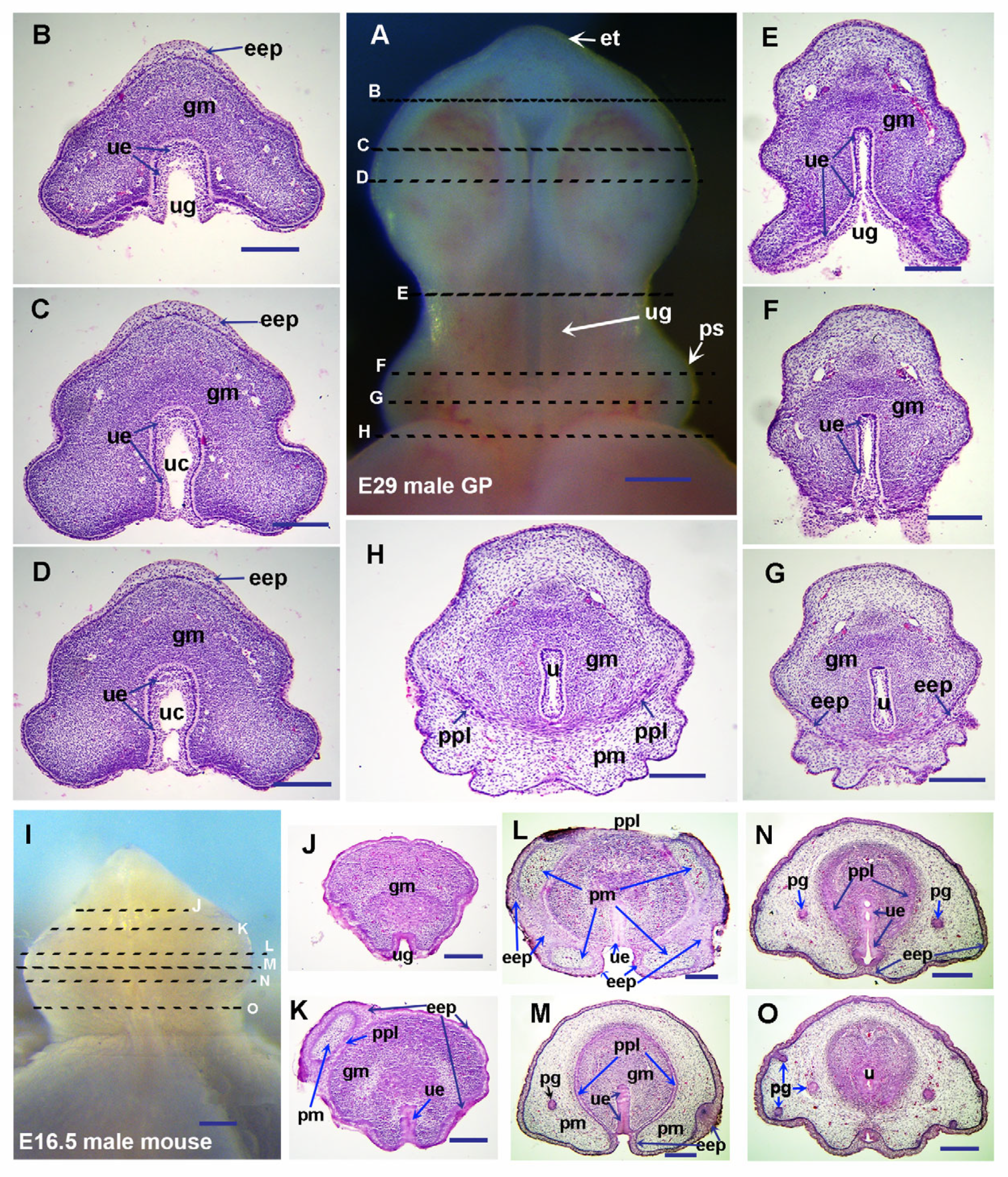
In guinea pigs, no preputial swelling was observed, and no preputial lamina or preputial glands were found in all transverse sections from proximal to distal GT at E27 (Figure 1A–H). We observed a mouse preputial swelling-like structure in E29 guinea pigs (Figure 2A), and preputial development is initiated around the same time as the initiation of sexual differentiation (Figure 2A–G and Ref. [3]). The epidermal epithelium folds to form the preputial lamina from both lateral sides in the proximal region and separates the glans from the preputial mesenchyme in E29 GT (Figure 2A–H). Compared to the sections in Figure 2G,H, we can see that epithelial ingrowth exists during the formation of the preputial lamina in guinea pigs. The opened urethral groove emerges at E28, and sexual differentiation is initiated at E28 to E29 in guinea pigs [3]. In mice, sexual differentiation of external genitalia is observed around E16, and the penile masculinization stage of E16.5 mice is comparable to that of E29 guinea pigs (Figure 2A–O). We can clearly see that the closing of the urethral tube is initiated in the proximal region of the E29 GT (Figure 2F–H), and then progressively extends distally, reaching the distal tip of the glans penis by E33 (Figure 3D–K).
As Figure 2 shows, we can see the fully opened urethral groove in the middle region of the glans penis (Figure 2E), not fully opened urethral epithelium with the urethral canal in the more distal region (close to the distal tip) (the opening zipper, Figure 2C,D), and the closed tubular urethra in the proximal region (the closing zipper, Figure 2F,G) in E29 guinea pigs. In E16.5 mice, the tubular urethra has formed in the proximal region (Figure 2O), indicating this is a comparable penile masculinization stage to the E29 guinea pig. In the middle region of the E16.5 mouse penis, the urethral epithelium in the glans region forms a urethral plate without a urethral canal. The medial boundary of epidermal epithelium-derived preputial epithelium from either side, together with the ventral surface epithelium of the GT, forms a shallow groove (Figure 2L,M), and a urethral groove-like structure is found only in the distal region without preputial covering (Figure 2J). The prepuce has covered more than half of the glans and reached the distal region in E16.5 mice (Figure 2K–O), but only a very small portion of the proximal region in E29 guinea pigs (Figure 2G,H). Interestingly, we found that in the mid-distal region of the E33 guinea pig penis, the epidermal epithelium has increased layers, and the thick epidermis is split and remodeled by the intrusion of preputial mesenchyme to form the preputial lamina and a thin surface epidermis (Figure 3F–N). When we compare the proximal (Figure 3I–K) with the distal (Figure 3D–H) region, the thicker epithelium in the distal region is apparently noticeable.
The evaginating preputial mesenchyme can be observed from the mid-distal boundary into the distal (Figure 3H,G,L–N). Similar processes can also be found in E16.5 mice (Figure 2L). In both E16.5 mice and E33 guinea pigs, we can see that the prepuce has fully covered the middle region of the glans penis, and the preputial lamina has almost formed a circular (tubular) shape, separating the preputial and glans mesenchyme in the same region (Figure 2M and Figure 3I,J). The only differences are the frenulum on the ventral side of the E33 guinea pig penis (Figure 3A,I,J), and the still unclosed urethra in the middle glans region of the E16.5 mouse penis (Figure 2M). Only fractional preputial lamina can be observed in the proximal (Figure 2N and Figure 3K) of the glans penis at this stage, which suggests that the epidermal epithelium might invaginate toward the proximal region of the glans penis to form preputial lamina in both guinea pigs and mice.
The preputial glands in guinea pigs develop even later, and no preputial glands have been found at any penile developmental stages before E30 (Figure 3A–H; Ref. [3]). The earliest stage we observed preputial glands in guinea pigs is E33, when the preputial glands are seen in the proximal penis (Figure 3J,K). In addition, the epithelial tag can be distinguished in the distal-most region of E29 (Figure 2A) and E33 (Figure 3A–C) guinea pig penises, but not in the penises of mice at comparable stages.

3.2. Differential Cell Proliferation in Epithelium and Mesenchyme Contributes to Preputial Development

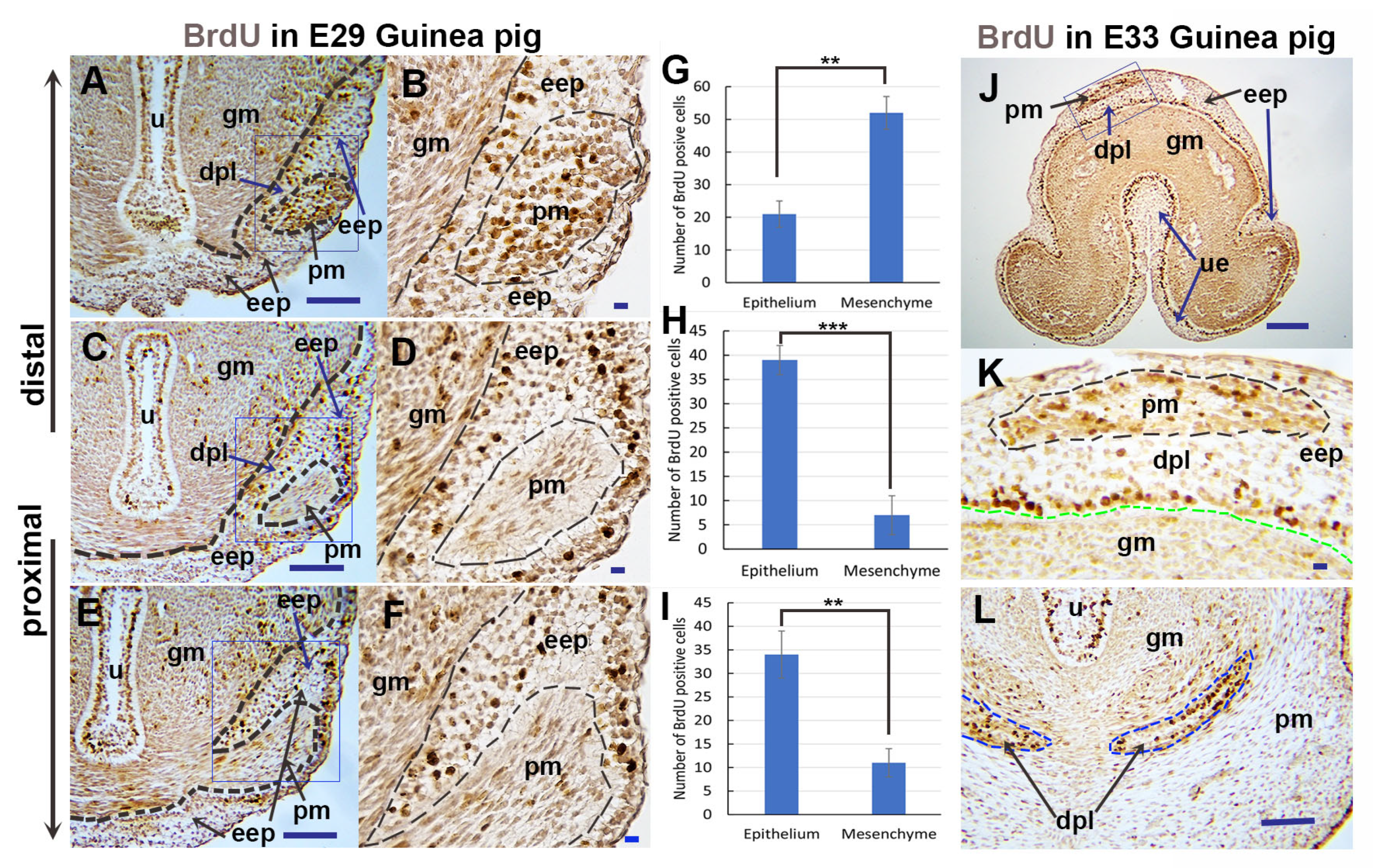
In light of our findings that both epithelial and mesenchymal cells may invaginate/evaginate to form the preputial lamina and prepuce during penile development, we hypothesized that differential cell proliferation may exist and contribute to epithelial ingrowth and mesenchymal evagination during preputial lamina formation in guinea pigs. To test this hypothesis, we performed a BrdU labeling cell proliferation assay. Figure 4E is the transverse section of the proximal (initiation of preputial development) GT of E29 guinea pigs. We found that there were more BrdU-positive cells (3.1 times, p = 0.0026, n = 3) in the epithelium than in the surrounded mesenchyme, and the epithelium invaginated and split the mesenchyme (Figure 4E,F,I). In a slightly distal section (Figure 4C), even more BrdU-positive cells (5.6 times, p = 0.0007, n = 3) were detected in the epithelium than in the mesenchyme; the epithelium increased in layers, and a small portion of mesenchymal cells was separated from the glans mesenchyme and formed the precursor cells of preputial mesenchyme (Figure 4C,D,H). In a more distal section (Figure 4A), the majority of BrdU-positive cells were restricted in the preputial mesenchyme and the epithelial layer adjacent to the glans mesenchyme (Figure 4A,B,G). The results indicate that epithelial cell proliferation leads to an increase in epithelial layers, and the evagination of preputial mesenchyme into the thickened distal epithelium is due to mesenchymal cell proliferation. In the distal region of the E33 penis, the majority of BrdU-positive cells are located in the basal epithelial layer adjacent to the glans mesenchyme and developing preputial mesenchyme (Figure 4J,K). In the proximal region of the E33 penis, more BrdU-positive cells are located in the basal layer of the preputial lamina epithelium (Figure 4L). The results indicate that the fractional preputial lamina in the proximal region of the E33 penis (Figure 3K) is mainly derived from epithelial invasion.

3.3. The Expression Patterns of Key Developmental Genes in Guinea Pig Genital Tubercle

We next sought to identify the molecules that mediate GT development in guinea pigs and performed a comparison with previously identified important genes in mouse GT development. We carried out an in situ hybridization screening on guinea pig GTs of those developmentally important genes identified from mice, focusing on the expression of Shh, Fgf, Bmp, Wnt, and Hox genes. We first present key gene expression patterns of these pathways in E23-23.5 guinea pig GTs, which are comparable to E12.5 mouse GTs.
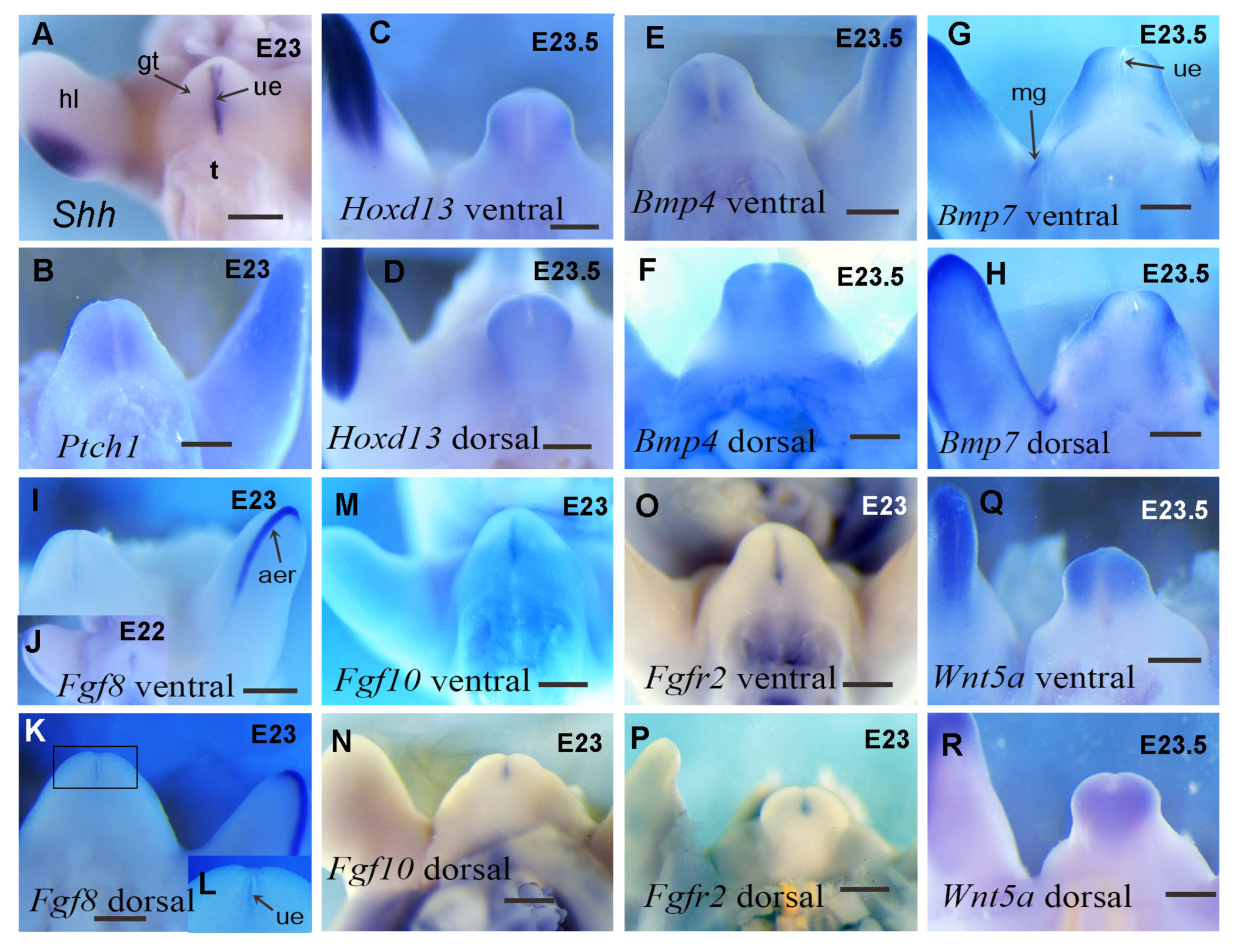
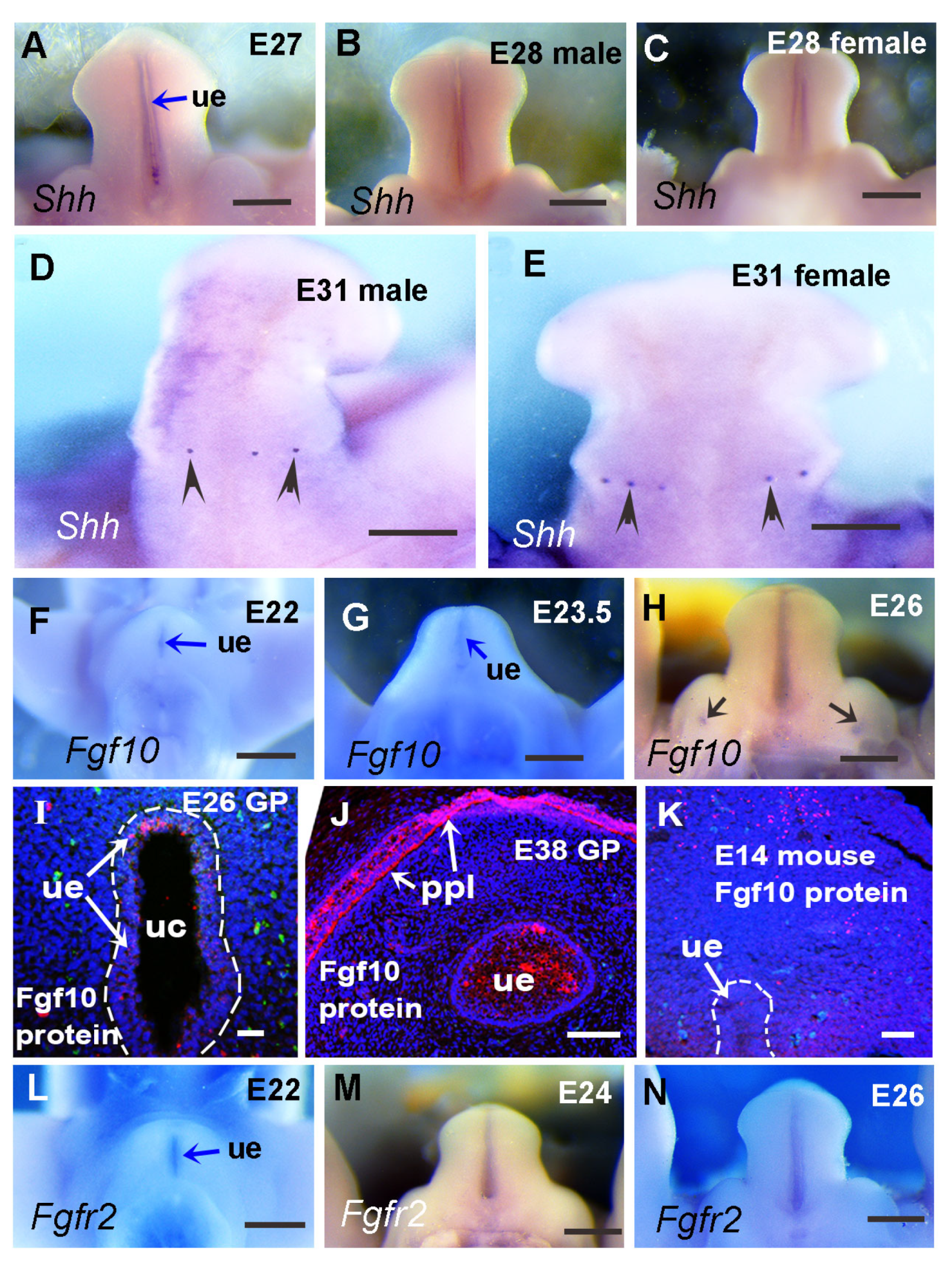
Previous work has reported the presence of Shh in developing guinea pig GT and found that the expression pattern was slightly different from that of mice at multiple stages [5]. Here, we show that at E23, Shh is expressed in the urethral epithelium of guinea pig GT (Figure 5A), and the pattern is similar to that of E12.5 mice [6]. The Shh receptor gene Ptch1 is expressed in a broad domain of the mesenchymal cells surrounding the Shh-expressing urethral epithelium at E23 in guinea pigs (Figure 5B), which is also similar to that of E12.5 mice [6]. In mouse GT, Shh is expressed in the urethral epithelium from E11.5 to E14.5 and in preputial glands from E13.5 to E14.5 [11,33]. To identify Shh expression at later stages in guinea pig GT, we performed Shh in situ hybridization from E27 to E33. Shh expression in the urethral plate can be detected as late as E28, and no preputial expression of Shh has been observed at any stage before E28 (Figure 6A–C and Ref. [5]). The earliest stage at which preputial Shh expression can be detected is around E32, when Shh is initially expressed in developing preputial glands as small dots in both male and female GT (Figure 6D,E).
Hoxd13 is expressed in the distal mesenchyme of E23.5 guinea pig GT (Figure 5C,D), and the expression pattern resembles that of E12.5-14.5 mouse GT [6,34]. Interestingly, the expression of Hoxd13 in GT is relatively weaker compared with the strong expression in the hindlimbs of guinea pigs (Figure 5C,D).
Bmp4 is expressed strongly in the distal tip mesenchyme of E23.5 guinea pig GT (Figure 5E,F), similar to that of E12.5 mouse GT [6]. The expression on the ventral side is localized to the mesenchyme surrounding the urethral epithelium, resembling the expression of Ptch1 in this region (compare Figure 5B,E). Additionally, Bmp4 expression on the dorsal side resembles the expression of Hoxd13 in the same region (compare Figure 5D,F). Bmp7 and Bmp4 have similar expression patterns in E23.5 hindlimbs (Figure 4E, H), but their expression patterns in GT are quite different. Bmp7 is expressed in both the urethral epithelium and mesenchyme of E23.5 GT (Figure 5G,H). The urethral expression is weak and confined only to the partial urethral epithelium near the distal tip, and the mesenchymal expression is not like that of Bmp4 and Ptch1, which are expressed in cells adjacent to the urethral epithelium but further away from the urethra, mainly on the side of the distal tip (Figure 5G,H).
Fgf8 expression is detected in a subset of the Shh expression domain in the guinea pig GT and is restricted to the anterior region of the urethral epithelium at E22 (Figure 5J). It remains at the distal tip of the urethral plate when the tubercle grows out at E23 and can only be seen from a dorsal view (Figure 5K,L). The pattern of Fgf8 in guinea pig GT also resembles the expression in mice [6,35]. Similarly to Hoxd13, when compared with the strong Fgf8 expression in limb buds, the signal is much weaker in developing GT.
Fgf10 is mainly restricted to the urethral plate in E23 guinea pig GT (Figure 5M,N), which differs from the reported mesenchymal expression in mouse GT [11,15]. The expression of the Fgf receptor Fgfr2 in E23 guinea pig GT is also found in the urethral epithelium (Figure 5O,P). The expression pattern of Fgfr2 in the guinea pig urethra is similar to that in mice at early stages [16], but there is a lack of preputial expression before E28 compared with mice at similar developmental stages (E13.5–14.5) due to the absence of preputial development until E29. As the urethral plate expression of Fgf10 is not the same as reported in mouse GT, we performed Fgf10 and Fgfr2 in situ hybridization on the GT of E22, E23.5, and E26 guinea pigs (comparable to E11.5, E12.5, and E13.5 mice, respectively, according to limb and GT morphology). We found that both Fgf10 and Fgfr2 expressions are mainly restricted to the urethral plate (Figure 6F–L). Interestingly, Fgf10 expression was also found in the dorsal and ventral regions of labioscrotal swellings in E26 guinea pig GT and was confined to four small, round areas (Figure 6H,I). The Fgf10 expression in labioscrotal swellings has never been reported before, and the function of Fgf10 in these domains is unidentified.
The Wnt5a expression domain is located in the mesenchyme of the GT, with the strongest expression at the distal tip. From both ventral and dorsal views, we can clearly see that Wnt5a expression is mainly located in the distal glans region (Figure 5Q,R), suggesting it may control the outgrowth of the glans at an early stage, as reported in mouse GT [21,36].
Taken together, these results reveal dynamic patterns of key developmental gene expression in guinea pig external genitalia, with Fgf8, Fgfr2, Bmp4, Bmp7, Hoxd13, and Wnt5a showing similar expression patterns during GT development between guinea pigs and mice. We identified that Shh and Fgf10 show differential expression patterns between the two species, which may contribute to the differential morphogenesis of external genitalia between guinea pigs and mice.

3.4. Relative Expression Levels of Shh, Fgf10, and Fgfr2 in Developing Guinea Pig GT Are Reduced Compared with the Comparable Stage of Mouse GT

Compared with mice, the major difference in guinea pig GT development is that the urethral plate opens to form the urethral groove before sexual differentiation (Figure 1, Figure 2 and Figure 3, Refs. [3,5]). Shh, Fgfs, Hoxd13, Hoxa13, Bmp4, and Wnt pathways play key roles in glans and preputial morphogenesis [11,12,13]. In light of our findings regarding the differential expression of Hoxd13 and Fgf8 between GT and limb buds and between guinea pigs and mice, we hypothesized that the expression levels of these developmental genes in developing guinea pig GT may be relatively weaker than those in mouse GT. To verify this hypothesis, we tested and compared the relative expression levels of these genes between mouse and guinea pig GTs with comparable developmental stages. Developing limb buds were widely used to determine the developmental stages in different species [37,38]. According to limb (Figure 7A–D) and external genital (Figure 7E–H) morphology, we found that E23 and E26.5 GTs of guinea pigs are comparable to E12.5 and E13.5 GTs of mice, respectively. As we compare relative gene expression levels between two different species, the selection of housekeeping genes is important. We compared Ct values of commonly used housekeeping genes, Gapdh and Actb, and found that the Ct values of Gapdh were closer to each other between mouse and guinea pig GTs and more stable when the same amount of total RNA was applied (Table S3). Thus, Gapdh was selected as the housekeeping gene to quantify the gene expression levels. Compared to the two housekeeping genes, the Ct values for all the selected developmental genes—Shh, Fgf8, Fgf10, Fgfr2, Hoxd13, Hoxa13, Bmp4, and Ctnnb1—at all the tested stages were found to be higher. Compared with E12.5 and E13.5 mice, lower expression levels of all eight developmental genes were detected in the GTs of E23 and E26.5 guinea pigs (p ≤ 0.0061, n = 5). Hoxa13, Bmp4, and Ctnnb1 were about 2-fold lower in guinea pig GTs compared with mice, and the expression of Shh, Fgf8, Fgf10, Fgfr2, and Hoxd13 was more than 4.5-fold lower compared with mice (Figure 7I,J). We also found that Actb relative expression in developing GTs showed no significant difference (E23, 1.23-fold, p = 0.163, n = 5; E26.5, 1.14-fold, p = 0.248, n = 5) between guinea pigs and mice (Figure 7I,J). Compared with the expression in E12.5 mice, Shh, Fgf8, Fgf10, Fgfr2, and Hoxd13 in E23 guinea pig GTs were downregulated by 5.54-, 4.5-, 14.62-, 6.47-, and 7.51-fold, respectively (Figure 7I). Likewise, all the five genes were dramatically upregulated in E13.5 mouse GT (Shh, 6.23-fold; Fgf8, 5.9-fold; Fgf10, 9.65-fold; Fgfr2, 7.24-fold; Hoxd13, 9.98-fold) compared with E26.5 guinea pigs (Figure 7J). Because these are relative gene expression levels in two species, we believe that the approximately 2-fold reductions in Hoxa13 (E23, 1.9-fold; E26.5, 2.8-fold), Bmp4 (E23, 2.1-fold; E26.5, 2.6-fold), and Ctnnb1 (E23, 2.5-fold; E26.5, 2.4-fold) may result from systemic differences. The dramatic differences (more than 4.5-fold) in the expression of Shh, Fgf8, Fgf10, Fgfr2, and Hoxd13 may play roles in patterning cellular processes and lead to differential morphological developments such as urethral groove formation and preputial development between the two species.

3.5. Shh and Fgf10/Fgfr2 Play Key Roles in Preputial and Urethral Groove Formation

Shh is a chief morphogen that organizes the structure in the ventral midline of multiple organs [6,39,40], and the deletion of Shh signaling genes at E13.5–15.5 led to an open urethral plate (hypospadias) and a reduction in glans and preputial development in mice [33,41]. Fgf10 and its receptor Fgfr2 also play important roles in urethral and preputial development [15,16,42]. Additionally, Shh and Fgf10/Fgfr2 signaling interact with the expression of other developmental genes, such as Fgf8, Hoxd13, Bmp4, and Wnt5a, to coordinately regulate morphogenesis during GT development; e.g., Shh negatively regulates Bmp and Wnt signaling molecules, which will define the limits of the range of each domain [11,40]. Based on our findings that Shh expression levels were reduced more than 5-fold in the developing GT of guinea pigs compared to that of mice, and considering the difference in Shh expression patterns between mouse and guinea pig GTs (Figure 6A–E and Ref. [5]), we observed that the Fgf10 expression pattern shifts from the mesenchyme of mouse GT to mainly the urethral epithelium of guinea pig GT. Furthermore, expression levels in guinea pig GT reduce more than 14-fold at E23 (compared with E12.5 mouse GT) and more than 9-fold at E26.5 (compared with E13.5 mouse GT). We hypothesized that the reduction in Shh and Fgf10/Fgfr2 signaling may induce urethral groove formation and delay preputial development in guinea pigs. To test this hypothesis, we performed GT organ culture and examined the roles of Shh and Fgf-10 in urethral groove and preputial formation using Fgf-10 and Shh proteins, the Fgf receptor inhibitor NVP-BGJ398, and the hedgehog signal inhibitor cyclopamine. Male GTs of E14.5 mice and E27 guinea pigs were dissected and maintained in culture using media supplemented with or without androgen (10 nM MT), Shh and Fgf10/Fgfr2 inhibitors (for mouse GT culture), and Shh and Fgf-10 proteins (for guinea pig GT culture) to test the function of Shh and Fgf-10 in urethral groove and preputial development. We first observed the effect on androgen’s mouse GT culture. After 48 h of culture, both the control male mouse GTs with and without androgen developed preputial swellings and a urethral plate (Figure 8A–C). However, compared to the GTs with 10 nM MT, there was a small hole remaining open in the middle of the urethral plate on the ventral side of the GTs without MT (Figure 8B). Thus, we added 10 nM MT in our male GT culture system when testing the effects of Shh, Fgf10/Fgfr2, and the inhibitors. We then performed the same stage of male mouse GT culture with the administration of 1.4 nM BGJ398, 200 nM cyclopamine, or 1.4 nM BGJ398 plus 200 nM cyclopamine to the media from the beginning. All three groups of GTs formed smaller preputial swellings and widely opened urethral grooves compared with controls (Figure 8D,E). After 48 h of culture with both 1.4 nM BGJ398 and 200 nM cyclopamine, the preputial swelling almost disappeared, and the GT grew its shape into a cylinder with an open urethral groove; in addition, the distal tip also malformed (Figure 8F). Next, we performed guinea pig GT culture (starting from E27) with or without 10 nM MT (Figure 8G–I). Because the guinea pig GT is very soft and larger/heavier compared with the mouse GT at this stage, when being cultured in a ventral side-up position, the distal part of the GT became flat, making it difficult to overcome the gravity to form a closed urethra using the same method as for mouse GT culture. We believe that additional support material should be applied in our guinea pig GT culture system in the future. After the administration of 50 ng/mL Fgf-10 protein, the guinea pig GT enlarged in size compared with the control and formed preputial swellings after 48 h, which have never been seen in controls (Figure 8J). After 48 h of culture with 200 ng/mL Shh protein, the guinea pig GT also formed preputial swellings; however, the elongation of the GT was not as obvious as that of the Fgf-10-treated ones (Figure 8K). When male guinea pig GT was cultured with both 50 ng/mL Fgf-10 and 200 ng/mL Shh proteins, the preputial swellings became more obvious after 48 h (Figure 8L). Our results suggest that Shh and Fgf10/Fgfr2 signaling play key roles in the urethral groove and preputial formation during penile development.

4. Discussion

During human penile development, the process of tubular urethral formation has been described as distal-opening-proximal-closing [1]. To date, the guinea pig is the only published animal model of penile development in the literature that can demonstrate a process similar to what is seen in humans [3]. The most commonly used animals, mice and rats, form urethral plates and a urethral opening at the ventral proximal region, rather than a fully open urethral groove like humans and guinea pigs [4,6]. From our perspective, preputial development in guinea pigs is more similar to the human preputial development model proposed by Glenister [8], and the detailed process of distal extension of the prepuce in guinea pigs is similar to a modified human preputial development model described by Cunha [9], suggesting that preputial development in guinea pigs and humans may share similar mechanisms. Interestingly, Liu et al. [43] reported that mouse external and internal prepuces occur via entirely different morphogenetic mechanisms. The structure they called internal prepuce develops postnatally, and humans have no similar structure in penile development. Based on our observations, the external prepuce development in E16.5 mice shows a similar developmental process to that of guinea pigs, with increasing epithelial layers and preputial mesenchyme evaginating distally. The major difference in preputial development between mice and guinea pigs is the timing of preputial development relative to tubular urethra formation. In guinea pigs, and humans as well, the urethral groove forms in the glans penis without preputial covering; in fact, the fully opened urethral groove forms at E28 in guinea pigs [3,5] and at 9.5 weeks of gestation in humans [1], before the initiation of preputial development in the proximal region of the glans penis (Figure 2 and Ref. [9]). In mice, the opened urethral groove without preputial covering can only be observed in the distal region of the E16.5 mouse penis (Figure 2 and Ref. [43]), suggesting a delay in the relative timing of preputial development to the formation of an opened urethral groove in guinea pigs and humans compared with mice.
Genetic control of external genital development has been well studied in mice, and genes in Shh, Fgf, Bmp, and Wnt pathways were found to be expressed in developing mouse GT before sexual differentiation. We compared these pathway genes in developing GT between guinea pigs and mice and found that the mRNA expression patterns of Shh and Fgf10 were different, while the expression levels of Shh, Fgf8, Fgf10, Hoxd13, Bmp4, and an important canonical Wnt signaling gene, Ctnnb1, were reduced in guinea pigs compared with stage-matched mouse GT. Shh signals are proposed to the ventral ectoderm to maintain the structural integrity of the epithelium, which is essential for the maintenance of a closed urethral tube [11]. Preputial Shh expressions were found almost at the same time of epithelial folding to form the preputial lamina in mice [6], and deletion of the Shh pathway gene Smo using Msx2cre disrupted urethral plate and preputial development [41]. If we compare normal human [44] and guinea pig [3] GTs with those of Smo conditional knockout mutant mice [41], they all have an open urethral groove, and the mutant mouse GT has a malformed prepuce (Figure 9A–D). The shift in the Shh expression domain from the urethral epithelium to the ventral ectodermal epithelium in developing guinea pig GT [5] disrupts the signals to ventral ectoderm, which may be one of the reasons that lead to urethral opening and urethral groove formation. Preputial Shh mRNA expression (Figure 7) was first found (around E31) two days after the initiation of preputial development (E28-E29), suggesting that Shh may play a role in preputial development, but not in its initiation, in guinea pigs.
Fgf8, Fgf10, and their receptor Fgfr2 were identified as candidate genes for human hypospadias [45,46]. In mice, several Fgf ligands and their receptors, including Fgf8, Fgf10, and Fgfr2 are expressed in multiple domains in developing GT [47]. Fgf8 is more likely a readout and not required for the outgrowth or normal patterning of the GT in mice [35], but Fgf10 and Fgfr2 have been revealed to play key roles in urethral tube closure and normal preputial development [15,16,42]. In mice, mesenchymal Fgf10 interacts with urethral epithelial Fgfr2 (Fgfr2IIIb) to maintain normal GT development with an unopened urethral plate [42,48]. Deletion of Fgf10 or Fgfr2IIIb leads to severe hypospadias in mice, in which the ventral side of the urethra is fully open, resembling a urethral groove in guinea pigs, but still with the malformed prepuce [15,42,48]. Loss-of-function mutations or deletions of Fgfr2 in the ectoderm result in the most severe hypospadias; the Fgfr2 mutant mice exhibit an open urethra [3,16,48], and the morphology of the developing GT looks similar to that of guinea pig [3] and human [44] GTs at the proximal-open stage, while the distal remains closed (Figure 9A,B,E). Our data showed differential expression patterns and levels of Shh, Fgf10, and Fgfr2 in GT between guinea pigs and mice, suggesting that Shh and Fgf10/Fgfr2 play key roles in urethral groove formation in guinea pigs, and possibly in humans as well. If we compare the morphology of normal human and guinea pig developing GTs with the GTs of Shh and Fgfr2 conditional knockout mutant mice, we can see the similarities; both mutant GTs showed an open urethral groove and a reduction in prepuce (Figure 9).
Hoxd13 is the gene with the most reduced expression level in the GT of guinea pigs compared with that of mice (Figure 7I,J). According to Lin et al. [41], Hoxd13 has been reported to be one of the downstream genes of Shh in the external genital and limb development [41,49]. Shh and Fgf signaling were found working together to control the expression of Hoxd genes in limb development [50]. Thus, we believe the reduction in the Hoxd13 expression in guinea pigs developing GT may be induced by the decreased Shh, Fgf8, Fgf10, and Fgfr2 signaling.
Our organ culture results clearly showed that both Shh and Fgf10/Fgfr2 signaling are required to maintain the prepuce and urethral plate in the developing mouse GT. The lack of either one will induce an open urethral groove (Figure 8). Block Shh or Fgf signaling has a detrimental effect on preputial development, and blocking both pathways results in the most severe reduction in developing prepuce (Figure 8). In the guinea pig GT culture system, since the E27 GT is much larger and softer compared with the E14.5 mouse GT, gravity caused the GT to grow into a kind of flat shape after 2 days of culture. We found that all the GTs (100%) formed preputial swellings in our Shh and Fgf-10 protein-treated groups, although they all have a fully opened urethral groove. Considering the delayed preputial Shh expression (Figure 6D,E) and the delayed preputial development in guinea pigs (Figure 2), we predict that, compared with mice, the ectodermal Fgf signaling may also be expressed later, coinciding with the initiation of proximal urethral closure, in response to the androgen signaling. During the development of the human penis, tubular urethral formation follows a process of distal-opening-proximal-closing, with the formation of the urethral groove being the first step. We hope that our findings can provide a basis for further research on the mechanism of distal-opening-proximal-closing, and provide a reference for clinical treatment of penile abnormalities such as hypospadias.

Supplementary Materials

The following supporting information can be downloaded at: https://www.mdpi.com/article/10.3390/cells14050348/s1, Table S1: PCR primers were used to clone guinea pig genes for making RNA probes. Table S2: QPCR primers for genes in developing genital tubercles of mice and guinea pigs. Table S3: Ct values of selected genes in developing genital tubercles of mice and guinea pigs.

Author Contributions

Conceptualization, Z.Z. and S.W.; Methodology, Validation, Formal Analysis, Data Curation, Writing—Original Draft Preparation, Review and Editing, and Visualization, S.W. and Z.Z.; Resources, Supervision, Project Administration and Funding Acquisition, Z.Z. All authors have read and agreed to the published version of the manuscript.

Funding

This work was supported by the National Institutes of Health (Eunice Kennedy Shriver National Institute of Child Health and Human Development; 1R15 HD095368).

Institutional Review Board Statement

The animal study protocol was approved by the Institutional Animal Care and Use Committee of Southern Illinois University Carbondale. (protocol codes: mouse, 23-011, approved on 7 June 2023; guinea pig, 20-014, approved on 1 May 2020).

Informed Consent Statement

Not applicable.

Data Availability Statement

All data are available upon reasonable request to the corresponding author.

Acknowledgments

We would like to acknowledge the financial support from the NIH and Southern Illinois University, School of Medicine (Research Seed Grant). We also would like to acknowledge the following publishers for their permission to use the published images: Development, published by The Company of Biologists; Differentiation, published by Elsevier; Urology, published by Elsevier.

Conflicts of Interest

The authors declare no conflicts of interest.

References

  1. Li, Y.; Sinclair, A.; Cao, M.; Shen, J.; Choudhry, S.; Botta, S.; Cunha, G.; Baskin, L. Canalization of the urethral plate precedes fusion of the urethral folds during male penile urethral development: The double zipper hypothesis. J. Urol. 2015, 193, 1353–1359. [Google Scholar] [CrossRef] [PubMed]
  2. Zheng, Z.; Armfield, B.A.; Cohn, M.J. Timing of androgen receptor disruption and estrogen exposure underlies a spectrum of congenital penile anomalies. Proc. Natl. Acad. Sci. USA 2015, 112, E7194–E7203. [Google Scholar] [CrossRef] [PubMed]
  3. Wang, S.; Shi, M.; Zhu, D.; Mathews, R.; Zheng, Z. External Genital Development, Urethra Formation, and Hypospadias Induction in Guinea Pig: A Double Zipper Model for Human Urethral Development. Urology 2018, 113, 179–186. [Google Scholar] [CrossRef] [PubMed]
  4. Anderson, C.A.; Clark, R.L. External genitalia of the rat: Normal development and the histogenesis of 5 alpha-reductase inhibitor-induced abnormalities. Teratology 1990, 42, 483–496. [Google Scholar] [CrossRef]
  5. Wang, S.; Zheng, Z. Differential cell proliferation and cell death during the urethral groove formation in guinea pig model. Pediatr. Res. 2019, 86, 452–459. [Google Scholar] [CrossRef]
  6. Perriton, C.L.; Powles, N.; Chiang, C.; Maconochie, M.K.; Cohn, M.J. Sonic hedgehog signaling from the urethral epithelium controls external genital development. Dev. Biol. 2002, 247, 26–46. [Google Scholar] [CrossRef]
  7. Hunter, R.H. Notes on the Development of the Prepuce. J. Anat. 1935, 70, 68–75. [Google Scholar]
  8. Glenister, T.W. A consideration of the processes involved in the development of the prepuce in man. Br. J. Urol. 1956, 28, 243–249. [Google Scholar] [CrossRef]
  9. Cunha, G.R.; Sinclair, A.; Cao, M.; Baskin, L.S. Development of the human prepuce and its innervation. Differentiation 2020, 111, 22–40. [Google Scholar] [CrossRef]
  10. Liu, X.; Liu, G.; Shen, J.; Yue, A.; Isaacson, D.; Sinclair, A.; Cao, M.; Liaw, A.; Cunha, G.R.; Baskin, L. Human glans and preputial development. Differentiation 2018, 103, 86–99. [Google Scholar] [CrossRef]
  11. Cohn, M.J. Development of the external genitalia: Conserved and divergent mechanisms of appendage patterning. Dev. Dyn. 2011, 240, 1108–1115. [Google Scholar] [CrossRef] [PubMed]
  12. Chang, J.; Wang, S.; Zheng, Z. Etiology of Hypospadias: A Comparative Review of Genetic Factors and Developmental Processes Between Human and Animal Models. Res. Rep. Urol. 2020, 12, 673–686. [Google Scholar] [CrossRef] [PubMed]
  13. Haller, M.; Ma, L. Temporal, spatial, and genetic regulation of external genitalia development. Differentiation 2019, 110, 1–7. [Google Scholar] [CrossRef] [PubMed]
  14. Haid, B.; Pechriggl, E.; Nagele, F.; Dudas, J.; Webersinke, G.; Rammer, M.; Fritsch, H.; Oswald, J. FGF8, FGF10 and FGF receptor 2 in foreskin of children with hypospadias: An analysis of immunohistochemical expression patterns and gene transcription. J. Pediatr. Urol. 2020, 16, 41.e1–41.e10. [Google Scholar] [CrossRef]
  15. Haraguchi, R.; Suzuki, K.; Murakami, R.; Sakai, M.; Kamikawa, M.; Kengaku, M.; Sekine, K.; Kawano, H.; Kato, S.; Ueno, N.; et al. Molecular analysis of external genitalia formation: The role of fibroblast growth factor (Fgf) genes during genital tubercle formation. Development 2000, 127, 2471–2479. [Google Scholar] [CrossRef]
  16. Gredler, M.L.; Seifert, A.W.; Cohn, M.J. Tissue-specific roles of Fgfr2 in development of the external genitalia. Development 2015, 142, 2203–2212. [Google Scholar] [CrossRef]
  17. Kajioka, D.; Suzuki, K.; Nakada, S.; Matsushita, S.; Miyagawa, S.; Takeo, T.; Nakagata, N.; Yamada, G. Bmp4 is an essential growth factor for the initiation of genital tubercle (GT) outgrowth. Congenit. Anom. 2020, 60, 15–21. [Google Scholar] [CrossRef]
  18. Ching, S.T.; Infante, C.R.; Du, W.; Sharir, A.; Park, S.; Menke, D.B.; Klein, O.D. Isl1 mediates mesenchymal expansion in the developing external genitalia via regulation of Bmp4, Fgf10 and Wnt5a. Hum. Mol. Genet. 2018, 27, 107–119. [Google Scholar] [CrossRef]
  19. Lin, C.; Yin, Y.; Long, F.; Ma, L. Tissue-specific requirements of beta-catenin in external genitalia development. Development 2008, 135, 2815–2825. [Google Scholar] [CrossRef]
  20. Miyagawa, S.; Satoh, Y.; Haraguchi, R.; Suzuki, K.; Iguchi, T.; Taketo, M.M.; Nakagata, N.; Matsumoto, T.; Takeyama, K.; Kato, S.; et al. Genetic interactions of the androgen and Wnt/beta-catenin pathways for the masculinization of external genitalia. Mol. Endocrinol. 2009, 23, 871–880. [Google Scholar] [CrossRef]
  21. Alcantara, M.C.; Suzuki, K.; Acebedo, A.R.; Sakamoto, Y.; Nishita, M.; Minami, Y.; Kikuchi, A.; Yamada, G. Stage-dependent function of Wnt5a during male external genitalia development. Congenit. Anom. 2021, 61, 212–219. [Google Scholar] [CrossRef] [PubMed]
  22. Person, A.D.; Beiraghi, S.; Sieben, C.M.; Hermanson, S.; Neumann, A.N.; Robu, M.E.; Schleiffarth, J.R.; Billington, C.J., Jr.; van Bokhoven, H.; Hoogeboom, J.M.; et al. WNT5A mutations in patients with autosomal dominant Robinow syndrome. Dev. Dyn. 2010, 239, 327–337. [Google Scholar] [CrossRef] [PubMed]
  23. Warot, X.; Fromental-Ramain, C.; Fraulob, V.; Chambon, P.; Dolle, P. Gene dosage-dependent effects of the Hoxa-13 and Hoxd-13 mutations on morphogenesis of the terminal parts of the digestive and urogenital tracts. Development 1997, 124, 4781–4791. [Google Scholar] [CrossRef] [PubMed]
  24. Goodman, F.R.; Bacchelli, C.; Brady, A.F.; Brueton, L.A.; Fryns, J.P.; Mortlock, D.P.; Innis, J.W.; Holmes, L.B.; Donnenfeld, A.E.; Feingold, M.; et al. Novel HOXA13 mutations and the phenotypic spectrum of hand-foot-genital syndrome. Am. J. Hum. Genet. 2000, 67, 197–202. [Google Scholar] [CrossRef]
  25. Tuzel, E.; Samli, H.; Kuru, I.; Turkmen, S.; Demir, Y.; Maralcan, G.; Guler, C. Association of hypospadias with hypoplastic synpolydactyly and role of HOXD13 gene mutations. Urology 2007, 70, 161–164. [Google Scholar] [CrossRef]
  26. Seifert, A.W.; Zheng, Z.; Ormerod, B.K.; Cohn, M.J. Sonic hedgehog controls growth of external genitalia by regulating cell cycle kinetics. Nat. Commun. 2010, 1, 23. [Google Scholar] [CrossRef]
  27. Sarac, M.; Canpolat, S.; Onalan Etem, E.; Tektemur, A.; Tartar, T.; Bakal, U.; Kazez, A. The role of sonic hedgehog homologue signal pathway in hypospadias aetiology. J. Pediatr. Urol. 2021, 17, 630.e631–630.e637. [Google Scholar] [CrossRef]
  28. Depreux, F.F.; Czech, L.; Whitlon, D.S. Sex Genotyping of Archival Fixed and Immunolabeled Guinea Pig Cochleas. Sci. Rep. 2018, 8, 5156. [Google Scholar] [CrossRef]
  29. Wang, S.; Lawless, J.; Zheng, Z. Prenatal low-dose methyltestosterone, but not dihydrotestosterone, treatment induces penile formation in female mice and guinea pigsdagger. Biol. Reprod. 2020, 102, 1248–1260. [Google Scholar] [CrossRef]
  30. Nieto, M.A.; Patel, K.; Wilkinson, D.G. In situ hybridization analysis of chick embryos in whole mount and tissue sections. Methods Cell Biol. 1996, 51, 219–235. [Google Scholar] [CrossRef]
  31. Tan, H.; Wu, G.; Wang, S.; Lawless, J.; Sinn, A.; Chen, D.; Zheng, Z. Prenatal exposure to atrazine induces cryptorchidism and hypospadias in F1 male mouse offspring. Birth Defects Res. 2021, 113, 469–484. [Google Scholar] [CrossRef] [PubMed]
  32. Livak, K.J.; Schmittgen, T.D. Analysis of relative gene expression data using real-time quantitative PCR and the 2−ΔΔCT Method. Methods 2001, 25, 402–408. [Google Scholar] [CrossRef] [PubMed]
  33. Seifert, A.W.; Bouldin, C.M.; Choi, K.S.; Harfe, B.D.; Cohn, M.J. Multiphasic and tissue-specific roles of sonic hedgehog in cloacal septation and external genitalia development. Development 2009, 136, 3949–3957. [Google Scholar] [CrossRef] [PubMed]
  34. Scott, V.; Morgan, E.A.; Stadler, H.S. Genitourinary functions of Hoxa13 and Hoxd13. J. Biochem. 2005, 137, 671–676. [Google Scholar] [CrossRef] [PubMed]
  35. Seifert, A.W.; Yamaguchi, T.; Cohn, M.J. Functional and phylogenetic analysis shows that Fgf8 is a marker of genital induction in mammals but is not required for external genital development. Development 2009, 136, 2643–2651. [Google Scholar] [CrossRef]
  36. Yamaguchi, T.P.; Bradley, A.; McMahon, A.P.; Jones, S. A Wnt5a pathway underlies outgrowth of multiple structures in the vertebrate embryo. Development 1999, 126, 1211–1223. [Google Scholar] [CrossRef]
  37. Hamburger, V.; Hamilton, H.L. A series of normal stages in the development of the chick embryo. J. Morphol. 1951, 88, 49–92. [Google Scholar] [CrossRef]
  38. Wanek, N.; Muneoka, K.; Holler-Dinsmore, G.; Burton, R.; Bryant, S.V. A staging system for mouse limb development. J. Exp. Zool. 1989, 249, 41–49. [Google Scholar] [CrossRef]
  39. Harfe, B.D.; Scherz, P.J.; Nissim, S.; Tian, H.; McMahon, A.P.; Tabin, C.J. Evidence for an expansion-based temporal Shh gradient in specifying vertebrate digit identities. Cell 2004, 118, 517–528. [Google Scholar] [CrossRef]
  40. Brady, M.V.; Vaccarino, F.M. Role of SHH in Patterning Human Pluripotent Cells towards Ventral Forebrain Fates. Cells 2021, 10, 914. [Google Scholar] [CrossRef]
  41. Lin, C.; Yin, Y.; Veith, G.M.; Fisher, A.V.; Long, F.; Ma, L. Temporal and spatial dissection of Shh signaling in genital tubercle development. Development 2009, 136, 3959–3967. [Google Scholar] [CrossRef] [PubMed]
  42. Satoh, Y.; Haraguchi, R.; Wright, T.J.; Mansour, S.L.; Partanen, J.; Hajihosseini, M.K.; Eswarakumar, V.P.; Lonai, P.; Yamada, G. Regulation of external genitalia development by concerted actions of FGF ligands and FGF receptors. Anat. Embryol. 2004, 208, 479–486. [Google Scholar] [CrossRef] [PubMed]
  43. Liu, G.; Liu, X.; Shen, J.; Sinclair, A.; Baskin, L.; Cunha, G.R. Contrasting mechanisms of penile urethral formation in mouse and human. Differentiation 2018, 101, 46–64. [Google Scholar] [CrossRef] [PubMed]
  44. Shen, J.; Overland, M.; Sinclair, A.; Cao, M.; Yue, X.; Cunha, G.; Baskin, L. Complex epithelial remodeling underlie the fusion event in early fetal development of the human penile urethra. Differentiation 2016, 92, 169–182. [Google Scholar] [CrossRef]
  45. Beleza-Meireles, A.; Lundberg, F.; Lagerstedt, K.; Zhou, X.; Omrani, D.; Frisen, L.; Nordenskjold, A. FGFR2, FGF8, FGF10 and BMP7 as candidate genes for hypospadias. Eur. J. Hum. Genet. 2007, 15, 405–410. [Google Scholar] [CrossRef]
  46. Carmichael, S.L.; Ma, C.; Choudhry, S.; Lammer, E.J.; Witte, J.S.; Shaw, G.M. Hypospadias and genes related to genital tubercle and early urethral development. J. Urol. 2013, 190, 1884–1892. [Google Scholar] [CrossRef]
  47. Tarulli, G.A.; Cripps, S.M.; Pask, A.J.; Renfree, M.B. Spatiotemporal map of key signaling factors during early penis development. Dev. Dyn. 2022, 251, 609–624. [Google Scholar] [CrossRef]
  48. Petiot, A.; Perriton, C.L.; Dickson, C.; Cohn, M.J. Development of the mammalian urethra is controlled by Fgfr2-IIIb. Development 2005, 132, 2441–2450. [Google Scholar] [CrossRef]
  49. Bangs, F.; Welten, M.; Davey, M.G.; Fisher, M.; Yin, Y.; Downie, H.; Paton, B.; Baldock, R.; Burt, D.W.; Tickle, C. Identification of genes downstream of the Shh signalling in the developing chick wing and syn-expressed with Hoxd13 using microarray and 3D computational analysis. Mech. Dev. 2010, 127, 428–441. [Google Scholar] [CrossRef]
  50. Rodrigues, A.R.; Yakushiji-Kaminatsui, N.; Atsuta, Y.; Andrey, G.; Schorderet, P.; Duboule, D.; Tabin, C.J. Integration of Shh and Fgf signaling in controlling Hox gene expression in cultured limb cells. Proc. Natl. Acad. Sci. USA 2017, 114, 3139–3144. [Google Scholar] [CrossRef]
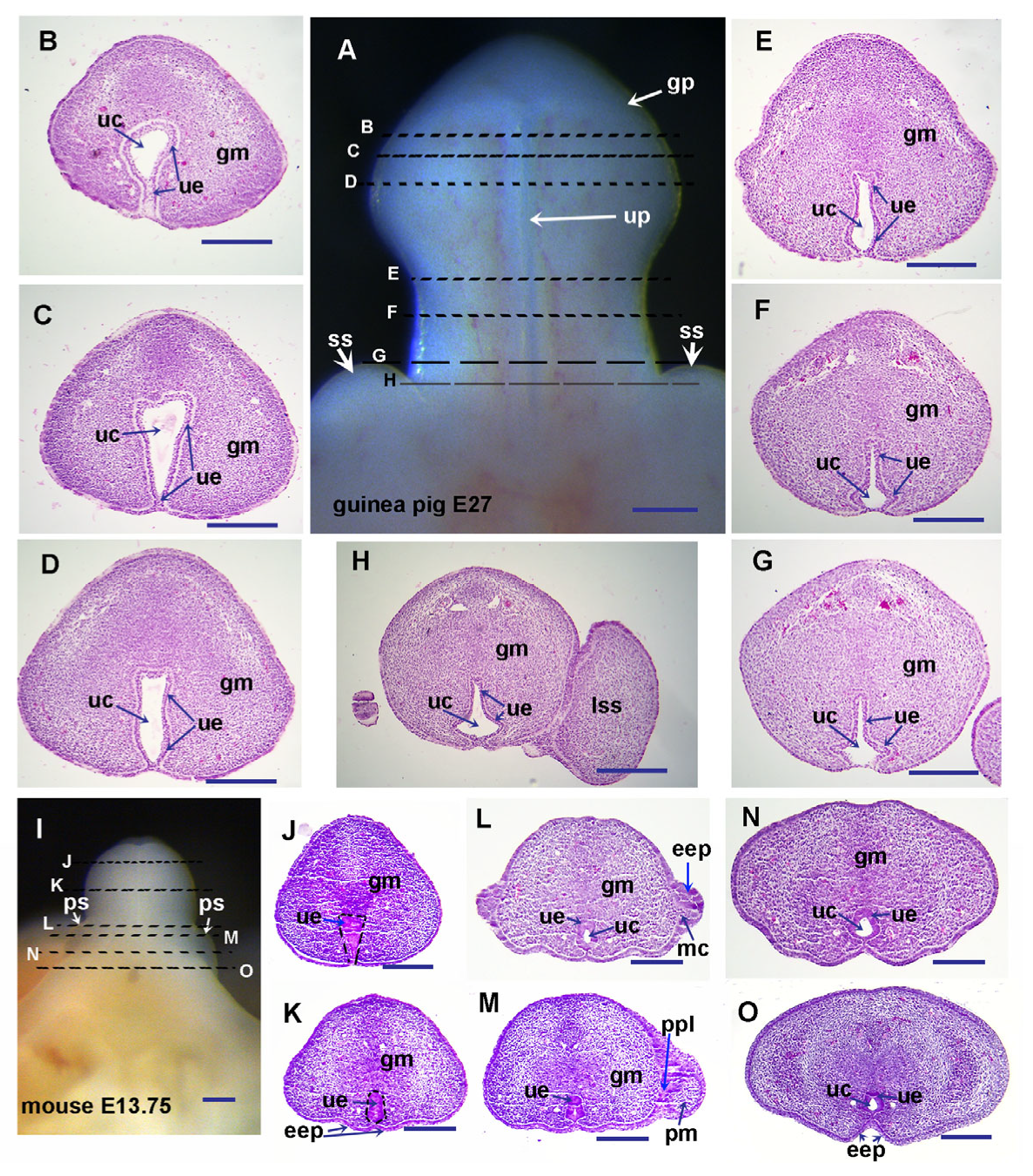
Figure 1. Histological structure of E27 guinea pig and E13.75 mouse genital tubercles (GTs). Images (A,I) are ventral views of E27 guinea pig (A) and E13.75 mouse (I) GTs with distal at the top. All sections of guinea pig (BH) and mouse (JO) are transverse through GT with dorsal at the top. Broken lines on images (A,I) indicate the planes of sections. Note the prepuce starts to form in E13.75 mice, but not in E27 guinea pigs. Abbrev: eep, epidermal epithelium; gm, glans mesenchyme; gp, glans penis; mc, mesenchyme; pm, preputial mesenchyme; ppl, preputial lamina; ps, preputial swelling; ss, scrotal swelling; uc, urethral canal; ue, urethral epithelium; up, urethral plate. Scale bars: 250 µm.
Cells 14 00348 g001
Figure 2. Histological structure of E29 guinea pig (GP) and E16.5 mouse penises. Images (A,I) are ventral views of E29 guinea pig (A) and E16.5 mouse (I) penises, with distal at the top. All sections of the guinea pig (BH) and mouse (JO) are transverse through penises, with dorsal at the top. Broken lines on images (A,I) indicate the planes of sections. Note that the prepuce starts to form at E29 in guinea pigs but reaches to the distal glans penis (K) at E16.5 in mice. Abbrev: et, epithelium tag; pg, preputial gland; u, urethra; ug, urethral groove; eep, gm, pm, ppl, ps, ss, uc, and ue are the same as in Figure 1. Scale bars: 250 µm.
Cells 14 00348 g002
Figure 3. Histological structure of the E33 guinea pig penis. Image (A) shows a ventral view of the E33 guinea pig penis with the distal end at the top. Sections of (BK) are transverse through the penis, with dorsal at the top. Broken lines on the image (A) indicate the planes of section. Sections of (LN) show sagittal planes of E33 guinea pig penis with dorsal at the left. Note that the prepuce reaches the distal glans penis at E33 in guinea pigs. Abbrev: eep, gm, pm, ppl, and ue are the same as in Figure 1; et, pg, and u are the same as in Figure 2. Scale bars: 250 µm.
Cells 14 00348 g003
Figure 4. Cell proliferation in guinea pig preputial development. Images show immunolocalization of BrdU (dark brown) on transverse sections of guinea pig penises at E29 (AF) and E33 (JL) with dorsal at the top. (A,C,E) are sections through the proximal region (See image in Figure 2A, between plane F and H) of the E29 guinea pig penis with the order from proximal to more distal as (E), (C), and (A). (B,D,F) are higher magnification images of the areas marked by blue boxes in (A,C,E), respectively. (G,H,I) show the number of BrdU-positive cells in a newly formed small region of preputial mesenchyme (pm) and surrounding epidermal epithelium (eep) counted from (B,D,F), respectively. Note that epidermal epithelia cells proliferate and invaginate to form original preputial lamina (B,F,I) and separate a small portion of preputial mesenchyme (C,D,H), and then the preputial mesenchymal cells proliferate and evaginate distally (A,B,G). (J,L) are distal (J) and proximal (L) sections of the E33 guinea pig penis, and (K) is a higher magnification image of the area marked by a blue box in (J). Note that the basal layer of epithelial cells (the deepest layer above the green line) proliferates to increase epidermal epithelial cell layers, and preputial mesenchymal cells (inside the black line) proliferate to evaginate distally (J,K), In the proximal section (L), BrdU-positive cells can be observed in developing preputial lamina (inside blue line). The data in (GI) are mean (n = 3) ± standard error (SE), ** p < 0.01, *** p < 0.001. Abbrev: dpl, developing preputial lamina; eep, gm, pm, and ue are the same as in Figure 1, u is the same as Figure 2. Scale bars: 100 μm.
Cells 14 00348 g004
Figure 5. In situ gene expression analysis of guinea pig genital tubercle (GT). Images are ventral (AC,E,G,I,J,M,O,Q) or dorsal (D,F,H,K,L,N,P,R) views of E23-E23.5 guinea pig (excerpt for J, which is E22) GTs with the distal at the top. The tail has been removed from all embryos. Purple or blue staining in each image indicates the gene expression domain. Developmental stages are labeled upright in each image. Note that Shh is expressed in the urethral epithelium (A) and its receptor is expressed in the adjacent mesenchyme (B). Hoxd13 (C,D), Bmp4 (E,F), and Wnt5a (Q,R) are expressed in genital mesenchyme. Bmp7 is expressed in the distal urethral epithelium (very weak), genital mesenchyme, and developing mammary glands (G,H). Fgf8 expression is located in the urethral epithelium at E22 (J), but it is only weakly expressed in the distal tip part of the urethral epithelium and can only be seen in dorsal view at E23 (I,K,L). Fgf10 expression in genital mesenchyme is very weak and is relatively strong in urethral epithelium (M,N). Fgfr2 expression is mainly in the urethral epithelium (O,P). Abbrev: aer, apical ectodermal ridge; gt, genital tubercle; hl, hindlimb; mg, mammary gland; ue is the same as Figure 1. Scale bars: 500 µm.
Cells 14 00348 g005
Figure 6. Spatiotemporal patterns of Shh, Fgf10, and Fgfr2 expression during guinea pig external genital development. Whole-mount images are ventral (AC,FH,LN) or dorsal (D,E) views of the developing external genitalia of guinea pigs (E,G) with the distal at the top, showing mRNA expression of Shh (AE), Fgf10 (FH) and Fgfr2 (LN). Images in (IK) are transverse sections of the developing penises of guinea pigs and mice, showing Fgf10 protein (in red; blue is Dapi) localization. Note that Shh mRNA is exclusively expressed in the urethral epithelium and prepuce during the later stages (AE) of developing EG in guinea pigs. Fgf10 mRNA and protein mainly localize in the urethral epithelium at early stages in the guinea pig genital tubercle (GT) (FI) and also in the preputial lamina (ppl) in E38 penis (J) of guinea pigs. However, in mouse GT, Fgf10 protein mainly localizes in the mesenchyme. Fgfr2 mRNA is mainly expressed in the urethral epithelium in guinea pig GT. Arrowheads in (D,E) point to the tiny Shh expression domain, while arrows in (H) indicate the Fgf10 expression domain in labioscrotal swellings. Abbrev: ppl and ue are the same as in Figure 1. Scale bars in (AH,LN): 500 µm; in (IK): 100 µm.
Cells 14 00348 g006
Figure 7. Comparison of relative expression levels of key developmental genes between guinea pig (GP) and mouse genital tubercles (GTs). Images in (AH) are GP and mouse hindlimb (H) buds (AD) and GTs (EH), showing developmental stage similarity. All limb buds are in a dorsal view with the anterior on the left, and all GTs are in a ventral view with the distal at the top. GT samples were cut from the level marked by dashed lines in (EH) for RNA extraction and quantitative PCR analysis. Data in (I,J) are mean ± standard error, n = 5, ** p ≤ 0.01, *** p ≤ 0.001. Scale bars in (AH): 500 µm.
Cells 14 00348 g007
Figure 8. The effect of Shh and Fgf-10 proteins on urethral groove and preputial development. Images in (AL) are ventral views of mouse (AF) and guinea pig (GL) genital tubercles (GTs) with the distal end at the top. Broken line in images (DF) shows the edge of urethral groove. Note that Hedgehog or Fgf inhibitors induce urethral groove formation but inhibit preputial development in cultured GTs of mice; the most significant effect is seen in cultured GTs with both inhibitors (DF). Shh or Fgf-10 protein induces the formation of preputial swellings in cultured GTs of guinea pigs, and the most obvious results were found in both protein-treated GTs (JL). Abbrev: uo, urethral opening; gp, ps, ss, and up are the same as in Figure 1, ug is the same as in Figure 2. Scale bars: 500 µm.
Cells 14 00348 g008
Figure 9. Conditional deletion of Smo and Fgfr2 in mouse genital tubercle (GT) resulted in a fully opened urethral groove resembling a normal human and guinea pig developing penis. Images (AE) are modified from published figures with permission. (A) Guinea pig E28 GT [5]. (B) Human 9-week-old developing penis [44]. (C,D) Mouse E15.5 normal male GT (C) and Msx2-rtTA;tetO-Cre;Smoc/c male GT (D) [41]. (E) Mouse E15.5 Msx2cre Fgfr2 c/c male GT [16]. Abbrev: gp, ps, ss and up are the same as in Figure 1, ug is the same as in Figure 2.
Cells 14 00348 g009
Disclaimer/Publisher’s Note: The statements, opinions and data contained in all publications are solely those of the individual author(s) and contributor(s) and not of MDPI and/or the editor(s). MDPI and/or the editor(s) disclaim responsibility for any injury to people or property resulting from any ideas, methods, instructions or products referred to in the content.