Article

Oligosaccharyltransferase Is Involved in Targeting to ER-Associated Degradation

by
Marina Shenkman
1,2,
Navit Ogen-Shtern
1,†,
Chaitanya Patel
1,2,
Haddas Saad
1,2,
Bella Groisman
1,
Metsada Pasmanik-Chor
3,
Sonya M. Schermann
4,‡,
Roman Körner
4 and
Gerardo Z. Lederkremer
1,2,*
1
The Shmunis School of Biomedicine and Cancer Research, Cell Biology Division, George Wise Faculty of Life Sciences, Tel Aviv University, Tel Aviv 69978, Israel
2
Sagol School of Neuroscience, Tel Aviv University, Tel Aviv 69978, Israel
3
Bioinformatics Unit, George Wise Faculty of Life Sciences, Tel Aviv University, Tel Aviv 69978, Israel
4
Max Planck Institute of Biochemistry, 82152 Martinsried, Germany
*
Author to whom correspondence should be addressed.
Current address: The Skin Research Institute, The Dead-Sea and Arava Science Centre, Masada 86910, Israel.
Current address: Ascend Advanced Therapies, Fraunhoferstraße 9b, 82152 Planegg, Germany.
Cells 2025, 14(20), 1593; https://doi.org/10.3390/cells14201593
Submission received: 30 June 2025 / Revised: 5 October 2025 / Accepted: 7 October 2025 / Published: 14 October 2025
(This article belongs to the Section Intracellular and Plasma Membranes)

Abstract

Most membrane and secretory proteins undergo N-glycosylation, catalyzed by oligosaccharyltransferase (OST), a membrane-bound complex in the endoplasmic reticulum (ER). Proteins failing quality control are degraded via ER-associated degradation (ERAD), involving retrotranslocation to cytosolic proteasomes, or relegated to ER subdomains and eliminated via ER-phagy. Using stable isotope labeling by amino acids in cell culture (SILAC) proteomics, we identified OST subunits as differential key interactors with a misfolded ER protein bait upon proteasomal inhibition, suggesting unexpected involvement in ERAD. Previous reports implied additional roles for OST subunits beyond N-glycosylation, such as quality control by ribophorin I. We tested OST engagement in glycoprotein and non-glycosylated protein ERAD; overexpression or partial knockdown of OST subunits interfered with ERAD in conditions that did not affect glycosylation. We studied the effects on model misfolded type I and II membrane-bound proteins, BACE476 and asialoglycoprotein receptor H2a, respectively, and on a soluble luminal misfolded glycoprotein, α1-antitrypsin NHK variant. OST subunits appear to participate in late ERAD stages, interacting with the E3 ligase HRD1 and facilitating retrotranslocation. Molecular dynamics simulations suggest membrane thinning by OST transmembrane domains, possibly assisting retrotranslocation via membrane distortion.

Graphical Abstract

1. Introduction

Secretory proteins are translocated into the endoplasmic reticulum (ER), where they undergo folding and quality control. Molecules that are terminally misfolded are retrotranslocated to the cytosol to be polyubiquitinated and degraded by proteasomes, in a process known as ER-associated degradation (ERAD) [1,2], or can be targeted to ER-to-lysosome-associated degradation (ERLAD) [3]. ERLAD has been studied only in recent years and usually involves autophagic degradation of ER subdomains (ER-phagy) [4,5]. In contrast, ERAD was identified about 30 years ago [6,7] and many studies have focused since then on this pathway. However, the precise mechanism of retrotranslocation is still unclear and remains a subject of ongoing study. It appears that various mechanisms may exist for different ERAD substrates, contingent upon the specific E3 ubiquitin ligase involved. Among these ligases, the most well-characterized participant in ERAD is HMG-CoA Reductase Degradation Protein 1 (HRD1), which forms a large protein complex mediating retrotranslocation [8,9]. Studies using genetic screens in yeast identified ERAD complexes involving HRD1 [10]. Subsequently, analogous components were found in mammals, including HRD1 itself [11]. The primary constituents of the mammalian HRD1 complex encompass transmembrane proteins such as Derlin 1–3, Homocysteine-inducible Endoplasmic Reticulum (ER) Stress Protein (Herp), and ubiquitin regulatory X domain-containing protein 8 (Ubxd8), along with Suppressor of lin-12-like protein 1 (SEL1L), Amplified in Osteosarcoma 9 (OS-9) and XTP3-transactivated gene B protein or ER lectin 1 (XTP3-B) on the luminal side. On the cytosolic side, components include Ubc6 or Ube2G2, p97 and NGLY1 [8,10,12]. Structural insight into the HRD1 complex has been reported in yeast [13,14] and very recently in mammalian cells [15]. Cryo-electron microscopy (CryoEM) studies of the yeast HRD1/HRD3 complex indicated a direct role in retrotranslocation [13]. Further evidence involves in vitro reconstitution experiments suggesting a regulation of retrotranslocation by HRD1 autoubiquitination [16,17]. Herp, a protein tethered to the ER membrane via a hydrophobic hairpin, with most of the protein on the cytosolic side, was shown to recruit HRD1 and misfolded proteins for ERAD at a specialized ER subcompartment, the ERQC [18,19].
An approach used to delineate the HRD1 complex and discover new components has been the pull-down of the complex through known ERAD components, such as Yos9 in yeast [20] and SEL1L [21], XTP-3B, HRD1, UBXD8, Derlin-1/2 and OS-9 in mammalian cells [22]. The caveat is that overexpression affects the function and interactions of these proteins. Furthermore, even when tagged by knock-in at the endogenous level, the isolated complexes might be non-functional [23]. This is because these complexes might be dormant, partial complexes, given the dynamic nature of complex assembly, influenced by the presence of an ERAD substrate [18,24]. Here we have used a SILAC proteomics approach to capture functional ERAD components. We accomplish this by pull-down of an ERAD substrate and analyzing its preferential interactors under conditions favoring the accumulation of molecules targeted for retrotranslocation and subsequent proteasomal degradation. Surprisingly, among the most highly enriched interactors were several subunits of the oligosaccharyltransferase (OST) complex.
OST plays a pivotal role in N-glycosylation, the primary post-translational modification of secretory proteins. It transfers a conserved oligosaccharide from a lipid-linked glycan donor to asparagine residues in defined sequons in newly synthesized proteins. In mammalian cells, the OST complex consists of a catalytic transmembrane subunit (STT3 oligosaccharyltransferase complex catalytic subunit A or B (STT3A) or (STT3B)) along with several additional transmembrane proteins including DAD1, OST48/DDOST, TMEM258, OST4, ribophorin1, ribophorin2, and subunits interacting with STT3A (KCP2, DC2/OSTC) or STT3B (TUSC3/N33 or IAP/MAGT1) [25,26]. While the primary function of the OST complex lies in protein N-glycosylation, studies suggest the participation of OST subunits, such as ribophorin I, in protein quality control [27,28], although the mode of action remains unclear. Here we show that OST subunits have a role in protein targeting to ERAD.

2. Materials and Methods

2.1. Materials

Streptavidin-agarose-beads, Biotin, tunicamycin, puromycin, MG-132, N-acetyl-Leu-Leu-norleucinal (ALLN), bortezomib (Bz) and digitonin were from Sigma. Lactacystin (Lac) was from Cayman Chemicals (Ann Arbor, MI, USA). Promix cell labeling mix ([35S]Met plus [35S]Cys), 1000 Ci/mmol, was from PerkinElmer Life Sciences. Predesigned shRNAs for Tusc3 (TRCN0000038065, TRCN0000038067, TRCN0000299038, TRCN0000299109, TRCN0000299107) and DAD1 (TRCN0000310240, TRCN0000200464, TRCN0000083039, TRCN0000200404, TRCN0000083041) were from Sigma (Saint Louis, MO, USA).

2.2. Antibodies

Rabbit anti-DAD1, anti-Tusc3 and anti-calnexin, and mouse anti-b tubulin and anti-actin were from Sigma. Mouse anti-HA was from Biolegend and anti-Myc was custom produced and used before [29]. Mouse anti-rRNA from Abcam was a kind gift from Orna Elroy-Stein. Rabbit anti-RPL26 was from Cell Signaling. Rabbit polyclonal anti-H2a carboxy-terminal and amino-terminal antibodies and mouse monoclonal anti–H2a carboxy-terminal antibody B9 were the ones used in earlier studies [18]. Mouse anti–S-tag was from Novagen (Gibbstown, NJ, USA). Mouse monoclonal anti-STT3A and rabbit anti-GFP were from Santa Cruz Biotech. Rabbit anti-RFP was from MBL. Rabbit anti-Sec61β was the one used previously [30]. Strep-HRP, goat anti-mouse IgG-HRP, goat anti-rabbit IgG-HRP and goat anti-mouse IgG conjugated to Cy2 or to Dylight 549 were from Jackson-Immuno-Research Labs.

2.3. Plasmids and Constructs

WT HRD1-myc, dominant negative mutant hsHRD1-myc (MutHRD1-myc), Sec61β-GFP, S-tagged-OS-9.1 and S-tagged-OS-9.2 in pcDNA3.1(+), H2a and H2a mutated in its three glycosylation sites (H2aΔgly), subcloned in pCDNA1, and HA-tagged beta-secretase 1 mutant 476 (BACE476-HA) were used before [18,31], as well as HA-tagged human α1-antitrypsin null Hong Kong (NHK-HA) in pcDNA3.1 [32]. Constructs encoding H2aG78R uncleavable mutant fused through its C-terminus to a 38-amino acid streptavidin binding peptide (H2aSBP), to monomeric red fluorescent protein (H2aRFP) or to GFP (H2aGFP), were described before [18,31,33]. DAD1 in pEGFP-C1 (GFP-DAD1) was a kind gift from Gert Kreibich. H2a-BAP-V5 was used before [34]. BirA and Sec-BirA plasmids were a kind gift from Gianlucca Petris and Oscar Burrone (ICGEB, Trieste, Italy) [35].

2.4. Cell Culture, Media and Transfections

NIH 3T3 and HEK293 cells were grown in DMEM supplemented with 10% bovine calf serum at 37 °C under 5% CO2. Transfections of NIH 3T3 cells were carried out using a Neon MP-100 microporator system (Life Technologies, Carlsbad, CA, USA). Transfection of HEK293 cells was performed using the calcium phosphate method. An HEK293 stable cell line expressing H2aSBP (SBP6) was described previously [33].

2.5. BAP Construct Transfection and Probing

H2a-BAP-V5 was transfected using the calcium phosphate method in HEK293 cells together with an equal amount of plasmid carrying BirA (cytosolic biotin ligase). Biotin (100 μM) was added 24 h post transfection overnight. The cells were then rinsed with PBS and lysed at 4 °C in Buffer A (1% Triton X-100, 0.5% NaDOC and 1× cOmplete Protease Inhibitor Cocktail (Roche) in PBS), containing 10 mM N-ethylmaleimide, to block BirA post-lysis activity. The probing of biotinylated proteins on blots was performed using Strep-HRP in PBS-TWEEN (0.5%), followed by detection by enhanced chemiluminescence (ECL).

2.6. Metabolic Labeling

Subconfluent (90%) cell monolayers in 60 mm dishes were labeled for 30 min with [35S]Cys and then chased for different periods of time with normal DMEM plus 10% FCS, lysed, and immunoprecipitated with anti-H2a carboxy-terminal antibody, as described previously [32]. Quantitation was performed in a Fujifilm FLA 5100 phosphorimager (Tokyo, Japan).

2.7. Immunoprecipitation, Crosslinking and Immunoblotting

Cell lysis in buffer A and immunoprecipitation methods are described in [33]. For GFP-DAD1 coexpressed with H2a, crosslinking was performed with DSP prior to cell lysis. Cells were washed 2× with PBS and incubated with PBS containing 1 mM DSP at 4 °C for 1 h. The reaction was stopped by incubation at 4 °C for 20 min with 50 mM Glycine, followed by a rinse with PBS and incubation with 20 mM Tris-HCl pH 7.4 in PBS for at RT 5 min.
For precipitation with streptavidin-agarose beads, cell lysates were incubated with the beads on a vertical rotating mixer at 4 °C overnight. After centrifugation, the beads were washed three times with 0.5% Triton X-100, 0.25% NaDOC and 0.5% SDS in PBS and once with PBS. Samples were boiled in sample buffer for 5 min and run on SDS-PAGE under reducing conditions.
Transfer to a nitrocellulose membrane, immunoblotting, detection by ECL and quantitation in a Bio-Rad ChemiDocXRS Imaging System (Hercules, CA, USA) were performed as described previously [29]. Quantity one (version 4.6.9) or ImageJ (version 1.53) software were used for the quantitation.

2.8. Iodixanol Equilibrium Sedimentation Gradient

Iodixanol gradients were performed similarly to methods previously described [32]. Briefly, HEK293 cells expressing H2a-RFP (untreated or treated with Bz) were washed with PBS, resuspended in a homogenization buffer (0.25 M Sucrose, and 10 mM HEPES pH 7.4). The cells were passed through a 21G needle five times before homogenization in a Dounce homogenizer (low clearance pestle, 30 strokes). The homogenates were centrifuged at 1000× g for 10 min at 4 °C to remove nuclei and cell debris and the supernatants were loaded on top of an iodixanol gradient (10 to 34%). The gradients were ultra-centrifuged at 24,000 rpm (98,500 g, Beckman SW41 rotor) at 4 °C for 16 h. Gradient fractions of 1 mL were collected from top to bottom, precipitated with 10% TCA and run on SDS-PAGE.

2.9. Immunofluorescence Microscopy

Immunofluorescence was performed as described previously [18,30]. Briefly, cells grown for 24 h after transfection on coverslips in 24-well plates were fixed with 3% paraformaldehyde for 30 min, incubated with 50 mM glycine in PBS, and permeabilized with 0.5% Triton X-100. After blocking with normal goat IgG in PBS/2% BSA, they were exposed to primary antibody for 60 min, washed and incubated for 30 min with a secondary antibody, followed by washes and staining of nuclei with DAPI. Specimens were observed on a Zeiss laser scanning confocal microscope (LSM 510; Carl Zeiss, Jena, Germany). ImageJ was used to quantify fluorescence intensity and to calculate Mander’s coefficients (using JACOP) for colocalization studies. ERQC localization was determined by the accumulation of H2a fluorescent fusion proteins or of OS-9.

2.10. Proteomics

SBP binding and elution conditions were as in [36]). For SILAC proteomics HEK293 or SBP6 cells were labeled with 13C(6)15N(2) L-lysine for 10 days. The labeling efficiency obtained was 97%. Then, 7 × 10⁸ cells (15 × 150 mm dishes) were treated with or without 50 μM MG-132 + 50 μM ALLN for 4 h and with 2 mM puromycin for the last 15 min before lysis. Cells were lysed in 1% digitonin in PBS + 2 mM PMSF + 5 μg/mL aprotinin. Labeled HEK293 and unlabeled SBP6 lysates were mixed at a 1:1 protein ratio. The combined lysates were subjected to ON precipitation with preblocked streptavidin beads (0.5% BSA, 0.2% digitonin in PBS for 2–3 h, washed once with 0.1% BSA in the same buffer composition). Precipitation was with beads in 0.1% BSA, 0.2% digitonin in PBS, followed by 4 washes with 0.2% digitonin in PBS, and protein interactors pulled down with H2aSBP were eluted for 1 h at +4 °C by competition with 250 μM free biotin dissolved in the wash buffer. Samples were heated at 65 °C with β-mercapto-ethanol containing sample buffer for 10 min and run on SDS-PAGE. In-gel digestion of SDS-page separated proteins was performed as described in [37] using 10 ng/μL of trypsin in 25 mM ammonium bicarbonate/1 mM CaCl at 37 °C for 16 h. Proteins were identified by nano LC-MS/MS using a nanoACQUITY UPLC nano liquid chromatography system (Waters, Eschborn, Germany) coupled to a LTQ orbitrap mass spectrometer (Thermo, Waltham, MA, USA) or an EasyLC system (Proxeon, Odense, Denmark) coupled to a Thermo LTQ-FT Ultra mass spectrometer. For nanoLC peptide separation, 15 cm long capillary columns (75 μm inner diameter) with a prepulled capillary tip (Proxeon) were packed in-house with Reprosil-Pur 3 μm C18 material (Dr. Maisch). Peptides were eluted at 0.3 μL/min using stepwise gradients from 2 to 60% acetonitrile, in 0.1% formic acid. MS/MS analysis on the LTQ orbitrap mass spectrometer was performed with standard settings using cycles of one high resolution MS scan from m/z 370–1750 followed by MS/MS scans of the 10 most intense ions with charge states of 2 or higher. For analysis on the Thermo LTQ-FT Ultra mass spectrometer, FT scans from m/z 400–2000 were taken at a resolution of 100,000, followed by collision induced dissociation (CID) scans in the LTQ of the eight most intense ions with signals greater than 2000 counts and charge states of 2 or higher.

2.11. Bioinformatics Analysis

Protein identification and SILAC-based quantitation was performed with MaxQuant (version 1.0.13.13) using the default settings. The UNIPROT human database (version 2009-04-14) was used for protein identification. MaxQuant uses a decoy version of the specified UNIPROT database to adjust the false discovery rates for proteins and peptides to below 1%. The mass spectrometry proteomics data have been deposited to the ProteomeXchange Consortium via the PRIDE partner repository [38], with the dataset identifier PXD067654.
Gene ontology enrichment of 86 differentially expressed proteins was performed using DAVID tool [39]. Selected enriched functions were presented, with −log2 pFDR from DAVID gene enrichment analysis (x-axis). Differentially expressed proteins were also applied to STRING protein-interaction tool [40]. Protein colors represent enriched functions circled in colors.

2.12. Statistical Analysis

Statistical analysis of proteomics MaxQuant data was performed with Perseus (version 1.6.13.0) [41]. A one-sided Student’s p-test for the treated subset of samples was employed with a Benjamini–Hochberg FDR of 0.05 to calculate p-values for Supplementary Table S4. Statistical analysis of the bioinformatic analysis was performed using included functions of the DAVID and STRING tools.
Other results are expressed as average ± SD. Student’s t-test (two-tailed) was used to compare the averages of two groups. Statistical significance was determined at p < 0.05 (*), p < 0.01 (**), p < 0.001 (***).

3. Results

3.1. Identification of OST as ERAD Substrate Interactor by a SILAC Proteomics Approach

To pull down ERAD machinery components interacting specifically with a protein undergoing ERAD, we used as a bait an established model ERAD substrate, H2a, linked to an affinity binding tag (streptavidin binding peptide, SBP). Molecules undergoing ERAD were caused to accumulate by inhibiting proteasomal degradation, and to reduce the number of nascent molecules undergoing cotranslational translocation into the ER, they were depleted by brief cell incubation with puromycin. Puromycin causes the termination of nascent polypeptide chains and their release into the lumen of the ER [42].
We used the model ERAD substrate H2aG78R as a bait because it is completely targeted to ERAD, with none of the substrate exiting the ER or being degraded by other systems. H2a, the uncleaved precursor of human asialoglycoprotein receptor (ASGPR) H2a, has been extensively studied. It is a type II membrane glycoprotein, completely retained in the ER [43], and is a substrate of the HRD1 complex [18,33]. As H2a can undergo natural cleavage, generating a soluble luminal fragment, which can be secreted, we used an uncleavable mutant, H2aG78R, which is entirely targeted to ERAD [31]. The use of the 38 amino acid SBP affinity tag allows for specific and high affinity binding and elution under mild conditions, to preserve complexes [44]. H2aG78R linked to SBP (H2aSBP) behaves like H2aG78R in its ER retention and targeting to ERAD, and H2aSBP precipitation with streptavidin beads allows for the coprecipitation of ERAD components, such as Derlin1, HRD1 and Skp1-Cullin1-F-box protein 2 (Fbs2), with the latter interaction being inhibited with kifunensin, which inhibits mannose trimming required for Fbs2 recognition and ERAD [33].
For the SILAC proteomics we prepared a stable HEK293 cell line (SBP6) expressing H2aSBP, which was cultured in the presence of media containing heavy amino acids (13C(6)15N(2) lysine) to uniformly label the proteins, while WT HEK293 cells were grown with normal light amino acids, or vice versa (Figure 1a). Thus, proteins become labeled with a known mass difference, allowing for accurate quantification of relative protein abundance by mixing the samples early in the process to minimize handling errors. Cell lysates in mild non-denaturing detergent (1% digitonin) (to preserve membrane protein complexes) from cells with or without H2aSBP expression were mixed at a 1:1 protein ratio, followed by precipitation with streptavidin-conjugated beads and elution by competition with biotin. Eluted proteins were subjected to SDS-PAGE, digested with trypsin and identified by electrospray tandem mass spectrometry (ESI-MS/MS). This experiment was performed on cells treated without or with proteasomal inhibitors for 4 h, followed by incubation with puromycin for 15 min (Figure 1a).
Prior to the proteomics experiment, we confirmed that the expression of H2aSBP increased by proteasomal inhibition and more robustly by overexpression of a dominant negative RING finger mutant of HRD1, as we had seen before with untagged H2a and as expected for an HRD1 substrate. The mutant HRD1 coprecipitated with H2aSBP upon pull-down of the latter with streptavidin-agarose beads (Figure 1b). We also tested whether the SBP tag allowed specific and efficient elution in non-denaturing conditions with a competitive concentration of free biotin. Indeed, a lysate of cells labeled with [35S] met + cys in the presence of the proteasome inhibitor showed many proteins coprecipitated specifically with H2aSBP (Figure 1c). Brief treatment with puromycin before lysis did not decrease the signal of these proteins. We used mild lysis conditions (see Materials and Methods Section), combined with the gentle biotin elution, to enable the preservation of protein complexes with little or no non-specific precipitation. Silver staining of the eluates after SDS-PAGE in a similar experiment showed an excellent signal to noise ratio, with only very weak bands in the control without H2aSBP, except for BSA, which was used as a blocker to reduce non-specific binding during the affinity pull-down. (Figure 1d).
After applying the SILAC proteomics protocol (Figure 1a), we selected as interactors only hits showing >2-fold ratio of labeled/unlabeled after normalization to the total amount of H2a-SBP (bait) obtained. We then compared the results from cells treated for 4 h with MG-132 and ALLN and 15 min with puromycin to those from cells treated only with puromycin. Gene ontology analysis showed that proteasomal inhibition led to a significant enrichment for cellular functions related to the ER, ER membrane and ubiquitin/proteasome processes, which was expected, and gave us confidence in the approach. One of the top function-enriched categories using the DAVID gene enrichment tool was the OST complex (p-value = 3.13 × 10⁻¹⁰), which was unexpected (Figure 1e and Supplementary Table S2). A STRING interaction map showed high enrichment of proteins belonging to a few categories related to protein quality control, ERAD (p-value = 1.28 × 10⁻⁷), protein folding (p-value = 1.28 × 10⁻⁷), glycosylation (p-value = 7.03 × 10⁻¹⁰) (including OST and calnexin (CNX) cycle genes), unfolded protein response (UPR) and vesicular transport (p-value = 8.00 × 10⁻⁶) (Figure 1f). This latter category perhaps relates to the participation of vesicular trafficking in ERAD, which we reported recently [32].
Among the most significant H2aSBP interactors, with enrichment values of 2-fold or higher in proteasomal inhibitor-treated cells, were most OST subunits (Supplementary Tables S1, S3 and S4). This was even more surprising considering that membrane proteins, some with multiple membrane spans such as OST subunits, are usually underrepresented in the MS analysis. All of these OST subunits were highly enriched upon proteasomal inhibition (Figure 1g). It should be stressed that the puromycin treatment before cell lysis should have reduced interactions with newly made translocated proteins, where H2aSBP could have encountered OST, which is associated with forward translocons [45]. Indeed, levels of forward translocon-related proteins, such as Sec61 subunits or SSRA, were similar or reduced compared to those in samples treated with proteasomal inhibitor versus those without (Figure 1g). Other enriched proteins in proteasomal inhibitor-treated samples include ERAD-related genes, such as XTP3-B, SEL1L and ubiquitin, calnexin cycle genes (CNX, Selenoprotein 15 (SEP15) and UDP-glucose glycoprotein glucosyltransferase 1 (UGGT1)) and other relevant genes such as the ER calcium pump Sarco(endo)plasmic reticulum calcium-ATPase 2 (SERCA2) and TAR DNA-binding protein 43 (TDP43) (Figure 1g). TDP43, normally localized in the nucleus, accumulates in the cytoplasm in several diseases, including ALS, and also upon proteasome inhibition [46].

3.2. Shared Subcellular Localization and Increased Interaction of OST Subunits with the ERAD Substrate upon Proteasomal Inhibition

Given the increase in the pull-down of OST subunits with the ERAD substrate upon proteasomal inhibition, we analyzed the subcellular localization of the main OST catalytic subunit, STT3A, in relation to the ERAD substrate and whether there is any change upon proteasomal inhibition. We used iodixanol density gradients, which we had previously optimized to separate ER subcompartments, to analyze the localization of these proteins in relation to the ER-derived quality control compartment (ERQC), a staging ground for ERAD [19,30,31]. As we had seen before [18], H2a linked to monomeric red fluorescent protein (H2a-RFP) appeared mainly in middle fractions of the gradient (4–7), with an increased peak at heavier fractions (6–8), the region of the ERQC, upon proteasomal inhibition (Figure 2a). A ribosomal subunit, RPL26, appeared at higher densities, marking regions of rough ER (RER) (fractions 8–10), with little or no overlap with the ERQC (Figure 2a), as we had seen before [18]. A subpopulation of endogenous STT3A appeared at the RER, as expected, but surprisingly, a larger subpopulation comigrated with H2a, separate from the RER, and peaking at the ERQC fractions, similarly to the ERAD substrate, upon proteasomal inhibition. We then examined another OST subunit, Defender Against apoptotic cell Death 1 (DAD1) [47], well characterized in previous studies, although it was not detected in our proteomics analysis, possibly due to its low abundance. Endogenous DAD1 showed a similar pattern to H2a-RFP in untreated cells, with a sharper accumulation upon proteasomal inhibition, as a peak in fraction 6, in the ERQC region, and an additional smaller peak in fraction 4 (Figure 2b). Surprisingly, there was very low abundance of DAD1 in the RER. Endogenous CNX and Sec61β also appeared in middle fractions, partially overlapping with DAD1 in untreated conditions and upon proteasomal inhibition where they concentrated in fraction 6. We analyzed another OST subunit, Tumor suppressor candidate 3 (Tusc3), a non-essential subunit that was also absent from our top proteomic hits. Endogenous Tusc3 showed a similar pattern to DAD1 in untreated conditions, although concentrating more at fraction 4, but it did not shift to higher densities upon proteasomal inhibition (Figure 2b).
We then compared the subcellular localization by fluorescence microscopy. We could not detect OST subunits by immunofluorescence with the available antibodies, and therefore we expressed a construct, GFP-DAD1. We chose GFP-DAD1 because, as mentioned above, it had been well studied before and had been shown to associate with forward translocons [48]. Both GFP-DAD1 and H2a-RFP show an ER pattern in untreated cells, although with some concentration in the juxtanuclear region (Figure 2c). Notably, under proteasomal inhibition, GFP-DAD1 accumulated together with H2a-RFP in the juxtanuclear ERQC (Figure 2d). This is similar to the effect of ERAD substrate accumulation (upon proteasomal inhibition or by other means) on the localization of ERAD machinery components (HRD1, Derlin-1 [33]). Several ER chaperones, such as CNX and CRT, also relocalize to the ERQC under these conditions, but not others, such as BiP [30]. As we had also seen before [31], the translocon subunit Sec61β linked to GFP accumulates at the ERQC with the ERAD substrate upon proteasomal inhibition (Figure 2e,f). To test whether the entire forward translocation complex linked to ribosomes (at the rough ER) accumulates in the ERQC along with the ERAD substrate, we immunostained cells transfected with H2a-RFP using an anti-ribosomal RNA (rRNA) antibody as a ribosomal marker. As opposed to DAD1 and Sec61β, the ribosomes did not accumulate together with H2a-RFP after proteasomal inhibition and remained in a cytosolic and ER pattern (Figure 2g,h), consistent with the results of the density gradients. Sec61 participates in the forward translocation complex, but has also been implicated in retrotranslocation [49].
To confirm the ERQC localization, we compared the localization of GFP-DAD1 with that of the ERAD-associated luminal lectin OS-9, which appears constitutively localized mainly at the ERQC [18,29]. Similarly to H2a-GFP, the GFP-DAD1 that accumulated under proteasomal inhibition colocalized with OS-9 (Figure 2j,m). However, while the colocalization of H2a-GFP with OS-9 increased significantly upon proteasomal inhibition, that of GFP-DAD1 did not change (Figure 2k,n). This suggests that a portion of GFP-DAD1 molecules localize to the ERQC in untreated cells and may already colocalize with OS-9 in peripheral regions.
We then analyzed whether DAD1 interacts with the ERAD substrate and at what stage. A specific interaction of GFP-DAD1 and H2a was detected by coimmunoprecipitation after crosslinking with DSP (Figure 2o). This interaction was significantly increased upon proteasomal inhibition, suggesting that molecules targeted for degradation have a higher probability of interacting than newly made ones.
In summary, a significant population of OST subunits colocalize with an ERAD substrate at the ERQC, separate from the rough ER, which contains forward translocons. Upon proteasomal inhibition, DAD1 accumulates in the ERQC, in association with an ERAD substrate.

3.3. OST Subunits Promote the Degradation of Glycosylated H2a and to a Lesser Extent of Its Non-Glycosylated Mutant

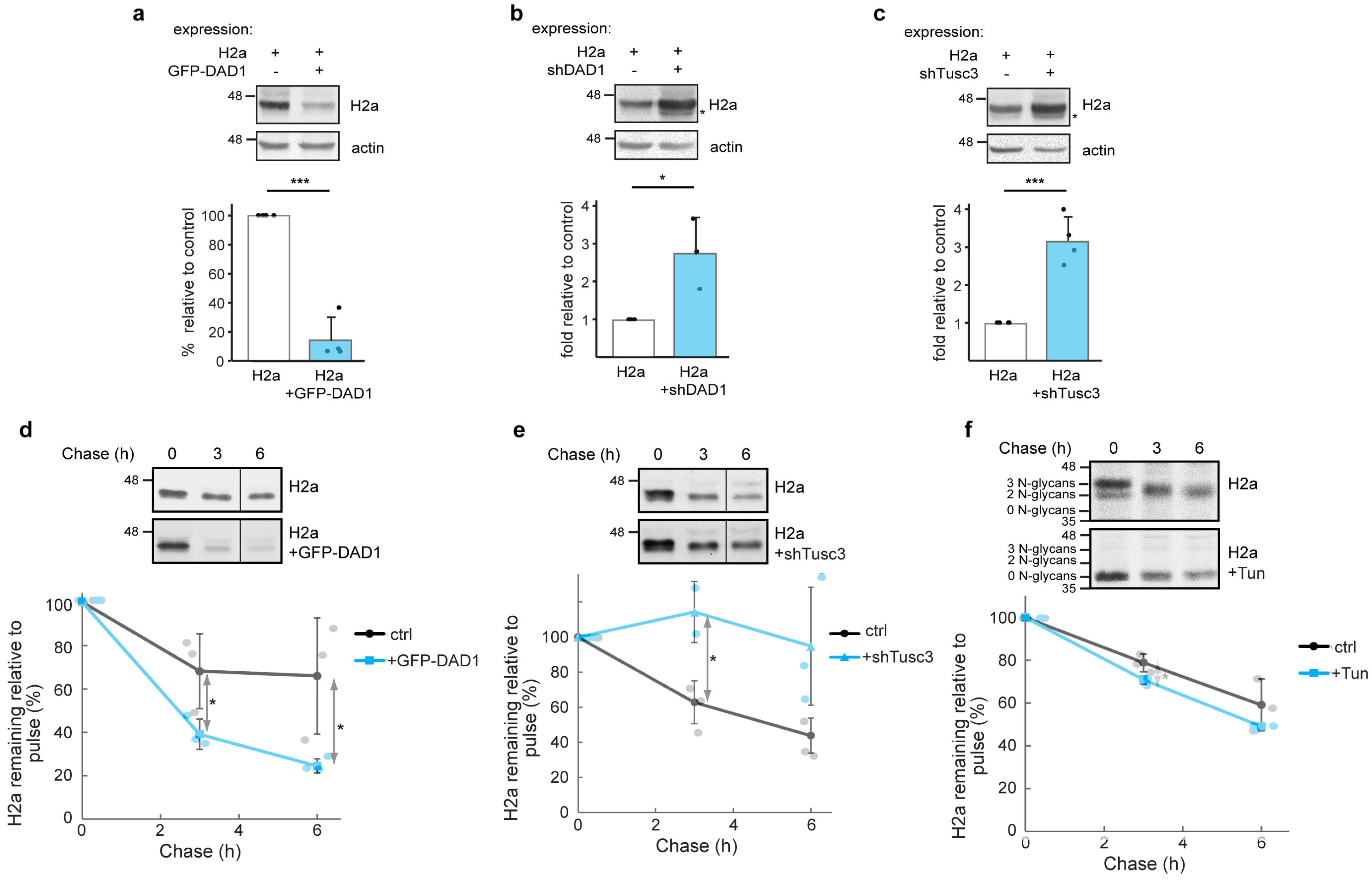
To investigate a possible role of OST in ERAD, we tried to manipulate the levels of OST subunits. This could not be performed with the essential catalytic subunits STT3A and STT3B, as it would result in an unwanted effect on the OST canonical activity on N-glycosylation. We first overexpressed GFP-DAD1 and analyzed its effect on H2a. Overexpression of GFP-DAD1 led to a dramatic decrease in the levels of H2a (Figure 3a), suggesting that the protein was degraded. There was no change in the migration of H2a, indicating that glycosylation was not affected. We then partially knocked down DAD1 (Supplementary Figure S1), which had a major effect in increasing the levels of H2a, suggesting that ERAD was inhibited (Figure 3b). Partial knockdown was intended to avoid a dramatic influence on OST canonical function. Indeed, despite its major effect on ERAD, the partial knockdown had only a very minor effect on glycosylation, as a weak band of faster migration appeared, suggesting the existence of a small population of underglycosylated molecules (two of the three glycosylation sites occupied according to our previous studies [50]). We then tried partially knocking down Tusc3 (Supplementary Figure S1), which also caused a major accumulation of H2a (Figure 3c). Tusc3 is a non-essential OST subunit, but nevertheless also caused the appearance of a minor population of underglycosylated H2a molecules. Therefore, partial knockdown of DAD1 or Tusc3, in conditions that affect minimally OST function in glycosylation, have a major effect on inhibiting ERAD.
To confirm that the effects on the steady state levels of H2a were due to changes in its degradation, we performed pulse-chase analysis experiments using [35S] cys metabolic labeling. The overexpression of GFP-DAD1 led to a significantly accelerated degradation of H2a (Figure 3d). Conversely, knockdown of Tusc3 strongly slowed down the degradation (Figure 3e).
Although glycosylation of H2a did not seem to be affected, the effect of Tusc3 knockdown on H2a degradation could be due to inhibition of N-glycosylation of key components of quality control or ERAD. To test this, we completely inhibited N-glycosylation via a long (18 h) incubation of cells with tunicamycin (Tun), after which we performed a pulse-chase experiment in the presence of Tun. The treatment caused a shift in the migration of H2a due to lack of its 3 N-glycan chains (Figure 3f). However, the rate of H2a degradation showed no significant change in the presence of Tun (Figure 3f graph), which suggests that the effect of Tusc3 knockdown in delaying degradation (Figure 3c,e) cannot be ascribed to inhibition of glycosylation of the ERAD substrate or of ERAD components.
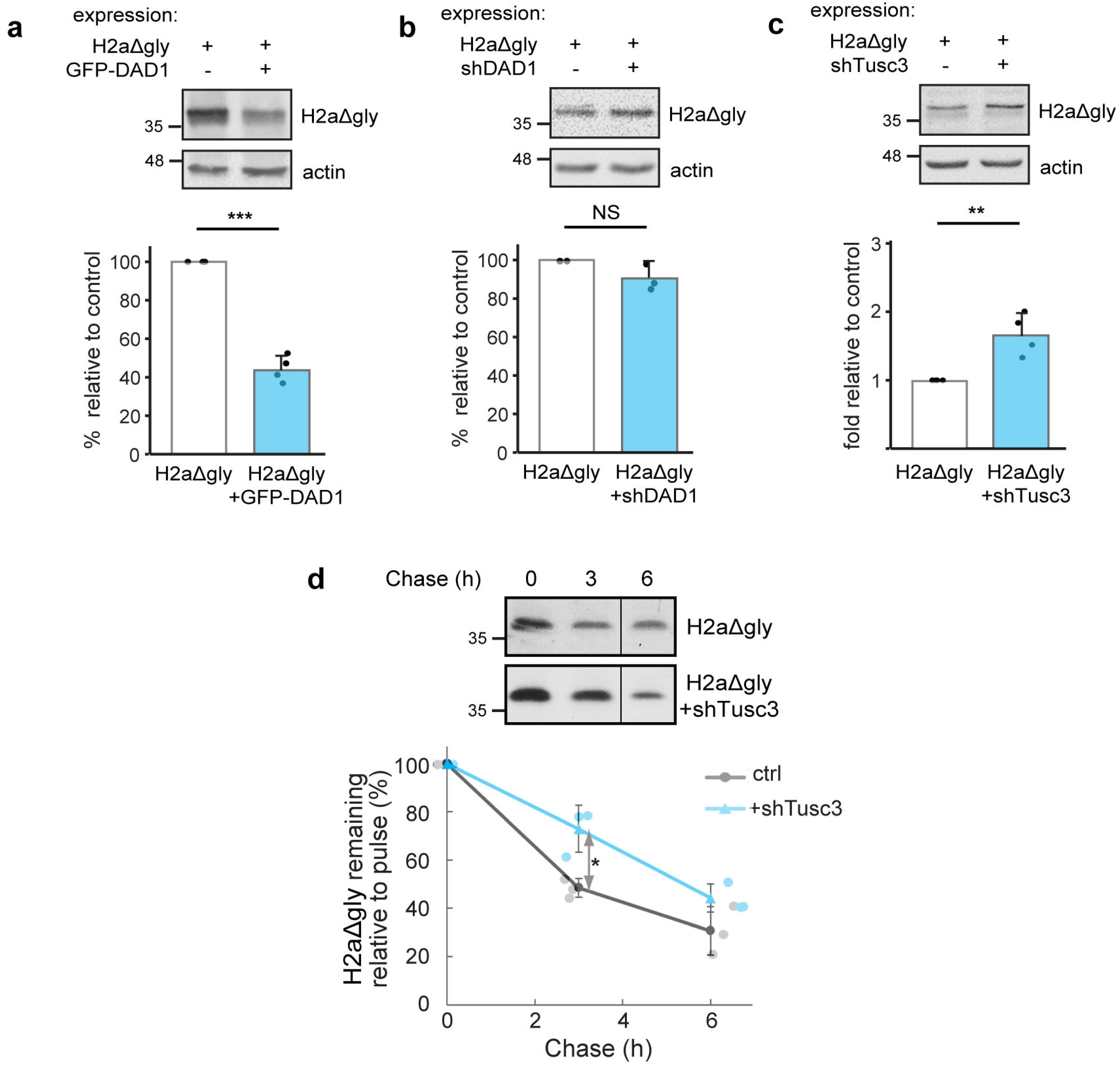
To test whether OST subunits are involved in ERAD of a non-glycosylated ERAD substrate, similar experiments were performed on an H2a mutant lacking its three glycosylation sites (H2aΔgly, [50]). GFP-DAD1 overexpression and Tusc3 knockdown had similar effects on H2aΔgly as on glycosylated H2a, but to a much lesser extent (compare Figure 4a,c,d with Figure 3a,c,e, respectively). DAD1 knockdown did not have any effect on H2aΔgly degradation (Figure 4b). DAD1 does not seem to participate in H2aΔgly ERAD at its endogenous levels but can target the non-glycosylated protein when DAD1 is overexpressed.

3.4. OST Subunits Participate in ERAD of Other Membrane and Luminal Substrates

To test whether OST subunits are more generally involved in ERAD, we analyzed other well-established ERAD substrates, BACE476, a mutant type I membrane-bound glycoprotein (in contrast with H2a, which is type II, [18]) and the soluble luminal misfolded glycoprotein human α1-antitrypsin variant null Hong Kong (NHK) [51]. Overexpression of GFP-DAD1 led to a decrease in the levels of BACE476 and NHK, although to a much lesser extent than for H2a (compare Figure 5a,d to Figure 3a). Consistent with a promotion of ERAD, the reduction in the levels of all ERAD substrates tested upon overexpression of GFP-DAD1 was inhibited by a proteasome inhibitor (Supplementary Figure S3).
Similarly, DAD1 or Tusc3 knockdown caused an accumulation of BACE476 and NHK, but again, less pronounced than for H2a (compare Figure 5b,c,e,f to Figure 3b,c).
Thus, OST subunits can promote ERAD of type I or type II transmembrane substrates and of a soluble luminal substrate, but with some differences in the extent of their action, depending on the substrate.

3.5. Interaction Between DAD1 and HRD1 and Promotion of Retrotranslocation by OST Subunits

As the ERAD substrates used in our experiments are targeted to HRD1, we wondered whether DAD1 and HRD1 interact. In a coimmunoprecipitation experiment in the absence of crosslinkers, we could see a robust and specific interaction between GFP-DAD1 and HRD1-myc (Figure 6a). Although both DAD1 and HRD1 accumulated upon proteasomal inhibition, the ratio of DAD1 coimmunoprecipitated in association with HRD1 decreased (Figure 6a graph). This possibly suggests that DAD1 associates with HRD1 in a functional ER membrane complex and does not interact with HRD1 molecules that are already targeted for proteasomal degradation. In fact, the coimmunoprecipitation does not exclude that the interaction could be through a third protein.
We next studied the requirement of OST subunits for the retrotranslocation step. We tested the effect of manipulating the levels of OST subunits on retrotranslocation using the BAP/BirA system [35]. This system utilizes the E. coli biotin ligase BirA expressed in the cytosol of mammalian cells to specifically biotinylate a 15 amino acid biotin-acceptor peptide (BAP). As shown by the group of Burrone, secretory proteins with a BAP tag on their ER luminal side are biotinylated only after retrotranslocation [35]. As previously shown, a construct of H2a with BAP attached to its luminal C-terminus behaved consistently as expected for a type II membrane protein [34]. H2a-BAP was biotinylated poorly when coexpressed with cytosolic BirA, but to a much higher extent when coexpressed with a luminally expressed secBirA, which contains a signal peptide for translocation into the ER [35] (Figure 6b). Molecules biotinylated by cytosolic BirA were fully glycosylated (including at a site near the C-terminus), indicating that they had previously entered the ER lumen. There was no biotinylation in a control without BirA or secBirA. As BAP is on the luminal C-terminus of H2a, it can be exposed to the cytosolic BirA only after H2a-BAP retrotranslocation. Overexposure of the cytosolic biotinylated retrotranslocated H2a signal allowed us to assess the effect of overexpression or knockdown of OST subunits (Figure 6c). GFP-DAD1 overexpression caused a decrease in total H2aBAP, as observed for H2a (Figure 6c lower panel, compared to Figure 3a), but there was a much smaller decrease in retrotranslocated biotinylated H2aBAP (Figure 6c upper panel), resulting in a large increase in the ratio of retrotranslocated molecules relative to total, as compared to the control (Figure 6c graph). Knockdown of either DAD1 or Tusc3 had the opposite effect, resulting in a significant decrease in the retrotranslocation ratio compared to the control.
Together, these results suggest that OST interacts with HRD1 and participates in the retrotranslocation process towards ERAD.

3.6. Molecular Dynamics Simulation Suggest Membrane Thinning by OST

To try to explain the involvement of OST in ERAD, we wondered whether it causes membrane thinning, promoting ERAD, as suggested for the HRD1 ERAD complex. The molecular architecture of the human OST complexes had been determined by cryo-EM [52], showing a dense region traversing the ER membrane with multiple transmembrane domains (Figure 7a,b). Molecular dynamics simulations (pre-rendered at the MemProtMD database [53]) indicate substantial membrane thinning by the OST transmembrane domains (Figure 7c,d). This may lower the energy barrier for protein retrotranslocation, or dislocation from the membrane, as proposed for the interphase between HRD1 and Der1 in yeast [54].

4. Discussion

In the present study we aimed to identify ERAD machinery components through their interaction with a model misfolded protein being targeted to this pathway. To this end, we developed an approach for isolation of functional protein complexes engaged in the retrotranslocation event, under conditions in which there is enrichment in retrotranslocation complexes, while minimizing the abundance of forward translocons. This approach employed proteasomal inhibition to accumulate the ERAD substrate just prior to degradation. Our previous studies with H2a, the model substrate used, indicated that in these conditions H2a accumulates in the ERQC membrane, because its retrotranslocation appears to be coupled to its degradation [18,19,30,31]. To reduce the presence of components of the forward translocon in the protein complexes obtained, we incubated the cells with puromycin. This antibiotic causes the release of nascent chains from the ribosome and, as a result, from the translocons. The combination of puromycin treatment with the C-terminal SBP tag used as bait was intended to prevent isolation of the machineries in charge of translation and forward translocation through incompletely translated H2aSBP molecules (which would lack a complete SBP). In these conditions, we observed an association of several of the OST subunits along with H2a in pull-down experiments. This association was substantially increased upon proteasome inhibition. Treatment with proteasome inhibitors was performed for only 4 h, to minimize induction of ER stress, which was reported to induce ER-phagy [55]. Moreover, no ER-phagy receptors were detected in our proteomics results, reinforcing the fact that the degradation pathway involved is ERAD. Notably, some of the OST subunits, OSTC, STT3A and MAGT1 were detected only after proteasome inhibition (Figure 1g); this, together with the puromycin treatment, suggests a preferential interaction with the ERAD substrate after its translation and translocation. Consistently, coimmunoprecipitation of DAD1 occurred after full glycosylation of H2a (Figure 2o).
OST association was reported with other ER proteins in different high MW complexes, one of them containing SSRD (Trap delta) [56]. Interestingly, we also detected SSRD only after puromycin treatment and proteasome inhibition (Figure 1g). Mutations in SSRD lead to defects in glycosylation [57], suggesting an early cotranslational event, as the SSR/Trap complex has been implicated in forward translocation. However, an alternative role has also been suggested for this complex in ERAD [58,59]. Other ER proteins have been suggested to participate in both forward translocation and in retrotranslocation towards ERAD, such as calumin (CCDC47) [60], and the Sec61 complex, for which interaction was found with ERAD factors [49]. Consistently, we found Sec61 subunits and CCDC47 at similar levels in cells with and without proteasomal inhibition (Figure 1g). It is noteworthy that TDP-43 appeared enriched in cells with proteasomal inhibition. TDP-43 is localized mainly in the nucleus but has been found sequestered at the cytosolic face of the ER in ALS [61].
For OST, noncanonical activities have also been suggested, such as that required for Dengue virus infection, which could be sustained with catalytically inactive OST subunits [62]. The OST subunit MAGT1 and its oxidoreductase activity [63] appeared to be essential for the infection. MAGT1 was present as one of the most significant interactors of our substrate under proteasome inhibition (Figure 1g). MAGT1 has been found to be associated with an STT3B containing complex [64]. The presence of MAGT1 and the effects of knockdown of Tusc3 (also associated with STT3B) would implicate the STT3B complex in our study. STT3B was not detected in the proteomics results, perhaps due to low abundancy, but STT3A and its associated OSTC were, suggesting also a participation of the STT3A complex in ERAD (Figure 1g). In fact, a substantial population of STT3A was observed to colocalize at similar ERQC subcellular densities as the ERAD substrate, segregated from the RER (Figure 2a).
There are previous studies that hint to the participation of OST subunits in quality control and ERAD. It was suggested that ribophorin I might be involved in protein quality control, as it interacted with a quality control substrate, a fragment of the amyloid precursor protein, after its translocation, remaining associated after puromycin release [28]. In the case of ERAD highjacking by CMV, the CMV US11 protein was found associated with OST subunits, ribophorin 1, ribophorin 2, and OST48 [65]. The ER protein malectin forms a complex with ribophorin 1 which retains misfolded NHK α1-antitrypsin and misfolded ribonuclease A but not the WT counterparts in the ER [27]. In a proteomics study using OS-9 as a bait, Christianson et al. pulled down ribophorin 1 with high score [66]. They also pulled down other OST subunits, such as STT3A, STT3B and DAD1 using UbxD8, Derlin-1 or Derlin-2 as baits [22]. However, these observations were not further investigated.
Our proteomics findings, along with insights from the effects of modulating DAD1 and Tusc3 levels, strongly suggest the participation of most, if not all, OST subunits in ERAD. However, we cannot exclude the possibility that only a subset of the subunits are involved in ERAD. This is a limitation of the study, as the knockout or knockdown of essential subunits, such as the catalytic STT3A and STT3B, would affect the canonical OST function in glycosylation, in addition to that in ERAD. There is a series of congenital disorders of glycosylation (CDGs) caused by mutations in several OST subunits, including STT3A and STT3B, which lead to severe neurodevelopmental deficiencies [67,68]. However, this could be the result of defects in the canonical or noncanonical OST functions.
It is likely that DAD1 and Tusc3 fell below the detection threshold in our proteomics analysis. Despite this, DAD1 exhibited clear associations with the ERAD substrate (Figure 2o) and HRD1 (Figure 6a) and showed strong effects of its overexpression. The effect of overexpression of this single OST subunit on ERAD might be explained by the limited quantity of its endogenous expression in relation to other subunits [69]. Interestingly, both DAD1 and Tusc3 partial knockdowns significantly affected ERAD without any significant change in glycosylation of the substrate. Although these knockdowns could still have affected the N-glycosylation of ERAD machinery components, thus inhibiting ERAD, this does not appear to be the case. Even upon complete inhibition of cellular N-glycosylation with Tun, ERAD was not affected (Figure 3f).
Both DAD1 and Sec61β relocalized to the ERQC under proteasomal inhibition (Figure 2), suggesting that most of their molecules are associated with the ERQC and not with forward translocons in these conditions. In a previous study, GFP-DAD1 had been observed to remain with relatively low mobility in the ER membrane, even after puromycin termination of protein synthesis and release from translocons. This observation led to the conclusion that GFP-DAD1 probably remains associated with inactive translocons [48]. However, the low mobility after puromycin treatment (as in this study) might instead signify a population of GFP-DAD1 molecules associated with a retrotranslocation complex. Indeed, GFP-DAD1 associates with HRD1, although the interaction could be indirect (Figure 6a). Intriguingly, other OST subunits were reported to undergo cleavage by the intramembrane rhomboid protease RHBDL4 before being targeted to ERAD [70]. Thus, if GFP-DAD1 were undergoing a similar targeting for ERAD, we would expect to observe cleaved molecules upon association with HRD1, but this was not the case. We can speculate that the association of GFP-DAD1 with HRD1 suggests a cooperative mechanism of the OST and HRD1 complexes for ERAD (Figure 7e).
In conclusion, OST appears to have a dual role of N-glycosylation at the initial stages of protein translocation into the ER and aiding ERAD at the final stages in the life of misfolded molecules (Figure 7e). We can only speculate, with the data that we have at the moment, about how OST precisely contributes to ERAD. The fact that the OST complex encompasses many transmembrane domains, interacts with HRD1, affects retrotranslocation and is predicted to cause membrane thinning suggests that it may participate directly in the mechanism of retrotranslocation. It might contribute to membrane distortion, directly aiding retrotranslocation, similarly to the mechanism proposed for the interphase between HRD1 and Der1 in yeast [54]. Experiments to study this mechanism should be undertaken in future research.
Targeting to ERAD has been best studied for glycoproteins and is linked to glycan processing and recognition steps [19]. OST could be a candidate for glycan recognition and targeting to ERAD. However, OST function for ERAD does not appear to be restricted to glycoproteins, although the results of OST subunit overexpression and knockdown suggest some preference for glycosylated substrates (Figure 3 and Figure 4). This dual targeting could be equated to the activities of OS-9, XTP3-B and EDEM1 in targeting both glycoproteins and non-glycoproteins to ERAD [50,71,72].

Supplementary Materials

The following supporting information can be downloaded at https://www.mdpi.com/article/10.3390/cells14201593/s1, Figure S1: Overexpression and knockdown of OST subunits; Figure S2: Degradation caused by GFP-DAD1 is proteasome-dependent; Figure S3: Gels in Figure 3 and Figure 4 including uncropped lanes; Table S1: OST subunits detected by mass spectrometry; Table S2: DAVID function enrichment analysis; Table S3: The table shows values for the 86 differentially expressed proteins resulting from the proteomics analysis; Table S4: Statistics analysis of the proteomics values.

Author Contributions

Conceptualization, G.Z.L.; bioinformatics, M.P.-C.; investigation, M.S., N.O.-S., C.P., H.S., B.G., S.M.S. and R.K.; writing—original draft preparation, G.Z.L.; writing—review and editing, G.Z.L., M.S., M.P.-C. and R.K.; supervision, G.Z.L. All authors have read and agreed to the published version of the manuscript.

Funding

This research was funded by grant 2577/20 from the Israel Science Foundation (GZL).

Data Availability Statement

The raw data supporting the conclusions of this article will be made available by the authors on request.

Acknowledgments

We would like to thank G. Kreibich, O. Elroy-Stein, G. Petris and O. Burrone for plasmids and reagents. We are very grateful to F.U. Hartl for ongoing advice and for critically reading and helping in the edition of this manuscript.

Conflicts of Interest

Sonya M. Schermann is currently an employee of Ascend Advanced Therapies, Martinsried, Germany. This manuscript is not an outcome of work at the company nor will it bring any benefits to this company’s products. The authors declare no conflicts of interest.

Abbreviations

The following abbreviations are used in this manuscript:
ALLNN-acetyl-Leu-Leu-norleucinal
BACE476beta-secretase 1 mutant 476
BAPbiotin-acceptor peptide
BirAE. coli biotin ligase BirA
BzBortezomib
CNXCalnexin
DAD1Defender Against apoptotic cell Death 1
ECLenhanced chemiluminescence
ERendoplasmic reticulum
ERADER-associated degradation
ERQCER-derived quality control compartment
Fbs2Skp1-Cullin1-F-box protein 2
HerpHomocysteine-inducible Endoplasmic Reticulum (ER) Stress Protein
HRD1HMG-CoA Reductase Degradation Protein 1
H2auncleaved precursor of human asialoglycoprotein receptor H2a. Gene: ASGPR2
H2aΔglyH2a mutated in its three glycosylation sites
H2aSBPH2aG78R uncleavable mutant fused through its C-terminus to a 38-amino acid streptavidin binding peptide
LacLactacystin
NHKhuman α1-antitrypsin variant null Hong Kong
OS-9Amplified in Osteosarcoma 9
OSToligosaccharyltransferase
SEL1LSuppressor of lin-12-like protein 1
SEP15Selenoprotein 15 or Selenof
SERCA2Sarco(endo)plasmic reticulum calcium-ATPase 2
SILACstable isotope labeling by amino acids in cell culture
STT3ASTT3 oligosaccharyltransferase complex catalytic subunit A
STT3BSTT3 oligosaccharyltransferase complex catalytic subunit B
TDP43TAR DNA-binding protein 43
TMDtransmembrane domain
Tusc3tumor suppressor candidate 3
TunTunicamycin
Ubxd8ubiquitin regulatory X domain-containing protein 8
UGGT1UDP-glucose glycoprotein glucosyltransferase 1
XTP3-BXTP3-transactivated gene B protein or ER lectin 1

References

  1. Sun, Z.; Brodsky, J.L. Protein quality control in the secretory pathway. J. Cell Biol. 2019, 218, 3171–3187. [Google Scholar] [CrossRef]
  2. Wu, X.; Rapoport, T.A. Mechanistic insights into ER-associated protein degradation. Curr. Opin. Cell Biol. 2018, 53, 22–28. [Google Scholar] [CrossRef]
  3. Salomo-Coll, C.; Jimenez-Moreno, N.; Wilkinson, S. Lysosomal Degradation of ER Client Proteins by ER-phagy and Related Pathways. J. Mol. Biol. 2025, 437, 169035. [Google Scholar] [CrossRef]
  4. Fregno, I.; Fasana, E.; Bergmann, T.J.; Raimondi, A.; Loi, M.; Soldà, T.; Galli, C.; D’Antuono, R.; Morone, D.; Danieli, A.; et al. ER-to-lysosome-associated degradation of proteasome-resistant ATZ polymers occurs via receptor-mediated vesicular transport. EMBO J. 2018, 37, e99259. [Google Scholar] [CrossRef]
  5. Mochida, K.; Nakatogawa, H. ER-phagy: Selective autophagy of the endoplasmic reticulum. EMBO Rep. 2022, 23, e55192. [Google Scholar] [CrossRef]
  6. Ward, C.L.; Omura, S.; Kopito, R.R. Degradation of CFTR by the ubiquitin-proteasome pathway. Cell 1995, 83, 121–127. [Google Scholar] [CrossRef]
  7. Werner, E.D.; Brodsky, J.L.; McCracken, A.A. Proteasome-dependent endoplasmic reticulum-associated protein degradation: An unconventional route to a familiar fate. Proc. Natl. Acad. Sci. USA 1996, 93, 13797–13801. [Google Scholar] [CrossRef] [PubMed]
  8. Christianson, J.C.; Jarosch, E.; Sommer, T. Mechanisms of substrate processing during ER-associated protein degradation. Nat. Rev. Mol. Cell Biol. 2023, 24, 777–796. [Google Scholar] [CrossRef]
  9. Lopata, A.; Kniss, A.; Löhr, F.; Rogov, V.V.; Dötsch, V. Ubiquitination in the ERAD Process. Int. J. Mol. Sci. 2020, 21, 5369. [Google Scholar] [CrossRef] [PubMed]
  10. Berner, N.; Reutter, K.R.; Wolf, D.H. Protein Quality Control of the Endoplasmic Reticulum and Ubiquitin-Proteasome-Triggered Degradation of Aberrant Proteins: Yeast Pioneers the Path. Annu. Rev. Biochem. 2018, 87, 751–782. [Google Scholar] [CrossRef] [PubMed]
  11. Kikkert, M.; Doolman, R.; Dai, M.; Avner, R.; Hassink, G.; van Voorden, S.; Thanedar, S.; Roitelman, J.; Chau, V.; Wiertz, E. Human HRD1 is an E3 ubiquitin ligase involved in degradation of proteins from the endoplasmic reticulum. J. Biol. Chem. 2004, 279, 3525–3534. [Google Scholar] [CrossRef] [PubMed]
  12. Wang, H.H.; Biunno, I.; Sun, S.; Qi, L. SEL1L-HRD1-mediated ERAD in mammals. Nat. Cell Biol. 2025, 27, 1063–1073. [Google Scholar] [CrossRef]
  13. Schoebel, S.; Mi, W.; Stein, A.; Ovchinnikov, S.; Pavlovicz, R.; DiMaio, F.; Baker, D.; Chambers, M.G.; Su, H.; Li, D.; et al. Cryo-EM structure of the protein-conducting ERAD channel Hrd1 in complex with Hrd3. Nature 2017, 548, 352–355. [Google Scholar] [CrossRef]
  14. Wu, X.; Siggel, M.; Ovchinnikov, S.; Mi, W.; Svetlov, V.; Nudler, E.; Liao, M.; Hummer, G.; Rapoport, T.A. Structural basis of ER-associated protein degradation mediated by the Hrd1 ubiquitin ligase complex. Science 2020, 368, eaaz2449. [Google Scholar] [CrossRef]
  15. Guo, L.; Liu, G.; He, J.; Jia, X.; He, Y.; Wang, Z.; Qian, H. Structural insights into the human HRD1 ubiquitin ligase complex. Nat. Commun. 2025, 16, 6007. [Google Scholar] [CrossRef] [PubMed]
  16. Peterson, B.G.; Glaser, M.L.; Rapoport, T.A.; Baldridge, R.D. Cycles of autoubiquitination and deubiquitination regulate the ERAD ubiquitin ligase Hrd1. Elife 2019, 8, e50903. [Google Scholar] [CrossRef]
  17. Vasic, V.; Denkert, N.; Schmidt, C.C.; Riedel, D.; Stein, A.; Meinecke, M. Hrd1 forms the retrotranslocation pore regulated by auto-ubiquitination and binding of misfolded proteins. Nat. Cell Biol. 2020, 22, 274–281. [Google Scholar] [CrossRef]
  18. Leitman, J.; Shenkman, M.; Gofman, Y.; Shtern, N.O.; Ben-Tal, N.; Hendershot, L.M.; Lederkremer, G.Z. Herp coordinates compartmentalization and recruitment of HRD1 and misfolded proteins for ERAD. Mol. Biol. Cell 2014, 25, 1050–1060. [Google Scholar] [CrossRef]
  19. Shenkman, M.; Lederkremer, G.Z. Compartmentalization and Selective Tagging for Disposal of Misfolded Glycoproteins. Trends Biochem. Sci. 2019, 44, 827–836. [Google Scholar] [CrossRef]
  20. Denic, V.; Quan, E.M.; Weissman, J.S. A luminal surveillance complex that selects misfolded glycoproteins for ER-associated degradation. Cell 2006, 126, 349–359. [Google Scholar] [CrossRef] [PubMed]
  21. Mueller, B.; Klemm, E.J.; Spooner, E.; Claessen, J.H.; Ploegh, H.L. SEL1L nucleates a protein complex required for dislocation of misfolded glycoproteins. Proc. Natl. Acad. Sci. USA 2008, 105, 12325–12330. [Google Scholar] [CrossRef] [PubMed]
  22. Christianson, J.C.; Olzmann, J.A.; Shaler, T.A.; Sowa, M.E.; Bennett, E.J.; Richter, C.M.; Tyler, R.E.; Greenblatt, E.J.; Wade Harper, J.; Kopito, R.R. Defining human ERAD networks through an integrative mapping strategy. Nat. Cell Biol. 2012, 14, 93–105. [Google Scholar] [CrossRef]
  23. Hwang, J.; Walczak, C.P.; Shaler, T.A.; Olzmann, J.A.; Zhang, L.; Elias, J.E.; Kopito, R.R. Characterization of protein complexes of the endoplasmic reticulum-associated degradation E3 ubiquitin ligase Hrd1. J. Biol. Chem. 2017, 292, 9104–9116. [Google Scholar] [CrossRef]
  24. Kny, M.; Standera, S.; Hartmann-Petersen, R.; Kloetzel, P.M.; Seeger, M. Herp regulates Hrd1-mediated ubiquitylation in a ubiquitin-like domain-dependent manner. J. Biol. Chem. 2011, 286, 5151–5156. [Google Scholar] [CrossRef]
  25. Cherepanova, N.; Shrimal, S.; Gilmore, R. N-linked glycosylation and homeostasis of the endoplasmic reticulum. Curr. Opin. Cell Biol. 2016, 41, 57–65. [Google Scholar] [CrossRef]
  26. Mohanty, S.; Chaudhary, B.P.; Zoetewey, D. Structural Insight into the Mechanism of N-Linked Glycosylation by Oligosaccharyltransferase. Biomolecules 2020, 10, 624. [Google Scholar] [CrossRef]
  27. Qin, S.Y.; Hu, D.; Matsumoto, K.; Takeda, K.; Matsumoto, N.; Yamaguchi, Y.; Yamamoto, K. Malectin forms a complex with ribophorin I for enhanced association with misfolded glycoproteins. J. Biol. Chem. 2012, 287, 38080–38089. [Google Scholar] [CrossRef]
  28. Wilson, C.M.; Kraft, C.; Duggan, C.; Ismail, N.; Crawshaw, S.G.; High, S. Ribophorin I associates with a subset of membrane proteins after their integration at the sec61 translocon. J. Biol. Chem. 2005, 280, 4195–4206. [Google Scholar] [CrossRef]
  29. Ron, E.; Shenkman, M.; Groisman, B.; Izenshtein, Y.; Leitman, J.; Lederkremer, G.Z. Bypass of glycan-dependent glycoprotein delivery to ERAD by up-regulated EDEM1. Mol. Biol. Cell 2011, 22, 3945–3954. [Google Scholar] [CrossRef] [PubMed]
  30. Kamhi-Nesher, S.; Shenkman, M.; Tolchinsky, S.; Fromm, S.V.; Ehrlich, R.; Lederkremer, G.Z. A novel quality control compartment derived from the endoplasmic reticulum. Mol. Biol. Cell 2001, 12, 1711–1723. [Google Scholar] [CrossRef] [PubMed]
  31. Kondratyev, M.; Avezov, E.; Shenkman, M.; Groisman, B.; Lederkremer, G.Z. PERK-dependent compartmentalization of ERAD and unfolded protein response machineries during ER stress. Exp. Cell Res. 2007, 313, 3395–3407. [Google Scholar] [CrossRef]
  32. Ogen-Shtern, N.; Chang, C.; Saad, H.; Mazkereth, N.; Patel, C.; Shenkman, M.; Lederkremer, G.Z. COP I and II dependent trafficking controls ER-associated degradation in mammalian cells. iScience 2023, 26, 106232. [Google Scholar] [CrossRef]
  33. Groisman, B.; Shenkman, M.; Ron, E.; Lederkremer, G.Z. Mannose trimming is required for delivery of a glycoprotein from EDEM1 to XTP3-B and to late endoplasmic reticulum-associated degradation steps. J. Biol. Chem. 2011, 286, 1292–1300. [Google Scholar] [CrossRef]
  34. Sharma, N.; Patel, C.; Shenkman, M.; Kessel, A.; Ben-Tal, N.; Lederkremer, G.Z. The Sigma-1 receptor is an ER-localized type II membrane protein. J. Biol. Chem. 2021, 297, 101299. [Google Scholar] [CrossRef]
  35. Petris, G.; Vecchi, L.; Bestagno, M.; Burrone, O.R. Efficient detection of proteins retro-translocated from the ER to the cytosol by in vivo biotinylation. PLoS ONE 2011, 6, e23712. [Google Scholar] [CrossRef]
  36. Call, M.E.; Pyrdol, J.; Wiedmann, M.; Wucherpfennig, K.W. The organizing principle in the formation of the T cell receptor-CD3 complex. Cell 2002, 111, 967–979. [Google Scholar] [CrossRef] [PubMed]
  37. Shevchenko, A.; Tomas, H.; Havlis, J.; Olsen, J.V.; Mann, M. In-gel digestion for mass spectrometric characterization of proteins and proteomes. Nat. Protoc. 2006, 1, 2856–2860. [Google Scholar] [CrossRef]
  38. Perez-Riverol, Y.; Bandla, C.; Kundu, D.J.; Kamatchinathan, S.; Bai, J.; Hewapathirana, S.; John, N.S.; Prakash, A.; Walzer, M.; Wang, S.; et al. The PRIDE database at 20 years: 2025 update. Nucleic Acids Res. 2025, 53, D543–D553. [Google Scholar] [CrossRef] [PubMed]
  39. Sherman, B.T.; Hao, M.; Qiu, J.; Jiao, X.; Baseler, M.W.; Lane, H.C.; Imamichi, T.; Chang, W. DAVID: A web server for functional enrichment analysis and functional annotation of gene lists (2021 update). Nucleic Acids Res. 2022, 50, W216–W221. [Google Scholar] [CrossRef] [PubMed]
  40. Szklarczyk, D.; Kirsch, R.; Koutrouli, M.; Nastou, K.; Mehryary, F.; Hachilif, R.; Gable, A.L.; Fang, T.; Doncheva, N.T.; Pyysalo, S.; et al. The STRING database in 2023: Protein-protein association networks and functional enrichment analyses for any sequenced genome of interest. Nucleic Acids Res 2023, 51, D638–D646. [Google Scholar] [CrossRef] [PubMed]
  41. Tyanova, S.; Temu, T.; Sinitcyn, P.; Carlson, A.; Hein, M.Y.; Geiger, T.; Mann, M.; Cox, J. The Perseus computational platform for comprehensive analysis of (prote)omics data. Nat. Methods 2016, 13, 731–740. [Google Scholar] [CrossRef]
  42. Adelman, M.R.; Sabatini, D.D.; Blobel, G. Ribosome-membrane interaction. Nondestructive disassembly of rat liver rough microsomes into ribosomal and membranous components. J. Cell Biol. 1973, 56, 206–229. [Google Scholar] [CrossRef]
  43. Shenkman, M.; Ayalon, M.; Lederkremer, G.Z. Endoplasmic reticulum quality control of asialoglycoprotein receptor H2a involves a determinant for retention and not retrieval. Proc. Natl. Acad. Sci. USA 1997, 94, 11363–11368. [Google Scholar] [CrossRef]
  44. Keefe, A.D.; Wilson, D.S.; Seelig, B.; Szostak, J.W. One-step purification of recombinant proteins using a nanomolar-affinity streptavidin-binding peptide, the SBP-Tag. Protein Expr. Purif. 2001, 23, 440–446. [Google Scholar] [CrossRef]
  45. Pfeffer, S.; Dudek, J.; Gogala, M.; Schorr, S.; Linxweiler, J.; Lang, S.; Becker, T.; Beckmann, R.; Zimmermann, R.; Förster, F. Structure of the mammalian oligosaccharyl-transferase complex in the native ER protein translocon. Nat. Commun. 2014, 5, 3072. [Google Scholar] [CrossRef]
  46. Scotter, E.L.; Vance, C.; Nishimura, A.L.; Lee, Y.B.; Chen, H.J.; Urwin, H.; Sardone, V.; Mitchell, J.C.; Rogelj, B.; Rubinsztein, D.C.; et al. Differential roles of the ubiquitin proteasome system and autophagy in the clearance of soluble and aggregated TDP-43 species. J. Cell Sci. 2014, 127, 1263–1278. [Google Scholar] [CrossRef] [PubMed]
  47. Kelleher, D.J.; Gilmore, R. DAD1, the defender against apoptotic cell death, is a subunit of the mammalian oligosaccharyltransferase. Proc. Natl. Acad. Sci. USA 1997, 94, 4994–4999. [Google Scholar] [CrossRef]
  48. Nikonov, A.V.; Snapp, E.; Lippincott-Schwartz, J.; Kreibich, G. Active translocon complexes labeled with GFP-Dad1 diffuse slowly as large polysome arrays in the endoplasmic reticulum. J. Cell Biol. 2002, 158, 497–506. [Google Scholar] [CrossRef]
  49. Pereira, F.; Rettel, M.; Stein, F.; Savitski, M.M.; Collinson, I.; Römisch, K. Effect of Sec61 interaction with Mpd1 on endoplasmic reticulum-associated degradation. PLoS ONE 2019, 14, e0211180. [Google Scholar] [CrossRef]
  50. Shenkman, M.; Groisman, B.; Ron, E.; Avezov, E.; Hendershot, L.M.; Lederkremer, G.Z. A Shared Endoplasmic Reticulum-associated Degradation Pathway Involving the EDEM1 Protein for Glycosylated and Nonglycosylated Proteins. J. Biol. Chem. 2013, 288, 2167–2178. [Google Scholar] [CrossRef] [PubMed]
  51. Lamriben, L.; Oster, M.E.; Tamura, T.; Tian, W.; Yang, Z.; Clausen, H.; Hebert, D.N. EDEM1’s mannosidase-like domain binds ERAD client proteins in a redox-sensitive manner and possesses catalytic activity. J. Biol. Chem. 2018, 293, 13932–13945. [Google Scholar] [CrossRef]
  52. Ramírez, A.S.; Kowal, J.; Locher, K.P. Cryo-electron microscopy structures of human oligosaccharyltransferase complexes OST-A and OST-B. Science 2019, 366, 1372–1375. [Google Scholar] [CrossRef] [PubMed]
  53. Newport, T.D.; Sansom, M.S.P.; Stansfeld, P.J. The MemProtMD database: A resource for membrane-embedded protein structures and their lipid interactions. Nucleic Acids Res. 2019, 47, D390–D397. [Google Scholar] [CrossRef]
  54. Wu, X.; Rapoport, T.A. Translocation of Proteins through a Distorted Lipid Bilayer. Trends Cell Biol. 2021, 31, 473–484. [Google Scholar] [CrossRef]
  55. Reggio, A.; Buonomo, V.; Berkane, R.; Bhaskara, R.M.; Tellechea, M.; Peluso, I.; Polishchuk, E.; Di Lorenzo, G.; Cirillo, C.; Esposito, M.; et al. Role of FAM134 paralogues in endoplasmic reticulum remodeling, ER-phagy, and Collagen quality control. EMBO Rep. 2021, 22, e52289. [Google Scholar] [CrossRef]
  56. Shibatani, T.; David, L.L.; McCormack, A.L.; Frueh, K.; Skach, W.R. Proteomic analysis of mammalian oligosaccharyltransferase reveals multiple subcomplexes that contain Sec61, TRAP, and two potential new subunits. Biochemistry 2005, 44, 5982–5992. [Google Scholar] [CrossRef]
  57. Losfeld, M.E.; Ng, B.G.; Kircher, M.; Buckingham, K.J.; Turner, E.H.; Eroshkin, A.; Smith, J.D.; Shendure, J.; Nickerson, D.A.; Bamshad, M.J.; et al. A new congenital disorder of glycosylation caused by a mutation in SSR4, the signal sequence receptor 4 protein of the TRAP complex. Hum. Mol. Genet. 2014, 23, 1602–1605. [Google Scholar] [CrossRef]
  58. Nagasawa, K.; Higashi, T.; Hosokawa, N.; Kaufman, R.J.; Nagata, K. Simultaneous induction of the four subunits of the TRAP complex by ER stress accelerates ER degradation. EMBO Rep. 2007, 8, 483–489. [Google Scholar] [CrossRef]
  59. Russo, A. Understanding the mammalian TRAP complex function(s). Open Biol. 2020, 10, 190244. [Google Scholar] [CrossRef] [PubMed]
  60. Yamamoto, S.; Yamazaki, T.; Komazaki, S.; Yamashita, T.; Osaki, M.; Matsubayashi, M.; Kidoya, H.; Takakura, N.; Yamazaki, D.; Kakizawa, S. Contribution of calumin to embryogenesis through participation in the endoplasmic reticulum-associated degradation activity. Dev. Biol. 2014, 393, 33–43. [Google Scholar] [CrossRef] [PubMed]
  61. Kon, T.; Mori, F.; Tanji, K.; Miki, Y.; Nishijima, H.; Nakamura, T.; Kinoshita, I.; Suzuki, C.; Kurotaki, H.; Tomiyama, M.; et al. Accumulation of Nonfibrillar TDP-43 in the Rough Endoplasmic Reticulum Is the Early-Stage Pathology in Amyotrophic Lateral Sclerosis. J. Neuropathol. Exp. Neurol. 2022, 81, 271–281. [Google Scholar] [CrossRef]
  62. Lin, D.L.; Cherepanova, N.A.; Bozzacco, L.; MacDonald, M.R.; Gilmore, R.; Tai, A.W. Dengue Virus Hijacks a Noncanonical Oxidoreductase Function of a Cellular Oligosaccharyltransferase Complex. mBio 2017, 8, e00939-17. [Google Scholar] [CrossRef]
  63. Patel, C.; Saad, H.; Shenkman, M.; Lederkremer, G.Z. Oxidoreductases in Glycoprotein Glycosylation, Folding, and ERAD. Cells 2020, 9, 2138. [Google Scholar] [CrossRef] [PubMed]
  64. Cherepanova, N.A.; Shrimal, S.; Gilmore, R. Oxidoreductase activity is necessary for N-glycosylation of cysteine-proximal acceptor sites in glycoproteins. J. Cell Biol. 2014, 206, 525–539. [Google Scholar] [CrossRef]
  65. Lilley, B.N.; Ploegh, H.L. A membrane protein required for dislocation of misfolded proteins from the ER. Nature 2004, 429, 834–840. [Google Scholar] [CrossRef] [PubMed]
  66. Christianson, J.C.; Shaler, T.A.; Tyler, R.E.; Kopito, R.R. OS-9 and GRP94 deliver mutant alpha1-antitrypsin to the Hrd1-SEL1L ubiquitin ligase complex for ERAD. Nat. Cell Biol. 2008, 10, 272–282. [Google Scholar] [CrossRef] [PubMed]
  67. Ng, B.G.; Freeze, H.H.; Himmelreich, N.; Blau, N.; Ferreira, C.R. Clinical and biochemical footprints of congenital disorders of glycosylation: Proposed nosology. Mol. Genet. Metab. 2024, 142, 108476. [Google Scholar] [CrossRef]
  68. Wilson, M.P.; Garanto, A.; Pinto e Vairo, F.; Ng, B.G.; Ranatunga, W.K.; Ventouratou, M.; Baerenfaenger, M.; Huijben, K.; Thiel, C.; Ashikov, A.; et al. Active site variants in STT3A cause a dominant type I congenital disorder of glycosylation with neuromusculoskeletal findings. Am. J. Hum. Genet. 2021, 108, 2130–2144. [Google Scholar] [CrossRef]
  69. Cherepanova, N.A.; Gilmore, R. Mammalian cells lacking either the cotranslational or posttranslocational oligosaccharyltransferase complex display substrate-dependent defects in asparagine linked glycosylation. Sci. Rep. 2016, 6, 20946. [Google Scholar] [CrossRef]
  70. Knopf, J.D.; Landscheidt, N.; Pegg, C.L.; Schulz, B.L.; Kühnle, N.; Chao, C.W.; Huck, S.; Lemberg, M.K. Intramembrane protease RHBDL4 cleaves oligosaccharyltransferase subunits to target them for ER-associated degradation. J. Cell Sci. 2020, 133, jcs243790. [Google Scholar] [CrossRef]
  71. Hosokawa, N.; Wada, I.; Nagasawa, K.; Moriyama, T.; Okawa, K.; Nagata, K. Human XTP3-B forms an endoplasmic reticulum quality control scaffold with the HRD1-SEL1L ubiquitin ligase complex and BiP. J. Biol. Chem. 2008, 283, 20914–20924. [Google Scholar] [CrossRef] [PubMed]
  72. Jaenicke, L.A.; Brendebach, H.; Selbach, M.; Hirsch, C. Yos9p assists in the degradation of certain nonglycosylated proteins from the endoplasmic reticulum. Mol. Biol. Cell 2011, 22, 2937–2945. [Google Scholar] [CrossRef] [PubMed]
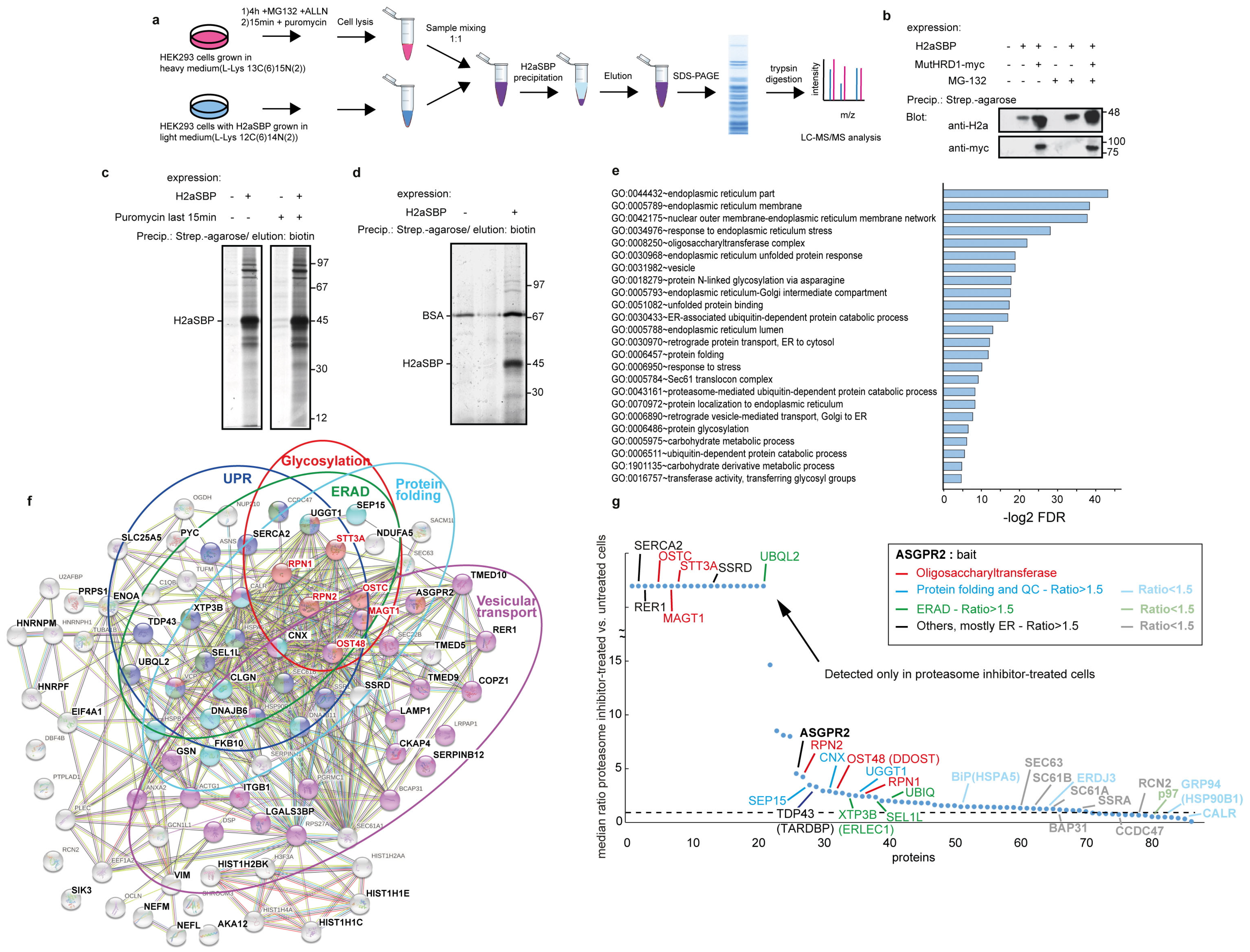
Figure 1. OST subunits are among top ERAD substrate interactors identified by a SILAC proteomics approach. (a) Experimental scheme of SILAC-based mass spectrometry analysis of HEK293 cells labeled with lysine containing heavy isotopes for 10 days or HEK293 stably expressing H2aSBP (SBP6 cell line) without heavy lysine (or vice versa). Cells were treated with proteasome inhibitors MG-132 (50 mM) and ALLN (50 mM) for 4 h and with puromycin (2 mM) for 15 min before lysis, or only with puromycin. Cells were solubilized in 1% digitonin in PBS and lysates from cells labeled with or without heavy lysine were mixed at a ratio of 1:1. Protein complexes pulled down with H2aSBP (using streptavidin-agarose beads) were eluted with 250 μM biotin, subjected to SDS-PAGE followed by trypsin digestion and LC-MS/MS analysis. (b) Effect of mutant HRD1 on H2aSBP. Dominant negative mutant myc-tagged hsHRD1 (mutHRD1-myc) was transfected into SBP6 cells. Two days post-transfection, these cells or control HEK293 cells were treated or not for 4 h with 20 μM MG132, lysed and subjected to precipitation with streptavidin beads. Biotin-eluted samples were subjected to 10% SDS-PAGE and immunoblotted with anti-H2a or anti-myc. MW markers in kDa are on the right. (c) Metabolically labeled proteins pulled down with H2aSBP. SBP6 cells or control HEK293 cells were labeled with [35S] Cys/Met mix for 5 h under proteasomal inhibition with 20 μM MG132 and treated or not with 2 mM puromycin for the last 15 min before lysis. Cells were lysed in digitonin buffer followed by precipitation with streptavidin beads, elution with biotin and SDS-PAGE with detection by fluorography. (d) Proteins pulled down with H2aSBP and silver stained. SBP6 cells or control HEK293 cells were treated for 4 h with 20 mM MG132 and 2 mM puromycin (last 15 min), lysed in digitonin buffer followed by precipitation with streptavidin beads (in the presence of 0.1% BSA), elution with biotin, SDS-PAGE and silver staining. (e) Gene ontology (GO) analysis of differentially expressed genes from the SILAC results of cells with or without proteasomal inhibition (ratio > 1.5). -log2 enriched p-values are shown for selected top entries with the lowest false discovery rates. (see also Supplementary Table S2). (f) STRING interaction map of proteins identified in the SILAC analysis, highlighting those with >1.5 ratio when comparing cells treated with or without proteasomal inhibitors. OST protein subunits are in red, other enriched functions are in colored circles. (g) Ratio of levels of proteins identified in cells treated with or without proteasomal inhibitors. The proteins on the left (up to UBQL2) were detected only in proteasomal inhibitor-treated cells. The dotted line is a value of 1, meaning equal presence in cells treated with or without proteasomal inhibitors. (see also Supplementary Tables S3 and S4).
Cells 14 01593 g001
Figure 2. OST subunits accumulate in the ERQC upon proteasomal inhibition separately from ribosomes and interact increasingly with an ERAD substrate. (a,b) Comigration of substantial populations of endogenous STT3A (a) and DAD1 and Tusc3 (b) with the ERAD substrate at the ERQC and separate from the rough ER (RER) in density gradients. HEK293 cells transiently transfected with H2a-RFP were incubated without (left) or with Bz (2 mM) for 3 h. Cells were homogenized and postnuclear supernatants were centrifuged on 10–34% iodixanol gradients. Fractions of the gradients were run on SDS–PAGE and immunoblotted with the indicated antibodies. A non-specific band is indicated (*). The peaks of migration of Sec61β, of the RER/ribosomes marker RPL26 and of the ER chaperone CNX are indicated in the graphs. Representative experiments out of three repeat experiments are shown. (cf) GFP-DAD1 and Sec61β -GFP accumulate at the ERQC. NIH 3T3 cells were transiently cotransfected with the ERAD substrate H2a-RFP and with GFP-DAD1 or Sec61β-GFP and were incubated with or without proteasome inhibitor (17 mM Lac) for 3 h. (g,h) Ribosomes do not accumulate at the ERQC. NIH 3T3 cells transiently transfected with H2a-RFP were treated as in (cf) and were fixed and immunostained using a primary mouse anti-rRNA antibody and secondary goat anti-mouse IgG antibody conjugated to Cy2. (in) GFP-DAD1 colocalizes with OS-9 with or without proteasomal inhibition. S-tagged OS-9.1/2 was expressed in NIH 3T3 cells together with H2a-GFP or GFP-DAD1, with or without treatment with proteasome inhibitor (1.5 mM Bz) for 3 h, followed by fixation and immunostaining with mouse anti-S tag antibody and secondary goat anti-mouse IgG antibody conjugated to Dylight 549. The graphs are box plots with inclusive medians of three independent experiments (n = 30). p value H2a-GFP = 2.03 × 10⁻⁹. p value GFP-DAD1 = 0.73. (o) Immunoprecipitation with a mouse monoclonal (B9) anti-H2a of lysates of HEK293 cells expressing H2a together with GFP-DAD1, with or without treatment with 2 mM Bz for 3 h. Crosslinking with 1 mM DSP was performed before cell lysis. Immunoprecipitates and 10% of the total lysates (input) were immunoblotted with rabbit anti-H2a amino-terminal antibody and anti-GFP. Asterisk = non-specific band. The graph shows increased coimmunoprecipitation of GFP-DAD1 with H2a upon proteasomal inhibition. Values are relative to control (=100), normalized by total GFP-DAD1 and H2a, the average of four independent experiments ± SD. p value = 0.0003. p < 0.05 (*), p < 0.001 (***).
Cells 14 01593 g002
Figure 3. OST subunits promote degradation of a glycosylated ERAD substrate. (ac) Overexpression of GFP-DAD1 strongly reduces the levels of H2a, knockdown of DAD1 or Tusc3 increases H2a levels. Cell lysates were immunoblotted with anti-H2a amino-terminal antibody. The asterisks indicate a band of underglycosylated H2a. The graphs show the averages of three or four independent experiments ± SD. p value ± GFP-DAD1 = 2 × 10⁻⁵, ±shDAD1 = 0.03, ±shTusc3 = 0.0004. (df) Pulse-labeling for 20 min with [35S]cysteine and chase for the indicated times of cells expressing H2a with or without overexpression of GFP-DAD1 (d) and knockdown of Tusc3 (e) or treatment with Tun (5 mg/mL) for 18 h prior to the pulse and during the pulse and chase times (f). The vertical lines indicate removal of irrelevant lanes (cropped lanes included in Supplementary Figure S3). H2a degradation (percentage remaining relative to pulse) was accelerated by GFP-DAD1, inhibited by Tusc3 knockdown and not affected by blocking glycosylation (+Tun). The graphs show the averages of three independent experiments ± SD. p value ± GFP-DAD1 3 h = 0.05, 6 h = 0.05; ±shTusc3 3 h = 0.03; ±Tun 3 h = 0.04. p < 0.05 (*), p < 0.001 (***).
Cells 14 01593 g003
Figure 4. Participation of OST subunits to a lesser extent in the degradation of an unglycosylated ERAD substrate. (ac) Overexpression of GFP-DAD1 reduces the levels of H2aΔgly; knockdown of DAD1 does not affect its levels, whereas Tusc3 knockdown moderately increases H2aΔgly levels. Cell lysates were immunoblotted with anti-H2a amino-terminal antibody. The graphs show the averages of three or four independent experiments ± SD. p value ± GFP-DAD1 = 3 × 10⁻⁶, ±shTusc3 = 0.004. (d) Pulse/chase with [35S]cysteine of cells expressing H2aΔgly shows a moderate inhibition of its degradation by Tusc3 knockdown. The vertical line indicates removal of an irrelevant lane (cropped lane included in Supplementary Figure S3). The graph shows an average of three independent experiments ± SD. p value ± shTusc3 3 h = 0.01. p < 0.05 (*), p < 0.01 (**), p < 0.001 (***).
Cells 14 01593 g004
Figure 5. OST subunits affect levels of other ERAD substrates. (ac) Overexpression of GFP-DAD1 moderately reduces the levels of BACE476-HA; knockdown of DAD1 or Tusc3 cause a minor increase in BACE476-HA levels. Cell lysates were immunoblotted with anti-HA and anti-actin as loading control. (df) Overexpression of GFP-DAD1 causes a minor reduction in the levels of NHK-HA, knockdown of DAD1 or Tusc3 moderately increase NHK-HA levels. Cell lysates were immunoblotted with anti-HA and anti-b tubulin as loading control. The graphs show the averages of three independent experiments ± SD. p value for BACE476-HA: ±GFP-DAD1 = 0.008, ±shDAD1 = 0.01, ±shTusc3 = 0.003; for NHK-HA: ±GFP-DAD1 = 0.05, ±shDAD1 = 2 × 10⁻⁶, ±shTusc3 = 0.03. p < 0.05 (*), p < 0.01 (**), p < 0.001 (***).
Cells 14 01593 g005
Figure 6. Interaction of an OST subunit with HRD1 and promotion of retrotranslocation of an ERAD substrate. (a) Immunoprecipitation with anti-myc of lysates of HEK293 cells expressing HRD1-myc together with GFP-DAD1 and treated with or without 2 mM Bz for 4 h. Immunoprecipitates and 10% of the total lysates (input) were immunoblotted with anti-myc and anti-GFP. The graph shows reduced coimmunoprecipitation of GFP-DAD1 with HRD1-myc under proteasomal inhibition. Values are relative to control (=100), normalized by total GFP-DAD1 and HRD1-myc, given as an average of three independent experiments ±SD. p value = 0.009. (b) Scheme of addition of biotin (star) to H2a-BAP by an ER luminal biotin ligase, sec-BirA or after retrotranslocation by a cytosolic one, BirA. HEK293 cells transiently expressing H2a-BAP with or without BirA or Sec-BirA were incubated 24 h post-transfection with biotin (100 μM) for 30 min before lysis. Strep-HRP was used to detect biotinylated H2a-BAP and anti-V5 to detect total H2a-BAP. (c) Cells expressing H2a-BAP together with BirA and GFP-DAD1 overexpression or knockdown of DAD1 or Tusc3 were incubated with biotin for 3 h. Cell lysates were reacted with Strep-HRP or with anti-H2a antibody. Retrotranslocated (biotinylated) H2a-BAP increased almost two-fold under GFP-DAD1 overexpression and was reduced to half under Tusc3 or DAD1 knockdown. Values are averages of four independent experiments ± SD, relative to total H2a-BAP and standardized by the control (=100). p value + shTusc3 vs. control = 0.02, +GFP-DAD1 vs. control = 0.04, +shDAD1 vs. control = 0.02. p < 0.05 (*), p < 0.01 (**).
Cells 14 01593 g006
Figure 7. Membrane thinning by OST complexes. (a,b) 3D structures of human oligosaccharyltransferase complexes OST-A and OST-B determined by cryo-EM at 3.5 Å resolution, indicating with different colors the observed subunits and illustrating the topology in the ER in relation to the transmembrane domain (TMD). From the RCSB database (6S7O and 6S7T). (c,d) Side views of OST-A and OST-B with membrane boundaries from simulations by molecular dynamics pre-rendered at the MemProtMD database. Distortions of the lipid bilayer indicate considerable membrane thinning by the OST transmembrane domains, highlighted by red lipid phosphate head groups, whereas blue illustrates thickening. (e) Model illustrating the interaction of OST with translocon components during forward translocation and its participation in N-glycosylation. OST also interacts with HRD1 and participates in retrotranslocation and targeting to ERAD.
Cells 14 01593 g007
Disclaimer/Publisher’s Note: The statements, opinions and data contained in all publications are solely those of the individual author(s) and contributor(s) and not of MDPI and/or the editor(s). MDPI and/or the editor(s) disclaim responsibility for any injury to people or property resulting from any ideas, methods, instructions or products referred to in the content.