
PKD1L1 Is Involved in Congenital Chylothorax

 , , ,
, , , 
Abstract
1. Introduction
2. Materials and Methods
2.1. Exome Analysis in Five CCT Families
2.2. Pathogenicity Prediction Using SIFT, PolyPhen-2, and MutPred
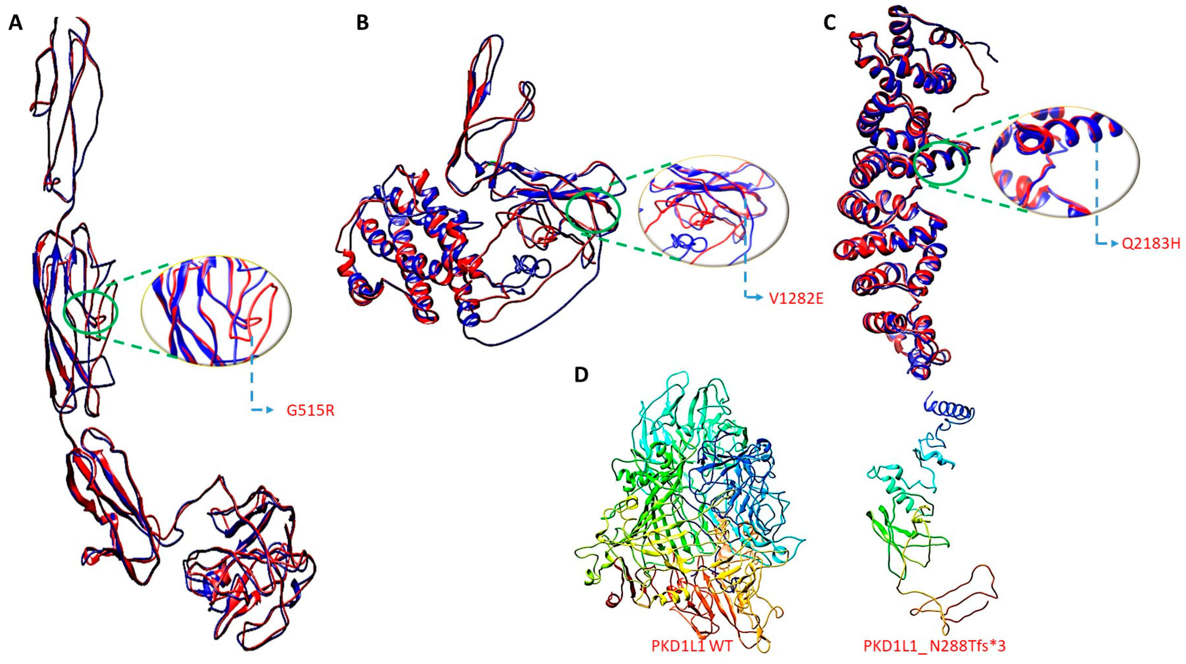
2.3. Three-Dimensional Structure Generation, Energy Minimization, and Superimposition
2.4. Mouse Husbandry
2.5. Mouse Genotyping
2.6. Mouse Embryo Phenotyping
2.7. Analysis of Lymphatic and Blood Vessels
2.8. MicroCT
2.9. Plasmid Construction
2.10. Antibody Production
2.11. Virion Production
2.12. Cell Culture and PKD1L1 Protein Localization
2.13. Individuals in the CCT Cohort
3. Results
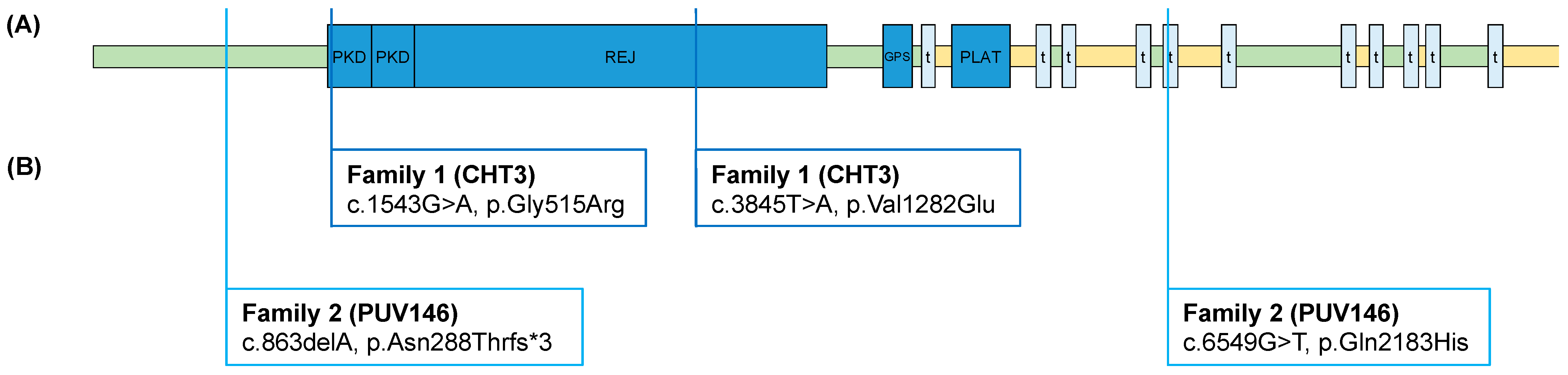
3.1. Putative Disease Variants Identified by Exome Survey
3.2. ES Results in Family 1
3.3. ES Results in Family 2
3.4. Cellular Localization of Identified PKD1L1 Variants
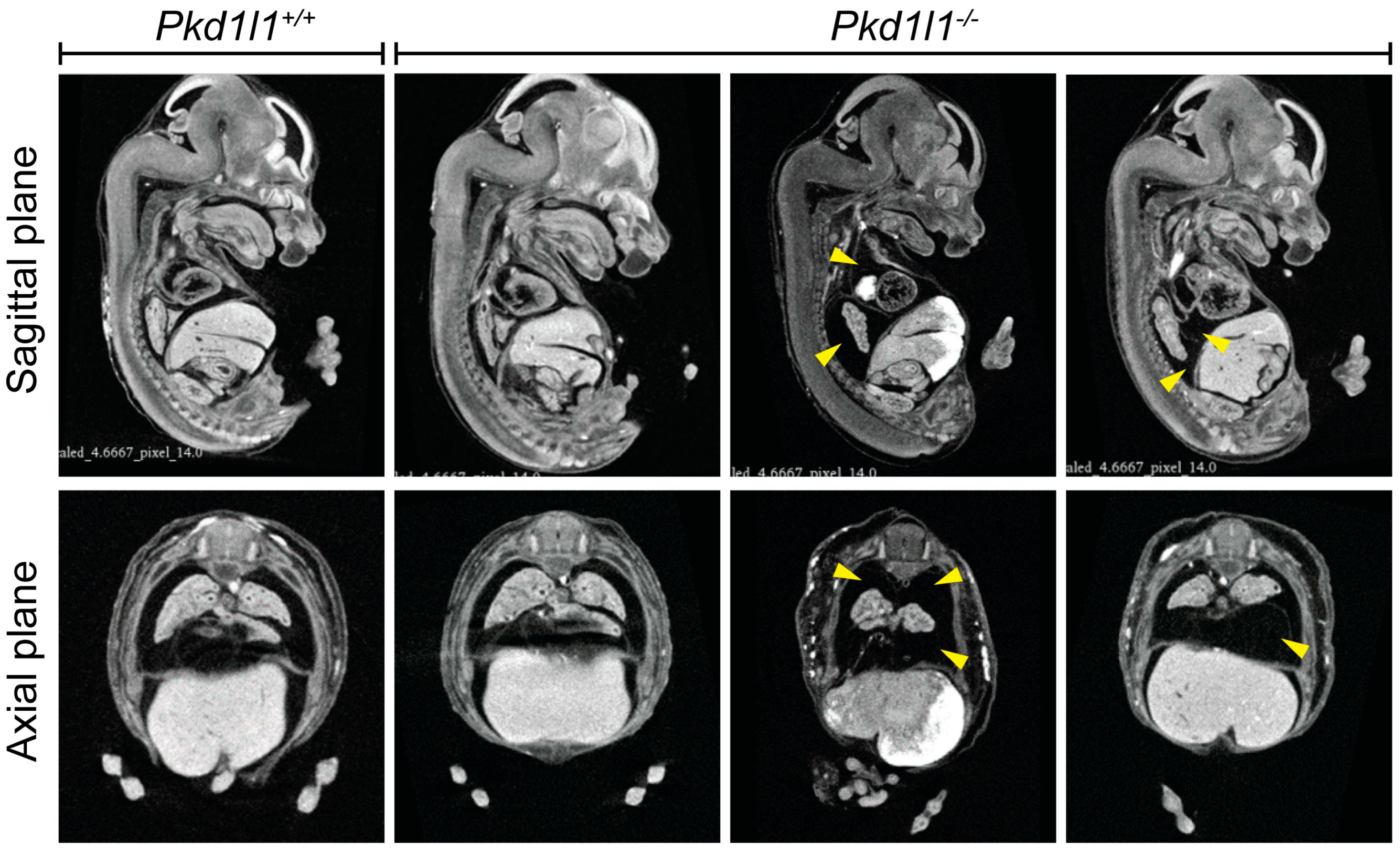
3.5. Incidence of Edema in Pkd1l1−/− Mouse Embryos
3.6. Loss of Pkd1l1 Can Result in Abnormal Lymphatic System Development


3.7. Observation of Pleural Effusion in Pkd1l1−/− Embryos
4. Discussion
5. Conclusions
Supplementary Materials
Author Contributions
Funding
Institutional Review Board Statement
Informed Consent Statement
Data Availability Statement
Acknowledgments
Conflicts of Interest
References
- Tutor, J.D. Chylothorax in Infants and Children. Pediatrics 2014, 133, 722–733. [Google Scholar] [CrossRef] [PubMed]
- Bengtsson, B.-O.S. Neonatal Lymphatic (Chylous) Disorders. NeoReviews 2013, 14, e600–e612. [Google Scholar] [CrossRef]
- Bialkowski, A.; Poets, C.F.; Franz, A.R.; the Erhebungseinheit für seltene pädiatrische Erkrankungen in Deutschland Study Group. Congenital chylothorax: A prospective nationwide epidemiological study in Germany. Arch. Dis. Child.-Fetal Neonatal Ed. 2014, 100, F169–F172. [Google Scholar] [CrossRef] [PubMed]
- Rocha, G.; Arnet, V.; Soares, P.; Gomes, A.C.; Costa, S.; Guerra, P.; Casanova, J.; Azevedo, I. Chylothorax in the neonate—A stepwise approach algorithm. Pediatr. Pulmonol. 2021, 56, 3093–3105. [Google Scholar] [CrossRef]
- Liviskie, C.J.; Brennan, C.C.; McPherson, C.C.; Vesoulis, Z.A. Propranolol for the Treatment of Lymphatic Malformations in a Neonate—A Case Report and Review of Literature. J. Pediatr. Pharmacol. Ther. 2020, 25, 155–162. [Google Scholar] [CrossRef] [PubMed]
- Swetman, G.L.; Berk, D.R.; Vasanawala, S.S.; Feinstein, J.A.; Lane, A.T.; Bruckner, A.L. Sildenafil for Severe Lymphatic Malformations. N. Engl. J. Med. 2012, 366, 384–386. [Google Scholar] [CrossRef]
- Hammill, A.M.; Wentzel, M.; Gupta, A.; Nelson, S.; Lucky, A.; Elluru, R.; Dasgupta, R.; Azizkhan, R.G.; Adams, D.M. Sirolimus for the treatment of complicated vascular anomalies in children. Pediatr. Blood Cancer 2011, 57, 1018–1024. [Google Scholar] [CrossRef]
- Krishnamurthy, M.B.; Malhotra, A. Congenital chylothorax: Current perspectives and trends. Res. Rep. Neonatol. 2017, 7, 53–63. [Google Scholar] [CrossRef]
- Mendola, A.; Schlögel, M.; Ghalamkarpour, A.; Irrthum, A.; Nguyen, H.; Fastré, E.; Bygum, A.; van der Vleuten, C.; Fagerberg, C.; Baselga, E.; et al. Mutations in the VEGFR3 Signaling Pathway Explain 36% of Familial Lymphedema. Mol. Syndr. 2013, 4, 257–266. [Google Scholar] [CrossRef]
- Dori, Y.; Smith, C.; Pinto, E.; Snyder, K.; March, M.E.; Hakonarson, H.; Belasco, J. Severe Lymphatic Disorder Resolved With MEK Inhibition in a Patient With Noonan Syndrome and SOS1 Mutation. Pediatrics 2020, 146, e20200167. [Google Scholar] [CrossRef]
- Pavel, M.A.; Lv, C.; Ng, C.; Yang, L.; Kashyap, P.; Lam, C.; Valentino, V.; Fung, H.Y.; Campbell, T.; Møller, S.G.; et al. Function and regulation of TRPP2 ion channel revealed by a gain-of-function mutant. Proc. Natl. Acad. Sci. USA 2016, 113, E2363–E2372. [Google Scholar] [CrossRef]
- DeCaen, P.G.; Delling, M.; Vien, T.N.; Clapham, D.E. Direct recording and molecular identification of the calcium channel of primary cilia. Nature 2013, 504, 315–318. [Google Scholar] [CrossRef] [PubMed]
- Vetrini, F.; D’alessandro, L.C.; Akdemir, Z.C.; Braxton, A.; Azamian, M.S.; Eldomery, M.K.; Miller, K.; Kois, C.; Sack, V.; Shur, N.; et al. Bi-allelic Mutations in PKD1L1 Are Associated with Laterality Defects in Humans. Am. J. Hum. Genet. 2016, 99, 886–893. [Google Scholar] [CrossRef] [PubMed]
- Field, S.; Riley, K.-L.; Grimes, D.T.; Hilton, H.; Simon, M.; Powles-Glover, N.; Siggers, P.; Bogani, D.; Greenfield, A.; Norris, D.P. Pkd1l1 establishes left-right asymmetry and physically interacts with Pkd2. Development 2011, 138, 1131–1142. [Google Scholar] [CrossRef] [PubMed]
- Tanaka, Y.; Morozumi, A.; Hirokawa, N. Nodal flow transfers polycystin to determine mouse left-right asymmetry. Dev. Cell 2023, 58, 1447–1461.e6. [Google Scholar] [CrossRef] [PubMed]
- Nauli, S.M.; Zhou, J. Polycystins and mechanosensation in renal and nodal cilia. BioEssays 2004, 26, 844–856. [Google Scholar] [CrossRef]
- Nauli, S.M.; Alenghat, F.J.; Luo, Y.; Williams, E.; Vassilev, P.; Li, X.; Elia, A.E.H.; Lu, W.; Brown, E.M.; Quinn, S.J.; et al. Polycystins 1 and 2 mediate mechanosensation in the primary cilium of kidney cells. Nat. Genet. 2003, 33, 129–137. [Google Scholar] [CrossRef]
- Pennekamp, P.; Karcher, C.; Fischer, A.; Schweickert, A.; Skryabin, B.; Horst, J.; Blum, M.; Dworniczak, B. The Ion Channel Polycystin-2 Is Required for Left-Right Axis Determination in Mice. Curr. Biol. 2002, 12, 938–943. [Google Scholar] [CrossRef]
- Karcher, C.; Fischer, A.; Schweickert, A.; Bitzer, E.; Horie, S.; Witzgall, R.; Blum, M. Lack of a laterality phenotype in Pkd1 knock-out embryos correlates with absence of polycystin-1 in nodal cilia. Differentiation 2005, 73, 425–432. [Google Scholar] [CrossRef]
- Kim, K.; Drummond, I.; Ibraghimov-Beskrovnaya, O.; Klinger, K.; Arnaout, M.A. Polycystin 1 is required for the structural integrity of blood vessels. Proc. Natl. Acad. Sci.USA 2000, 97, 1731–1736. [Google Scholar] [CrossRef]
- Wu, G.; Markowitz, G.S.; Li, L.; D’Agati, V.D.; Factor, S.M.; Geng, L.; Tibara, S.; Tuchman, J.; Cai, Y.; Park, J.H.; et al. Cardiac defects and renal failure in mice with targeted mutations in Pkd2. Nat. Genet. 2000, 24, 75–78. [Google Scholar] [CrossRef] [PubMed]
- Garcia-Gonzalez, M.A.; Outeda, P.; Zhou, Q.; Zhou, F.; Menezes, L.F.; Qian, F.; Huso, D.L.; Germino, G.G.; Piontek, K.B.; Watnick, T. Pkd1 and Pkd2 Are Required for Normal Placental Development. PLoS ONE 2010, 5, e12821. [Google Scholar] [CrossRef]
- Tallquist, M.D.; Soriano, P. Epiblast-restricted Cre expression in MORE mice: A tool to distinguish embryonic vs. extra-embryonic gene function. Genesis 2000, 26, 113–115. [Google Scholar] [CrossRef]
- Piontek, K.B.; Huso, D.L.; Grinberg, A.; Liu, L.; Bedja, D.; Zhao, H.; Gabrielson, K.; Qian, F.; Mei, C.; Westphal, H.; et al. A Functional Floxed Allele of Pkd1 that Can Be Conditionally Inactivated In Vivo. J. Am. Soc. Nephrol. 2004, 15, 3035–3043. [Google Scholar] [CrossRef] [PubMed]
- Dworschak, G.C.; Punetha, J.; Kalanithy, J.C.; Mingardo, E.; Erdem, H.B.; Akdemir, Z.C.; Karaca, E.; Mitani, T.; Marafi, D.; Fatih, J.M.; et al. Biallelic and monoallelic variants in PLXNA1 are implicated in a novel neurodevelopmental disorder with variable cerebral and eye anomalies. Anesth. Analg. 2021, 23, 1715–1725. [Google Scholar] [CrossRef] [PubMed]
- Ng, P.C.; Henikoff, S. SIFT: Predicting amino acid changes that affect protein function. Nucleic Acids Res. 2003, 31, 3812–3814. [Google Scholar] [CrossRef]
- Kumar, P.; Henikoff, S.; Ng, P.C. Predicting the effects of coding non-synonymous variants on protein function using the SIFT algorithm. Nat. Protoc. 2009, 4, 1073–1081. [Google Scholar] [CrossRef]
- Ramensky, V.; Bork, P.; Sunyaev, S. Human non-synonymous SNPs: Server and survey. Nucleic Acids Res. 2002, 30, 3894–3900. [Google Scholar] [CrossRef]
- Li, B.; Krishnan, V.G.; Mort, M.E.; Xin, F.; Kamati, K.K.; Cooper, D.N.; Mooney, S.D.; Radivojac, P. Automated inference of molecular mechanisms of disease from amino acid substitutions. Bioinformatics 2009, 25, 2744–2750. [Google Scholar] [CrossRef]
- Roy, A.; Kucukural, A.; Zhang, Y. I-TASSER: A unified platform for automated protein structure and function prediction. Nat. Protoc. 2010, 5, 725–738. [Google Scholar] [CrossRef]
- Xu, D.; Zhang, Y. Improving the Physical Realism and Structural Accuracy of Protein Models by a Two-Step Atomic-Level Energy Minimization. Biophys. J. 2011, 101, 2525–2534. [Google Scholar] [CrossRef]
- Zhang, J.; Liang, Y.; Zhang, Y. Atomic-Level Protein Structure Refinement Using Fragment-Guided Molecular Dynamics Conformation Sampling. Structure 2011, 19, 1784–1795. [Google Scholar] [CrossRef] [PubMed]
- Maiti, R.; Van Domselaar, G.H.; Zhang, H.; Wishart, D.S. SuperPose: A simple server for sophisticated structural superposition. Nucleic Acids Res. 2004, 32, W590–W594. [Google Scholar] [CrossRef] [PubMed]
- Finn, R.D.; Miller, B.L.; Clements, J.; Bateman, A. iPfam: A database of protein family and domain interactions found in the Protein Data Bank. Nucleic Acids Res. 2013, 42, D364–D373. [Google Scholar] [CrossRef] [PubMed]
- Zudaire, E.; Gambardella, L.; Kurcz, C.; Vermeren, S. A Computational Tool for Quantitative Analysis of Vascular Networks. PLoS ONE 2011, 6, e27385. [Google Scholar] [CrossRef] [PubMed]
- Grimes, D.T.; Keynton, J.L.; Buenavista, M.T.; Jin, X.; Patel, S.H.; Kyosuke, S.; Vibert, J.; Williams, D.J.; Hamada, H.; Hussain, R.; et al. Genetic Analysis Reveals a Hierarchy of Interactions between Polycystin-Encoding Genes and Genes Controlling Cilia Function during Left-Right Determination. PLOS Genet. 2016, 12, e1006070. [Google Scholar] [CrossRef]
- Ermakov, A.; Stevens, J.L.; Whitehill, E.; Robson, J.E.; Pieles, G.; Brooker, D.; Goggolidou, P.; Powles-Glover, N.; Hacker, T.; Young, S.R.; et al. Mouse mutagenesis identifies novel roles for left-right patterning genes in pulmonary, craniofacial, ocular, and limb development. Dev. Dyn. 2009, 238, 581–594. [Google Scholar] [CrossRef]
- Outeda, P.; Huso, D.L.; Fisher, S.A.; Halushka, M.K.; Kim, H.; Qian, F.; Germino, G.G.; Watnick, T. Polycystin Signaling Is Required for Directed Endothelial Cell Migration and Lymphatic Development. Cell Rep. 2014, 7, 634–644. [Google Scholar] [CrossRef]
- Human Protein Atlas Version 23.0. PKD2 RNA Expression Overview. Available online: Https://Www.Proteinatlas.Org/ENSG00000118762-PKD2/Subcellular (accessed on 12 October 2023).
- Arnaout, M.A. The vasculopathy of autosomal dominant polycystic kidney disease: Insights from animal models. Kidney Int. 2000, 58, 2599–2610. [Google Scholar] [CrossRef]


| Family 1 (CHT3) | Family 2 (PUV146) | ||
|---|---|---|---|
| Molecular Details | Individual | II-1 | II-1 |
| Zygosity | Compound Heterozygous | Compound Heterozygous | |
| Inheritance | Autosomal Recessive | Autosomal Recessive | |
| Variant location PKD1L1 (NM_138295.4) | c.1543G>A, p.Gly515Arg c.3845T>A, p.Val1282Glu | c.863delA, p.Asn288Thrfs*3 c.6549G>T, p.Gln2183His | |
| Variant consequence | Missense Missense | Frameshift Missense | |
| gDNA location Chr7 (NC_000007.13) | g.47944902C>T g.47913548A>T | g.47968998delT g.47869647C>A | |
| Exon | 11/57 24/57 | 7/57 43/57 | |
| gnomAD MAF | Not Reported 0.0003 (hom = 0) | Not Reported Not Reported | |
| Polyphen-2 | Probably Damaging Benign | N/A Benign | |
| SIFT | Tolerated Tolerated | N/A Tolerated | |
| Mutation Taster | Disease Causing Polymorphism | N/A Polymorphism | |
| CADD | 29.0 7.758 | N/A 23.6 | |
| ACMG Criteria | VUS (PM2, PP3) VUS (PM2) | Likely pathogenic (PVS1, PM2) VUS (PM2) | |
| Clinical Features | Sex | Female | Male |
| Age of Onset | Congenital | Congenital | |
| Primary Phenotype | Chylothorax, left Hydrops fetalis | Hydrothorax, bilateral Hydrops fetalis | |
| Secondary Phenotype | Persistent pulmonary hypertension, respiratory failure | Severe pulmonary hypoplasia, persistent pulmonary hypertension, cardio-respiratory failure | |
| Prenatal intervention | 3x shunt insertion, thoracocentesis | 3x shunt insertion |
| Mutant | SIFT | POLYPHEN-2 | MutPred | Overall Functional Impact | |||
|---|---|---|---|---|---|---|---|
| Prediction | Score | Prediction | Score | Prediction | Score | ||
| p.Gly515Arg | Deleterious | 0.02 | Probably damaging | 0.95 | Severe Functional Impact | 0.626 | Pathogenic |
| p.Val1282Glu | Neutral | 0 | Benign | 0.017 | Less Impact | 0.482 | Less pathogenic |
| p.Gln2183His | Neutral | 0 | Benign | 0.15 | Less Impact | 0.343 | Less pathogenic |
| p.Asn288Thrfs*3 | - | - | - | - | - | - | Pathogenic |
| Pfam Motifs | AA Position | Description | Independent E-Value | WT | Gly515Arg | Val1282Glu | Gln2183His | Asn288Thrfs*3 |
|---|---|---|---|---|---|---|---|---|
| REJ | 713–933 | PF02010, REJ domain | 3.70 × 10⁻¹⁸ | benign | benign | benign | benign | pathogenic |
| 1139–1304 | 1.20 × 10⁻¹⁶ | benign | pathogenic | benign | benign | pathogenic | ||
| PKD | 513–578 | PF00801, PKD domain | 6.90 × 10⁻⁶ | benign | pathogenic | benign | benign | pathogenic |
| 600–662 | 1.60 × 10⁻¹¹ | benign | benign | benign | benign | pathogenic | ||
| Polycystin_ dom | 2442–2512 | PF20519, Polycystin domain | 1.40 × 10⁻¹⁵ | benign | benign | benign | benign | pathogenic |
| PLAT | 1798–1904 | PF01477, PLAT/LH2 domain | 5.20 × 10⁻¹⁴ | benign | benign | benign | benign | pathogenic |
| PKD_4 | 618–661 | PF18911, PKD domain | 4.00 × 10⁻⁶ | benign | benign | benign | benign | pathogenic |
| PKD_channel | 2518–2662 | PF08016, Polycystin cation channel | 1.20 × 10⁻⁵ | benign | benign | benign | benign | pathogenic |
| Glyco_trans _2_3 | 2584–2676 | PF13632, Glycosyl transferase family group 2 | 0.013 | benign | benign | benign | benign | pathogenic |
| Ion_trans | 2528–2738 | PF00520, Ion transport protein | 0.11 | benign | benign | benign | benign | pathogenic |
| Structural Modeling | Alignment | Best Model Score | ||||
|---|---|---|---|---|---|---|
| PDB Matched | Coverage (%) | Normalized Z-Score | C-Score | Estimated TM-Score | Estimated RMSD (Å) | |
| PKD1L1_Gly0515_WT | 4m00A | 96 | 1.05 | −0.97 | 0.59 ± 0.14 | 09.1 ± 4.6 |
| PKD1L1_Arg0515_MT | 4m00A | 96 | 1.05 | −0.94 | 0.60 ± 0.14 | 09.1 ± 4.6 |
| PKD1L1_Val1282_WT | 7z01A | 88 | 1.27 | −2.60 | 0.41 ± 0.14 | 12.8 ± 4.2 |
| PKD1L1_Glu1282_MT | 7z01A | 86 | 1.28 | −3.00 | 0.37 ± 0.13 | 13.9 ± 3.9 |
| PKD1L1_Gln2183_WT | 6a70 | 73 | 6.40 | −0.71 | 0.62 ± 0.14 | 08.2 ± 4.5 |
| PKD1L1_His2183_MT | 6a70 | 74 | 4.66 | −0.89 | 0.60 ± 0.14 | 08.6 ± 4.5 |
| PKD1L1_Asn288Tfs*3 | −0.97 | 0.59 ± 0.14 | 9.1 ± 4.6 | |||
| Superimposing | Local RMSD (Å) | Global RMSD (Å) | |||
|---|---|---|---|---|---|
| Alpha-C | Backbone | Alpha-C | Backbone | ||
| PKD1L1_Gly515_WT | PKD1L1_Arg515_MT | 0.82 | 1.11 | 5.97 | 5.96 |
| PKD1L1_Val1282_WT | PKD1L1_Glu1282_MT | 0.71 | 0.86 | 9.18 | 9.23 |
| PKD1L1_Gln2183_WT | PKD1L1_His2183_MT | 2.82 | 2.84 | 2.82 | 2.84 |
Disclaimer/Publisher’s Note: The statements, opinions and data contained in all publications are solely those of the individual author(s) and contributor(s) and not of MDPI and/or the editor(s). MDPI and/or the editor(s) disclaim responsibility for any injury to people or property resulting from any ideas, methods, instructions or products referred to in the content. |
© 2024 by the authors. Licensee MDPI, Basel, Switzerland. This article is an open access article distributed under the terms and conditions of the Creative Commons Attribution (CC BY) license (https://creativecommons.org/licenses/by/4.0/).
Share and Cite
Whitchurch, J.B.; Schneider, S.; Hilger, A.C.; Köllges, R.; Stegmann, J.D.; Waffenschmidt, L.; Dyer, L.; Thiele, H.; Dhabhai, B.; Dakal, T.C.; et al. PKD1L1 Is Involved in Congenital Chylothorax. Cells 2024, 13, 149. https://doi.org/10.3390/cells13020149
Whitchurch JB, Schneider S, Hilger AC, Köllges R, Stegmann JD, Waffenschmidt L, Dyer L, Thiele H, Dhabhai B, Dakal TC, et al. PKD1L1 Is Involved in Congenital Chylothorax. Cells. 2024; 13(2):149. https://doi.org/10.3390/cells13020149
Chicago/Turabian StyleWhitchurch, Jonathan B., Sophia Schneider, Alina C. Hilger, Ricarda Köllges, Jil D. Stegmann, Lea Waffenschmidt, Laura Dyer, Holger Thiele, Bhanupriya Dhabhai, Tikam Chand Dakal, and et al. 2024. "PKD1L1 Is Involved in Congenital Chylothorax" Cells 13, no. 2: 149. https://doi.org/10.3390/cells13020149
APA StyleWhitchurch, J. B., Schneider, S., Hilger, A. C., Köllges, R., Stegmann, J. D., Waffenschmidt, L., Dyer, L., Thiele, H., Dhabhai, B., Dakal, T. C., Müller, A., Norris, D. P., & Reutter, H. M. (2024). PKD1L1 Is Involved in Congenital Chylothorax. Cells, 13(2), 149. https://doi.org/10.3390/cells13020149