Review

Prime Editing and DNA Repair System: Balancing Efficiency with Safety

by
Karim Daliri
1,2,*,
Jürgen Hescheler
1 and
Kurt Paul Pfannkuche
1,2,3
1
Institute for Neurophysiology, Centre for Physiology and Pathophysiology, Medical Faculty and University Hospital of Cologne, University of Cologne, 50931 Cologne, Germany
2
Marga and Walter Boll-Laboratory for Cardiac Tissue Engineering, University of Cologne, 50931 Cologne, Germany
3
Center for Molecular Medicine Cologne (CMMC), University of Cologne, 50931 Cologne, Germany
*
Author to whom correspondence should be addressed.
Cells 2024, 13(10), 858; https://doi.org/10.3390/cells13100858
Submission received: 24 March 2024 / Revised: 24 April 2024 / Accepted: 12 May 2024 / Published: 17 May 2024

Abstract

:
Prime editing (PE), a recent progression in CRISPR-based technologies, holds promise for precise genome editing without the risks associated with double-strand breaks. It can introduce a wide range of changes, including single-nucleotide variants, insertions, and small deletions. Despite these advancements, there is a need for further optimization to overcome certain limitations to increase efficiency. One such approach to enhance PE efficiency involves the inhibition of the DNA mismatch repair (MMR) system, specifically MLH1. The rationale behind this approach lies in the MMR system’s role in correcting mismatched nucleotides during DNA replication. Inhibiting this repair pathway creates a window of opportunity for the PE machinery to incorporate the desired edits before permanent DNA repair actions. However, as the MMR system plays a crucial role in various cellular processes, it is important to consider the potential risks associated with manipulating this system. The new versions of PE with enhanced efficiency while blocking MLH1 are called PE4 and PE5. Here, we explore the potential risks associated with manipulating the MMR system. We pay special attention to the possible implications for human health, particularly the development of cancer.

1. Introduction

The RNA-guided CRISPR (Clustered Regularly Interspaced Short Palindromic Repeats) endonuclease system was first identified in Escherichia coli (E. coli) in 1987, with its unique genomic structure providing evidence for its function. This structure evolved as an adaptive immune system employed by bacteria and archaea. It utilizes a set of CRISPR-associated (Cas) genes to incorporate exogenous material into the CRISPR locus. This incorporated material is then transcribed into RNA templates that guide the targeted destruction of mobile elements at the DNA or RNA level [1]. To date, three types of CRISPR systems have been identified, each with distinct mechanisms of action. Unlike the Type II CRISPR/Cas system, which uses a single endonuclease called Cas9 to identify and cleave target DNA, Type I and III systems employ a group of Cas genes to perform RNA processing, target recognition, and cleavage [1,2,3]. In the Type II system, a pair of non-coding RNAs guide the Cas9 endonuclease to its target DNA sequence. These RNAs include a CRISPR RNA (crRNA) and an auxiliary transactivating crRNA (tracrRNA). The crRNA contains two key components which are a 20-nucleotide guide sequence (often called spacer), which determines target specificity via Watson-Crick base-pairing with the target DNA and an invariant “direct repeat” component, which pairs with the “antirepeat” segment of the tracrRNA to form an RNA duplex. The paired repeat segments of both RNAs attract Cas9 to the complex [4,5].
CRISPR-Cas-based genome editing relies on four primary double-strand break (DSB) repair pathways in eukaryotic cells: non-homologous end joining (NHEJ), homologous recombination (HR), microhomology-mediated end joining (MMEJ), and single-strand annealing (SSA). Each pathway leads to different editing outcomes [6]. Therefore, researchers are actively developing strategies to control the pathway that cells use to repair DSBs during editing. However, the mechanisms governing DSB repair are intricately complex and remain incompletely understood [6,7]. A deeper understanding of these mechanisms promises fundamental insights into genome integrity and will pave the way for more sophisticated genome editing strategies.
Prime editors, as the latest generation of CRISPR-Cas9 -based technologies, can insert short DNA sequences without generating DSBs or requiring an external template. They consist of a nicking version of Cas9 fused to a reverse transcriptase (RT) domain, which is complexed with a prime editing guide RNA (pegRNA). The pegRNA consists of a primer-binding site homologous to the sequence in the target and a reverse transcriptase (RT) template, including the intended edit, all in the 3′ extension of a standard CRISPR-Cas9 guide RNA. At the target site, Cas9 nicks one strand of the DNA, which then anneals to the primer-binding site on the pegRNA, extending by the Cas9-fused reverse transcriptase (RT) using the pegRNA-encoded template sequence. Next, DNA repair mechanisms (miss match repair, MMR) resolve the conflicting sequences on the two DNA strands, ultimately writing the intended edit into the genome [8]. The prime editing (PE) process is complex, and the factors influencing its efficacy are not fully understood. It involves multiple somewhat independent stages, including three DNA binding events and successful MMR DNA repair, all necessary for successful editing. Each stage is potentially influenced by the edited sequence. Furthermore, the presence of a nick on the edited strand attracts the MMR machinery to that DNA strand, initiating the EXO1-mediated degradation of the mismatch generated by PE and restoring the original sequence. Consequently, compromising MMR protein function, inhibiting EXO1 activity, or blocking the MMR pathway can enhance prime editing efficiency [8,9].
The primitive PE system, referred to as PE1, was developed through the fusion of the Moloney murine leukemia virus (M-MLV) RT with the C-terminus of Cas9 (H840A) nickase. The pegRNA utilized in this system was an extension of the sgRNA, containing a PBS sequence and an RT template. PE1 demonstrated a maximum editing efficiency of 0.7–17% at various loci [8]. Building upon PE1, Liu et al. proposed enhancing prime editing efficacy by modifying the RT component (PE2) (Figure 1A). Compared to PE1, PE2 increased the efficiency of introducing point mutations by 1.6- to 5.1-fold on average, exhibited improved editing efficiency in indels, and was compatible with short PBS sequences, as evidenced by testing various RT mutations.Despite the enhanced efficacy of PE2, there remains a risk of edited insertions being excised due to DNA mismatch repair mechanisms targeting the edited strand. To mitigate this issue during DNA heteroduplex resolution, an additional single guide RNA (sgRNA) was introduced. This sgRNA was designed to complement the edited sequence introduced by the pegRNA while not targeting the original allele. It directed the Cas9 nickase component of the fusion protein to nick the unedited strand at a nearby location opposite to the original nick site. This improved prime editor was designated as PE3, exhibiting approximately three times the editing efficiency of PE2 but causing a higher rate of indels [8] (Figure 1C).
Motivated by the observation that pretreating cells with siRNAs targeting MMR can enhance PE editing efficiency, a novel strategy was proposed for the concurrent delivery of prime editors and MMR repressor units. This transient expression of the MMR repressor protein MLH1d in the PE2 and PE3 systems, now referred to as PE4 and PE5, respectively, significantly improved editing efficiency [9,10].
Chen et al. introduced PE4 (PE2 + MLH1 dominant negative, dn) (Figure 1B) and PE5 (PE3 + MLH1dn) (Figure 1D), increasing gene editing efficiency by transiently expressing an engineered MMR inhibitory protein. This resulted in improved efficiencies for substitutions, small insertions, and small deletions [10]. While optimizing the system is crucial, blocking MMR with MLH1dn at any stage presents significant risks, especially for translational applications, as discussed later. Notably, another group employed a similar approach to enhance PE efficiency through the absence of the MMR system [10].
Given their central role in diverse DNA transactions, MMR proteins can have severe consequences on various biological systems when inactivated. We will briefly summarize their key features in the following paragraphs.
The efficiency of various DNA repair processes, particularly the MMR system, should be a key consideration when designing PE experiments. A malfunction in this system can have wide-ranging consequences, both for individual cells and for the entire body. Notably, one major consequence is an increased level of point mutations across the genome due to unrepaired errors in DNA synthesis [11,12].
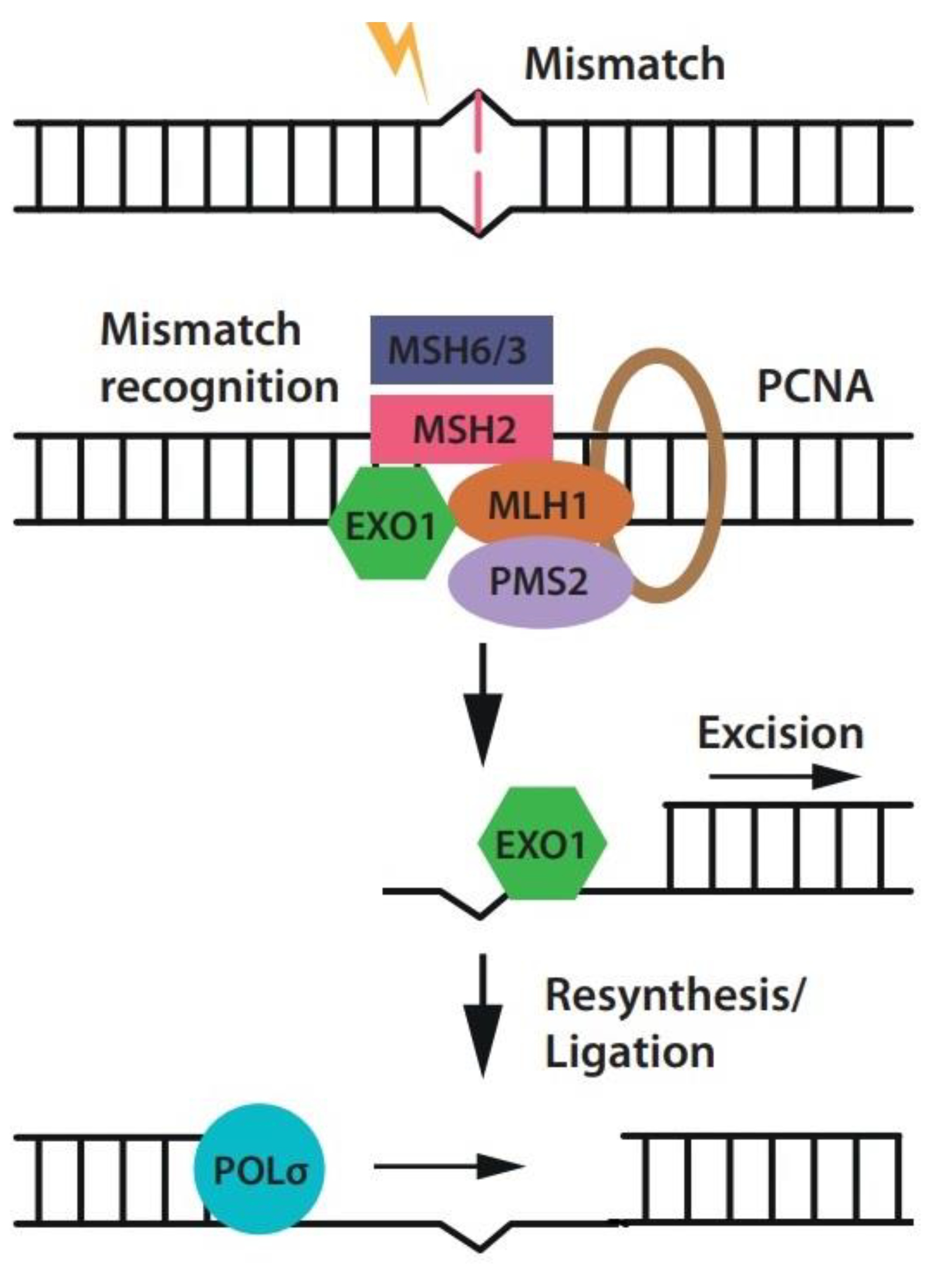
MLH1 is a key DNA mismatch repair (MMR) gene that plays a critical role in safeguarding genomic stability and preventing cancer development. MMR is a process that meticulously corrects errors arising during DNA replication (Figure 2). Uncorrected errors can lead to mutations that fuel cancer development. When mutations occur in MLH1 or other MMR genes, the entire MMR system becomes compromised, resulting in the accumulation of microsatellite instability (MSI) [13,14]. Microsatellites, consisting of repeated sequences of 1–6 base pairs, are abundant in higher organisms’ genomes and show significant genetic variation. Mutations in these regions typically involve the insertion or deletion of a few individual or repeated units, mirroring a pattern of gradual mutation accumulation. While the MMR system typically corrects most microsatellite mutations, cells lacking this system experience a drastic increase in mutation rate [15,16].

2. Potential Molecular and Cellular Risks Associated with MLH1 Disruption

2.1. Mitochondria

The critical role of mitochondria, ubiquitous organelles within eukaryotic cells, lies in their ability to generate cellular energy (adenosine triphosphate, ATP) via oxidative phosphorylation. Disruptions in mitochondrial function are increasingly recognized as contributing factors in various pathologies, including cancer [17,18]. A recent interesting finding suggests a previously unknown function for MLH1 within the mitochondria [19]. MLH1 exhibits robust mitochondrial localization, and its deficiency triggers synthetic lethality when combined with the inhibition of specific mitochondrial genes, such as POLG and PINK1. Moreover, MLH1 loss is associated with a decline in the oxygen consumption rate and reduced spare respiratory capacity [20,21].
MLH1, along with its partner proteins, plays a crucial role in maintaining mitochondrial function. Therefore, targeting these functions suggests a promising alternative therapeutic approach for MLH1-deficient diseases [22]. Interestingly, MLH1 frequently associates with POLG, a mitochondrial replication enzyme responsible for both safeguarding mitochondrial homeostasis and initiating apoptosis. Notably, disruptions in this interaction appear to be associated with the development of diabetic retinopathy, further highlighting the critical role of MLH1 in ensuring mitochondrial integrity [19,22,23].

2.2. Autophagy

Autophagy (“self-eating”) is a cellular recycling process that breaks down unnecessary or damaged components, like proteins and organelles, for degradation in lysosomes or vacuoles. Mutations in autophagy genes have been linked to various human disease [24].
MLH1, a key regulator of autophagy signaling, plays a crucial role in cellular survival. Studies have shown that MLH1 promotes autophagy, a self-preservation mechanism, while hindering apoptosis, a form of programmed cell death [25]. This allows cells to resist chemotherapy by activating the DNA mismatch repair (MMR) pathway through the mTOR/S6K1 signaling cascade [25]. Additionally, MLH1 is essential for autophagy activation in response to the chemotherapy drugs, acting alongside the p53 protein, Importantly, inhibiting MLH1 or the ATM-AMPK pathway diminishes autophagy, highlighting their significance in this process [26,27].
Despite long-standing beliefs that autophagy and DNA repair are distinct processes, it is increasingly apparent that they are closely related [27,28]. In other words, the cell ability to function efficiently and competently depends on mechanisms that regulate autophagy and DNA repair. The malfunction of either can result in dysregulation of the other, and vice versa, thus having adverse consequences for cells [29]. The MMR system triggers autophagy in tumor cells, which prevents chemotherapy-induced apoptosis if deficient. In addition, patients with DNA repair defects are thought to have problems with autophagy, leading to premature aging, developmental problems, and neurodegenerative diseases [27].

2.3. Variability in Cellular Responses to MMR Defects across Different Tissues

Although all human cells carry out common processes essential for survival, within their tissue environment, they also manifest unique functions contributing to their phenotype. These processes, both common and tissue-specific, are ultimately regulated by gene regulatory networks, which modulate gene expression and its extent [30]. Indeed, a single gene may exert different effects on cellular phenotype across different tissues.
According to Chen et al., Mlh1d-treated cells did not exhibit microsatellite instability (MSI) in the genome. However, clear evidence exists that tissue-specific haploinsufficiency of MLH1 causes MMR-associated cancer [31]. This explains why MMR-related cancers primarily affect the gastrointestinal tract. Therefore, tissue-specific investigations are crucial to analyze which organs or cells are at risk following MMR failure [32].
Additionally, MMR proteins have been shown to inhibit homologous recombination (recombination between similar or identical DNA sequences) [33]. In cells with MMR defects, recombination rates become dramatically higher, leading to gene conversions at the recombined sites, which can promote genome instability and tumorigenesis [34].

2.4. Folate

Folate, a key player in the methylation cycle which helps maintain healthy cells, appears to have a complex role in colorectal cancer. Low dietary folate intake has been linked to an increased risk of abnormal cell growth (neoplastic transformation) in the colon [35,36]. This may be due to its role as a methyl donor. One way folate deficiency can impact the colon is through methylation of the MLH1 gene promoter [37]. MLH1 is a critical component of the DNA repair system, and its inactivation through methylation has been implicated in the development of colon and lung cancers [38,39].
It is worth noting that the MMR system has been directly linked to folate-induced apoptosis [40]. Moreover, folate pathway genes might contribute to fertility complications in idiopathic (unexplained) infertility cases [41,42]. Notably, studies show that folate is the most common vitamin deficiency in general population diets [43]. Therefore, any alteration in the MMR system could potentially impact a large portion of the population, making this relevant for translational applications.

2.5. microRNA

MicroRNAs (miRNAs) are small, non-coding RNA molecules that regulate gene expression by binding to messenger RNA (mRNA) molecules and preventing them from being translated into proteins miRNAs are involved in various cellular processes, including metabolism, proliferation, cell cycle control, apoptosis, and autophagy [44]. Their dysregulation is linked to numerous human diseases, including diabetes, cancer, and cardiovascular diseases [45,46]. Moreover, making a connection between microRNA (miRNA) and MLH1 holds significant value. A study revealed an intriguing feedback loop between miRNAs and the MMR system. The MLH1-PMS2 heterodimer (MutLα) positively regulates both the processing of miR-422a and other miRNAs [44,47]. Further studies are needed to elucidate how specific miRNAs interact with the MMR system to answer key questions.

2.6. Wnt Signaling Pathway

The Wnt signaling pathway plays a crucial role in regulating cell growth, differentiation, and maintenance [48,49]. Alterations in genes within this pathway, including APC, are frequently observed in different pathological conditions. Notably, MLH1 can indirectly be involved in the Wnt signaling pathway by regulating the expression of other pathway components [50]. The MLH1 gene promoter harbors four specific binding sites for the transcription factor TCF7, a pivotal regulator of the Wnt signaling pathway, which regulates the expression of downstream target genes. When TCF7 binds to these sites, it triggers the transcriptional activation of the MLH1 gene [51]. More recently, a positive correlation between TCF7 and MLH1 expression was reported.

2.7. Interaction Networks

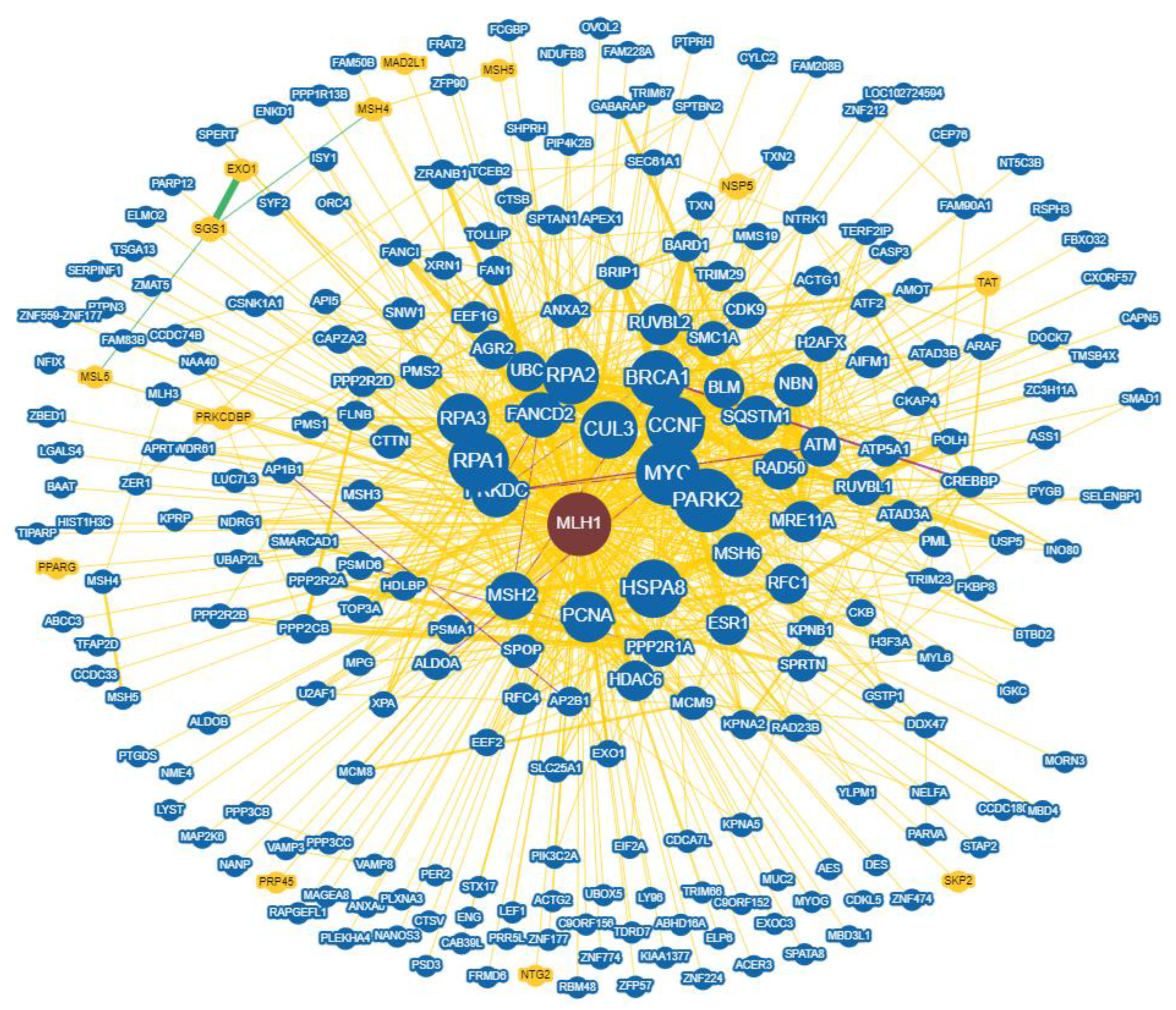
Another crucial perspective is considering gene interaction networks. These networks consist of genes linked by edges representing their functional relationships. These edges, called interactions, represent potential physical interactions between gene products, where one can alter or influence another. During genetic interactions, two gene variants collaborate to produce an effect that neither could achieve alone [52,53]. For MLH1, BioGRID, a systems biology database (Biological General Repository for Interaction Datasets), reports 416 interactions and 228 unique interactors [54]. This highlights the complex molecular interactions involving MLH1 within each cell, interactions that further change under different physiological and pathological conditions (Figure 3).

3. The Clinical Impact of Defective MLH1

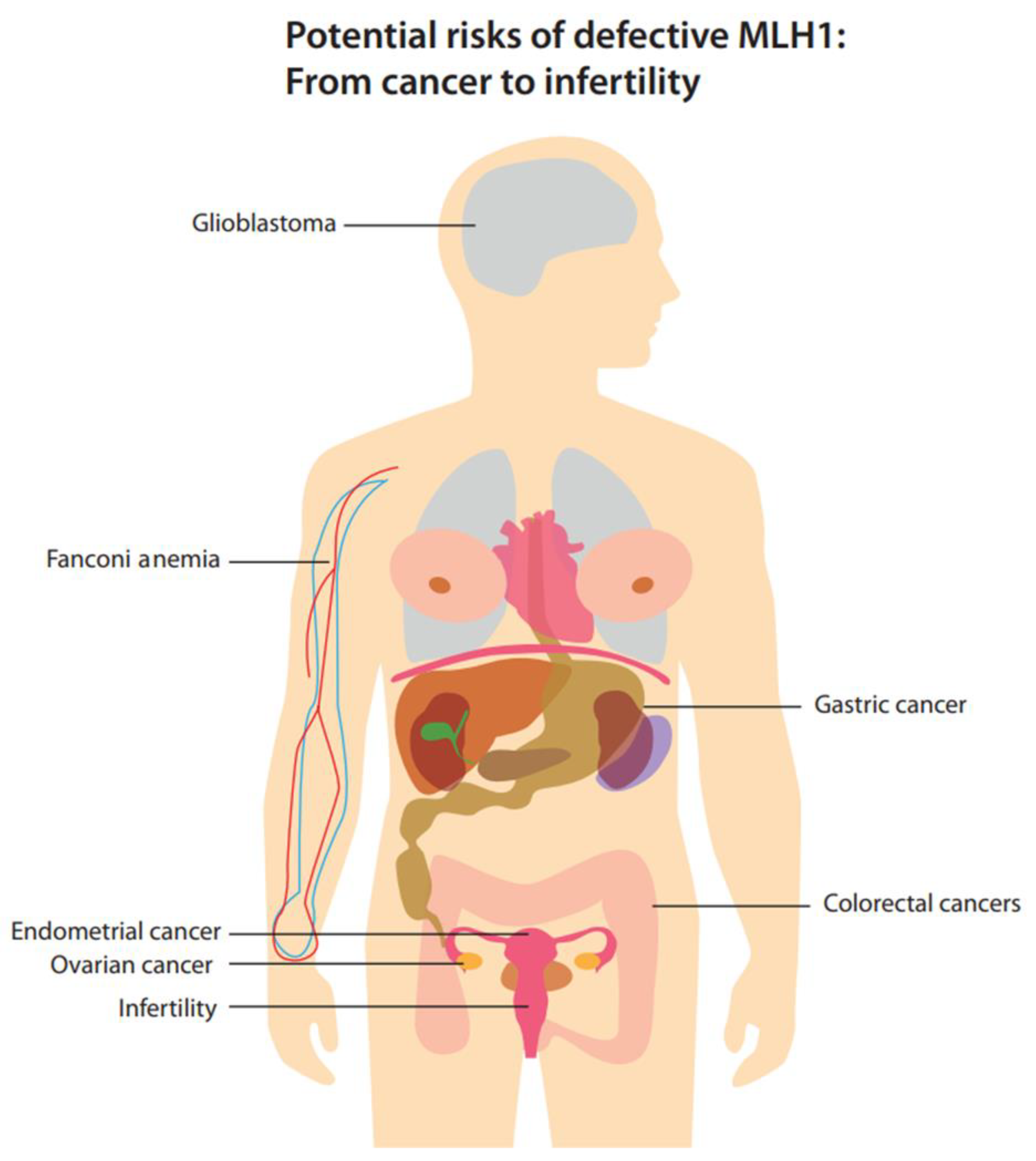
The integrity of the genome is critically safeguarded via the MMR system. Any malfunction within this system can lead to severe consequences, such as the development of cancer (Figure 4). Chen et al. found no signs of MSI in their cell lines. However, carcinogenesis is a multistage and long-term process, and oncogenic mutations (not necessarily MSI) in proto-oncogenes or tumor -suppressor genes can create malignant phenotypes several years after they are introduced [55]. By acting as lesion sensors, MMR proteins cause apoptosis and activate cell cycle checkpoints, contributing to tumorigenesis by allowing unchecked cell division [56].
In this section, we will discuss into the clinical outcomes of MMR deficiency, with a specific emphasis on MLH1.

3.1. Colorectal Cancer

Colorectal cancer (CRC) is considered the second leading cause of cancer-related mortalities, resulting from the accumulation of genetic alterations in oncogenes and tumor suppressor genes (TSGs) within the colorectal epithelium [57]. Two primary mechanisms of genomic instability have been identified in the progression of sporadic CRC. The first, called chromosomal instability, arises from a cascade of genetic changes that involve the activation of oncogenes, such as K-ras, and the inactivation of TSGs, including p53, DCC/Smad4, and APC [58,59,60].The second mechanism, referred to as microsatellite instability (MSI), originates from the inactivation of DNA mismatch repair (MMR) genes, such as MLH1 and/or MSH2, mostly via promoter hypermethylation (MLH1ph). This leads to mutations in genes containing coding microsatellites, such as transforming growth factor receptor II (TGF-RII) and BAX genes [38]. MLH1 plays a multifaceted role in CRC development [61]. MSI can accumulate mutations in other parts of the genome, including TSGs. These mutations contribute to the development of cancerous cells. Additionally, MSI allows cells to escape normal growth control mechanisms, enabling metastasis [58,62]. Furthermore, MMR deficiency may impair immune surveillance, allowing tumor cells to evade detection and eradication by the immune system [62].

3.2. Gastric Cancer

Gastric cancer (GC), a widespread form of cancer worldwide, is classified into two distinct histological subtypes: intestinal and diffuse, according to Lauren’s classification [63]. Intestinal-type gastric cancer is linked to various genetic alterations [64]. Its precursor, known as intestinal metaplasia, is marked by mutations in the p53 gene, reduced expression of retinoic acid receptor beta, and increased expression of telomerase reverse transcriptase [65,66].
Gastric adenomas, precancerous lesions that can progress to metastatic gastric cancer, also display genetic changes. These alterations include mutations in the APC gene, reduced expression of the p27 tumor suppressor protein, and amplification of the cyclin E oncohgene [67]. Furthermore, in more advanced GC, c-ErbB2 is often amplified and overexpressed, TGFBRI expression is decreased, and p27 expression is completely lost. While mutations in the hMLH1 gene are uncommon in gastric cancer, microsatellite instability (MSI) is more frequent than in colorectal cancer [68]. Here, MSI is primarily caused by hypermethylation of the MLH1 promoter; for example, 20% of primary GC patients have been reported to have hypermethylated MLH1 promoters [68,69].

3.3. Glioblastoma

Glioblastoma (GBM) is the most frequent and most malignant primary brain tumor in adults [70]. Growing evidence suggests DNA mismatch repair (MMR) gene expression may also be associated with the tumor response to alkylating agents. Notably, MLH1, MSH2, and MSH6 are considered intriguing candidate genes [71]. MMR deficiency, frequently observed in GBM, may contribute to therapy resistance and tumor recurrence. Notably, expression of MLH1 and PMS2 is reduced in recurrent GBM tumors compared to primary tumors [72,73].

3.4. Endometrial Cancer

Endometrial cancer (EC) is a prevalent gynecological malignancy, ranking fourth among women after breast, colorectal, and lung cancers [74].Two distinct types of endometrial carcinoma have been identified, exhibiting distinct pathological and clinical features. Type I carcinoma, linked to hyperestrogenism, commonly manifests as endometrial hyperplasia and is characterized by the frequent expression of estrogen and progesterone receptors, typically affecting younger women. In contrast, type II carcinoma, unrelated to estrogen, is associated with an atrophic endometrium, the infrequent expression of estrogen and progesterone receptors, and a higher prevalence in older women [75]. The distinct morphological features of these cancers are reflected in their molecular genetic profiles. Type I demonstrates defects in DNA repair mechanisms and mutations in PTEN, K-ras, and beta-catenin, while type II exhibits aneuploidy, p53 mutations, and amplification of the HER2/neu gene [76].
The underlying molecular dynamics of MMR-deficient/MSI-high endometrial cancers involve three primary classes: (MLH1ph), accounting for approximately 70–75% of cases; somatic mutations in MLH1, MSH2, MSH6, PMS2, and/or EPCAM occurring in 15–20% of cases; and germline mutations in these genes contributing 5–10% of patients [77]. A smaller percentage (around 3%) of ECs are associated with Lynch syndrome, which is inherited through mutations in one of the MMR genes [78]. It is worth mentioning that improved survival with an intact MMR system in EC has been reported by Cohn et al. [79].

3.5. Ovarian Cancer

Ovarian cancer (OC) is considered a primary contributor to mortality among gynecological cancers and ranks fifth as a cause of cancer-related mortalities in women [80]. Hereditary predisposition is identified as a risk factor for OC, with mutations in breast cancer susceptibility genes playing a role. Mutations in the MMR genes are the next most common cause of OCs [81].
This is further fueled by multiple studies reporting a prevalence of MLH1ph ranging from 10% to 50%, suggesting it’s a common feature in OC [82]. Additionally, research indicates a notable preference for MMR deficiency in endometrioid and serous carcinomas within OCs [83,84].
It should be mentioned that reported the safety of neoadjuvant pembrolizumab for patients with MMR-deficient solid tumors resulting in high rates of clinical outcome has drawn significant attention to the detection of MMR deficiency in tumors [85,86].

3.6. Fanconi Anemia

Fanconi anemia (FA) is a genetic syndrome that clinically affects several human systems. It results in progressive bone marrow failure and predisposes individuals to malignancies, particularly in the urogenital area and the head and neck [87].
In addition to the FANCJ-MLH1 interaction, several additional nodes link the FA and MMR pathways, suggesting that this crosstalk has functional importance. These interactions include BRCA1-MSH2, FANCD2-MLH1/MSH2, SLX4/FANCP-MSH2, and FAN1-MLH1. However, the full functional significance of these interactions remains unclear [88]. MSH2, a key protein in the MMR pathway, plays a critical role in the FA pathway by promoting the localization of FA core components to chromatin, and recruiting FANCJ to sites of DNA crosslinks [89]. Given the relationship between the FANCJ-MLH1 interaction and MSH2, a central question is whether other FA pathway components also function to balance the activity of MSH2 or other MMR proteins [90,91]. The ability of FA cells to cope with replication stress is linked to proteins in the MMR pathway. Furthermore, the MMR pathway plays a crucial role in the repair of DNA interstrand crosslinks (ICLs), toxic lesions that result in chromosomal instability and disrupt normal transcription, likely due to the direct interaction between FANCJ and MLH1 [92,93].

3.7. Fertility

From a fertility perspective, the MMR system plays a crucial role in many organisms by facilitating meiotic crossovers, which are essential for various processes during meiosis [94]. However, this involvement could lead to DNA rearrangements and an increased frequency of exchange between partially homologous sequences in germ cells, potentially causing infertility [94,95]. Spermatocytes deficient in MLH1 exhibit premature chromosome separation and arrest during the first meiotic division. Furthermore, human variants in MLH1 and MLH3 genes are associated with aneuploidy, pregnancy loss, and premature reproductive aging [95].

4. Conclusions and Perspectives

The genomic DNA of each cell is highly dynamic, undergoing an estimated 55,000 single-strand breaks and 25 double-strand breaks daily [96]. Therefore, even temporary inhibition of a multifunctional protein like MLH1 can have unpredictable consequences for individual cells and the organism. Indeed, the proposition that transient MLH1 inhibition enhances PE efficiency is intriguing but not overly surprising, as prolonged inhibition could result in detrimental effects. Of particular note, the discovery of novel roles for the MMR system is likely, adding a new layer of complexity to understanding how MMR interacts with other cellular components like DNA replication and transcription machinery. These issues all await further intensive studies in the MMR field, which could undoubtedly provide important insights into the system’s many other potential roles in DNA interactions.
There is a lack of literature concerning the consequences of the transient knockdown of MLH1 in various cell tissues, particularly in vivo, prior to any in vivo and clinical applications of PE4 and PE5. Universal molecular investigations, such as whole-genome sequencing and RNA sequencing, are suggested to observe any changes before and after MLH1 knockdown. Additionally, other experiments, such as apoptosis and autophagy evaluations, are recommended. Finally, long-term follow-ups in terms of clinical application is recommended to screen for possible side effects.
In summary, future experiments exploring the application of the new version of PE through MLH1 inhibition or targeting other members of the DNA repair system could lead to unpredictable and severe side effects in various cell types, potentially even including the development of cancers.

Author Contributions

K.D. wrote the manuscript, J.H. and K.P.P. provided financial support, and revised the manuscript. All authors have read and agreed to the published version of the manuscript.

Funding

This work was supported by the Hans and Gertie Fischer Foundation, 45133 Essen, Germany, and the Imhoff Foundation, 50678 Cologne, Germany.

Acknowledgments

This work was supported by the Hans and Gertie Fischer Foundation, Germany and the Imhoff Foundation, Germany.

Conflicts of Interest

The authors declare no conflict of interests.

References

  1. Koonin, E.V.; Makarova, K.S.; Zhang, F. Diversity, classification and evolution of CRISPR-Cas systems. Curr. Opin. Microbiol. 2017, 37, 67–78. [Google Scholar] [CrossRef] [PubMed]
  2. Makarova, K.S.; Wolf, Y.I.; Alkhnbashi, O.S.; Costa, F.; Shah, S.A.; Saunders, S.J.; Barrangou, R.; Brouns, S.J.J.; Charpentier, E.; Haft, D.H. An updated evolutionary classification of CRISPR–Cas systems. Nat. Rev. Microbiol. 2015, 13, 722–736. [Google Scholar] [CrossRef] [PubMed]
  3. Chylinski, K.; Makarova, K.S.; Charpentier, E.; Koonin, E. V Classification and evolution of type II CRISPR-Cas systems. Nucleic Acids Res. 2014, 42, 6091–6105. [Google Scholar] [CrossRef] [PubMed]
  4. Chylinski, K.; Le Rhun, A.; Charpentier, E. The tracrRNA and Cas9 families of type II CRISPR-Cas immunity systems. RNA Biol. 2013, 10, 726–737. [Google Scholar] [CrossRef] [PubMed]
  5. Lin, L.; He, X.; Zhao, T.; Gu, L.; Liu, Y.; Liu, X.; Liu, H.; Yang, F.; Tu, M.; Tang, L. Engineering the direct repeat sequence of crRNA for optimization of FnCpf1-mediated genome editing in human cells. Mol. Ther. 2018, 26, 2650–2657. [Google Scholar] [CrossRef] [PubMed]
  6. Xu, Y.; Xu, D. Repair pathway choice for double-strand breaks. Essays Biochem. 2020, 64, 765–777. [Google Scholar] [CrossRef] [PubMed]
  7. Karl, L.A.; Peritore, M.; Galanti, L.; Pfander, B. DNA double strand break repair and its control by nucleosome remodeling. Front. Genet. 2022, 12, 2833. [Google Scholar] [CrossRef] [PubMed]
  8. Anzalone, A.V.; Randolph, P.B.; Davis, J.R.; Sousa, A.A.; Koblan, L.W.; Levy, J.M.; Chen, P.J.; Wilson, C.; Newby, G.A.; Raguram, A. Search-and-replace genome editing without double-strand breaks or donor DNA. Nature 2019, 576, 149–157. [Google Scholar] [CrossRef]
  9. Ferreira da Silva, J.; Oliveira, G.P.; Arasa-Verge, E.A.; Kagiou, C.; Moretton, A.; Timelthaler, G.; Jiricny, J.; Loizou, J.I. Prime editing efficiency and fidelity are enhanced in the absence of mismatch repair. Nat. Commun. 2022, 13, 760. [Google Scholar] [CrossRef]
  10. Chen, P.J.; Hussmann, J.A.; Yan, J.; Knipping, F.; Ravisankar, P.; Chen, P.-F.; Chen, C.; Nelson, J.W.; Newby, G.A.; Sahin, M. Enhanced prime editing systems by manipulating cellular determinants of editing outcomes. Cell 2021, 184, 5635–5652. [Google Scholar] [CrossRef]
  11. Jiricny, J. The multifaceted mismatch-repair system. Nat. Rev. Mol. Cell Biol. 2006, 7, 335–346. [Google Scholar] [CrossRef] [PubMed]
  12. Kim, N.; Bozek, G.; Lo, J.C.; Storb, U. Different mismatch repair deficiencies all have the same effects on somatic hypermutation: Intact primary mechanism accompanied by secondary modifications. J. Exp. Med. 1999, 190, 21–30. [Google Scholar] [CrossRef] [PubMed]
  13. Mensenkamp, A.R.; Vogelaar, I.P.; van Zelst-Stams, W.A.G.; Goossens, M.; Ouchene, H.; Hendriks-Cornelissen, S.J.B.; Kwint, M.P.; Hoogerbrugge, N.; Nagtegaal, I.D.; Ligtenberg, M.J.L. Somatic mutations in MLH1 and MSH2 are a frequent cause of mismatch-repair deficiency in Lynch syndrome-like tumors. Gastroenterology 2014, 146, 643–646. [Google Scholar] [CrossRef] [PubMed]
  14. de’Angelis Gian, L.; Lorena, B.; Cinzia, A.; Gioacchino, L.; Federica, G.; Francesca, N. Microsatellite instability in colorectal cancer. Acta Bio Medica Atenei Parm. 2018, 89, 97. [Google Scholar]
  15. Hemminki, A.; Peltomäki, P.; Mecklin, J.-P.; Järvinen, H.; Salovaara, R.; Nyström-Lahti, M.; de la Chapelle, A.; Aaltonen, L.A. Loss of the wild type MLH1 gene is a feature of hereditary nonpolyposis colorectal cancer. Nat. Genet. 1994, 8, 405–410. [Google Scholar] [CrossRef] [PubMed]
  16. Ellegren, H. Microsatellites: Simple sequences with complex evolution. Nat. Rev. Genet. 2004, 5, 435–445. [Google Scholar] [CrossRef] [PubMed]
  17. Zong, W.-X.; Rabinowitz, J.D.; White, E. Mitochondria and cancer. Mol. Cell 2016, 61, 667–676. [Google Scholar] [CrossRef]
  18. Liu, Y.; Shi, Y. Mitochondria as a target in cancer treatment. MedComm 2020, 1, 129–139. [Google Scholar] [CrossRef]
  19. Rashid, S.; Freitas, M.O.; Cucchi, D.; Bridge, G.; Yao, Z.; Gay, L.; Williams, M.; Wang, J.; Suraweera, N.; Silver, A. MLH1 deficiency leads to deregulated mitochondrial metabolism. Cell Death Dis. 2019, 10, 1–11. [Google Scholar] [CrossRef]
  20. Martin, S.A.; Hewish, M.; Sims, D.; Lord, C.J.; Ashworth, A. Parallel high-throughput RNA interference screens identify PINK1 as a potential therapeutic target for the treatment of DNA mismatch repair–deficient cancers. Cancer Res. 2011, 71, 1836–1848. [Google Scholar] [CrossRef]
  21. Mootha, V.K.; Bunkenborg, J.; Olsen, J.V.; Hjerrild, M.; Wisniewski, J.R.; Stahl, E.; Bolouri, M.S.; Ray, H.N.; Sihag, S.; Kamal, M. Integrated analysis of protein composition, tissue diversity, and gene regulation in mouse mitochondria. Cell 2003, 115, 629–640. [Google Scholar] [CrossRef] [PubMed]
  22. Rashid, S. Targeting the mitochondria for the treatment of MLH1-deficient disease 2017. J. Clin. Oncol. 2016, 34 (Suppl. 15), e23182. [Google Scholar] [CrossRef]
  23. Mishra, M.; Kowluru, R.A. Retinal mitochondrial DNA mismatch repair in the development of diabetic retinopathy, and its continued progression after termination of hyperglycemia. Invest. Ophthalmol. Vis. Sci. 2014, 55, 6960–6967. [Google Scholar] [CrossRef] [PubMed]
  24. Yamamoto, H.; Zhang, S.; Mizushima, N. Autophagy genes in biology and disease. Nat. Rev. Genet. 2023, 24, 382–400. [Google Scholar] [CrossRef] [PubMed]
  25. Wang, Q.; Xiao, Z.; Lin, Z.; Zhou, J.; Chen, W.; Jie, W.; Cao, X.; Yin, Z.; Cheng, J. Autophagy influences the low-dose hyper-radiosensitivity of human lung adenocarcinoma cells by regulating MLH1. Int. J. Radiat. Biol. 2017, 93, 600–606. [Google Scholar] [CrossRef] [PubMed]
  26. McDaid, J.R.; Loughery, J.; Dunne, P.; Boyer, J.C.; Downes, C.S.; Farber, R.A.; Walsh, C.P. MLH1 mediates PARP-dependent cell death in response to the methylating agent N-methyl-N-nitrosourea. Br. J. Cancer 2009, 101, 441–451. [Google Scholar] [CrossRef]
  27. Zeng, X.; Kinsella, T.J. A novel role for DNA mismatch repair and the autophagic processing of chemotherapy drugs in human tumor cells. Autophagy 2007, 3, 368–370. [Google Scholar] [CrossRef] [PubMed]
  28. Guo, C.; Zhao, Y. Autophagy and DNA damage repair. Genome Instab. Dis. 2020, 1, 172–183. [Google Scholar] [CrossRef]
  29. Gomes, L.R.; Menck, C.F.M.; Leandro, G.S. Autophagy roles in the modulation of DNA repair pathways. Int. J. Mol. Sci. 2017, 18, 2351. [Google Scholar] [CrossRef]
  30. Sonawane, A.R.; Platig, J.; Fagny, M.; Chen, C.-Y.; Paulson, J.N.; Lopes-Ramos, C.M.; DeMeo, D.L.; Quackenbush, J.; Glass, K.; Kuijjer, M.L. Understanding tissue-specific gene regulation. Cell Rep. 2017, 21, 1077–1088. [Google Scholar] [CrossRef]
  31. Shrestha, K.S.; Aska, E.-M.; Tuominen, M.M.; Kauppi, L. Tissue-specific reduction in MLH1 expression induces microsatellite instability in intestine of Mlh1+/− mice. DNA Repair (Amst) 2021, 106, 103178. [Google Scholar] [CrossRef] [PubMed]
  32. Wang, L.; Tsutsumi, S.; Kawaguchi, T.; Nagasaki, K.; Tatsuno, K.; Yamamoto, S.; Sang, F.; Sonoda, K.; Sugawara, M.; Saiura, A. Whole-exome sequencing of human pancreatic cancers and characterization of genomic instability caused by MLH1 haploinsufficiency and complete deficiency. Genome Res. 2012, 22, 208–219. [Google Scholar] [CrossRef] [PubMed]
  33. Spies, M.; Fishel, R. Mismatch repair during homologous and homeologous recombination. Cold Spring Harb. Perspect. Biol. 2015, 7, a022657. [Google Scholar] [CrossRef]
  34. Elliott, B.; Jasin, M. Repair of double-strand breaks by homologous recombination in mismatch repair-defective mammalian cells. Mol. Cell. Biol. 2001, 21, 2671–2682. [Google Scholar] [CrossRef] [PubMed]
  35. Froese, D.S.; Fowler, B.; Baumgartner, M.R. Vitamin B12, folate, and the methionine remethylation cycle—Biochemistry, pathways, and regulation. J. Inherit. Metab. Dis. 2019, 42, 673–685. [Google Scholar] [CrossRef] [PubMed]
  36. Waki, T.; Tamura, G.; Tsuchiya, T.; Sato, K.; Nishizuka, S.; Motoyama, T. Promoter methylation status of E-cadherin, hMLH1, and p16 genes in nonneoplastic gastric epithelia. Am. J. Pathol. 2002, 161, 399–403. [Google Scholar] [CrossRef]
  37. Gay, L.J.; Arends, M.J.; Mitrou, P.N.; Bowman, R.; Ibrahim, A.E.; Happerfield, L.; Luben, R.; McTaggart, A.; Ball, R.Y.; Rodwell, S.A. MLH1 promoter methylation, diet, and lifestyle factors in mismatch repair deficient colorectal cancer patients from EPIC-Norfolk. Nutr. Cancer 2011, 63, 1000–1010. [Google Scholar] [CrossRef]
  38. Li, X.; Yao, X.; Wang, Y.; Hu, F.; Wang, F.; Jiang, L.; Liu, Y.; Wang, D.; Sun, G.; Zhao, Y. MLH1 promoter methylation frequency in colorectal cancer patients and related clinicopathological and molecular features. PLoS ONE 2013, 8, e59064. [Google Scholar] [CrossRef] [PubMed]
  39. Ma, Y.; Chen, Y.; Petersen, I. Expression and promoter DNA methylation of MLH1 in colorectal cancer and lung cancer. Pathol. Pract. 2017, 213, 333–338. [Google Scholar] [CrossRef]
  40. Li, G.-M.; Presnell, S.R.; Gu, L. Folate deficiency, mismatch repair-dependent apoptosis, and human disease. J. Nutr. Biochem. 2003, 14, 568–575. [Google Scholar] [CrossRef]
  41. Murphy, L.E.; Mills, J.L.; Molloy, A.M.; Qian, C.; Carter, T.C.; Strevens, H.; Wide-Swensson, D.; Giwercman, A.; Levine, R.J. Folate and vitamin B12 in idiopathic male infertility. Asian J. Androl. 2011, 13, 856. [Google Scholar] [CrossRef] [PubMed]
  42. Altmäe, S.; Stavreus-Evers, A.; Ruiz, J.R.; Laanpere, M.; Syvänen, T.; Yngve, A.; Salumets, A.; Nilsson, T.K. Variations in folate pathway genes are associated with unexplained female infertility. Fertil. Steril. 2010, 94, 130–137. [Google Scholar] [CrossRef] [PubMed]
  43. Hildebrand, L.A.; Dumas, B.; Milrod, C.J.; Hudspeth, J.C. Folate deficiency in an urban safety net population. Am. J. Med. 2021, 134, 1265–1269. [Google Scholar] [CrossRef] [PubMed]
  44. Mao, G.; Lee, S.; Ortega, J.; Gu, L.; Li, G.-M. Modulation of microRNA processing by mismatch repair protein MutLα. Cell Res. 2012, 22, 973–985. [Google Scholar] [CrossRef] [PubMed]
  45. Çakmak, H.A.; Demir, M. MicroRNA and cardiovascular diseases. Balkan Med. J. 2020, 37, 60. [Google Scholar] [CrossRef] [PubMed]
  46. Hussen, B.M.; Hidayat, H.J.; Salihi, A.; Sabir, D.K.; Taheri, M.; Ghafouri-Fard, S. MicroRNA: A signature for cancer progression. Biomed. Pharmacother. 2021, 138, 111528. [Google Scholar] [CrossRef] [PubMed]
  47. Mao, G.; Pan, X.; Gu, L. Evidence that a mutation in the MLH1 3′-untranslated region confers a mutator phenotype and mismatch repair deficiency in patients with relapsed leukemia. J. Biol. Chem. 2008, 283, 3211–3216. [Google Scholar] [CrossRef]
  48. Rao, T.P.; Kühl, M. An updated overview on Wnt signaling pathways: A prelude for more. Circ. Res. 2010, 106, 1798–1806. [Google Scholar] [CrossRef] [PubMed]
  49. Komiya, Y.; Habas, R. Wnt signal transduction pathways. Organogenesis 2008, 4, 68–75. [Google Scholar] [CrossRef]
  50. Lugli, A.; Zlobec, I.; Minoo, P.; Baker, K.; Tornillo, L.; Terracciano, L.; Jass, J.R. Prognostic significance of the wnt signalling pathway molecules APC, β-catenin and E-cadherin in colorectal cancer—A tissue microarray-based analysis. Histopathology 2007, 50, 453–464. [Google Scholar] [CrossRef]
  51. Wang, J.; He, T.; Gao, Q.; Chang, H.; Dai, X.; Yang, J.; Liu, S.; Zhang, S.; Shan, C.; Zhang, C. The dysfunctional Wnt pathway down-regulates MLH1/SET expression and promotes microsatellite instability and immunotherapy response in colorectal cancer. Genes Dis. 2024, 11, 542–545. [Google Scholar] [CrossRef] [PubMed]
  52. Bebek, G. Identifying gene interaction networks. Stat. Hum. Genet. Methods Protoc. 2012, 850, 483–494. [Google Scholar]
  53. Costanzo, M.; VanderSluis, B.; Koch, E.N.; Baryshnikova, A.; Pons, C.; Tan, G.; Wang, W.; Usaj, M.; Hanchard, J.; Lee, S.D. A global genetic interaction network maps a wiring diagram of cellular function. Science (80-) 2016, 353, aaf1420. [Google Scholar] [CrossRef] [PubMed]
  54. Chatr-Aryamontri, A.; Oughtred, R.; Boucher, L.; Rust, J.; Chang, C.; Kolas, N.K. Biological general repository for interaction datasets (BioGRID). FAIRsharing.
  55. Wilkins, A.; Corbett, R.; Eeles, R. Age distribution and a multi-stage theory of carcinogenesis: 70 years on. Br. J. Cancer 2023, 128, 404–406. [Google Scholar] [CrossRef] [PubMed]
  56. Pećina-Šlaus, N.; Kafka, A.; Salamon, I.; Bukovac, A. Mismatch repair pathway, genome stability and cancer. Front. Mol. Biosci. 2020, 7, 122. [Google Scholar] [CrossRef] [PubMed]
  57. Centelles, J.J. General aspects of colorectal cancer. Int. Sch. Res. Not. 2012, 2012, 139268. [Google Scholar] [CrossRef] [PubMed]
  58. Fearon, E.R. Molecular genetics of colorectal cancer. Annu. Rev. Pathol. Mech. Dis. 2011, 6, 479–507. [Google Scholar] [CrossRef] [PubMed]
  59. Rao, C.V.; Yamada, H.Y. Genomic instability and colon carcinogenesis: From the perspective of genes. Front. Oncol. 2013, 3, 130. [Google Scholar] [CrossRef] [PubMed]
  60. Morán, A.; Ortega, P.; de Juan, C.; Fernández-Marcelo, T.; Frías, C.; Sánchez-Pernaute, A.; Torres, A.J.; Díaz-Rubio, E.; Iniesta, P.; Benito, M. Differential colorectal carcinogenesis: Molecular basis and clinical relevance. World J. Gastrointest. Oncol. 2010, 2, 151. [Google Scholar] [CrossRef]
  61. Lawes, D.A.; Pearson, T.; Sengupta, S.; Boulos, P.B. The role of MLH1, MSH2 and MSH6 in the development of multiple colorectal cancers. Br. J. Cancer 2005, 93, 472–477. [Google Scholar] [CrossRef]
  62. Mestrallet, G.; Brown, M.; Bozkus, C.C.; Bhardwaj, N. Immune escape and resistance to immunotherapy in mismatch repair deficient tumors. Front. Immunol. 2023, 14, 1210164. [Google Scholar] [CrossRef] [PubMed]
  63. Ma, J.; Shen, H.; Kapesa, L.; Zeng, S. Lauren classification and individualized chemotherapy in gastric cancer. Oncol. Lett. 2016, 11, 2959–2964. [Google Scholar] [CrossRef] [PubMed]
  64. Tahara, E. Genetic pathways of two types of gastric cancer. IARC Sci. Publ. 2004, 157, 327–349. [Google Scholar]
  65. Vahidi, S.; Samadani, A.A. TERRA gene expression in gastric cancer: Role of hTERT. J. Gastrointest. Cancer 2021, 52, 431–447. [Google Scholar] [CrossRef] [PubMed]
  66. Busuttil, R.A.; Zapparoli, G.V.; Haupt, S.; Fennell, C.; Wong, S.Q.; Pang, J.-M.B.; Takeno, E.A.; Mitchell, C.; Di Costanzo, N.; Fox, S. Role of p53 in the progression of gastric cancer. Oncotarget 2014, 5, 12016. [Google Scholar] [CrossRef] [PubMed]
  67. Hamilton, J.P.; Meltzer, S.J. A review of the genomics of gastric cancer. Clin. Gastroenterol. Hepatol. 2006, 4, 416–425. [Google Scholar] [CrossRef]
  68. Falchetti, M.; Saieva, C.; Lupi, R.; Masala, G.; Rizzolo, P.; Zanna, I.; Ceccarelli, K.; Sera, F.; Mariani-Costantini, R.; Nesi, G. Gastric cancer with high-level microsatellite instability: Target gene mutations, clinicopathologic features, and long-term survival. Hum. Pathol. 2008, 39, 925–932. [Google Scholar] [CrossRef] [PubMed]
  69. Shokal, U.; Sharma, P.C. Implication of microsatellite instability in human gastric cancers. Indian J. Med. Res. 2012, 135, 599. [Google Scholar] [PubMed]
  70. Alexander, B.M.; Cloughesy, T.F. Adult glioblastoma. J. Clin. Oncol. 2017, 35, 2402–2409. [Google Scholar] [CrossRef]
  71. Stark, A.M.; Doukas, A.; Hugo, H.-H.; Hedderich, J.; Hattermann, K.; Maximilian Mehdorn, H.; Held-Feindt, J. Expression of DNA mismatch repair proteins MLH1, MSH2, and MSH6 in recurrent glioblastoma. Neurol. Res. 2015, 37, 95–105. [Google Scholar] [CrossRef]
  72. Indraccolo, S.; Lombardi, G.; Fassan, M.; Pasqualini, L.; Giunco, S.; Marcato, R.; Gasparini, A.; Candiotto, C.; Nalio, S.; Fiduccia, P. Genetic, epigenetic, and immunologic profiling of MMR-deficient relapsed glioblastoma. Clin. Cancer Res. 2019, 25, 1828–1837. [Google Scholar] [CrossRef] [PubMed]
  73. Caccese, M.; Ius, T.; Simonelli, M.; Fassan, M.; Cesselli, D.; Dipasquale, A.; Cavallin, F.; Padovan, M.; Salvalaggio, A.; Gardiman, M.P. Mismatch-repair protein expression in high-grade gliomas: A large retrospective multicenter study. Int. J. Mol. Sci. 2020, 21, 6716. [Google Scholar] [CrossRef] [PubMed]
  74. Kataki, A.C.; Baruah, U.; Maheshwari, A.; Medhi, P.; Kataki, K.J. Endometrial Cancer. In Fundamentals in Gynaecologic Malignancy; Springer: Berlin/Heidelberg, Germany, 2023; pp. 247–278. [Google Scholar]
  75. Murali, R.; Soslow, R.A.; Weigelt, B. Classification of endometrial carcinoma: More than two types. Lancet Oncol. 2014, 15, e268–e278. [Google Scholar] [CrossRef] [PubMed]
  76. Hecht, J.L.; Mutter, G.L. Molecular and pathologic aspects of endometrial carcinogenesis. J. Clin. Oncol. 2006, 24, 4783–4791. [Google Scholar] [CrossRef] [PubMed]
  77. Gordhandas, S.; Kahn, R.M.; Gamble, C.; Talukdar, N.; Maddy, B.; Nelson, B.B.; Askin, G.; Christos, P.J.; Holcomb, K.; Caputo, T.A. Clinicopathologic features of endometrial cancer with mismatch repair deficiency. Ecancermedicalscience 2020, 14, 1061. [Google Scholar] [CrossRef] [PubMed]
  78. Stelloo, E.; Jansen, A.M.L.; Osse, E.M.; Nout, R.A.; Creutzberg, C.L.; Ruano, D.; Church, D.N.; Morreau, H.; Smit, V.; Van Wezel, T. Practical guidance for mismatch repair-deficiency testing in endometrial cancer. Ann. Oncol. 2017, 28, 96–102. [Google Scholar] [CrossRef]
  79. Cohn, D.E.; Frankel, W.L.; Resnick, K.E.; Zanagnolo, V.L.; Copeland, L.J.; Hampel, H.; Kelbick, N.; Morrison, C.D.; Fowler, J.M. Improved survival with an intact DNA mismatch repair system in endometrial cancer. Obstet. Gynecol. 2006, 108, 1208–1215. [Google Scholar] [CrossRef] [PubMed]
  80. Siegel, R.L.; Miller, K.D.; Jemal, A. Cancer statistics, 2019. CA. Cancer J. Clin. 2019, 69, 7–34. [Google Scholar] [CrossRef]
  81. Toss, A.; Tomasello, C.; Razzaboni, E.; Contu, G.; Grandi, G.; Cagnacci, A.; Schilder, R.J.; Cortesi, L. Hereditary ovarian cancer: Not only BRCA 1 and 2 genes. Biomed Res. Int. 2015, 2015, 341723. [Google Scholar] [CrossRef]
  82. Samimi, G.; Fink, D.; Varki, N.M.; Husain, A.; Hoskins, W.J.; Alberts, D.S.; Howell, S.B. Analysis of MLH1 and MSH2 expression in ovarian cancer before and after platinum drug-based chemotherapy. Clin. cancer Res. 2000, 6, 1415–1421. [Google Scholar]
  83. Leskela, S.; Romero, I.; Cristobal, E.; Pérez-Mies, B.; Rosa-Rosa, J.M.; Gutierrez-Pecharroman, A.; Caniego-Casas, T.; Santón, A.; Ojeda, B.; López-Reig, R. Mismatch repair deficiency in ovarian carcinoma: Frequency, causes, and consequences. Am. J. Surg. Pathol. 2020, 44, 649–656. [Google Scholar] [CrossRef]
  84. Travaglino, A.; Raffone, A.; Gencarelli, A.; Saracinelli, S.; Riccardi, C.; Mollo, A.; Zullo, F.; Insabato, L. Clinico-pathological features associated with mismatch repair deficiency in endometrial undifferentiated/dedifferentiated carcinoma: A systematic review and meta-analysis. Gynecol. Oncol. 2021, 160, 579–585. [Google Scholar] [CrossRef]
  85. Ludford, K.; Ho, W.J.; Thomas, J.V.; Raghav, K.P.S.; Murphy, M.B.; Fleming, N.D.; Lee, M.S.; Smaglo, B.G.; You, Y.N.; Tillman, M.M. Neoadjuvant pembrolizumab in localized microsatellite instability high/deficient mismatch repair solid tumors. J. Clin. Oncol. 2023, 41, 2181–2190. [Google Scholar] [CrossRef] [PubMed]
  86. Bhamidipati, D.; Subbiah, V. Tumor-agnostic drug development in dMMR/MSI-H solid tumors. Trends Cancer 2023, 9, 828–839. [Google Scholar] [CrossRef] [PubMed]
  87. Moreno, O.M.; Paredes, A.C.; Suarez-Obando, F.; Rojas, A. An update on Fanconi anemia: Clinical, cytogenetic and molecular approaches. Biomed. Rep. 2021, 15, 74. [Google Scholar] [CrossRef] [PubMed]
  88. Peng, M.; Xie, J.; Ucher, A.; Stavnezer, J.; Cantor, S.B. Crosstalk between BRCA-F anconi anemia and mismatch repair pathways prevents MSH 2-dependent aberrant DNA damage responses. EMBO J. 2014, 33, 1698–1712. [Google Scholar] [CrossRef] [PubMed]
  89. Xie, J.; Guillemette, S.; Peng, M.; Gilbert, C.; Buermeyer, A.; Cantor, S.B. An MLH1 mutation links BACH1/FANCJ to colon cancer, signaling, and insight toward directed therapy. Cancer Prev. Res. 2010, 3, 1409–1416. [Google Scholar] [CrossRef] [PubMed]
  90. Williams, S.A.; Wilson, J.B.; Thomashevski, A.; Jones, N.J.; Kupfer, G. A Role for Mismatch Repair Factors in the FA-BRCA Pathway. Blood 2009, 114, 1095. [Google Scholar] [CrossRef]
  91. Xie, J.X. Regulation of BACH1/FANCJ Function in DNA Damage Repair: A Dissertation. Ph.D. Thesis, UMass Chan Medical School, Worcester, MA, USA, 2009. [Google Scholar]
  92. Cantor, S.B.; Xie, J. Assessing the link between BACH1/FANCJ and MLH1 in DNA crosslink repair. Environ. Mol. Mutagen. 2010, 51, 500–507. [Google Scholar] [CrossRef]
  93. Horan, T.S.; Ascenção, C.F.R.; Mellor, C.; Wang, M.; Smolka, M.B.; Cohen, P.E. The DNA helicase FANCJ (BRIP1) functions in double strand break repair processing, but not crossover formation during prophase I of meiosis in male mice. PLoS Genet. 2024, 20, e1011175. [Google Scholar] [CrossRef]
  94. Singh, P.; Fragoza, R.; Blengini, C.S.; Tran, T.N.; Pannafino, G.; Al-Sweel, N.; Schimenti, K.J.; Schindler, K.; Alani, E.A.; Yu, H. Human MLH1/3 variants causing aneuploidy, pregnancy loss, and premature reproductive aging. Nat. Commun. 2021, 12, 1–12. [Google Scholar] [CrossRef] [PubMed]
  95. Feitsma, H.; Leal, M.C.; Moens, P.B.; Cuppen, E.; Schulz, R.W. Mlh1 deficiency in zebrafish results in male sterility and aneuploid as well as triploid progeny in females. Genetics 2007, 175, 1561–1569. [Google Scholar] [CrossRef] [PubMed]
  96. Hume, S.; Dianov, G.L.; Ramadan, K. A unified model for the G1/S cell cycle transition. Nucleic Acids Res. 2020, 48, 12483–12501. [Google Scholar] [CrossRef] [PubMed]
Figure 1. Schematic representation of different types of prime editing (PE2, PE3, PE4, and PE5). (A) (1) The PE2 comprises a fusion protein of Cas9 nickase and a reverse transcriptase (RT). This complex is coupled with a prime editing guide RNA (pegRNA). (2) The PE complex binds to the target locus, nicks the PAM strand, and facilitates hybridization between the 3’ end of the PAM strand and the 3’ end of the pegRNA. The primer binding site (PBS) enables the initiation of reverse transcription by the RT. (3) The PE complex dissociates, and cellular repair mechanisms resolve DNA flaps to introduce the edit. (B) The PE4 comprises PE2 and a plasmid encoding the dominant negative MMR protein MLH1 (MLH1dn). The PE4 process mirrors PE2 but with higher efficiency due to the inhibition of MLH1 action, reducing the cellular MMR response. (C) The PE3 consists of PE2 and an additional sgRNA. (1) This sgRNA is designed to match the edited sequence introduced by the pegRNA but not the original allele. It directs the Cas9 nickase to cut the unedited strand at a nearby site, opposite to the original nick. (2) Nicking the non-edited strand causes the repair system to copy the information from the edited strand to the complementary strand, permanently installing the edit. (D) The PE5 consists of PE3 and a plasmid encoding MLH1dn which increases the editing efficiency.
Cells 13 00858 g001
Figure 2. An overview of the MMR process. The DNA damage is recognized by the MutSa (MSH2-MSH6) or MutSβ (MSH2-MSH3) complex following by the MutLa (MLH1-PMS2) complex attachment. Then, exonuclease 1 (EXO1) removes the impaired nucleotides. Finally, DNA polymerase δ (Polδ) incorporates accurate nucleotides.
Cells 13 00858 g002
Figure 3. Beyond the basics: MLH1 dynamic interactions beyond mismatch repair through 416 interacting partners.
Cells 13 00858 g003
Figure 4. The potential risks of a defective MLH1 gene, including cancer and infertility.
Cells 13 00858 g004
Disclaimer/Publisher’s Note: The statements, opinions and data contained in all publications are solely those of the individual author(s) and contributor(s) and not of MDPI and/or the editor(s). MDPI and/or the editor(s) disclaim responsibility for any injury to people or property resulting from any ideas, methods, instructions or products referred to in the content.