Article

A Disturbed Siderophore Transport Inhibits Myxobacterial Predation

1
Key Laboratory of Agricultural Microbiomics and Precision Application, Guangdong Provincial Key Laboratory of Microbial Culture Collection and Application, Key Laboratory of Agricultural Microbiome (MARA), State Key Laboratory of Applied Microbiology Southern China, Guangdong Microbial Culture Collection Center (GDMCC), Institute of Microbiology, Guangdong Academy of Sciences, Guangzhou 510070, China
2
College of Horticulture, Key Laboratory of Microbial Signals and Disease Control, South China Agricultural University, Guangzhou 510642, China
*
Author to whom correspondence should be addressed.
Cells 2022, 11(23), 3718; https://doi.org/10.3390/cells11233718
Submission received: 14 September 2022 / Revised: 11 November 2022 / Accepted: 15 November 2022 / Published: 22 November 2022

Abstract

:
Background: Understanding the intrinsic mechanisms of bacterial competition is a fundamental question. Iron is an essential trace nutrient that bacteria compete for. The most prevalent manner for iron scavenging is through the secretion of siderophores. Although tremendous efforts have focused on elucidating the molecular mechanisms of siderophores biosynthesis, export, uptake, and regulation of siderophores, the ecological aspects of siderophore-mediated competition are not well understood. Methods: We performed predation and bacterial competition assays to investigate the function of siderophore transport on myxobacterial predation. Results: Deletion of msuB, which encodes an iron chelate uptake ABC transporter family permease subunit, led to a reduction in myxobacterial predation and intracellular iron, but iron deficiency was not the predominant reason for the decrease in the predation ability of the ∆msuB mutant. We further confirmed that obstruction of siderophore transport decreased myxobacterial predation by investigating the function of a non-ribosomal peptide synthetase for siderophore biosynthesis, a TonB-dependent receptor, and a siderophore binding protein in M. xanthus. Our results showed that the obstruction of siderophores transport decreased myxobacterial predation ability through the downregulation of lytic enzyme genes, especially outer membrane vesicle (OMV)-specific proteins. Conclusions: This work provides insight into the mechanism of siderophore-mediated competition in myxobacteria.

1. Introduction

Bacteria rarely exist in isolation but instead more constantly inhabit complex microbial communities [1]. Microbial communities colonize different niches and exhibit different scales from hundreds to billions of cells [2]. The interspecies interactions between microbial communities can produce various outcomes ranging from cooperation to competition or commensalism. However, compared with cooperative or commensal interactions, competitive interactions appear to be more common [3,4]. Different bacterial species compete for scarce nutrients and limited space, and this competition serves as an important part of microbial life and is a pivotal evolutionary driver for various attack and defense mechanisms [5].
Iron is essential for all forms of life by functioning as cofactors involved in many biological processes. Meanwhile, it is also one of the essential nutrients that bacteria struggle for [6]. Bacteria capture iron by secreting siderophores. Siderophores are kinds of low molecular weight iron-chelating compounds, the role of which is thought to be in facilitating iron scavenging, transport, and uptake [7]. Generally, the outer-membrane receptors (OMRs), periplasmic binding proteins (PBPs), TonB complex, and ABC-type transporters are involved in the transport of iron–siderophore complexes in Gram-negative bacteria [8]. In contrast, due to the absence of an outer membrane or periplasmic zone, the iron–siderophore complexes are directly perceived by the PBPs’ receptors and then transported with the ABC transporter into the cytoplasm in Gram-positive bacteria [9]. Bacteria secrete siderophores with the dual effects of nutrient acquisition and limiting the iron available to its competitors. Bacteria’s fine-tuned production of siderophores may be an effective strategy to lock iron away from competing species. Under iron-limiting conditions, Vibrio fischeri ES114 can competitively inhibit V. harveyi by producing aerobactin, which does not produce and uptake aerobactin. When V. fischeri ES114 is unable to produce aerobactin, it will lose its competitive advantage over iron [10]. Pseudomonas aeruginosa plastically adjusts the production of pyoverdine in response to the level of iron competition imposed by Burkholderia cenocepacia [11]. Thus, siderophores might play an important role in mediating interactions between interspecific or intraspecific cells.
Myxococcus xanthus is a predatory bacterium that can prey on a broad range of bacteria and fungi and plays an important role in shaping microbial communities [12,13,14,15,16,17]. M. xanthus can prey collaboratively on other microbial cells in a wolf-pack pattern [18]. Although P. aeruginosa uses the Type 6 secretion system for killing the competitor in a contact-dependent manner, some studies have found that M. xanthus can prey on P. aeruginosa [12,19]. Myxochelin A, a catecholate siderophore, was originally isolated from a culture broth of myxobacterium Angiococcus disciformis An d30 [20]. The biosynthetic gene cluster of myxochelin-type siderophores was identified in Stigmatella aurantiaca Sg a15 and Sorangium cellulosum So ce56, and disruption of the myxochelin biosynthesis pathway leads to severe growth defects in myxobacteria under iron-limiting conditions [21,22]. A previous study demonstrated that M. xanthus can produce myxochelin A and B for iron acquisition in order to maintain intracellular iron homeostasis [23]. LC-MS analysis demonstrated that myxochelin is ubiquitous in M. xanthus, suggesting that it provides a significant fitness benefit to M. xanthus [23,24]. Alongside iron acquisition, the purified myxochelins also have antibacterial activity and antiproliferative activity by inhibiting human 5-lipoxygenase [25,26]. A recent study showed that M. xanthus increased the production of myxochelin and led Streptomyces coelicolor to experience iron-restricted conditions during coculture with S. coelicolor [27], indicating that siderophores derived from M. xanthus might have a stronger affinity to iron under microbial competition.
Bacterial outer membrane vesicles (OMVs) are spherical-, bilayered-, membranous structures (20–400 nm diameter) derived from the outer membrane of Gram-negative bacteria [28]. OMVs serve as a unique bacterial secretion pathway and are involved in bacterial interactions in microbial communities [29]. M. xanthus can produce prolific OMVs, which might be responsible for delivering a complex mixture of metabolites and enzymes to the prey [17]. A previous study has indicated that the OMVs generated by M. xanthus are able to kill Escherichia coli and a Pseudomonas sp. strain by fusing with their outer membranes and delivering cargo into the prey cytoplasm/periplasm [30]. Berleman and collaborators analyzed the secondary metabolite profile of myxobacterial OMVs using reversed phase liquid chromatography mass spectrometry (RP LC-MS) and confirmed that myxochelin A and myxochelin B were present in OMVs [31]. P. aeruginosa can enrich the highly hydrophobic iron chelator Pseudomonas quinolone signal (PQS) into OMVs for iron uptake [29]. However, the potential connection between siderophores and OMVs during myxobacterial competition has hitherto been unclear.
Here, we provide the first description of the msuABCD gene cluster encoding the ferric siderophore uptake ABC transporter and explore its functions in myxobacterial predation on P. aeruginosa. Our results showed that disruption of the msuB gene in M. xanthus led to a reduction in the predation ability and intracellular iron level, suggesting that disordered iron homeostasis may affect myxobacterial predation. We further confirmed that the interruption of siderophore transport, rather than iron deficiency, was the major reason for decreasing the predation ability of ∆msuB mutant. Next, our results demonstrated that the siderophores are involved in myxobacterial predation by affecting the expression of the outer membrane vesicle proteins.

2. Materials and Methods

2.1. Bacterial Strains and Growth Media

The strains and plasmids used in this study are listed in Supplementary Table S1. All M. xanthus strains are derived from DK1622 [32]. In-frame deletion mutants were constructed using the pBJ113 plasmid [33,34]. The pSWU30 plasmid for ectopic expression of the corresponding genes was transformed into the mutant and integrated into the chromosome at the attB site in M. xanthus [35]. Complementation of msuB deletion (∆msuB/msuB) was achieved by expressing msuB under the control of its own promoter. All plasmids were verified by sequencing. The deletion mutants were confirmed by PCR. M. xanthus strains were grown at 30 °C in a CTT medium (1% casitone, 10 mM Tris-HCl (pH 7.6), 1 mM KPO4 pH 7.6, 8 mM MgSO4) or on CTT 1.5% agar plates supplemented with kanamycin (40 µg mL−1) or tetracycline (10 µg mL−1) when required. E. coli strains were grown in LB broth. Plasmids were propagated in E. coli TOP10. P. aeruginosa PAO1 was grown in LB broth or on LB agar plates supplemented with tetracycline (20 µg mL−1).

2.2. Predation Assay on Agar Plates

A predation assay was performed on TPM plates (10 mM Tris-HCl (pH 7.6), 1 mM KPO4 pH 7.6, 8 mM MgSO4, 1.5% agar) using the colony-induced predation method as described [36,37]. Overnight cultures of P. aeruginosa PAO1 were resuspended in an LB medium and cultured to the mid-exponential phase. The cells were collected, washed twice with MMC buffer (10 mM MOPS pH 7.6, 4 mM MgSO4, 2 mM CaCl2), and resuspended in MMC to OD550 nm = 50. In total, 100 µL of PAO1 cells was inoculated on TPM (1.5% agar) plates and allowed to dry. For monitoring the effect of different iron concentrations on myxobacterial predation (200 µM, 500 µM and 800 µM FeCl3) was added to the TPM (1.5% agar) plates. The M. xanthus strains were grown in CTT for 24 h, and the cells were in the exponential growth phases (OD550 nm ~0.6). The cells were collected by centrifugation and washed twice with MMC buffer and resuspended in MMC buffer to OD550 nm = 10 (1 × 10¹⁰ cells mL−1). Next, 3 µL of the cells was spotted at a 2 mm distance from the PAO1 colony. The plates were cultured at 30 °C for the indicated time. The lysed area of P. aeruginosa was observed with a stereomicroscope (Olympus SZX10, Olympus Corporation, Tokyo, Japan).

2.3. Bacterial Competition Assay

The competition assay was performed as previously described with minor modifications [38]. The cells of P. aeruginosa PAO1 at the mid-exponential phase were harvested and resuspended in MMC buffer to OD550 nm = 10. M. xanthus was grown in CTT for 24 h and the cells were adjusted to OD550 nm = 10. Cells of different M. xanthus strains and PAO1 were mixed in a 1:1 ratio, and 100 µL of the mixture was spotted on dry TPM (1.5% agar) plates for coculture at 30 °C. As a control, 50 µL of PAO1 (OD550 nm = 10) was used. After 24 h, the cocultured M. xanthus strains and PAO1 were harvested, and the cells were resuspended in 1 mL of MMC buffer. Subsequently, the cellular suspension was serially diluted in MMC buffer, and 3 µL of the suspensions was spotted on LB agar plates supplemented with tetracycline (20 µg mL−1). The growth of M. xanthus was inhibited on LB agar plates containing tetracycline. Colonies, which represented the survival of P. aeruginosa PAO1, were photographed after 24 h of incubation at 30 °C. At least three biological replicates were performed. To assess the effect of different iron levels on myxobacterial predation, 1 mM, 2 mM, and 4 mM FeCl3, or 50 µM, 100 µM, and 200 µM bathophenanthroline disulfonate (BPS) was added to the CTT medium.

2.4. Iron Content Determination

Cellular iron concentrations were detected according to the BPS-based colorimetric method as described previously [39,40]. Different M. xanthus strains were grown in a CTT medium for 24 h. The cells were harvested and resuspended in 500 µL of 3% nitric acid, and completely lysed by boiling for 2 h. Next, 400 µL of the supernatants was then mixed with 160 µL of 38 mg mL−1 sodium ascorbate, 320 µL of 3 mM BPS, and 126 µL of 4 M sodium acetate. After incubation at room temperature for 10 min, the OD535 nm of the BPS-Fe complex was measured by Multiskan SkyHigh (ThermoFisher Scientific). OD680 nm was also detected as the nonspecific absorbance. The iron content was calculated via the formula: (OD535 nm—OD680 nm)/cell number, and displayed in arbitrary units (A.U.). The data are presented as means ± standard error (SE).

2.5. Chromeazurol S Overlay (O-CAS) Assay

The chromeazurol S overlay (O-CAS) assay was performed for detecting the content of siderophores as previously described, with some modifications [41]. Briefly, CAS Blue Dye was made by combining the following: 50 mL of Solution 1 (60.5 mg chromeazurol S dissolved in 50 mL deionized H2O), 10 mL of Solution 2 (16.2 mg ferric chloride hexahydrate dissolved in 100 mL 10 mM hydrochloric acid), 40 mL of Solution 3 (72.9 mg hexadecyl trimethyl ammonium bromide (HDTMA) dissolved in 40 mL of deionized H2O), and 900 mL of deionized H2O. The agarose was added as a solidifying agent. To make the CAS overlay, 1.0 g of an agarose solution was added to 100 mL of CAS Blue Dye and heated to melt the agarose. M. xanthus strains were grown in CTT for 24 h. The cells were harvested and resuspended in 1 mL of MMC buffer and adjusted to OD550 nm = 10, and 3 µL of the suspension was spotted on a CAA plate (5 g∙L−1 low-iron CAA (Difco), 1.46 g∙L−1 K2HPO4∙3H2O, 0.25 g∙L−1 MgSO4∙7H2O and 15 g∙L−1 agar) for 3 days. Next, 15 mL of the resulting O-CAS solution was overlaid onto the CAA plates. The plates were photographed after 24 h of incubation at 30 °C. At least three biological replicates were performed.

2.6. Transcriptome Analysis

M. xanthus DK1622 and ∆msuB mutants were grown in a CTT medium for 24 h at 30 °C. Total RNA was isolated and then the mRNA was purified using probes to remove rRNA for mRNA-seq library construction. The libraries were sequenced on an Illumina Novaseq platform to generate paired-end reads of 150 bp in length (Novogene Bioinformatics Technology Co., Ltd.). Clean reads were trimmed to remove adapter sequences and mapped to the DK1622 genome using Bowtie2-2.2.3. Differentially expressed genes (DEGs) were calculated with the cut-off of a fold change of >0 or a fold change of <0, and Padj < 0.05 using DESeq2. For the Kyoto Encyclopedia of Genes and Genomes (KEGG) analysis, DEGs with a fold change of >0 or a fold change of <0 (Padj < 0.01) were included and the statistical enrichment of differentially expressed genes in the KEGG pathways were analyzed using KOBAS software.

2.7. RNA Extraction and Quantitative Real-Time PCR (qRT-PCR)

M. xanthus was grown in CTT for 24 h at 30 °C. The samples were harvested by centrifuging a 12,000× g at 4 °C, flash-frozen in liquid nitrogen, and stored at −80 °C. Total RNA was extracted using a HiPure Bacterial RNA Kit (Guangzhou Magen Biotechnology Co., Ltd., Guangzhou, China). Total RNA (1 µg) was used to synthesize cDNA using the TransScript Uni One-Step gDNA Removal and cDNA Synthesis SuperMix (TransGenBiotech, Beijing, China), according to the manufacturer’s instructions. Quantitative RT-PCR (qRT-PCR) was performed in 20 µL reaction mixtures using SYBR Green PCR master mix (TransGenBiotech), 2.0 µL of cDNA product, and 0.2 µM of each primer in a QuantStudio 6 Flex system (Applied Biosystems, Waltham, MA, USA). The relative expression of the target genes was calculated using the 2−ΔΔCt method [42]. The gapA (MXAN_2815) gene was used as the internal standard. The data are presented as means ± standard error (SE).

2.8. Isolation of OMVs

M. xanthus DK1622 and ∆msuB mutants were grown in a CTT medium for 24 h at 30 °C. In total, 100 mL of the liquid culture was vortexed for 30 s and then centrifuged for 10 min at 5000× g to obtain a supernatant containing OMVs. Subsequently, the supernatant was sequentially passed through 0.45 µm and 0.22 µm filters for removing the cellular debris. The cell-free liquid was centrifuged at 150,000× g for 1 h at 4 °C. The resulting pellet was resuspended in 500 µL of PBS to obtain the OMVs.

2.9. Data Availability

All data supporting this study are available within the article and the supplemental information files. Raw RNA-seq data for this study were deposited at the National Center for Biotechnology Information website SRA under PRJNA849267.

3. Results

3.1. Loss of msuB Function Affects Myxobacterial Predation

Iron is essential for nearly all organisms and is also a crucial micronutrient that bacteria compete for [6]. Previous studies have demonstrated that siderophores can serve as competitive agents involved in microbial competition [6,43,44]. Here, we characterized a cluster of ferric siderophore uptake ABC transporter genes by performing BLAST searches in the M. xanthus DK1622 Genome Database. The cluster included four genes MXAN_0684–MXAN_0687 (denoted msuA–msuD), which encoded an ATP-binding protein, two ABC transporter permease, and a ferric siderophore-binding protein, respectively (Figure 1A). Reverse transcription PCR analysis showed that the four genes were co-transcribed as an operon (Figure 1B). The myxobacterial predation ability and the effect of the msuB deletion were assessed in two ways: (i) by the lysed area of P. aeruginosa and (ii) by the ability to kill the P. aeruginosa prey. No significant difference was detected between the lysed area of P. aeruginosa caused by wild-type DK1622 and ∆msuB/msuB on the TPM (1.5% agar) plates, while the lysed area was significantly reduced in the ∆msuB mutant, suggesting that the deletion of msuB decreased the predation ability (Figure 1C). We further tested the ability to kill P. aeruginosa by a bacterial competition assay. As expected, the ∆msuB mutant showed a significantly reduced ability to kill the P. aeruginosa prey compared with wild-type DK1622 and ∆msuB/msuB (Figure 1D). We further found that the fruiting body development of the ∆msuB mutant was delayed in comparison with wild-type DK1622 and ∆msuB/msuB during predation on P. aeruginosa (Figure 1E). Interestingly, we observed a similar delay in the fruiting body development of the ∆msuB mutant on plain TPM (1.5% agar) plates (Figure S1), which depended on the programmed cell death of M. xanthus cells to provide nutrients to the fruiting/sporulating sub-population [45]. We could not rule out a general fitness disadvantage of the ∆msuB mutant on the TPM (1.5% agar) plates, and tested the effect of musB deletion on growth and gliding motility. The results showed that disruption of the msuB gene did not cause significant growth and motility defects in the presence of nutrients (0.5–1% CTT, 1.5% agar) (Figures S2 and S3). Taken together, these results suggest that MsuB affects predation in M. xanthus.

3.2. Deletion of msuB Leads to a Significant Decrease in Intracellular Iron Levels

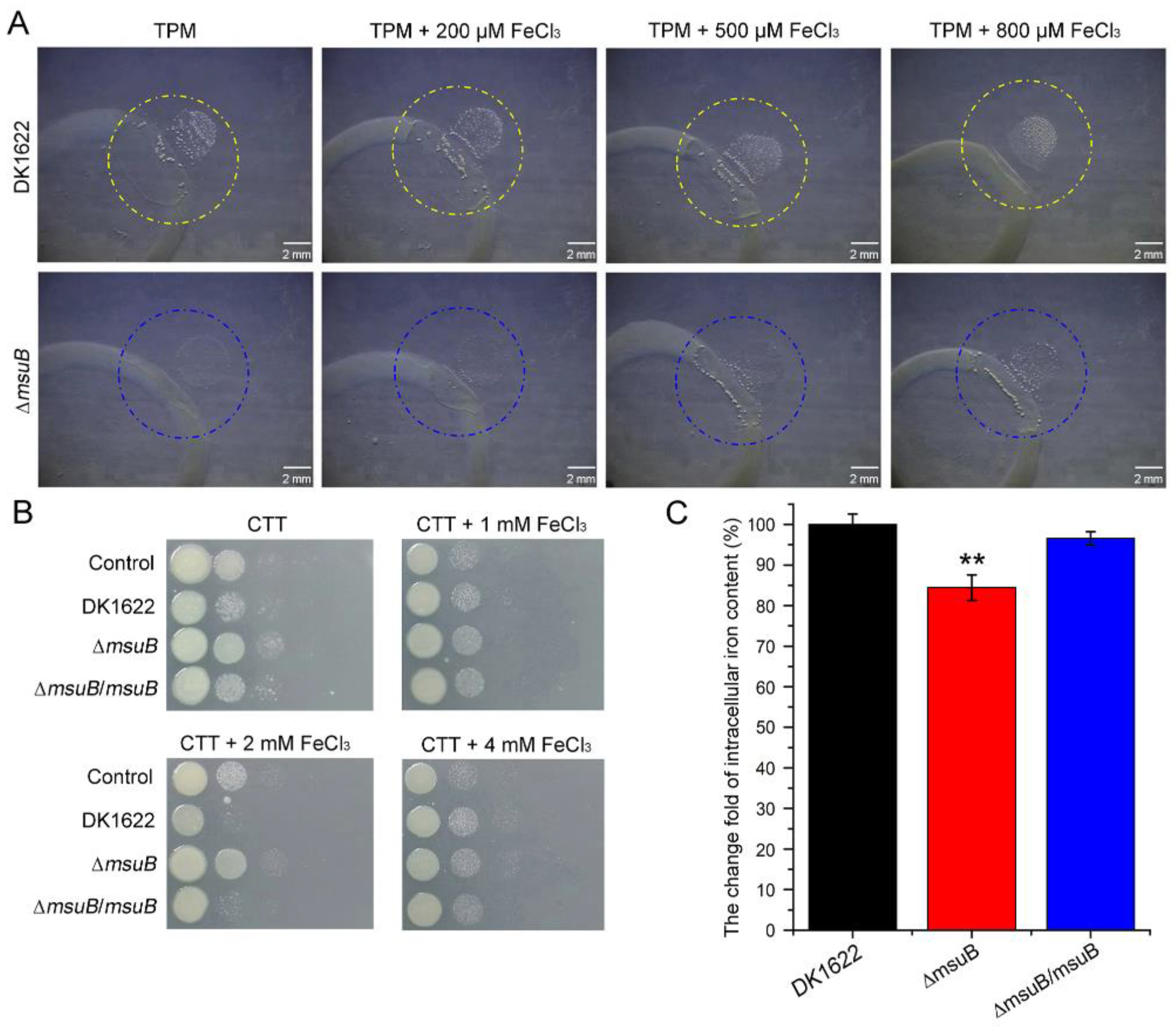
Since the msuB gene encodes a permease of the ferric-siderophore uptake ABC transporter, it is reasonable that the deletion of msuB affects iron uptake. Thus, we hypothesized that intracellular iron levels may be essential for myxobacterial predation. To test this hypothesis, we performed a predation assay on TPM (1.5% agar) plates and a bacterial competition assay on CTT (1.5% agar) plates. The results suggested that the lysed area of P. aeruginosa gradually increased with an increase in the iron level in the medium in the ∆msuB mutant, and DK1622 showed the opposite trend on TPM (1.5% agar) plates (Figure 2A). Furthermore, we also found that the ability of DK1622 and ∆msuB mutants to kill P. aeruginosa increased with lower iron levels and decreased at a higher iron level on CTT (1.5% agar) plates. Especially, 1 mM FeCl3 increased the predation ability of ∆msuB mutants, and 2 mM FeCl3 significantly enhanced the ability of DK1622 to kill P. aeruginosa (Figure 2B). Intracellular iron levels were assessed by the bathophenanthroline disulfonate (BPS)-based colorimetric method. The results showed that disruption of the msuB gene resulted in a decrease in intracellular iron levels (Figure 2C). On the basis of these observations, we concluded that intracellular iron levels might be involved in regulating the predation of M. xanthus.

3.3. MsuB Is Involved in the Maintenance of Myxobacterial Intracellular Iron Homeostasis

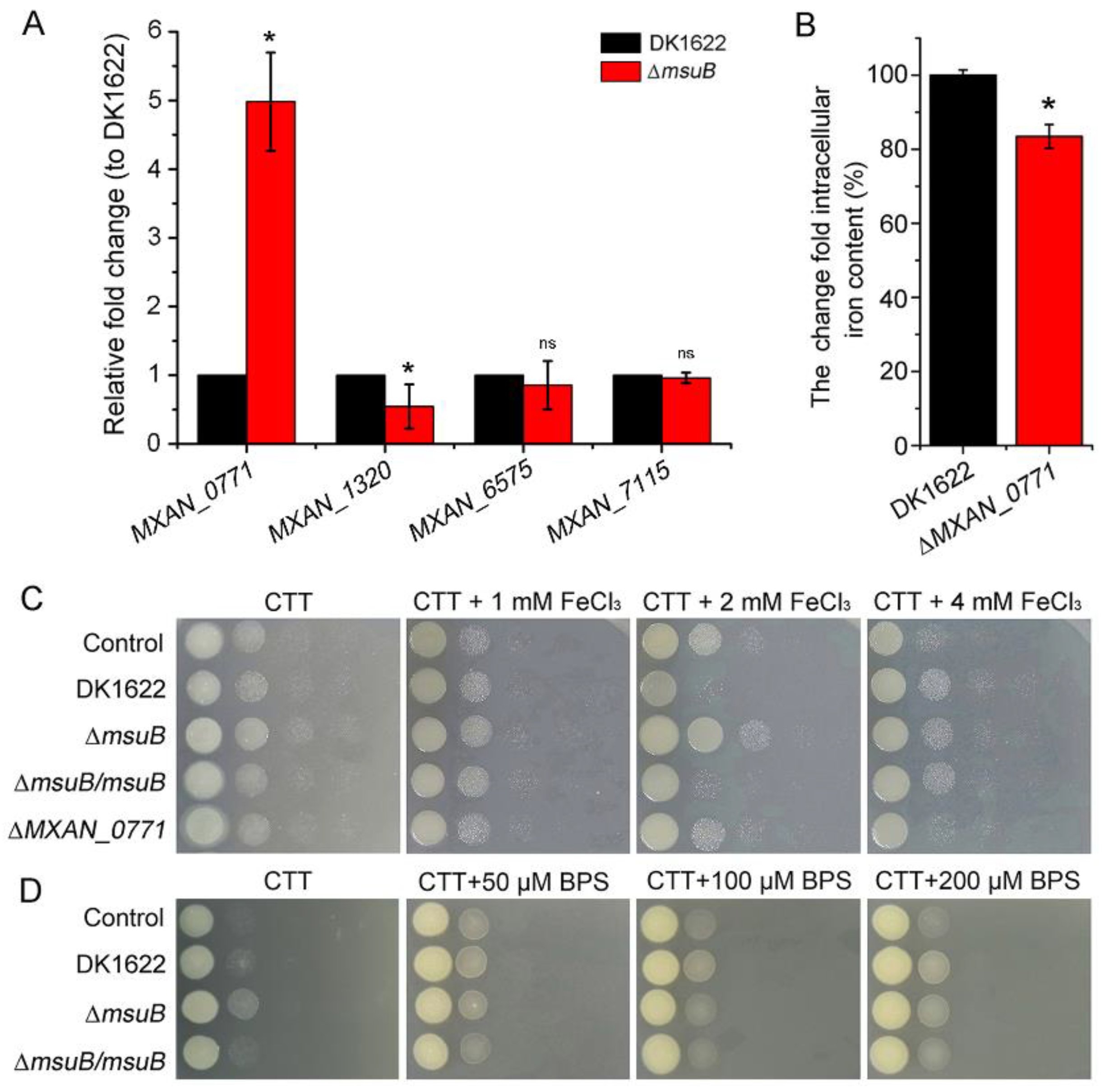
The results presented so far indicated that the ΔmsuB mutant had a significantly reduced ability. In addition to the msuABCD gene cluster, there were four iron ABC transporters in M. xanthus’s genome. We further investigated the effect of deleting msuB on the expression levels of other iron ABC transporter permease genes. The results revealed that the expression of MXAN_0771 was significantly upregulated in the ∆msuB mutant (Figure 3A). MXAN_0771 is predicted to be an ABC-type Fe3+ transport system, permease component [46]. We found that the intracellular iron level was also significantly decreased in the ∆MXAN_0771 mutant, which was similar to the ∆msuB mutant (Figure 3B). However, the bacterial competition assay demonstrated that disruption of MXAN_0771 did not result in a significant decrease in the ability to kill P. aeruginosa, even at CTT (1.5% agar) plates with different iron concentrations (Figure 3C). We also found that the deletion of MXAN_0771 did not lead to a significant decrease in the lysed area of P. aeruginosa at TPM (1.5% agar) plates with different iron concentrations (Figure S4). Subsequently, we assessed the ability to kill P. aeruginosa under low iron levels and found that iron deficiency did not alter the ability of ∆msuB mutants to kill P. aeruginosa, even though iron deficiency led to a significant increase in the colony growth diameter of DK1622 (Figure 3D and Figure S5). These findings suggest that MsuB plays an important role in maintaining myxobacterial intracellular iron homeostasis, but iron deficiency might be not the major reason for the decreased predation ability of the ∆msuB mutant.

3.4. Disruption of Siderophore Synthesis and Transport Decreased Predation in M. xanthus

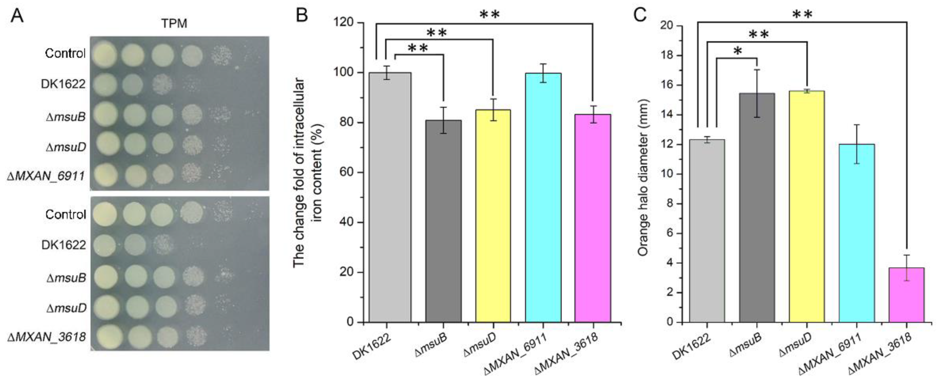
To further elucidate the mechanistic details of the reduction in predation ability in the ∆msuB mutant, we constructed ∆MXAN_3618, ∆MXAN_6911, and ∆msuD mutants, which encoded non-ribosomal peptide synthetase, TonB-dependent receptor, and substrate-binding protein in charge of siderophore synthesis, recognition, and binding in M. xanthus, respectively. As expected, the disruption of MXAN_3618 and msuD resulted in a significant decrease in the ability to kill P. aeruginosa, and the intracellular iron levels of ∆MXAN_3618 and ∆msuD mutants also significantly decreased, consistent with the ∆msuB mutant (Figure 4A,B). Furthermore, the production of siderophores was detected by a chromeazurol S overlay (O-CAS) assay. The results showed that the deletion of msuB and msuD resulted in a significant increase in the diameter of the orange halo, while the deletion of MXAN_3618 led to a decrease in the diameter of the orange halo compared with DK1622 (Figure 4C). Although deletion of MXAN_6911 did not affect the intracellular iron levels and the production of siderophores, the ∆MXAN_6911 mutant showed an attenuated ability to kill P. aeruginosa (Figure 4A–C). These results support the view that obstruction of the siderophore transport and synthesis pathway results in a reduction in predation in M. xanthus.

3.5. MsuABCD Is Closely Related to the Secretion of Lytic Enzymes

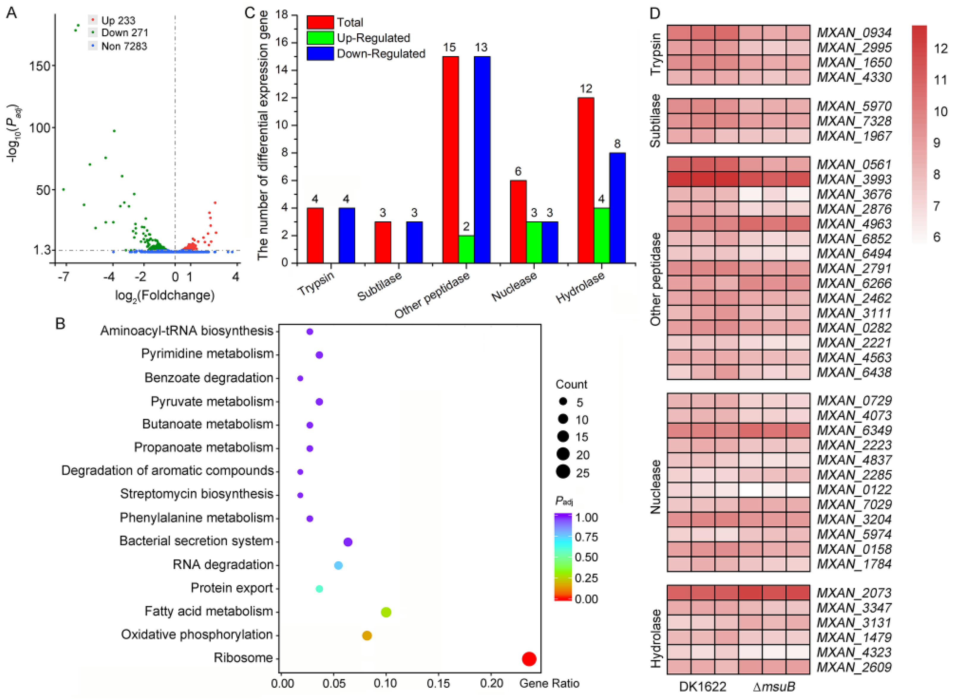
In order to shed light on the prevailing mechanistic uncertainties in siderophores’ interference with myxobacterial predation, we probed the overall transcriptional profile of DK1622 and ∆msuB mutants by transcriptome analysis. Our data showed that deletion of msuB significantly altered the gene expression profile, with 504 differentially expressed genes (DEGs), including 271 upregulated and 233 downregulated genes (Figure 5A, Supplementary Table S2). The upregulated genes were enriched in the ribosome, protein export, and bacterial secretion system pathways, and the deletion of msuB also enhanced the secretion of extracellular protein (Figure 5B and Figure S6). Surprisingly, most of these DEGs encoding lytic enzymes such as proteases, peptidases, nucleases, and hydrolase had significantly downregulated expression (Figure 5C). Among these, 13 DEGs encoding protease HtpX (MXAN_0561), M36 family metallopeptidase (MXAN_3676), S8 family serine peptidase (MXAN_5970), and trypsin-like peptidase (MXAN_2995) were downregulated by two- to fourfold and were involved in extracellular degradation of prey cells (Figure 5D). Collectively, these results suggest that the downregulation of lytic enzyme genes might impair the predation ability of the ∆msuB mutant.

3.6. Interruption of Siderophore Transport Decreased the Expression of OMV-Specific Proteins

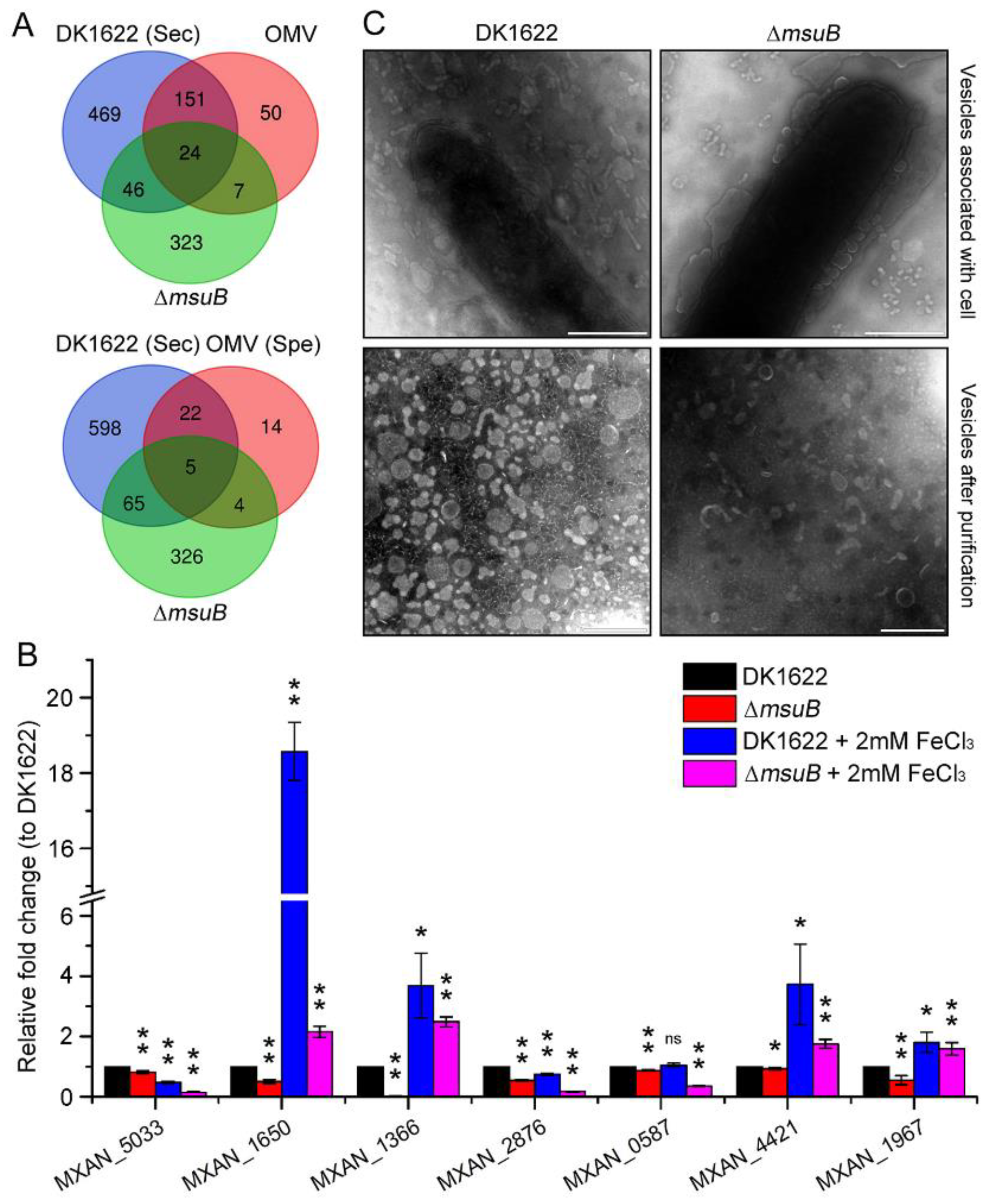
Several studies have suggested that outer membrane vesicles (OMVs) play an important role in the delivery of lytic factors onto the prey cells [15,31,47]. On the basis of the DK1622 secretion proteins (Supplementary Table S3) and public proteomic data sets [31], the common DEGs between secretion protein and OMV protein were analyzed. The results showed that nine DEGs were common, including that five OMV-specific proteins, which encode two trypsin-like serine proteases, two S8 family serine peptidases, and one endopeptidase (Figure 6A and Figure S7, Table 1). Our results confirmed that the expression of genes related to OMVs’ lytic factors was significantly decreased in the ∆msuB mutant (Figure 6B). Some of the DEGs, which encoded trypsin-like serine protease (MXAN_1650) and S8 family serine peptidase (MXAN_1967), were significantly upregulated under iron-rich conditions in DK1622, implying that iron increased myxobacterial predation by activating the OMVs’ lytic factors. To investigate the effect of deleting msuB on the formation of OMVs, the morphology of the OMVs was observed by transmission electron microscopy (TEM). The results found that the deletion of msuB reduced OMVs’ biogenesis (Figure 6C). Together, these results demonstrate that the disruption of siderophore transport results in the reduction in the predation ability of M. xanthus by affecting the expression of OMV-specific proteins.

4. Discussion

Scarce nutrients have been confirmed to be crucial driving forces that shape the composition of the microbial community [3,48,49]. Iron is a scarce essential nutrient that bacteria compete for in many niches. M. xanthus can generate specialized cells by phase variation under iron limitations [50]. These specialized cells decrease the biosynthesis of antibiotics and pigments and increase expression of the siderophores, hemin binding proteins, and iron transport proteins for acquiring iron. Our results showed that the color of colonies was different between DK1622 and ∆msuB mutants, and the deletion of the msuB gene led to a decrease in intracellular iron level and an increase in the production of siderophores (Figure S2 and Figure 4C). Most bacteria use siderophores to chelate ferric iron (Fe3+), and iron–siderophore complexes are transported via the metal-type ABC transporter [51,52]. There are 57 complete ABC transporters in the genome of M. xanthus, including 20 importers and 37 exporters [53]. On the basis of our bioinformatic analysis, we found that five ABC transporters might be involved in iron uptake in M. xanthus. In this study, we investigated the functions of two ABC transporters in myxobacterial predation by constructing in-frame deletion mutants, MsuABCD and MXAN_0770–MXAN_0772. The results showed that deletion of the msuB and MXAN_0771 genes caused a dramatic decrease in intracellular iron levels compared with DK1622 (Figure 2B and Figure 3B). However, there was an obvious difference between the ∆msuB and ∆MXAN_0771 mutants in their predation ability (Figure 3C). A previous study demonstrated that virus capsid-like nano-compartments are assembled for storing iron and protecting cells from oxidative stress in M. xanthus [54], implying that fluctuations in intracellular iron are not the major determinants of reduced myxobacterial predation. Actually, deletion of the msuB gene significantly altered the gene expression profile (Figure 5A). Therefore, we hypothesized that it may be attributed to siderophores transport. According to our analysis of the mutant (∆MXAN_3618), we found that disruption of siderophore biosynthesis led to a reduction in predation ability (Figure 4B). According to an analysis of the siderophore content secreted by the ∆msuB, ∆msuD, and ∆MXAN_3618 mutants, it was further confirmed that the interruption of siderophore transport resulted in a decrease in predation ability in M. xanthus.
As mentioned above, siderophores are involved in myxobacterial predation. However, this also raises an important question of how siderophore transport alters myxobacterial predation behavior. To address this question, we used transcriptome analysis to show that the upregulated DEGs were significantly enriched in the ribosome, protein export, and bacterial secretion systems (Figure 5B), and extracellular proteins obviously increased in the ∆msuB mutant (Figure S6). These results implied that loss of msuB may affect the synthesis and export of protein. In bacteria, the Sec machinery is responsible for translocating proteins across the cytoplasmic membrane [55]. Our data showed that MXAN_4691, MXAN_4692, and MXAN_7509 were distinctively upregulated, which encode SecD, YajC, and YidC, respectively. The three proteins form a complex with SecYEG in vivo known as the holotranslocon and assist the core Sec machinery [56]. The unfolded polypeptides are mainly secreted through the Sec translocon, and the fully folded proteins are transported through the twin-arginine translocation translocase [57,58]. A previous study found that Fur regulon was involved in secretory pathways by binding to the promoter of secY in Neisseria gonorrhoeae [59]. Even though the ferric uptake regulator (Fur) regulon has not been identified in M. xanthus, we suggested that deletion of the msuB gene might alter the expression of Fur, thereby indirectly affecting the Sec-dependent secretion of extracellular proteins (Figure S6). M. xanthus cells form thin biofilms which facilitate predation and food gathering. The cluster of cells cooperates to produce a large number of digestive enzymes, which digest prokaryotic and eukaryotic microorganisms [17,60]. Surprisingly, some lytic enzymes were significantly downregulated in the ∆msuB mutant, including proteases (MXAN_1650), peptidases (MXAN_7328, MXAN_1967), nucleases (MXAN_3347, MXAN_4323), and hydrolase (MXAN_4073, MXAN_4837) (Figure 5C,D). Prey cells can be lysed by using antibiotics, hydrolytic enzymes, and extracellular OMVs that may facilitate delivery [17]. MXAN_3564 (mepA), an M36 protease homolog, contributes to predation by degrading the proteins that are released from prey cells [31]. The enzymes with peptidoglycan-degrading activity have emerged as a class of antimicrobial proteins against pathogens by inducing prey cell lysis [61]. β-1,6-Glucanase GluM from the Corallococcus sp. strain EGB was essential for lysing the chitinous cell wall of certain fungi [62]. M. xanthus secretes a glycoside hydrolase 19 family LlpM with lysozyme-like activity, which displays bacteriolytic activity in vivo and in vitro [47]. Thus, our results support the conclusion that a reduction in the secretion of lytic enzymes leads to a decrease in predation ability in ∆msuB mutants.
OMVs are involved in multiple biological processes, including bacterial competition and nutrition acquisition, through delivering various biologically active molecules in high concentrations [63,64]. OMVs are a unique bacterial secretion pathway, termed Type 0 secretion system (T0SS) [65]. Transcriptome analysis showed that the OMVs secreted by M. xanthus could induce changes in the expression of large numbers of E. coli genes [66]. In P. aeruginosa, the Type VI secretion system effector TseF secreted by H3-T6SS can be incorporated into OMVs. TseF can be recognized by the TonB-dependent iron transporter FptA, and then facilitates the transport of iron into the cell via the OMV complex [67,68]. TonB-dependent receptors, such as CirA and MXAN_6911, have also been found in E. coli and M. xanthus OMVs, respectively [31,69]. Our results suggested that deletion of the MXAN_6911 gene led to a decrease in predation ability, although it had no effect on intracellular iron levels and siderophore synthesis (Figure 4A–C). These results further confirm that the disruption of siderophore transport results in a decrease in predation ability in M. xanthus. By using reversed phase liquid chromatography mass spectrometry (RP LC/MS) analysis, Berleman et al. identified a conservative set of 46 OMV-specific and 188 OMV-contained proteins from the purifying vesicles and whole cell membranes [31]. The siderophores myxochelin A and myxochelin B were also identified in OMVs by LC/MS [31]. On basis of these data, we explored the relationship between OMV-related proteins and proteins encoded by DEGs. The results demonstrated that 31 common DEGs were identified, including 9 DEGs encoding OMV-specific proteins (Figure 6A). Interestingly, the expression of nine DEGs was significantly downregulated, implying that the interruption of siderophore transport affected the production of OMV-specific proteins. Taken together, these results demonstrated that siderophores manipulate myxobacterial predation by interfering with the expression of outer membrane vesicle proteins.

Supplementary Materials

The following supporting information can be downloaded at: https://www.mdpi.com/article/10.3390/cells11233718/s1, Figure S1: Effect of msuB deletion on M. xanthus fruiting body development. Figure S2: Effect of msuB deletion on M. xanthus growth. Colony diameter (A) and morphology (B) of DK1622 and ∆msuB mutants was measured on CTT (1.5% agar) plates. Figure S3: Effect of msuB deletion on M. xanthus gliding motility and exopolysaccharide accumulation. (A) Strains were incubated on 0.5% CTT with 1.5% agar to detect gliding motility. Scale bars, 5 mm (top plane), 1 mm (down plane). The same results were obtained in two independent experiments. (B) Lack of MsuB does not affect exopolysaccharide accumulation. ΔpilA strain served as negative control. The same results were obtained in two independent experiments. Figure S4: The effects of different iron concentrations on myxobacterial predation on TPM (1.5% agar) plates were assessed by a predation experiment. Scale bars = 2 mm. Figure S5: Effect of msuB deletion on M. xanthus growth under low iron level. Colony diameter of DK1622 and ∆msuB mutants was measured on CTT (1.5% agar) plates with different concentration of BPS (bathophenanthroline disulfonate). Figure S6: Effect of msuB deletion on extracellular proteins. DK1622 and ∆msuB mutants were grown in CTT medium for 24 h at 30 °C, 200 rpm. The optical density (OD550nm) of DK1622 and ∆msuB mutants reached to 0.8. The supernatants were obtained by centrifuging at 12,000× g. The extracellular proteins were precipitated with ammonium sulphate and resuspended in PBS. The samples were detected by SDS-PAGE. Loading volumes were 10 µL (A), 20 µL (B), and 30 µL (C), respectively. Figure S7: The expression of OMVs related genes in ∆msuB mutant, compared with DK1622. * p-value < 0.05 and ** p-value < 0.01 compared with the DK1622 as measured by a two-tailed unpaired Student’s t-test. Table S1: Strains and plasmids used in this work; Table S2: Deletion of msuB significantly altered the differentially expressed genes (DEGs); Table S3: The secretion proteins derived from wild type DK1622.

Author Contributions

Conceptualization, Y.D., Q.Y. and H.Z.; methodology, Y.D., H.D., Z.F. and X.W.; formal analysis, Y.D., H.D., Z.F. and X.W.; writing—original draft preparation, Y.D.; writing—review and editing, Y.D., H.D., Z.F., X.W., Q.Y. and H.Z.; visualization, Y.D.; supervision, Q.Y. and H.Z.; funding acquisition, Y.D and H.D. All authors have read and agreed to the published version of the manuscript.

Funding

This work was supported by the Guangdong Basic and Applied Basic Research Foundation (2021A1515011143), the National Natural Science Foundation of China (32102276), the GDAS’ Project of Science and Technology Development (2021GDASYL-20210103016) and Guangdong Special Support Program (2021JC06N628).

Institutional Review Board Statement

Not applicable.

Informed Consent Statement

Not applicable.

Data Availability Statement

The original contributions presented in the study are included in the article/Supplementary Materials, further inquiries can be directed to the corresponding authors. The raw data associated with this study has been deposited in the Sequence Read Archive (SRA) under the accession PRJNA849267.

Conflicts of Interest

The authors declare no conflict of interest.

References

  1. Klein, T.A.; Ahmad, S.; Whitney, J.C. Contact-dependent interbacterial antagonism mediated by protein secretion machines. Trends Microbiol. 2020, 28, 387–400. [Google Scholar] [CrossRef]
  2. Stubbendieck, R.M.; Straight, P.D. Multifaceted interfaces of bacterial competition. J. Bacteriol. 2016, 198, 2145–2155. [Google Scholar] [CrossRef]
  3. Hibbing, M.E.; Fuqua, C.; Parsek, M.R.; Peterson, S.B. Bacterial competition: Surviving and thriving in the microbial jungle. Nat. Rev. Microbiol. 2010, 8, 15–25. [Google Scholar] [CrossRef]
  4. Ghoul, M.; Mitri, S. The ecology and evolution of microbial competition. Trends Microbiol. 2016, 24, 833–845. [Google Scholar] [CrossRef]
  5. Kamal, F.; Liang, X.; Manera, K.; Pei, T.T.; Kim, H.; Lam, L.G.; Pun, A.; Hersch, S.J.; Dong, T.G. Differential cellular response to translocated toxic effectors and physical penetration by the Type VI Secretion System. Cell Rep. 2020, 31, 107766. [Google Scholar] [CrossRef]
  6. Kramer, J.; Ozkaya, O.; Kummerli, R. Bacterial siderophores in community and host interactions. Nat. Rev. Microbiol. 2020, 18, 152–163. [Google Scholar] [CrossRef]
  7. Kurth, C.; Kage, H.; Nett, M. Siderophores as molecular tools in medical and environmental applications. Org. Biomol. Chem. 2016, 14, 8212–8227. [Google Scholar] [CrossRef]
  8. Gorska, A.; Sloderbach, A.; Marszall, M.P. Siderophore-drug complexes: Potential medicinal applications of the ‘Trojan horse’ strategy. Trends Pharmacol. Sci. 2014, 35, 442–449. [Google Scholar] [CrossRef]
  9. Clarke, T.E.; Tari, L.W.; Vogel, H.J. Structural biology of bacterial iron uptake systems. Curr. Top. Med. Chem. 2001, 1, 7–30. [Google Scholar] [CrossRef]
  10. Eickhoff, M.J.; Bassler, B.L. Vibrio fischeri siderophore production drives competitive exclusion during dual-species growth. Mol. Microbiol. 2020, 114, 244–261. [Google Scholar] [CrossRef]
  11. Leinweber, A.; Weigert, M.; Kummerli, R. The bacterium Pseudomonas aeruginosa senses and gradually responds to interspecific competition for iron. Evolution 2018, 72, 1515–1528. [Google Scholar] [CrossRef] [PubMed]
  12. Sutton, D.; Livingstone, P.G.; Furness, E.; Swain, M.T.; Whitworth, D.E. Genome-wide identification of myxobacterial predation genes and demonstration of formaldehyde secretion as a potentially predation-resistant trait of Pseudomonas aeruginosa. Front. Microbiol. 2019, 10, 2650. [Google Scholar] [CrossRef] [PubMed]
  13. Akbar, S.; Phillips, K.E.; Misra, S.K.; Sharp, J.S.; Stevens, D.C. Differential response to prey quorum signals indicates predatory specialization of myxobacteria and ability to predate Pseudomonas aeruginosa. Environ. Microbiol. 2022, 24, 1263–1278. [Google Scholar] [CrossRef] [PubMed]
  14. Livingstone, P.G.; Morphew, R.M.; Whitworth, D.E. Myxobacteria are able to prey broadly upon clinically-relevant pathogens, exhibiting a prey range which cannot be explained by phylogeny. Front. Microbiol. 2017, 8, 1593. [Google Scholar] [CrossRef]
  15. Thiery, S.; Kaimer, C. The predation strategy of Myxococcus xanthus. Front. Microbiol. 2020, 11, 2. [Google Scholar] [CrossRef]
  16. Morgan, A.D.; MacLean, R.C.; Hillesland, K.L.; Velicer, G.J. Comparative analysis of Myxococcus predation on soil bacteria. Appl. Environ. Microbiol. 2010, 76, 6920–6927. [Google Scholar] [CrossRef]
  17. Keane, R.; Berleman, J. The predatory life cycle of Myxococcus xanthus. Microbiology 2016, 162, 1–11. [Google Scholar] [CrossRef]
  18. Velicer, G.J.; Kroos, L.; Lenski, R.E. Developmental cheating in the social bacterium Myxococcus xanthus. Nature 2000, 404, 598–601. [Google Scholar] [CrossRef]
  19. Stolle, A.S.; Meader, B.T.; Toska, J.; Mekalanos, J.J. Endogenous membrane stress induces T6SS activity in Pseudomonas aeruginosa. Proc. Natl. Acad. Sci. USA 2021, 118, e2018365118. [Google Scholar] [CrossRef]
  20. Kunze, B.; Bedorf, N.; Kohl, W.; Hofle, G.; Reichenbach, H. Myxochelin A, a new iron-chelating compound from Angiococcus disciformis (Myxobacterales). Production, isolation, physico-chemical and biological properties. J. Antibiot. 1989, 42, 14–27. [Google Scholar] [CrossRef]
  21. Silakowski, B.; Kunze, B.; Nordsiek, G.; Blocker, H.; Hofle, G.; Muller, R. The myxochelin iron transport regulon of the myxobacterium Stigmatella aurantiaca Sg a15. Eur. J. Biochem. 2000, 267, 6476–6485. [Google Scholar] [CrossRef]
  22. Gaitatzis, N.; Kunze, B.; Muller, R. Novel insights into siderophore formation in myxobacteria. Chembiochem 2005, 6, 365–374. [Google Scholar] [CrossRef]
  23. Findlay, B.L. The chemical ecology of predatory soil bacteria. ACS Chem. Biol. 2016, 11, 1502–1510. [Google Scholar] [CrossRef] [PubMed]
  24. Krug, D.; Zurek, G.; Revermann, O.; Vos, M.; Velicer, G.J.; Muller, R. Discovering the hidden secondary metabolome of Myxococcus xanthus: A study of intraspecific diversity. Appl. Environ. Microbiol. 2008, 74, 3058–3068. [Google Scholar] [CrossRef]
  25. Weissman, K.J.; Muller, R. Myxobacterial secondary metabolites: Bioactivities and modes-of-action. Nat. Prod. Rep. 2010, 27, 1276–1295. [Google Scholar] [CrossRef]
  26. Sester, A.; Winand, L.; Pace, S.; Hiller, W.; Werz, O.; Nett, M. Myxochelin- and Pseudochelin-derived lipoxygenase inhibitors from a genetically engineered Myxococcus xanthus strain. J. Nat. Prod. 2019, 82, 2544–2549. [Google Scholar] [CrossRef]
  27. Lee, N.; Kim, W.; Chung, J.; Lee, Y.; Cho, S.; Jang, K.S.; Kim, S.C.; Palsson, B.; Cho, B.K. Iron competition triggers antibiotic biosynthesis in Streptomyces coelicolor during coculture with Myxococcus xanthus. ISME J. 2020, 14, 1111–1124. [Google Scholar] [CrossRef]
  28. Dhurve, G.; Madikonda, A.K.; Jagannadham, M.V.; Siddavattam, D. Outer membrane vesicles of Acinetobacter baumannii DS002 are selectively enriched with TonB-Dependent transporters and play a key role in iron acquisition. Microbiol. Spectr. 2022, 10, e00293-22. [Google Scholar] [CrossRef]
  29. Li, C.; Zhu, L.; Wang, D.; Wei, Z.; Hao, X.; Wang, Z.; Li, T.; Zhang, L.; Lu, Z.; Long, M.; et al. T6SS secretes an LPS-binding effector to recruit OMVs for exploitative competition and horizontal gene transfer. ISME J. 2022, 16, 500–510. [Google Scholar] [CrossRef]
  30. Evans, A.G.L.; Davey, H.M.; Cookson, A.; Currinn, H.; Cooke-Fox, G.; Stanczyk, P.J.; Whitworth, D.E. Predatory activity of Myxococcus xanthus outer-membrane vesicles and properties of their hydrolase cargo. Microbiology 2012, 158, 2742–2752. [Google Scholar] [CrossRef]
  31. Berleman, J.E.; Allen, S.; Danielewicz, M.A.; Remis, J.P.; Gorur, A.; Cunha, J.; Hadi, M.Z.; Zusman, D.R.; Northen, T.R.; Witkowska, H.E.; et al. The lethal cargo of Myxococcus xanthus outer membrane vesicles. Front. Microbiol. 2014, 5, 474. [Google Scholar] [CrossRef] [PubMed]
  32. Kaiser, D. Social gliding is correlated with the presence of pili in Myxococcus xanthus. Proc. Natl. Acad. Sci. USA 1979, 76, 5952–5956. [Google Scholar] [CrossRef] [PubMed]
  33. Yang, Y.J.; Wang, Y.; Li, Z.F.; Gong, Y.; Zhang, P.; Hu, W.C.; Sheng, D.H.; Li, Y.Z. Increasing on-target cleavage efficiency for CRISPR/Cas9-induced large fragment deletion in Myxococcus xanthus. Microb. Cell Fact. 2017, 16, 142. [Google Scholar] [CrossRef]
  34. Julien, B.; Kaiser, A.D.; Garza, A. Spatial control of cell differentiation in Myxococcus xanthus. Proc. Natl. Acad. Sci. USA 2000, 97, 9098–9103. [Google Scholar] [CrossRef]
  35. Wu, S.S.; Wu, J.; Kaiser, D. The Myxococcus xanthus pilT locus is required for social gliding motility although pili are still produced. Mol. Microbiol. 1997, 23, 109–121. [Google Scholar] [CrossRef]
  36. Dong, H.; Xu, X.; Gao, R.; Li, Y.; Li, A.; Yao, Q.; Zhu, H. Myxococcus xanthus R31 suppresses tomato bacterial wilt by inhibiting the pathogen Ralstonia solanacearum with secreted proteins. Front. Microbiol. 2021, 12, 801091. [Google Scholar] [CrossRef]
  37. Berleman, J.E.; Chumley, T.; Cheung, P.; Kirby, J.R. Rippling is a predatory behavior in Myxococcus xanthus. J. Bacteriol. 2006, 188, 5888–5895. [Google Scholar] [CrossRef]
  38. Basler, M.; Ho, B.T.; Mekalanos, J.J. Tit-for-tat: Type VI secretion system counterattack during bacterial cell-cell interactions. Cell 2013, 152, 884–894. [Google Scholar] [CrossRef]
  39. Hsu, P.C.; Yang, C.Y.; Lan, C.Y. Candida albicans Hap43 is a repressor induced under low-iron conditions and is essential for iron-responsive transcriptional regulation and virulence. Eukaryot. Cell 2011, 10, 207–225. [Google Scholar] [CrossRef]
  40. Dong, Y.; Zhang, D.; Yu, Q.; Zhao, Q.; Xiao, C.; Zhang, K.; Jia, C.; Chen, S.; Zhang, B.; Zhang, B.; et al. Loss of Ssq1 leads to mitochondrial dysfunction, activation of autophagy and cell cycle arrest due to iron overload triggered by mitochondrial iron-sulfur cluster assembly defects in Candida albicans. Int. J. Biochem. Cell Biol. 2017, 85, 44–55. [Google Scholar] [CrossRef]
  41. Kominek, J.; Doering, D.T.; Opulente, D.A.; Shen, X.X.; Zhou, X.; DeVirgilio, J.; Hulfachor, A.B.; Groenewald, M.; McGee, M.A.; Karlen, S.D.; et al. Eukaryotic acquisition of a bacterial operon. Cell 2019, 176, 1356–1366. [Google Scholar] [CrossRef]
  42. Livak, K.J.; Schmittgen, T.D. Analysis of relative gene expression data using real-time quantitative PCR and the 2−ΔΔCT Method. Methods 2001, 25, 402–408. [Google Scholar] [CrossRef]
  43. Niehus, R.; Picot, A.; Oliveira, N.M.; Mitri, S.; Foster, K.R. The evolution of siderophore production as a competitive trait. Evolution 2017, 71, 1443–1455. [Google Scholar] [CrossRef]
  44. Lee, W.; van Baalen, M.; Jansen, V.A. Siderophore production and the evolution of investment in a public good: An adaptive dynamics approach to kin selection. J. Theor. Biol. 2016, 388, 61–71. [Google Scholar] [CrossRef]
  45. Popp, P.F.; Mascher, T. Coordinated cell death in isogenic bacterial populations: Sacrificing some for the benefit of many? J. Mol. Biol. 2019, 431, 4656–4669. [Google Scholar] [CrossRef] [PubMed]
  46. Goldman, B.S.; Nierman, W.C.; Kaiser, D.; Slater, S.C.; Durkin, A.S.; Eisen, J.A.; Ronning, C.M.; Barbazuk, W.B.; Blanchard, M.; Field, C.; et al. Evolution of sensory complexity recorded in a myxobacterial genome. Proc. Natl. Acad. Sci. USA 2006, 103, 15200–15205. [Google Scholar] [CrossRef]
  47. Arend, K.I.; Schmidt, J.J.; Bentler, T.; Luchtefeld, C.; Eggerichs, D.; Hexamer, H.M.; Kaimer, C. Myxococcus xanthus predation of Gram-positive or Gram-negative bacteria is mediated by different bacteriolytic mechanisms. Appl. Environ. Microbiol. 2020, 87, e02382-20. [Google Scholar] [CrossRef]
  48. Kamada, N.; Chen, G.Y.; Inohara, N.; Nunez, G. Control of pathogens and pathobionts by the gut microbiota. Nat. Immunol. 2013, 14, 685–690. [Google Scholar] [CrossRef] [PubMed]
  49. Knauf, G.A.; Powers, M.J.; Herrera, C.M.; Trent, M.S.; Davies, B.W. Acinetobactin-mediated inhibition of commensal bacteria by Acinetobacter baumannii. mSphere 2022, 7, e00016–e00022. [Google Scholar] [CrossRef]
  50. Dziewanowska, K.; Settles, M.; Hunter, S.; Linquist, I.; Schilkey, F.; Hartzell, P.L. Phase variation in Myxococcus xanthus yields cells specialized for iron sequestration. PLoS ONE 2014, 9, e95189. [Google Scholar] [CrossRef]
  51. Huang, W.; Wilks, A. Extracellular heme uptake and the challenge of bacterial cell membranes. Annu. Rev. Biochem. 2017, 86, 799–823. [Google Scholar] [CrossRef] [PubMed]
  52. Andrews, S.C.; Robinson, A.K.; Rodriguez-Quinones, F. Bacterial iron homeostasis. FEMS Microbiol. Rev 2003, 27, 215–237. [Google Scholar] [CrossRef]
  53. Yan, J.; Bradley, M.D.; Friedman, J.; Welch, R.D. Phenotypic profiling of ABC transporter coding genes in Myxococcus xanthus. Front. Microbiol. 2014, 5, 352. [Google Scholar] [CrossRef]
  54. McHugh, C.A.; Fontana, J.; Nemecek, D.; Cheng, N.; Aksyuk, A.A.; Heymann, J.B.; Winkler, D.C.; Lam, A.S.; Wall, J.S.; Steven, A.C.; et al. A virus capsid-like nanocompartment that stores iron and protects bacteria from oxidative stress. EMBO J. 2014, 33, 1896–1911. [Google Scholar] [CrossRef] [PubMed]
  55. Dalal, K.; Duong, F. The SecY complex: Conducting the orchestra of protein translocation. Trends Cell Biol. 2011, 21, 506–514. [Google Scholar] [CrossRef]
  56. Cranford-Smith, T.; Huber, D. The way is the goal: How SecA transports proteins across the cytoplasmic membrane in bacteria. FEMS Microbiol. Lett. 2018, 365, fny093. [Google Scholar] [CrossRef]
  57. Costa, T.R.; Felisberto-Rodrigues, C.; Meir, A.; Prevost, M.S.; Redzej, A.; Trokter, M.; Waksman, G. Secretion systems in Gram-negative bacteria: Structural and mechanistic insights. Nat. Rev. Microbiol. 2015, 13, 343–359. [Google Scholar] [CrossRef]
  58. Goosens, V.J.; van Dijl, J.M. Twin-Arginine protein translocation. Curr. Top. Microbiol. Immunol. 2017, 404, 69–94. [Google Scholar] [CrossRef]
  59. Sebastian, S.; Agarwal, S.; Murphy, J.R.; Genco, C.A. The gonococcal fur regulon: Identification of additional genes involved in major catabolic, recombination, and secretory pathways. J. Bacteriol. 2002, 184, 3965–3974. [Google Scholar] [CrossRef]
  60. Zusman, D.R.; Scott, A.E.; Yang, Z.; Kirby, J.R. Chemosensory pathways, motility and development in Myxococcus xanthus. Nat. Rev. Microbiol. 2007, 5, 862–872. [Google Scholar] [CrossRef] [PubMed]
  61. Sobieraj, A.M.; Huemer, M.; Zinsli, L.V.; Meile, S.; Keller, A.P.; Rohrig, C.; Eichenseher, F.; Shen, Y.; Zinkernagel, A.S.; Loessner, M.J.; et al. Engineering of long-circulating peptidoglycan hydrolases enables efficient treatment of systemic Staphylococcus aureus infection. mBio 2020, 11, e01781-20. [Google Scholar] [CrossRef] [PubMed]
  62. Li, Z.; Ye, X.; Liu, M.; Xia, C.; Zhang, L.; Luo, X.; Wang, T.; Chen, Y.; Zhao, Y.; Qiao, Y.; et al. A novel outer membrane β-1,6-glucanase is deployed in the predation of fungi by myxobacteria. ISME J. 2019, 13, 2223–2235. [Google Scholar] [CrossRef] [PubMed]
  63. Biller, S.J.; Schubotz, F.; Roggensack, S.E.; Thompson, A.W.; Summons, R.E.; Chisholm, S.W. Bacterial vesicles in marine ecosystems. Science 2014, 343, 183–186. [Google Scholar] [CrossRef] [PubMed]
  64. Rivera, J.; Cordero, R.J.; Nakouzi, A.S.; Frases, S.; Nicola, A.; Casadevall, A. Bacillus anthracis produces membrane-derived vesicles containing biologically active toxins. Proc. Natl. Acad. Sci. USA 2010, 107, 19002–19007. [Google Scholar] [CrossRef]
  65. Guerrero-Mandujano, A.; Hernandez-Cortez, C.; Ibarra, J.A.; Castro-Escarpulli, G. The outer membrane vesicles: Secretion system type zero. Traffic 2017, 18, 425–432. [Google Scholar] [CrossRef] [PubMed]
  66. Livingstone, P.G.; Millard, A.D.; Swain, M.T.; Whitworth, D.E. Transcriptional changes when Myxococcus xanthus preys on Escherichia coli suggest myxobacterial predators are constitutively toxic but regulate their feeding. Microb. Genom. 2018, 4, e000152. [Google Scholar] [CrossRef]
  67. Lin, J.; Zhang, W.; Cheng, J.; Yang, X.; Zhu, K.; Wang, Y.; Wei, G.; Qian, P.Y.; Luo, Z.Q.; Shen, X. A Pseudomonas T6SS effector recruits PQS-containing outer membrane vesicles for iron acquisition. Nat. Commun. 2017, 8, 14888. [Google Scholar] [CrossRef]
  68. Behrens, H.M.; Lowe, E.D.; Gault, J.; Housden, N.G.; Kaminska, R.; Weber, T.M.; Thompson, C.M.A.; Mislin, G.L.A.; Schalk, I.J.; Walker, D.; et al. Pyocin S5 import into Pseudomonas aeruginosa reveals a generic mode of bacteriocin transport. mBio 2020, 11, e03230-19. [Google Scholar] [CrossRef]
  69. Lee, E.Y.; Bang, J.Y.; Park, G.W.; Choi, D.S.; Kang, J.S.; Kim, H.J.; Park, K.S.; Lee, J.O.; Kim, Y.K.; Kwon, K.H.; et al. Global proteomic profiling of native outer membrane vesicles derived from Escherichia coli. Proteomics 2007, 7, 3143–3153. [Google Scholar] [CrossRef]
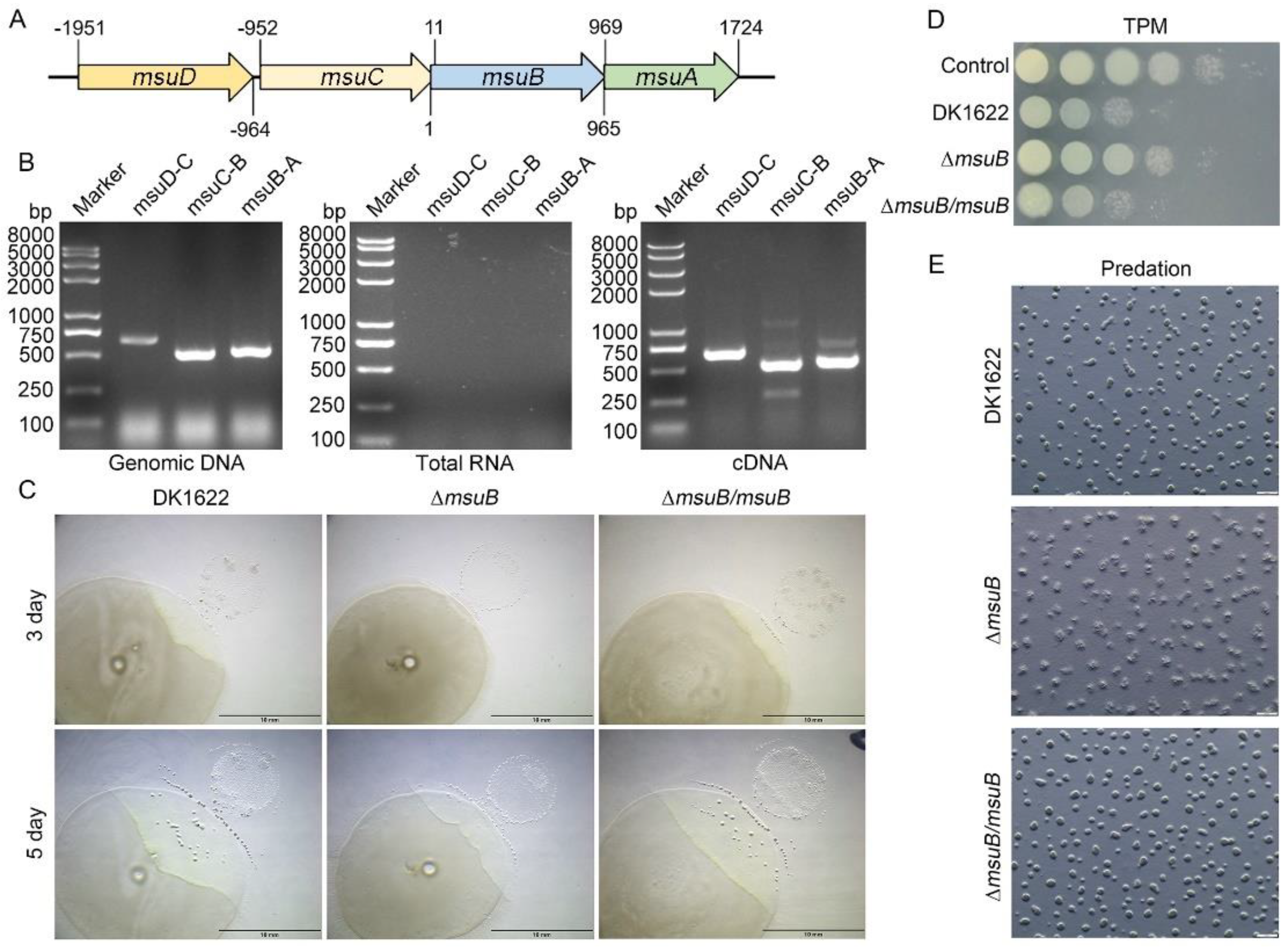
Figure 1. MsuB plays an important role in myxobacterial predation. (A) msuABCD locus. Start and stop codons are indicated; +1 indicates the first nucleotide in the msuB start codon. (B) Cotranscriptional analysis of the msuABCD cluster by reverse transcription–PCR. The templates were genomic DNA, RNA removed from genomic DNA, and cDNA, respectively. (C) Disruption of msuB affects the ability of M. xanthus to prey on P. aeruginosa. Predation assay on TPM (1.5% agar) plates. Scale bars = 10 mm. (D) The competition assay analyzed myxobacterial predation. After coculture of M. xanthus strains and PAO1 for 24 h, the cells were harvested and resuspended in 1 mL of MMC buffer. Subsequently, the cellular suspension was serially diluted in MMC buffer. For this, 3 µL of the suspensions (from 10−1 to 10−6) was spotted on LB agar plates supplemented with tetracycline (20 µg mL−1) from left to right. The plates were grown at 37 °C and photographed after 24 h. (E) Fruiting body formation was analyzed during the predation of P. aeruginosa. The plates were photographed by a stereomicroscope after 24 h of coculture. Scale bars = 500 µm.
Cells 11 03718 g001
Figure 2. Differences in predation ability between DK1622 and ∆msuB mutants under different iron concentrations. (A) The effects of different iron concentrations on myxobacterial predation on TPM (1.5% agar) plates were assessed by a predation experiment. Scale bars = 2 mm. (B) The survival of P. aeruginosa co-cultured with DK1622, ∆msuB, and ∆msuB/msuB at different iron concentrations for 24 h was calculated by a competitive experiment. The cocultured M. xanthus strains and PAO1 were harvested and the cells were resuspended in 1 mL of MMC buffer. Subsequently, the cellular suspension was serially diluted in MMC buffer. Next, 3 µL of the suspensions (from 10−2 to 10−6) was spotted on LB agar plates supplemented with tetracycline (20 µg mL−1) from left to right. The plates were grown at 37 °C and photographed after 24 h. (C) The relative content of intracellular iron was detected by the BPS method. ** p-value < 0.01 compared with the DK1622 as measured by a two-tailed unpaired Student’s t-test.
Cells 11 03718 g002
Figure 3. MsuB has a profound effect on iron homeostasis in M. xanthus. (A) qRT-PCR was used to measure the expression of other iron ABC transporter permease genes, which were normalized to the expression of gapA. DK1622 and the ∆msuB mutant were grown on CTT plates. * p-value < 0.01 compared with the DK1622; ns, not significant. (B) The intracellular iron level of ∆MXAN_0771 mutant was detected by the BPS method. * p-value < 0.01 compared with the DK1622 as measured by a two-tailed unpaired Student’s t-test. (C) The competition assay analyzed the predation of different mutants on CTT (1.5% agar) plates containing different concentrations of FeCl3. After 24 h of coculture, the colony was harvested, and the cells were resuspended in 1 mL of MMC buffer. Subsequently, the cellular suspension was serially diluted in MMC buffer. Next, 3 µL of the suspension (from 10−2 to 10−6) was spotted on LB agar plates supplemented with tetracycline (20 µg mL−1) from left to right. The plates were grown at 37 °C and photographed after 24 h. (D) The competition assay analyzed the predation of mutants on CTT (1.5% agar) plates containing different concentrations of BPS. After 24 h of coculture, the colony was harvested, and the cells were resuspended in 1 mL of MMC buffer. Subsequently, the cellular suspension was serially diluted in MMC buffer, and then 3 µL of the suspension (from 10−2 to 10−6) was spotted on LB agar plates supplemented with tetracycline (20 µg mL−1) from left to right. The plates were grown at 37 °C and photographed after 24 h.
Cells 11 03718 g003
Figure 4. The effects of deleting siderophore synthesis and transport-related genes on intracellular iron levels, predation, and siderophore production. (A) The competition assay analyzed the predation of different mutants on TPM (1.5% agar) plates. After coculture of M. xanthus strains and PAO1 for 24 h, the cells were harvested and resuspended in 1 mL of MMC buffer. Subsequently, the cellular suspension was serially diluted in MMC buffer, and 3 µL of the suspension (from 10−1 to 10−6) was spotted on LB agar plates supplemented with tetracycline (20 µg mL−1) from left to right. The plates were grown at 37 °C and photographed after 24 h. (B) The relative content of intracellular iron was detected by the BPS method. ** p-value < 0.01 compared with DK1622 as measured by a two-tailed unpaired Student’s t-test. (C) Determination of siderophore production by the chromeazurol S overlay (O-CAS) assay. * p-value < 0.05 and ** p-value < 0.01 compared with DK1622 as measured by a two-tailed unpaired Student’s t-test.
Cells 11 03718 g004
Figure 5. Global transcriptional profiles of DK1622 and the ∆msuB mutant. (A) Volcano plot of differentially expressed genes (DEGs). Red dots and green dots represent the up- and down-regulated genes with significant differences, respectively (Padj < 0.05). The blue dots represent the genes that did not change significantly. (B) KEGG pathway enrichment results of DEGs. The y–axis represents the categories of the KEGG pathways. The x–axis is the richness factor (richness factor = amount of DEGs in the pathway divided by the amount of all genes in the background gene set). (C) Analysis of the DEGs encoding lytic enzymes. (D) Heatmap of RNA sequencing, showing the expression pattern of genes encoding lytic enzymes in DK1622 and the ∆msuB mutant, respectively. The normalized FPKM value of differentially expressed genes was used for plotting.
Cells 11 03718 g005
Figure 6. Effects of msuB deletion on the expression of OMV-specific proteins in M. xanthus. (A) Venn diagram analysis of DEGs and the genes encoding secretion proteins (DK1622) and OMV proteins. (B) DK1622 and ∆msuB mutants were grown in CTT medium or CTT medium with 2 mM FeCl3 for 24 h at 30 °C. The sample was harvested by centrifuging 12,000× g at 4 °C and total RNA was extracted for detecting the expression of OMV related-genes, which were normalized to the expression of gapA. * p-value < 0.05; ** p-value < 0.01 compared with DK1622; ns, not significant. (C) DK1622 and the ∆msuB mutant were grown in a CTT medium for 24 h at 30 °C. OMVs were observed by transmission electron microscopy (TEM) (scale bar = 500 nm) (top panel). OMVs were isolated by centrifuging at 150,000× g and were observed by transmission electron microscopy (TEM) (scale bar = 200 nm) (bottom panel).
Cells 11 03718 g006
Table 1. OMV-specific expression genes.
No.Old LocusLocusGene Function
1MXAN_0587MXAN_RS02845trypsin-like serine protease: Trypsin
2MXAN_1967MXAN_RS09535S8 family serine peptidase: Subtilase family
3MXAN_1650MXAN_RS08015trypsin-like serine protease: Trypsin
4MXAN_2876MXAN_RS13930endopeptidase
5MXAN_5970MXAN_RS28960S8 family serine peptidase: Subtilase family
Publisher’s Note: MDPI stays neutral with regard to jurisdictional claims in published maps and institutional affiliations.