Article

Evaluation of the Reproductive Toxicity of Fluopimomide in Meloidogyne incognita and Caenorhabditis elegans

1
Key Laboratory of Pesticide Toxicology & Application Technique, College of Plant Protection, Shandong Agricultural University, Tai’an 271018, China
2
Shandong Institute of Pomology, Tai’an 271000, China
3
Shandong Huayang Technology Co., Ltd., Tai’an 271411, China
4
College of Food Science and Engineering, Shandong Agricultural University, Tai’an 271018, China
5
Tropical Research and Education Center, Department of Plant Pathology, University of Florida, Institute of Food and Agricultural Sciences (IFAS), Homestead, FL 33031, USA
*
Authors to whom correspondence should be addressed.
These authors contributed equally to this work.
Agronomy 2023, 13(10), 2471; https://doi.org/10.3390/agronomy13102471
Submission received: 29 August 2023 / Revised: 13 September 2023 / Accepted: 21 September 2023 / Published: 25 September 2023
(This article belongs to the Special Issue Integrated Nematode Management in Sustainable Agricultural Production)

Abstract

:
Fluopimomide is a new pesticide that is widely applied in agriculture; however, the effects and molecular mechanisms of fluopimomide in inhibiting nematode reproduction remain unknown. In this study, the effects of fluopimomide on the development and infection of Meloidogyne incognita and the reproductive toxicity in Caenorhabditis elegans were evaluated. Results showed that, in comparison to inoculated control, fluopimomide at 0.33, 0.67, and 1.0 mg/kg soil significantly (p < 0.05) delayed M. incognita development and decreased the reproduction in pot experiments. Fluopimomide notably reduced the galls index with a control effect of 78.6%, 67.9%, and 50.0%, respectively. In addition, a dose–response relationship existed between the brood size and germ cell number of C. elegans and fluopimomide concentrations. Compared with the control group, fluopimomide at 1.0 and 5.0 mg/L notably (p < 0.001) increased the number of cell corpses per gonad in the N2 strain of C. elegans by 8.8- and 14.4-fold, respectively. The number of cell corpses per gonad was similar between the fluopimomide treated worms and the control group in mutants of ced-3, ced-4, and ced-9. Further evidence revealed fluopimomide significantly enhanced the expression of cep-1, egl-1, and clk-2, while no obvious effects were observed in their mutants. Taken together, these results indicated that fluopimomide inflicted DNA damage and induced the core apoptosis pathway caused by germ-cell apoptosis, leading to the reduction of the brood size of C. elegans.

1. Introduction

Root-knot nematodes (RKNs; Meloidogyne spp.) are important plant-parasitic nematodes causing significant crop yield losses worldwide [1]. In China, the protected agriculture for high value vegetable crops is an important measure for agriculture and the economy. Meloidogyne incognita is the main high-value vegetable crop damaged by RKNs in China [2]. Crop yield and quality is usually severely affected after continuous cropping cultivation, with losses of 20–40% or sometimes as high as 60% [3]. M. incognita is becoming a primary limiting factor for crop production. Mature females of M. incognita have excellent reproductive ability and can lay up to 1000 eggs per egg mass [4]. The capacity of large-scale offspring of M. incognita poses a major threat to vegetable crop production.
At present, chemical nematicides are widely used in the prevention and control of M. incognita [5,6]. In China, fosthiazate and abamectin are the most commonly used nematicides to control nematodes in vegetable crops. However, the emergence of resistance to fosthiazate and the poor soil mobility of abamectin affect their nematocidal effects [7,8]. New, non-fumigant nematocides, including fluopyram, fluensulfone, and fluazaindolizine, were discovered in the last decade [9,10]. However, fluopyram had a longer soil half-life than conventional nematicides, and fluensulfone had phytotoxic effects to crops, not to mention that fluazaindolizine is not registered in China [11,12]. Therefore, it is of great importance to develop effective and environment-friendly approaches to use against M. incognita.
Fluopimomide (CAS No. 1309859-39-9) is a novel fungicide released by Sino-Agri Union Biotechnology Co. Ltd. (Jinan, Shandong, China) in 2010. Recently, many studies reported that fluopimomide had an inhibitory effect on M. incognita [3,13]. Ji et al. [14] found that fluopimomide was highly toxic to M. incognita, and its field efficacy was comparable to that of abamectin. Reproductive toxicity is one of the most important toxic effects of pesticides [15]. Many studies have shown that pesticide applications cause reproductive toxicity in various organisms [16,17]. However, the effects and molecular mechanisms of fluopimomide in inhibiting nematode reproduction remain unknown.
Caenorhabditis elegans is a model in reproductive toxicity studies owing to its transparent body, low cost, and rapid reproduction [18]. In previous studies, C. elegans was used to determine the reproductive toxicity of many compounds [19,20]. Yu et al. [21] employed C. elegans to determine the effect of long-term plastic pollution on reproductive toxicity, which found that a single long-term maternal nanoplastics exposure caused multi- and trans-generational reproduction decline in the nematodes. In addition, indicators such as brood size, DNA damage, and cell apoptosis are also widely used in reproductive research [22,23]. It is still unknown whether fluopimomide has effects on DNA damage and cell apoptosis in C. elegans. As a promising nematicide, we hypothesized that fluopimomide had reproductive toxicity to nematodes. The objectives of this study were to (i) assess the effect of fluopimomide on the development and infection of M. incognita; and (ii) investigate the reproductive toxicity of fluopimomide to C. elegans by brood size, number of germ cells, cell apoptosis, and associated gene expressions.

2. Materials and Methods

2.1. Chemicals and Nematodes

2.1.1. Chemicals

Fluopimomide was provided by the Sino-Agri Union. A 50% water dispersible granule (WDG) and 98% active ingredient of fluopimomide were used in subsequent pot and in vitro experiments, respectively.

2.1.2. Nematodes

Meloidogyne incognita was obtained from infected tomato roots in Beiteng, Taian, Shandong, China. M. incognita was maintained on tomatoes (cv. Jingpeng No. 8) cultured in a greenhouse at 26 °C. Galled roots were washed and cut into 2 cm pieces. After being placed in 1 L of 3% of NaClO solution for 1 min, M. incognita eggs were collected on a 500 mesh sieve. Egg suspension was incubated at 25 °C. The hatched J2s were collected daily for further study.
Caenorhabditis elegans strains, including the wild type N2, hus-1(op241), clk-2(qm37), cep-1(ep347), egl-1(n987), ced-3(n717), ced-4(n1894), ced-9(n1950), and Escherichia coli OP50, were obtained from Caenorhabditis Genetics Center (CGC, University of Minnesota, Minneapolis, MN, USA). Nematodes were grown on nematode growth medium (NGM) at 20 °C with live E. coli OP50 as the food source [24]. Synchronized nematodes were collected using a bleaching mixture (5 M NaOH and 5% NaClO), and the larval stage four (L4) worms were prepared for the following tests after 48 h of synchronization.

2.2. Pot Experiments

The pot experiments were performed to evaluate the effects of fluopimomide for M. incognita control at a greenhouse in Shandong Agricultural University in 2022. Two kg sterilized sandy soil was poured into 20 cm diameter and 16 cm height pots and wetted before transplanting [3]. A single one-month-old tomato cultivar Jingpeng No. 8 was transplanted into each pot, and then 15 mL sterilized water suspension containing 1500 freshly hatched J2s was injected into three 5–10 cm deep holes in the root zone. Tomato plants were grown in a 12:12 photoperiod with a temperature between 20 and 30 °C. One week after transplantation, pots were soil drenched with the following treatments: 50% fluopimomide WDG at (1) 0.33 mg/kg soil, (2) 0.67 mg/kg soil, (3) 1.0 mg/kg soil, and (4) sterile water was used as untreated control (CK). To detect nematodes inside the roots, infected roots were boiled for 2 min in 0.8% acetic acid and 0.013% acid fuchsin at 3 days post inoculation (dpi). They were rinsed with tap water and destained with acidified glycerol. The numbers of nematodes within the roots in different developmental stages (J2, J3/J4, and female) were recorded at 14 and 42 dpi under the stereomicroscope (Olympus TH4-200, Tokyo, Japan). Numbers of egg masses per g root and eggs per egg mass were recorded at 42 dpi. Moreover, root gall evaluation was assessed using a 0 to 10 scale (0 = no gall and 10 = 90–100% of roots galled) [25]. Each experiment consisted of 10 tomato plants with five replicates per treatment. Pot experiments were performed twice.

2.3. Effects of Fluopimomide on Reproduction of C. elegans

Fluopimomide stock solution (1 × 10⁴ mg/L) was dissolved with dimethyl sulfoxide (DMSO) and diluted in the M9 solution (20 mM KH2PO4, 40 mM Na2HPO4, 68 mM NaCl) with the final DMSO concentration lower than 0.25%. The working concentrations of fluopimomide (0.2, 1.0, and 5.0 mg/L) were selected according to our previous study [26].
Brood size was determined according to Hou et al. [27]. L4 C. elegans were picked and incubated on NGM plates containing 0 (0.25% DMSO), 0.2, 1.0, and 5.0 mg/L of fluopimomide for 24 h. After exposure, the nematodes were randomly picked to another NGM plates seeded with E. coli OP50 and allowed to lay eggs, one nematode for each plate. The adult nematodes were transferred to fresh NGM plates daily until the nematodes stopped laying eggs, and the eggs were recorded daily. The brood size was counted under a dissecting microscope (Olympus SZX10, Tokyo, Japan). The experiments were repeated thrice, and 30 nematodes were randomly selected for each treatment.

2.4. Germ Cells Assay

The number of germ cells in the gonad were recorded using the method reported previously [28]. Synchronous L4-stage worms were transferred to individual 24-well plates with fluopimomide for 24 h. Then, the worms were washed with M9 buffer and transferred to centrifuge tubes with 20 μL 2 μg/mL 4,6-diamino-2-phenyl indole (Sigma Aldrich, Shanghai, China) and incubated at 20 °C for 10 min in the dark. Then, the nematodes were washed with PBS buffer for three times to remove excess 4,6-diamino-2-phenyl indole and fixed by 1 mM levamisole and placed on 2% agar pads. The number of germ cells was assessed under an Olympus TH4-200 fluorescence microscope (Tokyo, Japan). The experiments were repeated thrice, and 30 nematodes were randomly selected for each treatment.

2.5. Germ Cell Apoptosis Assay

The apoptosis experiment was performed based on Liu et al. [29]. The N2, ced-3(n717), ced-4(n1894), ced-9(n1950), hus-1(op241), clk-2(qm37), cep-1(ep347), and egl-1(n987) nematodes were exposed to fluopimomide as described above. Nematodes were washed three times with M9 buffer and transferred to centrifuge tubes with 1 mL of 25 mg/mL acridine orange (AO) dye (Sigma Aldrich, Shanghai, China) at 20 °C for 60 min in the dark. After AO dyeing, the nematodes were transferred to NGM plate with food and recovered for 60 min to repel the excessive dye. Then, the nematodes were fixed by 1 mM levamisole and placed on 2% agar pads. The germ cell apoptosis was observed by the Olympus TH4-200 fluorescence microscope (Tokyo, Japan) with a suitable filter set (excitation, 485 nm; emission, 520 nm). The apoptotic cells appeared as light green spots, representing increased DNA fragmentation, whereas intact cells were uniformly green in color. The experiments were repeated three times, and 30 nematodes were randomly selected for each treatment.

2.6. RNA Isolation and qPCR

At the end of exposure, N2 nematodes (~8000) were washed in each group with M9 buffer three times and then put in 2 mL RNase-free centrifuge tubes (Axygen Scientific, Union, CA, USA). Total RNA was extracted from treated N2 nematodes using RNAiso Plus (TaKaRa, Beijing, China), and RNA concentrations were measured by the absorbance at 260 nm. Then, the first strand cDNA was synthesized using M-MLV reverse transcriptase (Takara, Beijing, China) for reverse transcription to generate cDNA. Universal SYBR Green qPCR SuperMix was used to perform a real-time quantitative polymerase chain reaction (qPCR) assay. Q-PCR was performed on an iCycler (Bio-Rad Laboratories, Hercules, CA, USA). First, the reaction system was pre-incubated at 95 °C for 120 s, and it then entered a cycle of denaturation at 95 °C for 5 s and annealing/stretching at 60 °C for 30 s. The above two steps formed a cycle, which was repeated 40 times. All sample reactions were performed in triplicate, and analysis of the dissociation curve confirmed that each replicate contained a single amplification product. The mRNA expression levels of cep-1, egl-1, hus-1, and clk-2 related to the germ cell apoptosis were tested, and the fold changes of all of the samples were calculated using the 2−△△Ct method [30]. The sequences of qPCR primers are listed in Table 1, with act-1 as the internal reference. Experiments were performed at least twice independently.

2.7. Statistical Analyses

The results were presented as mean ± standard error (SE) of the mean and analyzed with SPSS 25.0 statistical software (SPSS Inc., Chicago, IL, USA). Parameters used were continuous variables, and the Agostino D test was used to check the normality before the parameter statistics. A one-way analysis of variance was used to compare mean and SE values, and Tukey’s test (p = 0.05) was performed to determine statistical significance. No statistical differences were found between repeats of the two pot experiments; hence, data were pooled for analysis.

3. Results

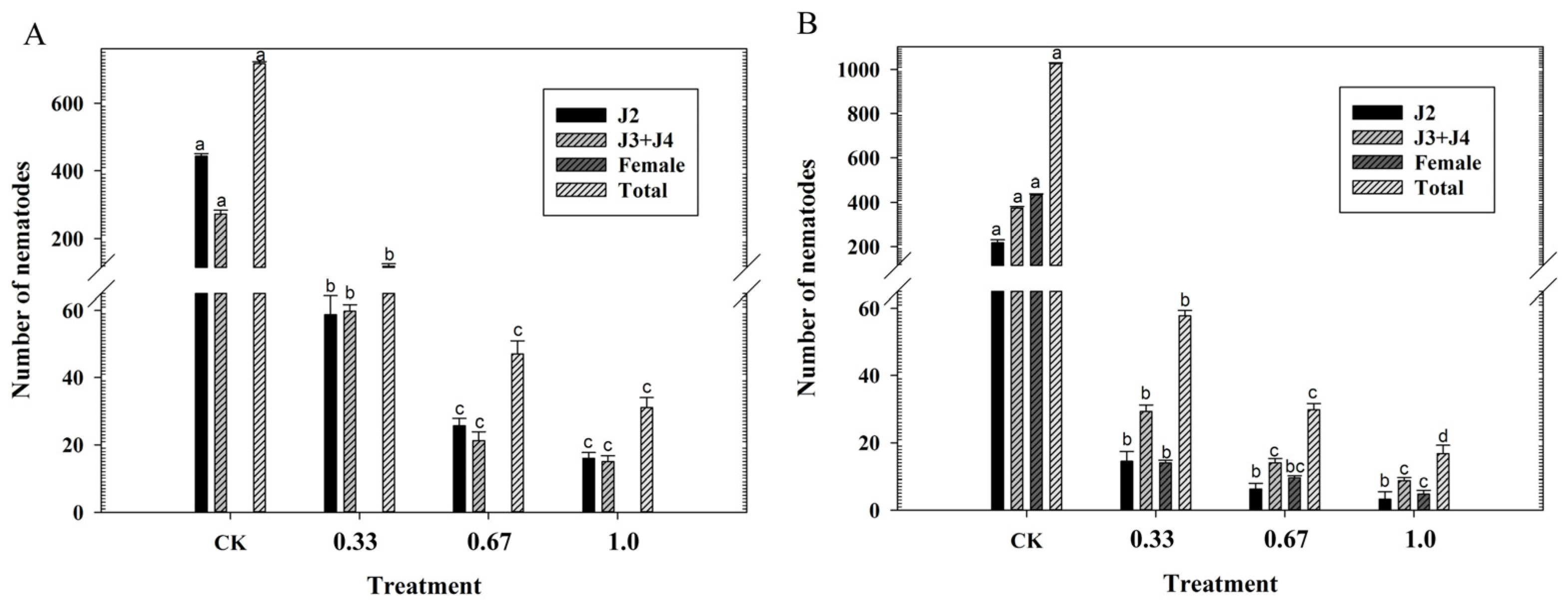
3.1. Effects of Fluopimomide on the Development of M. incognita

At 14 days post inoculation (dpi), compared to the inoculated control, populations of M. incognita second-stage juveniles (J2s) and third-stage/fourth-stage juveniles (J3s/J4s) were notably (p < 0.05) decreased by all treatments (Figure 1A). Fluopimomide at 1.0 and 0.67 mg/kg soil were the most effective in reducing the populations of J2s and J3s/J4s in tomato roots, followed by fluopimomide at 0.33 mg/kg soil. No females were detected at 14 dpi. Results at 42 dpi had a similar tendency (Figure 1B). In comparison to the inoculated control, M. incognita at all developmental stages were notably (p < 0.05) decreased by all treatments. No statistical differences were noted in the number of J2s in tomato roots between fluopimomide treatments. For the number of J3s/J4s, fluopimomide at higher concentrations was more effective than fluopimomide at 0.33 mg/kg soil. For the number of females, fluopimomide at 1.0 mg/kg soil was the most effective, followed by fluopimomide at other concentrations. A dose–response relationship existed between the total number of M. incognita and fluopimomide doses.

3.2. Effects of Fluopimomide on M. incognita Reproduction

In comparison to the inoculated control, fluopimomide significantly reduced the number of egg masses and eggs per egg mass (Figure 2). For the number of egg masses per g root, fluopimomide at higher concentrations was the most effective, followed by fluopimomide at 0.33 mg/kg soil (Figure 2A). In comparison to the inoculated control, fluopimomide notably decreased the number of eggs per egg mass by 85.8%, 81.7%, and 58.7%, respectively (Figure 2B).

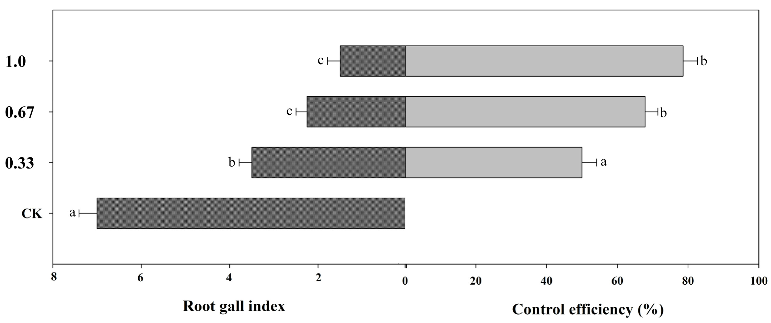
3.3. Effects of Fluopimomide on the Root Gall Index of Tomato

Compared to the inoculated control, all treatments notably decreased the root gall index (Figure 3). Fluopimomide at higher concentrations, with galling indexes of 1.50 and 2.25, were the most effective in decreasing the root gall index, with control effects of 78.6% and 67.9%, followed by fluopimomide at 0.33 mg/kg soil with a control effect of 50.0%.

3.4. Effects of Fluopimomide on the Brood Size and Germ Cell Number of C. elegans

A dose–response relationship existed between the brood size and germ cell number of C. elegans and fluopimomide concentrations (Figure 4). Compared to the untreated control, fluopimomide notably (p < 0.05) decreased the brood size by 6.4%, 32.7%, and 47.8% (Figure 4A). Compared to the untreated control, all nematicide treatments notably (p < 0.001) decreased the number of germ cell by 42.6%, 58.1%, and 77.0% (Figure 4B).

3.5. Effects of Fluopimomide on the Germ Cell Apoptosis of C. elegans

Compared to the control group, fluopimomide at 1.0 and 5.0 mg/L notably (p < 0.001) increased the number of cell corpses per gonad in the N2 strain of C. elegans by 8.8- and 14.4-fold, respectively, while no statistical effects (p = 0.10) were noted in fluopimomide at 0.2 mg/L (Figure 5A,B). Further studies on the germ cell apoptosis induced by fluopimomide were conducted in ced-3, ced-4, and ced-9 mutants (Figure 5C). These results indicated that there were no statistical differences in the number of cell corpses per gonad between the fluopimomide-treated worms and the control group in all mutants.

3.6. Effects of Fluopimomide on the DNA Damage of C. elegans

Compared to the control, fluopimomide at higher concentrations notably enhanced the expression of cep-1, egl-1, and clk-2, with fluopimomide at 5.0 mg/L being the highest (Figure 6A). No significant differences (p = 0.68; p = 0.09; p = 0.68, respectively) were observed between fluopimomide at 0.2 mg/L and the untreated control. Germline DNA damage was detected by cep-1, egl-1, hus-1, and clk-2 mutants. Similarly, no obvious differences were found between all fluopimomide treatments and the control group in germ cell apoptosis of the cep-1, egl-1, hus-1, and clk-2 mutants (Figure 6B).

4. Discussion

Root-knot nematodes possess strong reproductive ability and pose a devastating threat to agriculture. Our previous study indicated that fluopimomide caused a high M. incognita mortality and showed high control efficacy in pot and field trials [3,13]. However, the effects and molecular mechanisms of fluopimomide in inhibiting nematode reproduction remain unknown.
In previous studies, fluopimomide was found to be toxic to M. incognita [14]. In the present study, fluopimomide at tested concentrations significantly delayed M. incognita development in tomato roots and reduced gall numbers. Therefore, fluopimomide is a potential substitution for currently available nematocides. The results were also consistent with Ji et al. [31], who reported that β-aminobutyric acid treatment of rice plants inhibited the penetration of M. graminicola and resulted in delayed development of nematodes and giant cells. Zhan et al. [32] demonstrated that silicon reduced the infection of M. graminicola and delayed the development of nematodes inside roots. According to our results, one of the actions of fluopimomide against M. incognita is probably based on direct toxic effects on nematode development and reproductive capacity.
Reproductive toxicity is one of the most significant negative effects of pesticides. It is important to evaluate this and the underlying mechanisms. However, it is difficult to elucidate the mechanism of reproductive toxicity of fluopimomide directly through RKNs, because RKNs require a host with a long life cycle to complete their onset cycle [33]. C. elegans has become a model to study the mechanism of reproductive toxicity instead of RKNs, owing to its short life cycle and large-scale offspring [19]. Thus, the reproductive-toxicity molecular mechanism of fluopimomide was further studied in C. elegans. Brood size is commonly used to assess the reproductive capacity in C. elegans [34]. In the present study, fluopimomide significantly decreased the brood size, which suggested that fluopimomide induced the reproductive toxicity in C. elegans. Some studies reported that exposure to pesticides reduced brood size and induced reproductive toxicity in C. elegans [29,35]. Moya et al. [19] showed that chlorpyrifos and 2,4-dichlorophenoxyacetic acid decreased the brood size, caused abnormal vitellogenin distribution in C. elegans.
After clarifying that fluopimomide reduced the brood size of C. elegans, whether this phenomenon was related to the reduction of the lower total number of germ cells was further investigated. Two U-shaped gonads constitute the reproductive system of C. elegans, and germ cells are distributed in the gonads. Since the intestines covers one of the gonad arms, only germ cells in one gonad arm are counted [36]. In this study, fluopimomide significantly decreased the number of germ cells, which confirmed that fluopimomide induced reproductive toxicity. Similarly, Cheng et al. [22] reported that exposure to furfural acetone significantly decreased the number of germ cells of C. elegans.
Whether germ cell apoptosis led to the decrease in germ cells in C. elegans was further studied. In the present study, fluopimomide increased the number of cell corpses per gonad, indicating that fluopimomide reduced the brood size of C. elegans by inducing germ-cell apoptosis. In line with this observation, a previous study reported that endosulfan significantly increased the apoptosis level of germ cells in C. elegans, inducing reproductive toxicity [37].
The apoptosis of germ cells induced by fluopimomide was further studied in the mutants of ced-3, ced-4, and ced-9. In C. elegans, the anti-apoptotic protein CED-9, the caspase protein CED-3, and the Apaf-1 homolog CED-4 act as a core apoptotic pathway to co-regulate germ cell apoptosis [38]. CED-3 can bind to CED-4 to initiate cell apoptosis, while CED-9 combines with CED-4 to prevent CED-3 activation, thereby inhibiting germ cell apoptosis [39]. In the present study, germ cell apoptosis was significantly increased after fluopimomide exposure in N2 nematodes, whereas the changes disappeared in ced-3, ced-4, and ced-9 mutants, indicating that these genes were necessary in inducing cell apoptosis. This result was in agreement with Wang et al. [40], who reported that bisphenol A induced the germ-cell apoptosis of N2 nematodes, while this induction was lost in ced-3 and ced-4 mutants.
During reproduction, germ cells carry genetic information to keep the genome complete [41]. When toxicants damage the genome, the DNA repair system can judge and remove it [42]. However, DNA damage can interrupt replication, lead to the reduction of germ cells and damage reproductive ability, and, finally, induce reproductive toxicity in the body [43]. Cep-1, encoding a p53-like protein and activating the transcription of egl-1, plays a key role in DNA damage-induced germ cell apoptosis in C. elegans [44]. Egl-1, a member of the apoptosis activators family, directly inhibits its activity after binding with CED-9 and releasing the apoptosis activator CED-4 from the protein complex containing CED-9/CED-4, thereby activating cell apoptosis [45]. In addition, the DNA damage checkpoint proteins HUS-1 and CLK-2 together activate the function of CEP-1 and also play essential roles in the process of DNA damage in C. elegans [46]. Apoptosis of germ cells is commonly used to detect DNA damage [47]. In this study, the mRNA expression of cep-1, egl-1, hus-1, and clk-2 were significantly increased after exposure to higher concentrations of fluopimomide, while no obvious effects in germ cell apoptosis were observed in their mutants, indicating that these genes were essential for induction of DNA damage in C. elegans. This agreed with Ni et al. [48], who reported that 2,4,6-trinitrotoluene (TNT) altered the expression of cep-1, egl-1, and hus-1 in N2 nematodes, while these changes disappeared in the loss-function mutants, indicating that TNT might induce DNA damage in germ cell by activating hus-1, cep-1, and egl-1 genes.

5. Conclusions

In the present study, the results indicated that fluopimomide inhibited M. incognita development in tomatoes and reduced the number of galls. Moreover, further investigation of the mechanism of reproductive toxicity was performed using C. elegans. After C. elegans are exposed to fluopimomide, it might activate the expression of p53-like protein CEP-1 by inducing the expression of the DNA damage checkpoint proteins HUS-1 and CLK-2. CEP-1 further activates the transcription of EGL-1. And EGL-1 binds with CED-9, thus releasing the apoptosis activator CED-4 from the protein complex containing CED-9/CED-4, activating CED-3 caspase, promoting germ-cell death, and ultimately leading to the reduction of brood size in C. elegans.

Author Contributions

K.Q. conceived and designed research. H.L., B.L., S.Z. (Siqi Zhang), and X.J. conducted experiments and analyzed the data. All authors contributed to the discussion of the results. K.Q. and Z.W. supervised the research. K.Q. and S.Z. (Shouan Zhang) wrote and revised the paper. All authors have read and agreed to the published version of the manuscript.

Funding

This research was funded by the Shandong Innovation Capability Enhancement Project for Technological Small and Medium-sized Enterprises (2023TSGC0613), the Shandong Provincial Natural Science Foundation (ZR2021MC065), the Shandong Province Key Research and Development Plan (2021TZXD011), and the National Natural Science Foundation of China (31601661).

Institutional Review Board Statement

Not applicable.

Informed Consent Statement

Not applicable.

Data Availability Statement

The data are available from the corresponding author on reasonable request.

Acknowledgments

We thank the Caenorhabditis Genetics Center (University of Minnesota, Minneapolis, MN, USA), Ye Hong (Shandong University, Shandong, China), and Huixin Li (Nanjing Agricultural University, Nanjing, Jiangsu) for providing the C. elegans strains used in this study.

Conflicts of Interest

The authors declare no conflict of interest.

References

  1. Rutter, W.B.; Franco, J.; Gleason, C. Rooting out the mechanisms of root-knot nematode-plant interactions. Annu. Rev. Phytopathol. 2022, 60, 43–76. [Google Scholar] [CrossRef] [PubMed]
  2. Qiao, K.; Liu, X.; Wang, H.; Xia, X.; Ji, X.; Wang, K. Effect of abamectin on root-knot nematodes and tomato yield. Pest Manag. Sci. 2012, 68, 853–857. [Google Scholar] [CrossRef] [PubMed]
  3. Li, J.; Meng, Z.; Li, N.; Ji, X.; Dong, B.; Zhang, S.; Qiao, K. Evaluating a new non-fumigant nematicide fluopimomide for management of southern root-knot nematodes in tomato. Crop. Prot. 2020, 129, 105040. [Google Scholar] [CrossRef]
  4. Jones, J.; Haegeman, A.; Danchin, E.; Gaur, H.; Helder, J.; Jones, M.; Kikuchi, T.; Manzanilla-López, R.; Palomares-Rius, J.; Wesemael, W.; et al. Top 10 plant-parasitic nematodes in molecular plant pathology. Mol. Plant Pathol. 2013, 14, 946–961. [Google Scholar] [CrossRef]
  5. Mao, L.; Wang, Q.; Yan, D.; Liu, P.; Shen, J.; Fang, W.; Hu, X.; Li, Y.; Ouyang, C.; Guo, M.; et al. Application of the combination of 1,3-dichloropropene disulfide by soil injection or chemigation: Effects against soilborne pests in cucumber in China. J. Integr. Agric. 2016, 15, 145–152. [Google Scholar] [CrossRef]
  6. Desaeger, J.; Wram, C.; Zasada, I. New reduced-risk agricultural nematicides-rationale and review. J. Nematol. 2020, 52, 1–16. [Google Scholar] [CrossRef]
  7. Cao, J.; Guenther, R.; Sit, T.; Lommel, S.; Opperman, C.; Willoughby, J. Development of abamectin loaded lignocellulosic matrices for the controlled release of nematicide for crop protection. Cellulose 2016, 23, 673–687. [Google Scholar] [CrossRef]
  8. Huang, B.; Qian, W.; Guo, M.; Fang, W.; Wang, X.; Wang, Q.; Yan, D.; Ouyang, C.; Li, Y.; Cao, A. The synergistic advantage of combining chloropicrin or dazomet with fosthiazate nematicide to control root-knot nematode in cucumber production. J. Integr. Agric. 2019, 18, 2093–2106. [Google Scholar] [CrossRef]
  9. Qiao, K.; Liu, Q.; Zhang, S. Evaluation of fluazaindolizine, a new nematicide for management of Meloidogyne incognita in squash in calcareous soils. Crop Prot. 2021, 143, 105469. [Google Scholar] [CrossRef]
  10. Watson, T.T. Sensitivity of Meloidogyne enterolobii and M. incognita to fluorinated nematicides. Pest Manag. Sci. 2022, 78, 1398–1406. [Google Scholar] [CrossRef]
  11. Giannakou, I.O.; Panopoulou, S. The use of fluensulfone for the control of root-knot nematodes in greenhouse cultivated crops: Efficacy and phytotoxicity effects. Cogent Food Agric. 2019, 5, 1643819. [Google Scholar] [CrossRef]
  12. Rathod, P.H.; Shah, P.G.; Parmar, K.D.; Kalasariya, R.L. The fate of fluopyram in the Soil-Water-Plant ecosystem: A Review. Rev. Environ. Contam. Toxicol. 2022, 260, 1. [Google Scholar] [CrossRef]
  13. Zhang, W.; Sun, W.; Wang, Y.; Liu, H.; Zhang, S.; Dong, B.; Qiao, K. Management of Meloidogyne incognita on cucumber with a new nonfumigant nematicide fluopimomide. Plant Dis. 2022, 106, 151–155. [Google Scholar] [CrossRef] [PubMed]
  14. Ji, X.; Li, J.; Meng, Z.; Li, N.; Dong, B.; Zhang, S.; Qiao, K. Fluopimomide effectively controls Meloidogyne incognita and shows a growth promotion effect in cucumber. J. Pest Sci. 2020, 93, 1421–1430. [Google Scholar] [CrossRef]
  15. Yu, Y.; Chen, H.; Hua, X.; Wang, C.; Dong, C.; Xie, D.; Tan, S.; Xiang, M.; Li, H. A review of the reproductive toxicity of environmental contaminants in Caenorhabditis elegans. Hyg. Environ. Health Adv. 2022, 3, 100007. [Google Scholar] [CrossRef]
  16. El-Nahhal, Y. Pesticide residues in honey and their potential reproductive toxicity. Sci. Total Environ. 2020, 741, 139953. [Google Scholar] [CrossRef]
  17. Cooper, R.L. Current developments in reproductive toxicity testing of pesticides. Reprod. Toxicol. 2009, 28, 180–187. [Google Scholar] [CrossRef]
  18. Kaletta, T.; Hengartner, M. Finding function in novel targets: C. elegans as a model organism. Nat. Rev. Drug Discov. 2006, 5, 387–399. [Google Scholar] [CrossRef]
  19. Moya, A.; Tejedor, D.; Manetti, M.; Clavijo, A.; Pagano, E.; Munarriz, E.; Kronberg, M. Reproductive toxicity by exposure to low concentrations of pesticides in Caenorhabditis elegans. Toxicology 2022, 475, 153229. [Google Scholar] [CrossRef]
  20. Yin, J.; Hong, X.; Wang, J.; Li, W.; Shi, Y.; Wang, D.; Liu, R. DNA methylation 6mA and histone methylation involved in multi-/trans-generational reproductive effects in Caenorhabditis elegans induced by atrazine. Ecotoxicol. Environ. Saf. 2023, 249, 114348. [Google Scholar] [CrossRef]
  21. Yu, C.W.; Luk, T.C.; Liao, V.H.C. Long-term nanoplastics exposure results in multi and trans-generational reproduction decline associated with germline toxicity and epigenetic regulation in Caenorhabditis elegans. J. Hazard. Mater. 2021, 412, 125173. [Google Scholar] [CrossRef] [PubMed]
  22. Cheng, W.; Yang, X.; Xue, H.; Huang, D.; Cai, M.; Huang, F.; Zheng, L.; Yu, Z.; Zhang, J. Reproductive toxicity of furfural acetone in Meloidogyne incognita and Caenorhabditis elegans. Cells 2022, 11, 401. [Google Scholar] [CrossRef] [PubMed]
  23. Wamucho, A.; Unrine, J.; May, J.; Tsyusko, O. Global DNA adenine methylation in Caenorhabditis elegans after multigenerational exposure to silver nanoparticles and silver nitrate. Int. J. Mol. Sci. 2023, 24, 6168. [Google Scholar] [CrossRef]
  24. Brenner, S. The genetics of Caenorhabditis elegans. Genetics 1974, 77, 71–94. [Google Scholar] [CrossRef] [PubMed]
  25. Bridge, J.; Page, L. Estimation of root-knot nematode infestation levels on roots using a rating chart. Int. J. Pest Manag. 1980, 26, 296–298. [Google Scholar] [CrossRef]
  26. Zhang, W.; Liu, H.; Fu, G.; Li, Y.; Ji, X.; Zhang, S.; Qiao, K. Exposure to fluopimomide at sublethal doses causes oxidative stress in Caenorhabditis elegans regulated by insulin/IGF-1-like signaling pathway. Environ. Toxicol. 2022, 37, 2529–2539. [Google Scholar] [CrossRef] [PubMed]
  27. Hou, L.; Jiang, M.; Guo, Q.; Shi, W. Zymolytic grain extract (ZGE) significantly extends the lifespan and enhances the environmental stress resistance of Caenorhabditis elegans. Int. J. Mol. Sci. 2019, 20, 3489. [Google Scholar] [CrossRef]
  28. Qu, Z.; Ji, S.; Zheng, S. BRAF controls the effects of metformin on neuroblast cell divisions in C. elegans. Int. J. Mol. Sci. 2021, 22, 178. [Google Scholar] [CrossRef]
  29. Liu, Y.; Zhang, W.; Wang, Y.; Liu, H.; Zhang, S.; Ji, X.; Qiao, K. Oxidative stress, intestinal damage, and cell apoptosis: Toxicity induced by fluopyram in Caenorhabditis elegans. Chemosphere 2022, 286, 131830. [Google Scholar] [CrossRef]
  30. Livak, K.J.; Schmittgen, T.D. Analysis of relative gene expression data using real-time quantitative PCR and the 2−ΔΔCT method. Methods 2001, 25, 402–408. [Google Scholar] [CrossRef]
  31. Ji, H.; Kyndt, T.; He, W.; Vanholme, B.; Gheysen, G. β-Aminobutyric acid-induced resistance against root-knot nematodes in rice is based on increased basal defense. Mol. Plant Microbe Interact. 2015, 28, 519–533. [Google Scholar] [CrossRef] [PubMed]
  32. Zhan, L.; Peng, D.; Wang, X.; Kong, L.; Peng, H.; Liu, S.; Liu, Y.; Huang, W. Priming effect of root-applied silicon on the enhancement of induced resistance to the root-knot nematode Meloidogyne graminicola in rice. BMC Plant Biol. 2018, 18, 50. [Google Scholar] [CrossRef] [PubMed]
  33. Shivakumara, T.; Dutta, T.; Chaudhary, S.; von Reuss, S.; Williamson, V.; Rao, U. Homologs of Caenorhabditis elegans chemosensory genes have roles in behavior and chemotaxis in the root-knot nematode Meloidogyne incognita. Mol. Plant-Microbe Interact. 2019, 32, 876–887. [Google Scholar] [CrossRef] [PubMed]
  34. Chen, H.; Wang, C.; Li, H.; Ma, R.; Yu, Z.; Li, L.; Xiang, M.; Chen, X.; Hua, X.; Yu, Y. A review of toxicity induced by persistent organic pollutants (POPs) and endocrine-disrupting chemicals (EDCs) in the nematode Caenorhabditis elegans. J. Environ. Manag. 2019, 237, 519–525. [Google Scholar] [CrossRef]
  35. Lu, Q.; Bu, Y.; Ma, L.; Liu, R. Transgenerational reproductive and developmental toxicity of tebuconazole in Caenorhabditis elegans. J. Appl. Toxicol. 2020, 40, 578–591. [Google Scholar] [CrossRef]
  36. Yin, J.; Liu, R.; Jian, Z.; Yang, D.; Pu, Y.; Yin, L.; Wang, D. Di (2-ethylhexyl) phthalate-induced reproductive toxicity involved in dna damage-dependent oocyte apoptosis and oxidative stress in Caenorhabditis elegans. Ecotoxicol. Environ. Saf. 2018, 163, 298–306. [Google Scholar] [CrossRef]
  37. Du, H.; Wang, M.; Dai, H.; Hong, W.; Wang, M.; Wang, J.; Weng, N.; Nie, Y.; Xu, A. Endosulfan isomers and sulfate metabolite induced reproductive toxicity in Caenorhabditis elegans involves genotoxic response genes. Environ. Sci. Technol. 2015, 49, 2460–2468. [Google Scholar] [CrossRef]
  38. Salinas, L.S.; Maldonado, E.; Navarro, R.E. Stress-induced germ cell apoptosis by a p53 independent pathway in Caenorhabditis elegans. Cell Death Differ. 2006, 13, 2129–2139. [Google Scholar] [CrossRef]
  39. Boag, P.; Nakamura, A.; Blackwell, T. A conserved RNA-protein complex component involved in physiological germline apoptosis regulation in C. elegans. Development 2005, 132, 4975–4986. [Google Scholar] [CrossRef]
  40. Wang, Y.; Zhang, L.; Luo, X.; Wang, S.; Wang, Y. Bisphenol A exposure triggers apoptosis via three signaling pathways in Caenorhabditis elegans. RSC Adv. 2017, 7, 32624–32631. [Google Scholar] [CrossRef]
  41. Jackson, S.P.; Bartek, J. The DNA-damage response in human biology and disease. Nature 2009, 461, 1071–1078. [Google Scholar] [CrossRef] [PubMed]
  42. Norbury, C.J.; Hickson, I.D. Cellular responses to DNA damage. Annu. Rev. Pharmacol. Toxicol. 2001, 41, 367–401. [Google Scholar] [CrossRef] [PubMed]
  43. Luo, Y.; Hartford, S.A.; Zeng, R.; Southard, T.L.; Shima, N.; Schimenti, J.C. Hypersensitivity of primordial germ cells to compromised replication-associated DNA repair involves ATM-p53-p21 signaling. PLoS Genet. 2014, 10, e1004471. [Google Scholar] [CrossRef] [PubMed]
  44. Schumacher, B.; Hofmann, K.; Boulton, S.; Gartner, A. The C. elegans homolog of the p53 tumor suppressor is required for DNA damage-induced apoptosis. Curr. Biol. 2001, 11, 1722–1727. [Google Scholar] [CrossRef]
  45. Conradt, B.; Horvitz, H.R. The C. elegans protein EGL-1 is required for programmed cell death and interacts with the Bcl-2-like protein CED-9. Cell 1998, 93, 519–529. [Google Scholar] [CrossRef]
  46. Lettre, G.; Hengartner, M.O. Developmental apoptosis in C. elegans: A complex CEDnario. Nat. Rev. Mol. Cell Biol. 2006, 7, 97–108. [Google Scholar] [CrossRef]
  47. Lans, H.; Vermeulen, W. Tissue specific response to DNA damage: C. elegans as role model. DNA Repair 2015, 32, 141–148. [Google Scholar] [CrossRef]
  48. Ni, S.; Zhang, H.; Sun, L.; Zhao, Y.; Pei, C.; Nie, Y.; Liu, X.; Wu, L.; Xu, A. Transgenerational reproductive toxicity of 2, 4, 6-trinitrotoluene (TNT) and its metabolite 4-ADNT in Caenorhabditis elegans. Environ. Toxicol. Pharmacol. 2022, 92, 103865. [Google Scholar] [CrossRef]
Figure 1. Effects of fluopimomide on the number of different stages of Meloidogyne incognita in tomato roots (A: 14 dpi; B: 42 dpi). The values represent mean ± SE from five replicates. Different letters above the bars indicate statistical significance at p < 0.05. CK = non-treated control with nematodes; 0.33 = fluopimomide at 0.33 mg/kg soil; 0.67 = fluopimomide at 0.67 mg/kg soil; 1.0 = fluopimomide at 1.0 mg/kg soil; dpi = days post inoculation.
Agronomy 13 02471 g001
Figure 2. Effects of fluopimomide on the number of egg masses per g root (A) and eggs per egg mass (B) of Meloidogyne incognita. The values represent mean ± SE from five replicates. Different letters above the bars indicate statistical significance at p < 0.05. CK = non-treated control with nematodes; 0.33 = fluopimomide at 0.33 mg/kg soil; 0.67 = fluopimomide at 0.67 mg/kg soil; 1.0 = fluopimomide at 1.0 mg/kg soil.
Agronomy 13 02471 g002
Figure 3. Effects of fluopimomide on the root gall index and control efficacy of Meloidogyne incognita. The values represent mean ± SE from five replicates. Different letters above the bars indicate statistical significance at p < 0.05. CK = non-treated control with nematodes; 0.33 = fluopimomide at 0.33 mg/kg soil; 0.67 = fluopimomide at 0.67 mg/kg soil; 1.0 = fluopimomide at 1.0 mg/kg soil.
Agronomy 13 02471 g003
Figure 4. Effects of fluopimomide on brood size and number of germ cells in Caenorhabditis elegans. (A) Brood size, (B) germ cell number. The values represent mean ± SE from five replicates. Different letters above the bars indicate statistical significance at p < 0.05.
Agronomy 13 02471 g004
Figure 5. Effects of fluopimomide on germ cell apoptosis in Caenorhabditis elegans. (A) Microscopic fluorescence imaging of germ cells. Red arrows indicated apoptotic germ cells. (B) Number of apoptotic germ cells in N2 nematodes, (C) number of apoptotic germ cells in ced-3, ced-4, and ced-9 mutants. The values represent mean ± SE from five replicates. Different letters above the bars indicate statistical significance at p < 0.05.
Agronomy 13 02471 g005
Figure 6. Effects of fluopimomide on expression of genes related to DNA damage of C. elegans. (A) The expression of cep-1, egl-1, hus-1, and clk-2 in N2 nematodes, (B) number of apoptotic germ cells in cep-1, egl-1, hus-1, and clk-2 mutants. The values represent mean ± SE from five replicates. Different letters above the bars indicate statistical significance at p < 0.05.
Agronomy 13 02471 g006
Table 1. The base sequences of the primers.
GenesPrimers
ForwardReverse
act-1CCAGGAATTGCTGATCGTATGCAGAATGGAGAGGGAAGCGAGGATAGA
cep-1TACCCGATTCGCAGGACATCGCATCGGAAATCTTTGGCGT
egl-1GCCTCAACCTCTTCGGATCTGCACATTGCTGCTAGCTTGG
hus-1GCGGCAATCGACGTGTTTATCCGGGCAGAACACGTACTAA
clk-2CACAGTGCCCAACAAAGTCGTGACATGCTCGCCAGACAAT
Disclaimer/Publisher’s Note: The statements, opinions and data contained in all publications are solely those of the individual author(s) and contributor(s) and not of MDPI and/or the editor(s). MDPI and/or the editor(s) disclaim responsibility for any injury to people or property resulting from any ideas, methods, instructions or products referred to in the content.

