Article

In Vitro Organogenesis of Critically Endangered Lachenalia viridiflora

Department of Ornamental Plants and Garden Art, Faculty of Biotechnology and Horticulture, University of Agriculture in Krakow, 29-Listopada 54, 31-425 Krakow, Poland
*
Author to whom correspondence should be addressed.
Agronomy 2022, 12(2), 475; https://doi.org/10.3390/agronomy12020475
Submission received: 30 December 2021 / Revised: 27 January 2022 / Accepted: 9 February 2022 / Published: 14 February 2022

Abstract

:
Micropropagation via organogenesis of Lachenalia viridiflora W. F. Barker, a native endangered plant from South Africa, is presented in this study. Leaves, collected from in vivo plants, were a source for culture initiation to obtain three types of explants (leaf, bulb, callus). They were grown on Murashige and Skoog (MS) medium supplemented with two concentrations of sucrose (3 or 6%, marked as S3 and S6) and different concentrations of benzylaminopurine (BA) (0.0–5.0 μM) and naphthalene acetic acid (NAA) (0.0–0.5 μM). The results demonstrated that bulbing was more efficient on media with 3% sucrose and this process was closely related to explant type—irrespective of media type twice more adventitious bulbs (5.1–5.6) were regenerated on leaf and bulb explants than on callus (2.6). The highest value of adventitious bulb number was recorded for interaction: leaf × S3/2.5 BA/0.5 NAA (9.3 bulbs per explant). Irrespective of media type, the higher the number of adventitious bulbs (obtained from leaf and bulb explants), the lower their diameter. Moreover, adventitious bulbs regenerated from bulb explants formed the longest leaves (18.9 mm) and in the greatest percentage (52.3%). The highest biomass weight increase indicator (50 times) was recorded for interaction: leaf × S6/2.5 BA/0.25 NAA. Various combinations of plant growth regulators favor biomass increase.

1. Introduction

Lachenalia is a genus of bulbous geophytes from Asparagaceae family consisting of 136 species restricted to the winter rainfall regions of southern Africa [1,2]. Lachenalia viridiflora, named for its unusual, extremely rare in nature, viridian flowers (Figure 1A), is critically endangered with an extent of occurrence less than 100 km2 [3]. Its population is threatened by anthropogenic activities [1,3]. Turquoise flowers combined with spotted leaves (Figure 1A) [1] make Lachenalia viridiflora an ideal and extraordinary garden plant. This species, as many other wild lachenalias, was used in breeding programs to obtain the new line of cultivars with global horticultural potential [4]. It was a male parent for obtaining ‘Aqua Lady’ cultivar with turquoise blue flowers and maroon spots on leaves [5]. It is likely that the economic importance of Lachenalia viridiflora will increase in the near future due to its unusual decorativeness. Therefore, it is necessary to look for other, more efficient techniques than traditional methods of propagation. In vitro methods, such as organogenesis, allow for mass plant propagation and were described for selected lachenalia species and cultivars [6,7,8,9,10,11,12,13]. Most often the leaf tissue [11,13,14,15,16,17] and less frequently bulb scales [12,18,19] were used as initial plant material to obtain adventitious bud formation. The aforementioned type of explants are frequently used in different geophytes for the induction of storage organs, but in the case of endangered species, choosing storage organ for culture initiation may be problematic due to the destruction of the mother plant [20]. In such a situation, the choice of a leaf explant seems more appropriate. In case of lachenalia, leaf cutting has also been described as a main and sufficient in vivo method of lachenalia propagation [4,21].
Apart from various type of explant, different media compositions are used for propagation. Formation of bulbs in vitro requires exogenous application of appropriate plant growth regulators (PGRs), in which the endogenous level is insufficient to that process [22]. Proper type and concentration of PGRs are essential to develop efficient clonal regeneration protocol for Lachenalia viridiflora. PGRs like: BA (cytokinin) and NAA (auxin) are routinely applied to induce various geophytes’ organogenesis [20,22,23,24,25,26,27], also in lachenalia propagation [9,15,18,28]. According to Aremu et al. (2017) [11], BA belongs to the best cytokinin for propagation of Lachenalia montana. Simultaneously, Niederwieser and van Staden [15] claim that NAA is crucial for adventitious bud formation in Lachenalia hybrids. The same PGRs are often applied for micropropagation of other plants from the Asparagaceae family [26].
Sucrose, as a carbon source, is commonly used in micropropagation and influences bulb formation in vitro [20,29]. In the majority of geophytes, high sucrose concentration (6% or higher) in medium is needed for bulb development [22]. According to Van Rensburg and Vcelar [30] optimal sucrose concentration for bud formation in Lachenalia cultures amounts to 50–70 g/L. However, in the Hyacinthus experiments, 3% of sucrose was the sufficient concentration which even favored the formation of bulbs, as opposed to 6% [31].
Plants obtained by micropropagation are more likely to be free of viruses. This is particularly important in the context of lachenalia’s susceptibility to the Ornithogalum mosaic virus (OMV), causing negative symptoms on flowers and leaves which completely eliminate plants from further cultivation [32], or to the Veltheimia mosaic virus [33]. Plant tissue cultures are also extremely useful for the rapid multiplication of rare or endangered genotypes [10,26,34] due to the small amount of initial plant tissue required and the high number of plants obtained [35]. The preservation of rare genotypes is in line with maintaining plant biodiversity, which can be supported by introducing and developing new, more efficient and rapid methods of in vitro multiplication [36].
So far, only two protocols for in vitro propagation of Lachenalia viridiflora have been described. The first refers to plantlet regeneration through somatic embryogenesis [37], second to seed germination under in vitro conditions [38]. In this study, for the first time, the effect of explant type, sucrose and PGRs’ concentration on in vitro organogenesis threatened with extinction of Lachenalia viridiflora was investigated. Based on the results obtained, the preliminary propagation protocol for its mass propagation and conservation will be proposed.

2. Materials and Methods

2.1. Plant Material and Culture Conditions

The experiment was conducted using Lachenalia viridiflora W. F. Barker material, obtained from the greenhouse collection at the University of Agriculture in Krakow. In vitro cultures were initiated in March 2018 using leaves, which were previously prepared as follows: surfaced-disinfected with 70% ethanol (1 min) (Avantor Performance Materials, Gliwice, Poland) and 15% solution Domestos (15 min) (Unilever, Warszawa, Poland), rinsed 3 times with sterile water. The leaves were then plated and cultured on solid MS [39] (Sigma-Aldrich, Merck, Poland) medium, supplemented with 30 g L−1 sucrose (Avantor Performance Materials, Poland) in the dark at 20 ± 2 °C.
Within 10 weeks, callus had occurred on the surface of the leaves and after the next 14 weeks, in the callus, development of adventitious bulblets (with leaves) was observed (Figure 1B). From these cultures, 3 types of explants were taken for the experiment: leaf (10–11 mm length, 2–3 mm width) (Figure 1C), bulb (4–5 mm diameter) (Figure 1D) and callus (portions of friable, undifferentiated tissue, 7–8 mm diameter) (Figure 1E). Bulbs and leaves were cut lengthwise (V-shaped). Each type of horizontal explant was put on 8 combinations of solid MS media with different concentration of PGRs: BA (0–5 µM) (Sigma-Aldrich, Merck, Poland), NAA (0–0.5 µM) (Sigma-Aldrich, Merck, Poland) and sucrose: 3% (S3) or 6% (S6) (Table 1). The media were adjusted to pH 5.8, gelled with 0.5% Lab-agar (Biocorp, Warszawa, Poland) and autoclaved. Each screw glass jar (110 mm × 60 mm, volume 300 mL) with 40 mL of MS medium contained five explants of the same type. Five jars for each type of explant were exposed to each of the eight media combinations.
All types of the explants were weighted at the beginning of the experiment to determine initial explant fresh weight. The cultures were maintained in darkness at 20 ± 2 °C for 12 weeks, after which biometrical observations were made.

2.2. Biometrical Observations

Induction of adventitious bulbs: their number (Bulbs No.) and diameter (Bulb D), percentage of bulbs forming leaves (Bulbs FL), length of the leaves (Leaf L) and biomass (initial explant fresh weight + fresh weight of adventitious bulbs with or without leaves) were recorded for all types of explants from all combinations of media (3 explants × 8 media), after 12 weeks of culture. Additionally, biomass weight increase indicator (WI) was calculated in accordance to Equation (1):
WI   % = biomass     initial   explant   fresh   weight   ×   100   initial   explant   fresh   weight
During the experiment, the dynamics of adventitious bulb formation on each type of explant and on all combinations of media were observed every 4 weeks.

2.3. Statistical Analysis

The results were analyzed using Statistica 13.3 software (TIBCO Software Inc., Palo Alto, CA, USA) [40]. Statistical analyses were applied to all data that were previously checked for normality by the Shapiro–Wilk test. Analysis of Variance (two-way ANOVA) followed by Duncan’s multiple range test (at a significance level p ≤ 0.05) were performed to determine the effects of the type of explant and medium on the examined features of Lachenalia viridiflora, as well as to identify the significant differences among explant-medium combinations. The analysis of dynamics was presented using descriptive statistics. Then, for more in-depth study of interrelations among factors, in particular the individual media components, and studied culture traits data were subjected to Principal component analysis (PCA) and Pearson’s correlation coefficients were computed. PCA was calculated using total dataset including all studied traits, media and explant types. Variables were previously standardized. The number of principal components capturing most relevant data variation was determined according to Kaiser rule and percentage of variance criterion [41,42]. As a part of the analysis, a PCA biplot was created that allowed for the investigation of relationships between variables and observations.

3. Results and Discussion

Many studies have focused on the optimization of in vitro culture conditions for effective regeneration, thereby restoration of endangered geophytes and their conservation [20]. In the present study, formation of adventitious bulbs via organogenesis of Lachenalia viridiflora under various culture conditions was observed. This issue, to our knowledge, has not been reported before.
In this study, the analysis of the main effects proved that interactions between treatment groups were significantly different (Table 2).
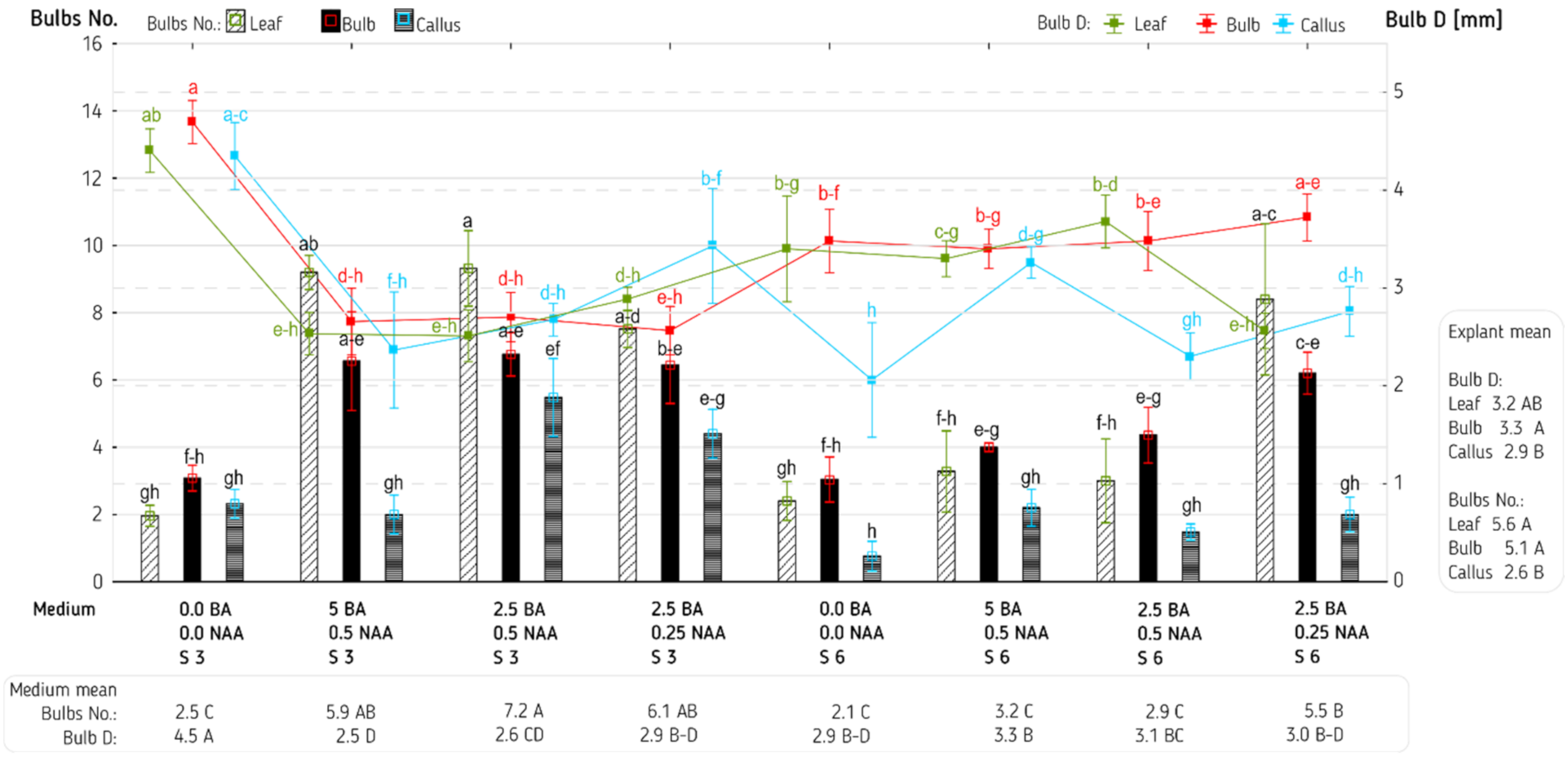
In the present study, leaf and bulb explants were cut lengthwise due to the information that wounding may enhance forming of buds in some lachenalia’s genotypes [14,17]. According to Grace and Van Staden (2003) [9], the genotype strongly influences the period of adventitious bud formation, whereas Niederwieser and Ndou (2002) [18] claim that in Lachenalia hybrid cultures, number and size of bulblets depended on tissue age. Bulbs are desirable regenerants in Lachenalia due to faster transfer to ex vitro conditions and easier transport, as storage organs characterize higher survival rate than shoots [17]. Similar conclusions were drawn in the case of other geophytes [22,43,44,45]. In present study, the adventitious bulbs of Lachenalia viridiflora developed on the explants treated with BA and NAA via direct organogenesis, similarly to other experiments with geophytes: Tulipa tarda, Allium aflatunense, Lilium ledebourii [46,47,48] and slight callus tissue had appeared only on the surface of leaf and bulb explants. The explant types that were studied here showed different ability of bulbing, which additionally depended on media composition (Figure 2). The lowest value of adventitious bulbs number (0.8 bulb per explant) was noticed for interaction callus × S6/without PGRs but this value did not differ statistically from values assigned to 11 other two-factor objects (1.5–3.3 bulbs per explant). The highest value of this feature was recorded for leaf explant × S3/2.5 BA/0.5 NAA (9.3 bulbs per explant), and together with other values (6.6–9.2 bulbs per explant) assigned to five objects, formed one statistical group which did not include variables such as callus and media without PGRs. It proves that these variables should not be used in the propagation procedure of Lachenalia viridiflora. The highest value of adventitious bulb number (9.3) noticed in this study was nearly the same to that recorded for bulbs number (9.7) formed in vivo on lachenalia ‘Namakwa’ leaf cuttings [21], however, the length of such a cutting (100 mm) corresponds to the length of 10 leaf explants used here. This implies that in vitro organogenesis may be highly efficient compared to traditional methods of lachenalia propagation. In our study, comparing the explant types, irrespective of media combination, can be divided into two statistical groups. The first consists of callus, which has proved to be a less favorable bulb-forming explant (2.6 bulbs), and the second of bulb and leaf explants which formed nearly twice more bulbs (5.1–5.6 bulbs). Most Lachenalia species form not enough daughter bulbs [4], thus high regeneration potential of leaf tissue within this genus [14] is used by growers in commercial lachenalia propagation via leaf cuttings [49]. Leaf explants were also most often applied during in vitro organogenesis within this genus [11,15,28]. In the present experiment, bulb explants (the counterpart of a modified leaf) were also used with sufficient results, whereas callus explant, probably due to its structure and characteristics, turned out to be a worse plant material, although it was used successfully in an experiment with others geophytes [50,51]. According to Fehér [52], not every but only certain callus cells are involved in organogenesis. This can explain why callus was a less favorable bulb-forming explant in this study. Analyzing the impact of media combination, irrespective of explant type, it was found that more adventitious bulbs were formed on medium S3/2.5 BA/0.5NAA (7.2 bulbs per explant) than on medium S6/2.5 BA/0.25 NAA (5.5 bulbs per explant) and on four other media combinations which formed a homogeneous statistical group in terms of this feature; they were: S3 or S6/without PGRs, S6/2.5 BA/0.5 NAA and S6/5 BA/0.5 NAA (2.0–3.2 bulbs per explant). Phenotypic responses of Lachenalia montana in vitro regenerants also depended on exogenous PGRs; 1 µM BA positively influenced the formation of the adventitious bulbs from leaf explants, while 10 µM BA treatment entirely inhibited bulb production and caused formation of callus [11]. Highest number of buds (20.6) in Lachenalia cultures were recorded on lower BA concentration (0.5 and 1.0 mg L−1) [15]. Additionally, 1.0 mg L−1 BA with 0.1 mg L−1 NAA occurred to be the best combination for bulbing in Lachenalia ‘Romaud’ culture [28]. High level of BA or NAA inhibits formation of bulbs in Lilium cultures as well [53,54], however, slight differences were observed on Lilium regale scale explants, between 5 µM and 10 µM BA treatment in bulbing, which amounted to 1.7 and 2.4 bulbs, respectively [27]. On the contrary to the above experiments, the highest number (16) of adventitious bulblets in Muscari muscarimi cultures were noticed when scale explants were treated with 18 µM BA and 3 µM NAA [55]. In the experiments with Tulipa tarda, most adventitious bulbs were formed on callus explants cultured on 0.5 μM BA and 3% sucrose [46,51], but when the same medium contained 6% sucrose, the number of adventitious bulbs significantly decreased (similarly to the present results with Lachenalia viridiflora). Moreover, if the medium was free of BA, there were no differences between 3 and 6% sucrose in the bulb regeneration [46]. In other Lachenalia cultures [17], there were no differences in bulbing between 3% and 6% sucrose, although larger bulblets formed under higher sucrose concentration. Sucrose is regarded as the main component of medium responsible for bulb development [17,22]. Bach et al. [31] in Hyacinthus culture demonstrated that high level of both sucrose and glucose had positive impact on storage organ formation. Some Tulipa cultivars formed bulbs after glucose alone or with fructose treatment [56], whereas in Lilium culture, starch was the main component involved in bulblet formation [45]. High sucrose concentration promoted formation of bulb in: Tulipa [57], Lilium [58], Hyacinthus [59,60] and Narcissus [61].
Taking into account the two examined factors (explant × medium), the adventitious bulbs had a diameter ranging from 2.1 to 4.7 mm (Figure 2). The distinguishing medium for all explants was the one without PGRs and with 3% sucrose. Analysis irrespective of explant type confirmed that adventitious bulbs formed on this medium had significantly higher diameter (4.5 mm) compared to the diameter of the adventitious bulbs regenerated on the other media (2.5–3.3 mm). Irrespective of media combination, higher diameter was noticed for adventitious bulbs obtained from bulb explants (3.3 mm) than from callus (2.9 mm). With regard to leaf and bulb explants, a relationship was observed—the more bulbs are formed, the smaller their diameter (Figure 6B). Similar observation was made by Cheesman et al. (2010) [62] during Eucomis zambesiaca bulbing. Lachenalia ‘Ronina’ formed, on average, 5.4 adventitious bulbs/explant after 16 weeks of treatment with 3% sucrose; 2.5 μM BA, 0.5 μM NAA with 4.1 mm in diameter [12]. Diameter of Lilium regale adventitious bulbs 6 weeks after cultivation on medium with 3% sucrose, 5–10 µM BA and 0.5 µM NAA, ranged between 4.0–4.2 mm [27]. In Fritillaria imperialis culture, treatment with 3% sucrose and 0.2 mg L−1 BA caused the largest bulb diameter which amounted to 10.1 mm [63]. In Hyacinthus culture, elevated sucrose concentration to 9% was the main factor that increased bulb diameter to 1.21 cm [64].
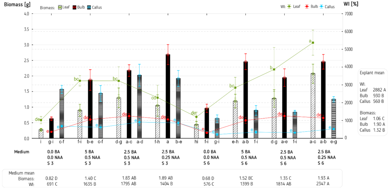
Biomass (defined as a sum of initial explant fresh weight and fresh weight of adventitious bulb with or without leaf) ranged from 0.27 g for leaf explant × S3/without PGRs to 2.68 g for bulb explant × S3/2.5 BA/0.25 NAA (Figure 3). Irrespective of media combinations, the bulb explant was the most favorable to obtain the highest value of biomass (1.9 g), next in this regard was callus (1.3 g) and the last, the leaf explant (1.1 g). Analyzing only the effect of media type, it was noticed that lack of PGRs resulted in obtaining approximately half the biomass (0.68–0.82 g) compared to the media containing the regulators (1.35–1.93 g depending on media combination), and sucrose seems to have no impact on this feature. These trends are confirmed by correlation analysis, presented in Figure 6B, revealing most often a positive relationship between explant biomass and PGRs content, and lack of correlation between explant biomass and sucrose. On the contrary, in Tulipa tarda cultures, there were no significant differences between biomass fresh weight obtained on MS media with or without PGRs [51]. In the case of Lapiedra martinezii, 6% sucrose was the main factor increasing biomass of bulblets, nearly to 1.5 g [65]. Whereas, in an experiment with Hippeastrum hybridum an average weight of bulbs, equal to 2.17 g, was achieved under 9% sucrose treatment [66], but in Eucomis zambesiaca cultures, the greatest impact on bulb weight, which amounted to 57 mg, had the temperature of 20 °C [62].
WI was inserted to compare the fresh weight increase on different explant type that had different initial fresh weight. Generally, on leaf explants regenerated on media with PGRs, the value of biomass increase (WI) was higher compared to other explant types, but these differences were not always of statistical significance (Figure 3). The highest value of WI among all objects was noticed for interaction leaf × S6/2.5 BA/0.25 NAA—the biomass increased by 50 compared to the initial fresh weight of the explant, whereas the combination of callus and S6/without PGRs only doubled the fresh biomass weight. The data also indicated that irrespective of media combination, the WI was higher for leaf explant (2882%) than for bulb explant (930%) and callus (560%). Irrespective of explant type, the lowest biomass increase was recorded for media without PGRs (575–691%), the highest for medium S6/2.5 BA/0.25 NAA (2347%), however, this value was on the same statistical level with two other media combinations: S6/2.5 BA/0.5 NAA (1814%) and S3/2.5 BA/0.5 NAA (1795%). In the experiment with Tulipa tarda, there were no differences in increase of fresh weight of callus tissue between media with or without 0.5 μM BA [46].
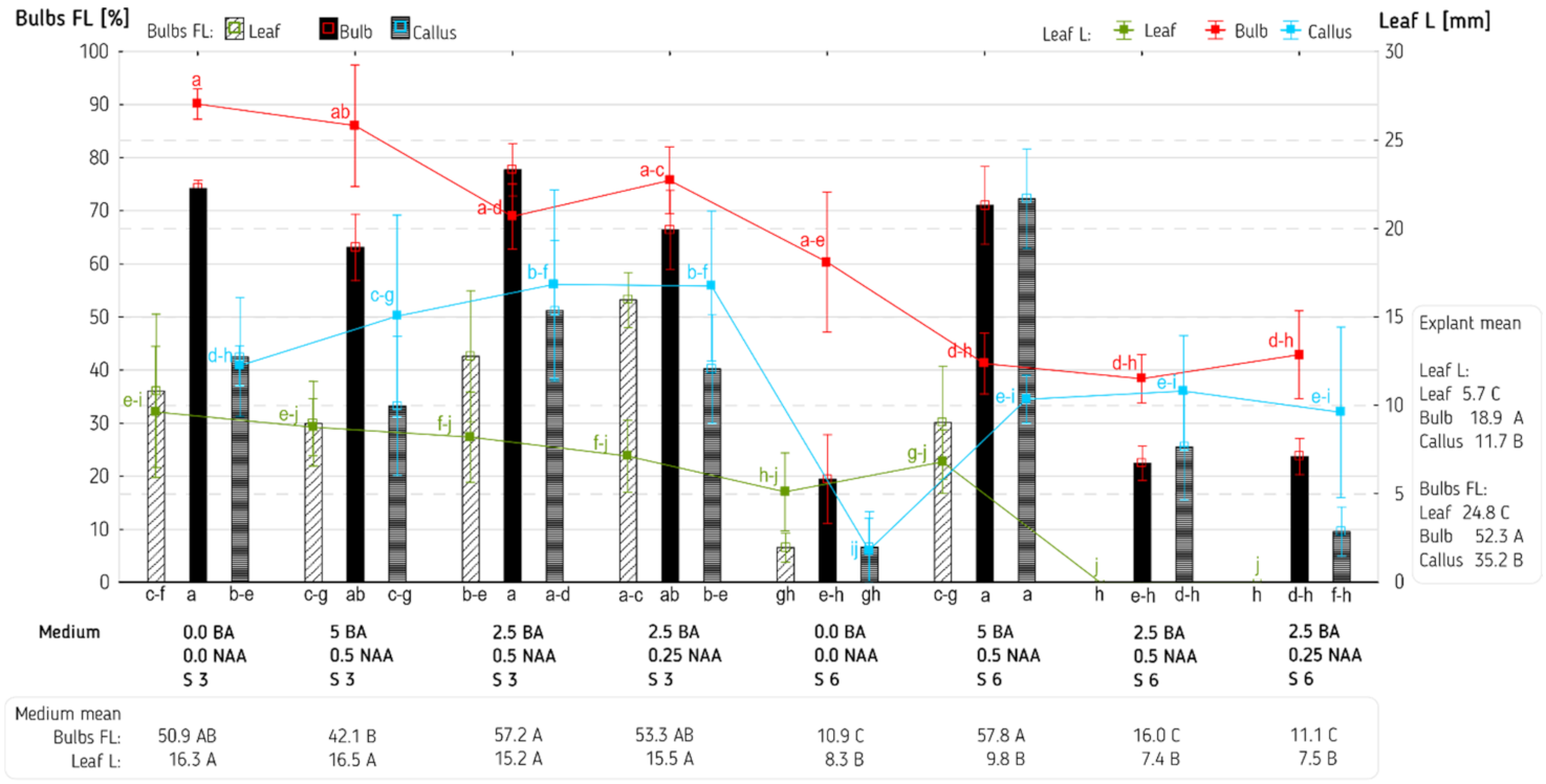
The response of adventitious bulbs in relation to the ability to form leaves depended on the interaction of two studied factors, but no clear tendency could be defined in which of these factors might have been more important in this interaction (Figure 4). Using two combinations: leaf explant × S6/2.5 BA/0.25 NAA and leaf explant × S6/2.5 BA/0.5 NAA, resulted in no leaf formation. In other objects, 6.6–77.7% (depending on factors combination) of adventitious bulbs showed the ability to form leaves. The analysis, carried out irrespective of individual factors, revealed some statistically significant relationships with regard to this feature. Irrespective of media combination, the adventitious bulbs regenerated from three explant types displayed different potential for leaf formation. Leaves were formed most frequently by adventitious bulbs originated from bulb explants (52.3%), then from callus (35.2%) and, last often, from leaf explants (24.8%). Irrespective of the media, adventitious bulbs on the bulb explants developed earlier than on other explants (Figure 1G,J), therefore, the leaves also developed earlier and faster, and were longer. Irrespective of explant type, less adventitious bulbs cultivated on media S6/without PGRs, S6/2.5 BA/0.25 NAA and S6/2.5 BA/0.5 NAA (10.9/11.1/16.0%, respectively) formed leaves compared to adventitious bulbs cultivated on the other five media combinations (42.1–57.8 depending on medium type). Bulbing was the main goal of this experiment, and in this context, leaf formation can be viewed as a negative phenomenon. However, if further in vitro propagation of Lachenalia viridiflora was planned, then the leaves could be used as explants.
The highest value of leaf length (27 mm) was recorded in the case of adventitious bulbs regenerated from bulb explants cultivated on medium with 3% sucrose and without the PGRs (Figure 4). Statistically, the same length of leaf was observed for bulbs obtained also from bulb explants but grown on media supplemented with S3/5 BA/0.5 NAA (25.8 mm), S3/2.5 BA/0.25 NAA (22.7 mm), S3/2.5 BA/0.5 NAA (20.7 mm) and 6% sucrose without PGRs (18.1 mm). Leaf explants cultivated on media S6/2.5BA/0.5NAA and on S6/2.5BA/0.25NAA, proved to be explants forming bulbs without leaves. Two-factor analysis of leaf length showed also a stronger influence (not always confirmed by statistical differences) of the type of explant and sucrose concentration than of the PGRs’ concentration. This tendency in relation to the type of explant was statistically confirmed by an analysis, irrespective of media type—bulbs obtained from bulb explants formed the longest leaves (18.9 mm), from callus with average length leaves (11.7 mm) and from leaf explants, the shortest ones (5.7 mm). In relation to sucrose concentration, this tendency was confirmed by an analysis irrespective of explant type—the longest leaves were observed for adventitious bulbs cultivated on medium with 3% sucrose (15.2–16.5 mm depending on the PGRs’ concentration) than on medium with 6% sucrose (7.4–9.8 mm depending on the PGRs’ concentration). A longer leaf may provide more explants that can be used for further micropropagation (which was not the purpose of this study).
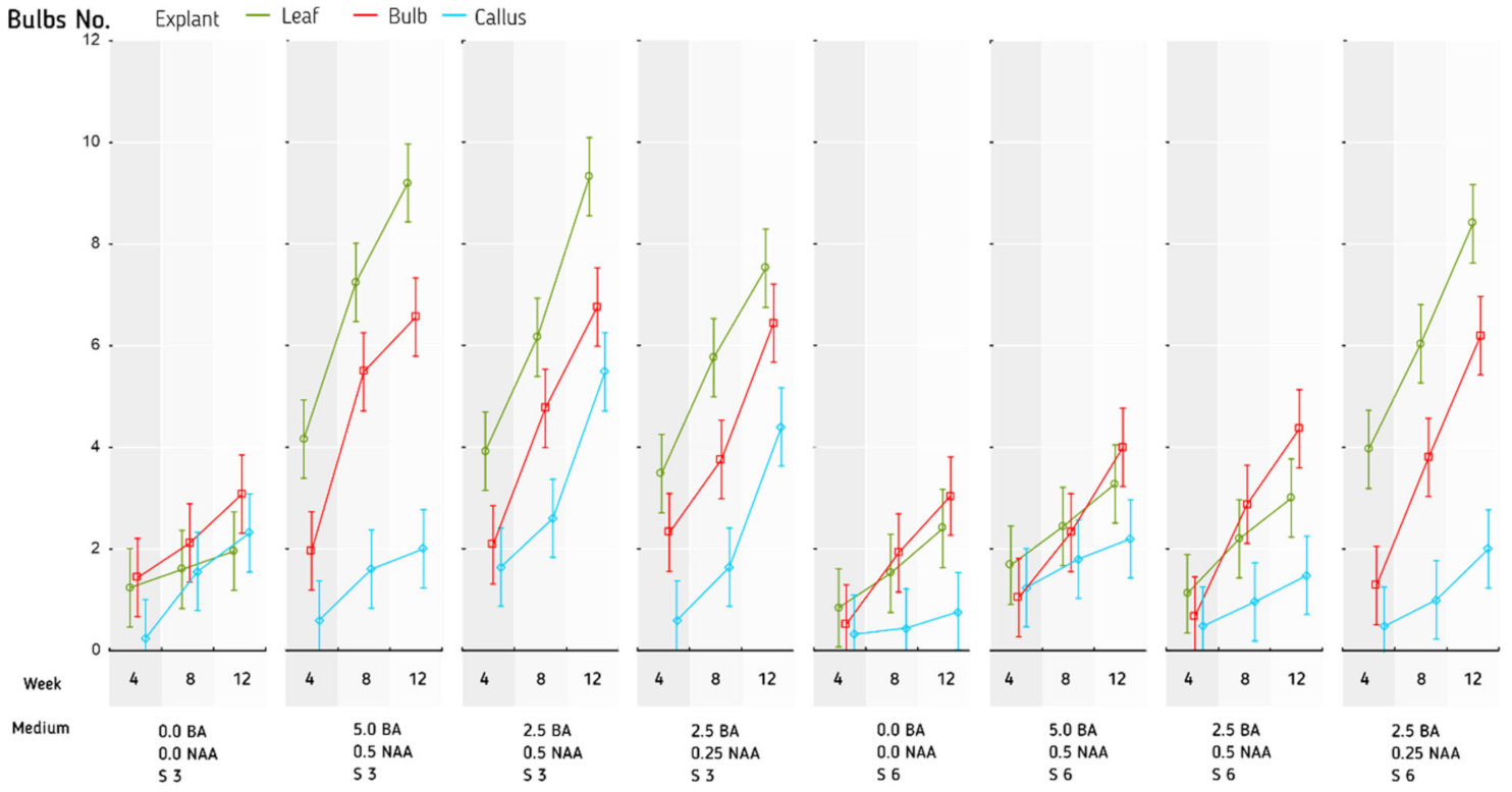
An analysis of dynamics of adventitious bulbs’ formation revealed that with the lapse of the duration of the culture, the number of arising adventitious bulbs on the explants increased, but the course of this process depended on the explant type and media composition (Figure 5). Rising trend in bulbing was more visible between the fourth and eighth week for leaf and bulb explants cultivated on medium S3/5 BA/0.5 NAA, which increased from 4.16 to 7.24 and from 1.96 to 5.48, respectively. Generally, from eighth to twelfth week, higher growth dynamics of bulbs was noticed for all explant types cultivated on S3/2.5 BA/0.5 NAA, S3/2.5 BA/0.25 NAA and S6/2.5 BA/0.25 NAA.
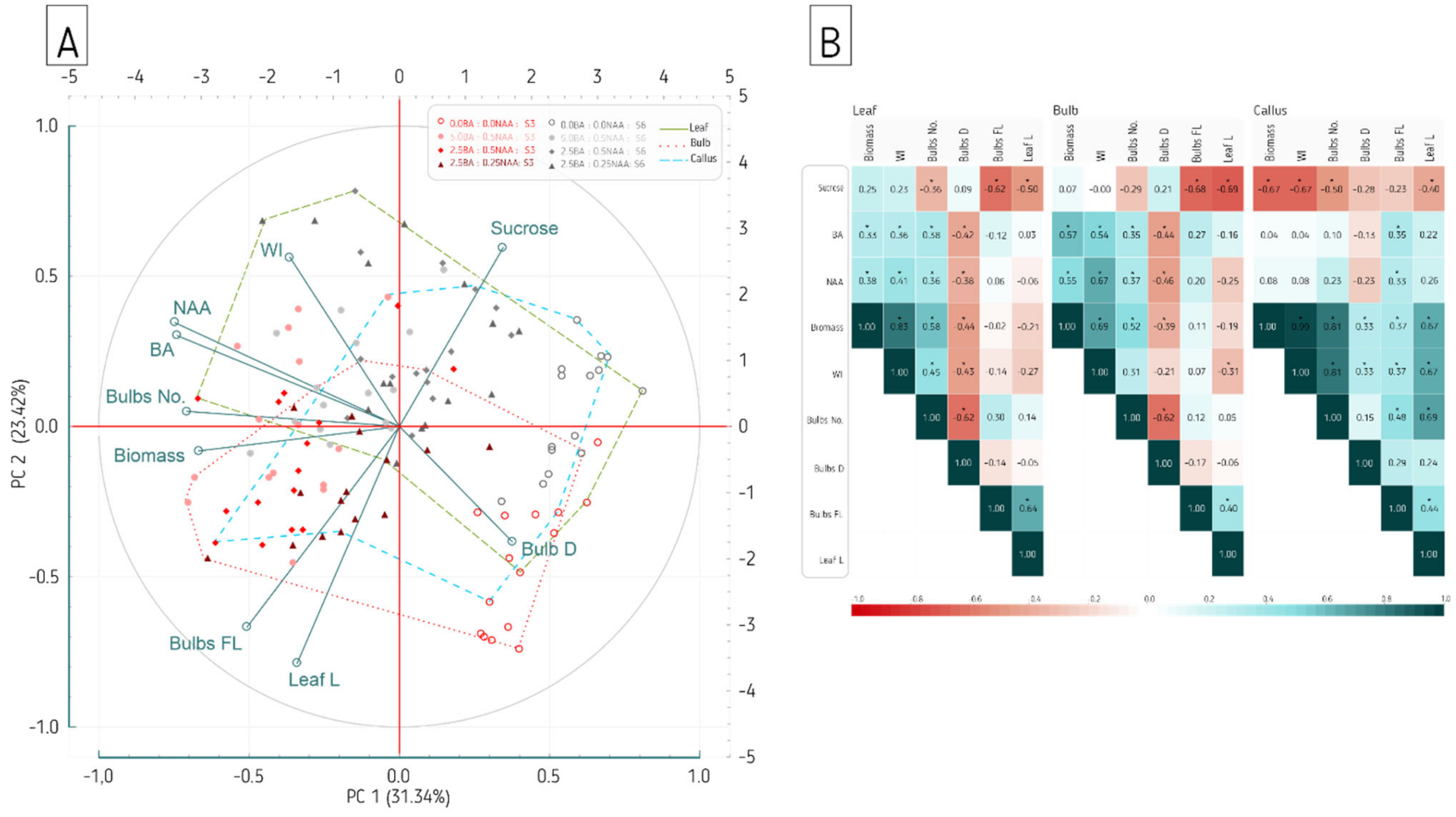
The principal component analysis (PCA) method is used to reduce the size of a statistical dataset and create a smaller set of new variables (principal components) containing most of the information of a larger group of original variables [67,68]. In this study, the principal component analysis was performed to assess the general structure of interrelations between studied traits of Lachenalia viridiflora, individual PGRs (BA, NAA) and sucrose in medium.
The first three principal components (PCs) explain 68.78% of total variation (Table 3), of which PC1 and PC2 account for 55%. The PCA outcomes and their graphical representation on biplot enable to distinguish two main groups of strongly correlated variables (Table 3, Figure 6A). The first group encompasses variables constituting the most important contributors to the PC1 and correlated negatively such as Biomass (−0.67), Bulbs No. (−0.71) and both PGRs: BA (−0.74) and NAA (−0.75). This shows that biomass and the number of adventitious bulbs are related to PGRs (Table 3, Figure 6A). The PCA indicated medium-related clustering of observation points, separated by the first principal component (Figure 6A). The observations representing explants regenerated on media with PGRs are closer to the vectors of variables of Biomass, Bulb No. and WI, which confirms the above assumptions. The second group includes variables that have the greatest contribution to the PC2. Those are negatively correlated Bulbs FL (−0.67) and Leaf L (−0.79), as well as positively correlated Sucrose (0.60) and WI (0.56) (Table 3, Figure 6A). This suggests the association between sucrose and the intensity of leaf formation and their elongation. The second principal component slightly divide observations into groups according to explant type (Figure 6A). Observations that represent bulb explants are located closer to variable Bulbs FL and Leaf L vectors, which indicate that this type of explant mostly tend to form leaves. Leaf-derived explant observations slightly cluster towards vector of WI variable, which is in line with previous analysis and confirm the highest fresh weight increase of this explant type.
As an additional way of studying PCA results, Pearson correlation coefficients were calculated separately for each explant type. This analysis confirms what was demonstrated by PCA correlations between variables in the case of leaf and bulb explants (Figure 6B). It indicates significant and strong negative correlation of Bulb FL and Leaf L with sucrose as well as significant positive correlation of Biomass, WI and Bulb No. with PGRs. The culture derived from callus showed different pattern of variables’ correlation. The Pearson correlation analysis revealed also significant, moderate negative correlation between Bulb D and both PGRs among leaf and bulb explants. On PCA biplot, these variables’ vectors are located in opposite quadrants of the PC1 axis, however, positive correlation of Bulb D variable with PC1 is weak to moderate (Figure 6A,B).

4. Conclusions

The application of the in vitro technique proved to be an effective method for micropropagation via organogenesis of Lachenalia viridiflora. Based on the obtained results, we concluded that twice as many adventitious bulbs were obtained from leaf and bulb explants as compared to callus. The more adventitious bulbs regenerated on the leaf and bulb explants, the smaller their diameter was. Bulbing and leafing was promoted by the medium containing 3% of sucrose. The adventitious bulbs derived from bulb explants formed the leaves most often. No leaves were noticed on adventitious bulbs from leaf explants when medium contained 6% sucrose, 2.5 µM BA, 0.5 or 0.25 µM NAA. The highest biomass was obtained from bulb explants, and the addition of various combinations of PGRs favored obtaining higher biomass compared to media without PGRs.
Considering the propagation efficiency (adventitious bulb number per explant) and the economic aspect (lower proportion of PGRs and sucrose in the medium), leaf explants cultivated on MS medium supplemented with 3% sucrose and 2.5 µM BA, and 0.25–0.5 µM NAA or bulb explants cultivated on MS medium supplemented with 3% sucrose and 2.5 µM BA and 0.5 µM NAA are combinations recommended for the production of bulbs of Lachenalia viridiflora. The results may support mass propagations and, consequently, the conservation of this valuable species.

Author Contributions

Conceptualization, M.M. and A.K.; methodology, M.M.; validation, M.M., J.M. and A.K.; formal analysis, J.M.; investigation, M.M.; resources, M.M. and A.K.; data curation, M.M., J.M. and A.K.; writing—original draft preparation, M.M., J.M. and A.K.; writing—review and editing, M.M., J.M. and A.K.; visualization, M.M., J.M. and A.K.; supervision, M.M. and A.K.; funding acquisition, M.M. and A.K. All authors have read and agreed to the published version of the manuscript.

Funding

This research was supported by the Ministry of Science and Higher Education of the Republic of Poland from subvention funds for University of Agriculture in Krakow.

Institutional Review Board Statement

Not applicable.

Informed Consent Statement

Not applicable.

Data Availability Statement

Not applicable.

Conflicts of Interest

The authors declare no conflict of interest. The funders had no role in the design of the study; in the collection, analyses, or interpretation of data; in the writing of the manuscript, or in the decision to publish the results.

References

  1. Duncan, G.D. The Genus Lachenalia: Botanical Magazine Monograph, 1st ed.; Royal Botanic Gardens: Kew, UK, 2012; ISBN 9781842463826. [Google Scholar]
  2. Duncan, G.D. Two new species, two rediscoveries and a range extension in Lachenalia (Asparagaceae: Scilloideae) from southern and western South Africa. Phytotaxa 2017, 316, 261–270. [Google Scholar] [CrossRef]
  3. Raimondo, D.; von Staden, L.; Foden, W.; Victor, J.; Helme, N.; Turner, R.; Kamundi, D.; Manyama, P. Red List of South African Plants.Series: Strelitzia; South African National Biodiversity Institute (SANBI Publishing): Pretoria, South African, 2009; Volume 25, ISBN 78-1-919976-52-5. [Google Scholar]
  4. Kleynhans, R. Lachenalia spp. In Flower Breeding and Genetics: Issues, Challenges and Opportunities for the 21st Century; Anderson, N.O., Ed.; Springer: Dordrecht, The Netherlands, 2006; pp. 491–516. [Google Scholar]
  5. Kleynhans, R. Lachenalia Jacq. f. ex Murray Named “Aqua Lady”. U.S. Plant Patent Application No. US 2017/0112037 P1, 20 April 2017. [Google Scholar]
  6. Nel, D.D. Rapid propagation of Lachenalia hybrids in vitro. S. Afr. J. Bot. 1983, 2, 245–246. [Google Scholar] [CrossRef] [Green Version]
  7. Ault, J.R. In vitro rooting and greenhouse acclimatization of Lachenalia shoots. HortScience 1995, 30, 1304–1305. [Google Scholar] [CrossRef] [Green Version]
  8. McCartan, S.A.; van Staden, J. Micropropagation of members of the Hyacinthaceae with medicinal and ornamental potential—A review. S. Afr. J. Bot. 1999, 65, 361–369. [Google Scholar] [CrossRef] [Green Version]
  9. Grace, O.M.; van Staden, J. A horticultural history of Lachenalia (Hyacinthaceae): Review article. S. Afr. J. Sci. 2003, 99, 526–531. [Google Scholar]
  10. Moyo, M.; Bairu, M.W.; Amoo, S.O.; van Staden, J. Plant biotechnology in South Africa: Micropropagation research endeavours, prospects and challenges. S. Afr. J. Bot. 2011, 77, 996–1011. [Google Scholar] [CrossRef] [Green Version]
  11. Aremu, A.O.; Plačková, L.; Masondo, N.A.; Amoo, S.O.; Moyo, M.; Novák, O.; Doležal, K.; Van Staden, J. Regulating the regulators: Responses of four plant growth regulators during clonal propagation of Lachenalia montana. Plant Growth Regul. 2017, 82, 305–315. [Google Scholar] [CrossRef]
  12. Bach, A.; Kapczyńska, A.; Dziurka, K.; Dziurka, M. Phenolic compounds and carbohydrates in relation to bulb formation in Lachenalia ‘Ronina’ and ‘Rupert’ in vitro cultures under different lighting environments. Sci. Hortic. 2015, 188, 23–29. [Google Scholar] [CrossRef]
  13. Bach, A.; Kapczyńska, A.; Dziurka, K.; Dziurka, M. The importance of applied light quality on the process of shoot organogenesis and production of phenolics and carbohydrates in Lachenalia sp. cultures in vitro. S. Afr. J. Bot. 2018, 114, 14–19. [Google Scholar] [CrossRef]
  14. Niederwieser, J.G.; Vcelar, B.M. Regeneration of Lachenalia species from leaf explants. HortScience 1990, 25, 684–687. [Google Scholar] [CrossRef] [Green Version]
  15. Niederwieser, J.G.; van Staden, J. Interaction between benzyladenine, naphthaleneacetic acid and tissue age on adventitious bud formation on leaf sections of Lachenalia hybrids. S. Afr. J. Bot. 1992, 58, 13–16. [Google Scholar] [CrossRef] [Green Version]
  16. Van Staden, J.; Drewes, F.E. The effect of benzyladenine and its glucosides on adventitious bud formation on Lachenalia leaf sections. S. Afr. J. Bot. 1994, 60, 191–192. [Google Scholar] [CrossRef] [Green Version]
  17. Slabbert, M.M.; Niederwieser, J.G. In vitro bulblet production of Lachenalia. Plant Cell Rep. 1999, 18, 620–624. [Google Scholar] [CrossRef]
  18. Niederwieser, J.G.; Ndou, A.M. Review on adventitious bud formation in Lachenalia. Acta Hortic. 2002, 570, 135–140. [Google Scholar] [CrossRef] [Green Version]
  19. Bach, A.; Kapczyńska, A. The initiation of the in vitro cultures of some Lachenalia cultivars. Ann. Warsaw Univ. Life Sci.—SGGW Hortic. Landsc. Archit. 2013, 34, 13–20. [Google Scholar]
  20. Ascough, G.D.; van Staden, J.; Erwin, J.E. In vitro Storage Organ Formation of Ornamental Geophytes. Hortic. Rev. 2008, 417–445. [Google Scholar] [CrossRef]
  21. Kapczyńska, A. Propagation of Lachenalia cultivars with leaf cuttings. Acta Sci. Pol. Hortorum Cultus 2019, 18, 189–195. [Google Scholar] [CrossRef]
  22. Podwyszyńska, M. The mechanisms of in vitro storage organ formation in ornamental geophytes. Floric. Ornam. Biotechnol. 2012, 6, 9–23. [Google Scholar]
  23. Fennell, C.W.; van Staden, J.; Bornman, C.H. Biotechnology of southern African bulbs. S. Afr. J. Bot. 2004, 70, 37–46. [Google Scholar] [CrossRef] [Green Version]
  24. Kumar, S.; Awasthi, V.; Kanwar, J.K. Influence of growth regulators and nitrogenous compounds on in vitro bulblet formation and growth in oriental lily. Hortic. Sci. 2007, 34, 77–83. [Google Scholar] [CrossRef] [Green Version]
  25. Malik, M.; Bach, A. Morphogenetic pathways from Narcissus L. ‘Carlton’ in vitro cultures of PC stage flower bud explants according to cytokinin and auxin rations. Acta Sci. Pol. Hortorum Cultus 2016, 15, 101–111. [Google Scholar]
  26. Qadir, J.; Singh, S.; Kaloo, Z.A. A review on in vitro propagation of some medicinally important plant species of family Asparagaceae. Int. J. Biosci. Technol. 2017, 10, 1–12. [Google Scholar]
  27. Prokopiuk, B.; Cioć, M.; Maślanka, M.; Pawłowska, B. Effects of light spectra and benzyl adenine on in vitro adventitious bulb and shoot formation of Lilium regale E.H. Wilson. Propag. Ornam. Plants 2018, 18, 12–18. [Google Scholar]
  28. Niederwieser, J.G.; van Staden, J.; Upfold, S.J.; Drewes, F.E. Metabolism of 6-benzyladenine by leaf explants of Lachenalia during adventitious bud formation. S. Afr. J. Bot. 1992, 58, 236–238. [Google Scholar] [CrossRef] [Green Version]
  29. Bach, A.; Sochacki, D. Propagation of ornamental geophytes: Physiology and management systems. In Ornamental Geophytes: From Basic Science to Sustainable Production; Kamenetsky, R., Okubo, H., Eds.; CRC Press: Boca Raton, FL, USA, 2013; pp. 261–286. [Google Scholar]
  30. Van Rensburg, J.G.J.; Vcelar, B.M. The effect of the sucrose concentration on the initiation and growth of adventitious buds from leaf tissue of Lachenalia. S. Afr. J. Bot. 1989, 55, 117–121. [Google Scholar] [CrossRef] [Green Version]
  31. Bach, A.; Pawlowska, B.; Pulczynska, K. Utilization of soluble carbohydrates in shoot and bulb regeneration of Hyacinthus orientalis L. in vitro. Acta Hortic. 1992, 325, 487–492. [Google Scholar] [CrossRef]
  32. Niederwieser, J.G.; Bornman, C.H. Role of biotechnology in the development and production of Lachenalia and Ornithogalum cultivars in South Africa. S. Afr. J. Bot. 2004, 70, 47–51. [Google Scholar] [CrossRef] [Green Version]
  33. Pearson, M.N.; Cohen, D.; Cowell, S.J.; Jones, D.; Blouin, A.; Lebas, B.S.M.; Shiller, J.B.; Clover, G.R.G. A survey of viruses of flower bulbs in New Zealand. Australas. Plant Pathol. 2009, 38, 305–309. [Google Scholar] [CrossRef]
  34. Pence, V.C. Evaluating costs for the in vitro propagation and preservation of endangered plants. Vitr. Cell. Dev. Biol. Plant 2011, 47, 176–187. [Google Scholar] [CrossRef]
  35. Bhojwani, S.S.; Dantu, P.K. Micropropagation. In Plant Tissue Culture: An Introductory Text; Springer: New Delhi, India, 2013; pp. 245–274. [Google Scholar]
  36. Hesami, M.; Naderi, R.; Yoosefzadeh-Najafabadi, M.; Maleki, M. In vitro culture as a powerful method for conserving Iranian ornamental geophytes. BioTechnologia 2018, 99, 73–81. [Google Scholar] [CrossRef]
  37. Kumar, V.; Moyo, M.; Van Staden, J. Enhancing plant regeneration of Lachenalia viridiflora, a critically endangered ornamental geophyte with high floricultural potential. Sci. Hortic. 2016, 211, 263–268. [Google Scholar] [CrossRef]
  38. Maślanka, M.; Kapczyńska, A. Cold treatment promotes in vitro germination of two endangered Lachenalia species. S. Afr. J. Bot. 2021, 142, 430–432. [Google Scholar] [CrossRef]
  39. Murashige, T.; Skoog, F. A revised medium for rapid growth and bioassays with tobacco tissue culture. Physiol. Plant. 1962, 15, 473–497. [Google Scholar] [CrossRef]
  40. Statistica 13.3; Data Analysis Software System. TIBCO Software Inc.: Palo Alto, CA, USA, 2017.
  41. Hair, J.F.; Black, W.C.; Babin, B.J.; Anderson, R.E. Multivariate Data Analysis, 8th ed.; Cengage: Hampshire, UK, 2019. [Google Scholar]
  42. Jolliffe, I.T. Principal Component Analysis, Springer Series in Statistics, 2nd ed.; Springer: New York, NY, USA, 2002; ISBN 0-387-95442-2. [Google Scholar]
  43. De Klerk, G.J.M. Micropropagation of bulbous crops: Technology and present state. Floric. Ornam. Biotechnol. 2012, 6, 1–8. [Google Scholar]
  44. Askari, N.; Visser, R.G.F.; De Klerk, G.-J. Growth of lily bulblets in vitro, a review. Int. J. Hortic. Sci. Technol. 2018, 5, 133–143. [Google Scholar] [CrossRef]
  45. Wu, Y.; Ren, Z.; Gao, C.; Sun, M.; Li, S.; Min, R.; Wu, J.; Li, D.; Wang, X.; Wei, Y.; et al. Change in sucrose cleavage pattern and rapid starch accumulation govern lily shoot-to-bulblet transition in vitro. Front. Plant Sci. 2021, 11, 564713. [Google Scholar] [CrossRef]
  46. Maślanka, M.; Bach, A. Induction of bulb organogenesis in in vitro cultures of tarda tulip (Tulipa tarda Stapf.) from seed-derived explants. Vitr. Cell. Dev. Biol. Plant 2014, 50, 712–721. [Google Scholar] [CrossRef] [Green Version]
  47. Evenor, D.; Levi-Nissim, A.; Afgin, L.; Lilien-Kipnis, H.; Watad, A.A. Regeneration of plantlets and bulblets from explants and callus of Allium aflatunense cultivars and selection from indigenous Israeli Allium ampeloprasum. Acta Hortic. 1997, 430, 325–330. [Google Scholar] [CrossRef]
  48. Bakhshaie, M.; Babalar, M.; Mirmasoumi, M.; Khalighi, A. Somatic embryogenesis and plant regeneration of Lilium ledebourii (Baker) Boiss., an endangered species. Plant Cell Tissue Organ Cult. 2010, 102, 229–235. [Google Scholar] [CrossRef] [Green Version]
  49. Kleynhans, R.; Niederwieser, J.G.; Hancke, F.L. Lachenalia: Development and commercialization of a new flower bulb crop. Acta Hortic. 2002, 570, 81–85. [Google Scholar] [CrossRef]
  50. Gheisari, M.; Miri, S.M. In vitro callus induction and bulblet regeneration of hyacinth (Hyacinthus orientalis L.). Plant Cell Biotechnol. Mol. Biol. 2017, 18, 145–155. [Google Scholar]
  51. Maślanka, M.; Prokopiuk, B. Bulb organogenesis of Tulipa tarda in vitro cultures in relation to light environment. Acta Agric. Scand. Sect. B 2019, 69, 398–404. [Google Scholar] [CrossRef]
  52. Fehér, A. Callus, Dedifferentiation, Totipotency, Somatic Embryogenesis: What These Terms Mean in the Era of Molecular Plant Biology? Front. Plant Sci. 2019, 10, 536. [Google Scholar] [CrossRef] [PubMed] [Green Version]
  53. Azadi, P.; Khosh-Khui, M. Micropropagation of Lilium ledebourii (Baker) Boiss as affected by plant growth regulators, sucrose concentrations, harvesting season and cold treatments. Electron. J. Biotechnol. 2007, 10, 582–591. [Google Scholar] [CrossRef]
  54. Nhut, D.T. Micropropagation of lily (Lilium longiflorum) via in vitro stem node and pseudo-bulblet culture. Plant Cell Rep. 1998, 17, 913–916. [Google Scholar] [CrossRef]
  55. Ozel, C.A.; Khawar, K.M.; Unal, F. Factors affecting efficient in vitro micropropagation of Muscari muscarimi Medikus using twin bulb scale. Saudi J. Biol. Sci. 2015, 22, 132–138. [Google Scholar] [CrossRef] [Green Version]
  56. Podwyszyńska, M. Effect of carbohydrates on shoot multiplication and bulb formation of tulip in vitro. Roczniki Akademii Rolniczej w Poznaniu Ogrodnictwo 2001, 33, 119–126. [Google Scholar]
  57. Taeb, A.G.; Alderson, P.G. Effect of low temperature and sucrose on bulb development and on the carbohydrate status of bulbing shoots of tulip in vitro. J. Hortic. Sci. 1990, 65, 193–197. [Google Scholar] [CrossRef]
  58. Kumar, S.; Kashyap, M.; Sharma, D.R. In vitro regeneration and bulblet growth from lily bulbscale explants as affected by retardants, sucrose and irradiance. Biol. Plant. 2005, 49, 629–632. [Google Scholar] [CrossRef]
  59. Bach, A. Micropropagation of hyacinths (Hyacinthus orientalis L.). In High-Tech and Micropropagation, Biotechnology in Agriculture and Forestry; Bajaj, Y.P.S., Ed.; Springer: New York, NY, USA; Berlin/Heidelberg, Germany, 1992; pp. 144–159. [Google Scholar]
  60. Chung, C.H.; Chung, Y.M.; Yang, S.J.; Ko, E.K.; Jeong, S.J.; Nam, J.S.; Kim, G.T.; Yi, Y.B. Effects of storage temperature and sucrose on bulblet growth, starch and protein contents in in vitro cultures of Hyacinthus orientalis. Biol. Plant. 2006, 50, 346–351. [Google Scholar] [CrossRef]
  61. Squires, W.M.; Langton, F.A.; Fenlon, J.S. Factors influencing the transplantation success of micropropagated narcissus bulbils. J. Hortic. Sci. 1991, 66, 661–671. [Google Scholar] [CrossRef]
  62. Cheesman, L.; Finnie, J.F.; van Staden, J. Eucomis zambesiaca baker: Factors affecting in vitro bulblet induction. S. Afr. J. Bot. 2010, 76, 543–549. [Google Scholar] [CrossRef] [Green Version]
  63. Rahimi, M.; Daneshvar, M.H.; Heidari, M. Propagation and bulb formation of Fritillaria (Fritillaria imperialis L.) via in vitro culture. Int. J. Plant Anim. Environ. Sci. 2014, 4, 707–710. [Google Scholar]
  64. Kizil, S.; Sogut, T.; Acay, U.; Sarihan, B.; Sesiz, U.; Khawar, K.M. Effects of sucrose concentrations on increase in bulb size of in vitro regenerated Hyacinth (Hyacinthus orientalis L.) bulblets. Sci. Bull. Ser. F Biotechnol. 2017, 2285, 51–55. [Google Scholar]
  65. Juan-Vicedo, J.; Pavlov, A.; Ríos, S.; Casas, J.L. In vitro culture and micropropagation of the Baetic-Moroccan endemic plant Lapiedra martinezii Lag. (Amaryllidaceae). Vitr. Cell. Dev. Biol. Plant 2019, 55, 725–732. [Google Scholar] [CrossRef]
  66. Sutlana, J.; Sutlana, N.; Siddique, M.; Islam, A.; Hossain, M.; Hossain, T. In vitro bulb production in Hippeastrum (Hippeastrum hybridum). J. Cent. Eur. Agric. 2010, 11, 469–474. [Google Scholar] [CrossRef] [Green Version]
  67. Iezzoni, A.F.; Pritts, M.P. Applications of Principal Component Analysis to Horticultural Research. HortScience 1991, 26, 334–338. [Google Scholar] [CrossRef] [Green Version]
  68. Rotaru, A.S.; Pop, I.D.; Vatcă, A.; Cioban, A. Usefulness of principal components analysis in agriculture. Bull. Univ. Agric. Sci. Vet. Med. Cluj-Napoca. Hortic. 2012, 69, 54–59. [Google Scholar]
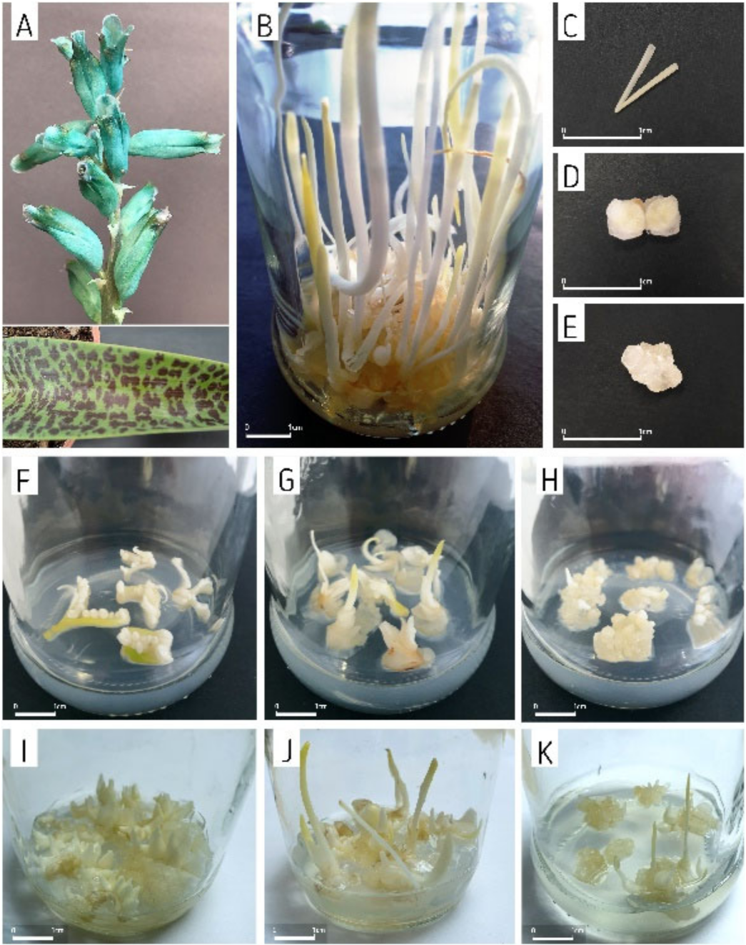
Figure 1. Lachenalia viridiflora: (A) Turquoise flowers and spotted leaf; (B) The starting culture from which the explants were taken; (C) Leaf explant; (D) Bulb explant; (E) Callus explant. Adventitious bulb formed after: (F) 4 weeks of cultivation of leaf explants × S3/5 BA/0.5 NAA; (G) 4 weeks of cultivation of bulb explants × S6/2.5 BA/0.5 NAA; (H) 4 weeks of cultivation of callus explants × S3/2.5 BA/0.25 NAA; (I) 12 weeks of cultivation of leaf explants × S3/2.5 BA/0.25 NAA; (J) 12 weeks of cultivation of bulb explants × S3/2.5 BA/0.5 NAA; (K) 12 weeks of cultivation of callus explants × S6/without PGRs.
Agronomy 12 00475 g001
Figure 2. The effect of different explant and medium type on the number of adventitious bulbs (Bulb No.) and their diameter (Bulb D). Mean values ± SE with different small letters as well as mean values with different large letters are significantly different according to Duncan’s multiple range test at p ≤ 0.05.
Agronomy 12 00475 g002
Figure 3. The effect of different explant and medium type on the biomass and biomass weight increase indicator (WI). Mean values ± SE with different small letters as well as mean values with different large letters are significantly different according to Duncan’s multiple range test at p ≤ 0.05.
Agronomy 12 00475 g003
Figure 4. The effect of different explant and medium type on the percentage of adventitious bulbs forming leaves (Bulb FL) and on the leaves’ length (Leaf L). Mean values ± SE with different small letters as well as mean values with different large letters are significantly different according to Duncan’s multiple range test at p ≤ 0.05.
Agronomy 12 00475 g004
Figure 5. The dynamics of adventitious bulbs’ formation after 4, 8 and 12 weeks of cultivation, depending on the explant and medium type. The shown values are means ± SE.
Agronomy 12 00475 g005
Figure 6. (A) Biplot of the Principal component analysis (PCA) representing the projection of variable vectors and observation points on the plane defined by the first two PCs, showing associations between studied traits, explant types, PGRs and sucrose. The symbols representing the observation points relate to the different combinations of medium, lines connecting the observation points determine the range of location of different explants (see legend chart); (B) Pearson correlation coefficient matrix between studied traits, PGRs and sucrose, presented separately for three types of explant. The asterisks mean significance at p < 0.05. Correlation direction is emphasized in color (positive in blue, negative in red) and strength in color saturation (the stronger correlation, the more intense color).
Agronomy 12 00475 g006
Table 1. Combination of the media.
Sucrose (%)Plant Growth Regulators (µM)
BANAA
300
50.5
2.50.5
2.50.25
600
50.5
2.50.5
2.50.25
Table 2. Two-way ANOVA results indicating the main effects of explant type, medium and their interaction on studied traits.
Source
of Variation
dfMean Squares
Biomass (g)WIBulbs No.Bulb D (mm)Bulbs FL (%)Leaf L (mm)
Explant (E)2 7.32 ***62,252,188 ***105.30 ***1.93 *7689.6 ***1743.53 ***
Medium (M)7 3.35 ***5,219,339 ***58.28 ***5.48 ***6673.0 ***260.01 ***
E × M14 1.12 ***2,928,211 ***9.80 **1.04 *489.9 NS60.87 NS
Error96 0.27808,7663.980.53347.141.65
* The asterisks mean significant at the * 0.05, ** 0.01, *** 0.001 probability level; NS not significant.
Table 3. Component loadings of 9 variables, eigen values, proportion of total variability represented by the first three principal components (PCs) and cumulative variability.
CharactersPrincipal Components
PC1PC2PC3
Biomass−0.668545−0.081330.220848
WI−0.3646870.5635260.594663
Bulbs No.−0.7072690.0502110.560183
Bulb D0.376447−0.3843640.160915
Bulbs FL−0.507503−0.668136−0.148332
Leaf L−0.340757−0.788754−0.084753
Sucrose0.3450830.595031−0.255695
BA−0.7393980.302067−0.479799
NAA−0.7477780.345689−0.440573
Eigen Value2.822.121.26
Percentage of Variance31.3423.4214.01
Cumulative % of Variance31.3354.7568.76
Publisher’s Note: MDPI stays neutral with regard to jurisdictional claims in published maps and institutional affiliations.