Article

Temporal and Spatial Dynamics of Dark Septate Endophytes in the Roots of Lycium ruthenicum in the Desert Region of Northwest China

1
School of Life Sciences, Hebei University, Baoding 071002, China
2
Institute of Life Sciences and Green Development, Hebei University, Baoding 071002, China
3
College of Pharmaceutical Sciences, Hebei University, Baoding 071002, China
4
Institute of Medicinal Plant Development, Chinese Academy of Medical Sciences & Peking Union Medical College, Beijing 100005, China
*
Author to whom correspondence should be addressed.
Li Han and Jingxin Shi are co-first authors and contribute equally to the article.
Agronomy 2021, 11(4), 648; https://doi.org/10.3390/agronomy11040648
Submission received: 15 February 2021 / Revised: 25 March 2021 / Accepted: 26 March 2021 / Published: 28 March 2021
(This article belongs to the Section Soil and Plant Nutrition)

Abstract

:
With the intensification of desertification in northwest China, drought has become a serious environmental problem restricting plant growth and ecological restoration. Recently, dark septate endophytes (DSEs) have attracted more attention because of their ability to improve plants’ resistance to drought. Here, we investigated DSE colonization and species diversity in roots of Lycium ruthenicum collected from Anxi and Minqin, in northwest China, during July, September, and December 2019. This study aimed to evaluate the influence of seasonality and sampling sites on DSEs. In different seasons, DSE colonization varied with the phenology of L. ruthenicum. At different sites, DSE colonization significantly differed. Four isolates were reported in desert ecosystems for the first time. The results showed microsclerotial colonization was directly affected by changing seasons, while hyphal colonization and species diversity were directly affected by sampling sites. The soil organic carbon, pH, alkaline phosphatase, and alkali-hydrolyzable nitrogen were the main predictors of DSE colonization and species diversity. We conclude that DSE colonization and diversity showed significant spatial–temporal heterogeneity and were closely related to soil factors. This research provides a basis for the further understanding of the ecological functions of DSEs and their application potential for vegetative restoration and agricultural cultivation in drylands.

1. Introduction

Drought and desertification are rapidly intensifying in northwest China, where approximately 27% of the land is exposed to desertification [1]. Drought has become a serious environmental problem restricting plant growth and ecological restoration [2,3]. Therefore, it is particularly urgent to select appropriate plants with adaption to the arid environment. Fortunately, in order to survive in these conditions many plant species have evolved specialized ways to adapt to desert ecosystems [4]. Thus, special attention has been given to the utilization of these native plants for promoting ecological restoration in desert ecosystems [5]. Lycium ruthenicum, as an economically important traditional medicinal plant, belongs to Solanaceae and it is widely distributed in arid and semi-arid regions of northwest China as well as regions of Eastern Europe [6]. With the special characteristics of drought and salt resistance, L. ruthenicum is particularly well suited for increasing the nutrient and organic matter content of surface mineral soils in this arid ecosystem and may have the potential to improve soil conditions, facilitate ecosystem development, and delay further desertification. In addition to a direct drought resistance of plants, studies have shown that endophytic fungi may also affect the response of plants to drought stress, which are generally believed to improve the ability of plants to cope with environmental stress [7,8]. Therefore, the study of plant-related fungi in arid environments could provide a basis for the application of fungi in agricultural drylands.
Dark septate endophytes (DSEs) are a miscellaneous group of fungal endophytes that generally colonize the living plant roots and are characterized by dark septate hyphae and melanized microsclerotia [9,10]. They are found in diverse ecosystems, especially under stressful environments, including arid and semiarid environments [11,12,13]. As important root endophytes, arbuscular mycorrhizal fungi (AMF) are well documented for their resistance to drought and promotion of plant growth [14]. However, DSEs, which may have similar functions to AMF, have recently received more attention [10,15]. Zhang et al. [16] found that the roots of L. barbarum in arid area were colonized by typical DSEs and inoculation with DSEs increased the total biomass of L. barbarum by 39.2% [17]. In our previous investigations in northwest China, DSE fungi were found to co-occur with multiple desert plants such as Ammopiptanthus mongolicus and Hedysarum scoparium [18,19]. Subsequent experiments have proven that DSEs could promote the growth of A. mongolicus and H. scoparium [20,21]. DSEs, as an important endophytic fungi of plant roots, can promote water acquisition and nutrient transport in host plants in arid environments by forming a continuous integrated network in roots, and therefore increase the stress tolerance of host plants [11,22,23]. In addition, melanin in DSE hyphae might play an important role in arid ecosystems, which has been considered to provide structural rigidity to cell walls and can increase resistance against drought stress [24,25]. Although they have such potential functions as compared with AMF, research on DSEs is still in its infancy. Studies evaluating the taxonomic diversity and ecological adaptability of DSEs in different environments, especially in desert regions, are still relatively scarce.
DSEs form close ecological relationships with host plants, which are related to a plant’s dependency on DSEs and abiotic factors such as spatial–temporal factors and other related abiotic factors [8,26,27,28]. Such a dynamic and diverse endophyte-host plant relationship has meant that predictable patterns of variation are still elusive and poorly understood. Xie et al. [19] found that DSE colonization in roots of Hedysarum scoparium was spatially predictable. Ruotsalainen et al. [29] reported there was no correlation between fungal colonization and season, while Mandyam and Jumpponen [26] found that DSEs showed seasonal correlation and that DSE colonization was the highest in spring. Rayment et al. [30] pointed out that abiotic variables caused by different seasons may affect DSE colonization and DSE colonization presented a non-random distribution due to the effect of seasons, regions, soil factors, and plant species. Although not all studies have reached a consensus, DSE colonization in host roots appears to be influenced by changes in environmental conditions [26,30,31]. In order to increase the understanding of how DSE colonization and species diversity respond to abiotic factors, in this study, we collected root samples of L. ruthenicum from two sites, during three seasons in northwest China. The objective of the present study was to evaluate DSE spatial–temporal dynamics in the roots of L. ruthenicum. Specifically, we aimed (1) to detect DSE distribution and resources of L. ruthenicum and (2) to explore the influence of both season and sampling sites on DSE colonization and species diversity, thus providing a basis for ecological function in desert ecosystems and for applied potential in the cultivation of medicinal plants in northwest China. In our study, 114 strains belonging to 17 taxa were isolated, and the results demonstrated that DSE colonization and diversity showed significant spatial and temporal heterogeneity and were closely related to soil factors.

2. Materials and Methods

2.1. Study Sites and Sampling

The sampling sites were located in the desert region of northwest China. Soil and root samples were collected from Anxi (40.75° N, 95.75° E) and Minqin (38.75° N, 103.25° E) in Gansu Province, China, during July, September, and December 2019. These areas have a typical arid continental climate, with considerable seasonal and diurnal temperature variation. The altitude, monthly average precipitation, and temperature of sampling sites are shown in Table S1 in Supplementary Materials. The studied soils were entisols and aridisols [32]. The selected plots are characterized by desert sands where L. ruthenicum is abundant. Psammophytic shrubs and subshrubs also occur at each plot.
Three sample plots were selected, and the distances between the plots were ≥10 km. Five replicate soil and L. ruthenicum root samples in each plot were randomly sampled from a depth of 0–30 cm. The distances between the plants that were sampled was ≥100 m. The samples were sealed in plastic bags and transported to the laboratory in an insulated container. Subsequently, all samples were sieved (<2 mm mesh) to remove litter and coarse roots. Fresh fine roots were extracted from each sample and were immediately processed for DSE morphological observation and DSE isolation. Soil samples for enzyme analyses were dried at 15–25 °C and stored at 4 °C until analyses. Other subsamples were air-dried and used for determination of soil physicochemical properties.

2.2. Soil Analysis

Soil pH was measured with a digital pH meter (PHS-3C, Shanghai Lida Instrument Factory, Shanghai, China) on a soil/water (1:2.5) suspension. Soil organic carbon (SOC) was measured by the combustion method [33]. The samples were heated in a muffle furnace (TMF-4-10 T, Shanghai Gemtop Scientific Instrument Corp., Shanghai, China) for 4 h at 550 °C. Alkali-hydrolyzable nitrogen (AN) was estimated using the alkaline hydrolysis diffusion method. The samples with sodium hydroxide and boric acid indicators were placed in a Petri dish and incubated at 40 °C for 24 h. Available phosphorus (AP) was determined by the chlorostannous reduced molybdophosphoric blue color method [34]. Soil alkaline phosphatase activity was determined by the method reported by Tarafdar and Marschner [35]. Soil urease activity was determined using the method of Hoffmann and Teicher [36], and urease activity was expressed as μg NH4+-N released from 1 g soil within 1 h [36].

2.3. Quantification of Fungal Colonization

Fresh roots were cut into 0.5 cm segments and cleaned using sterile water. The segments were placed in 10% (w/v) potassium hydroxide for 1 h at 100 °C and stained with 0.5% (w/v) acid fuchsin for 30 min at 90 °C [37]. For each sample, 30 segments were selected randomly and examined microscopically at 20× and 40× magnification [38]. The colonization rate of DSEs (%) (total, hyphal, and microsclerotial) and DSE colonization intensity were analyzed, according to Trouvelot et al. [39] as follows:
Colonization rate (%) = (number of colonized root segments/total number of root segments) × 100%.
Colonization intensity (%) = (length of colonized root segments/total length of root segments) × 100%.

2.4. Isolation of Endophytic Fungi

For each plant individual, 15 randomly selected 0.5 cm fine root segments were disinfected with 75% ethanol for 5 min and 10% sodium hypochlorite for 2 min, and then washed with sterile water 3 times. The roots were placed in potato dextrose agar (PDA) culture medium supplemented with the antibiotics ampicillin and streptomycin sulphate and kept at 27 °C in the dark. In the meantime, water from the final rinse was plated in PDA as a negative control to ensure successful root surface sterilization. After incubation for 15 days, colonies with dark mycelium were isolated onto PDA to observe colony morphology and microscopic morphological characteristics [40].

2.5. Molecular Identification of Endophytic Fungi

DNA was extracted from 50 mg mycelium per colony with a genomic DNA extraction kit (SolarBio, Beijing, China). PCR sequencing was conducted with 20 μL reaction volumes each containing 3.5 μL genomic DNA, 0.5 μL of each primer ITS4 (5′-TCCTCCGCTTATTGATATGC-3′) and ITS5 (5′-GGAAGTAAAAGT- CGTAACAAGG-3′), 10 μL of 2 × Es Taq Master Mix (CoWin Biosciences, Beijing, China), and 5.5 μL ddH2O. PCR was performed in a LifeECOTM thermocycler (Hangzhou Bioer Technology Co. Ltd., Hangzhou, China) using the following program: initial denaturation at 94 °C for 5 min, followed by 35 cycles of 94 °C for 1 min, 55 °C for 1 min, 72 °C for 1 min, and a final incubation at 72 °C for 10 min [19]. Finally, the PCR products were purified and sequenced. The sequences were analyzed with the BLAST tool in NCBI with the option “type strain material” to determine taxonomic identifications. Sequence alignment was completed using Clustal X v. 1.81. A phylogenetic tree based on maximum likelihood was plotted with MEGA v. 6 [41]. DNA sequences were compiled and deposited in GenBank under accession numbers MW548074, MW548075, MW548076, MW548077, MW548078, MW548079, MW548080, MW548081, MW548082, MW548083, MW548085, MW548087, MW548088, MW548089, MW548090, MW548091, and MW548092.

2.6. DSE Species Diversity

The isolation rates (IRs) were calculated as the total number of plant segments infected by ≥1 type of fungus divided by the total number of incubated segments [42]. The isolation frequency (IF) was calculated as the number of strains of a fungus divided by the total number of strains isolated [43].
Diversity of DSEs was assessed using indices of the Shannon–Weaver index (H) [44]. Dominance was calculated with Simpson’s dominance (D) measures [45]. An evenness index (J) was used for the determination of uniformity of the endophytic fungi [46]. The formulae were as follows:
H = −∑(Pi)(InPi)
D = ∑(Pi)2
where the ratio “Pi” is the frequency of colonization of the taxon in the sample.
J = H/In(s)
where “s” is the total number of fungi isolated.

2.7. Statistical Analysis

The DSE colonization of L. ruthenicum was assessed using one-way analysis of variance (ANOVA), and two-way analysis was performed to analyze the effects in different seasons and sampling sites on DSE colonization and soil variables; comparisons among means were performed using the least significant difference method (p < 0.05). Pearson correlation analysis was used to test the relationships among soil variables and DSE colonization and to evaluate the influence of sampling time, sample sites, and the interaction on DSE colonization. SPSS 21.0 software (SPSS Inc., Chicago, IL, USA) was used for all of the above analyses. Principal component analyses (PCA) of DSE colonization were assessed by Canoco 4.5 based on the principle of the cumulative contribution rate of variance being greater than 80% if correlation matrix eigenvalues exceeded 1. Structural equation model (SEM) was established by AMOS 21.0 to test the effects of environmental variables on DSE hyphal, microsclerotial colonization rate, and DSE diversity.

3. Results

3.1. Spatial–Temporal Distribution of DSE Colonization

Typical DSE hyphae and microsclerotia structures were observed in the roots of L. ruthenicum, at Anxi and Minqin (Figure 1). Brown to dark brown septate hyphae invaded the epidermal, cortical cells (Figure 1A,G), or vascular tissue (Figure 1C,I). Chainlike (Figure 1E,K), tufted (Figure 1D,F,L) and discrete (Figure 1J) microsclerotia filled single cortical cells or colonized more than one cell.
With the change of seasons, the diameter of hyphae became thicker, the septum became shorter, and swollen hyphae formed at the end of the hyphae (Figure 1A–C). The DSE colonization showed significant (p < 0.05) differences among seasons (Figure 2). Total colonization and colonization intensity showed a V-shaped distribution, and the lowest value was in September. Hyphal colonization was July > December > September, and the highest values measured in July were 86.7% (at Anxi) and 54.4% (at Minqin). However, the microsclerotial colonization increased gradually with season changes, with the following order: July < September < December, and the highest values measured in December were 45.0% (at Anxi) and 34.4% (at Minqin). For different sampling sites, hyphal colonization, total colonization, and colonization intensity at Anxi were significantly (p < 0.05) higher than that at Minqin in July, while colonization intensity at Anxi were significantly (p < 0.05) lower than that at Minqin in September. However, microsclerotial colonization showed no significant differences among Anxi and Minqin in July, September, or December (Figure 2).
Two-way ANOVA showed that DSE hyphal, microsclerotial, total colonization, and colonization intensity were significantly (p < 0.01) influenced by seasons. DSE hyphal, total colonization, and colonization intensity were significantly (p < 0.05) influenced by the sampling sites. DSE total colonization and colonization intensity were significantly (p < 0.05) influenced by the interaction of seasons and sampling sites (Table 1).

3.2. Morphological Characteristics and Identification of Endophytic Fungi

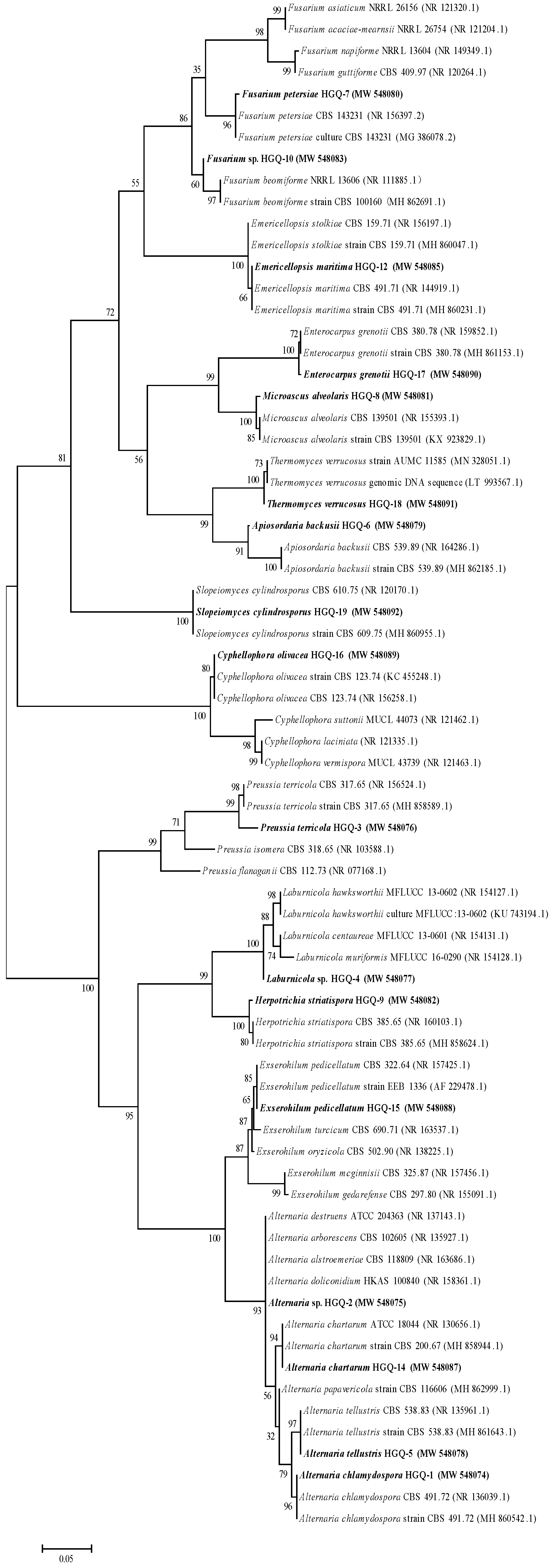
A total of 114 DSE strains were isolated from the roots of L. ruthenicum. The colonial and microscopic morphologies of the DSEs isolated are illustrated in Figure S1. The DSE colonies were black, dark brown, or grey. Microscopic observation showed that all the DSE hyphae were dark and had septum. DSE HGQ-1 (Figure S1A, HGQ-2 (Figure S1B), HGQ-5 (Figure S1E), HGQ-8 (Figure S1G), HGQ-14 (Figure S1I), and HGQ-15 (Figure S1J) produced spores in the dark at 27 °C. A comparative analysis of the fungal sequences in the GenBank database assigned the DSEs to 17 taxa within 13 genera and 14 species (Figure 3 and Table S2). The DSEs detected here included Alternaria chlamydospore, Alternaria sp., Preussia terricola, Laburnicola sp., Alternaria tellustris, Apiosordaria backusii, Fusarium petersiae, Microascus alveolaris, Herpotrichia striatispora, Fusarium sp., Emericellopsis maritima, Alternaria chartarum, Exserohilum pedicellatum, Cyphellophora olivacea, Enterocarpus grenotii, Thermomyces verrucosus and Slopeiomyces cylindrosporus. Among the 17 taxa, A. chlamydospore was the most frequent isolate, being present in multiple seasons and sites, with IF values of 21.05% (Table 2).
Isolation rate, community composition, and diversity of DSEs were varied among different seasons and sampling sites. The DSE diversity and dominance, at both Anxi and Minqin, were the highest in July, while the evenness index showed the maximum values in December. At Anxi, the order of the isolation rate was July > September > December, and the order, at Minqin, was July > December > September (Table S3).
DSE species compositions differed among sampling sites; 84 strains classified into 12 taxa were isolated from the Anxi site (IF = 73.68 %, Table 2) and isolation frequencies (IFs) in July, September, and December were 29.82%, 22.81%, and 21.05%, respectively. The dominant isolate was A. chlamydospore (IF = 19.30%) and endemic isolates were A. tellustris (IF = 3.51%), A. backusii (IF = 1.75%), F. petersiae (IF = 5.26%), E. maritima (IF = 3.51%), and E. pedicellatum (IF = 1.75%) (Table 2). By contrast, 30 strains classified into 6 taxa were isolated from the Minqin site (IF = 26.32%, Table 2), and the isolation frequencies (IFs) in July, September, and December were 12.28%, 5.26%, and 8.77%, respectively. The dominant isolate was M. alveolaris (IF = 10.52 %) and endemic isolates were M. alveolaris (IF = 10.52%), H. striatispora (IF = 3.51%), C. olivacea (IF = 3.51%), and S. cylindrosporus (IF = 3.51%) (Table 2). In general, the isolation rate and diversity were higher at Anxi than that at Minqin in each sampling season while the evenness index was higher at Minqin than at Anxi (Table 2 and Table S3).

3.3. Spatial–Temporal Distribution of Soil Factors

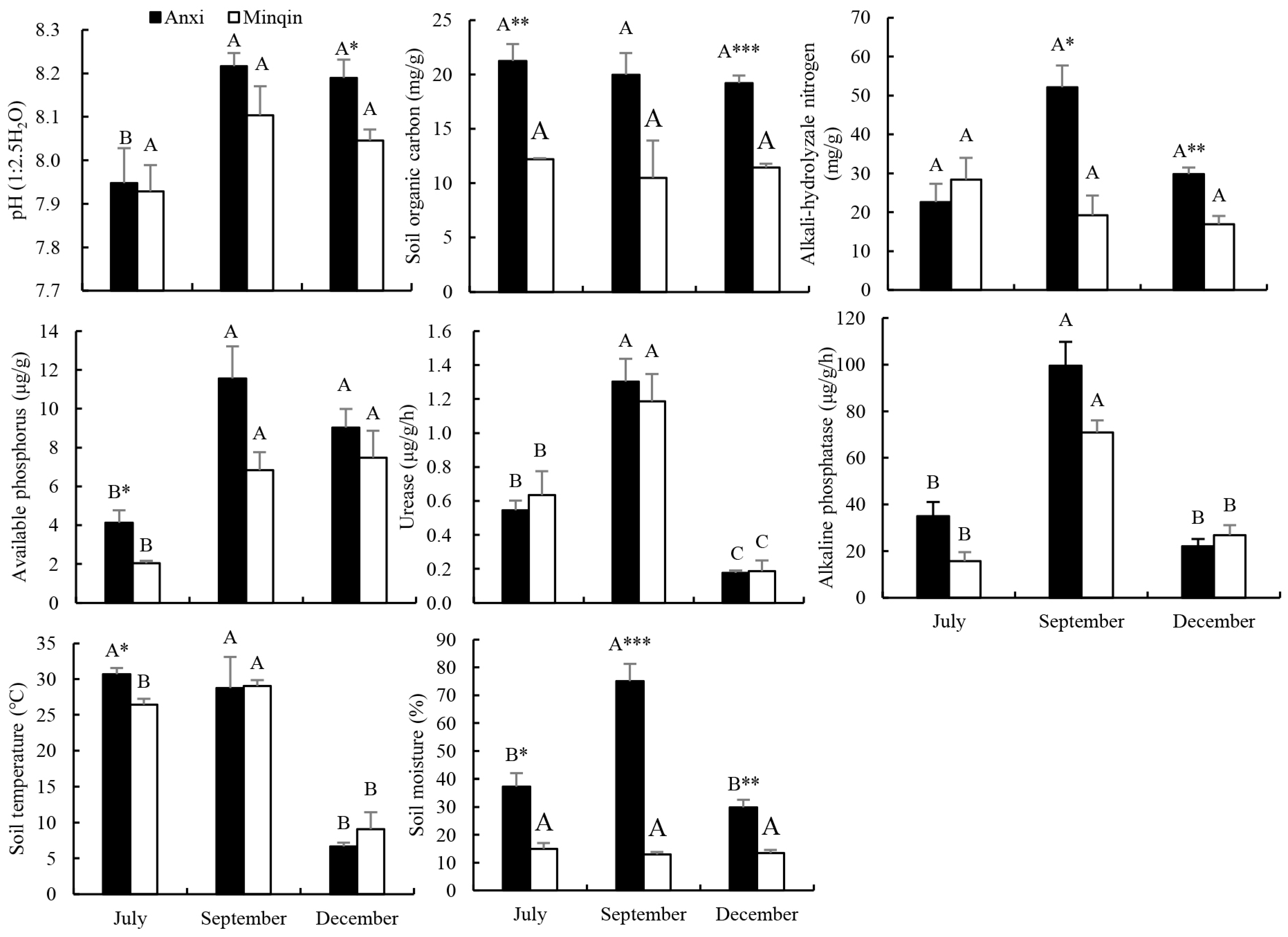
Soil factors significantly (p < 0.05) varied among seasons and sampling sites. The lowest pH was recorded in July at both Anxi and Minqin and the lowest soil temperature was measured in December (Figure 4). Soil urease and alkaline phosphatase were significantly (p < 0.05) higher in September than that in July and in December. Available phosphorus was significantly (p < 0.05) lower in July than that in September and December. Soil moisture content in September was significantly (p < 0.05) higher than that in July and December at Anxi, while there was no significant difference at Minqin. For different sampling sites, SOC was significantly higher at Anxi than Minqin in July (p < 0.01) and December (p < 0.001). Alkali-hydrolyzable nitrogen was significantly higher at Anxi than Minqin in September (p < 0.05) and December (p < 0.01). Available phosphorus and soil temperature were significantly (p < 0.05) higher at Anxi than Minqin in July. Soil moisture contents were significantly (p < 0.05) higher at Anxi than at Minqin in each sampling season. Two-way ANOVA showed that all soil factors, except SOC, were significantly (p < 0.05) influenced by the seasons. SOC, available phosphorus, alkali-hydrolyzable nitrogen, alkaline phosphatase, and soil moisture contents were significantly (p < 0.05) correlated by the sampling sites. Soil moisture content, alkali-hydrolyzable nitrogen, and alkaline phosphatase were significantly (p < 0.05) influenced by interaction of seasons and sampling sites (Table S4).

3.4. Principal Component Analysis of DSE Colonization

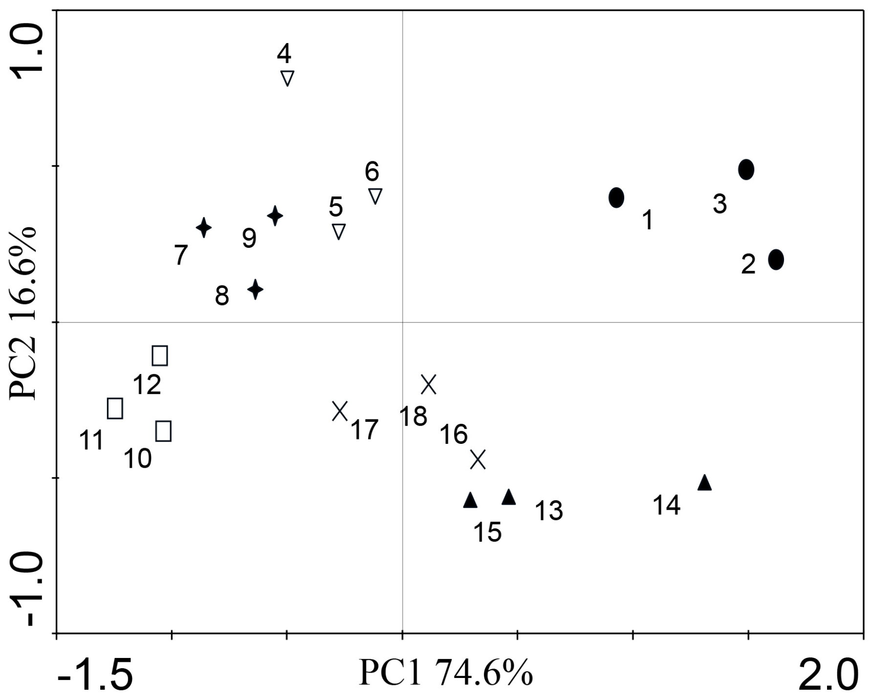
The PCA results for DSE colonization (Figure 5) showed that the cumulative contribution of the two principal components (PCs) related to DSE colonization reached 91.0% (PC1 and PC2). Along the PC2 axis, the Anxi plots in July and December occurred on the right, and the plot in September occurred on the left. Along the PC1 axis, the Minqin plots in September and December were below the axis, and the plot in July was above the axis. The result indicated DSE colonization was significantly affected by seasons and sampling sites.

3.5. Correlation Analyses

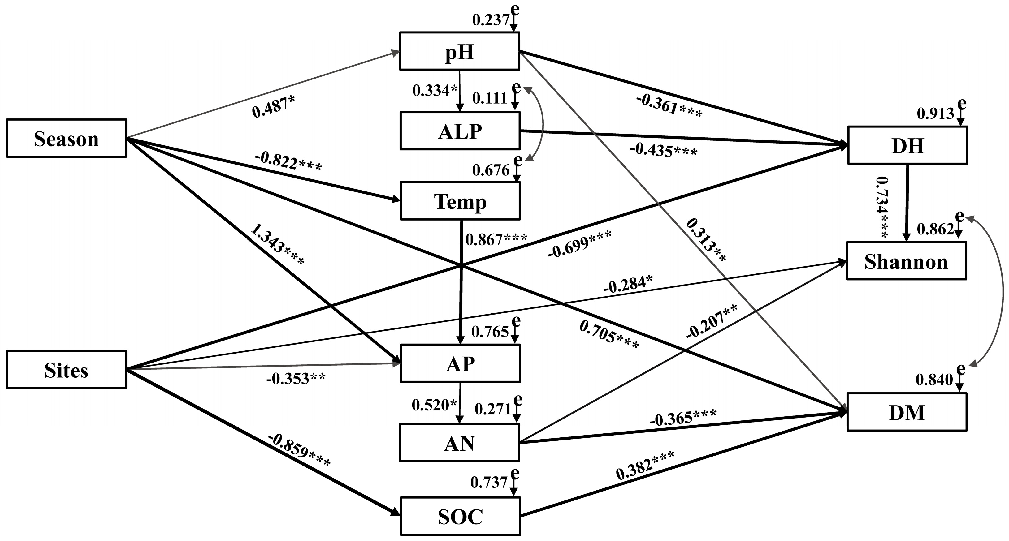
Pearson’s correlation analyses demonstrated the strong correlations among soil factors and DSE colonization (Table S5). Referring to the correlation coefficients (R values), we used SEM to quantify the relative effects of soil temperature, pH, SOC, alkali-hydrolyzable nitrogen, alkaline phosphatase, and available phosphorus on DSE hyphal colonization, microsclerotial colonization, and species diversity (χ2 = 46.384, df = 35, p = 0.094, RMSEA = 0.138, GFI = 0.705, AIC = 108.384, see Figure 6). The results revealed that sampling seasons were significantly directly correlated with pH (p < 0.05), temperature (p < 0.001), available phosphorus (p < 0.001), and microsclerotial colonization (p < 0.001). The sampling sites had significant direct effects on SOC (p < 0.001), available phosphorus (p < 0.01), hyphal colonization (p < 0.001), and species diversity (P < 0.05). Soil pH and alkaline phosphatase had significant negative effects on hyphal colonization (p < 0.001). The microsclerotial colonization was positively affected by SOC (p < 0.001) and negatively affected by alkali-hydrolyzable nitrogen (p < 0.001). Overall, DSE colonization and species diversity showed significant spatial and temporal heterogeneity and was closely related to soil factors.

4. Discussion

4.1. DSE Colonisation Status

DSEs, as root-associated fungi, have a wide ecological distribution and high colonization in a variety of desert plants [13,18,21,47]. In the present study, we found the roots of L. ruthenicum to be infected by typical dark septate hyphae and microsclerotia structures, which suggests that DSEs might be vital components of the roots of L. ruthenicum in desert habitats. The DSE morphology and colonization varied with seasons and sampling sites, which may be an effective adaptive adjustment of L. ruthenicum to adversity environments [20]. With a change of seasons, the morphology of DSE hyphae became correspondingly thicker and shorter, which was consistent with Zuo et al. [21]. Hyphae, being a channel for nutrient exchange between DSEs and plants, might protect the host against drought stress through a more effective absorption and translocation of limited nutrients that might otherwise not be accessible to the roots of the plant [48,49]. Since the sampling sites are desert regions with low rainfall, any additional feature that can improve nutrient status is a necessity. In this study, the PCA results revealed that DSE colonizations were correlated to the seasons. The hyphal colonization reached the highest in summer while the microsclerotinia colonization reached the maximum in winter, results that are consistent with the results of Rayment et al. [30]. Several studies have indicated that DSEs can promote plant biomass, improve root growth and development, and increase the number of root hairs, especially in arid environments [16,20,50]. L. ruthenicum grow actively in July and need abundant nutrients, while the DSE hyphae may promote the absorption and utilization of nutrients for host plants. DSE microsclerotia may play important roles in substrate storage, and act as a protective device for host plants to withstand severe conditions [51]. In December, with a reduction in temperature, the leaves and stems of L. ruthenicum gradually wither, and thus an increased number of microsclerotia may help host plants to resist the increasingly challenging environment [26]. In general, the changes of DSE colonization were consistent with the phenology of host plants. Moreover, except for the direct effect of season on microsclerotia colonization, season influences hyphal and microsclerotial colonization by adjusting soil factors such as pH, available phosphorus, soil temperature, and other abiotic factors, which may also lead to seasonal heterogeneity of DSE distribution [30].
For different sampling sites, the DSE hyphal colonization, total colonization, and colonization intensity showed obvious spatial heterogeneity, which was consistent with the results of Xie et al. [19], who reported that DSE infection was spatially predictable. In this work, SEM indicated that the hyphal colonization was directly affected by sampling sites. U’Ren et al. [52] declared climatic factors could drive fungi special heterogeneity. Nevertheless, in terms of climatic conditions, Anxi and Minqin were similar, both typically arid with low precipitation. Thus, we speculate that other factors can drive fungal spatial heterogeneity, such as soil characteristics. Li et al. [18] investigated the colonization of Ammopiptanthus mongolicus in three sampling sites in desert and concluded that the dynamics of DSEs had a highly spatial pattern, and were influenced by soil nutrient availability, which supported our results. Our results showed that sampling sites indirectly affect DSE microsclerotial colonization by influencing SOC, which is known as a vital factor in promoting the colonization of endophytic fungi [53,54]. In addition, soil moisture content is another trigger that causes increases in fungi colonization [55]. In this study, the soil moisture content, which was mainly the result of the unpredictable rainfall events, had positive effects on DSE colonization. The SOC and soil moisture content was higher at Anxi than at Minqin. Thus, relatively abundant soil moisture content and SOC can lead to the colonization differences among sampling sites.

4.2. DSE Species Diversity

In the present study, DSE strains were subdivided into 13 genera and 14 species based on their morphological and molecular identification. To our best knowledge, A. chartarum, C. olivacea, S. cylindrosporus, and T. verrucosus were reported here in desert ecosystems for the first time. The presence of these DSEs may be related to the arid environment and specific host plants. DSEs are generally believed to improve the ability of plants to cope with environmental stress. Meanwhile, previous studies have shown that endophytic fungal communities may have host specificity [56]. Nevertheless, the mechanism of interaction between DSEs and host plants is not clear [56,57]. In further studies we should pay more attention to the ecological functions of these DSEs and their relationship with host plants. A. chlamydospore was the dominant isolate of L. ruthenicum, which was frequently detected among different seasons and sites. Alternaria sp. has been frequently identified in the roots of desert plants and seemingly has no host specificity [13,58,59,60]. A recent study has shown that Alternaria sp. can promote the growth of Hedysarum scoparium [21]. H. striatispora, E. pedicellatum, Fusarium sp., and Microascus sp. were previously isolated in arid or semi-arid environments [13,21,61]. Interestingly, M. alveolaris is not only the dominant species but also an endemic species at Minqin. The results of our study suggested that DSEs are generalists of host plants in desert regions, where they harbor abundant fungal resources.
The DSE species composition inhabiting the roots of L. ruthenicum were shown to differ geographically, and there was no overlapping or crossing strains for dominant isolates and endemic isolates at the Anxi and Minqin sites. The DSE isolation rate and species diversity were higher at Anxi than at Minqin, which suggests that environmental selection and habitat heterogeneity may mainly account for the difference [62,63]. In our research, SEM revealed that the species diversity was directly related to sampling sites. Previously, Knapp et al. [12] suggested that endophytic fungi composition may change with locations of host plants, and this postulate has been corroborated by other relevant studies [19,21,60,64]. Moreover, our results showed that sampling sites also affect the species diversity by influencing available phosphorus and alkaline hydrolyzable nitrogen. Another study found that soil phosphorus and nitrogen content was the key factor impacting the abundance and diversity of fungi [63]. When the carbon supply in the substrate is sufficient, phosphorus, nitrogen, and other nutrient elements become the limiting factors for microbial biomass [65]. Under low-phosphorus stress, plants can release insoluble soil phosphorus [66] for microbial to utilize, which ultimately changes the physical and chemical properties of the soil by improving nutrition. Changes in these properties further affect the fungi species composition and diversity. In addition, our results showed that sampling sites indirectly affect species diversity by influencing hyphae colonization. In terms of DSE colonization, the colonization of DSEs was significantly higher at Anxi than at Minqin; thus, this may be one of the explanations for the higher species diversity at Anxi than at Minqin.
Martins et al. [67] reported that the diversity and richness of endophytic fungi isolated from olive trees in spring were higher than in autumn, and the diversity of endophytic fungi was affected by seasonal variation. However, in our study, despite seasonal differences in species diversity, the species diversity was not directly correlated to the seasons, which suggests that other factors could drive fungal temporal heterogeneity. We speculate the variation of DSE species compositions over time might be due to changes in the chemistry of plant tissues in the course of the phenological growth stages of the tree and to the interspecific competition among the fungi [68]. Thus, more research is required to confirm the mechanism by which interactive associations form between the DSEs and host plants.

4.3. DSE and Soil Factors

Root-associated fungi play a pivotal role in nutrient exchange between plants and soil [69] and can alter the physical and chemical properties of the soil [70]. Numerous studies have confirmed that there is a significant relationship between soil nutrient and fungal infection [13,19,21]. Here, SOC was positively correlated with DSE microsclerotial colonization. Several studies have identified soil carbon sources as key ecological drivers of microbial community dynamics [71]. SOC provides the dominant carbon metabolic sources for endophytic fungi [72]. Abundant organic carbon can promote DSE colonization and can even improve the viability of host plants [53,54]. It has been well recorded that pH is an important factor that strongly affects colonization of soil fungi [73,74]. The optimal pH of most fungi are in the range of 5.5–7.5. With an increase in soil pH, absorption ability of host plants is limited [75,76]. Favorable changes in soil pH are likely to improve the nutritional status and health of the host by influencing fungal distribution [77]. Correlation analysis showed that DSE hyphal colonization was negatively correlated with soil pH, which was consistent with several studies about Solanaceae [78,79]. In addition, our data suggest that soil alkali-hydrolyzable nitrogen has a negative correlation with DSE microsclerotia, in part supporting the finding that the concentration and content of root N are related to infection by DSE structures [15]. Moreover, alkali-hydrolyzable nitrogen was also negatively correlated with species diversity. Li et al. [20] determined plants infected with DSE fungi increased N absorption. Although DSEs cannot improve uptake of N through direct transfer, they can allow increased access to a host plant [80]. For growth, host plants need N to provide nutrients [81], which is mainly acquired from soil substance. In the case of insufficient soil N source, host plants likely need more endophytic fungi to assist absorption of nutrients. In general, DSE colonization and composition may be affected by host plant identity, which is a substrate for the survival of fungi [56]. Meanwhile, environmental factors may also lead to spatial and temporal heterogeneity of DSEs [31,82,83]. Our research showed that changing seasons and sampling sites can directly impact DSE colonization and species composition and can indirectly influence DSEs by adjusting soil microenvironments.
Our results demonstrated that desert plants can be colonized by DSEs, which may have the potential to promote the growth of desert plants. This finding provides a new direction for the DSE fungi application to cultivation of L. ruthenicum. Meanwhile, agricultural ecological environments are facing drought stress. Some studies have shown that DSEs can promote plant growth under drought stress [8]. Therefore, it is crucial to understand the spatial and temporal distribution of DSEs in L. ruthenicum, which could provide the basis for the application of DSEs in agricultural drylands.

5. Conclusions

Overall, root–fungal associations were found between L. ruthenicum and DSEs in desert regions of northwest China. The DSE colonization and species diversity showed significant spatial and temporal heterogeneity and was closely related to soil factors. Among the identified DSE species, A. chartarum, C. olivacea, S. cylindrosporus, and T. verrucosus were reported here for the first time in a desert environment. The morphology and colonization of DSEs might be useful indicators for evaluating soil quality and function of desert ecosystems. Future research should investigate the function of DSE associations in L. ruthenicum to improve the understanding of the role of DSE fungi in desert ecosystems. Therefore, a better understanding of the complex interactions between L. ruthenicum and DSEs would help to enhance ecological restoration in desert ecosystems and for practical application in the agriculture cultivation of L. ruthenicum in northwest China.

Supplementary Materials

The following are available online at https://www.mdpi.com/article/10.3390/agronomy11040648/s1, Figure S1: A-L Colonies of endophytic fungi isolated from the roots of L. ruthenicum. a-l Microscopic morphology of endophytic fungi. Arrows indicate the following: Hy = DSE hyphae, S = DSE spore. A-E: HGQ-1, 2, 3, 4, 5. F-H: HGQ-7, 8, 9. I-K: HGQ-14, 15, 16. L: HGQ-18, Table S1: Environmental condition in the sites in Gansu, China, Table S2: DSE isolates from medicinal plant roots and results of sequence Blast from type material database, Table S3: DSE species diversity of L. ruthenicum, Table S4: Two-way analysis of variance of the effects in different months and sites on soil factors, Table S5: Correlation relationship among DSE colonization and soil factors.

Author Contributions

Conceptualization, L.H., J.S. and X.H.; methodology, L.H. and J.S.; formal analysis, L.H. and J.S.; investigation, L.H. and J.S.; resources, X.H.; writing—original draft preparation, L.H. and J.S.; writing—review and editing, L.H., J.S., C.H., and X.H.; supervision, X.H.; funding acquisition, X.H. All authors have read and agreed to the published version of the manuscript.

Funding

This research was funded by the National Natural Science Foundation of China (Project 31770561).

Acknowledgments

We are grateful to students of Yiling Zuo and Lifeng Hou for sampling and laboratory work.

Conflicts of Interest

The authors declare no conflict of interest.

References

  1. Tang, Z.; An, H.; Deng, L.; Wang, Y.; Zhu, G.; Shangguan, Z. Effect of desertification on productivity in a desert steppe. Sci. Rep. 2016, 6, 27839. [Google Scholar] [CrossRef]
  2. Chaves, M.M.; Maroco, J.P.; Pereira, J.S. Understanding plant responses to drought-from genes to the whole plant. Funct. Plant Biol. 2003, 30, 239–264. [Google Scholar] [CrossRef]
  3. Niu, R.; Liu, J.; Zhao, X.; Qin, Y. Ecological benefit of different revegetated covers in the middle of Hexi corridor, northwestern China. Environ. Earth Sci. 2015, 74, 5699–5710. [Google Scholar] [CrossRef]
  4. Lioubimtseva, E.; Henebry, G.M. Climate and environmental change in arid Central Asia: Impacts, vulnerability, and adaptations. J. Arid Environ. 2009, 73, 963–977. [Google Scholar] [CrossRef]
  5. Brooker, R.W.; Maestre, F.T.; Callaway, R.M.; Lortie, C.L.; Cavieres, L.A.; Kunstler, G.; Michalet, R. Facilitation in plant communities: The past, the present, and the future. J. Ecol. 2008, 96, 18–34. [Google Scholar] [CrossRef] [Green Version]
  6. Jalali, G.A.; Akbarian, H.; Rhoades, C.; Yousefzadeh, H. The effect of the halophytic shrub lycium ruthenicum (mutt) on selected soil properties of a desert ecosystem in central iran. Pol. J. Ecol. 2012, 845–850. [Google Scholar] [CrossRef]
  7. Kivlin, S.N.; Emery, S.M.; Rudgers, J.A. Fungal symbionts alter plant responses to global change. Am. J. Bot. 2013, 100, 1445–1457. [Google Scholar] [CrossRef]
  8. Li, X.; He, X.L.; Hou, L.F.; Ren, Y.; Wang, S.J.; Su, F. Dark septate endophytes isolated from a xerophyte plant promote the growth of Ammopiptanthus mongolicus under drought condition. Sci. Rep. 2018, 8, 239–264. [Google Scholar] [CrossRef] [Green Version]
  9. Jumpponen, A.; Trappe, J.M. Dark septate endophytes: A review of facultative biotrophic root-colonizing fungi. New Phytol. 1998, 140, 295–310. [Google Scholar] [CrossRef]
  10. Mandyam, K.; Jumpponen, A. Seeking the elusive function of the root-colonising dark septate endophytic fungi. Stud. Mycol. 2005, 53, 173–189. [Google Scholar] [CrossRef] [Green Version]
  11. Barrow, J.R. A typical morphology of dark septate fungal root endophytes of Bouteloua in arid southwestern USA rangelands. Mycorrhiza 2003, 13, 239–247. [Google Scholar] [CrossRef]
  12. Knapp, D.G.; Pintye, A.; Kovács, G.M. The dark side is not fastidious-dark septate endophytic fungi of native and invasive plants of semiarid sandy areas. PLoS ONE 2012, 7, e32570. [Google Scholar] [CrossRef] [Green Version]
  13. Hou, L.F.; He, X.L.; Li, X.; Wang, S.J.; Zhao, L.L. Species composition and colonization of dark septate endophytes are affected by host plant species and soil depth in the Mu Us sandland, northwest China. Fungal Ecol. 2019, 39, 276–284. [Google Scholar] [CrossRef]
  14. Van der Heij, M.G.; Martin, F.M.; Selosse, M.A.; Sanders, I.R. Mycorrhizal ecology and evolution: The past, the present, and the future. New Phytol. 2015, 205, 1406–1423. [Google Scholar] [CrossRef] [PubMed]
  15. Newsham, K.K. A meta-analysis of plant responses to dark septate root endophytes. New Phytol. 2011, 190, 783–793. [Google Scholar] [CrossRef] [PubMed]
  16. Zhang, H.H.; Tang, M.; Chen, H.; Wang, Y.J.; Ban, Y. Arbuscular mycorrhizas and dark septate endophytes colonization status in medicinal plant Lycium barbarum L. in arid Northwestern China. Afr. J. Microbiol. Res. 2010, 4, 1914–1920. [Google Scholar] [CrossRef] [Green Version]
  17. Zhang, H.H.; Tang, M.; Chen, H.; Wang, Y.J. Effects of a dark-septate endophytic isolate LBF-2 on the medicinal plant Lycium barbarum L. J. Microbiol. 2012, 50, 91–96. [Google Scholar] [CrossRef]
  18. Li, B.K.; He, X.L.; He, C.; Chen, Y.; Wang, X. Spatial dynamics of dark septate endophytes and soil factors in the rhizosphere of Ammopiptanthus mongolicus in Inner Mongolia, China. Symbiosis 2015, 65, 75–84. [Google Scholar] [CrossRef]
  19. Xie, L.L.; He, X.L.; Wang, K.; Hou, L.F.; Sun, Q. Spatial dynamics of dark septate endophytes in the roots and rhizospheres of Hedysarum scoparium in northwest China and the influence of edaphic variables. Fungal Ecol. 2017, 26, 135–143. [Google Scholar] [CrossRef]
  20. Li, X.; He, C.; He, X.L.; Su, F.; Hou, L.F.; Ren, Y.; Hou, Y.T. Dark septate endophytes improve the growth of host and non-host plants under drought stress through altered root development. Plant Soil 2019, 439, 259–272. [Google Scholar] [CrossRef]
  21. Zuo, Y.L.; Su, F.; He, X.L.; Li, M. Colonization by dark septate endophytes improves the growth of Hedysarum scoparium under multiple inoculum levels. Symbiosis 2020, 82, 201–214. [Google Scholar] [CrossRef]
  22. Likar, M.; Regvar, M. Isolates of dark septate endophytes reduce metal uptake and improve physiology of Salix caprea L. Plant Soil 2013, 370, 593–604. [Google Scholar] [CrossRef]
  23. Dos Santos, S.G.D.; Silva, P.R.A.D.; Garcia, A.C.; Zilli, J.É.; Berbara, R.L.L. Dark septate endophyte decreases stress on rice plants. Braz. J. Microbiol. 2017, 48, 333–341. [Google Scholar] [CrossRef]
  24. Butler, M.; Day, A. Fungal melanins: A review. Can. J. Microbiol. 1998, 44, 1115–1136. [Google Scholar] [CrossRef]
  25. Zhan, F.; He, Y.; Zu, Y.; Li, T.; Zhao, Z. Characterization of melanin isolated from a dark septate endophyte (DSE), Exophiala pisciphila. World J. Microbiol. Biotechnol. 2011, 27, 2483–2489. [Google Scholar] [CrossRef]
  26. Mandyam, K.; Jumpponen, A. Seasonal and temporal dynamics of arbuscular mycorrhizal and dark septate endophytic fungi in a tallgrass prairie ecosystem are minimally affected by nitrogen enrichment. Mycorrhiza 2008, 18, 145–155. [Google Scholar] [CrossRef]
  27. Hazard, C.; Gosling, P.; Mitchell, D.T.; Doohan, F.M.; Bending, G.D. The role of local environment and geographical distance in determining community composition of arbuscular mycorrhizal fungi at the landscape scale. ISME J. 2013, 7, 498–508. [Google Scholar] [CrossRef] [PubMed] [Green Version]
  28. Dasila, K.; Pandey, A.; Samant, S.; Pande, V. Endophytes associated with Himalayan silver birch (Betula-utilis D. Don) roots in relation to season and soil parameters. Appl. Soil Ecol. 2020, 149, 103513. [Google Scholar] [CrossRef]
  29. Ruotsalainen, A.; Väre, H.; Vestberg, M. Seasonality of root fungal colonization in low-alpine herbs. Mycorrhiza 2002, 12, 29–36. [Google Scholar] [CrossRef] [PubMed]
  30. Rayment, J.T.; Jones, S.; French, K. Seasonal patterns of fungal colonisation in Australian native plants of different ages. Symbiosis 2020, 80, 169–182. [Google Scholar] [CrossRef]
  31. Huusko, K.; Ruotsalainen, A.L.; Markkola, A.M. A shift from arbuscular mycorrhizal to dark septate endophytic colonization in Deschampsia flexuosa roots occurs along primary successional gradient. Mycorrhiza 2017, 27, 1–10. [Google Scholar] [CrossRef] [PubMed]
  32. Soil Survey Staff. Keys to Soil Taxonomy, 12th ed.; USDA-Natural Resources Conservation Service: Washington, DC, USA, 2014. [Google Scholar]
  33. Heiri, O.; Lotter, A.F.; Lemcke, G. Loss on ignition as a method for estimating organic and carbonate content in sediments: Reproducibility and comparability of results. J. Paleolimnol. 2001, 25, 101–110. [Google Scholar] [CrossRef]
  34. Bever, J.D.; Morton, J.B.; Antonovics, J.; Schultz, P.A. Host-dependent sporulation and species diversity of arbuscular mycorrhizal fungi in a mown grassland. J. Ecol. 1996, 84, 71–82. [Google Scholar] [CrossRef]
  35. Tarafdar, J.C.; Marschner, H. Phosphatase activity in the rhizosphere and hyphosphere of VA mycorrhizal wheat supplied with inorganic and organic phosphorus. Soil Biol. Biochem. 1994, 26, 387–395. [Google Scholar] [CrossRef]
  36. Hoffmann, G.G.; Teicher, K. A colorimetric technique for determining urease activity in soil. Dung Boden 1961, 95, 55–63. [Google Scholar] [CrossRef]
  37. Berch, S.M.; Kendrick, B. Vesicular-arbuscular mycorrhizae of southern Ontario ferns and fern-allies. Mycologia 1982, 74, 769–776. [Google Scholar] [CrossRef]
  38. Biermann, B.; Linderman, R.G. Quantifying vesicular-arbuscular mycorrhizae: A proposed method towards standardization. New Phytol. 1981, 87, 63–67. [Google Scholar] [CrossRef]
  39. Trouvelot, A.; Kough, J.L.; Gianinazzi-Pearson, V. Mesure du taux demycorhization V A d’un système radiculaire. Recherche de methods d’estimation ayant une signification fonctionnelle. In Physiological and Genetical Aspects of Mycorrhizae; Gianinazzi-Pearson, V., Gianinazzi, S., Eds.; INRA Press: Paris, France, 1986; pp. 217–221. [Google Scholar]
  40. Maadon, S.N.; Wakid, S.A.; Zainudin, I.I.; Rusli, L.S.; Mohd Zan, M.S.; Hasan, N.A.; Abu Shah, N.A.; Rohani, E.R. Isolation and identification of endophytic fungi from UiTM reserve forest, Negeri Sembilan. Sains Malays. 2018, 47, 3025–3030. [Google Scholar] [CrossRef]
  41. Tamura, K.; Stecher, G.; Peterson, D.; Filipski, A.; Kumar, S. MEGA6: Molecular evolutionary genetics analysis version 6.0. Mol. Biol. Evol. 2013, 30, 2725–2729. [Google Scholar] [CrossRef] [PubMed] [Green Version]
  42. Sun, Y.; Wang, Q.; Lu, X.; Okane, I.; Kakishima, M. Endophytic fungal community in stems and leaves of plants from desert areas in China. Mycol. Prog. 2011, 11, 781–790. [Google Scholar] [CrossRef]
  43. Yuan, Z.L.; Rao, L.B.; Chen, Y.C.; Zhang, C.L.; Wu, Y.G. From pattern to process: Species and functional diversity in fungal endophytes of Abies beshanzuensis. Fungal Biol. 2011, 115, 197–213. [Google Scholar] [CrossRef]
  44. Shannon, C.E.; Weaver, W. The Mathematical Theory of Communication; University of Illinois Press: Urbana, IL, USA, 1949. [Google Scholar]
  45. Caruso, T.; Pigino, G.; Bernini, F.; Bargagli, R.; Migliorini, M. The Berger—Parker index as an effective tool for monitoring the biodiversity of disturbed soils: A case study on Mediterranean oribatid (Acari: Oribatida) assemblages. Biodivers. Conserv. 2008, 7, 35–43. [Google Scholar] [CrossRef]
  46. Samaga, P.V.; Rai, V.R. Diversity and bioactive potential of endophytic fungi from Nothapodytes foetida, Hypericum mysorense and Hypericum japonicum collected from Western Ghats of India. Ann. Microbiol. 2015, 66, 229–244. [Google Scholar] [CrossRef]
  47. Ali, S.; Ganai, B.A.; Kamili, A.N.; Bhat, A.A.; Mir, Z.A.; Bhat, J.A.; Grover, A. Pathogenesis-related proteins and peptides as promising tools for engineering plants with multiple stress tolerance. Microbiol. Res. 2018, 212–213, 29–37. [Google Scholar] [CrossRef]
  48. Orłowska, E.; Orłowski, D.; Mesjasz-Przybyłowicz, J.; Turnau, K. Role of mycorrhizal colonization in plant establishment on an alkaline gold mine tailing. Int. J. Phytoremediat. 2010, 13, 185–205. [Google Scholar] [CrossRef]
  49. Saito, K.; Kuga-Uetake, Y.; Saito, M.; Peterson, R.L. Vacuolar localization of phosphorus in hyphae of Phialocephala fortinii, a dark septate fungal root endophyte. Can. J. Microbiol. 2006, 52, 643–650. [Google Scholar] [CrossRef]
  50. Wu, L.Q.; Guo, S.X. Interaction between an isolate of dark septate fungi and its host plant Saussurea involucrata. Mycorrhiza 2008, 18, 79–85. [Google Scholar] [CrossRef] [PubMed]
  51. Yu, T.; Nassuth, A.; Peterson, R.L. Characterization of the interaction between the dark septate fungus Phialocephala fortinii and Asparagus officinalis roots. Can. J. Microbiol. 2001, 47, 741–753. [Google Scholar] [CrossRef]
  52. U’Ren, J.M.; Lutzoni, F.; Miadlikowska, J.; Laetsch, A.D.; Arnold, E.A. Host and geographic structure of endophytic and endolichenic fungi at a continental scale. Am. J. Bot. 2012, 99, 898–914. [Google Scholar] [CrossRef] [PubMed] [Green Version]
  53. Penton, C.R.; Derek, S.L.; Amanda, P.; Cole, J.R.; Wu, L.Y.; Schuur, E.A.G.; Zhou, J.Z.; Tiedje, J.M. Denitrifying and diazotrophic community responses to artifcial warming in permafrost and tallgrass prairie soils. Front. Microbiol. 2015, 6, 746. [Google Scholar] [CrossRef] [PubMed]
  54. Zhang, H.S.; Zhou, M.X.; Zai, X.M.; Zhao, F.G.; Qin, P. Spatio-temporal dynamics of arbuscular mycorrhizal fungi and soil organic carbon in coastal saline soil of China. Sci. Rep. 2020, 10, 9781. [Google Scholar] [CrossRef]
  55. He, X.L.; Mouratov, S.; Steinberger, Y. Temporal and spatial dynamics of vesicular-arbuscular mycorrhizal fungi under the canopy of Zygophyllum dumosum Boiss. in the Negev Desert. J. Arid Environ. 2002, 52, 379–387. [Google Scholar] [CrossRef]
  56. Sudová, R.; Kohout, P.; Rydlová, J.; Čtvrtlíková, M.; Suda, J.; Voříšková, J.; Kolaříková, Z. Diverse fungal communities associated with the roots of isoetid plants are structured by host plant identity. Fungal Ecol. 2020, 45, 100914. [Google Scholar] [CrossRef]
  57. Selim, K.A.; El-Beih, A.A.; Abdel-Rahman, T.M.; El-Diwany, A.I. Biodiversity and antimicrobial activity of endophytes associated with Egyptian medicinal plants. Mycosphere 2011, 6, 669–678. [Google Scholar] [CrossRef]
  58. Bezerra, J.D.P.; Santos, M.G.S.; Barbosa, R.N.; Svedese, V.M.; Lima, D.M.M. Fungal endophytes from cactus Cereus jamacaru in Brazilian tropical dry forest: A first study. Symbiosis 2013, 60, 53–63. [Google Scholar] [CrossRef]
  59. Fonseca-García, C.; Devin, C.D.; Etzel, G.; Axel, V.; Tringe, S.G.; Partida-Martínez, L.P. The cacti microbiome: Interplay between habitat-filtering and host-specificity. Front. Microbiol. 2016, 7, 150. [Google Scholar] [CrossRef]
  60. Pieterse, Z.; Aveling, T.A.S.; Jacobs, A.; Cowan, D.A. Seasonal variability in fungal endophytes from Aizoaceae plants in the Succulent Karoo biodiversity hotspot, South Africa. J. Environ. 2018, 156, 19–26. [Google Scholar] [CrossRef] [Green Version]
  61. ÖZer, G.; Göre, M.E.; Alkan, M.; Yaman, M.; Dababat, A.A. First report of Exserohilum pedicellatum causing root rot of wheat in azerbaijan. Plant Dis. 2019, 103, 1416. [Google Scholar] [CrossRef]
  62. Lundberg, D.S.; Lebeis, S.L.; Paredes, S.H.; Yourstone, S.; Gehring, J.; Malfatti, S.; Tremblay, J.; Engelbrektson, A.; Kunin, V.; Rio, T.G.; et al. Defining the core Arabidopsis thaliana root microbiome. Nature 2012, 488, 86–90. [Google Scholar] [CrossRef] [Green Version]
  63. Qiang, W.; He, X.L.; Wang, J.J.; Zhao, L.L. Temporal and spatial variation of arbuscular mycorrhizal fungi under the canopy of Hedysarum scoparium in the northern desert, China. Appl. Soil Ecol. 2019, 136, 139–147. [Google Scholar] [CrossRef]
  64. Zhao, D.; Li, T.; Shen, M.; Wang, J.; Zhao, Z. Diverse strategies conferring extreme cadmium (Cd) tolerance in the dark septate endophyte (DSE), Exophiala pisciphila: Evidence from RNA-seq data. Microbiol. Res. 2015, 170, 27–35. [Google Scholar] [CrossRef]
  65. Ilstedt, U.; Singh, S. Nitrogen and phosphorus limitations of microbial respiration in a tropical phosphorus-fixing acrisol ultisol. compared with organic compost. Soil Biol. Biochem. 2005, 37, 1407–1410. [Google Scholar] [CrossRef]
  66. Lambers, H.; Shane, M.W.; Cramer, M.D.; Pearse, S.J.; Veneklaas, E.J. Root structure and functioning for efficient acquisition of phosphorus, matching morphological and physiological traits. Ann. Bot-Lond. 2006, 98, 693–713. [Google Scholar] [CrossRef] [PubMed] [Green Version]
  67. Martins, F.; Pereira, J.A.; Bota, P.; Bento, A.; Baptista, P. Fungal endophyte communities in above- and belowground olive tree organs and the effect of season and geographic location on their structures. Fungal Ecol. 2016, 20, 193–201. [Google Scholar] [CrossRef]
  68. Fang, W.; Yang, L.; Zhu, X.; Zeng, L.; Li, X. Seasonal and habitat dependent variations in culturable endophytes of Camellia sinensis. J. Plant Pathol. Microbiol. 2013, 4, 169. [Google Scholar] [CrossRef] [Green Version]
  69. Peterson, R.L.; Wagg, C.; Pautler, M. Associations between microfungal endophytes and roots: Do structural features indicate function? Botany 2008, 86, 445–456. [Google Scholar] [CrossRef]
  70. Casazza, G.; Lumini, E.; Ercole, E.; Dovana, F.; Guerrina, M.; Arnulfo, M.; Fusconi, A.; Mucciarelli, M. The abundance and diversity of arbuscular mycorrhizal fungi are linked to the soil chemistry of screes and to slope in the Alpic paleo-endemic Berardia subacaulis. PLoS ONE 2017, 12, e0171866. [Google Scholar] [CrossRef] [PubMed]
  71. Vries, F.T.; Manning, P.; Tallowin, J.R.B.; Mortimer, S.R.; Pilgrim, E.S.; Harrison, K.A.; Hobbs, P.J.; Quirk, H.; Shipley, B.; Cornelissen, J.H.C.; et al. Abiotic drivers and plant traits explain landscape-scale patterns in soil microbial communities. Ecol. Lett. 2012, 15, 1230–1239. [Google Scholar] [CrossRef]
  72. Paterson, E.; Gebbing, T.; Abel, C.; Telfer, G. Rhizodeposition shapes rhizosphere microbial community structure in organic soil. New Phytol. 2007, 173, 600–610. [Google Scholar] [CrossRef] [PubMed]
  73. Ju, T.; Chen, Y.; Chang, C.; An, L. The diversity of soil fungi and its relations with fertility factors in Taxus chinenesis (Pilg.) Rehd community of Xiaolongshan of Tianshui City. Res. Environ. Sci. 2008, 21, 128–132. [Google Scholar] [CrossRef]
  74. Wang, M.; Shi, S.; Lin, F.; Jiang, P. Response of the soil fungal community to multi-factor environmental changes in a temperate forest. Appl. Soil Ecol. 2014, 81, 45–56. [Google Scholar] [CrossRef]
  75. Asghar, M.N.; Khan, S.; Mushtaq, S. Management of treated pulp and paper mill effluent to achieve zero discharge. J. Environ. Manag. 2008, 88, 1285–1299. [Google Scholar] [CrossRef]
  76. Ruotsalainen, A.L.; Eskelinen, A. Root fungal symbionts interact with mammalian herbivory, soil nutrient availability and specific habitat conditions. Oecologia 2011, 166, 807–817. [Google Scholar] [CrossRef] [PubMed]
  77. Glassman, S.I.; Wang, I.J.; Bruns, T.D. Environmental filtering by pH and soil nutrients drives community assembly in fungi at fine spatial scales. Mol. Biol. 2017, 26, 6960–6973. [Google Scholar] [CrossRef] [Green Version]
  78. Rajeshkumar, P.P.; Thomas, G.V.; Gupta, A.; Gopal, M. Diversity, richness and degree of colonization of arbuscular mycorrhizal fungi in coconut cultivated along with intercrops in high productive zone of Kerala, India. Symbiosis 2015, 65, 125–141. [Google Scholar] [CrossRef]
  79. Muthukumar, T.; Sathya, R. Endorhizal fungal association and colonization patterns in Solanaceae. Pol. Bot. J. 2017, 62, 287–299. [Google Scholar] [CrossRef] [Green Version]
  80. Caldwell, B.A.; Jumpponen, A.; Trappe, J.M. Utilization of major detrital substrates by dark septate, root endophytes. Mycologia 2000, 92, 230–232. [Google Scholar] [CrossRef]
  81. Zuo, Y.L.; He, C.; He, X.L.; Li, X.; Xue, Z.K.; Li, X.M.; Wang, S.J. Plant cover of Ammopiptanthus mongolicus and soil factors shape soil microbial community and catabolic functional diversity in the arid desert in Northwest China. Appl. Soil Ecol. 2019, 147, 103389. [Google Scholar] [CrossRef]
  82. Dumbrell, A.J.; Ashton, P.D.; Aziz, N.; Feng, G.; Nelson, M.; Dytham, C.; Alastair, H. Distinct seasonal assemblages of arbuscular mycorrhizal fungi revealed by massively parallel pyrosequencing. New Phytol. 2011, 190, 794–804. [Google Scholar] [CrossRef]
  83. Montero Sommerfeld, H.; Díaz, L.M.; Alvarez, M.; Añazco, V.C.; Matus, F.; Boon, N.; Boeckx, P.; Huygens, D. High winter diversity of arbuscular mycorrhizal fungal communities in shallow and deep grassland soils. Soil Biol. Biochem. 2013, 65, 236–244. [Google Scholar] [CrossRef]
Figure 1. Dark septate endophytes associated with the roots of L. ruthenicum. Arrows indicate the following: Hy, dark septate endophytes (DSE) hyphae and Mi, DSE microsclerotia. Images are of roots sampled from Anxi (AF) and Minqin (GL). (A,D,G,J) are sampled in July, (B,E,H,K) are sampled in September, and (C,F,I,L) are sampled in December.
Agronomy 11 00648 g001
Figure 2. Temporal and spatial changes of DSE colonization in L. ruthenicum roots. Significant (p < 0.05) differences among different months marked with different letters. Significant (p < 0.05) differences among different sites marked with * (* indicates a significant difference at p < 0.05 and ** indicates a significant difference at p < 0.01).
Agronomy 11 00648 g002
Figure 3. Maximum likelihood tree generated from ITS region sequences of isolated strains and their closest matches. Scale bar indicates distance = 5% nucleotide diversity. Sequences that were determined in the course of this study appear in bold.
Agronomy 11 00648 g003
Figure 4. Spatial and temporal distribution of soil factors of L. ruthenicum. p < 0.05, significant differences among different mouths marked with different letters; p < 0.05, significant differences among sampling sites marked with *; * indicates a significant difference at P < 0.05, ** indicates a significant difference at p < 0.01, and *** indicates a significant difference at P < 0.001.
Agronomy 11 00648 g004
Figure 5. Principal component analysis of DSE colonization in different sampling times and sampling sites. (1–3) July, Anxi; (4–6) July, Minqin; (7–9) September, Anxi; (10–12) September, Minqin; (13–15) December, Anxi; (16–18) December, Minqin.
Agronomy 11 00648 g005
Figure 6. Structural equation model showing the causal relationships among different months, sites, edaphic factors, and DSE colonization. The final model fits the data well: maximum likelihood χ2 = 46.384, df = 35, P = 0.094, goodness-of-fit index = 0.705, Akaike information criteria = 108.384, and root mean square error of approximation = 0.138. The numbers near the arrows indicate the standardized path coefficients, and the width of the solid lines indicates the strength of the causal effect (* p < 0.05, ** p < 0.01, and *** p < 0.001). ALP, alkaline phosphatase; AP, available phosphorus; AN, alkali-hydrolyzable nitrogen; SOC, soil organic carbon; Temp, temperature; DH, DSE hyphal colonization; DM, DSE microsclerotial colonization; Shannon, the Shannon–Wiener index; e, the values of residuals.
Agronomy 11 00648 g006
Table 1. Two-way analysis of variance of the effects of different seasons and sites on DSE colonization.
ItemSeasonSitesSeason * Sites
FpFpFp
Hyphae19.023<0.00122.6<0.0011.9950.179
Microsclerotia19.634<0.0012.1770.1661.6340.236
Total colonisation9.7090.0036.4870.0264.3260.038
Colonization intensity33.738<0.0019.1260.01121.259<0.001
* means interation effect of two factors.
Table 2. The number of isolates recovered and isolation frequency (IF) of DSEs in the roots of L. ruthenicum.
Sampling TimeSampling SitesStrain NumberClassificationNumber of StrainsIF%
JulyAnxiHGQ-1Alternaria chlamydospora1210.53
HGQ-2Alternaria sp.43.51
HGQ-3Preussia terricola21.75
HGQ-4Laburnicola sp.65.26
HGQ-5Alternaria tellustris43.51
HGQ-6Apiosordaria backusii21.75
HGQ-7Fusarium petersiae43.51
Total number of strains3429.82
MinqinHGQ-8Microascus alveolaris65.26
HGQ-3Preussia terricola21.75
HGQ-9Herpotrichia striatispora43.51
HGQ-10Fusarium sp.21.75
Total number of strains1412.28
SeptemberAnxiHGQ-1Alternaria chlamydospora21.75
HGQ-2Alternaria sp.65.26
HGQ-14Alternaria chartarum87.02
HGQ-15Exserohilum pedicellatum21.75
HGQ-3Preussia terricola87.02
Total number of strains2622.81
MinqinHGQ-8Microascus alveolaris21.75
HGQ-16Cyphellophora olivacea43.51
Total number of strains65.26
DecemberAnxiHGQ-1Alternaria chlamydospora87.02
HGQ-7Fusarium petersiae21.75
HGQ-12Emericellopsis maritima43.51
HGQ-17Enterocarpus grenotii43.51
HGQ-18Thermomyces verrucosus65.26
Total number of strains2421.05
MinqinHGQ-1Alternaria chlamydospora21.75
HGQ-8Microascus alveolaris43.51
HGQ-19Slopeiomyces cylindrosporus43.51
Total number of strains108.77
Total 114100
Publisher’s Note: MDPI stays neutral with regard to jurisdictional claims in published maps and institutional affiliations.