Article

Inhibition of the Growth and Development of Sclerotinia sclerotiorum (Lib.) De Bary by Combining Azoxystrobin, Penicillium chrysogenum VKM F-4876d, and Bacillus Strains

by
Vera V. Yaderets
,
Nataliya V. Karpova
,
Elena V. Glagoleva
,
Alexander I. Ovchinnikov
,
Kseniya S. Petrova
and
Vakhtang V. Dzhavakhiya
*
Laboratory of Biotechnology of Physiologically Active Compounds, Federal Research Center “Fundamentals of Biotechnology”, Russian Academy of Sciences, pr-t 60-letiya Oktyabrya, 7, bld. 1, 117312 Moscow, Russia
*
Author to whom correspondence should be addressed.
Agronomy 2021, 11(12), 2520; https://doi.org/10.3390/agronomy11122520
Submission received: 20 October 2021 / Revised: 30 November 2021 / Accepted: 9 December 2021 / Published: 11 December 2021

Abstract

:
Sclerotinia sclerotiorum (Lib.) de Bary is a plant pathogen with a wide host range, which causes significant yield and storage losses of edible roots and other plant products. Due to its ability to sclerotia formation, the efficient control of this pathogen is complicated. The study of five Bacillus strains (B. subtilis VKM B-3154D, VKM B-3155D, VKM B-3505D, VKM B-2998D, and B. amyloliquefaciens VKM B-3153D) showed their ability to produce polyene antibiotics suppressing the growth and development of plant pathogenic fungi. The maximum concentration of polyene compounds was revealed for B. subtilis VKM B-2998D. A high in vitro antifungal activity of a dry mycelium biomass (DMP) of Penicillium chrysogenum VKM F-4876D, B. subtilis VKM B-2998D, and their combination has been demonstrated in relation to S. sclerotiorum. A combined application of DMP (0.3 g/L) and azoxystrobin at low dosage (2.5 mg/L) showed a high suppressing activity towards S. sclerotiorum (100% growth inhibition) including inhibition of a sclerotia formation that may be useful for the development of efficient methods of crop protection against this plant pathogen. A high performance liquid chromatography (HPLC) analysis of DMP revealed the presence of mevastatin suggesting the mechanism of the DMP antifungal activity is based on the blocking of the ergosterol (the main component of fungal cell walls) biosynthesis. The results of the study provide a prerequisite to the development of biopreparations to control S. sclerotiorum, whose use may provide a reduction of concentrations of fungicides used in agriculture and the corresponding reduction of their negative xenobiotic impact on the environment and recovery of the ecological balance in the soil.

1. Introduction

Sclerotinia sclerotiorum (Lib.) de Bary is a universal necrotrophic plant pathogen infecting aboveground or underground parts of more than 500 plant species from 75 families [1,2]. The most sensitive to this pathogen crops include leguminous [3,4], oilseed [5,6], and some other crops [7,8]. Crop infection with S. sclerotiorum results in the reduction of the yield quality as well as yield losses in the field or during long-term storage, i.e., causes significant economic losses [2,9].
Due to the ability of S. sclerotiorum to synthesize oxalic acid [1,10] and lytic enzymes, such as cellulase, hemicellulase, pectinase, and polygalacturonase [1,11,12], it provokes the development of various rots of roots, stems, and other plant organs characterized by a formation of soft watery areas with well-distinguished edges [13,14]. Another typical sign of infection of plants or edible roots with S. sclerotiorum (especially during a storage period) is the development of a white or light-grey scurf with cotton-like mycelium aggregations [13,15].
The control of the development and spreading of infection caused by S. sclerotiorum is extremely difficult since the fungus can form sclerotia consisting of dense aggregations of mycelial hyphae and intended to preserve and spread the pathogen [16]. Under certain environmental conditions (temperature, humidity, and aeration), sclerotia remain viable for up to 8–10 years and can initiate new disease cycles [2,17,18]. Thus, efficient control of the plant infection with S. sclerotiorum and the further spreading of this pathogen in agrobiocoenoses or storage facilities requires the application and integration of several protective approaches. A wide range of host plants infected with S. sclerotiorum significantly limits the efficiency of various agrotechnical approaches (crop rotation, soil treatments.) intended to minimize pathogen development and to reduce disease-associated economic losses [19,20].
One of the most efficient ways to prevent and control crop diseases caused by S. sclerotiorum is the use of chemical fungicides with different chemical structures and modes of action [21,22,23,24]. However, the use of synthetic fungicides resulted in the reduction of their efficiency due to the appearance of fungicide-resistant forms of this pathogen [25,26,27] and the accumulation of residual fungicides in the soil, water, and agricultural products that pose a threat to humans, animals, and the ecosystem as a whole [28,29]. Moreover, chemical protection is not suitable for the treatment of long-stored vegetables. Therefore, in recent years, the concept of biological protection of crops based on the use of microorganisms or their metabolites attracted a lot of attention. Biopreparations can be easily used in integrated crop protection systems as they are quite efficient, selective, and relatively safe for nature and humans [30]. Among various microorganisms used for crop protection, special attention is paid to those from the genera Penicillium and Bacillus synthesizing a lot of biologically active compounds and possessing a wide range of fungicidal activity [31,32,33,34]. In addition, preparations developed based on these two genera are often able to stimulate the growth of plants and induce plant immunity.
However, despite a successful practice of the use of biofungicides for crop protection against plant pathogenic microorganisms, including S. sclerotiorum [2], a complete rejection of chemical fungicides under current conditions of plant growth is rather complicated, as only such fungicides can provide the most complete and efficient control of plant diseases [35]. Thus, the combined use of fungicides with biologically active compounds of microbial or plant origin (or their synthesized analogs) could become a promising approach in crop protection [36,37,38]. The resulting synergistic or additive effect of both components provides a significant reduction of the efficient concentrations of chemical fungicides up to a level, at which they would be ineffective if applied alone.
Azoxystrobin is a wide-range fungicide used in agriculture to protect crops from plant pathogenic fungi including S. sclerotiorum [2]. This compound blocks the electron transfer in mitochondria resulting in the inhibition of the adenosine triphosphate production and the development of oxidative stress in fungi [39]. It is also able to block the development of ascospores that can be considered as an indirect way to control S. sclerotiorum spreading, since sclerotia, which provides the development of the apothecium and ascospores, may initiate new disease development cycles thus complicating the control of this pathogen [40]. These properties allow one to consider azoxystrobin as a fungicide suitable for plant treatment during vegetation. However, multiple and excessive applications of azoxystrobin may provoke the development of resistant forms of the pathogen thus sharply reducing its efficiency. There are also some data on the toxicity of this fungicide for both vertebrates and invertebrates [41]. Thus, the search for ways to reduce working concentrations of azoxystrobin (and fungicides as a whole) represents a very relevant task.
Earlier we demonstrated a high fungicidal activity of the dry mycelial biomass of P. chrysogenum F-24-28 (DMP), alone and in combination with azoxystrobin, in relation to some plant pathogenic Fusarium fungi [42]. The mentioned strain was deposited in the All-Russian Collection of Microorganisms (Russia) as P. chrysogenum VKM F-4876D. Therefore, it was interesting to study the antifungal activity of DMP towards S. sclerotiorum, both alone and in combination with azoxystrobin. Since the search for possible ways for a complete rejection of chemical crop protectants is dictated by some objective factors, we also considered carrying out an experimental evaluation of the antifungal activity of DMP in combination with bacteria from the genus Bacillus, some strains of which are widely used as biocontrol agents to control plant pathogens [32].
Taking into account all aforesaid, the purpose of this study was the evaluation of the ability of P. chrysogenum VKM F-4876D and its dry mycelium mass to suppress the growth and development of S. sclerotiorum both used alone and in combination with azoxystrobin or some Bacillus strains.

2. Materials and Methods

2.1. Reagents

The used inorganic salts, glycerin, and glucose were manufactured by Acros Organics (Geel, Belgium). Agar and meat peptone were manufactured by Difco (Detroit, Michigan, USA) and HiMedia Laboratories (Mumbai, India), respectively.
A commercial fungicide Quadris (Syngenta, Basel, Switzerland) containing 250 g/L of azoxystrobin and widely used for crop protection was used as a tested fungicide.

2.2. Microorganisms

Strains of B. subtilis VKM B-3154D, B. subtilis VKM B-3155D, and B. amyloliquefaciens VKM B-3153D were kindly provided by the Altbiotech company (Moscow, Russia). Strains of B. subtilis VKM-B3505D and B. subtilis VKM B-298D were kindly provided by the Fermilab company (Moscow, Russia). The P. chrysogenum strain VKM F-4876D was taken from the work collection of the Laboratory of Biotechnology of Physiologically Active Compounds of the Federal Research Center “Fundamentals of Biotechnology” of the Russian Academy of Sciences (Moscow, Russia).
Genetic identification of the S. sclerotiorum strain and the further sequencing of PCR fragments of its ribosomal operon were performed using two primer systems (18S F566-18S R1200, ITS4-ITS5). The analysis of the obtained nucleotide sequences was performed using BLAST software and the NCBI GenBank database (http://www.ncbi.nlm.nih.gov, access on 10 September 2021), as well as the RDP database classifier (http://rdp.cme.msu.edu, access on 10 September 2021). The assembling and editing of the obtained sequences were carried out using BioEdit software (http://jwbrown.mbio.ncsu.edu/BioEdit/bioedit.html, access on 10 September 2021). To exclude chimeras, the obtained sequences were checked using an online CHECK_CHIMERA tool of the Ribosomal Database Project resource (http://rdp.cme.msu.edu, accessed 10 September 2021).

2.3. Nutrient Media and Cultivation Conditions

2.3.1. Nutrient Media Compositions to Maintain Strains

P. chrysogenum VKM F-4876D was maintained on a solid medium consisting of the following components (g/L): agar, 20.0; glucose, 30.0; glycerin, 70.0; soybean flour, 10.0; meat peptone, 10.0; NaNO3, 2.0; MgSO4·7H2O, 1.0 (pH 6.3–6.5).
Bacillus strains used in the study were grown and maintained on the solid medium consisting of the following components (g/L): agar, 20.0; yeast extract, 5.0; meat peptone, 15.0; NaCl, 10.0; MgSO4·7H2O, 2.5 (pH 7.0–7.2).
S. sclerotiorum F-1-21 was maintained on a ready potato dextrose agar PDA medium (Pronadisa Conda, Madrid, Spain).
All strains used in this study were maintained for 1 month at 4 °C and then reinoculated on fresh agar media.

2.3.2. Cultivation of P. chrysogenum VKM F-4876D in a Liquid Nutrient Medium

To obtain an inoculum of P. chrysogenum, 10 mL of a sterile physiological solution was added into a tube with a fungal culture grown on the corresponding maintenance medium. The top spore layer was accurately removed by an inoculation loop and transferred into 750-mL flasks containing 100 mL of the liquid nutrient medium of the following composition (g/L): sucrose, 100.0; soybean flour, 20.0; trypton, 10.0; NaNO3, 2.0; MgSO4·7H2O, 1.0 (pH 5.7–6.0). The flasks were placed on an Innova 44 incubation shaker (New Brunswick, Edison, NJ, USA) and incubated for 48 h at 25°C and 220 rpm. After completion of the process, the obtained inoculate was transferred (10% v/v) into 1-L flasks containing 250 mL of fermentation medium of the following composition (g/L): sucrose, 100.0; soybean flour, 30.0; trypton, 10.0; NaNO3, 2.0; MgSO4·7H2O, 1.0 (pH 5.7–6.0). Incubation was performed for 96 h under the same conditions.

2.3.3. Cultivation of Bacillus Strains in a Liquid Nutrient Medium

Bacillus strains were cultivated in a liquid medium of the following composition (g/L): soybean flour, 15.0; soluble starch, 15.0; glucose, 50.0; CoCl2 ·6H2O, 0.005; CaCO3, 10.0. The cultivation was performed in 750-mL flasks containing 50 mL of liquid nutrient medium. Flasks were placed on an Innova 44 incubation shaker (New Brunswick, Edison, NJ, USA) and incubated for 72 h at 28 °C and 250 rpm.

2.3.4. Cultivation of S. sclerotiorum

The study of morphophysiological characteristics of S. sclerotiorum F-1-21 was carried out using a modified malt extract agar medium (HiMedia Laboratories, Mumbai, India) or an oat agar medium of the following composition (g/L): oatmeal, 20.0; agar, 20.0; FeSO4·7H2O, 1.0; ZnSO4, 0.1, MnCl, 0.1 (native pH).
Cultivation of S. sclerotiorum F-1-21 in a liquid medium was carried out using ready potato dextrose broth (HiMedia Laboratories, Mumbai, India). To obtain inoculate, 10 mL of a sterile physiological solution was added into a tube with fungal culture, and the top spore layer was accurately removed by an inoculation loop and transferred into 750-mL flasks containing 100 mL of liquid medium. The flasks were placed on an Innova 44 incubation shaker (New Brunswick, Edison, New Jersey, USA) and incubated for 72 h at 25 °C and 220 rpm. After completion of the process, 10 mL of the obtained seed material was transferred into each of 750-mL flasks containing 100 mL of liquid medium and incubated under the same conditions for 72 h.
To study the effect of DMP on the growth and development of S. sclerotiorum F-1-21, a sterile PDA medium was supplemented with a suspension of inactivated biomass of P. chrysogenum prepared as described in Section 2.4 up to the final concentrations equal to 0.075, 0.3, and 2.5 g/L.

2.4. Evaluation of the Antagonistic Activity by a Dual Culture Assay

The antagonistic activity of the studied strains was evaluated by a dual culture assay as described in [43]. Petri plates were incubated in the dark at 24 °C. After a 7-day incubation, a visual evaluation of the growth of the plant pathogen and its antagonists was carried out.

2.5. Obtaining a Dry Mycelium Biomass of P. chrysogenum VKM F-4876D (DMP)

After 96 h of incubation, the biomass of the P. chrysogenum was inactivated at 80 °C for 30 min and freeze-dried. The obtained DMP was used for the evaluation of its antifungal activity.

2.6. Evaluation of the DMP Antifungal Activity

2.6.1. Preparation of DMP Suspension

Five grams of DMP was added to 50 mL of sterile physiological solution. The resulted suspension was hydrated by 1-h mixing at 28 °C and 220 rpm.

2.6.2. Preparation of Agar Medium Containing DMP, Azoxystrobin, or Their Combination

Ready DMP suspension was added to cooled sterilized agar medium using a sterile pipette up to a final concentration corresponding to the experimental conditions. Azoxystrobin solved in sterile water was added to the sterilized agar medium up to a final concentration of 0.1, 0.5, 1.0, or 2.5 mg/L. The resulting media were thoroughly mixed and poured into Petri plates.

2.6.3. Antifungal Activity Evaluation by a Radial Growth Method

The antifungal activity of the tested substances and their combination was evaluated by the radial growth method as described in [44]. The prepared Petri plates were incubated at 24 °C. Fungal colony diameters were measured on the 3rd, 7th, and 14th day of incubation. Plates with pathogen colonies grown on PDA only were used as a control.
The antifungal activity (AFA) of the tested preparations was calculated using the following formula
A F A = ( 1 D E D C ) 100 % ,
where DE and DC are the colony diameters in the experimental and control variants, respectively (mm).
A possible synergistic effect was determined using Limpel’s formula [45]
E E = X + Y X Y 100 < E R ,
where X and Y represent inhibiting activities provided by each of the tested components (DMP and azoxystrobin) separately (%), EE is their expected summarized (additive) effect (%) and ER is the effect obtained experimentally for a joint application of both components (%). If ER > EE, then the interaction of the components is considered to be synergistic.

2.6.4. Effect of DMP on the Germination of S. sclerotiorum F-1-21 Sclerotia

Sclerotia of a 14-day culture of S. sclerotiorum F-1-21 were collected from a Petri plate by sterile pincers, sterilized in 70% ethanol for 2–3 min, washed with sterile water at least three times, and placed onto Petri plates (3 sclerotia per plate) with PDA (control) or PDA supplemented with DMP at different concentrations within the range of 0.003–3 g/L. The plates were incubated at 25 °C in the dark. After 14 days, the colony radial growth was measured.

2.7. Examination of the Antifungal Activity of DMP Combined with Bacillus Strains towards Sclerotia sclerotiorum F-1-21

Sterile PDA medium was supplemented with DMP (0.15 or 0.3 g/L) and poured into Petri plates. An agar disk with the S. sclerotiorum F-1-21 mycelium prepared according to [44] was placed at the center of the Petri plate, and a suspension of B. subtilis or B. amyloliquefaciens was inoculated into four points located in the centers of four Petri plate sectors. The volume of each inoculation was 20 μL (109 CFU). The plates were incubated at 25 °C in the dark. After 5 days, the pathogen development was evaluated.

2.8. Determination of Secondary Metabolites Produced by the Studied Strains

Determination of the total hexane content in the culture broth of Bacillus strains was carried out according to [46].
The content of mevastatin in the culture broth of P. chrysogenum VKM F-4876D was determined by HPLC using a Waters 1525 Binary HPLC Pump chromatograph (Conquer Scientific, Poway, CA, USA) equipped with a reverse-phase Phenomenex column (type Luna 5 µm C8(2) 100 Å, 250 × 4.6 mm) and a Waters 2487 Dual λ Absorbance Detector. Mevastatin was extracted from culture broth by a double volume of ethyl acetate. The extraction was carried out on a shaker for 2 h at 24 °C and 290 rpm. The resulting extract (50 µL) was added to 950 µL of methanol (Merck, Darmstadt, Germany), centrifuged for 3 min at 7500 g, and analyzed. The acetonitrile: H2O (65:35, v/v) mix was used as a mobile phase. A flow rate of 1 mL/min was used, and peaks were detected by absorbance at 237 nm. A commercial 95% mevastatin sample (Sigma-Aldrich, Burlington, MA, USA) was used as a standard.

2.9. Data Treatment

All experiments with microbial cultures were repeated three times, each in three replications. The arithmetical means of colony diameters were used for further calculations. The statistical treatment of data was performed using a “STATISTICA 6.0” program package (StatSoft, Inc., Tulsa, OK, USA). The diagrams show arithmetical means and standard errors. The significance of differences between the control and experimental values was determined using a Student’s t-test for independent variables (p < 0.05).

3. Results

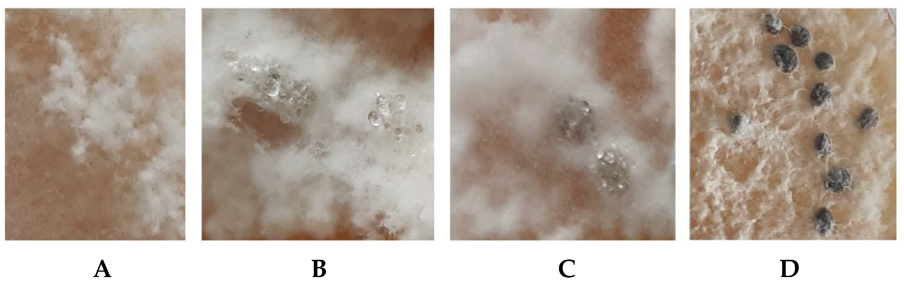
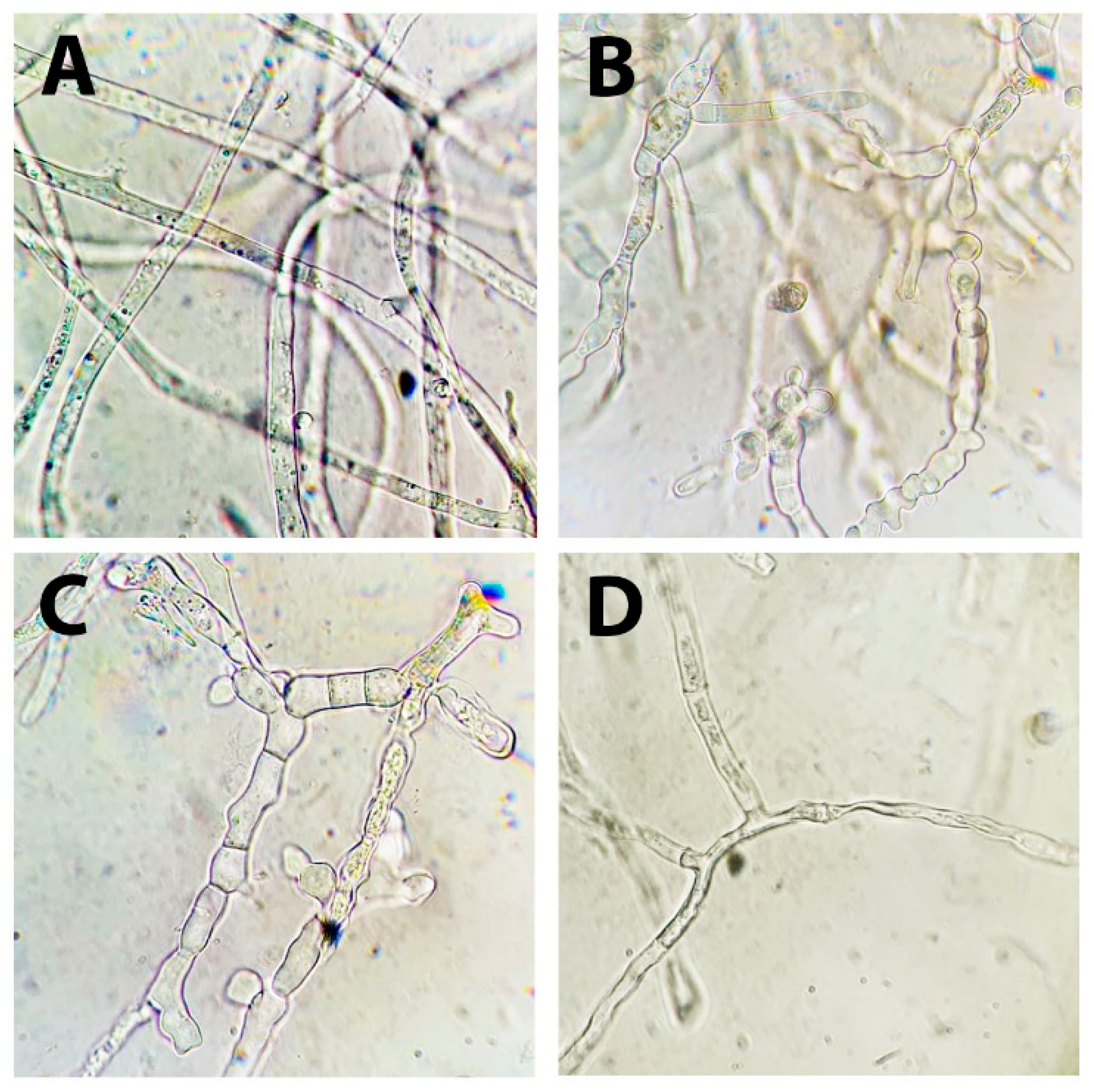
3.1. Identification of the S. sclerotiorum Strain F-1-21

The Sclerotinia sclerotiorum strain F-1-21 was isolated at the Laboratory of Biotechnology of Physiologically Active Compounds from infected carrot samples (cv. Olimpo, Nizhny Novgorod region, Russia) and stored at the work collection of this department. Strain identification was performed at the Laboratory of Molecular Diagnostics of the Federal Research Center “Fundamentals of Biotechnology” of the Russian Academy of Sciences (Moscow, Russia).
Using the 18S F566-18S R1200r primer system, a partial 629-bp sequence of a gene encoding the conservative region of 18S rRNA of the studied strain was determined (Figure S1). Using the ITS4–ITS5 primer system a 551-bp sequence encoding the intergenic region of the ribosomal operon in the studied strain was determined (Figure S2).
The resulting sequence from the 18S rRNA gene of S. sclerotiorum F-1-21 was 100% identical to those of the genera Monilinia, Sclerotinia, Dumontinia, and Botrytis (the search algorithm included all nucleotide sequences from the NCBI database (https://www.ncbi.nlm.nih.gov/, access on 10 September 2021) excepting non-cultured microorganisms).
The intergenic region of the strain characterized by a high variability was the closest (99.64%) to those of S. sclerotiorum representatives (the search algorithm included all nucleotide sequences from the NCBI database (https://www.ncbi.nlm.nih.gov/, access on 10 September 2021) excepting non-cultured microorganisms).
At the next stage, the growth characteristics of the S. sclerotiorum F-1-21 on different nutrient media were evaluated. Among the chosen media (PDA, malt agar, and oat agar), PDA was the most optimal: the strain spread over the whole surface of an 85-mm Petri plate after a 6-day incubation at 24–25 °C and formed white cobweb-like mycelium gradually transformed to the flaky form. The growth of the strain on the oat agar after 6 days of incubation reached 55 mm, while in the case of the malt agar, it was minimal (no more than 35 mm). The optimum growth temperature for the PDA medium was 25 °C (Figure 1).
In the case of the strain cultivation on PDA at the optimum temperature, a formation of small mycelial aggregations and early sclerotia (loose white knobs) was observed at the 6–7 days of cultivation. They gradually increased in size; 24 h after their appearance, their exudation was registered. During further incubation, these knobs became more dense and dark, and finally formed mature sclerotia (Figure 2). The average sclerotium length was 2–5 mm, while the average thickness was 1–2 mm. They had a roundish or cylindrical, sometimes irregular form.
A typical feature of the S. sclerotiorum development was a certain pattern of sclerotia location. During formation, they formed concentric rings on the medium surface. Summarizing the obtained data on the genetic identification and appearance of the growth and development of the studied pathogen strain, a conclusion was made that the studied F-1-21 strain belongs to the species S. sclerotiorum [47].

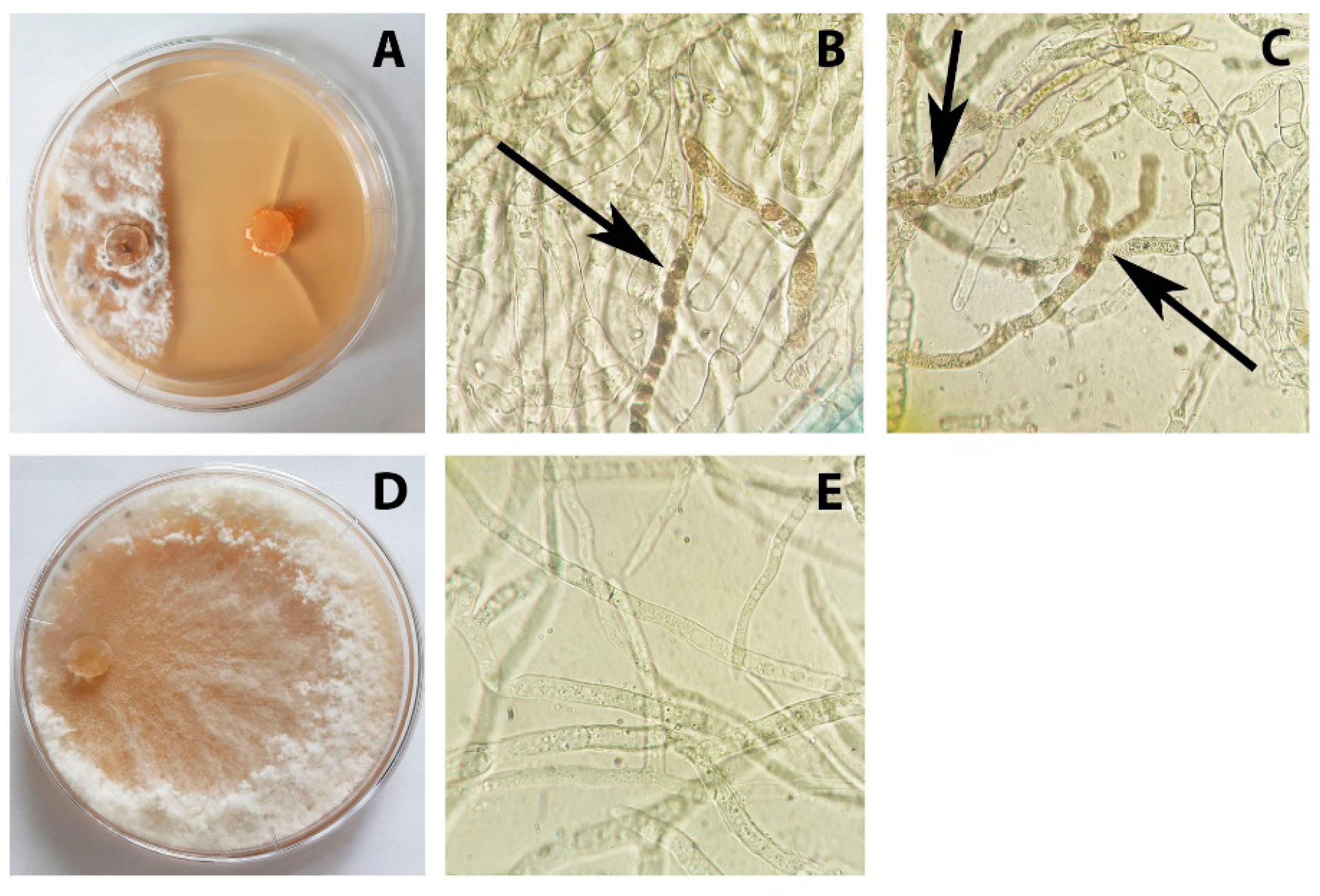
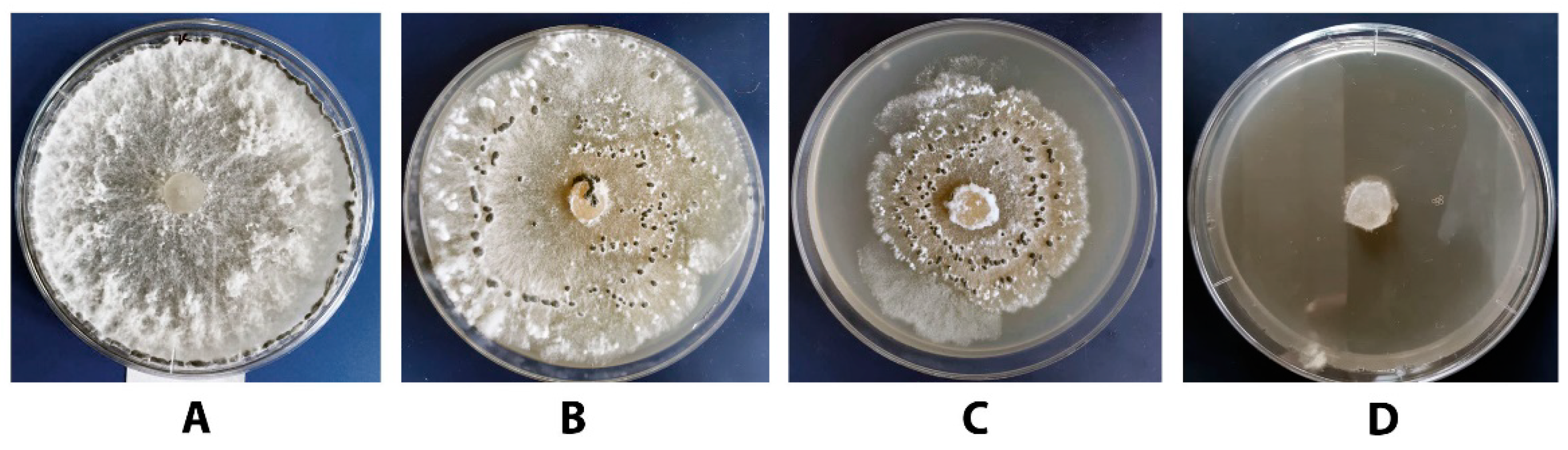
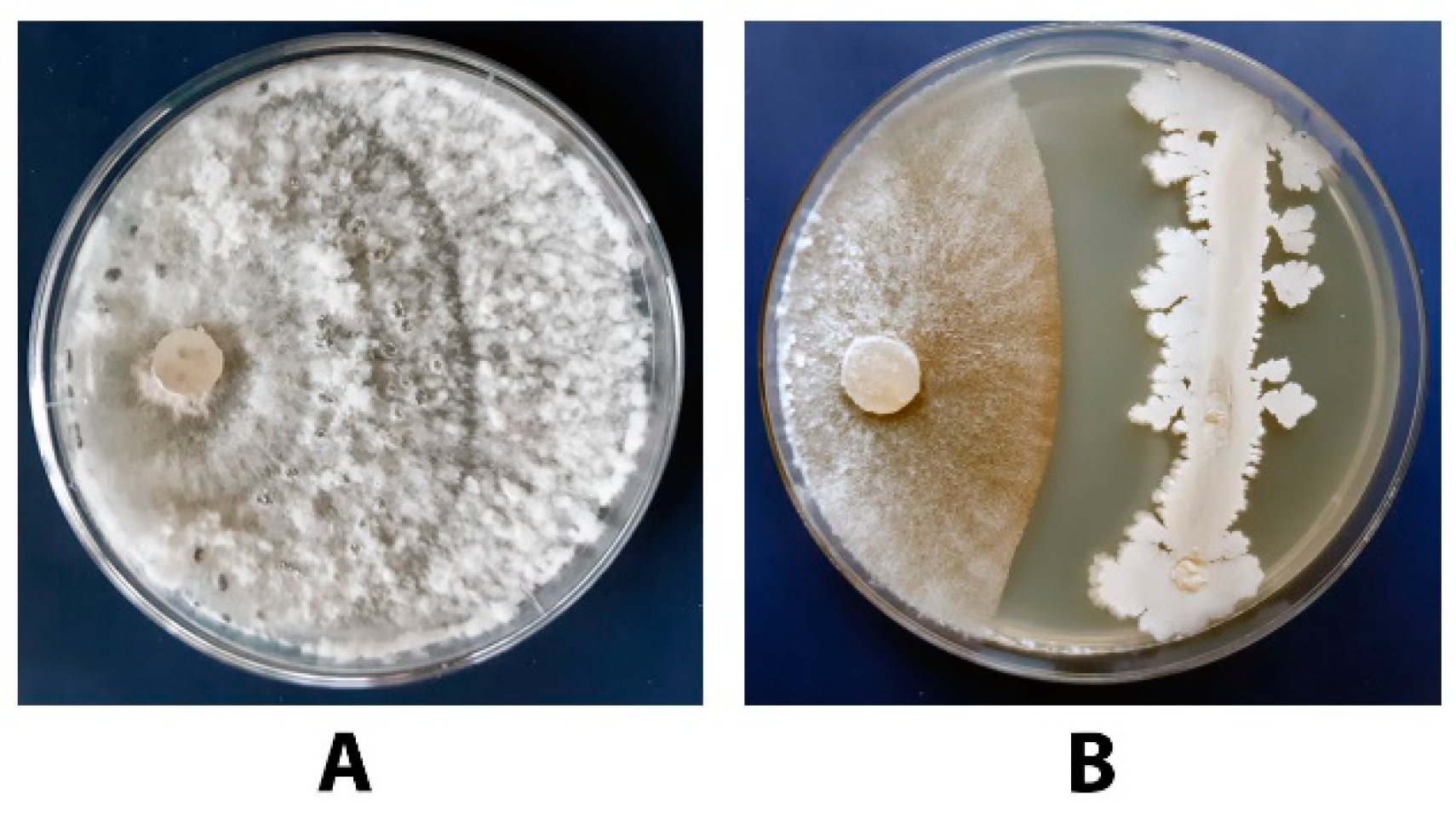
3.2. Antagonistic Activity of P. chrysogenum VKM F-4876D towards S. sclerotiorum F-1-21

The antagonistic activity of the P. chrysogenum towards S. sclerotiorum F-1-21 was tested by the dual culture assay. The assessment of this activity was carried out after a 7-day co-cultivation of microorganisms. The results of this assessment are shown in Figure 3.
Despite the almost complete absence of a P. chrysogenum growth on PDA, its metabolites provided a rather strong effect on the growth of the S. sclerotiorum strain. The inhibiting activity calculated according to Section 2.5 reached 87%. On the 7th day of incubation, the inhibition zone reached 15–20 mm on average and did not change to the 14th day of the experiment. Note that in the case of a co-cultivation of P. chrysogenum and S. sclerotiorum, an accelerated sclerotia formation occurred already after 7 days of incubation (instead of 10 days in the control). A microscopic study of the S. sclerotiorum mycelium in the area adjacent to the inhibition zone showed that compared to the control (Figure 3E), hyphae of the fungus were thickened and septate, and their cells were characterized by an increased content of vacuoles and produced melanin (Figure 3B,C), which synthesis was enhanced to the 14th day of the experiment. The accelerated formation of sclerotia and induction of melanogenesis can be considered as one of the possible ways to protect fungal cells from the impact of unfavorable environmental factors including fungitoxic metabolites secreted by P. chrysogenum.

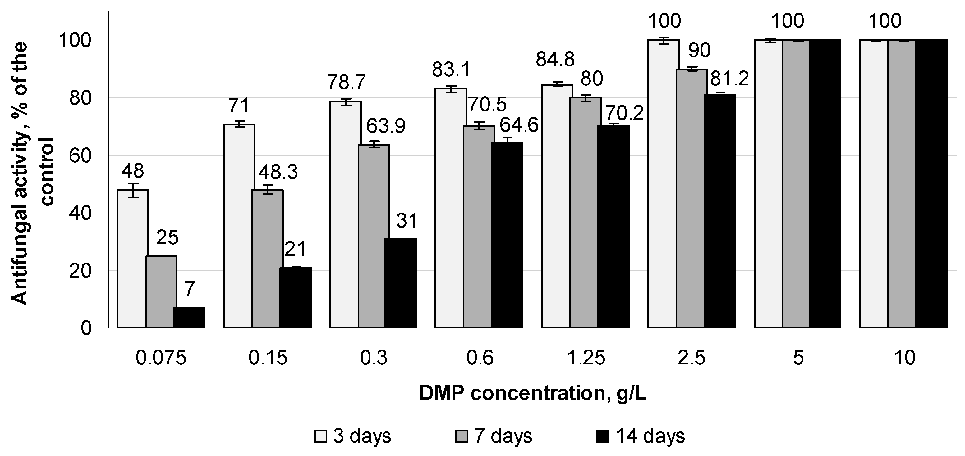
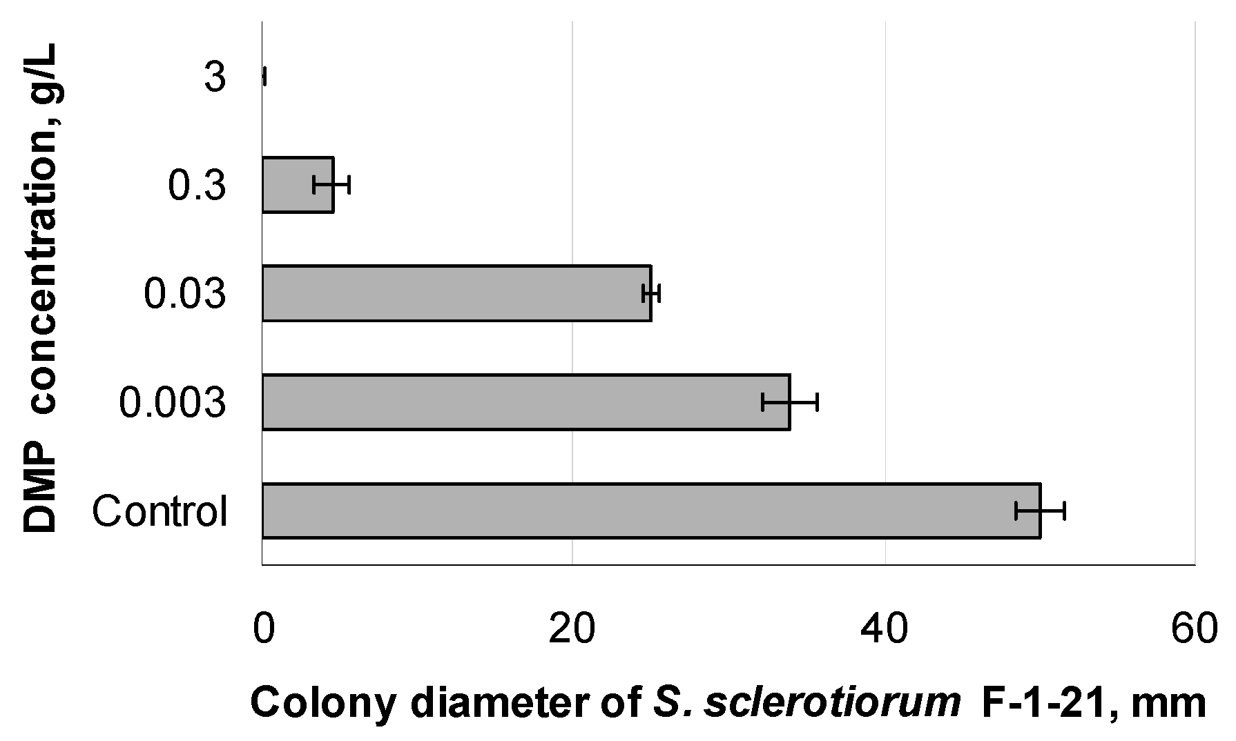
3.3. Effect of Different DMP Concentrations on the Growth of S. sclerotiorum F-1-21

At the next stage of the study, the amount of DMP added to the nutrient medium and providing a complete suppression of the S. sclerotiorum F-1-21 growth should be determined. The results of the radial growth experiments are shown in Figure 4. At DMP concentrations equal to 5 and 10 g/L, a complete suppression of the S. sclerotiorum F-1-21 growth was observed for the whole period of observation. The further reduction of the DMP concentration resulted in a gradual decrease of the antifungal effect. At the minimum studied DMP concentration (0.075 g/L), the level of pathogen suppression on the 3rd day of the experiment was 48%; at the end of the experiment, S. sclerotiorum F-1-21 almost completely covered the surface of the Petri plate (similar to the control). At the DMP concentrations equal to 0.15 and 0.3 g/L, the antifungal effect close to 50% was observed for the first 7 days; to the end of the experiment, the pathogen spread over the Petri plate surface indicating the reduction of the antifungal effect to 21 and 31%, respectively.
Thus, the obtained data allowed us to conclude that the DMP addition to PDA at a final concentration equal to or exceeding 0.6 g/L resulted in significant and prolonged suppression of the pathogen’s growth under in vitro conditions.
The DMP effect on the growth of S. sclerotiorum F-1-21 on a liquid nutrient medium was studied for several DMP concentrations (0.075, 0.3, and 2.5 g/L) differing in the level of a pathogen growth suppression on the solid PDA medium. The pathogen’s growth on a liquid nutrient medium containing different DMP concentrations is shown in Figure 5. According to the obtained data, the effect of a DMP-induced suppression of the pathogen growth on a liquid medium is dose-dependent. The level of suppression of the S. sclerotiorum F-1-21 growth at DMP concentration equal to 0.075 and 0.3 g/L reached 65.2 and 94.6%, respectively.
A microscopic study of S. sclerotiorum F-1-21 mycelium showed that, unlike in the control (Figure 6A), mycelial hyphae grown in the presence of DMP were deformed, septated, and significantly thickened (Figure 6B,C for DMP concentrations of 0.075 and 0.3 g/L, respectively). Complete inhibition of the pathogen’s growth was observed at the DMP concentration equal to 2.5 g/L; the observed mycelial hyphae were very thin, deformed, and lysed (Figure 6D).

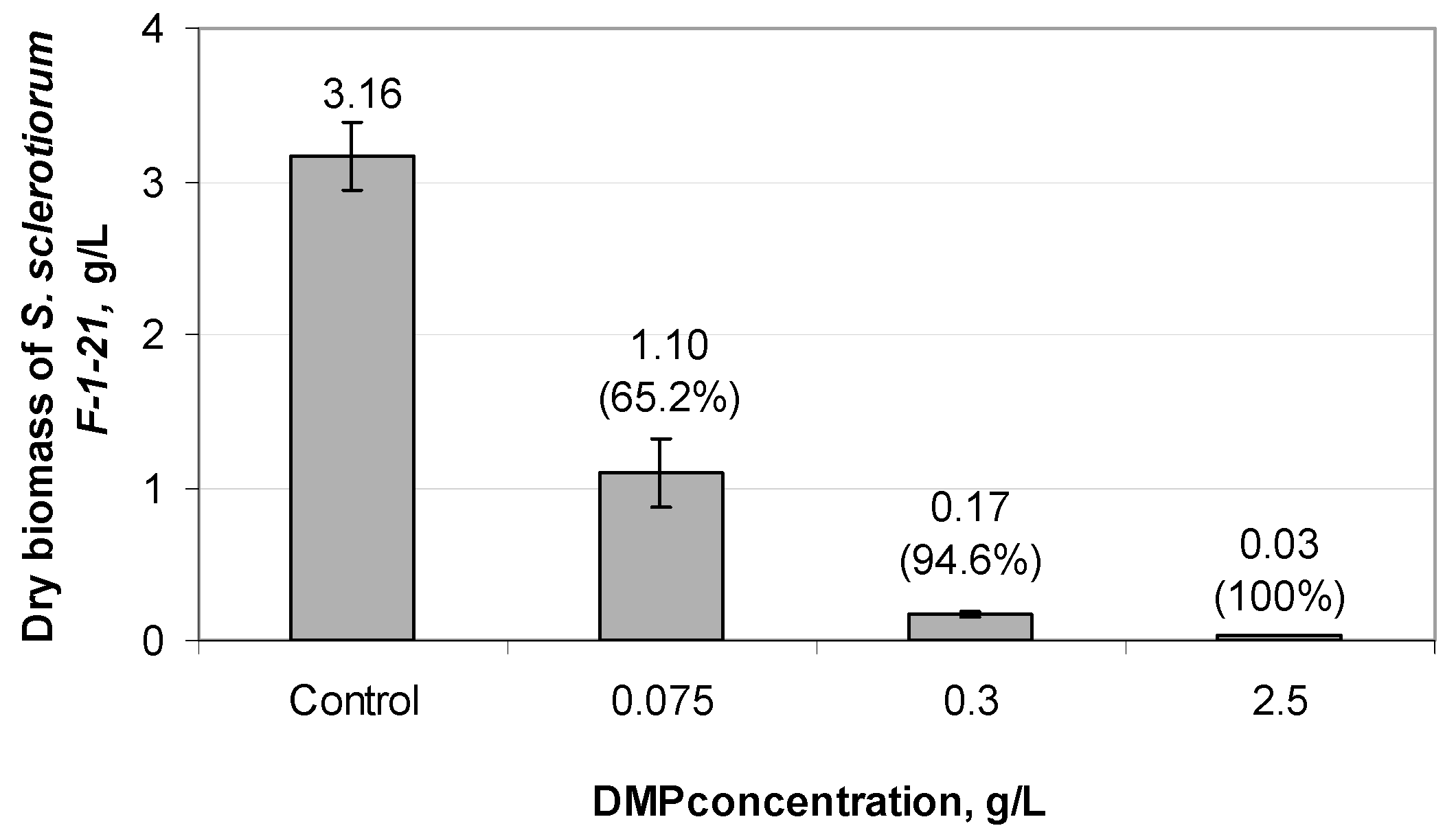
3.4. Effect of DMP on the Germination of S. sclerotiorum F-1-21 Sclerotia

Sclerotia of S. sclerotiorum F-1-21 were placed onto PDA medium supplemented with DMP at concentrations varied from 0.003 to 3 g/L, incubated for 5 days at 25 °C, and then evaluated for their germination. The results of the experiments are shown in Figure 7 and Figure 8. In the case of DMP-free medium (control, Figure 7 and Figure 8A), all sclerotia formed colonies with typical morphological traits. DMP addition to PDA suppressed germination of sclerotia at a degree directly depending on the DMP concentration. A zero growth of S. sclerotiorum F-1-21 was observed at 3 g/L DMP, whereas at DMP concentrations below 0.003 g/L the colony diameter becomes close to that in the control.
Summarizing these data with the data described in Section 3.2 and Section 3.3, one can conclude that DMP possesses a high antifungal activity towards S. sclerotiorum F-1-21 and can be used for efficient control of the growth and development of a pathogen irrespectively of its life stage.

3.5. Antifungal Activity of DMP Combined with Azoxystrobin towards S. sclerotiorum F-1-21

To determine the optimal combination of DMP and fungicide, two DMP concentrations, which demonstrated average antifungal activity, were chosen (0.15 and 0.3 g/L; see Figure 4). The results of the experiments are shown in Figure 9 and Figure 10. During the first three days of the experiment, azoxystrobin within the chosen concentration range (0.5–2.5 mg/L) inhibited the growth of S. sclerotiorum F-1-21. Then the mycelium gradually spread on the Petri plate surface. At the same time, the combination of the fungicide with DMP provided a more prominent suppression effect. The most efficient combination (0.3 g/L DMP + 2.5 mg/L azoxystrobin) resulted in a complete (100%) inhibition of the S. sclerotiorum F-1-21 within the whole observation period (Figure 9D and Figure 10B,D,F). In the case of the lower DMP concentration (0.15 g/L) combined with 2.5 mg/L of azoxystrobin, a high antifungal effect (100% inhibition) was observed within the first 7 days of the experiment (Figure 10A,C); later an insignificant mycelial growth occurred resulting in the reduction of the antifungal efficiency to 70% (Figure 9C and Figure 10E).
Note that the real antifungal effect (ER) observed on the 7th day of the experiment for the combined use of DMP and azoxystrobin was almost similar or (in the case of DMP + 2.5 g/L azoxystrobin) exceeded the expected additive effect (EE) calculated by Equation (2). At the end of the experiment, the difference between EE and ER became more significant; in the case of the maximum fungicide concentration, the real antifungal activity ER thrice exceeded the expected (calculated) additive value (EE).
Thus, the obtained results may be considered as evidence of a high inhibiting efficiency of the DMP + azoxystrobin combination in relation to S. sclerotiorum F-1-21.

3.6. Antagonistic Activity of the Tested Bacillus Strains towards S. sclerotiorum F-1-21

At the first step, the analysis of antagonistic activity of five Bacillus strains (B. subtilis VKM B-3154D, B. amyloliquefaciens VKM B-3155D, B. subtilis VKM B-3153D, B. subtilis VKM B-3505D, and B. subtilis VKM B-2998D) towards S. sclerotiorum F-1-21 was performed to reveal the most efficient strain. The obtained results demonstrated a different sensitivity of the pathogen strain to metabolites secreted by the tested strains (Figure 11).
Based on these data, the tested strains can be arranged in the following way according to their target activity: B. subtilis VKM B-2998D > B. subtilis VKM B-3505D > B. subtilis VKM B-3153D > B. amyloliquefaciens VKM B-3155D > B. subtilis VKM B-3154 D. Note that in the case of a co-cultivation with B. subtilis VKM B-2998D or B. subtilis VKM B-3505D strains, mycelium of S. sclerotiorum F-1-21 became greyish-brown (Figure 12A,B); at the same time, the mycelium of the control variant remained white during the whole observation period. This fact evidences an increased melanin production in cells of the pathogen induced by B. subtilis metabolites secreted to the agar medium.

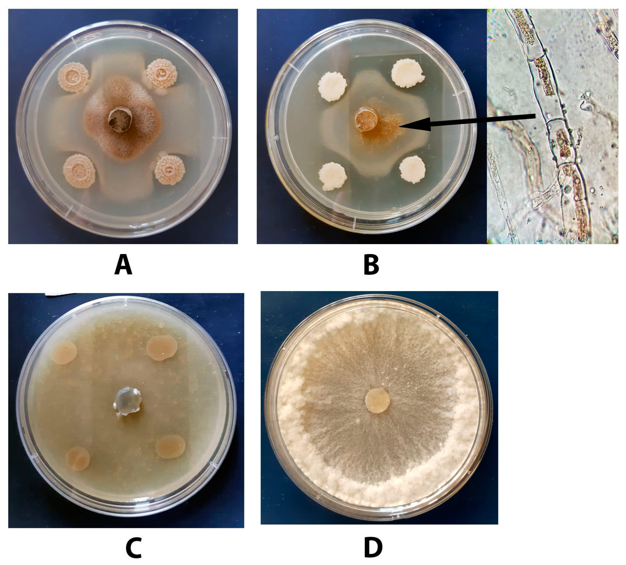
3.7. Antagonistic Activity of a Combination of DMP and B. subtilis VKM B-2998D towards S. sclerotiorum F-1-21

The results of the study of the antagonistic effect of a combination of DMP (0.3 g/L) added to PDA according to Section 2.6.2 and B. subtilis VKM B-2998D inoculated on the same medium according to the Section 2.6 in relation to S. sclerotiorum F-1-21 are shown in Figure 12C. A combined application of the above-mentioned components resulted in a complete inhibition of a pathogen’s growth for the whole period of observation (14 days). Note that in the case of S. sclerotiorum F-1-21 co-cultivation with B. subtilis VKM B-2998D and B. subtilis VKM B-3505D for 14 or 21 days, no sclerotia formation was observed that represents one of the criteria used to evaluate the efficiency of the examined biocontrol approach to control S. sclerotiorum.

3.8. Determination of the Hexaene Content in Culture Broth of Bacillus Strains

A study of the total hexaene content in culture broth showed a good level of correlation between this parameter and the observed antifungal activity. The only exception was B. subtilis VKM B-3505D demonstrated a high suppressing activity towards S. sclerotiorum F-1-21, but contained the minimum amount of hexaenes in the culture broth (Table 1).
Combination of B. subtilis VKM B-2998D with 0.5 mg/L azoxystrobin resulted in a suppression of the in vitro pathogen growth, active formation of intracellular melanin, and disorders in the sclerotia formation process (Figure 13); these disorders were observed even at the 21–28 days of observation.

3.9. Mevastatin Determination in DMP

The review of the published studies showed that some secondary metabolites of Penicillium fungi, such as statins, possess a high antifungal and antiviral activity [33,48]. The ability of statins to the efficient inhibition of the growth and development of plant pathogens is already known [49]. As Penicillium fungi, including P. chrysogenum, can produce statins [50], the putative presence of such types of compounds in DMP was examined. A comparative analysis of DMP and a commercial statin sample confirmed the presence of mevastatin in the amount of no more than 3.4 mg/g of dry weight (Figure 14).

4. Discussion

Microscopic fungi of the genus Penicillium represent one of the most valuable sources of biologically active compounds—alkaloids, antibiotics, hormones, etc., which differ in their structure and range of application and can suppress the development of a wide range of plant pathogenic fungi [33,51]. The mechanisms of a fungicide action of Penicillium metabolites may vary including, for example, hyperpolarization of the plasma membrane, which results in disorders of cell homeostasis, especially osmotic balance, activation of ion channels with the further increase of reactive oxygen forms within cells, and induction of cell apoptosis [33].
B. subtilis and B. amyloliquefaciens represent valuable tools for biological control of plant pathogenic microorganisms due to their ability to produce various biologically active compounds including antibiotics [52]. For example, some B. subtilis strains can produce polyene antibiotics with conjugated double bonds, such as hexaenes inhibiting the growth of plant pathogenic fungi [46]. These antibiotics have a well-known mechanism of action. Interacting with ergosterol, they can impair the permeability of cell walls of plant pathogens. As a result, additional pores are formed in cell membranes followed by the release of univalent cations from a cell that causes cell death.
In this study, we determined the total content of polyene antibiotics, which amount in culture broth correlated with the level of antifungal activity. In vitro study of the target activity of a combination of dry mycelium biomass of P. chrysogenum VKM F-4876D and B. subtilis VKM B-2998D showed the joint action of biologically active compounds, synthesized by both microorganisms and possessing antimicrobial activity due to their ability to break the integrity of a cell structure, resulting in complete and long-term inhibition of the S. sclerotium F-1-21 development (Figure 12C). The obtained result may serve as a basis for the further development of an efficient and environmentally safe approach for crop protection without any use of chemical fungicides.
Evaluation of the antagonistic activity of the studied P. chrysogenum strain showed that, despite a very slow growth of the strain on PDA, its metabolites significantly inhibited the growth of S. sclerotiorum F-1-21, but, at the same time, stimulated the process of sclerotia formation. In this case, sclerotia were formed on the 7th day of incubation (Figure 3A,D), while in the control their formation was completed on the 10th day of incubation. In addition, the presence of P. chrysogenum VKM F-4876D stimulated melanin production in the pathogen cells (Figure 3B,C).
The fact of enhanced melanin production by microscopic fungi under extreme conditions is well-known. A long-term study of fungal melanin pigments demonstrated their importance for the survival of fungi under extreme conditions including protection of plant pathogens from reactive oxygen species generated by host cells [53]. Therefore, we can suggest that metabolites secreted by the P. chrysogenum strain may induce some protective processes in S. sclerotiorum cells resulting in the accelerated formation of sclerotia and melanin.
The determination of the optimal efficient concentration of azoxystrobin showed a complete inhibition of the S. sclerotiorum F-1-21 growth at the fungicide concentrations exceeding 5 mg/L (data not shown). Based on this value, the lower fungicide concentrations (0.1, 0.5, 1, and 2.5 mg/L), which provided an incomplete suppression of pathogen growth, were chosen to evaluate the effect of a joint application of azoxystrobin and DMP. Our study showed that DMP itself possesses a high antifungal activity at concentrations exceeding 0.6 g/L. Therefore, DMP concentrations chosen for the experiment were 0.15 and 0.3 g/L. The results of the experiment showed that the maximum (100%) inhibition of the S. sclerotiorum F-1-21 growth over the whole period of observation was provided by a combination of the maximal (for this experiment) concentrations of DMP (0.3 g/L) and azoxystrobin (2.5 mg/L; Figure 8). In addition, a high (> 50%) antifungal effect maintained for 14 days was registered for the combinations of DMP (0.15 and 0.3 g/L) with 0.5 and 1 mg/L of the fungicide.
The effect obtained due to such joint application of two compounds can be synergistic or additive. The theoretical additive effect (EE) calculated by Limpel’s formula for the 7th day of incubation was insignificantly lower than the experimental value (ER). However, on the 14th day, when the pathogen mycelium covered the whole surface of the Petri plate with azoxystrobin-containing PDA, the ER value exceeded EE 1.5–3 times. Higher antifungal activity of the combination of two above-mentioned compounds can be explained by a suggestion that metabolites synthesized by the studied P. chrysogenum strain increased the sensitivity of S. sclerotiorum F-1-21 to the fungicide via the influence on metabolic pathways controlling cell response to oxidative stress and enhanced this stress-provoking generation of toxic reactive oxygen species, as well as a break of the integrity of the cell and vacuolar membranes causing osmotic stress and apoptosis [54].
Taking into account all aforesaid, we considered the investigation of the mechanisms of the DMP antifungal effect should start from the determination of the range of metabolites synthesized by P. chrysogenum VKM F-4876D. At the current stage, we identified mevastatin in the culture broth of this fungus. The analysis for the presence of this compound was based on the existing data on its ability to block the growth and development of plant pathogenic fungi [49]. Some Penicillium fungi may produce this compound [55,56]. Antimicrobial properties of statins in relation to a wide range of pathogenic yeasts and fungi are already known. The mechanism of their action is based on the ability to inhibit hydroxymethylglutaryl-CoA resulting in the impaired ergosterol biosynthesis in fungal cells and, therefore, changes in the cell membrane properties, required for the maintenance of the cell homeostasis, and cell growth inhibition [57]. Statins are also able to prevent the germination of fungal spores [58].
The above-mentioned facts are confirmed by morphological changes in hyphae of S. sclerotiorum F-1-21 grown in the presence of DMP (Figure 5). Thus, induced changes in the cell wall integrity, together with the inhibition of mitochondrial respiration caused by azoxystrobin, resulted in the maximal inhibition of the pathogen growth even at minimal concentrations of the studied compounds.
Destruction of sclerotia or limitation of their formation or germination represents the basis for a successful S. sclerotiorum control. The performed experimental study demonstrated the potential of the DMP use for suppression of the growth and development of the pathogen at both vegetation and dormant stages. The observed inhibition of sclerotia germination (by DMP) or even their formation (by metabolites produced by B. subtilis) may turn to be a very useful and valuable factor in the development of biological methods of plant protection.

5. Conclusions

The revealed high antifungal activity of the dry mycelial biomass of P. chrysogenum combined with Bacillus subtilis VKM B-2998D or azoxystrobin provide a prospect for the development of an efficient and environmentally safe approach for the crop protection against S. sclerotiorum irrespectively of the pathogen’s developmental stages. The obtained results make prerequisites for the replacement or significant reduction of the amounts of fungicides used in agriculture.

Supplementary Materials

The following are available online at https://www.mdpi.com/article/10.3390/agronomy11122520/s1, Figure S1: Nucleotide sequence of the fragment of an 18S RNA gene from Sclerotia sclerotiorum F-1-21 determined using the 18S F566-18S R1200r primer system. Figure S2: Nucleotide sequence (551 bp) encoding the intergenic region of the ribosomal operon from Sclerotia sclerotiorum F-1-21 (ITS1-ITS4 primers).

Author Contributions

Conceptualization, V.V.D. and E.V.G.; methodology, N.V.K.; software, A.I.O.; validation, E.V.G., N.V.K. and V.V.Y.; formal analysis, V.V.D.; investigation, N.V.K., A.I.O. and K.S.P.; data curation, V.V.Y.; writing—original draft preparation, N.V.K.; writing—review and editing, V.V.Y. and E.V.G.; visualization, K.S.P.; supervision, E.V.G.; project administration, V.V.D.; and funding acquisition, V.V.D. All authors have read and agreed to the published version of the manuscript.

Funding

The study was supported by the Ministry of Science and Higher Education of the Russian Federation.

Institutional Review Board Statement

Not applicable.

Informed Consent Statement

Not applicable.

Data Availability Statement

The authors declare that the data supporting the findings of this study are available within the main text of the manuscript and supplementary information. Raw data are available from the corresponding author upon reasonable request.

Conflicts of Interest

The authors declare no conflict of interest.

References

  1. Kabbage, M.; Yarden, O.; Dickman, M.B. Pathogenic attributes of Sclerotinia sclerotiorum: Switching from a biotrophic to necrotrophic lifestyle. Plant Sci. 2015, 233, 53–60. [Google Scholar] [CrossRef] [PubMed]
  2. Kamal, M.M.; Savocchia, S.; Lindbeck, K.D.; Ash, G.J. Biology and biocontrol of Sclerotinia sclerotiorum (Lib.) de Bary in oilseed Brassicas. Australas. Plant Pathol. 2016, 45, 1–14. [Google Scholar] [CrossRef]
  3. Kamvar, Z.N.; Amaradasa, B.S.; Jhala, R.; McCoy, S.; Steadman, J.R.; Everhart, S.E. Population structure and phenotypic variation of Sclerotinia sclerotiorum from dry bean (Phaseolus vulgaris) in the United States. Peer J. 2017, 5, 41–52. [Google Scholar] [CrossRef] [PubMed] [Green Version]
  4. Oliveira, C.S.; Lião, L.M.; Alcantara, G.B. Metabolic response of soybean plants to Sclerotinia sclerotiorum infection. Phytochemistry 2019, 167, 112099. [Google Scholar] [CrossRef]
  5. Ding, L.-N.; Li, T.; Guo, X.-J.; Li, M.; Liu, X.-Y.; Cao, J.; Tan, X.-L. Sclerotinia Stem Rot Resistance in Rapeseed: Recent Progress and Future Prospects. J. Agric. Food Chem. 2021, 69, 2965–2978. [Google Scholar] [CrossRef] [PubMed]
  6. Fass, M.I.; Rivarola, M.; Ehrenbolger, G.F.; Maringolo, C.A.; Montecchia, J.F.; Quiroz, F.; García-García, F.; Blázquez, J.D.; Hopp, H.E.; Heinz, R.A.; et al. Exploring sunflower responses to Sclerotinia head rot at early stages of infection using RNA-seq analysis. Sci. Rep. 2020, 10, 13347. [Google Scholar] [CrossRef] [PubMed]
  7. Ojaghian, M.R.; Zhang, J.-Z.; Zhang, F.; Qiu, W.; Li, X.-L.; Xie, G.-L.; Zhu, S.-J. Early detection of white mold caused by Sclerotinia sclerotiorum in potato fields using real-time PCR. Mycol. Progress 2016, 5, 959–965. [Google Scholar] [CrossRef]
  8. Mao, X.-W.; Li, J.-S.; Chen, Y.-L.; Song, X.-S.; Duan, Y.-B.; Wang, J.-X.; Chen, C.-J.; Zhou, M.-G.; Hou, Y.-P. Resistance risk assessment for fluazinam in Sclerotinia sclerotiorum. Pestic. Biochem. Phys. 2018, 144, 27–35. [Google Scholar] [CrossRef]
  9. Elsherbiny, E.A.; Taher, M.A. Silicon induces resistance to postharvest rot of carrot caused by Sclerotinia sclerotiorum and the possible defense mechanisms. Postharv. Biol. Technol. 2018, 140, 11–17. [Google Scholar] [CrossRef]
  10. Guimarães, R.L.; Stotz, H.U. Oxalate production by Sclerotinia sclerotiorum deregulates guard cells during infection. Plant Physiol. 2004, 136, 3703–3711. [Google Scholar] [CrossRef] [Green Version]
  11. Riou, C.; Freyssinet, G.; Fevre, M. Production of cell wall-degrading enzymes by the phytopathogenic fungus Sclerotinia sclerotiorum. Appl. Environ. Microbiol. 1991, 57, 1478–1484. [Google Scholar] [CrossRef] [PubMed] [Green Version]
  12. Oliveira, M.B.; Barbosa, S.C.; Petrofeza, S. Comparative in vitro and in planta analyses of extracellular enzymes secreted by the pathogenic fungus Sclerotinia sclerotiorum. Genet. Mol. Res. 2013, 12, 1796–1807. [Google Scholar] [CrossRef] [PubMed]
  13. Bolton, M.D.; Thomma, B.P.H.J.; Nelson, B.D. Sclerotinia sclerotiorum (Lib.) de Bary: Biology and molecular traits of a cosmopolitan pathogen. Mol. Plant Pathol. 2006, 7, 1–16. [Google Scholar] [CrossRef] [PubMed]
  14. Monazzah, M.; Soleimani, M.J.; Enferadi, S.T.; Rabiei, Z. Effects of oxalic acid and culture filtrate of Sclerotinia sclerotiorum on metabolic changes in sunflower evaluated using FT-IR spectroscopy. J. Gen. Plant. Pathol. 2018, 84, 2–11. [Google Scholar] [CrossRef]
  15. Purdy, L.H. Sclerotinia sclerotiorum: History, diseases and symptomatology, host range, geographic distribution, and impact. Phytopathology 1979, 69, 875–880. [Google Scholar] [CrossRef]
  16. Sousa, B.; Aline, M.; Voltan, R.; Arruda, W.; Lopes, F.A.C.; Georg, R.C.; Ulhoa, C.J. Morphological and molecular aspects of sclerotial development in the phytopathogenic fungus Sclerotinia sclerotiorum. Microbiol. Res. 2019, 229, 126326. [Google Scholar] [CrossRef] [PubMed]
  17. Smolińska, U.; Kowalska, B. Biological control of the soil-borne fungal pathogen Sclerotinia sclerotiorum—A review. J. Plant Pathol. 2018, 100, 1–12. [Google Scholar] [CrossRef] [Green Version]
  18. O’Sullivan, C.A.; Belt, K.; Thatche, L.F. Tackling control of a cosmopolitan phytopathogen: Sclerotinia. Front. Plant Sci. 2021, 12, 707509. [Google Scholar] [CrossRef] [PubMed]
  19. Kolkman, J.M.; Kelly, J.D. Agronomic traits affecting resistance to white mold in common bean. Crop Sci. 2002, 42, 693–699. [Google Scholar] [CrossRef]
  20. Rousseau, G.; Rioux, S.; Dostaler, D. Effect of crop rotation and soil amendments on Sclerotinia stem rot on soybean in two soils. Can. J. Plant Sci. 2007, 87, 605–614. [Google Scholar] [CrossRef] [Green Version]
  21. Huzar-Novakowiski, J.; Paul, P.A.; Dorrance, A.E. Host resistance and chemical control for management of Sclerotinia stem rot of soybean in Ohio. Phytopathology 2018, 107, 937–949. [Google Scholar] [CrossRef] [Green Version]
  22. Duan, Y.; Ge, C.; Liu, S.; Chen, C.; Zhou, M. Effect of phenyl pyrrole fungicide fludioxonil on morphological and physiological characteristics of Sclerotinia sclerotiorum. Pestic. Biochem. Phys. 2013, 106, 61–67. [Google Scholar] [CrossRef]
  23. Mahoney, K.J.; McCreary, C.M.; Gillard, C.L. Response of dry bean white mould [Sclerotinia sclerotiorum (Lib.) de Bary, causal organism] to fungicides. Can. J. Plant Sci. 2014, 94, 905–910. [Google Scholar] [CrossRef] [Green Version]
  24. Mc Creary, C.M.; Depuydt, D.; Vyn, R.J.; Gillard, C.L. Fungicide efficacy of dry bean white mold [Sclerotinia sclerotiorum (Lib.) de Bary, causal organism] and economic analysis at moderate to high disease pressure. Crop Protect. 2016, 82, 75–81. [Google Scholar] [CrossRef]
  25. Zhou, F.; Zhang, X.L.; Li, J.L.; Zhu, F.X. Dimethachlon resistance in Sclerotinia sclerotiorum in China. Plant Dis. 2014, 98, 1221–1226. [Google Scholar] [CrossRef] [Green Version]
  26. Hou, Y.-P.; Mao, X.-W.; Qu, X.-P.; Wang, J.-X.; Chen, C.-J.; Zhou, M.-G. Molecular and biological characterization of Sclerotinia sclerotiorum resistant to the anilinopyrimidine fungicide cyprodinil. Pestic. Biochem. Phys. 2018, 146, 80–89. [Google Scholar] [CrossRef] [PubMed]
  27. Tleuova, A.B.; Wielogorska, E.; Talluri, V.S.S.L.P.; Štěpánek, F.; Elliott, C.T.; Grigoriev, D.O. Recent advances and remaining barriers to producing novel formulations of fungicides for safe and sustainable agriculture. J. Control. Release 2020, 326, 468–481. [Google Scholar] [CrossRef]
  28. Komárek, M.; Čadková, E.; Chrastný, V.; Bordas, F.; Bollinger, J.-C. Contamination of vineyard soils with fungicides: A review of environmental and toxicological aspects. Environ. Int. 2010, 36, 138–151. [Google Scholar] [CrossRef] [PubMed]
  29. Teló, G.M.; Marchesan, E.; Zanella, R.; Peixoto, S.C.; Prestes, O.D.; de Oliveira, M.L. Fungicide and insecticide residues in rice grains. Acta Sci. Agron. 2017, 39, 9–15. [Google Scholar] [CrossRef] [Green Version]
  30. Pérez-García, A.; Romero, D.; Vicente, A. Plant protection and growth stimulation by microorganisms: Biotechnological applications of Bacilli in agriculture. Curr. Opin. Biotechnol. 2011, 22, 187–193. [Google Scholar] [CrossRef] [PubMed]
  31. Ma, H.-G.; Liu, Q.; Zhu, G.-L.; Liu, H.-S.; Zhu, W.-M. Marine natural products sourced from marine-derived Penicillium fungi. J. Asian Nat. Products Res. 2016, 18, 92–115. [Google Scholar] [CrossRef]
  32. Stein, T. Bacillus subtilis antibiotics: Structures, syntheses and specific functions. Mol. Microbiol. 2005, 56, 845–857. [Google Scholar] [CrossRef] [PubMed]
  33. Huber, A.; Galgoczy, L.; Varadi, G.; Holzknecht, J.; Kakar, A.; Malanovic, N.; Leber, R.; Koch, J.; Keller, M.A.; Batta, G.; et al. Two small, cysteine-rich and cationic antifungal proteins from Penicillium chrysogenum: A comparative study of PAF and PAFB. BBA Biomembr. 2020, 1862, 183246. [Google Scholar] [CrossRef] [PubMed]
  34. Li, Y.; Qin, L.; Roberts, D.P.; Hu, X.; Xie, L.; Gu, C.; Liao, X.; Han, P.; Liao, X. Biological fertilizer containing Bacillus subtilis BY-2 for control of Sclerotinia sclerotiorum on oilseed rape. Crop Protect. 2020, 138, 105340. [Google Scholar] [CrossRef]
  35. Xu, T.; Wang, Y.-T.; Liang, W.-S.; Yao, F.; Li, Y.-H.; Li, D.-R.; Wang, H.; Wang, Z.-Y. Involvement of alternative oxidase in the regulation of sensitivity of Sclerotinia sclerotiorum to the fungicides azoxystrobin and procymidone. J. Microbiol. 2013, 51, 352–358. [Google Scholar] [CrossRef]
  36. Dzhavakhiya, V.; Shcherbakova, L.; Semina, Y.; Zhemchuzhina, N.; Campbell, B. Chemosensitization of plant pathogenic fungi to agricultural fungicides. Front. Microbiol. 2012, 3, 87. [Google Scholar] [CrossRef] [PubMed] [Green Version]
  37. Kim, K.; Lee, Y.; Ha, A.; Kim, J.-I.; Park, A.R.; Yu, N.H.; Son, H.; Choi, G.J.; Park, H.W.; Lee, C.W.; et al. Chemosensitization of Fusarium graminearum to chemical fungicides using cyclic lipopeptides produced by Bacillus amyloliquefaciens strain JCK-12. Front. Plant Sci. 2017, 8, 2010. [Google Scholar] [CrossRef] [PubMed] [Green Version]
  38. Shcherbakova, L.; Mikityuk, O.; Arslanova, L.; Stakheev, A.; Erokhin, D.; Zavriev, S.; Dzhavakhiya, V. Studying the ability of thymol to improve fungicidal effects of tebuconazole and difenoconazole against some plant pathogenic fungi in seed or foliar treatments. Front. Microbiol. 2021, 12, 331. [Google Scholar] [CrossRef]
  39. Wong, F.P.; Wilcox, W.F. Comparative physical modes of action of azoxystrobin, mancozeb, and metalaxyl against Plasmopara viticola (grapevine downy mildew). Plant Dis. 2001, 85, 649–656. [Google Scholar] [CrossRef] [Green Version]
  40. Zeng, W.; Kirk, W.; Hao, J. Field management of Sclerotinia stem rot of soybean using biological control agents. Biol. Control. 2012, 60, 141–147. [Google Scholar] [CrossRef]
  41. Imura, N.; Ae, M.; Hoshino, R.; Abe, M.; Yamamuro, T.; Oyama, K.; Oyama, Y. Membrane hyperpolarization and depolarization of rat thymocytes by azoxystrobin, a post harvest fungicide. Chem. Biol. Interact. 2019, 300, 35–39. [Google Scholar] [CrossRef]
  42. Karpova, N.V.; Yaderets, V.V.; Glagoleva, E.V.; Petrova, K.S.; Ovchinnikov, A.I.; Dzhavakhiya, V.V. Antifungal activity of the dry biomass of Penicillium chrysogenum F-24-28 and its application in combination with azoxystrobin for efficient crop protection. Agriculture 2021, 11, 935. [Google Scholar] [CrossRef]
  43. Elazab, N.T. Diversity and biological activities of endophytic fungi at Al-Qassim region. J. Mol. Biol. Res. 2019, 9, 160–171. [Google Scholar] [CrossRef]
  44. Karpova, N.; Shagdarova, B.; Lunkov, A.; Il’ina, A.; Varlamov, V. Antifungal action of chitosan in combination with fungicides in vitro and chitosan conjugate with gallic acid on tomatoes against Botrytis cinerea. Biotechnol. Lett. 2021, 43, 1565. [Google Scholar] [CrossRef] [PubMed]
  45. Richer, D.L. Synergism: A patent view. Pestic. Sci. 1987, 19, 309–315. [Google Scholar] [CrossRef]
  46. Kudryashova, E.B.; Vinokurova, N.G.; Ariskina, E.V. Bacillus subtilis and phenotypically similar strains producing hexaene antibiotics. Appl. Biochem. Microbiol. 2005, 41, 486–489. [Google Scholar] [CrossRef]
  47. Jeon, Y.-J.; Kwon, H.-W.; Nam, J.-S.; Kim, S.H. Characterization of Sclerotinia sclerotiorum isolated from paprika. Mycobiology 2006, 34, 154–157. [Google Scholar] [CrossRef] [PubMed] [Green Version]
  48. Zhong, Y.; Peng, J.-J.; Chen, Z.-Z.; Xie, H.; Luo, D.; Dai, J.-R.; Yan, F.; Wang, J.-G.; Dong, H.-Z.; Chen, S.-Y. Dry mycelium of Penicillium chrysogenum activates defense responses and restricts the spread of Tobacco Mosaic Virus in tobacco. Physiol. Mol. Plant Pathol. 2015, 92, 28–37. [Google Scholar] [CrossRef]
  49. Galgóczy, L.; Papp, T.; Lukács, G.; Leiter, É.; Pócsi, I.; Vágvölgyi, C. Interactions between statins and Penicillium chrysogenum antifungal protein (PAF) to inhibit the germination of sporangiospores of different sensitive Zygomycetes. FEMS Microbiol. Lett. 2007, 270, 109–115. [Google Scholar] [CrossRef] [Green Version]
  50. Martín, J.F. Key role of LaeA and velvet complex proteins on expression of β-lactam and PR-toxin genes in Penicillium chrysogenum: Cross-talk regulation of secondary metabolite pathways. J. Ind. Microbiol. Biotechnol. 2017, 44, 525–535. [Google Scholar] [CrossRef] [PubMed]
  51. Kozlovskii, A.G.; Zhelifonova, V.P.; Antipova, T.V. Fungi of the genus Penicillium as producers of physiologically active compounds (review). Appl. Biochem. Microbiol. 2013, 49, 1–10. [Google Scholar] [CrossRef]
  52. Abbas, A.; Khan, S.U.; Khan, W.U.; Saleh, T.A.; Khan, M.H.U.; Ullah, S.; Ali, A.; Ikraf, M. Antagonist effects of strains of Bacillus spp. against Rhizoctonia solani for their protection against several plant diseases: Alternatives to chemical pesticides. Comptes Rendus Biol. 2019, 342, 124–135. [Google Scholar] [CrossRef] [PubMed]
  53. Gessler, N.N.; Egorova, A.S.; Belozerskaya, T.A. Melanin pigments of fungi under extreme environmental conditions (review). Appl. Biochem. Microbiol. 2014, 50, 125–134. [Google Scholar] [CrossRef]
  54. Shcherbakova, L.A. Fungicide resistance of plant pathogenic fungi and their chemosensitization as a tool to increase anti-disease effects of triazoles and strobilurines (review). Sel’skokhozyaistvennaya Biol. 2019, 54, 875–891. [Google Scholar] [CrossRef]
  55. Visagie, C.M.; Frisvad, J.C.; Houbraken, J.; Visagie, A.; Samson, R.A.; Jacobs, K. A re-evaluation of Penicillium section Canescentia, including the description of five new species. Persoonia 2021, 46, 163–187. [Google Scholar] [CrossRef]
  56. Grijseels, S.; Nielsen, J.C.; Nielsen, J.; Larsen, T.O.; Frisvad, J.C.; Nielsen, K.F.; Frandsen, R.J.N.; Workman, M. Physiological characterization of secondary metabolite producing Penicillium cell factories. Fungal Biol. Biotechnol. 2017, 4, 8. [Google Scholar] [CrossRef] [Green Version]
  57. Lefebvre, M.; Alshawa, K.; Dupont, B. L’activité antifongique des statins. Antifungal activity of statins. J. Mycol. Médic. 2010, 20, 212–217. [Google Scholar] [CrossRef]
  58. Galgóczy, L.; Papp, T.; Vágvölgyi, C. In vitro interaction between suramin and fluvastatin against clinically important zygomycetes. Mycoses 2009, 52, 447–453. [Google Scholar] [CrossRef]
Figure 1. Dynamics of the Sclerotinia sclerotiorum F-1-21 colony growth on PDA medium at different temperatures.
Agronomy 11 02520 g001
Figure 2. Sclerotia formation stages in the studied S. sclerotiorum strain F-1-21 at different days of cultivation. (A), 6–7 days; (B), 7–8 days; (C), 8–10 days; (D), 12–14 days.
Agronomy 11 02520 g002
Figure 3. (A) Antagonistic activity of Penicillium chrysogenum VKM F-4876D towards Sclerotia sclerotiorum F-1-21 in the dual culture assay after 7-day incubation. (B,C) Melanin formation in S. sclerotiorum cells contacting with the inhibition zone. (D) Growth of S. sclerotiorum on the PDA medium (control). (E) Mycelium of S. sclerotiorum grown on the PDA medium. The pictures were obtained on a Zeiss light microscope at an 800× magnification.
Agronomy 11 02520 g003
Figure 4. Concentration dependence of the antifungal activity of dry mycelium biomass of Penicillium chrysogenum VKM F-4876D (DMP) against Sclerotinia sclerotiorum F-1-21.
Agronomy 11 02520 g004
Figure 5. Growth of Sclerotinia sclerotiorum F-1-21 after 72 h of incubation in a liquid potato medium containing different concentrations of dry mycelium biomass of Penicillium chrysogenum VKM F-4876D (DMP). The growth of the pathogen on a DMP-free medium was used as a control. Numbers in brackets correspond to the antifungal activity levels calculated according to Formula (1) (see Section 2.6.2).
Agronomy 11 02520 g005
Figure 6. Mycelium of Sclerotinia sclerotiorum F-1-21 grown on a liquid potato medium (A) and the same medium containing 0.075 (B), 0.3 (C), or 2.5 (D) g/L of dry mycelium biomass of Penicillium chrysogenum VKM F-4876D (DMP). The pictures were obtained on a Zeiss light microscope at an 800× magnification.
Agronomy 11 02520 g006
Figure 7. Diameters of 7-day colonies developed from sclerotia of Sclerotia sclerotiorum F-1-21 on the PDA medium supplemented with different concentrations of dry mycelium biomass of Penicillium chrysogenum VKM F-4876D (DMP).
Agronomy 11 02520 g007
Figure 8. Five-day Sclerotia sclerotiorum F-1-21 colonies developed from sclerotia on the PDA containing dry mycelium biomass of Penicillium chrysogenum VKM F-4876D (DMP). (A) Control (DMP-free medium), (B) 0.03 g/L DMP, (C) 3 g/L DMP.
Agronomy 11 02520 g008
Figure 9. 14-day colonies of Sclerotia sclerotiorum F-1-21 on the PDA medium supplemented with a dry mycelium biomass of Penicillium chrysogenum VKM F-4876D (DMP), azoxystrobin, or their combination. (A) Control (PDA), (B) PDA + azoxystrobin (2.5 mg/L), (C) PDA+DMP (0.3 g/L), (D) PDA + azoxystrobin (2.5 mg/L) + DMP (0.3 mg/L).
Agronomy 11 02520 g009
Figure 10. Antifungal activity of dry mycelium biomass of Penicillium chrysogenum VKM F-4876D (DMP) (A,C,E—0.15 g/L; B,D,F—0.3 g/L) combined with azoxystrobin (0–2.5 mg/L) towards Sclerotia sclerotiorum F-1-21. Regular numbers above bars indicate the experimental values of antifungal activity (ER, p ≤ 0.05), and numbers in brackets correspond to the expected values (EE, p ≤ 0.05) of this activity calculated according to [45]. The data were obtained after 3 (A,B), 7 (C,D), and 14 (E,F) days of incubation.
Agronomy 11 02520 g010
Figure 11. Antagonistic activity of the tested Bacillus strains towards S. sclerotiorum F-1-21.
Agronomy 11 02520 g011
Figure 12. Antagonistic activity of (A) B. subtilis VKM B-3505D, (B) B. subtilis VKM B-2998D, and (C) combination of DMP (0.3 g/L) and B. subtilis VKM B-2998D towards S. sclerotiorum F-1-21 after a 7-day incubation at 25 °C. The fungus development in the control variant is shown as (D).
Agronomy 11 02520 g012
Figure 13. (A) Growth of Sclerotia sclerotiorum F-1-21 on PDA, 14th day (control); (B) antifungal activity of Bacillus subtilis VKM B-2998D combined with azoxystrobin (0.5 mg/L) towards S. sclerotiorum F-1-21, 14th day.
Agronomy 11 02520 g013
Figure 14. HPLC analysis of a dry mycelium biomass of Penicillium chrysogenum VKM F-4876D (DMP). Left: mevastatin standard sample, right: DMP.
Agronomy 11 02520 g014
Table 1. Hexaene content in the culture broth of Bacillus subtilis strains and the inhibiting activity of these strains towards S. sclerotiorum F-1-21.
StrainHexaene Content in Culture Broth, mg/LInhibiting Activity Towards S. Sclerotiorum F-1-21
Bacillus subtilis VKM В-2998D 38.1+++++
Bacillus subtilis VKM В-3505D 10.8+++
Bacillus subtilis VKM В-3154D 13.5+
Bacillus subtilis VKM В-3155D 28.2+++
Bacillus subtilis VKM В-3153D26.4++
Publisher’s Note: MDPI stays neutral with regard to jurisdictional claims in published maps and institutional affiliations.

