Article

Determination of Enzyme Inhibition Potential and Anticancer Effects of Pistacia khinjuk Stocks Raised in In Vitro and In Vivo Conditions

1
Department of Biology, Science and Literature Faculty, Batman University, Batman 72060, Turkey
2
Department of Analytical Chemistry, Faculty of Pharmacy, Dicle University, Diyarbakir 21280, Turkey
3
Department of Analytical Chemistry, Faculty of Pharmacy, Istanbul University, Istanbul 34116, Turkey
4
Diyarbakir Group Chairmanship, The Council of Forensic Medicine, Diyarbakır 21070, Turkey
5
Department of Horticulture, Faculty of Agriculture, Ataturk University, Erzurum 25240, Turkey
6
Department of Pharmacognosy, Faculty of Pharmacy, Dicle University, Diyarbakır 21280, Turkey
7
Department of Gene Bank, Crop Research Institute Prague, Drnovská 507, 161 06 Praha 6, Ruzyně, Czech Republic
*
Authors to whom correspondence should be addressed.
Agronomy 2021, 11(1), 154; https://doi.org/10.3390/agronomy11010154
Submission received: 15 December 2020 / Revised: 25 December 2020 / Accepted: 12 January 2021 / Published: 15 January 2021
(This article belongs to the Special Issue Use of Wild Crop Relatives as Genetic Resources in Crop Breeding)

Abstract

:
In this study, antihypertensive, anticholinesterase, antiurease, antityrosinase and antielastase enzyme inhibition and anticancer activities of in vivo (male and female) and in vitro samples (root, stem and leaf parts) of the Pistacia khinjuk Stocks were investigated comparatively. In this context, in vitro shoot cultures were obtained from germinated mature seeds. Then, the juvenile shoots were proliferated in Murashige and Skoog (MS) medium supplemented with 1 mg/L 6-Benzylaminopurine (BAP). In terms of anticancer activity, the whole of the samples studied was found to have apoptotic effects against MCF-7 (breast cancer) and HT-29 (colon cancer) cell lines. The extracts obtained from in vivo female root parts showed better cytotoxicity than all the other tested extracts on MCF-7 (IC50: 31.86 ± 1.40 µg/mL) and HT-29 cell series (IC50: 59.60 ± 0.69 µg/mL). Even though all the samples showed a strong butyrylcholinesterase enzyme inhibition (BChE) activity, it was detected that none of the samples had shown acetylcholinesterase enzyme inhibition (AChE). It was also determined that in vivo leaf samples of female trees had the highest BChE activity (Inhibition%: 75.20 ± 1.50). All the samples showed a low-moderate level of urease and tyrosinase enzyme activity, while in vivo samples showed a significant level of the elastase enzyme activities (Inhibition%: 58.72 for female root extracts; 58.25 for female leaf extracts, at 50 µg/mL concentration), and they were more active than the standard oleanolic acid (Inhibition%: 39.46 ± 0.52). The antihypertensive activities as the inhibition of angiotensin I-converting enzyme (ACE) of in vivo samples (Inhibition%: 95.88 for female stem extracts; 95.18 for female root extracts) were detected as close to the standard (Inhibition%: 96.64 ± 1.85) used. In general, it can be stated that in vivo samples had higher biological activities compared to in vitro ones. Consequently, according to our results, it was concluded that in vitro stem parts of khinjuk pistachio could also be evaluated as an alternative new antihypertensive, antielastase and anticancer agent source.

1. Introduction

It is well-known that many plants have been used for medicinal purposes since ancient times. The earliest information of medicinal plants and their uses dates back to historical records of the Chinese, Egyptian and Greek traditional medicine [1]. Today, it is estimated that 4000 drugs are widely used around the world, and approximately 400 of them are utilized commercially [2,3]. Some plants contain commercially important secondary metabolites (SMs) (terpenes, phenolic compounds, nitrogen-containing secondary metabolites, etc.) and these SMs supply raw materials for scientific, technological and commercial applications. In addition to being used as pharmaceutical raw material, SMs are increasingly used in food additives as well as the cosmetic, perfume and agricultural industries for therapeutic, flavouring and culinary purposes [4].
Many bioactive phytochemicals, such as terpenoids, phenolic compounds, fatty acids and sterols, have been isolated and identified from different Pistacia species [5]. The genus Pistacia L. belongs to the Anacardiaceae family, which contains 70 genera and more than 600 species worldwide. The trees of this genus produce plenty of resins in the trunk and branches. Resins are mixtures of many components with essential oils. Resin, which is composed of phytochemicals and enriched with these essential oils, has various pharmacological properties, such as antimicrobial, anti-inflammatory, hypocholesterolemic, antiatherogenic and anticancer activities [6,7,8]. Various parts of Pistacia species, including resin, fruit and stems, are used for a wide variety of purposes in traditional medicine. Different parts of P. vera L., P. atlantica Desf., P. khinjuk Stocks, P. terebinthus L. and P. lentiscus L. have been used for a long time as useful remedies for different diseases, for example, the fruit kernel of P. vera as a cardiac, stomach, hepatic, and brain tonic, the fruits of P. atlantica, P. khinjuk and P. terebinthus for their aphrodisiac activity and treatment of liver, kidney, heart, and respiratory system disorders and the gum resin of P. lentiscus, P. atlantica, P. khinjuk and P. terebinthus for their wound healing activity and treatment of brain and gastrointestinal disorders [5]. Myricetin-3-glucoside, myricetin-3-galactoside and myricetin-3-rutinoside are the major flavonoid glycosides from P. khinjuk. Significant antifungal activities were observed in different extracts of P. khinjuk leaves. Some active constituents of the essential oil from the aerial parts of P. khinjuk responsible for its antibacterial and antifungal activity are α-pinene, β-pinene, myrcene, b-caryophyllene, germacrene B and spathulenol. Triterpenoids are important components of the essential oil and have been associated with the anticancer potential of the resinous oil [5,9,10].
Cancer has become an increasingly important health and life problem. Chemotherapy, surgery and radiotherapy are the most common types of cancer treatments available nowadays. On the other hand, recent studies showed that consumption of nuts such as pistachio reduced cancer mortality; moreover, nuts may protect individuals from prostate cancer (about 31%), colorectal and colon cancers in women, endothelial cancer (about 27%), pancreatic cancer and breast cancer (with a stronger effect on postmenopausal breast cancer than premenopausal breast cancer) [11]. Hypertension (HT) is also regarded as a major risk factor, according to the Global Burden of Disease study. The World Health Organization predicts that 1.5 billion people will suffer from HT by 2025, and more than 7 million deaths yearly are likely to be caused by HT. According to a WHO report, about 70–80% of the world’s population relies on non-conventional medicine mainly from herbal sources in their primary health care [12].
There are various types of antihypertensive medications such as angiotensin, converting enzyme (ACE) inhibitors, beta-blockers, and calcium channel blockers, owing to many physiological mechanisms of blood pressure control, including cardiac output, peripheral vascular resistance and circulating blood volume. These antihypertensive drugs are extensively used for the treatment of HT and related cardiovascular diseases, but they are reported to have adverse side effects as well [13]. These side effects, such as diuretics, may cause muscle cramps, dizziness, extreme tiredness, dehydration, blurred vision, abnormal heart rate, skin rash and others. The use of medicinal plants for the treatment of HT is widespread. They do not cause side effects such as weakness, tiredness, drowsiness, impotence, cold hands and feet, depression, insomnia, abnormal heartbeats, skin rash, dry mouth, dry cough, stuffy nose, headache, dizziness, swelling around eyes, constipation or diarrhoea, fever, etc. [13]. It has been reported that polyunsaturated fatty acids of pistachio show an antihypertensive effect. They are shown to be effective in lowering total cholesterol levels, reducing the risk of breast cancer and preventing cardiovascular and neurodegenerative diseases [14].
Numerous chemically diverse SMs that are optimized for exerting biological functions are still far from being exhaustively investigated. Primarily, there are just a few studies about anticancer, antihypertensive effects and/or enzyme inhibition activities of Pistacia species [15,16,17,18,19]. For that reason, this is the first study to report the cytotoxic, antihypertensive, anticholinesterase, antiurease, antityrosinase and antielastase enzyme inhibition activities of different parts of P. khinjuk Stocks (root, stem and leaves) regenerated under in vitro and in vivo (male and female) conditions.

2. Materials and Methods

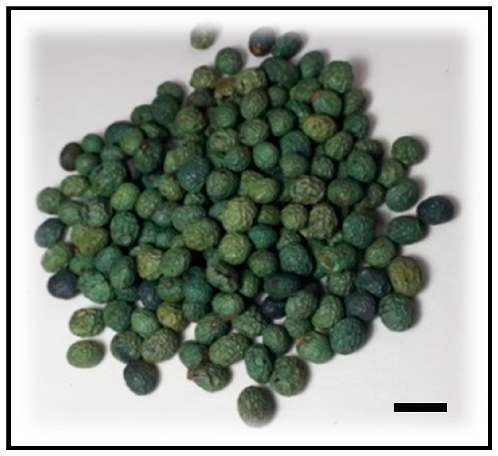
2.1. Plant Materials

In this study, the root, stem, leaves and seeds of in vivo male and female P. khinjuk were used as the plant material, obtained from Gaziantep Pistachio Research Institute between July and September 2018. The seeds were put into dry plastic containers and kept in a refrigerator at 4 °C until use for the in vitro studies (Figure 1).

2.2. Methodology

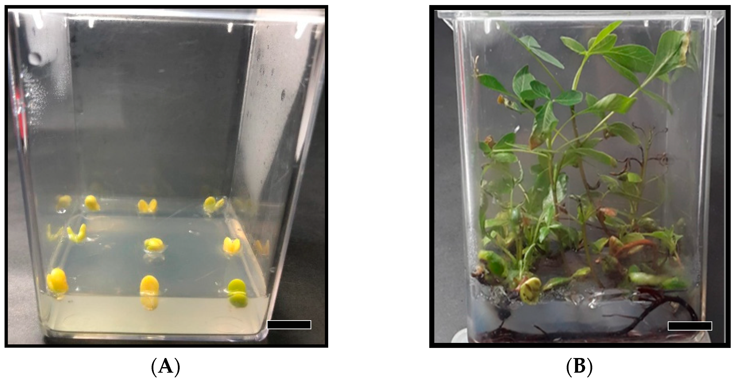
2.2.1. Obtaining In Vitro Shoot Cultures

The protocol developed by Tilkat et al. [20] was used for the surface sterilization of seeds, culture initiation and shoot proliferation stages. The seeds were surface sterilized by immersion in a 20% (w/v) commercial bleach solution (NaOCl) for 20 min. In this context, the seed coats were removed, and the kernels were washed three times with sterile distilled water before inoculating into the MS basal medium. For culture initiation, surface-sterilized seeds were inoculated into MS basal medium supplemented with 100 mg/L ascorbic acid and 30 g/L sucrose and solidified with 5.7 g/L agar. Media were adjusted to pH 5.7 before autoclaving (120 °C for 20 min). For the shoot proliferation, germinated seedlings were inoculated into MS basal medium supplemented with 1 mg/L BAP, 30 g/L sucrose and 5.7 g/L agar. All the cultures were maintained at 25 ± 2 °C with 16 h photoperiod (40 µmol/m2s). After a culture period, the parts of the plantlets (root, shoot and leaves) were dried separately and stored at 4 °C, for further anticancer and antihypertensive analyses.

2.2.2. Preparation of Extracts

Besides the root, stem and leaf parts of in vivo female and male genotypes, the same plant parts of in vitro germinated P. khinjuk plants were used for the extract preparation. After being powdered separately with the help of a grinder, adequate amounts from each portion were weighed, and the values were noted. Then, ethanol (50 mL, 3 × 24 h) was added to the dried plant samples and extracted with a shaker at 260 rpm. Crude extracts were obtained after filtration and solvent evaporation. These extracts were dissolved in ethanol, and solutions at a concentration of 4000 mg/L with a volume of 10 mL were prepared. Then, these solutions were diluted to 1000 mg/L with final volumes of 5 mL [21].

2.2.3. Cytotoxicity Analysis Using an MTT Test

In this study, human cancerous cell lines and Primary Dermal Fibroblasts series were used. For this purpose, breast cancer cell series (MCF-7), colon cancer series (HT-29) and Primary Dermal Fibroblasts series (PDF) were obtained from ATCC. The cell lines were cultured so that molecular and biochemical analyses will be able to reach sufficient numbers and in order to be frozen and used in the subsequent studies. Cell lines were grown in cell culture medium in full medium DMEM containing 10% FBS, 2 mM L-Glutamine and 100 units/mL penicillin/streptomycin. For MCF-7 cells, 0.01 mg/mL human recombinant insulin was also added into DMEM. Cell cultures were maintained at 37 °C, 5% CO2 humid environment (Thermo Steri-Cycle 371), and MCF-7 and HT-29 were centrifuged three times a week, Primary Dermal Fibroblasts twice a week at 130 g for 7 min, and the passages were diluted by diluting 3 × 10⁶ cells/mL in a sterile cabinet. Moreover, in order to preserve the cell series due to possible future studies, 2 × 10⁶ cells, 70% DMEM, 20% FBS and 10% dimethyl sulfoxide (DMSO) (Sigma-Aldrich) were kept in liquid nitrogen after standing for 1 day at −80 °C.

2.2.4. Investigation of the Effects of Samples on Cell Proliferation

The effects of in vivo and in vitro extracts of P. khinjuk on cancer cells (MCF-7 and HT-29) and healthy cell line (PDF) proliferation were observed using the MTT Cell Proliferation Kit (Sigma, Turkey) following the Sigma-Aldrich instructions for use.
Briefly, 1 × 10⁴ cells were placed in a 96-well plate and kept at 37 °C, 5% CO2 humid environment for 24 h for cells to stick to the plate. After 24 h, the cells were treated with plant samples at various concentrations for 48 h. After 48 h of treatment, cells were incubated with 10 µL of MTT solution for 4 h. After incubation, dark blue formazan dye was formed.
After washing the cells with the wash solution that came with the mass and keeping it at room temperature for 2 h, the absorbance of the formazan dye at 570 nm was measured with a plate reader (Thermo/MultiskanGo). MTT assay was performed in three parallel for each concentration, and each MTT assay was repeated three times [22,23].

2.2.5. Determination of Antihypertensive Activity (Angiotensin I-Converting Enzyme, ACE, Inhibition)

The method developed by Kwon et al. [24] (2006) was modified and used. A 50 μL sample solution was incubated for 10 min at 25 °C with 200 µL NaCl-borate buffer solution (0.3 M NaCl, pH 8.3) containing 2 mU ACE. After incubation, 100 µL of 5.0 mM substrate (hippuryl-histidyl-leucine) was added to the reaction solution, and the solution was incubated at 37 °C for 1 h. A sample blank (containing buffer solution instead of enzyme and substrate), a control (containing pure water instead of sample extract) and a blank (containing buffer instead of sample extract and enzyme) were also analyzed. The reaction was terminated by adding 150 µL of 0.5 N HCl, and the determination of the resulting hippuric acid was carried out by high-performance liquid chromatography with an ultraviolet detector (HPLC-UV) at 228 nm wavelength. Lisinopril was used as the standard. The ACE inhibition was calculated using the peak areas with the following equation:
Inhibition% = [Areacontrol − (Areasample − Areasample blank)]/(Areacontrol − Areablank) × 100

2.2.6. Determination of Anticholinesterase Activity

Acetylcholinesterase enzyme (AChE) was used as the enzyme for AChE inhibition activity, and acetylthiocoline iodide (AcI) was used as the substrate [25]. The absorbance of the yellow 5-thio-2-nitrobenzoate anion was measured with a microplate reader at 412 nm. Then, 130 µL of phosphate buffer (pH = 8) was added to the wells of the microplate. Then, 10 µL of the solution of the extracts were prepared at 1000 µg/mL concentration in ethanol, and 20 µL of enzyme solution were added. The solution was incubated for 10 min at 25 °C. After 10 min, 20 µL of DTNB reagent and 20 µL of the substrate (AcI) were added. Galantamine was used as the standard. The microplate was placed in the reader, and the absorbance at 412 nm wavelength was read. AChE activity (Inhibition%) was calculated using the following equation:
Inhibition% = (Acontrol − Asample)/Acontrol × 100
Three parallel studies were performed from each sample. The method applied in the AChE activity test was also used for butyrylcholinesterase (BChE) activity. Differently, the BChE enzyme, obtained from horse serum as an enzyme for BChE inhibition activity and butyrylthiocoline iodide (BuI), was used as the substrate [21,25].

2.2.7. Determination of Antiurease Activity

Urease enzyme is used as the enzyme for urease inhibition activity, and urea is used as the substrate [26]. First, 10 µL of extract solution was prepared at a concentration of 1000 µg/mL in ethanol and 25 µL of enzyme solution was placed into the microplate wells; then, 50 µL of the substrate (urea) was added. The microplate was placed in the ELISA reader, and the first absorbance at 630 nm wavelength was read. This solution was incubated for 15 min at 30 °C. After 15 min, 45 µL of phenol reagent and 70 µL of alkaline reagent were added on that solution. After 20 min of incubation, the final reading at 630 nm wavelength was done, and absorbance was read. Thiourea was used as the standard. Urease activity (Inhibition %) was calculated using the following equation:
Inhibition% = (Acontrol − Asample)/Acontrol × 100

2.2.8. Determination of Antityrosinase Activity

Tyrosinase enzyme was used as the enzyme for tyrosinase inhibition activity, and L-DOPA was used as the substrate [27]. A 150 μL phosphate buffer (pH = 6.8), 10 µL of the solution of the extract prepared at a concentration of 1000 µg/mL in ethanol and 20 µL of the enzyme solution were added into the well in the microplate. The microplate was placed in the ELISA reader and mixed for 3 min, and the first absorbance at the wavelength of 475 nm was read. Then, the solution was incubated for 10 min at 37 °C. After 10 min, 20 µL of a substrate (L-DOPA) was added. After incubation at 37 °C for 10 min, the final absorbance was read at 475 nm wavelength. Kojic acid was used as the standard. Tyrosinase activity (Inhibition %) was calculated using the following equation:
Inhibition% = (Acontrol − Asample)/Acontrol × 100

2.2.9. Determination of Antielastase Activity

The elastase inhibitor effect was determined spectrophotometrically [28]. Solutions of the samples in different concentrations were prepared. Later, 0.05 mL was added onto the enzyme elastase, by taking 0.05 mL of the solutions of different concentrations prepared. Then, 0.9 mL of Tris-HCl buffer (pH: 7.8; 0.2 M) was added. A portion of 0.1 mL enzyme solution and 0.9 mL Tris-HCl mixture were added as the control solution. The mixture is incubated for 15 min at 37 °C, incubation on sample tubes and control solutions after, 0.05 mL of 5 mM N-succinyl-(Ala) 3-nitroanilide solution was added and incubated at 37 °C for 30 min. Absorbance values were read at 410 nm against a reagent blank.

2.3. Statistical Analysis

All statistical calculations were performed by statistical software Minitab 16.2.1. (MINITAB Inc., State College, Pennsylvania, USA 2010). The results of the chemical contents of P. khinjuk were performed with multivariate analysis, principal component analysis (PCA) and hierarchical cluster analysis (HCA) techniques.

3. Results

3.1. Evaluation of Seed Germination and Proliferation Studies

Plant tissue culture techniques have the potential to synthesize very valuable phytochemicals that can naturally be obtained through agricultural methods. Since agricultural production for many plants is seasonal, and also depends on geographical location, climate and growth conditions, instead of traditional practices, biotechnological production is generally preferred [29].
Since the ultimate aim of this study was not to make a new micropropagation technique from khinjuk pistachio seeds, the protocols previously developed by Tilkat et al. [20] were used to obtain in vitro shoot cultures. The seeds are inoculated in PGR-free MS medium for germination and proliferated axenic shoots in MS medium supplemented with 1 mg/L BAP, 30 g/L sucrose and 6.2 g/L agar are shown in Figure 2A,B, respectively.

3.2. Cytotoxic Activity by MTT Test

The toxic effects of the extracts of in vivo grown P. khinjuk (male and female genotypes) and in vitro samples of this plant on healthy cell lines (PDF) were studied.
The cytotoxic effects of these extracts were demonstrated using the MTT method on cancerous MCF-7 (breast cancer) and HT-29 (colon cancer) cell lines. As seen from Table 1, all samples showed cytotoxic effects against MCF-7 and HT-29 cell lines. The extracts from in vivo samples gave higher results than in vitro ones in terms of cytotoxic activity, and extracts obtained from in vivo female root parts were found to have significantly higher cytotoxic effects on MCF-7 cell lines. In general terms, it was determined that root extracts belonging to both male and female genotypes showed better cytotoxic activity on MCF-7 (male IC50: 36.11 ± 0.44 µg/mL, female IC50: 31.86 ± 1.40 µg/mL) and HT-29 (male IC50: 68.47 ± 2.41 µg/mL, female IC50: 59.60 ± 0.69 µg/mL) cell lines compared to stem and leaf extracts.

3.3. Antihypertensive Activity

Antihypertensive activity of extracts obtained from in vivo grown P. khinjuk (female and male genotypes) and in vitro samples of this plant were determined. The highest antihypertensive activity was obtained from the root extracts of in vivo female genotypes among all the samples examined (Inhibition: 95.88%). When the in vivo samples were evaluated within themselves, it was observed that the root and stem parts had higher activity compared to the leaf parts. The root extracts of in vivo male genotypes took second place in terms of antihypertensive activity. For in vitro samples, it was determined that only the stem extracts were active parts (Inhibition: 85.16%); the root and leaf parts were not functional. However, in general, it should be noted that the in vivo extracts were more useful compared to in vitro extracts in terms of their antihypertensive effect (Table 2).

3.4. Anticholinesterase Activity

Anticholinesterase activities of all prepared ethanol extracts (root, stem, leaf extracts of in vivo female and male genotypes and in vitro samples) were determined according to AChE and BChE enzyme inhibition methods.
In general, in vivo female and male genotype samples showed active results close to galantamine used as a standard against BChE enzyme inhibition, but they were not active in the AChE enzyme inhibition method (Table 2). It was determined that in vitro samples showed lower activity than the in vivo samples of female and male genotypes. In this context, the ethanol extracts of the samples were compared among themselves, and it was determined that leaf extracts of female genotype showed the highest activity against BChE enzyme with 75.20 ± 1.50% inhibition, while the root extracts of male genotype with 70.86 ± 1.41%, at 50 µg/mL concentration. When the results are over-viewed, the extracts obtained from in vitro root and leaf samples and those obtained from in vivo stem samples were found as more effective in terms of anticholinesterase activity.

3.5. Antiurease and Antityrosinase Activity

As seen in Table 2, the in vivo samples were found to have higher urease and tyrosinase enzyme inhibition activities than in vitro samples. The in vitro samples had no antiurease activity. The root extract of in vivo male genotype showed the highest antiurease activity (56.31 ± 1.12%, at 50 µg/mL concentration) and the second-highest antityrosinase activity (54.81 ± 1.09, at 50 µg/mL concentration). It was determined that root extracts of in vivo female genotypes showed the highest antityrosinase activity (54.81 ± 1.09%, at 50 µg/mL concentration). When the extracts are envisaged in terms of aerial parts and underground plant parts, it becomes evident that the root extracts of both in vivo and in vitro samples showed comparatively better results than stem and leaf parts. It is also noteworthy that in the study where kojic acid (81.54 ± 0.63%, at 50 µg/mL concentration) and thiourea (98.85 0.54%, at 50 µg/mL concentration) were used as standards, only the root extract belonging to in vitro sample had low antityrosinase activity.

3.6. Antielastase Activity

In Table 2, elastase enzyme inhibition results of ethanol extracts from in vivo female and male genotypes and in vitro samples of P. khinjuk were given. It was observed that the samples of leaf extracts of in vitro origin did not show antielastase activity, but all of the extracts except them gave active results. Similar to the previous enzyme inhibition results, it was found that extracts from in vivo female and male genotypes had higher activity compared to in vitro samples in terms of antielastase activity. Indeed, ethanol extracts of roots of in vivo female (58.72 ± 1.17% at 50 µg/mL concentration) and male (57.54 ± 1.15%, at 50 µg/mL concentration) genotypes had higher antielastase activity than leaf and stem ethanolic extracts. Additionally, it was determined that in vitro root extracts had the lowest value, namely 6.65 ± 0.13 at a 50 µg/mL concentration. In this context, the root and leaf of in vivo samples appeared to be more effective than the oleanolic acid, which was used as standard.

4. Discussion

Several studies have been conducted to determine the cytotoxic effects of extracts and compounds obtained from Pistacia species. It has been shown that most of them show cytotoxic activity. According to the first of these studies, the crude methanol extract of P. chinensis Bunge subsp. integerrima (J.L.Stewart) Rech.f. showed good antitumor (IC50: 125 μg/mL) activity. Moreover, the crude stem extract inhibited MCF-7 cell viability in a dose-dependent manner. The IC50 values calculated were 90.9 μg/mL. The ethyl acetate and chloroform fractions at a concentration of 200 μg/mL showed ~100 and 97.4% inhibition against MCF-7 cell line, respectively [30].
In another example, the essential oil from P. atlantica galls has been reported to have moderate cytotoxic activity against mycobacteria because of MTT testing on C3A and Vero monkey kidney cells [31]. Moreover, it has been reported that the fruit extracts of P. atlantica induced apoptosis and showed cytotoxic effects without observing a significant necrosis formation on human epidermoid carcinoma and HGFcell line [32]. Apart from these studies, P. vera hull extracts were tested for the cytotoxic effects of human colon cancer (HT-29) and breast adenocarcinoma (MCF-7) using the MTT method, resulting in significant inhibition of angiogenesis at high concentrations [16].
In the same species, a study has been designed to evaluate the cytotoxic effects of methanol extract obtained from fruit stem, red outer shell cover and resin of P. vera on human prostate cancer (PC3, DU-145) and breast cancer (MDA-MB-231, MCF-7) using the MTT method. The results showed cytotoxic effects on cancer cells at a low dose [33]. Among the extracts (leaves, fruits and roots), the methanol extracts of the leaves had the most substantial cytotoxic effect obtained from P. lentiscus [34]. Mirian et al. [35] investigated the cytotoxic effects of the gum extracts of P. khinjuk on the human umbilical vein endothelial cell (HUVEC) and Y79 cell lines and reported that P. khinjuk extracts inhibited angiogenesis. In the same species, it was reported that the ethanol extracts of the leaves had moderate cytotoxic activity against human PC3 prostate cancer, A549 lung cancer, MCF7 breast cancer and HepG2 liver cancer [36].
Extracts of the P. khinjuk plant grown under in vitro conditions were investigated for the first time in this study. It has been determined that ethanol extracts (especially root extracts) belonging to both in vitro and in vivo samples had good cytotoxic activity. In this context, it can be said that the findings in this study are compatible with the studies mentioned above. It was observed that in vivo samples, especially the root parts, showed more effective cytotoxic activity. In terms of cytotoxic activity results performed with the MTT test, it has been determined that the extracts obtained from in vivo female root parts showed better cytotoxicity on MCF-7 (IC50: 31.86 ± 1.40μg/mL) and HT-29 cell series (IC50: 59.60 ± 0.69μg/mL). Moreover, it was concluded that in vivo male genotype root extracts followed this sequence, and the root parts were more active and useful in terms of cytotoxic activity.
According to the Turkish Society of Cardiology National Hypertension Treatment and Follow-up Guide, with a single drug (monotherapy) in the treatment of hypertension, 30–50% unsuccessful results are obtained [37]. In this case, there are three options for the physicians: (a) to increase the dose of the drug used, (b) to switch to another drug group, (c) to apply combination therapy.
It is also vital to research new therapeutics in order to reduce the side effects of the disease symptoms and medications and to provide physical and psychological support, as they bring along side effects. Indeed, Ahmed et al. [38] reported that eight peaks were observed in the leaf extracts of P. atlantica that inhibit ACE activity. Another study investigated the in vitro and in vivo antihypertensive effect of mango leaves (Mangifera indica L.), belonging to the Anacardiaceae family; it was reported that the dichloromethane fraction could have an antihypertensive potential [39]. This current study had similar results with other Pistacia species, and root extracts belonging to in vivo female genotypes were the closest samples to the standard with 95.88% inhibition. Since in vivo extracts had more beneficial activity compared to in vitro extracts and possessed high levels of antihypertensive activity, it was concluded that in vivo stem and root parts could be evaluated in pharmacological studies.
Drugs that inhibit the enzyme acetylcholinesterase are called acetylcholinesterase inhibitors or anticholinesterases. Cholinesterase inhibitors are used clinically in diseases such as myasthenia gravis and glaucoma, primarily Alzheimer’s disease (AD). In advanced AD, a 55–67% reduction in brain AChE activity was recorded. Today, the most frequently used strategy in the symptomatic treatment of the disease is the use of cholinesterase enzyme inhibitors. Galantamine, which has recently been used in the treatment of AD, is a phenanthrene alkaloid isolated from common snowdrop, Galanthus nivalis L., is a reversible acetylcholinesterase inhibitor. Acetylcholinesterase inhibition is the only mechanism in which the success rate is achieved in the symptomatic treatment of AD; the drugs used in the treatment are donepezil, rivastigmine and galantamine. Apart from these, studies on anticholinergics that may be effective in treatment continue rapidly. Ongoing studies are aimed at researching new anticholinesterases of plant origin, and the discovery of therapeutic agents with low levels of toxicity and side effects [40]. It was observed that the P. khinjuk extracts examined in this study did not respond to the acetylcholinesterase enzyme (AChE) inhibition. However, leaf extracts of the in vivo female genotype gave the closest result to galantamine used as the standard for butyrylcholinesterase enzyme (BChE) inhibition. In this context, it is suggested that aerial parts of in vitro samples and underground parts of in vivo samples could be used for therapeutic purposes for anti-BChE activity.
Urease is an enzyme that causes hepatic coma as well as kidney stones and infections. In the treatment of such diseases, urease inhibitors are being studied that may inhibit the urease enzyme. In recent years, studies on the inhibition effects of various plant extracts on the urease enzyme have gained importance for this purpose [41]. Lithraea molleoides (Vell.) Engl., a species of Anacardiaceae family that grows in South America, has been reported to exhibit a strong antiurease activity on Helicobacter pylori [42]. Likewise, the methanol extracts of Rhus coriaria L. (sumac) in the same family have been reported to have a high level of inhibition activity on the Canavalia ensiformis (L.) DC. (Jack bean) urease enzyme [43]. In this current study, almost all in vivo samples were in line with the findings of the above-mentioned study and the in vivo root extracts of male genotypes especially gave the closest results to the thiourea used as the standard, and showed the highest antiurease activity among these samples.
Polyphenols are the most common ones among enzyme inhibitors. Especially, flavonoids (stilbenes, chalcones, isoflavonoids, isoflavones), long-chain lipids and steroids are well-known inhibitors. Kojic acid is the most studied inhibitor of tyrosinase; it is also used in the cosmetic field for skin whitening and in the food industry to prevent enzymatic browning. Investigation of tyrosinase enzyme activity is crucial since it is commonly found in the leaves, seeds and flowers of many plants [40]. In terms of tyrosinase enzyme inhibition, there are no studies on literature related to Pistacia species. At this point, looking at the findings, it was found that the root extracts of in vivo female genotypes showed the closest antityrosinase activity to kojic acid used as the standard. Furthermore, it has been concluded that the tyrosinase inhibitor activities of P. khinjuk could be useful for both the development of new pharmaceuticals and industrial applications.
Elastin is one of the main elements of connective tissue, and it creates wrinkles that accompany skin ageing as a result of its breakdown with the elastase enzyme. Increasing elastase activities with age leads to a decrease in skin elasticity and skin sagging. Inhibition of this enzyme is becoming more and more important for clinical and cosmetic industries due to its positive effects both on preventing skin ageing and on connective tissue diseases [41]. In this study, it was observed that most of the samples showed antielastase activity. In terms of antielastase activity, especially in vivo female genotypes showed higher activity. The activities of in vivo samples on elastase enzyme inhibition were found to be hierarchically ranked in terms of effectiveness as root > leaf > stem, based on the elastase enzyme inhibition results. Antielastase activities of root and leaf extracts of in vivo female genotypes were found to be 58.72% and 58.25%, respectively, at 50 µg/mL concentration. When considering the activity of oleanolic acid (39.46 ± 0.52%, at 50 µg/mL concentration) used in this method, one can be asserted that especially in vivo root samples of both genotypes have significant antiaging potential. Accordingly, it was determined that the root and leaf extracts of both genotypes showed much higher activity than the standard oleanolic acid (39.46%), thus indicating a possible new antiaging natural product.

5. Conclusions

In this study, the in vivo samples were found to be generally more active than the in vitro samples in all the methods tested in this study. When considering the high antihypertensive, antielastase and cytotoxic effects of the in vivo samples, it can be asserted that these samples have the potential to be used in the pharmaceutical, cosmetic and food industry. In this context, it has been concluded that detailed isolation and animal testing studies should be conducted using extracts of the studied species herein.

Author Contributions

Conceptualization, E.A.T., H.B. and I.Y.; data curation E.A.T., H.B. and I.Y.; formal analysis, P.K.Y., M.A. and A.K.; methodology, P.K.Y., M.A. and A.K.; project administration, E.A.T. and H.B.; visualization, V.H. and S.E.; writing—original draft, V.H., A.E. and S.E.; writing—review and editing, E.A.T., A.E., H.B. and I.Y. All authors have read and agreed to the published version of the manuscript.

Funding

The study was funded by a grant from BTÜBAP—Batman University Research Project Council (Project No: BTÜBAP-2018-FED-1) and partly by the National programme No. 51834/2017-MZE-17253/6.2.14, MZE_RO0418, Czech Republic. We also thank Israt Jahan for linguistically reviewing the manuscript.

Institutional Review Board Statement

Not applicable.

Informed Consent Statement

Not applicable.

Data Availability Statement

All new research data were presented in this contribution.

Conflicts of Interest

The authors declare that they have no conflict of interest.

References

  1. Bozyel, M.E.; Merdamert, E. Antiurolithiatic activity of medicinal plants in Turkey. In Science, Ecology and Engineering Research in the Globalizing World; St. Kliment Ohridski University Press: Sofia, Bulgaria, 2018; pp. 152–167. [Google Scholar]
  2. Borris, R.P. Natural products research: Perspectives from a major pharmaceutical company. J. Ethnopharmacol. 1996, 51, 29–38. [Google Scholar] [CrossRef]
  3. Sarı, A.O.; Oğuz, B.; Bilgiç, A.; Tort, N.; Güvensen, A.; Şenol, S.G. Batı Anadolu’da halk ilacı olarak kullanılan Asteraceae türleri. Anadolu Jaari. 2008, 18, 1–15. [Google Scholar]
  4. Yang, L.; Wen, K.S.; Ruan, X.; Zhao, Y.X.; Wei, F.; Wang, Q. Response of plant secondary metabolites to environmental factors. Molecules 2018, 23, 762. [Google Scholar] [CrossRef] [PubMed] [Green Version]
  5. Bozorgi, M.; Memariani, Z.; Mobli, M.; Surmaghi, M.H.S.; Shams-Ardekani, M.R.; Rahimi, R. Five Pistacia species (P. vera, P. atlantica, P. terebinthus, P. khinjuk and P. lentiscus): A review of their traditional uses, phytochemistry, and pharmacology. Sci. World J. 2013, 219815, 1–33. [Google Scholar] [CrossRef] [PubMed] [Green Version]
  6. Dimas, K.; Hatziantoniou, S.; Wyche, J.H.; Pantazis, P. A mastic gum extract induces suppression of growth of human colorectal tumour xenografts in immunodeficient mice. In Vivo 2009, 23, 63–68. [Google Scholar]
  7. Konstantinos, S.D.; Pantazis, P.; Ramanujam, R. Chios mastic gum: A Plant-produced resin exhibiting numerous diverse pharmaceutical and biomedical properties. In Vivo 2012, 26, 777–785. [Google Scholar]
  8. Paraschos, S.; Mitakou, S.; Skaltsounis, A.-L. Chios gum mastic: A review of its biological activities. Curr. Med. Chem. 2012, 19, 2292–2302. [Google Scholar] [CrossRef]
  9. Remila, S.; Kilani, D.A.; Delemasure, S.; Atmani, D. Antioxidant, cytoprotective, anti-inflammatory and anticancer activities of Pistacia lentiscus (Anacardiaceae) leaf and fruit extracts. Eur. J. Integr. 2015, 7, 274–286. [Google Scholar] [CrossRef]
  10. Hashemi, L.; Samani, M.A.; Moradi, T.M.; Alidadi, S. Anticancer activity and phenolic compounds of Pistacia atlantica extract. Int. J. Pharm. Phytopharm. Res. 2017, 7, 26–31. [Google Scholar]
  11. Ghasemynasab Parizi, M.; Ahmadi, A.; Mazloomi, S.M. A review on pistachio: Its composition and benefits regarding the prevention or treatment of diseases. JOHE 2016, 4, 57–69. [Google Scholar]
  12. Chan, K. Some aspects of toxic contaminants in herbal medicines. Chemosphere 2003, 52, 1361–1371. [Google Scholar] [CrossRef] [Green Version]
  13. Singh, M.; Singh, A.K.; Singh, S.; Pandey, P.; Chandra, S.; Gambhir, I.S. Angiotensin-converting enzyme gene I/D polymorphism increases the susceptibility to hypertension and additive diseases: A study on north Indian patients. Clin. Exp. Hypertens. 2016, 38, 305–311. [Google Scholar] [CrossRef] [PubMed]
  14. Labdelli, A.; Zemour, K.; Simon, V.; Cerny, M.; Adda, A.; Merah, O. Pistacia atlantica Desf., a source of healthy vegetable oil. Appl. Sci. 2019, 9, 2552. [Google Scholar] [CrossRef] [Green Version]
  15. Rahman, H.S. Phytochemical analysis and antioxidant and anticancer activities of mastic gum resin from Pistacia atlantica subspecies kurdica. Onco Targets Ther. 2018, 11, 4559–4572. [Google Scholar] [CrossRef] [PubMed] [Green Version]
  16. Seifaddinipour, M.; Farghadani, R.; Namvar, F.; Mohamad, J.; Abdul Kadir, H. Cytotoxic effects and anti-angiogenesis potential of pistachio (Pistacia vera L.) Hulls against MCF-7 human breast cancer cells. Molecules 2018, 23, 110. [Google Scholar] [CrossRef] [PubMed] [Green Version]
  17. Buriani, A.; Fortinguerra, S.; Vincenzo, S.; Dall’Acqua, S.; Innocenti, G.; Montopoli, M.; Gabbia, D.; Carrara, M. Human adenocarcinoma cell line sensitivity to essential oil phytocomplexes from Pistacia species: A multivariate approach. Molecules 2017, 22, 1336. [Google Scholar] [CrossRef] [Green Version]
  18. Almehdar, H.; Abdallah, H.; Osman, A.; AbdelSattar, E. In vitro cytotoxic screening of selected Saudi medicinal plants. J. Nat. Med. 2012, 66, 406–412. [Google Scholar] [CrossRef]
  19. Sakagami, H.; Kishino, K.; Kobayashi, M.; Hashimoto, K.; Iida, S.; Shimetani, A.; Nakamura, Y.; Takahashi, K.; Ikarashi, T.; Fukamachi, H.; et al. Selective antibacterial and apoptosis modulating activities of mastic. In Vivo 2009, 23, 215–223. [Google Scholar]
  20. Tilkat, E.; Işıkalan, Ç.; Onay, A. In vitro propagation of khinjuk pistachio (Pistacia khınjuk Stocks) through seedling apical shoot tip culture. Propag. Ornam. Plants 2005, 5, 1–5. [Google Scholar]
  21. Ertas, A.; Boga, M.; Yilmaz, M.A.; Yesil, Y.; Tel, G.; Temel, H.; Hasimi, N.; Gazioglu, I.; Ozturk, M.; Ugurlu, P. A detailed study on the chemical and biological profiles of essential oil and methanol extract of Thymus nummularius (Anzer tea): Rosmarinic acid. Ind. Crop. Prod. 2015, 67, 336–345. [Google Scholar] [CrossRef]
  22. Mojarraba, M.; Langzian, M.S.; Emamic, S.A.; Asilic, J.; Tayarani-Najaranb, Z. In vitro antiproliferative and apoptotic activity of different fractions of Artemisia armeniaca. Rev. Bras. Farm. 2013, 23, 783–788. [Google Scholar] [CrossRef] [Green Version]
  23. Yener, I.; Tokul Olmez, O.; Ertas, A.; Yilmaz, M.A.; Firat, M.; Irtegun Kandemir, S.; Ozturk, M.; Kolak, U.; Temel, H. A detailed study on chemical and biological profile of nine Euphorbia species from Turkey with chemometric approach: Remarkable cytotoxicity of E. fistulasa and promising tannic acid content of E. eriophora. Ind. Crop. Prod. 2018, 123, 442–453. [Google Scholar] [CrossRef]
  24. Kwon, Y.I.; Vattem, D.A.; Shetty, K. Evaluation of clonal herbs of Lamiaceae species for management of diabetes and hypertension. Asia Pac. J. Clin. Nutr. 2006, 15, 107–118. [Google Scholar] [PubMed]
  25. Ellman, G.L.; Courtney, K.D.; Andres, V.; Featherstone, R.M. A new and rapid colourimetric determination of acetylcholinesterase activity. Biochem. Pharm. 1961, 7, 88–95. [Google Scholar] [CrossRef]
  26. Hina, Z.; Ghazala, H.R.; Arfa, K.; Huma, S.; Sabiha, T.; Ajmal, K. Anti-urease Activity of Mimusops elengi L. (Sapotaceae). E. J. Med. Pl. 2015, 6, 223–230. [Google Scholar]
  27. Hearing, V.J.; Jiménez, M. Mammalian tyrosinase—The critical regulatory control point in melanocyte pigmentation. Int. J. Biochem. 1987, 19, 1141–1147. [Google Scholar] [CrossRef]
  28. Melzig, M.F.; Löser, B.; Ciesielski, S. Inhibition of neutrophil elastase activity by phenolic compounds from plants. Pharmazie 2011, 56, 967–970. [Google Scholar]
  29. Güven, A.; Gürsul, I. Bitki doku kültürlerinde sekonder metabolit sentezi. Gıda 2014, 39, 299–306. [Google Scholar]
  30. Bibi, Y.; Nisa, S.; Zia, M.; Waheed, A.; Ahmed, S.; Chaudhary, M.F. The study of anticancer and antifungal activities of Pistacia integerrima extract in vitro. Indian J. Pharm. Sci. 2012, 74, 375–379. [Google Scholar]
  31. Sifia, I.; Dzoyem, J.P.; Ouinten, M.; Yousfi, M.; McGaw, L.J.; Eloff, J.N. Antimycobacterial, antioxidant and cytotoxic activities of essentıal oil of gall of Pistacia atlantica Desf. from Algeria. Afr. J. Tradit. Complement Altern. Med. 2015, 12, 150–155. [Google Scholar] [CrossRef] [Green Version]
  32. Jaafari-Ashkvandi, Z.; Shirazi, S.Y.; Rezaeifard, S.; Hamedi, A.; Erfani, N. Cytotoxic effects of Pistacia atlantica (Baneh) fruit extract on human KB cancer cell line. Acta Med. (Hradec Králové) 2019, 62, 30–34. [Google Scholar] [CrossRef]
  33. Koyuncu, İ. Urfa fıstığından (Pistacia vera L.) elde edilen çeşitli ekstraktların antikanser özelliklerinin incelenmesi. Harran Ünv. Tıp Fak. Derg. 2018, 15, 72–75. [Google Scholar]
  34. Yemmen, M.; Landolsi, A.; Ben Hamida, J.; Mégraud, F.; Trabelsi Ayadi, M. Antioxidant activities, anticancer activity and polyphenolics profile, of leaf, fruit and stem extracts of Pistacia lentiscus from Tunisia. Cell. Mol. Biol. 2017, 63, 87–95. [Google Scholar] [CrossRef] [PubMed]
  35. Mirian, M.; Behrooeian, M.; Ghanadian, M.; Dana, N.; Sadeghi-Aliabadi, H. Cytotoxicity and antiangiogenic effects of Rhus coriaria, Pistacia vera and Pistacia khinjuk oleoresin methanol extracts. Res. Pharm. Sci. 2015, 10, 233–240. [Google Scholar] [PubMed]
  36. Kearney, P.M.; Whelton, M.; Reynolds, K.; Muntner, P.; Whelton, P.K.; He, J. Global burden of hypertension: Analysis of worldwide data. Lancet 2005, 365, 217–223. [Google Scholar] [CrossRef]
  37. Mancia, G.; Sega, R.; Milesi, C.; Cesana, G.; Zanchetti, A. Blood pressure control in the hypertensive population. Lancet 1997, 349, 454–457. [Google Scholar] [CrossRef]
  38. Ahmed, Z.B.; Yousfi, M.; Viaene, J.; Dejaegher, B.; Demeyer, K.; Mangelings, D.; Heyden, Y.V. Potentially antidiabetic and antihypertensive compounds identified from Pistacia atlantica leaf extracts by LC fingerprinting. J. Pharm. Biomed. Anal. 2018, 5, 149–547. [Google Scholar] [CrossRef]
  39. Ronchi, S.N.; Brasil, G.A.; Marques do Nascimento, A.; Miranda de Lima, E.; Scherer, R.; Helber, B.; Romão, C.W.; Boëchat, G.A.P.; Lenz, D.; Fronza, M.; et al. Phytochemical and in vitro and in vivo biological investigation on the antihypertensive activity of mango leaves (Mangifera indica L.). Ther. Adv. Cardiovasc. Dis. 2015, 9, 244–256. [Google Scholar]
  40. Şener, S.Ö.; Korkmaz, N.; Akkaya, Ş.; Badem, M.; Aliyazıcıoğlu, R.; Özgen, U.; Alpay Karaoğlu, Ş. Investigation of phenolic compounds by RP-HPLC and antioxidant, antimicrobial, tyrosinase inhibitor activities of Clinopodium vulgare L. subsp. vulgare extract. Güfbed Gustij 2018, 8, 230–238. [Google Scholar]
  41. Bilgin, S.B.; Aydın, S.; Şahin, Y.; Akyurt, I. Üreaz ve elastaz aktivitelerine Giresun’dan toplanan Cystoseira barbata (Stackhouse) C. Agardh deniz yosununun inhibisyon etkisinin incelenmesi. Karadeniz Fen Bilim. Derg. 2016, 6, 124–131. [Google Scholar]
  42. Salinas Ibáñez, A.G.; Arismendi Sosa, A.C.; Ferramola, F.F.; Paredes, J.; Wendel, G.; Maria, A.O.; Vega, A.E. Inhibition of Helicobacter pylori and its associated urease by two regional plants of San Luis Argentina. Int. J. Curr. Microbiol. Appl. Sci. 2017, 6, 2097–2106. [Google Scholar] [CrossRef] [Green Version]
  43. Mahernia, S.; Bagherzadeh, K.; Mojab, F.; Amanlou, M. Urease inhibitory activities of some commonly consumed herbal medicines. Iran J. Pharm. Res. 2015, 14, 943–947. [Google Scholar] [PubMed]
Figure 1. The seeds of Pistacia khinjuk Stocks. Bar: 0.90 cm.
Agronomy 11 00154 g001
Figure 2. (A) Pistacia khinjuk Stocks seeds germinating in vitro in MS medium without any plant growth regulators (PGRs) (B) Proliferated plantlets in MS medium supplemented with 1 mg/L BAP; Bar: 0.90 cm.
Agronomy 11 00154 g002
Table 1. Toxic and cytotoxic activity results of in vivo and in vitro samples of Pistacia khinjuk Stocks. a: viability (%) values at 200 µg/mL concentration; b: The IC50 is the concentration of extracts required for 50% inhibition, IC50 values represent the means ± standard deviation meaning of three parallel measurements (p < 0.05).
SamplesHT-29MCF-7PDF
(200 µg/mL) a(IC50) b(200 µg/mL) a(IC50) b(200 µg/mL) a(IC50) b
In vitroR67.51 ± 1.12125.45 ± 2.3969.11 ± 1.70129.58 ± 0.3795.72 ± 1.82≥200
S74.17 ± 2.02132.34 ± 1.6954.06 ± 2.67114.47 ± 2.6192.81 ± 0.49≥200
L76.60 ± 2.49150.29 ± 1.5074.02 ± 1.15152.39 ± 1.3092.65 ± 0.61≥200
In vivo FemaleR33.17 ± 1.2959.60 ± 0.6929.63 ± 0.5431.86 ± 1.4093.51 ± 1.95≥200
S40.58 ± 2.7878.59 ± 1.1936.26 ± 2.0754.45 ± 1.1895.40 ± 1.69≥200
L38.48 ± 2.7274.10 ± 2.4533.19 ± 0.8742.25 ± 2.2593.39 ± 0.96≥200
In vivo MaleR35.95 ± 1.3868.47 ± 2.4131.34 ± 0.4836.11 ± 0.4492.69 ± 0.46≥200
S41.15 ± 0.5981.65 ± 2.3934.18 ± 1.3744.25 ± 1.5492.27 ± 0.20≥200
L37.65 ± 0.4972.36 ± 2.2632.80 ± 0.2041.64 ± 0.6497.19 ± 1.42≥200
Table 2. Antihypertensive, anticholinesterase, antiurease, antityrosinase and antielastase enzyme inhibition activities resulted from prepared ethanol extracts of Pistacia khinjuk Stocks in vivo and in vitro samples. a: Values are given as the mean and standard deviation of three parallel measurements (200 µg/mL). I.A.: Inactive, b: Standard compound.
Enzyme Activity (50 µg/mL) a
Inhibition %
SamplesAntihypertensive (ACE)AChEBChEAntiureaseAntityrosinaseAntielastase
In vitroRI.A.I.A.23.44 ± 0.46I.A.6.88 ± 0.136.65 ± 0.13
S85.16 ± 1.53I.A.37.65 ± 0.75I.A.I.A.37.37 ± 0.74
LI.A.I.A.38.10 ± 0.76I.A.I.A.I.A.
In vivo FemaleR95.18 ± 2.19I.A.71.76 ± 1.4343.66 ± 0.8754.81 ± 1.0958.72 ± 1.17
S95.88 ± 1.96I.A.33.85 ± 0.67I.A.23.42 ± 0.4622.36 ± 0.44
L41.11 ± 0.84I.A.75.20 ± 1.5029.86 ± 0.5917.44 ± 0.3858.25 ± 1.16
In vivo MaleR84.78 ± 1.05I.A.70.86 ± 1.4156.31 ± 1.1250.89 ± 1.0157.54 ± 1.15
S77.05 ± 0.48I.A.38.73 ± 0.7711.08 ± 0.2226.82 ± 0.5322.83 ± 0.45
L33.15 ± 0.64I.A.46.70 ± 0.9326.03 ± 0.5215.82 ± 0.3146.52 ± 0.93
Lisinopril b 96.64 ± 1.85-----
Galanthamine b-85.32 ± 0.7278.28 ± 0.26---
Kojic acid b----81.54 ± 0.63-
Thiourea b---98.85 ± 0.54--
Oleanolic acid b-----39.46 ± 0.52
Publisher’s Note: MDPI stays neutral with regard to jurisdictional claims in published maps and institutional affiliations.

