Article

Overexpression of Nepenthesin HvNEP-1 in Barley Endosperm Reduces Fusarium Head Blight and Mycotoxin Accumulation

by
Zelalem Eshetu Bekalu
1,*,
Claus Krogh Madsen
1,
Giuseppe Dionisio
1,
Inger Bæksted Holme
1,
Lise Nistrup Jørgensen
2,
Inge S. Fomsgaard
2 and
Henrik Brinch-Pedersen
1
1
Department of Molecular Biology and Genetics, Research Center Flakkebjerg, Aarhus University, DK-4200 Slagelse, Denmark
2
Department of Agroecology, Research Center Flakkebjerg, Aarhus University, DK-4200 Slagelse, Denmark
*
Author to whom correspondence should be addressed.
Agronomy 2020, 10(2), 203; https://doi.org/10.3390/agronomy10020203
Submission received: 21 December 2019 / Revised: 23 January 2020 / Accepted: 29 January 2020 / Published: 1 February 2020

Abstract

:
Fusarium head blight (FHB) causes substantial losses of yield and quality in grains, both in the field and in post-harvest storage. To date, adequate natural genetic resistance is not available for the control of FHB. This study reports the cloning and overexpression of a barley (Hordeum vulgare L.) antifungal gene, nepenthesin 1 (HvNEP-1), in the endosperm of barley grains. Transgenic barley lines overexpressing HvNEP-1 substantially reduced FHB severity and disease progression after inoculation with Fusarium graminearum or Fusarium culmorum. The transgenic barley also showed reduced accumulation of the mycotoxin deoxynivalenol (DON) in grain, far below the minimum value allowable for food. Semi-field evaluation of four HvNEP-1 transgenic lines revealed substantial reduction of FHB severity and progression as compared with the control H. vulgare cultivar Golden promise (GP) plants. Our study demonstrated the utility of HvNEP-1 for the control of FHB in barley, and possibly other grains such as wheat and maize.

1. Introduction

Fungal pathogens cause considerable loss of yield and quality in economically important crops. Fusarium head blight (FHB) or scab is one of the major fungal diseases of the Triticeae family in temperate and warm, humid regions of the world [1]. FHB is caused by several Fusarium species, but Fusarium graminearum and Fusarium culmorum are seen as two of the most economically relevant species [1,2,3]. In the 1990s, an FHB epidemic caused an estimated loss of 2.7 billion USD in the US alone [4]. In 2010, a report on the occurrence of FHB in the US indicated that the disease was on the decline in most states. However, in parts of Ohio, the FHB incidence reached up to 60% [5]. In the same year, the overall impact of the disease in the state of Kansas was valued at 13 million USD. More recently, the state of Kansas experienced an estimated 2.1% yield loss, due to FHB in 2019 [6]. Overall, FHB could lead to >1 billion USD economic losses per year [7].
FHB causes shriveled grains and contamination of grain with mycotoxins, which are harmful to humans and animals if consumed. Mycotoxins produced by Fusarium species belong to the trichothecenes and include deoxynivalenol (DON), nivalenol (NIV), and their derivatives 3-acetyldeoxynivalenol (3-ADON), 15-acetyldeoxynivalenol (15-ADON), and 4-acetylnivalenol (4-ANIV) [8]. Fusarium species also produce other mycotoxins such as zearalenone (ZEA), moniliformin, fumonisins, and butenolide. Most of these mycotoxins are associated with fungal virulence and cause toxicosis in humans and other animals [9,10,11].
Genetic resistance is a highly effective strategy for managing most diseases; however, to date, few wheat (Triticum spp.) and barley accessions with even moderate resistance to FHB have been reported [12]. Resistance to FHB is a quantitative trait that is governed by the combined effects of several quantitative trait loci (QTL), epistasis, and the environment [13]. A major QTL (Fhb1) located on chromosome 3BS and several minor QTL derived from the Chinese cultivar Sumai 3 are the main sources of genetic resistance to FHB in wheat [12,14]. Recently, fine mapping of the Fhb1 locus identified the casual genes associated with Fhb1-mediated resistance in wheat [15,16,17,18]. In contrast, sources of FHB resistance are more limited in barley and provide only a modest level of resistance. Due to the polygenic nature of FHB resistance, the development of resistant cultivars with suitable agronomic traits is a challenge [19,20]. Hence, the discovery of antifungal or antitoxin genes in barley would be of considerable interest for the development of FHB resistant cultivars.
Aspartic proteases play important roles in biotic and abiotic responses in plants [21,22]. Nepenthesin (EC 3.4.23.12), named after the carnivorous plant Nepenthes, represents a distinct group of aspartic proteases from the A1B subfamily [23]. They have primarily been reported from the pitcher fluid of Nepenthes plants [24,25], and account for the majority of protease activity in the fluid [26]. Purification and characterization of proteases from the pitcher fluid has confirmed that the protease activity was indeed mainly ascribed to nepenthesins [27,28,29]. Subsequently, cDNA clones of nepenthesins were characterized from Nepenthes alata [30] and N. gracilis [31,32]. Furthermore, nepenthesins were also directly purified and characterized from the pitcher fluid of N. distillatoria [31] and other carnivorous plants [33,34].
Recently, we reported that barley crude protein extracts inactivate microbial phytases via aspartic acid proteinase activity, rather than through classical enzyme inhibition [35]. Activity inhibition-based purification and MS analysis of inhibitory fractions identified HvNEP-1 as a potent inhibitor of fungal phytases [36]. Moreover, the addition of recombinant HvNEP-1 (rHvNEP-1) to the in vitro grown fungus caused a significant reduction of fungal biomass and complete suppression of 15-ADON accumulation [36]. Our recent report presented as a patent described the implication of HvNEP-1 mediated fungal disease resistance in major crop plants [37]. Here, we describe the in vivo characterization of the barley HvNEP-1. Transgenic barley lines overexpressing HvNEP-1 in the endosperm limited FHB disease progression and, substantially, reduced levels of mycotoxin accumulation in the grain. Our findings indicate that HvNEP-1 is an antifungal gene that can be used to control FHB in barley, and possibly other cereals.

2. Materials and Methods

2.1. Plasmid Construction for Plant Transformation

For plant transformation, the sequence for the signal peptide lacking HvNEP-1 was PCR amplified from gDNA using primer pairs P1 (see primer information in Table S1). PCR was carried out in a 50 μL reaction mixture containing 100 ng gDNA template (2 μL), 10 μl 5× Herculase II Buffer, 10 μL 2 mM dNTP mixture, 4 μL primer mixture P1 (10 pmol/μL), 0.5 μL Herculase II DNA polymerase (2 U/μL), 3 μL DMSO, and 20.5 μL MQ. The PCR conditions were a denaturation step at 96 °C for 2 min, then, 40 cycles of 96 °C for 1 min, 60 °C for 20 sec, 72 °C for 2.30 min, and a final extension step at 72 °C for 2.30 min. Amplicons were detected by electrophoresis in 1% agarose gels stained with ethidium bromide. An endoplasmic reticulum (ER) sorting sequence (SE) KDEL was introduced at the sequence for the C-terminal end of HvNEP-1. The barley HordD signal peptide (HordDsp) was used in place of the HvNEP-1 native signal peptide. The HordD promoter (HordDp) and signal peptide were PCR amplified with primer pairs P2 under the following conditions: initial denaturation at 96 °C for 2 min, then 40 cycles of 96 °C for 1 min, 60 °C for 20 sec, 72 °C for 1 min, and a final extension step at 72 °C for 2.30 min, and a 4 °C hold. The amplicon was fused upstream of the ΔHvNEP-1 and inserted into the barley transformation vector pWBVec8 by In-fusion cloning [38]. In the resulting construct, pWBVec8: HordDp: HordDsp: ΔHvNEP-1: NOS, HvNEP-1 was fused downstream of the HordD promoter and upstream of the Agrobacterium tumefaciens-derived NOS terminator.

2.2. Generation of Transgenic Barley Plants

The construct was used to transform competent Agrobacterium strain AGL0 using the method already described by An et al. [39]. The bacteria were grown on LB plates containing 100 µg/mL spectinomycin and 25 µg/mL rifampicin for 72 h at 28 °C. Positive colonies were identified by PCR using gene-specific primers. A positive clone was inoculated into 5 ml MG/L medium (5 g/L mannitol, 1 g/L L-glutamic acid, 0.25 g/L KH2PO4, 0.1 g/L NaCl, 0.1 g/L MgSO4·7H2O, 1ng/L biotin, 5 g/L tryptone, and 2.5 g/L yeast extract) [40], containing 100 µg/mL spectinomycin and 25 µg/mL rifampicin and grown by shaking (150 rpm, 28 °C, 48 h). Sterile 15% glycerol stocks were prepared and stored at −80 °C. For barley transformation, 200 µL of the glycerol stock was used to inoculate 5 ml MG/L medium and grown overnight by shaking (180 rpm, 28 °C). The culture was used for transformation of immature barley embryos isolated from barley cultivar Golden promise (GP) following the procedure described by Holme et al. [41] and Bartlett et al. [42].

2.3. PCR Analysis of Transformed Barley Plants

After transformation, selection, and regeneration of T0 plants, the gDNA was isolated from young leaves using the procedure described in [43]. PCR was performed to confirm the presence of the hygromycin resistance gene hpt using primer pair P3. Positive transformants were further confirmed with PCR using primer pair P4 designed inside the HordDp and HvNEP-1. The resulting PCR fragment size was 759 bp. Untransformed GP plants used as the negative control were analyzed similarly using both primer pairs.

2.4. Inoculation of Transgenic Barley Lines with Fusarium

Two DON chemotype Fusarium isolates, F. graminearum 7775 and F. culmorum 8984, were used to inoculate barley plants. Spore suspensions of both strains were prepared according to Etzerodt et al. [44]. Eight weeks after germination (Zadoks growth stage 60 [45] ), spikes of 10 T0 plants were spray inoculated with a spore suspension (1 × 10⁵ spores per ml in water containing 0.04% tween 20) (Figure S1a). Briefly, tillers of the T0 plants were subdivided into three sections. Two of the sections were inoculated with the two Fusarium isolates and the remaining section was sprayed with Milli-Q water (MQ water) (Milli-Q® Direct Water Purification System, Merck, Germany) as a control. Two untransformed GP plants at the same stage of development were treated similarly with Fusarium spores and MQ water. Subsequently, the plants were covered with plastic bags for 3 days and transferred into the greenhouse (18 to 21 °C and relative humidity 70% to 75%).

2.5. Disease Scoring and Mycotoxin Analysis

Spikes of the individual transgenic lines and GP control plants were scored for disease symptoms (FHB severity) at 1, 2, and 3 weeks after inoculation. Percent disease severity was scored as the percentage of infected seeds in a spike. Since the transgene segregated in the T0 plants, the first 3 matured spikes from each transgenic and GP control plants were selected and scored for disease severity. Grains from the selected spikes were used for analysis of mycotoxins and HvNEP-1 expression. Mycotoxin accumulation (DON, NIV, ZEA, T-2, and HT-2) in the seeds of the transgenic lines and control GP plants was quantified in an LC-MS/MS system consisting of an Agilent 1260 infinity chromatographic system (Santa Clara, CA, USA) coupled to an AB Sciex 3200 triple-quadrupole trap mass spectrometer (QTRAP/MS) (AB Sciex, Framingham, USA) according to Etzerodt et al. [46]. Analyst 1.6.3 software (AB Sciex, Framingham, USA) was used to control the LC-MS/MS system. The same numbers of seeds per treatment were used for the analysis of mycotoxins and HvNEP-1 expression. In the Fusarium-treated transgenic and GP control plants, seeds were collected and analyzed separately for F. graminearum (FG) and F. culmorum (FC). For each treatment, 3 seeds per spike (9 seeds per treatment) were collected in liquid nitrogen and dissected longitudinally into equal parts. The seeds were pooled by treatment and one half of each pooled sample was freeze dried, ground, and used for mycotoxin analysis; the other pooled half was used for analysis of HvNEP-1 expression. Similarly, seeds from the non-inoculated control and the GP control plants were collected and analyzed for mycotoxins.

2.6. HvNEP-1 Expression in the Transgenic Barley Lines

Total RNA was extracted from the pooled seeds using the method described by Chomczynski and Sacchi [47]. RNA samples were treated with DNase according to the manufacturer’s recommendation (Roche). Reverse transcription of mRNA was performed using Superscript III-RT (Invitrogen) and oligo (dT) 21T-anchor-containing primers. The qPCR was performed in a final volume of 12 µL containing 6 µL Power SYBR Green master mix (Applied Biosystems), 1 µL diluted cDNA, 2.4 µL of 1.5 µM primer mix, and 2.6 µL sterile Milli Q water. The samples were set up in 384-well plates and the qPCR was run and detected in an AB7900HT sequence detection system (Applied Biosystems, USA). Gene-specific qPCR primers were designed according to the available HvNEP-1 sequence. Expression analysis of the seeds from Fusarium (FG, FC) and water (MQ) treated spikes were conducted separately for the transgenic and control plants. The level of Splicing factor 2 (SP2) gene expression was used to normalize HvNEP-1 expression. The qPCR primers are described in the Table S1.

2.7. Field Soil Plot Analysis of Transgenic Barley Lines for Fusarium Resistance

The segregating T1 generation from 4 selected lines (NEP02-01, NEP16, NEP18, and NEP20) and the GP-1 control plants were grown on 5 plots (0.6 m × 2.2 m) in a controlled semi-field facility at Aarhus University (Flakkebjerg, 55.321547° N) (Figure S1b). Transgene-positive plants from the segregating T1 generation were selected by PCR using hpt primers. The transgenic and non-transgenic control plants (Zadoks stage 60) were spray-inoculated with F. graminearum 7775 spores and covered with nylon mesh for 3 days. Ten transgene-positive plants from the transgenic lines and 53 GP-1 control plants were used to study disease infection rate and progression. The plants were assessed for disease severity on a scale of 1 to 9. In addition, the disease progression (AUDPC) was also examined at 1, 2, and 3 weeks after inoculation.

3. Results

3.1. HvNEP-1-Overexpressing Barley Lines and Assessment of FHB Resistance

The overexpression construct was designed to express HvNEP-1 specifically in the endosperm of barley grains (Figure 1a). Immature barley embryos were transformed with the construct using the Agrobacterium strain AGL0. A total of 525 immature embryos were infected with Agrobacterium and 82 T0 plants were regenerated. Among the 82 T0 plants, 24 plants were selected and tested for the insertion of HvNEP-1 by PCR. All tested plants were positive for the transgene. Plants regenerated from 20 out of the 24 lines were transferred into soil and grown in the greenhouse. Ten of the transgenic lines were randomly selected for downstream analysis (Figure 1b,c). The lines analyzed by qPCR for seed HvNEP-1 expression showed varying expression of the transgene between the lines. The highest relative expression was seen in line NEP20 (0.4166 ΔCt) and the lowest was detected in line NEP20-02 (0.0114 ΔCt) as compared with the reference gene SP2 (Figure 1d).
The transgenic lines were assessed for FHB resistance and mycotoxin accumulation at the 85 to 87 growth stage (in the Zadoks scale, [45]). Disease severity of the 10 T0 lines was compared to two untransformed control GP plants. FHB severity was scored as the percentage of infected seeds in a spike after one, two, and three weeks after inoculation. The transgenic lines showed a substantial reduction in FHB severity as compared with the control plants. The mean percent of infection in the transgenic plants varied from 3.41% to 23.08%, whereas the mean percent of infection in the control GP plants ranged from 31.88% to 50% for both F. graminearum and F. culmorum strains (Figure 2a). The progression of FHB in the spikes of the transgenic lines and control GP plants was assessed for the first three weeks after inoculation, and the area under the disease progress curve (AUDPC) was calculated (Figure 2b). The mean AUDPC of FHB progress was higher in the control GP plants than that in the transgenic lines.

3.2. Mycotoxin Analysis of the HvNEP-1-Overexpressing Lines

The mycotoxins levels were analyzed in the grains of Fusarium-infected and water-treated transgenic (T0) and non-transformed GP plants three weeks after inoculation (Table 1). Fusarium-infected seeds were sorted into seeds with FHB symptoms (FC/FG, FHB+) and seeds with no FHB symptoms (FC/FG, FHB). In FHB- seeds from transgenic plants, mycotoxin levels were below detection level in all samples, except from some NIV accumulation in NEP02-01 (FC, 77 µg/kg DM and FG, 124 µg/kg DM) and NEP06 (FG, 171 µg/kg DM). Water-treated control plants showed no sign of infections and mycotoxin accumulation. Similarly, water-treated transgenic plants did not develop infections, although a minor NIV level (163 µg/kg DM) was seen in line NEP16. In the FHB+ non-transgenic plants, both F. culmorum and F. graminearum caused a strong accumulation of DON, and also a strong accumulation of NIV (except for GP-2) and ZEA (except for GP-1). The highest levels of DON were seen in F. culmorum-infected (FC, FHB+) GP-1 control seeds. In the transgenic plants infected with F. culmorum (FC, FHB+), DON levels were substantially reduced as compared with the F. culmorum-infected (FC, FHB+) non-transgenic GP plants. For nine out of the 10 transgenic plants, DON levels were below the detection level. For the NEP20 line, in which DON levels were detected, we measured 152 µg DON/kg DW, which was still far below the DON levels in the controls. For the F. graminearum-infected (FG, FHB+) seeds of transgenic plants, DON levels were below the detection level in six out of the 10 lines. The NEP04 line had a DON content just above the detection level, but NEP19 and NEP20 had high levels of DON (8170 and 40,073 µg/kg DW, respectively). ZEA levels were also strongly reduced in the transgenic lines. Only line NEP20 had a detectable ZEA level (256 µg/kg DW). In contrast, high levels of NIV were still seen in the transgenic lines. For some lines (NEP02-01, NEP06, and NEP20 for F. culmorum-infected (FC, FHB+) seeds and NEP02-01, NEP05, and NEP06 for F. graminearum-infected (FG, FHB+) seeds), NIV levels were higher than in the untransformed GP plants. In the non-transgenic FHB plants, higher levels of DON were detected in both F. culmorum and F. graminearum infected plants as compared with transgenic FHB plants.

3.3. Semi-Field Analysis of the HvNEP-1 Transgenic Lines

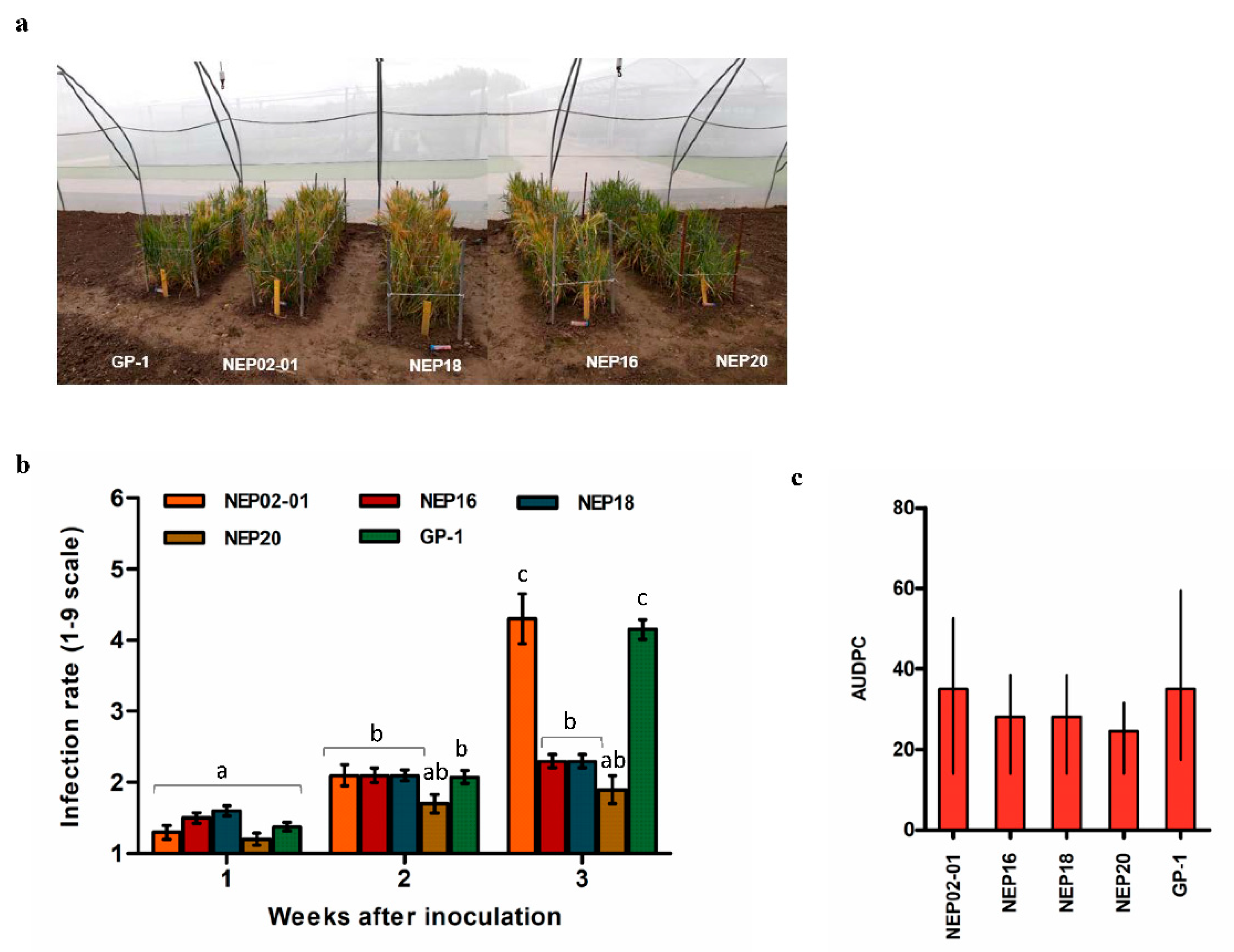
To test the reliability of HvNEP-1-mediated resistance to FHB, four Fusarium-infected transgenic lines and control GP plants were grown in a field plot covered with an insect-proof net (Figure S1b). Ten transgene-positive plants from each transgenic line were compared to 53 untransformed GP-1 plants. Line NEP20 showed the lowest disease infection rate and progression as compared with the other transgenic lines (NEP02-01, NEP16 and NEP18) and the untransformed control plant GP-1 (Figure 3a). Three weeks after infection, line NEP20 had an average infection score of 1.9, whereas the control GP plants were scored at 4.15 (on a one to nine scale) (Figure 3b). AUDPC was calculated at one, two, and three weeks after inoculation. After three weeks, the AUDPC was significantly higher in the GP control plants (35%) than in the transgenic NEP20 plants (24.5%) (Figure 3c).

4. Discussion

Fusarium head blight is a devastating disease of barley and other crops worldwide. Resistant cultivars could reduce damage from FHB. However, so far, only limited resources of resistance genes are available. In the current study, the antifungal potential of barley HvNEP-1 was investigated by transgenic overexpression in the developing barley endosperm. The ten investigated T0 barley plants had variable levels of HvNEP-1 expression, but no noticeable phenotypic differences were observed between the transgenic and control GP plants, even in the highest expressing lines (Figure 1b). Studies of Fusarium-infected plants showed that overexpression of HvNEP-1 in transgenic barley improved resistance to FHB both by limiting primary infection (Type-I resistance) and the spread of the disease (AUDPC) (Type-II resistance) as compared with the untransformed control plants [48]. Limiting infection and spread of disease is important, particularly, from a yield perspective. However, accumulation of mycotoxins is another unfortunate effect of Fusarium infections. The current study demonstrated that, endosperm-specific overexpression of HvNEP-1 reduced the accumulation of mycotoxins in the infected grains significantly (Type-IV resistance) [49]. In contrast to the non-transgenic control plants infected with F. culmorum, in which the level of DON was high, the accumulation of DON in the infected transgenic grains was very low (below the detection level in nine out of the ten lines). This was in agreement with our previous in vitro experiments, where the accumulation of 15-ADON was reduced in cultures with rHvNEP-1 [36]. For the F. graminearum-infected plants, seven transgenic lines had DON levels below the detection level. The lower mycotoxin formation in the transgenic seeds was accompanied by decreased disease progression in the transgenic plants as compared with the non-transgenic controls. Contamination of food and feed with mycotoxins is a worldwide problem. Although acute mycotoxicosis caused by high doses is rare in animals and humans, exposure to Fusarium mycotoxins can alter the human and animal susceptibility to infectious diseases by affecting the intestinal health and the innate and adaptive immune system [50]. The current study demonstrates that Fusarium resistance in barley induced by HvNEP-1 can facilitate a protection against mycotoxins.
Furthermore, the reliability of HvNEP-1 as an antifungal gene was examined under semi-field conditions where plants were grown in soil plots covered by an insect net (Figure 3a, Figure S1b). The result from the semi-field experiment was consistent with the greenhouse study where the overexpression of HvNEP-1 strongly suppressed FHB disease severity and progression (Figure 3b,c). On the basis of the semi-field analysis, the expression level of HvNEP-1 in line NEP20 was sufficient to reduce the infection rate and disease progression.
The current study describes a new resistance mechanism against FHB in barley. HvNEP-1 is demonstrated to have a strong potential as an FHB resistance gene and for reducing mycotoxins in infected seed. The transgenic approach to manage FHB used here can potentially minimize fungicide use, post-harvest sorting of infected grains, and combat mycotoxin-associated health risks. The use of azole fungicides for Fusarium control has aroused growing concern over the last years. Fungicides such as tebuconazole induces triazole-resistance in Aspergillus fumigatus [51]. At the same time, broad-spectrum triazoles are the primary drugs in the treatment of patients with prophylaxis of aspergillosis [52]. Fusarium resistant plants require less fungicide, and thereby alleviate the formation of triazole-resistant microorganisms in the environment.
Since the HvNEP-1 gene was isolated from barley, it can reduce safety concerns and could potentially also be used in cis- and intra-genesis approaches [41]. Similarly, NEP-based strategies can be developed in other species where NEP candidates are available. Further studies are essential to understand the exact mechanism of HvNEP-1-based resistance, as well as to show if variation in HvNEP-1 levels is available in the natural germplasm and the extent to which these variants have the potential to protect the crop against FHB.

5. Conclusions

The results of this study provide insights into the novel HvNEP-1 based Fusarium resistance mechanism in barley that limits primary infection, the spread of the disease, and synthesis of mycotoxins in the grains. Utilization of HvNEP-1 based Fusarium resistance in our crops could help to reduce the use of azole fungicides in agriculture. Future studies would show to what extent HvNEP-1 based Fusarium resistance can be utilized in other crops.

Supplementary Materials

The following are available online at https://www.mdpi.com/2073-4395/10/2/203/s1.

Author Contributions

Z.E.B. performed most of the experiments, data analysis and manuscript preparation; C.K.M. and G.D. participated in the HvNEP-1 cloning and review of the manuscript; I.B.H. was involved in Agrobacterium-mediated transformation of HvNEP-1 in barley; I.S.F. analysis of transgenic grains for mycotoxin accumulation; L.N.J. participated in the inoculation and scoring of transgenic plants with Fusarium; H.B.-P. developed the study concept, designed the experiment and manuscript preparation. All authors have read and agreed to the published version of the manuscript.

Funding

This research received no external funding.

Acknowledgments

The authors acknowledge Lis Bagnkop Holte, Ole Bråd Hansen, and Kirsten Heinrichson for technical assistance in the lab and greenhouse.

Conflicts of Interest

The authors declare no competing financial interests.

References

  1. Parry, D.W.; Jenkinson, P.; McLeod, L. Fusarium ear blight (scab) in small-grain cereals—A review. Plant Pathol. 1995, 44, 207–238. [Google Scholar] [CrossRef]
  2. Stenglein, S.A. Fusarium Poae: A pathogen that needs more attention. J. Plant Pathol. 2009, 91, 25–36. [Google Scholar]
  3. Toth, B.; Mesterhazy, A.; Horvath, Z.; Bartok, T.; Varga, M.; Varga, J. Genetic variability of central European isolates of the Fusarium graminearum species complex. Eur. J. Plant Pathol. 2005, 113, 35–45. [Google Scholar] [CrossRef]
  4. Nganje, W.E.; Bangsund, D.A.; Leistritz, F.L.; Wilson, W.W.; Tiapo, N.M. Regional economic impacts of Fusarium Head Blight in wheat and barley. Rev. Agric. Econ. 2004, 26, 332–347. [Google Scholar] [CrossRef]
  5. Roth, G. Mid-Atlantic Soft Winter Wheat Region; Wheat and Barley Scab Initiative: Fusarium Focus; Academic: Milwaukee, WI, USA, 2010; Volume 10, pp. 1–8. [Google Scholar]
  6. Hollandbeck, G.F.; Dewolf, E.; Todd, T. Preliminary 2019 Kansas Wheat Disease Loss Estimates; Kansas cooperative plant disease survey report; Kansas Department of Agriculture: Manhattan, KS, USA, 2019.
  7. McMullen, M.; Bergstrom, G.; De Wolf, E.; Dill-Macky, R.; Hershman, D.; Shaner, G.; Van Sanford, D. A Unified Effort to Fight an Enemy of Wheat and Barley: Fusarium Head Blight. Plant Dis. 2012, 96, 1712–1728. [Google Scholar] [CrossRef] [Green Version]
  8. Desjardins, A.E. Fusarium Mycotoxins: Chemistry, Genetics and Biology. Plant Pathol. 2007, 56, 337. [Google Scholar]
  9. Harris, L.J.; Desjardins, A.E.; Plattner, R.D.; Nicholson, P.; Butler, G.; Young, J.C.; Weston, G.; Proctor, R.H.; Hohn, T.M. Possible role of trichothecene mycotoxins in virulence of Fusarium graminearum on maize. Plant Dis. 1999, 83, 954–960. [Google Scholar] [CrossRef] [Green Version]
  10. Proctor, R.H.; Hohn, T.M.; McCormick, S.P. Reduced virulence of Gibberella-zeae caused by disruption of a trichothecene toxin biosynthetic gene. Mol. Plant-Microbe Interact. 1995, 8, 593–601. [Google Scholar] [CrossRef] [Green Version]
  11. Placinta, C.M.; D’Mello, J.P.F.; Macdonald, A.M.C. A review of worldwide contamination of cereal grains and animal feed with Fusarium mycotoxins. Anim. Feed Sci. Technol. 1999, 78, 21–37. [Google Scholar] [CrossRef]
  12. Bai, G.H.; Shaner, G. Management and resistance in wheat and barley to Fusarium head blight. Annu. Rev. Phytopathol. 2004, 42, 135–161. [Google Scholar] [CrossRef] [PubMed]
  13. Buerstmayr, H.; Ban, T.; Anderson, J.A. QTL mapping and marker-assisted selection for Fusarium head blight resistance in wheat: A review. Plant Breed. 2009, 128, 1–26. [Google Scholar] [CrossRef]
  14. Jin, F.; Zhang, D.D.; Bockus, W.; Baenziger, P.S.; Carver, B.; Bai, G.H. Fusarium Head Blight Resistance in US Winter Wheat Cultivars and Elite Breeding Lines. Crop Sci. 2013, 53, 2006–2013. [Google Scholar] [CrossRef] [Green Version]
  15. Li, G.; Zhou, J.; Jia, H.; Gao, Z.; Fan, M.; Luo, Y.; Zhao, P.; Xue, S.; Li, N.; Yuan, Y.; et al. Mutation of a histidine-rich calcium-binding-protein gene in wheat confers resistance to Fusarium head blight. Nat. Genet. 2019, 51, 1106–1112. [Google Scholar] [CrossRef] [PubMed]
  16. Su, Z.; Bernardo, A.; Tian, B.; Chen, H.; Wang, S.; Ma, H.; Cai, S.; Liu, D.; Zhang, D.; Li, T.; et al. A deletion mutation in TaHRC confers Fhb1 resistance to Fusarium head blight in wheat. Nat. Genet. 2019, 51, 1099–1105. [Google Scholar] [CrossRef]
  17. Rawat, N.; Pumphrey, M.O.; Liu, S.; Zhang, X.; Tiwari, V.K.; Ando, K.; Trick, H.N.; Bockus, W.W.; Akhunov, E.; Anderson, J.A.; et al. Wheat Fhb1 encodes a chimeric lectin with agglutinin domains and a pore-forming toxin-like domain conferring resistance to Fusarium head blight. Nat. Genet. 2016, 48, 1576–1580. [Google Scholar] [CrossRef]
  18. Gadaleta, A.; Colasuonno, P.; Giove, S.L.; Blanco, A.; Giancaspro, A. Map-based cloning of QFhb.mgb-2A identifies a WAK2 gene responsible for Fusarium Head Blight resistance in wheat. Sci. Rep. 2019, 9, 6929. [Google Scholar] [CrossRef] [Green Version]
  19. Liu, S.Y.; Hall, M.D.; Griffey, C.A.; McKendry, A.L. Meta-Analysis of QTL Associated with Fusarium Head Blight Resistance in Wheat. Crop Sci. 2009, 49, 1955–1968. [Google Scholar] [CrossRef]
  20. Loffler, M.; Schon, C.C.; Miedaner, T. Revealing the genetic architecture of FHB resistance in hexaploid wheat (Triticum aestivum L.) by QTL meta-analysis. Mol. Breed. 2009, 23, 473–488. [Google Scholar] [CrossRef]
  21. Simoes, I.; Faro, C. Structure and function of plant aspartic proteinases. Eur. J. Biochem. 2004, 271, 2067–2075. [Google Scholar] [CrossRef] [Green Version]
  22. Simoes, I.; Faro, R.; Bur, D.; Faro, C. Characterization of recombinant CDR1, an Arabidopsis aspartic proteinase involved in disease resistance. J. Biol. Chem. 2007, 282, 31358–31365. [Google Scholar] [CrossRef] [Green Version]
  23. Rawlings, N.D.; Alan, J.; Thomas, P.D.; Huang, X.D.; Bateman, A.; Finn, R.D. The MEROPS database of proteolytic enzymes, their substrates and inhibitors in 2017 and a comparison with peptidases in the PANTHER database. Nucleic Acids Res. 2018, 46, D624–D632. [Google Scholar] [CrossRef] [PubMed]
  24. Vines, S.H. The Proteolytic Enzyme of Nepenthes. Ann. Bot. 1897, 11, 563–584. [Google Scholar] [CrossRef]
  25. Vines, S.H. The Proteolytic Enzyme of Nepenthes. Ann. Bot. 1901, 15, 563–573. [Google Scholar] [CrossRef] [Green Version]
  26. Owen, T.P.; Lennon, K.A. Structure and development of the pitchers from the carnivorous plant Nepenthes alata (Nepenthaceae). Am. J. Bot. 1999, 86, 1382–1390. [Google Scholar] [CrossRef]
  27. Amagase, S.; Nakayama, S.; Tsugita, A. Acid protease in nepenthes 2. Study on specificity of nepenthesin. J. Biochem. 1969, 66, 431–439. [Google Scholar] [CrossRef]
  28. Amagase, S. Digestive enzymes in insectivorous plants part 3 Acid proteases in the genus Nepenthes and Drosera-peltata. J. Biochem. 1972, 72, 73–81. [Google Scholar] [CrossRef]
  29. Nakayama, S.; Amagase, S. Acid protease in nepenthes - partial purification and properties of enzyme. Proc. Jpn. Acad. 1968, 44, 358. [Google Scholar] [CrossRef]
  30. An, C.I.; Fukusaki, E.; Kobayashi, A. Aspartic proteinases are expressed in pitchers of the carnivorous plant Nepenthes alata Blanco. Planta 2002, 214, 661–667. [Google Scholar] [CrossRef]
  31. Athauda, S.B.P.; Matsumoto, K.; Rajapakshe, S.; Kuribayashi, M.; Kojima, M.; Kubomura-Yoshida, N.; Iwamatsu, A.; Shibata, C.; Inoue, H.; Takahashi, K. Enzymic and structural characterization of nepenthesin, a unique member of a novel subfamily of aspartic proteinases. Biochem. J. 2004, 381, 295–306. [Google Scholar] [CrossRef]
  32. Kadek, A.; Mrazek, H.; Halada, P.; Rey, M.; Schriemer, D.C.; Man, P. Aspartic Protease Nepenthesin-1 as a Tool for Digestion in Hydrogen/Deuterium Exchange Mass Spectrometry. Anal. Chem. 2014, 86, 4287–4294. [Google Scholar] [CrossRef] [PubMed]
  33. Lee, L.; Zhang, Y.; Ozar, B.; Sensen, C.W.; Schriemer, D.C. Carnivorous Nutrition in Pitcher Plants ( Nepenthes spp.) via an Unusual Complement of Endogenous Enzymes. J. Proteome Res. 2016, 15, 3108–3117. [Google Scholar] [CrossRef] [PubMed]
  34. Buch, F.; Kaman, W.E.; Bikker, F.J.; Yilamujiang, A.; Mithöfer, A.; Mithoefer, A. Nepenthesin Protease Activity Indicates Digestive Fluid Dynamics in Carnivorous Nepenthes Plants. PLoS ONE 2015, 10, 3. [Google Scholar] [CrossRef] [PubMed] [Green Version]
  35. Bekalu, Z.E.E.; Madsen, C.K.K.; Dionisio, G.; Brinch-Pedersen, H. Aspergillus ficuum phytase activity is inhibited by cereal grain components. PLoS ONE 2017, 12, 13. [Google Scholar] [CrossRef] [PubMed]
  36. Bekalu, Z.E.E.; Dionisio, G.; Krogh Madsen, C.; Etzerodt, T.; Brinch-Pedersen, H. A novel barley nepenthesin-like aspartic protease HvNEP-1 inhibits fungal phytases and suppress fungal growth and toxin production. Status (Unpublished, under preparation).
  37. Bekalu, Z.E.; Dionisio, G.; Madsen, C.K.; Holme, I.B.; Etzerodt, T.P.; Fomsgaard, I.; Jørgensen, L.N.; Brinch-Pedersen, H. Nepenthesin-1 Derived Resistance to Fungal Pathogens in Major Crop Plants; WO 2019/057845 A1, filed 20 September 2018, and issued 28 March 2019; World Intellectual Property Organization: Geneva, Switzerland, 2019. [Google Scholar]
  38. Zhu, B.; Cai, G.; Hall, E.O.; Freeman, G.J. In-fusion assembly: Seamless engineering of multidomain fusion proteins, modular vectors, and mutations. Biotechniques 2007, 43, 354–359. [Google Scholar] [CrossRef] [PubMed]
  39. An, G.; Ebert, P.R.; Mitra, A.; Ha, S.B. Binary vectors. In Plant Molecular Biology Manual; Springer: Dordrecht, The Netherlands, 2014; pp. 45–63. [Google Scholar]
  40. Jones, H.D.; Doherty, A.; Wu, H. Review of methodologies and a protocol for the Agrobacterium-mediated transformation of wheat. Plant Methods. 2005, 1, 5. [Google Scholar] [CrossRef] [Green Version]
  41. Holme, I.B.; Dionisio, G.; Brinch-Pedersen, H.; Wendt, T.; Madsen, C.K.; Vincze, E.; Holm, P.B. Cisgenic barley with improved phytase activity. Plant Biotechnol. J. 2012, 10, 237–247. [Google Scholar] [CrossRef]
  42. Bartlett, J.G.; Alves, S.C.; Smedley, M.; Snape, J.W.; Harwood, W.A. High-throughput Agrobacterium-mediated barley transformation. Plant Methods 2008, 4, 22. [Google Scholar] [CrossRef] [Green Version]
  43. Doyle, J.J.; Doyle, J.L. Isolation of plant DNA from fresh tissue. Focus (Madison) 1990, 12, 13–15. [Google Scholar]
  44. Etzerodt, T.; Maeda, K.; Nakajima, Y.; Laursen, B.; Fomsgaard, I.S.; Kimura, M. 2,4-Dihydroxy-7-methoxy-2H-1,4-benzoxazin-3(4H)-one (DIMBOA) inhibits trichothecene production by Fusarium graminearum through suppression of Tri6 expression. Int. J. Food Microbiol. 2015, 214, 123–128. [Google Scholar] [CrossRef]
  45. Zadoks, J.C.; Changi, T.T.; Konzak, C.F. A decimal code for the growth stages of cereals. Weed Res. 1974, 14, 415–421. [Google Scholar] [CrossRef]
  46. Etzerodt, T.; Gislum, R.; Laursen, B.B.; Heinrichson, K.; Gregersen, P.L.; Jorgensen, L.N.; Fomsgaard, I.S. Correlation of Deoxynivalenol Accumulation in Fusarium-Infected Winter and Spring Wheat Cultivars with Secondary Metabolites at Different Growth Stages. J. Agric. Food Chem. 2016, 64, 4545–4555. [Google Scholar] [CrossRef] [PubMed]
  47. Chomczynski, P.; Sacchi, N. The single-step method of RNA isolation by acid guanidinium thiocyanate-phenol-chloroform extraction: Twenty-something years on. Nat. Protoc. 2006, 1, 581–585. [Google Scholar] [CrossRef] [PubMed]
  48. Schroeder, H.W.; Christensen, J.J. Factors affecting resistance of wheat to scab caused by Gibberella zeae. Phytopathology 1963, 53, 831. [Google Scholar]
  49. Miller, J.D.; Young, J.C.; Sampson, D.R. Deoxynivalenol and fusarium head blight resistance in spring cereals. Phytopathol. Zeitschrift J. Phytopathol. 1985, 113, 359–367. [Google Scholar] [CrossRef]
  50. Antonissen, G.; Martel, A.; Pasmans, F.; Ducatelle, R.; Verbrugghe, E.; Vandenbroucke, V.; Li, S.; Haesebrouck, F.; Van Immerseel, F.; Croubels, S. The impact of Fusarium Mycotoxins on human and animal host susceptibility to infectious diseases. Toxins (Basel) 2014, 6, 430–452. [Google Scholar] [CrossRef] [Green Version]
  51. Cui, N.; He, Y.; Yao, S.; Zhang, H.; Ren, J.; Fang, H.; Yu, Y. Tebuconazole induces triazole-resistance in Aspergillus fumigatus in liquid medium and soil. Sci. Total Environ. 2019, 648, 1237–1243. [Google Scholar] [CrossRef]
  52. Mortensen, K.L.; Mellado, E.; Lass-Flörl, C.; Rodriguez-Tudela, J.L.; Johansen, H.K.; Arendrup, M.C. Environmental study of azole-resistant Aspergillus fumigatus and other aspergilli in Austria, Denmark, and Spain. Antimicrob. Agents Chemother. 2010, 54, 4545–4549. [Google Scholar] [CrossRef] [Green Version]
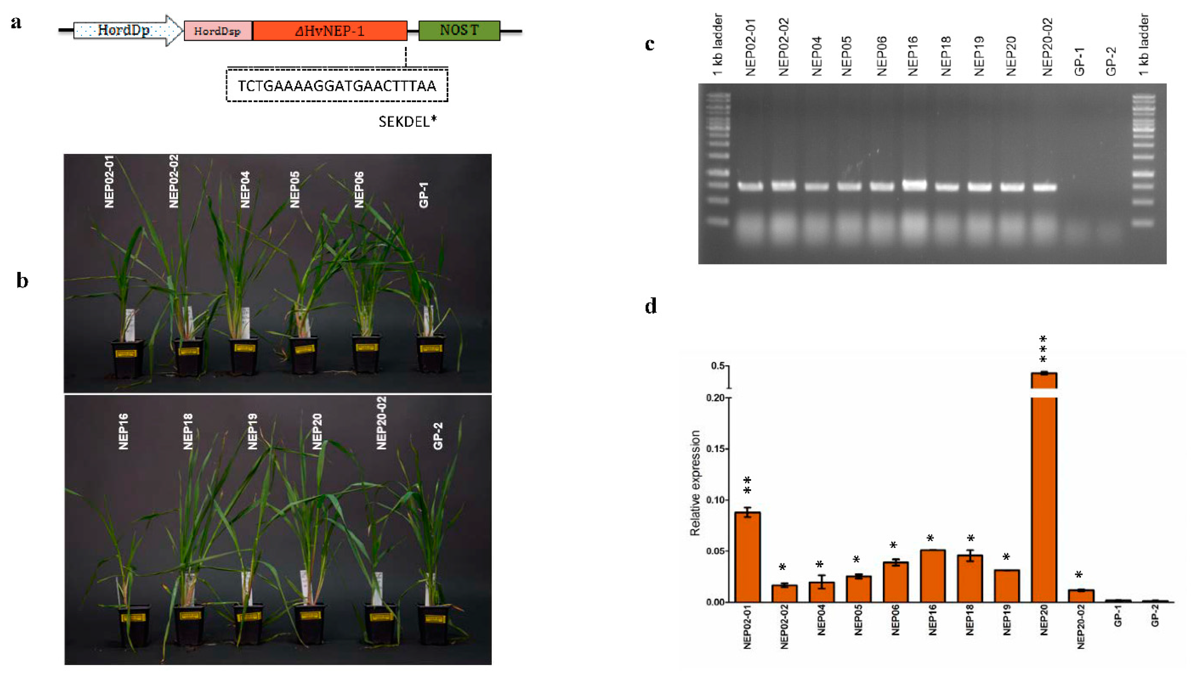
Figure 1. Analysis of transgenic barley lines overexpressing HvNEP-1. (a) Gene expression construct used for Agrobacterium-mediated transformation, HordDp: D-hordeum promoter, HordDsp: D-hordeum signal peptide, ΔHvNEP-1: signal peptide lacking HvNEP-1, and NOS terminator; (b) transgenic HvNEP-1-overexpressing barley lines used for downstream studies; (c) PCR analysis of transgenic HvNEP-1-overexpressing barley lines using P5 amplifying fragments inside the HordD promoter and HvNEP-1 gene; and (d) qPCR analysis of transgenic lines with variable expression of the transgene. Values in d are the mean of 3 independent technical replicates and were analyzed by Student’s t-test. Error bars = mean + sd. Significant differences between transgenic and non-transgenic plants are labelled with asterisks: * p < 0.05, ** p < 0.01, and *** p < 0.001.
Agronomy 10 00203 g001
Figure 2. Transgenic barley lines overexpressing HvNEP-1 showed reduced severity of Fusarium head blight (FHB) in the greenhouse. (a) FHB disease severity scoring 3 weeks after inoculation with F. graminearum (FG) and F. culmorum (FC) spores and the water control Milli-Q water (MQ). Values are the mean of 3 independent technical replicates. Error bars = mean + sd. Letters on the top of the column bars indicate statistically significant differences (p = 0.05, least significant difference (LSD) test); (b) area under the disease progress curve (AUDPC) analysis of FHB disease progression evaluated for 3 weeks after inoculation. Bars refer to the median AUDPC, and the minimum and maximum values are indicated with error bars.
Agronomy 10 00203 g002
Figure 3. Semi-field analysis of transgenic barley lines overexpressing HvNEP-1 against FHB infection. (a) Semi-field evaluation of Fusarium-infected four T1 transgenic lines (NEP02-01, NEP16, NEP18, and NEP20) and control Golden promise (GP) plants in the semi-field plots 3 weeks after infection; (b) Fusarium infection level of T1 transgenic and control GP plants scored on a scale of 1–9. Values are the mean of 10 and 53 biological replicates for transgenic (NEP02-01, NEP16, NEP18, and NEP20) and GP plants, respectively. Letters on the top of the column bars indicate statistically significant differences (p = 0.05, LSD test); (c) AUDPC for the semi-field-grown T1 transgenic lines and control GP plants. Error bars = mean + sem. Bars refer to the median AUDPC, and the minimum and maximum values are indicated with error bars.
Agronomy 10 00203 g003
Table 1. Mycotoxin analyses of transgenic and control GP plants treated with F. graminearum and F. culmorum spores or water.
HvNEP-1 transgenic lines and GP controls
NEP02-01NEP02-02NEP04NEP05NEP06NEP16NEP18NEP19NEP20NEP20-02GP-1GP-2
Samples used for analysisFCFHB+159387941627123191989251257<5025043462726477NIVMycotoxins (µg/kg DW)
<50<50<50<5052<50<50<50152<50370912324DON
<5<5<5<5<5<5<5<5<5<518655ZEA
FHB77<50<5055<50<50<50<50<50<50<50<50NIV
<50<50<50<50<50<50<50<50<50<50142855DON
<5<5<5<5<5<5<5<5<5<5<515ZEA
FGFHB+3551062431543163772972759797<5053239913050<50NIV
5295<5056<50<50<50<50817040073<5060221664DON
<5<5<5<5<5<5<5<5256<5<538ZEA
FHB124<50<50<50171<50<50<50<50<50<50<50NIV
<50<50<50<50<50<50<50<50<50<50569240DON
<5<5<5<5<5<5<5<5<5<5<5<5ZEA
MQ <5052<50<50<50163<50<50<50<50<50<50NIV
<50<50<50<50<50<50<50<50<50<50<50<50DON
<5<5<5<5<5<5<5<5<5<5<5<5ZEA
One half of 9 grains (3 per spike) collected from 3 spikes per treatment in a plant were pooled together for mycotoxin analysis. GP-1 and GP-2 stands for the two Golden promise cultivar plants used as controls. The FC and FG denote for spikes infected with spores of F. culmorum (FC) and F. graminearum (FG) fungal species, respectively. FHB+ and FHB stand for grains that show and do not show FHB symptoms, respectively. MQ stands for grains treated with water. The levels of deoxynivalenol (DON), nivalenol (NIV), and zearalenone (ZEA) mycotoxins were measured. Detectable limits for DON, NIV, and ZEA are >50 µg, >50 µg, and >5 µg per kg of DW, respectively.