Article

Amino Acid-Modified Polyethylenimines with Enhanced Gene Delivery Efficiency and Biocompatibility

1
Key Laboratory of Green Chemistry and Technology (Ministry of Education), College of Chemistry, Sichuan University, Chengdu 610064, China
2
National Key Laboratory of Shock Wave and Detonation Physics, Institute of Fluid Physics, Chinese Academy of Engineering Physics, Mianyang 621900, China
3
West China School of Medicine, Sichuan University, Chengdu 610041, China
4
Humanwell Healthcare (Group) Co., LTD., 666 Gaoxin Road, East Lake High-Tech Development Zone, Wuhan 430075, China
*
Authors to whom correspondence should be addressed.
Polymers 2015, 7(11), 2316-2331; https://doi.org/10.3390/polym7111516
Submission received: 25 August 2015 / Revised: 2 November 2015 / Accepted: 10 November 2015 / Published: 17 November 2015
(This article belongs to the Special Issue Functional Polymers for Medical Applications)

Abstract

:
The development of gene delivery vectors with high efficiency and biocompatibility is one of the key points of gene therapy. A series of polycations were prepared from polyethylenimine (PEI) with several amino acids or their analogs. The target polymers have different charge and hydrophilic/hydrophobic properties, which may affect their performance in the gene transfection process. Gel retardation and DLS assays showed that these polymers may condense DNA into nanoparticles with positive zeta potentials and proper sizes for cellular uptake. Luciferase reporter gene transfection results revealed their higher transfection efficiency than PEI; especially in the presence of serum, in which up to 23 times higher efficiency was achieved by employing glycolic acid-grafted PEI. Moreover, it was found that the degree of substitution on PEI has an apparent influence on the transfection, and the balance between electron-positive/negative groups largely affects the delivery process. The higher serum tolerance was also proven by BSA adsorption, flow cytometry and confocal microscopy assays. Results demonstrate that such type of polycations may serve as promising non-viral gene delivery vectors.

Graphical Abstract

1. Introduction

As an important type of non-viral gene delivery vectors, cationic polymers have received attention for their good stability, easy preparation, and modification [1,2]. Cationic polymers can efficiently condense DNA via electrostatic interaction, and their cationic properties may also facilitate the contact between the polymer/DNA complex (polyplex) and negatively charged cell membrane, leading to better cell uptake. The leading polymeric materials for gene delivery include polyethylenimine (PEI) [3,4], poly(l-lysine) [5], poly(tertiaryamine methacrylate) [3], and polyamidoamine [6], etc. Among these polymers, PEI was most widely studied due to its easy availability. High molecular weight (HMW) PEI, especially that with the Mw of 25 kDa, has relatively higher tranfection efficiency (TE) both in vitro and in vivo. This made it the golden standard for the design of novel polymeric vectors [7,8,9]. However, along with the higher TE, significant cytotoxicity and limited biocompatibility are also found in HMW PEI-mediated gene delivery [10,11]. Thus, to develop novel PEI-based materials with both high TE and improved biocompatibility is of great importance.
In recent years, large efforts were made to overcome the shortcomings of PEI [12,13,14,15,16,17]. Wang et al. showed that the introduction of hydrophobic dodecyl chains to PEI would improve the TE by enhanced escape of DNA from the endosome to the cytoplasm, but unfortunately the biocompatibility was not effectively improved [12]. Ramezani and co-workers revealed that alkyl-oligoamine derivatives of PEI without reduction of primary amine quantity is an effective strategy for the balance of hydrophobic-hydrophilic property and for the improvement of TE while maintaining low toxicity [13]. Zhuo found that the incorporation of a hydroxyl-enriched “skin” would afford PEI-g-5-ethyl-5-(hydroxymethyl)-1,3-dioxan-2-oxo (PEI-g-EHDO) with remarkably improved biocompatibility and stronger resistance against the serum-associated detrimental effects, such as protein adsorption, particle aggregation, and polycation-protein exchange [11]. In addition, poly(ethyleneglycol) (PEG) has been shown to have the ability to shield the positive charges on the complex, improving the serum tolerance [14,15,16]. However, the strong charge shielding and volume exclusion from such a polymer coating would interfere with the polycation/DNA complexation and hinder their efficient internalization into cells [17]. With these concerns, the modification of PEI, aiming for higher TE together with improved biocompatibility is not an easy task. The balances between the hydrophobic and hydrophilic properties and between the DNA binding and release abilities are of much significance.
Taking clues from these studies, the present investigation was undertaken with the objective to incorporate hydrophobic, hydrophilic, or charged elements in the polymeric structure and evaluate their effects on the TE, cytotoxicity, and serum resistance. For this purpose, we selected serine (Ser), glycolic acid (Ga), glycine (Gly), N,N-bis(2′-aminoethyl)glycine (Deta) and leucine (Leu) to modify 25 kDa PEI with various degrees of substitution (DS). Among these novel polymers, compared to PEI, Ser-PEI introduces hydrophilic hydroxyl groups without the reduction of amines; Ga-PEI replaces some amines with hydroxyls; Gly-PEI maintains the amount of amines; Deta-PEI may increase the primary amine groups; while Leu-PEI introduces hydrophobic alkyl groups. Using HEK293, HeLa and U-2OS cells as models, experiments revealed that these modified polymers showed better biocompatibility, serum resistance, and TE.

2. Experimental Section

2.1. Materials

Unless otherwise stated, all chemicals and reagents were obtained commercially and used without further purification. Absolute chloroform (CHCl3) and dichloromethane (CH2Cl2) were distilled after being dried with calcium hydride (CaH2). Column chromatography was performed using 200–300 mesh silica gel or 200–300 mesh Al2O3. All aqueous solutions were prepared from deionized or distilled water. The 1H NMR spectra was measured on a Bruker AM400 NMR spectrometer (Zurich, Switzerland). Proton chemical shifts of NMR spectra were given in ppm relative to internal reference TMS (1H, 0.00 ppm). N-(tert-Butoxycarbonyl)-l-serine, N-(tert-Butoxycarbonyl)-l-Glycine and N-(tert-Butoxycarbonyl)-l-leucine were purchased from Aladdin Industrial Corporation (Shanghai, China), MicroBCA protein assay kit was obtained from Pierce (Rockford, IL, USA). Luciferase assay kit was purchased from Promega (Madison, WI, USA). Endotoxin-free plasmid purification kit was purchased from TIANGEN (Beijing, China). Cell counting Kit-8 (CCK-8) was purchased from Dojindo Laboratories (Kumamoto, Japan). The plasmids used in the study were pGL-3 (Promega, Madison, WI, USA) coding for luciferase DNA, and pEGFP-N1 (Clontech, Palo Alto, CA, USA) coding for EGFP DNA. The Dulbecco′s modified Eagle′s medium (DMEM) and fetal bovine serum were purchased from Invitrogen Co. (Chengdu, China). Cy5™ was obtained from Molecular Probe (Mirus, Madison, WI, USA). HEK293T human embryonic kidney cell lines, HeLa cell lines, and U-2OS human osteosarcoma cancer cells were purchased from Shanghai Institute of Biochemistry and Cell Biology, Chinese Academy of Sciences (Shanghai, China). N′,N″-di-Boc-N,N-bis(2′-aminoethyl)glycine was synthesized as previously reported [18].

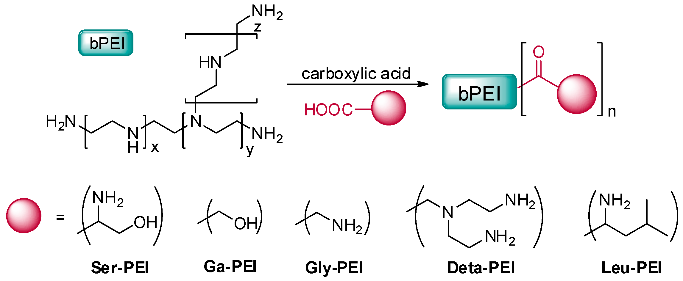
2.2. Preparation of Polymers

N-(tert-Butoxycarbonyl)-carboxylic acid (2 mmol), EDCI (2.4 mmol), HOBT (2.4 mmol), and DIEA (2.4 mmol) in dry methylene chloride (50 mL) was stirred for 0.5 h in ice bath. Then, the desired amount of PEI (25 kDa) based on molar ratios solved in dry methylene chloride was added and the reaction mixture was stirred for two days at room temperature. After completion of the reaction, MeOH/HCl solution was added to remove the protecting t-butyloxycarbonyl (Boc) group. The residue was dissolved in a small amount of water and dialyzed (MWCO 8000–14,000 kDa) against deionized water for three days. The product was obtained as a white or pale-yellow solid after lyophilization. Yield: 46%–62%.

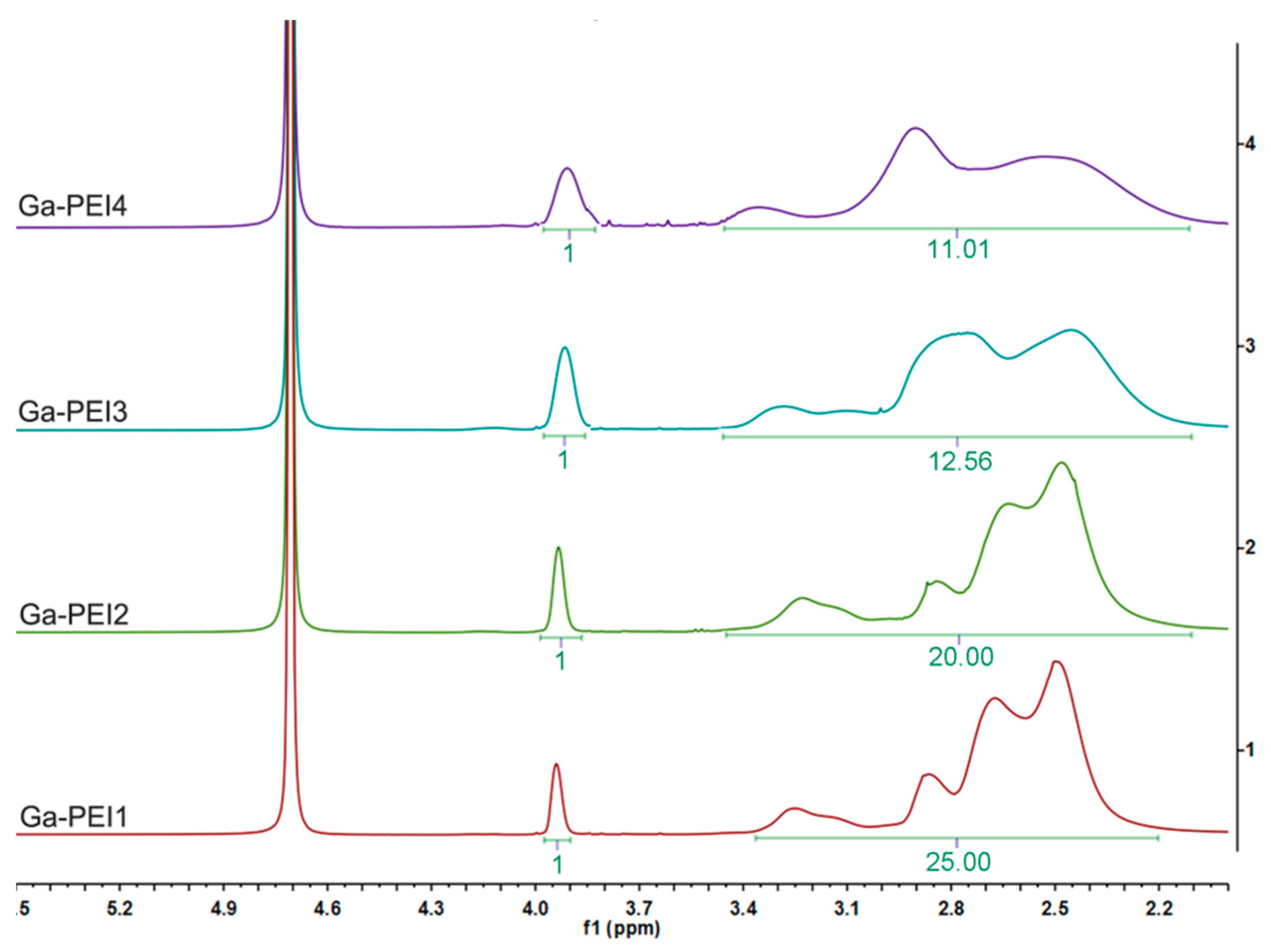
2.3. Polymer Characterization

1H NMR spectra in D2O were obtained on a Bruker AV 400-MHz instrument at 25 °C. The molecular weights (Mw) and polydispersity (PDI, Mw/Mn) of prepared cationic polymers were determined by a gel permeation chromatography (GPC) system, which consisted of a Waters 515 pump, a Linear 7.8 mm × 300 mm column (Waters Corp, Milford, MA, USA), an 18-angle laser scattering instrument (Wyatt Technology Corporation, Santa Barbara, CA, USA), and an OPTILAB DSP interferometric refractometer (Wyatt Technology Corporation, Santa Barbara, CA, USA). NaAc (0.3 M, pH 4.4) passed through a 0.02 μm film filter was used as the eluent. A flow rate of 0.5 mL/min was applied.
  • Ser-PEI: 1H NMR (400 MHz, D2O, TMS): δ = 2.64–3.50 ppm (m, PEI–H and –CH(NH2)CH2OH), 3.76 ppm (s, –CH2OH).
  • Ga-PEI: 1H NMR (400 MHz, D2O, TMS): δ = 2.52–3.25 ppm (m, PEI–H), 3.94 ppm (s, –CH2OH).
  • Gly-PEI: 1H NMR (400 MHz, D2O, TMS): δ = 2.64–3.51 ppm (m, PEI–H), 3.67 ppm (m, –CH2NH2).
  • Deta-PEI: 1H NMR (400 MHz, D2O, TMS): δ = 2.64–3.13 ppm (m, –CH2CH2NH2, –CONCH2CH2), 3.25 ppm (m, –COCH2, –CONCH2).
  • Leu-PEI: 1H NMR (400 MHz, D2O, TMS): δ = 0.86 ppm (d, –CH3), 1.58–1.64 ppm (m, –CHCH3), 2.68–3.60 ppm (m, PEI–H), 3.9 ppm (m, –CHNH2).
The calculation of DS of the polymers: Taking Ga-PEI for example, the characteristic singlet of δ 3.9 ppm represents the 2H on CH2OH on the substituents, while the broad multiplet represents the C–H of PEI (4H for each ethylenimine unit). Consequently, if the DS were 100%, the area ratio of the two peaks would be 1:2. Therefore, the DS may be calculated by (real peak area ratio)/(1:2). For Ga-PEI1, the DS would be (1:25)/(1:2) = 0.08, i.e., 8%.

2.4. Amplification and Purification of Plasmid DNA

pGL-3 and pEGFP-N1 plasmids were used. The former was seed as the luciferase reporter gene, which was transformed in M109 Escherichia coli, and the latter was used as the enhanced green fluorescent protein reporter gene, which was transformed in E. coli DH5α. Both plasmids were amplified in E. coli grown in LB medium at 37 °C and 220 rpm overnight. The plasmids were purified by an EndoFree Tiangen™ Plasmid Kit. Then, the purified plasmids were dissolved in TE (Tris+EDTA) buffer solution and stored at −80 °C. The integrity of plasmids was confirmed by agarose gel electrophoresis. The purity of plasmids was determined by the ratio of ultraviolet (UV) absorbances at 260 to 280 nm, and the result was approximately 1.91.

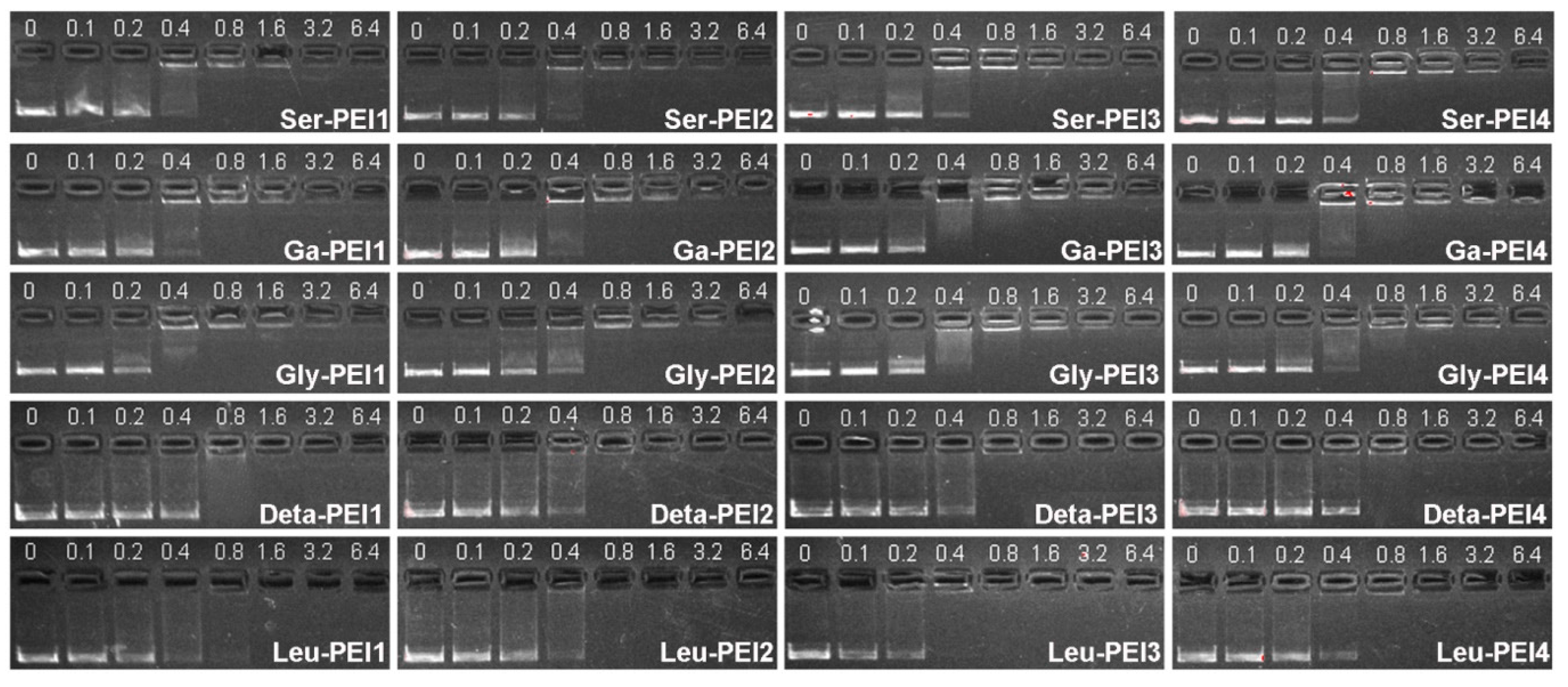
2.5. Agarose Gel Retardation Assay

Polyplexes at different w/w ratios (weight ratio of polymer relative to pDNA) were prepared by adding an appropriate volume of the polymer solution to 5 μL of pUC 19 (0.025 mg/mL). The obtained complex solution was then diluted to the total volume of 15 μL. After incubation at 37 °C for 0.5 h, the polyplexes were electrophoresed on a 1% (w/v) agarose gel containing GelRed™ in Triseacetate (TAE) running buffer at 120 V for 0.5 h. Then, DNA was visualized under an ultraviolet lamp using a Vilber Lourmat imaging system.

2.6. Particle Size and ζ-Potential Measurement in Water

Zeta potential (ζ-potential) and particle size were measured by a Nano-ZS ZEN3600 apparatus (Malvern Instruments) at 25 °C. This instrument is equipped with a red laser of wavelength 630 nm and measures the electrophoretic mobility of the particles using phase analysis of scattered light in an experimental set up similar to Laser Doppler Velocimetry (M3PALS technique, Malvern Instruments Ltd., Worcestershire, UK). Polyplexes with various w/w ratios were prepared by adding 1 mg of pUC-19 to the appropriate volume of the polymer solution (in PBS). Then, the solution of polyplexes was incubated at 37 °C for 0.5 h and diluted with deionized water to 1 mL prior to measurement. Data were shown as mean ± standard deviation (SD) based on triplicate independent experiments.

2.7. Cell Culture

Human osteosarcoma (U-2OS) cells, HeLa cells, and human embryonic kidney transformed HEK293 cells were incubated in DMEM with 10% (v/v) fetal bovine serum (FBS) and 1% antibiotics (penicillin-streptomycin, 10,000 U·mL−1) at 37 °C in a humidified atmosphere containing 5% CO2. The medium was replenished every other day.

2.8. Transfection Procedure

Gene transfection of a series of complexes was investigated in U-2OS, HeLa and HEK293 cells. Cells were seeded in 24-well plates (10 × 104 cells/well for U-2OS, 8.5 × 10⁴ cells/well for HeLa and HEK293) and grown to reach 70%–80% cell confluence at 37 °C for 24 h in 5% CO2. Before transfection, the medium was replaced with a serum-free or a 10% serum-containing culture medium containing polymer/pDNA (1 μg) complexes at various weight ratios. After 4 h under standard incubator conditions, the medium was replaced with fresh medium containing serum and incubated for another 20 h.
For fluorescent microscopy assays, cells were transfected by complexes containing pEGFP-N1. After 24 h incubation, GFP-expressed cells were observed with an inverted fluorescence microscope (Nikon Eclipse TE 2000E) equipped with a cold Nikon camera at a magnification of 100×. Control transfection was performed in each case using a commercially available transfection reagent bPEI (25 kDa) based on the standard conditions specified by the manufacturer.
For luciferase assays, cells were transfected by complexes containing pGL-3. For a typical assay in a 24-well plate, 24 h post transfection as described above, cells were washed with cold PBS and lysed with 100 μL 1× Lysis reporter buffer (Promega, Madison, WI, USA). The luciferase activity was measured by microplate reader (Model 550, Bio-Rad, Hercules, CA, USA). Protein content of the lysed cell was determined by BCA protein assay. Gene transfection efficiency was expressed as the relative fluorescence intensity per mg protein (RLU/mg protein). All the experiments were done in triplicate.

2.9. Cytotoxicity Assay

Toxicity toward Human osteosarcoma (U-2OS) cells, HeLa cells, and human embryonic kidney transformed HEK293 cells was determined by cell counting kit-8 (CCK-8). About 7000 cells per well were seeded in 96-well plates and cultured overnight for 70%–80% cell confluence. The medium was replaced with 50 μL of fresh medium, to which 50 μL polyplexes at various concentrations were added to achieve a final volume of 100 μL. Twenty-four hours later, 10 μL CCK-8 mixed in 90 μL PBS was added to each well for additional 1 h incubation. The absorbance was measured in an ELISA plate reader (model 550, BioRad, Hercules, CA, USA) at a wavelength of 450 nm. The metabolic activity of the polyplex-treated cells was expressed as a relative to untreated cell controls taken as 100% metabolic activity. In addition, the cell viability of PEI was performed with a CCK-8 assay as unmodified control.

2.10. Protein Adsorption Assay

In brief, 1 mL of polymer solution (1 mg/mL) was added to 1 mL of bovine serum albumin (BSA) solution (2 mg/mL). After shaking at 37 °C for 0.5 h, the polymer adsorbed BSA to form white cotton-shaped precipitates, which were removed by centrifugation. The supernatant was carefully collected and the concentration of BSA in it was determined by BCA protein assay. The protein adsorbed on the polyplexes was calculated using the following equation:
q = (CiCs) × V/m
where Ci and Cs are the initial BSA concentration and the BSA concentration in the supernatant after adsorption experiments, respectively; V is the total volume of the solution (2 mL); and m is the weight of the polymer (1 mg) added into the solution.

2.11. Cellular Uptake of Plasmid DNA

The cellular uptake of the polymer/fluorescein-labeled DNA complexes was analyzed by flow cytometry. The Label IT Cy5 Labeling Kit was used to label pDNA with Cy5 according to the manufacturer’s protocol. Briefly, U-2OS cells were seeded in 12-well plates (2 × 10⁵ cells/well) and allowed to attach and grow for 24 h. For transfection in the absence of serum, the medium was exchanged with serum-free medium. As for transfection in the presence of serum, the medium was exchanged with serum-containing medium. Cells were incubated with Cy5 labeled DNA complexes (2 μg DNA/well, optimal weight ratio of each sample) in media for 4 h at 37 °C. Subsequently, the cells were washed with 1× PBS containing heparin (120 U/mL) and harvested with 0.25% Trypsin/EDTA and resuspended in PBS. Mean fluorescence intensity was analyzed using a FC 500 flow cytometer (Beckman Coulter, Brea, CA, USA). Cy5-labeled plasmid DNA uptake was measured in the FL4 channel using the red diode laser (633 nm). Data from 10,000 events were gated using forward and side-scatter parameters to exclude cell debris. The flow cytometer was calibrated for each run to obtain a background level of ~1% for control samples (i.e., untreated cells).

2.12. Confocal Laser Scanning Microscopy (CLSM) Analysis

U-2OS cells were seeded at a density of 2 × 10⁴ cells per well in a 35 mm confocal dish (Φ = 15 mm), 24 h prior to transfection. For transfection in the absence of serum, the medium was exchanged with serum-free medium. As for transfection in the presence of serum, the medium was exchanged with serum-containing medium. Cells were incubated with Cy5-labeled DNA complexes (2 μg DNA/well, optimal weight ratio of each sample) in media for 4 h at 37 °C. Subsequently, cells were rinsed twice with PBS (pH 7.4) to remove complexes that were not taken up by cells, fixed with 4% paraformaldehyde (dissolved with PBS buffer) for 10 min, nuclear staining was done with DAPI. The CLSM observation was performed using Leica TCS SP5 at excitation wavelengths of 405 nm for DAPI (blue), and 633 nm for Cy5 (red), respectively.

3. Results and Discussion

3.1. Synthesis and Characterization of Acylated Polymers

The aim of this study was to rationally design and evaluate a class of potentially safe non-viral gene delivery vectors based on modified PEI. As shown in Scheme 1, target polymers were prepared from PEI 25 kDa and a series of functional acids with different amino densities and hydrophilic/hydrophobic properties. Except glycolic acid (Ga), other acids with primary amine group(s) need to be protected by di-tert-butyl dicarbonate (Boc2O) before their reaction with PEI. The final polymers were obtained by dialysis (MWCO: 8000–14,000) against water for 3 days and lyophilization. Adjusting the feeding ratio of acid carboxyls to amines on PEI led to different DS (for example, feeding ratios of 0.1, 0.2, 0.3, and 0.5 resulted in Ga-PEI1 to Ga-PEI4, respectively), which could be calculated from the specific peak integrals in the 1H NMR spectra (Figure 1, also see the Experimental Section). The calculated DS are listed in Table 1. Considering the steric effect and the reactivity diversity of 1°/2°/3° amines in PEI, the DS is always lower than the carboxyl/amine feeding ratio. The molecular weights of target polymers were measured by GPC, and results in Table 1 show that the Mw increased with the rise of DS.
Scheme 1. Cationic polymers modified from branched 25 kDa polyethylenimine (PEI).
Polymers 07 01516 g011
Figure 1. 1H NMR spectra of Ga-PEI. The degree of substitution (DS) could be calculated by the ratio between the peak areas. The singlet of δ 3.9 ppm represents CH2OH on the substituents, while the broad multiplet represents the C–H near the amino groups of PEI.
Polymers 07 01516 g001
Table 1. Characterization of the polymers. DS was obtained from 1H NMR and represents the DS of the entire amino groups in PEI. Mw and polydispersity (PDI) were obtained from GPC.
PolymerDS (%)Mw (kDa)PDI
Ser-PEI16.831.91.67
Ser-PEI29.332.61.66
Ser-PEI314.835.81.80
Ser-PEI419.037.01.71
Ga-PEI18.029.41.55
Ga-PEI210.031.01.63
Ga-PEI315.932.61.68
Ga-PEI418.233.31.68
Gly-PEI14.028.51.54
Gly-PEI28.730.51.58
Gly-PEI313.332.71.60
Gly-PEI425.035.21.70
Deta-PEI16.533.21.88
Deta-PEI210.536.01.77
Deta-PEI312.537.31.96
Deta-PEI415.442.11.90
Leu-PEI17.032.31.56
Leu-PEI210.334.61.71
Leu-PEI313.336.61.78
Leu-PEI421.146.42.37

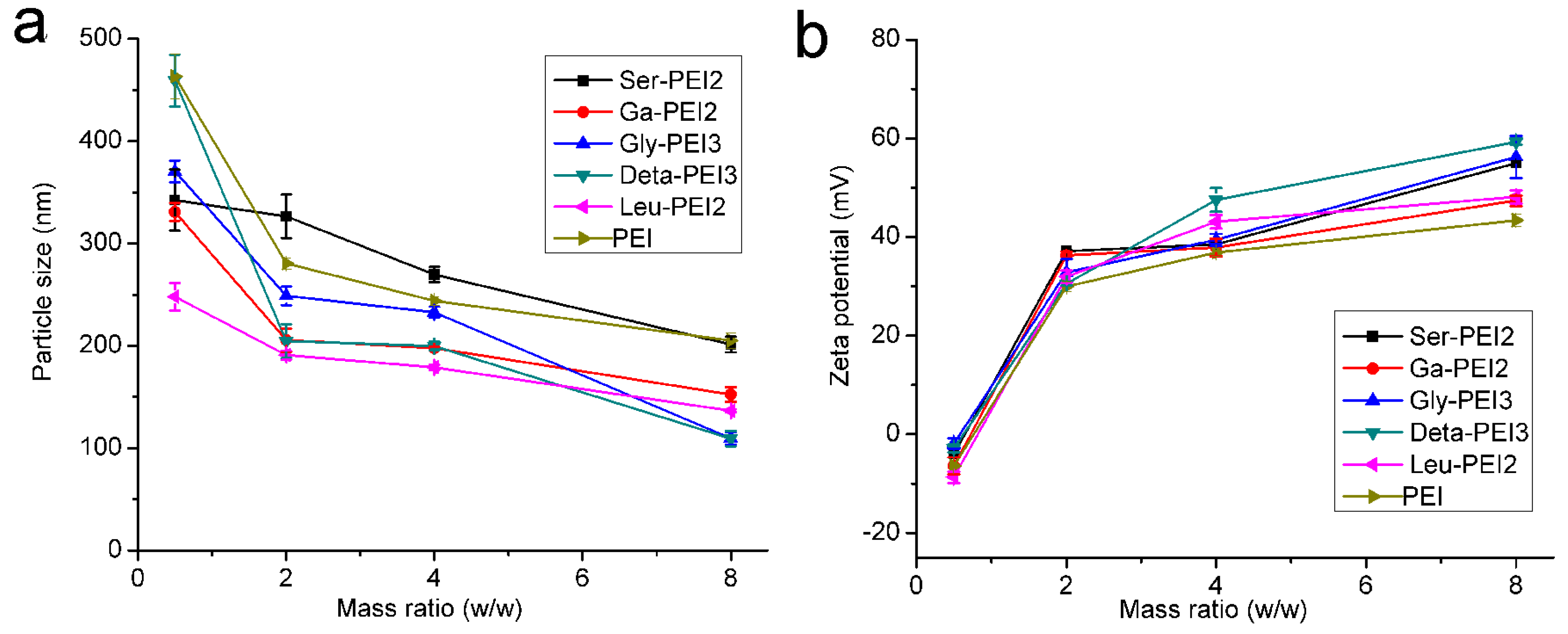
3.2. Formation of Polymer/DNA Complexes (Polyplexes)

Condensation of DNA into nano-sized particles is a prerequisite for efficient delivery of DNA into cells [13]. The binding strength of the modified PEIs to plasmid DNA was determined by gel retardation assay at various weight ratios (polymer/DNA, w/w), and the binding ability was indicated by the ratio at which full retardation was achieved [19]. Figure 2 shows that the complete DNA retardation induced by these new materials could be observed at a w/w ratio of 0.8, and the DS did not affect the DNA condensation ability [20]. Among the five modifications, Deta-PEI shows slightly lower binding ability. The stability of the polyplexes was also studied, and it was found that DNA could be gradually released from the polyplex with the increase of heparin (Figure S1). However, a greater amount of heparin than the polymer was needed for release, indicating a tight binding of polymer to DNA. Moreover, DNA protection against nuclease was investigated, and the results are shown in Figure S2. After the treatment of DNase, naked DNA was degraded and no band was observed on the gel. On the contrary, after condensation with the polymers, DNA could resist DNase and be released by subsequent addition of heparin. Such results suggest that these polymers may well protect DNA from degradation in the circumstance with nuclease [21].
Figure 2. Agarose gel electrophoresis of plasmid DNA (pUC 19) complexed with the cationic polymers at different weight ratios. In each image, the first lane is DNA control.
Polymers 07 01516 g002
Since cells typically take up particles ranging from micrometers to nanometers [22], it is necessary for the cationic polymers to compact nucleic acids into nanoparticles with proper zeta potentials, which also play an important role in endocytosis and material cytotoxicity [23]. A dynamic light scattering (DLS) assay was used to measure the diameters and zeta potential of polyplexes formed by the modified-PEI and plasmid DNA at w/w ratios of 0.5, 2, 4, and 8, and unmodified PEI was used for comparison. The DS of each polymer was chosen according to the optimized DS from the luciferase gene transfection experiments (Section 3.3). As shown in Figure 3, in general, similar to PEI, these polymers may condense DNA into particles with diameters ranging from 100 to 450 nm, and the zeta-potential of the polyplexes reversed to positive at w/w ratio of ~1. The particle size decreased whereas the zeta potential increased with the rise of weight ratio. Polyplexes formed from Ga-PEI or Leu-PEI, in which hydroxyl or hydrophobic groups were introduced, showed relatively lower zeta potentials. Meanwhile, polyplex derived from Deta-PEI had the highest amino density, leading to the highest positive charge. However, the particle size seemed to be unaffected by the density of amino groups. Deta-PEI only gave the smallest particles at a relatively higher w/w ratio of 8. The influence of DS on polyplex size and potential was also studied by using Ga-PEI as model, and the results (Figure S3) show that with the rise of DS, an increase in particle size and a decreased zeta potential were observed. This may be logically explained by the positive charge screening from the increased amount of hydroxyl groups.
Figure 3. Particle size (a) and zeta potential (b) of polyplexes obtained from different modified PEIs by dynamic light scattering (DLS). Data represent mean ± SD (n = 3).
Polymers 07 01516 g003

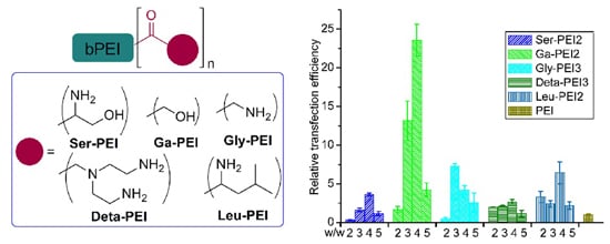
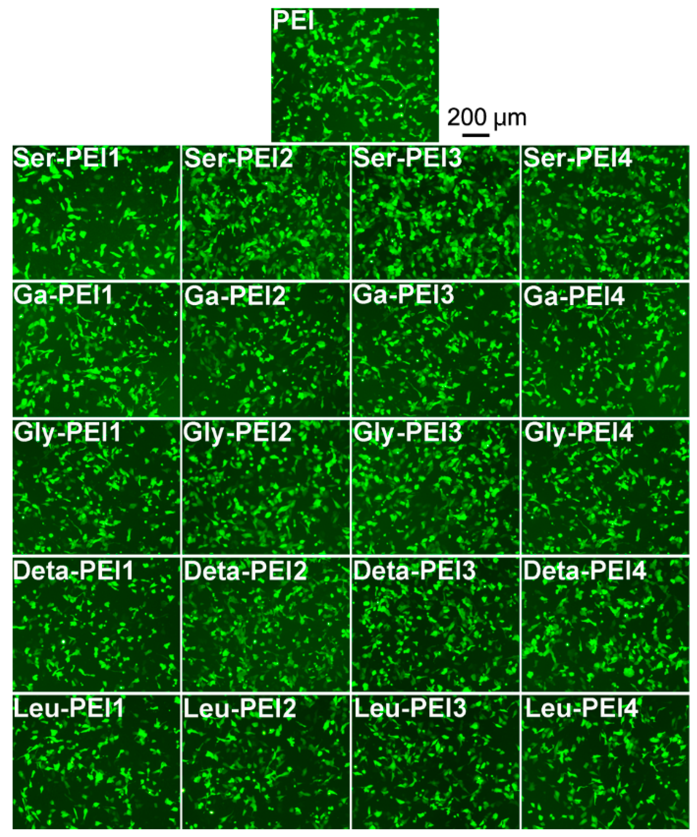
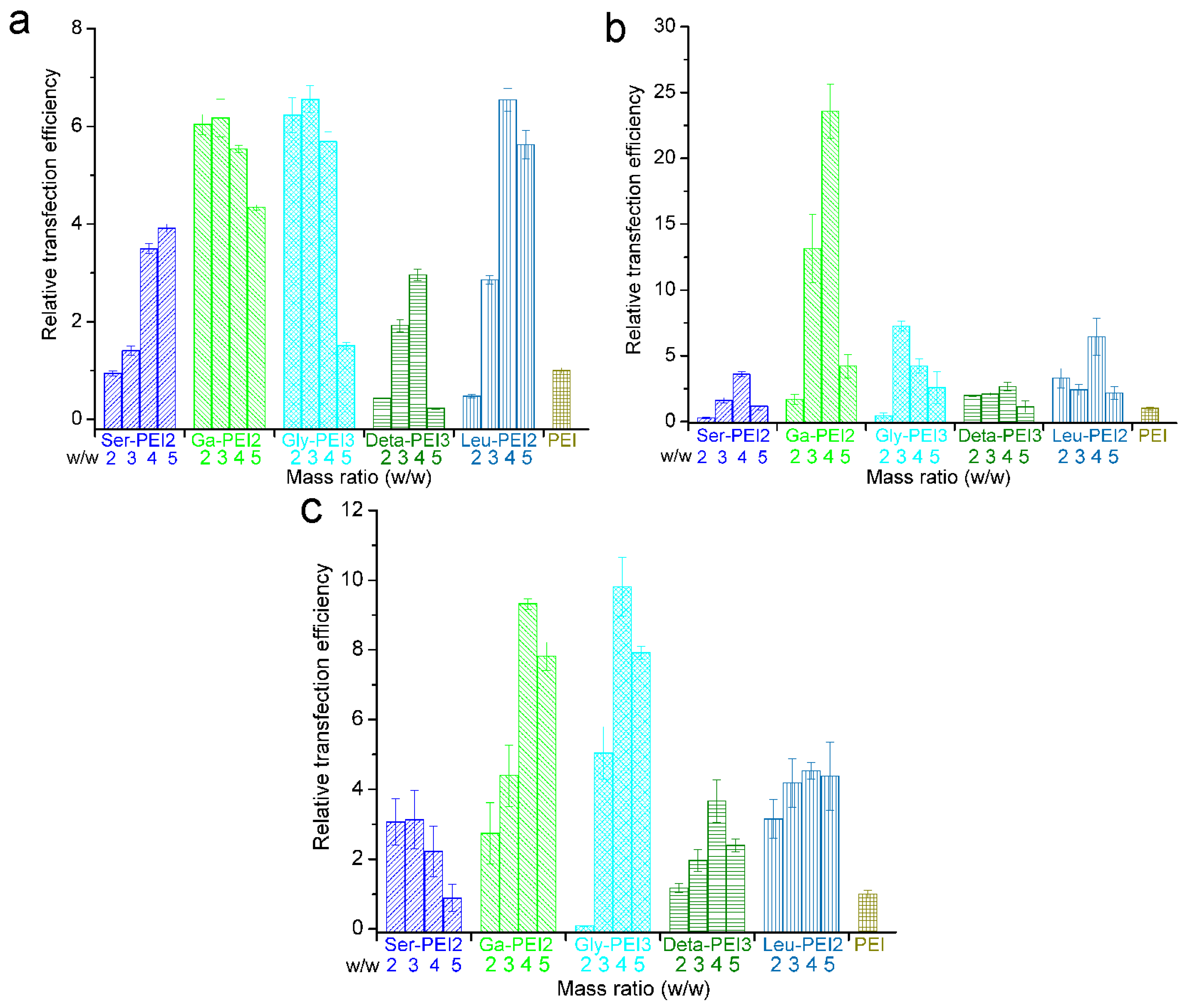
3.3. In Vitro Gene Transfection

Luciferase reporter gene was used to quantitatively assess the in vitro TE of these modified-PEIs in U-2OS cells. Figure 4 shows the relative TE of these polymers at various w/w ratios (2, 3, 4, 5) in comparison with PEI (25 kDa) at its optimal weight ratio of 1.4 (N/P ratio = 10) [24]. The conjugation of amino acids on PEI definitely enhanced the luciferase expression, and glycine-modified Gly-PEI showed the best TE, which was about 10 times higher than PEI. The DS also affects their gene delivery behavior: for each polymer, about 10% DS is most suitable for the transfection.
It is known that the TE of PEI dramatically decreases in serum-supplemented medium [25,26,27]. Thus, the development of non-viral gene vectors that may retain their efficiency in serum is very important. In this study, the TEs of the newly prepared polymers with optimal DS were also tested in the presence of 10% serum. As shown in Figure 5a, compared to the results without the use of serum (Figure 4), polymers modified with hydroxyls showed better serum tolerance than PEI. For example, Ga-PEI gave six times higher TE than PEI, while the value was four in the absence of serum. Further, Ga-PEI also showed excellent TE toward other cell lines including HeLa (Figure 5b) and HEK293 (Figure 5c). In Hela cells, up to 23 times higher TE than PEI was achieved. In each cell line, the TE of Ga-PEI was higher than Ser-PEI, which also has hydroxyl groups but maintains the amount of amino groups. This indicates that the balance between electron-positive/negative groups, which also called the “hydroxyl effect” [11,28] or the charge screening effect [29,30], is important for the transfection by such types of polycations, especially in the serum circumstance. On the other hand, Gly-PEI also showed good TE, but the relative TE compared to PEI was lower than that obtained without serum.
Figure 4. Luciferase gene expression transfected by polyplexes at different weight ratios in comparison with 25 kDa PEI (w/w = 1.4, N/P = 10) in U-2OS cells in the absence of serum. In each group of columns, the w/w are 2, 3, 4, and 5, consecutively. Data represent mean ± SD (n = 3).
Polymers 07 01516 g004
Figure 5. Luciferase gene expression transfected in the presence of 10% serum in different cell lines. (a) U-2OS; (b) HeLa; (c) HEK293. Data represent mean ± SD (n = 3). 25 kDa PEI (w/w = 1.4, N/P = 10) was used as control.
Polymers 07 01516 g005
In addition, enhanced green fluorescent protein (EGFP) expression assays were performed to directly visualize the infected cells expressing the pEGFP-Nl reporter gene. Figure 6 shows the density of transfected cells by modified polymers under the optimal weight ratios obtained in luciferase assays. The results indicate that the transfection mediated by these polymers was more effective than that obtained in the experiment involving PEI. In addition, the same assay was carried out in the presence of serum (Figure S4) or in other cell lines (HEK293, Figures S5 and S6; HeLa, Figures S7 and S8). All results further demonstrate the good serum tolerance of these vectors.
Figure 6. Fluorescence microscope images of pEGFP-transfected U-2OS cells by the polycations at their optimal weight ratio.
Polymers 07 01516 g006

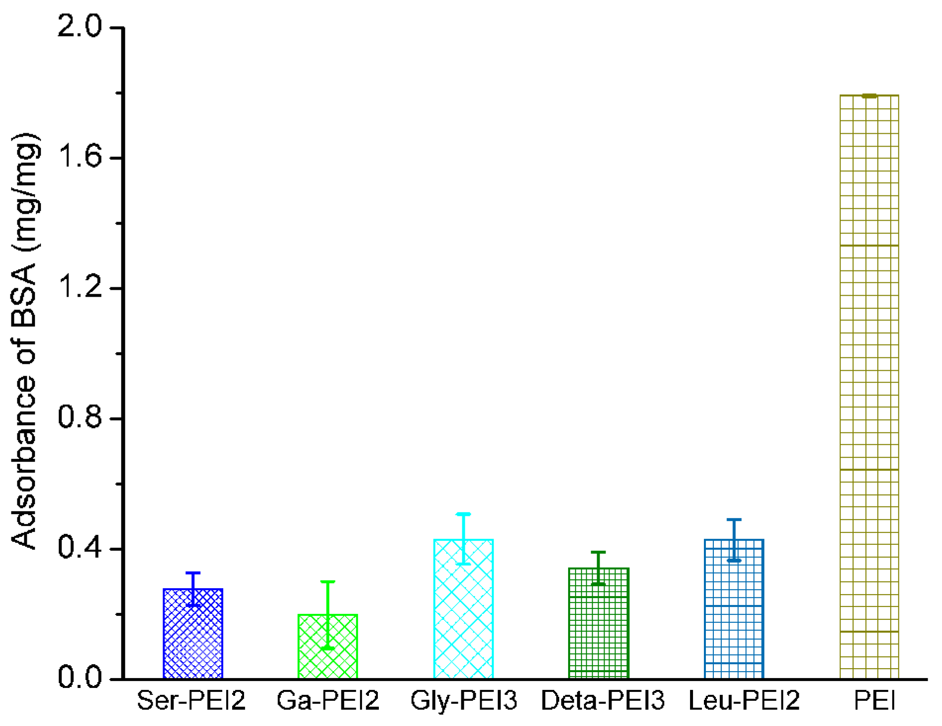
3.4. Biocompatibility Studies

The negatively-charged cellular and blood components would also interact with the cationic polyplexes, leading to inherent toxicity [31]. Transfection involving polycations is usually accompanied by some degree of cell toxicity, which may limit their use as gene delivery vectors [32]. The cytotoxicities of these PEI derivatives were studied and compared with that of unmodified 25 kDa bPEI by CCK-8 assay at various concentrations. The effect of DS was first investigated. For Ser-PEI and Ga-PEI, the viability of U-2OS cells slightly increased with the rise of DS (Figure S9), indicating that the introduction of hydroxyl groups might benefit the biocompatibility of the polymeric materials [14,19,28]. Meanwhile, for other PEI derivatives, such a DS-depended trend was not found [13]. The cytotoxicities of these five polymers with their optimized DS, which were the same with those used in DLS studies, were subsequently measured within different cell lines, and the results are shown in Figure 7. As anticipated, these modified-PEI conjugates exhibited obviously lower cytotoxicity than PEI, especially in normal cells HEK293. Leu-PEI with hydrophobic side chains showed higher cytotoxicity than other polymers, suggesting that hydrophobic modification is prone to induce toxicity, which may come from the enhanced interaction with the cell membrane [33,34,35]. Among the five polymers, Ga-PEI showed the lowest cytotoxicity. This might be attributed to the replacement of amines with hydroxyls, which would shield the positive surface charge of the PEI (Figure 3b).
Figure 7. Cytotoxicity of the polycations at different concentrations toward (a) U-2OS; (b) HeLa and (c) HEK293 cells. Data represent mean ± SD (n = 3).
Polymers 07 01516 g007
The serum-induced inhibition in gene transfection largely depends on the non-specific interaction between the polyplex and negatively charged serum protein [36,37,38]. The resistance against protein adsorption may favor the competitive approach of DNA cargo onto cell membrane, resulting in enhanced endocytosis and gene expression [39]. To verify this assumption, bovine serum albumin (BSA) was used as a model protein to simulate non-specific protein adsorption onto the surfaces of the polymers. As shown in Figure 8, modified PEI showed far lower protein adsorption than the unmodified one, especially for Ga-PEI. Such higher resistance against the negatively charged protein may contribute to its higher relative TE in serum condition (Figure 5).
Figure 8. Comparison of protein adsorption between the polymers. 1 Milliliter of polymer solution (1 mg/mL) was added to 1 mL bovine serum albumin (BSA) solution (2 mg/mL) and co-incubated with shaking at 37 °C for 0.5 h prior to measurement. Data represent mean ± SD (n = 3).
Polymers 07 01516 g008

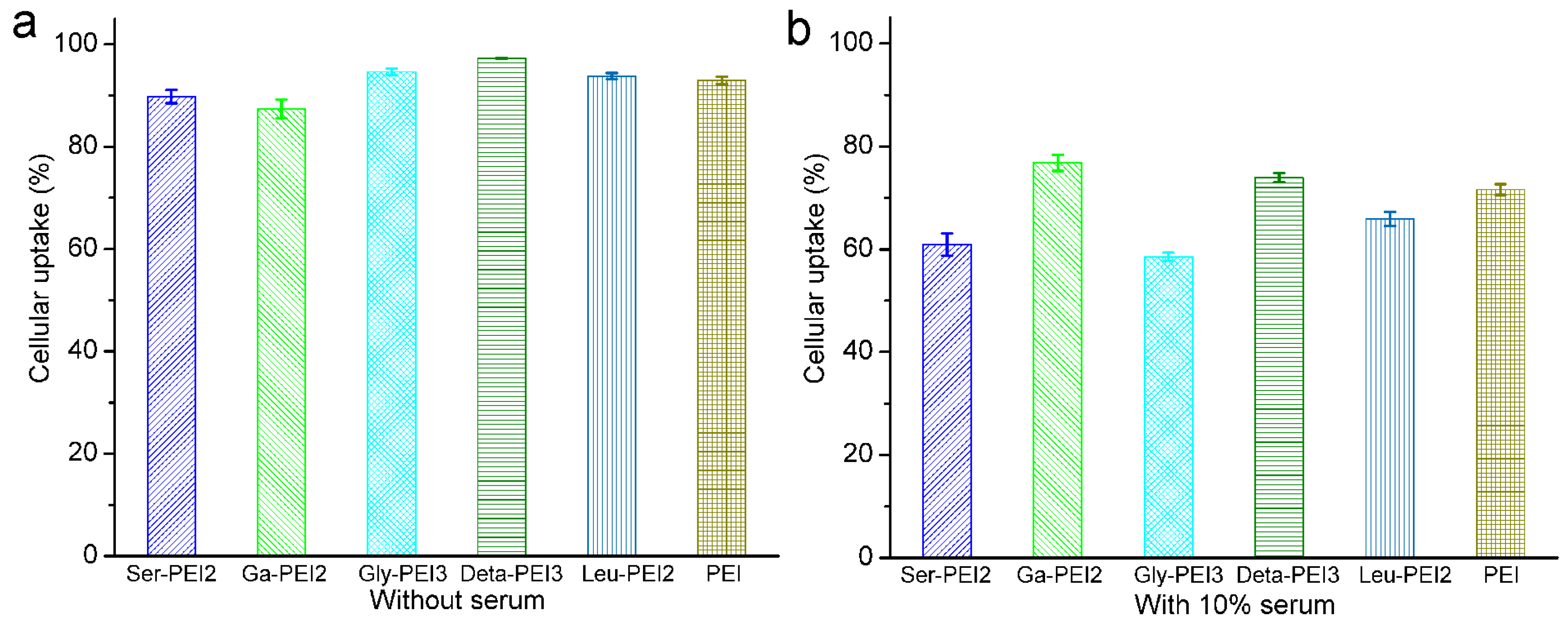
3.5. Cellular Uptake and Intracellular Distribution

To gain insight into the transfection mechanisms promoted by the title polymers, the cellular uptake of DNA was analyzed by flow cytometry. After 4 h incubation of polyplexes with U-2OS cells, the percentage of cells positive for Cy5-labelled pDNA was calculated and shown in Figure 9a. All of the polyplexes showed good cellular uptake, and about 90% of tested cells were positive for Cy5-labled DNA. Although Gly-PEI gave the best TE in luciferase assay (Figure 4), its cellular uptake was not the highest. Since cellular uptake is only one of the several barriers in the gene delivery process [28,40], we speculate that Gly-PEI may deliver the nucleic acid with higher intracellular efficiency. Moreover, the cellular uptake experiments were also carried out in the presence of 10% serum in the same cell line. The results shown in Figure 9b reveal that the cellular uptake of all polyplexes was negatively affected. However, it’s clearly shown that Ga-PEI gave the least uptake decrease, which might be attributed to its higher charge screening effect (Figure 3b) and better protein adsorption resistance (Figure 8). This also contributes to its higher serum tolerance than other polymers (Figure 5).
Subsequently, the internalization and intracellular location of the delivered DNA (Cy5-labelled) by these polymers at the optimal transfection weight ratio were studied in U-2OS cells with CLSM. The nuclei were stained with 4’,6-diamidino-2-phenylindole (DAPI, blue). As shown in Figure 10, in the absence of serum, all polyplexes may effectively deliver a considerable amount of DNA (red) into the peri-nucleic region as well as nuclei after 4 h transfection. However, the red fluorescence decreased for the PEI-mediated transfection in the presence of serum. Meanwhile, there was no obvious change of the fluorescence density in the transfection mediated by the modified PEIs. For Ga-PEI mediated transfection, many more red signals were found in the nuclei, indicating its potential as non-viral gene delivery vector with higher TE, especially in the serum circumstance.
Figure 9. Cellular uptake of polyplexes at optimal weight ratio in U-2OS cells quantified by flow cytometry analysis. (a) Without serum; (b) In the presence of 10% serum. Data represent mean ± SD (n = 3).
Polymers 07 01516 g009
Figure 10. CLSM images of U-2OS cells transfected with Cy5-labelled DNA by polymers at their respective optimal weight ratio in the absence (upper) and presence (lower) of serum. PEI as control (w/w = 1.4, N/P = 10). For each triad image, left: cell nuclei stained by DAPI (blue); middle: Cy5-labeled pDNA (red); right: merged image.
Polymers 07 01516 g010

4. Conclusions

Branched PEI (25 kDa) was modified with several amino acids or other functional acids to give the derivative polymers with various DS. Different carboxylic acids were employed to endue the new polymers with different charge and hydrophilic/hydrophobic properties. These polymers may condense DNA into nanoparticles with proper sizes and zeta potentials. In vitro experiments showed that compared to PEI, they could give higher TE and lower cytotoxicity. Up to 23 times higher TE than PEI was achieved by employing Ga-PEI as transfection reagent. The effects of DS and substituted groups were also investigated, and 10% DS was found to be most suitable for transfection. Ga-PEI, in which some amino groups were replaced by hydroxyls, showed the highest serum tolerance and also the highest TE in several cell lines with the presence of serum. BSA adsorption and flow cytometry assays also proved its higher biocompatibility. Such results may guide a way to design PEI derivatives as non-viral gene vectors with higher efficiency and lower toxicity.

Supplementary Materials

Supplementary materials can be found at www.mdpi.com/2073-4360/7/11/1516/s1.

Acknowledgments

This work was financially supported by the National Science Foundation of China (Nos. 21472131, 21232005, J1310008 and J1103315), and the Specialized Research Fund for the Doctoral Program of Higher Education in China (20120181130006).

Author Contributions

Qin-Fang Zhang, Ji Zhang and Xiao-Qi Yu designed the studies. Qin-Fang Zhang performed the chemical and biological experiments, Chao-Ran Luan and Dong-Xiao Yin performed the chemical experiments, Yan-Hong Liu and Qi Peng performed the biological experiments, Qin-Fang Zhang, Ji Zhang, Yong Xu and Xiao-Qi Yu prepared the manuscript.

Conflicts of Interest

The authors declare no conflict of interest.

References

  1. Hunt, K.K.; Vorburger, S.A. Gene therapy: Hurdles and hopes for cancer treatment. Science 2002, 297, 415–416. [Google Scholar] [CrossRef] [PubMed]
  2. Niidome, T.; Huang, L. Gene therapy progress and prospects: Nonviral vectors. Gene Ther. 2002, 9, 1647–1652. [Google Scholar] [CrossRef] [PubMed]
  3. Xu, F.J.; Yang, W.T. Polymer vectors via controlled/living radical polymerization for gene delivery. Prog. Polym. Sci. 2011, 36, 1099–1131. [Google Scholar] [CrossRef]
  4. Bisht, H.S.; Manickam, D.S.; You, Y.; Oupicky, D. Temperature-controlled properties of DNA complexes with poly(ethylenimine)-graft-poly(N-iso-propylacrylamide). Biomacromolecules 2006, 7, 1169–1178. [Google Scholar] [CrossRef] [PubMed]
  5. Wagner, E.; Ogris, M.; Zauner, W. Polylysine-based transfection systems utilizing receptor-mediated delivery. Adv. Drug Deliv. Rev. 1998, 30, 97–113. [Google Scholar] [PubMed]
  6. Kukowska-Latallo, J.F.; Bielinska, A.U.; Johnson, J.; Spindler, R.; Tomalia, D.A.; Baker, J.R., Jr. Efficient transfer of genetic material into mammalian cells using starburst polyamidoamine dendrimers. Proc. Natl. Acad. Sci. USA 1996, 93, 4897–4902. [Google Scholar] [CrossRef] [PubMed]
  7. Mintzer, M.A.; Simanek, E.E. Nonviral vectors for gene delivery. Chem. Rev. 2009, 109, 259–302. [Google Scholar] [CrossRef] [PubMed]
  8. Baker, A.; Saltik, M.; Lehrmann, H.; Killisch, I.; Mautner, V.; Lamm, G.; Christofori, G.; Cotton, M. Polyethylenimine (PEI) is a simple, inexpensive and effective reagent for condensing and linking plasmid DNA to adenovirus for gene delivery. Gene Ther. 1997, 4, 773–782. [Google Scholar] [CrossRef] [PubMed]
  9. Boussif, O.; Lezoualch, F.; Zanta, M.A.; Mergny, M.D.; Scherman, D.; Demeneix, B.; Behr, J.P. A versatile vector for gene and oligonucleotide transfer into cells in culture and in vivo: Polyethylenimine. Proc. Natl. Acad. Sci. USA 1995, 92, 7297–7301. [Google Scholar] [CrossRef] [PubMed]
  10. Tian, H.Y.; Lin, L.; Chen, J.; Chen, X.S.; Park, T.G.; Maruyama, A. RGD targeting hyaluronic acid coating system for PEI-PBLG polycation gene carriers. J. Control. Release 2011, 155, 47–53. [Google Scholar] [CrossRef] [PubMed]
  11. Luo, X.H.; Huang, F.W.; Qin, S.Y.; Wang, H.F.; Feng, J.; Zhang, X.Z.; Zhuo, R.X. A strategy to improve serum-tolerant transfection activity of polycation vectors by surface hydroxylation. Biomaterials 2011, 32, 9925–9939. [Google Scholar] [CrossRef] [PubMed]
  12. Yan, X.B.; Zhang, Y.; Zhang, H.; Wang, P.G.; Chua, X.; Wang, X. Amphiphilic polyethylenimine (PEI) as highly efficient non-viral gene carrier. Org. Biomol. Chem. 2014, 12, 1975–1982. [Google Scholar] [CrossRef] [PubMed]
  13. Dehshahri, A.; Oskuee, R.K.; Shier, W.T.; Hatefi, A.; Ramezani, M. Gene transfer efficiency of high primary amine content, hydrophobic, alkyl-oligoamine derivatives of polyethylenimine. Biomaterials 2009, 30, 4187–4194. [Google Scholar] [CrossRef] [PubMed]
  14. Clements, B.A.; Bai, J.; Kucharski, C.; Farrell, L.L.; Lavasanifar, A.; Ritchie, B.; Ghahary, A.; Uludag, H. RGD conjugation to polyethyleneimine does not improve DNA delivery to bone marrow stromal cells. Biomacromolecules 2006, 7, 1481–1488. [Google Scholar] [CrossRef] [PubMed]
  15. Dong, X.; Tian, H.Y.; Chen, L.; Chen, J.; Chen, X.S. Biodegradable mPEG-b-P(MCC-g-OEI) copolymers for efficient gene delivery. J. Control. Release 2011, 152, 135–142. [Google Scholar] [CrossRef] [PubMed]
  16. Yang, C.; Ong, Z.Y.; Yang, Y.Y.; Ee, P.L.; Hedrick, J.L. Novel biodegradable block copolymers of poly(ethylene glycol) (PEG) and cationic polycarbonate: Effects of PEG configuration on gene delivery. Macromol. Rapid Commun. 2011, 32, 1826–1833. [Google Scholar] [CrossRef] [PubMed]
  17. Petersen, H.; Merdan, T.; Kunath, K.; Fischer, D.; Kissel, T. Poly(ethylenimine-co-l-lactamide-co-succinamide): A biodegradable polyethylenimine derivative with an advantageous pH-dependent hydrolytic degradation for gene delivery. Bioconjug. Chem. 2002, 13, 812–821. [Google Scholar] [CrossRef] [PubMed]
  18. Makino, A.; Hara, E.; Hara, I.; Ozeki, E.; Kimura, S. Size control of core-shell-type polymeric micelle with a nanometer precision. Langmuir 2014, 30, 669–674. [Google Scholar] [CrossRef] [PubMed]
  19. Tripathi, S.K.; Yadav, S.; Gupta, K.C.; Kumar, P. Synthesis and evaluation of N-(2,3-dihydroxypropyl)-PEIs as efficient vectors for nucleic acids. Mol. Biosyst. 2012, 8, 1426–1434. [Google Scholar] [CrossRef] [PubMed]
  20. Layek, B.; Haldar, M.K.; Sharma, G.; Lipp, L.; Mallik, S.; Singh, J. Hexanoic acid and polyethylene glycol double grafted amphiphilic chitosan for enhanced gene delivery: Influence of hydrophobic and hydrophilic substitution degree. Mol. Pharm. 2014, 11, 982–994. [Google Scholar] [CrossRef] [PubMed]
  21. Xun, M.-M.; Xiao, Y.-P.; Zhang, J.; Liu, Y.-H.; Peng, Q.; Guo, Q.; Wu, W.-X.; Xu, Y.; Yu, X.-Q. Low molecular weight PEI-based polycationic gene vectors via Michael addition polymerization with improved serum-tolerance. Polymer 2015, 65, 45–54. [Google Scholar] [CrossRef]
  22. Liu, Y.M.; Reineke, T.M. Hydroxyl stereo chemistry and amine number within poly(glycoamidoamine)s affect intracellular DNA delivery. J. Am. Chem. Soc. 2005, 127, 3004–3015. [Google Scholar] [CrossRef] [PubMed]
  23. Petersen, H.; Fechner, P.M.; Martin, A.L.; Kunath, K.; Stolnik, S.; Roberts, C.J.; Fischer, D.; Davies, M.C.; Kissel, T. Polyethylenimine-graft-poly(ethylene glycol) copolymers: Influence of copolymer block structure on DNA complexation and biological activities as gene delivery system. Bioconjug. Chem. 2002, 13, 845–854. [Google Scholar] [CrossRef] [PubMed]
  24. Xiu, K.M.; Yang, J.J.; Zhao, N.N.; Li, J.S.; Xu, F.J. Multiarm cationic star polymers by atom transfer radical polymerization from β-cyclodextrin cores: Influence of arm number and length on gene delivery. Acta Biomater. 2013, 9, 4726–4733. [Google Scholar] [CrossRef] [PubMed]
  25. Dai, F.; Liu, W. Enhanced gene transfection and serum stability of polyplexes by PDMAEMA-polysulfobetaine diblock copolymers. Biomaterials 2011, 32, 628–638. [Google Scholar] [CrossRef] [PubMed]
  26. He, Y.; Cheng, G.; Xie, L.; Nie, Y.; He, B.; Gu, Z. Polyethyleneimine/DNA polyplexes with reduction-sensitive hyaluronic acid derivatives shielding for targeted gene delivery. Biomaterials 2013, 34, 1235–1245. [Google Scholar] [CrossRef] [PubMed]
  27. Xu, P.; Quick, G.K.; Yeo, Y. Gene delivery through the use of a hyaluronate-associated intracellularly degradable crosslinked polyethyleneimine. Biomaterials 2009, 30, 5834–5843. [Google Scholar] [CrossRef] [PubMed]
  28. Zhang, Q.-F.; Yi, W.-J.; Wang, B.; Zhang, J.; Ren, L.; Chen, Q.-M.; Guo, L.; Yu, X.-Q. Linear polycations by ring-opening polymerization as non-viral gene delivery vectors. Biomaterials 2013, 34, 5391–5401. [Google Scholar] [CrossRef] [PubMed]
  29. Chen, C.-K.; Jones, C.H.; Mistriotis, P.; Yu, Y.; Ma, X.; Ravikrishnan, A.; Jiang, M.; Andreadis, S.T.; Pfeifer, B.A.; Cheng, C. Poly(ethylene glycol)-block-cationic polylactide nanocomplexes of differing charge density for gene delivery. Biomaterials 2013, 34, 9688–9699. [Google Scholar] [CrossRef] [PubMed]
  30. Tian, H.; Guo, Z.; Lin, L.; Jiao, Z.; Chen, J.; Gao, S.; Zhu, X.; Chen, X. pH-responsive zwitterionic copolypeptides as charge conversional shielding system for gene carriers. J. Control. Release 2014, 174, 117–125. [Google Scholar] [CrossRef] [PubMed]
  31. Fischer, D.; Li, Y.X.; Ahlemeyer, B.; Krieglstein, J.; Kissel, T. In vitro cytotoxicity testing of polycations: Influence of polymer structure on cell viability and hemolysis. Biomaterials 2003, 24, 1121–1131. [Google Scholar] [CrossRef]
  32. Tao, X.; Wu, R.; Xia, Y.; Huang, H.; Chai, W.; Feng, T.; Gan, Y.; Zhang, W. Biotemplated fabrication of Sn@C anode materials based on the unique metal biosorption behavior of microalgae. ACS Appl. Mater. Interfaces 2014, 6, 3969–3702. [Google Scholar] [CrossRef] [PubMed]
  33. Chang, H.; Wang, H.; Shao, N.; Wang, M.; Wang, X.; Cheng, Y. Surface-engineered dendrimers with a diaminododecane core achieve efficient gene transfection and low cytotoxicity. Bioconjugate Chem. 2014, 25, 342–350. [Google Scholar] [CrossRef] [PubMed]
  34. Zhang, Q.-F.; Wang, B.; Yin, D.-X.; Zhang, J.; Wu, W.-X.; Yu, Q.-Y.; Yu, X.-Q. Linear TACN-based cationic polymers as non-viral gene vectors. RSC Adv. 2014, 4, 59164–59174. [Google Scholar] [CrossRef]
  35. Yi, W.-J.; Zhang, Q.-F.; Zhang, J.; Liu, Q.; Ren, L.; Chen, Q.-M.; Guo, L.; Yu, X.-Q. Cyclen-based lipidic oligomers as potential gene delivery vehicles. Acta Biomater. 2014, 10, 1412–1422. [Google Scholar] [CrossRef] [PubMed]
  36. Funhoff, A.M.; van Nostrum, C.F.; Lok, M.C.; Fretz, M.M.; Crommelin, D.J.A.; Hennink, W.E. Poly(3-guanidinopropyl methacrylate): A novel cationic polymer for gene delivery. Bioconjug. Chem. 2004, 15, 1212–1220. [Google Scholar] [CrossRef] [PubMed]
  37. Son, S.; Kim, W.J. Biodegradable nanoparticles modified by branched polyethylenimine for plasmid DNA delivery. Biomaterials 2010, 31, 133–143. [Google Scholar] [CrossRef] [PubMed]
  38. Jones, C.H.; Chen, C.-K.; Jiang, M.; Fang, L.; Cheng, C.; Pfeife, B.A. Synthesis of cationic polylactides with tunable charge densities as nanocarriers for effective gene delivery. Mol. Pharm. 2013, 10, 1138–1145. [Google Scholar] [CrossRef] [PubMed]
  39. Liu, Z.; Janzena, J.; Brooks, D.E. Adsorption of amphiphilic hyperbranched polyglycerol derivatives onto human red blood cells. Biomaterials 2010, 31, 3364–3373. [Google Scholar] [CrossRef] [PubMed]
  40. Gabrielson, N.P.; Pack, D.W. Acetylation of polyethylenimine enhances gene delivery via weakened polymer/DNA interactions. Biomacromolecules 2006, 7, 2427–2435. [Google Scholar] [CrossRef] [PubMed]

